Embed Size (px)
Citation preview

J N E R JOURNAL OF NEUROENGINEERING AND REHABILITATION
Tkach et al. Journal of NeuroEngineering and Rehabilitation 2010, 7:21http://www.jneuroengrehab.com/content/7/1/21
Open AccessR E S E A R C H
ResearchStudy of stability of time-domain features for electromyographic pattern recognitionDennis Tkach1,2, He Huang*1,3 and Todd A Kuiken1,4
AbstractBackground: Significant progress has been made towards the clinical application of human-machine interfaces (HMIs) based on electromyographic (EMG) pattern recognition for various rehabilitation purposes. Making this technology practical and available to patients with motor deficits requires overcoming real-world challenges, such as physical and physiological changes, that result in variations in EMG signals and systems that are unreliable for long-term use. In this study, we aimed to address these challenges by (1) investigating the stability of time-domain EMG features during changes in the EMG signals and (2) identifying the feature sets that would provide the most robust EMG pattern recognition.
Methods: Variations in EMG signals were introduced during physical experiments. We identified three disturbances that commonly affect EMG signals: EMG electrode location shift, variation in muscle contraction effort, and muscle fatigue. The impact of these disturbances on individual features and combined feature sets was quantified by changes in classification performance. The robustness of feature sets was evaluated by a stability index developed in this study.
Results: Muscle fatigue had the smallest effect on the studied EMG features, while electrode location shift and varying effort level significantly reduced the classification accuracy for most of the features. Under these disturbances, the most stable EMG feature set with combination of four features produced at least 16.0% higher classification accuracy than the least stable set. EMG autoregression coefficients and cepstrum coefficients showed the most robust classification performance of all studied time-domain features.
Conclusions: Selecting appropriate EMG feature combinations can overcome the impact of the studied disturbances on EMG pattern classification to a certain extent; however, this simple solution is still inadequate. Stabilizing electrode contact locations and developing effective classifier training strategies are suggested to further improve the robustness of HMIs based on EMG pattern recognition.
IntroductionElectromyographic (EMG) signals represent neuromus-cular activity and are effective biological signals forexpressing movement intent for external device control.EMG-based human-machine interfaces (HMIs) havebeen widely applied in biomedicine, industry, and aero-space. In the field of rehabilitation engineering, EMG sig-nals are one of the major neural control sources forpowered upper-limb prostheses [1,2], powered orthoses/exoskeletons [3,4], rehabilitation robots [5,6], roboticwheelchairs [7], and assistive computers [8].
Various EMG signal processing algorithms have beenused to decipher movement intent. Simple HMI systemsemploy methods such as computing root mean square(RMS) to estimate the EMG magnitude. When the EMGmagnitude is above a set value, the user's movementintent is identified, which triggers the HMI system todrive an external device. Such algorithms have been usedin robotic devices [5-7] and upper-limb prostheses [9],but with limited function. For example, EMG signalsfrom a residual pair of agonist/antagonist muscles wereused to proportionally drive a prosthetic joint [9]. EachEMG signal controlled motor rotation in one direction.Although such prostheses have been widely used in clin-ics, they do not provide sufficient information to reliablycontrol more than one degree of freedom. In addition,
* Correspondence: [email protected] Neural Engineering Center for Artificial Limbs, Rehabilitation Institute of Chicago, 345 E. Superior Street, Suite 1309, Chicago, IL, 60611, USAFull list of author information is available at the end of the article
BioMed Central© 2010 Tkach et al; licensee BioMed Central Ltd. This is an Open Access article distributed under the terms of the Creative CommonsAttribution License (http://creativecommons.org/licenses/by/2.0), which permits unrestricted use, distribution, and reproduction inany medium, provided the original work is properly cited.

Tkach et al. Journal of NeuroEngineering and Rehabilitation 2010, 7:21http://www.jneuroengrehab.com/content/7/1/21
Page 2 of 13
users must be trained to avoid co-contracting the twomuscles in order to drive the artificial joints smoothly.
EMG pattern recognition is an advanced, intelligentsignal processing technology and has been proposed as apotential method for reliable user intent classification[8,10]. Beyond signal magnitude, a typical pattern recog-nition algorithm extracts a set of features that character-ize the acquired EMG signals and then classifies theuser's intended movement for external device control.The benefit of pattern recognition algorithms are thatthey can increase the neural information extracted fromEMG signals using a small number of monitored musclesand allow intuitive control of external devices. Previousstudies have evaluated the ability of various EMG featuresand classifiers to recognize user intent [6,8,11-14]. Thesestudies were mainly done on able-bodied subjects or onsubjects with transradial amputations. The results dem-onstrated over 90% classification accuracy for eitheroffline or online testing. The comparison of classificationaccuracies resulting from utilization of different types ofclassifiers and EMG features demonstrated that the typeof classifier used does not significantly affect the classifi-cation performance, while the choice of features has a sig-nificant impact on classification performance [11-13].
Although these previous studies reported high classifi-cation accuracies in single-session experiments con-ducted in research laboratories, the robustness over timeof HMIs based on EMG pattern recognition has rarelybeen evaluated [15]. Our research group attempted toimplement HMIs based on EMG pattern recognition inclinics. In our experience, the performance of these sys-tems can degrade within hours after initial classifiertraining [16]. This significantly challenges the clinicalapplication of such systems. This performance degrada-tion could be the result of EMG signal variations causedby undesired disturbances. One simple solution is toidentify EMG features that are not only insensitive to thechanges in EMG signals caused by these disturbances,but also maintain a high level of class separability. Zard-oshti-Kermani et al. [12] defined high-quality features asthose that produce maximum class separability, robust-ness, and less computational complexity. In their study,robustness of features was tested by a repeat measure-ment of the classifier's performance with artificiallyadded white noise. However, the factors affecting EMGpattern recognition the most may be more complex thanadditional noise and might be due to physical and physio-logical changes that directly interfere with the EMG sig-nal sources.
In this study, we investigated the general impact ofEMG signal variations on 11 commonly used EMG fea-tures and identified the most robust EMG feature sets forreliable EMG pattern recognition. To keep the computa-tional complexity low, our investigation focused only on
time-domain (TD) features that do not require additionalsignal transformation. Additionally, instead of using com-puter simulation, we collected EMG data from humansubjects with three changing physical or physiologicalconditions: EMG electrode location change (physicalchange of electrodes), muscle contraction effort (cogni-tive variations in users), and muscle fatigue (electrophysi-ological changes in users). These three factors arecommon disturbances of EMG signal sources in EMGpattern recognition.
Changing electrode location: Unlike the self-adhesiveEMG electrodes used in a laboratory, the EMG elec-trodes used in prostheses or exoskeletons are usuallymetal contacts mounted on the inside wall of a socketor robotic limb. Sliding motion between the rigidstructure and the user's limb causes shifts in the elec-trode contact location and therefore affects therecorded EMG signals [17].Variability of muscle contraction effort: Pattern recog-nition is composed of two procedures: training andtesting. During the training procedure, the classifiermust "learn" the patterns of EMG signals generatedwhen the user performs different tasks. The EMGclassifier can then be used to identify user intent.However, maintaining the same effort of muscle con-traction while controlling an external device as thatused when training the classifier could be difficult. Itis well known that the muscle contraction force deter-mines the number and type of recruited musclefibers, thus directly affecting the magnitude and fre-quency of surface EMG signals [18].Muscle fatigue: Muscle fatigue is another factor thatinfluences the EMG signal [19,20]. Muscle fatigue iscommon for users with neuromotor deficits, evenwith the assistance of robots or exoskeletons. Ampu-tee users also experienced fatigue after several hoursof myoelectric prosthesis use, mostly due to the sus-tained muscle contraction.
The outcomes of this study could inform the design ofmore robust and clinically viable EMG pattern recogni-tion systems for specific rehabilitation applications andeventually benefit individuals with motor deficits.
MethodsParticipants and Experimental ProtocolThis study was approved by the Institutional ReviewBoard at Northwestern University. Eight able-bodied sub-jects (four male and four female, 35 ± 15 years in age) par-ticipated in the study and provided written and informedconsent.
Two four-by-three grids of monopolar surface elec-trodes were placed on each subject, one over the bicepsmuscle and one over the triceps muscle (Figure 1A). Eachmonopole Ag/AgCl electrode (TMS International B.V.,

Tkach et al. Journal of NeuroEngineering and Rehabilitation 2010, 7:21http://www.jneuroengrehab.com/content/7/1/21
Page 3 of 13
the Netherlands) was circular with a diameter of 10 mm.The center-to-center distance between two poles was 15mm. Before electrode placement, the skin was shaved,lightly abraded, and cleaned with alcohol. Conductive gelwas applied to each monopole. The center of the elec-trode grids were positioned over the anatomical locationsdescribed by Delagi and Perotto [21]. A reference elec-trode was placed on the abdomen of each subject. Thesubjects were asked to perform four types of isometriccontractions with their preferred arm: elbow flexion,elbow extension, forearm pronation, and forearm supina-tion. They were also asked to complete resting trials. Anexperimental apparatus (Figure 1B) was constructed tomaintain a consistent arm posture and normalize thelevel of effort exerted by all subjects. Subjects sat com-fortably in front of a desk with their elbow resting on anarmrest, such that their elbow joint was at a right angleand their hand was level with the top of the desk. Theirhand gripped the handle of the experimental apparatus.Elbow flexion and extension were performed by pressingthe handle upward or downward against force sensorswithin the upper or lower enclosure of the apparatus,respectively. Pronation and supination were achieved bygripping a handle connected to a torque wrench androtating the forearm against the resistance of the devicewhile maintaining proper arm posture. No effort wasrequired for the subjects to maintain their nominal pos-ture in the experimental apparatus.
To study the effect of different levels of muscle contrac-tion effort on classifier performance, we defined two dif-ferent effort levels--high and low. At the beginning ofeach experiment, subjects were asked to perform each ofthe four actions at their own pace and maintain maximalvoluntary contractile force (MVC) for five seconds. Lowand high effort levels were defined as 25% and 65% ofMVC, respectively, in congruence with effort protocolsseen in literature [20,22]. Although 25% to 65% effort lev-els are high compared to the effort required by able-bod-ied subjects to naturally move a joint without load,powered prostheses, wheelchairs, or exoskeletons areusually driven by EMG signal amplitudes (or muscle con-traction effort), and therefore patients with motor deficitsuse these effort levels to drive these machines. OnceMVC was established, subjects were asked to performflexion, extension, supination, or pronation at their ownpace and to hold the contraction at the defined effortlevel for 5 s. Subjects were given feedback on their effortlevel via either the force sensors or the torque wrench(Figure 1B).
In order to study the effect of muscle fatigue on EMGfeatures, we instructed the subjects to perform isometriccontractions that induced short-term muscle fatigue.Subjects were asked to maintain an isometric contractionof the respective muscle at low effort (25% MVC) for 90seconds [23,24]. All subjects verbally reported musclesoreness and presented some difficulties in maintainingthe required amount of constant force at the end of thissession. The EMG signals measured after this 90 secondscontraction were from fatigued muscles.
The experiment was divided into ten trials--a baseline/rest trial and nine action trials. During the first trial, thesubjects remained relaxed for 2 min. while baseline EMGactivity was recorded. Each of the remaining trials con-sisted of 10 isometric contractions, either 5 flexions and 5extensions, or 5 pronations and 5 supinations. The typeof action and desired effort level were specified randomlywithin each trial. For each action, subjects wereinstructed to maintain a target level of contraction--either low or high effort, depending on the trial--for 5 s,with 5 s breaks between low effort contractions and 1.5min. breaks between high effort contractions to avoidmuscle fatigue [19,23]. During the first seven of the nineaction trials the subjects were instructed to perform iso-metric contractions at either low or high levels of effortwhile the muscles remained unfatigued. The last two ofthe nine action trials required the subjects to performonly low effort actions while the muscles were in afatigued state. A rest was allowed between trials.
EMG Data Collection and Pre-ProcessingThe Refa System (TMS International B.V., the Nether-lands) was used to acquire the EMG signals. The monop-
Figure 1 Experimental apparatus and the placement of elec-trodes. (A) Electrode grids were placed on the biceps and triceps mus-cles of the participants. Single differential EMG signals were obtained by subtracting data from two longitudinally neighboring electrodes (e.g. green box). (B) Subjects grip a handle that is pressed upward or downward against the enclosure of the apparatus to achieve flexion or extension, respectively. Force sensors encased in the upper and lower enclosure provide force feedback. To achieve pronation or supination, subjects twist a handle. The handle is attached to a torque wrench pro-viding the subjects with torque feedback.

Tkach et al. Journal of NeuroEngineering and Rehabilitation 2010, 7:21http://www.jneuroengrehab.com/content/7/1/21
Page 4 of 13
olar analog signals were low-pass filtered with a 625 Hzcut-off frequency and pre-amplified with the gain of 60dB. The common mode was removed by subtracting theaverage of the connected monopole signals. The EMGsignals were digitally sampled at 2500 Hz and band-passfiltered from 15 to 450 Hz using a digital, eighth-orderButterworth filter. The data coinciding with muscle con-tractions were manually segmented and concatenatedbased on the type of intended movement [25]. Manualdata segmentation allowed us to select transient EMGsignals in the initial movement state, compared to auto-matic method. Note that the data segmentation was notrequired in real-time EMG pattern recognition. Singledifferential EMG signal (bipolar) recordings from longi-tudinally neighboring electrodes were subtracted fromeach other (see, e.g., Figure 1A). Single differential EMGsignals are referred to below as EMG signal channels.
Investigation of Time-Domain FeaturesEleven frequently suggested time-domain features withhigh computational efficiency [10,12,15,26-28] for real-time EMG pattern recognition were assessed. These fea-tures were extracted within an N-sample analysis timewindow.Mean Absolute Value (mAV)This feature is the mean absolute value of signal x in ananalysis time window with N samples. xk is the kth samplein this analysis window.
Zero Crossings (ZC)ZC is the number of times signal x crosses zero within ananalysis window; it is a simple measure associated withthe frequency of the signal. To avoid signal crossingcounts due to low-level noise, a threshold ε was included(ε = 0.015 V) [27]. The ZC count increased by one if
Slope Sign Changes (slopeSign)Slope sign change is related to signal frequency and isdefined as the number of times that the slope of the EMGwaveform changes sign within an analysis window. Acount threshold ε was used to reduce noise-inducedcounts (ε = 0.015 V) [27]. The slopeSign count increasedby one if
Waveform Length (waveLen)This feature provides a measure of the complexity of thesignal. It is defined as the cumulative length of the EMGsignal within the analysis window:
Willison Amplitude (wAmp)This feature is defined as the amount of times that thechange in EMG signal amplitude exceeds a threshold; it isan indicator of the firing of motor unit action potentialsand is thus a surrogate metric for the level of muscle con-traction [12]. A threshold between 50 and 100 mV hasbeen reported in the literature [12]. In this study, thethreshold ε was defined for each subject as the EMG sig-nal value that had a 50% probability of occurrence asdefined by a computed cumulative distribution functionfor each type of intended movement:
where f(x) = {1 if x > ε; 0 otherwise}.Variance (var)This feature is the measure of the EMG signal's power.
v-Order (vOrder)
This metric yields an estimation of the exerted muscle
force [12]. The optimal EMG signal processor consists of
a pre-whitening filter, a nonlinear detector, a smoothing
filter, and a re-linearizer [12]. The nonlinear detector
here is characterized by the absolute value of EMG signal
to the vth power. The applied smoothing filter is the mov-
ing average window. Therefore, this feature is defined as
, where E is the expectation operator
applied on the samples in one analysis window. One study
[12] indicates that the best value for v is 2, which leads to
the definition of the EMG v-Order feature as the same as
the square root of the var feature.
mAVN
xk
k
N
==
∑1
1
(1)
x and x or x and x
and x x
k k k k
k k
> <{ } < >{ }− ≥
+ +
+
0 0 0 01 1
1
)
e
(2)
x x and x x or x x and x x
and x x or x x
k k k k k k k k
k k k
> >{ } < <{ }− ≥ −− + − +
+
1 1 1 1
1 e kk− ≥1 e
waveLen x where x x xk
k
N
k k k= = −=
−∑ Δ Δ1
1; (4)
wamp f x xk k
k
N
= − +=
∑ ( )1
1
(5)
var =−
=∑1
12
1N
xk
k
N
(6)
vOrder E xkvv= { }

Tkach et al. Journal of NeuroEngineering and Rehabilitation 2010, 7:21http://www.jneuroengrehab.com/content/7/1/21
Page 5 of 13
log-Detector (logDetect)Like the vOrder feature, this feature also provides an esti-mate of the exerted muscle force [12]. The nonlineardetector is characterized as log(|xk|) and the logDetectfeature is defined as
Aside from the single-value features described above,we also studied three features with multiple dimensions.Each of them captured one or more characteristics of theEMG process. To be consistent, the dimensionality ofthese features was constrained to nine.EMG Histogram (emgHist)This feature provides information about the frequencywith which the EMG signal reaches various amplitudes[12]. For each subject, a minimum and a maximum volt-age value of the EMG signal were determined and used asthe data range for a histogram with nine data bins. Werefer to this feature as emgHist. Although the data rangefor computing emgHist was different among subjects, thisdid not bias the classification result because the classifierwas adaptive to the EMG patterns for individual subjects.Autoregression Coefficient (AR)This feature models individual EMG signals as a linearautoregressive time series and provides informationabout the muscle's contraction state. It is defined as
where ai represents autoregressive coefficients, p is theAR model order, and ek is the residual white noise [26].Cepstrum coefficients (Ceps)A cepstrum of a signal is the result of taking the Fouriertransform of the decibel spectrum as if it were a signal.This measure provides information about the rate ofchange in different frequency spectrum bands of a signal.Cepstrum coefficients were derived from the autoregres-sive model [15] and were computed as
where ai is the ith AR coefficient as (8), ci is the ith Ceps-trum coefficient, i is the dimensionality of the model.Note that computing this feature does not require a Fou-rier transform, and this feature is still considered a time-domain feature.
Analysis of Disturbance Impact on EMG FeaturesThe impact of the studied disturbances on individual fea-tures and combined features was quantified by thechange in classification performance. A simple linear dis-criminant analysis (LDA) classifier was used because it isa computationally efficient real-time operation and hasclassification performance similar to more complex algo-rithms [10,29,30]. One EMG channel from the biceps andone channel from the triceps were input to the LDA clas-sifier to identify five intended movements. For eachmovement class, the concatenated signals were separatedinto 150 ms analysis windows with 75 ms (50% of dura-tion) of overlap [25]. EMG features were calculated foreach analysis window for each EMG channel. Features fortwo EMG signals were concatenated into a vector andpassed to the LDA classifier. EMG features were furtherseparated into a training data set (to train the classifier)and a testing data set (to evaluate the classifier). The clas-sification performance was quantified by the overall clas-sification accuracy (CA):
To investigate feature stability with respect to the threestudied disturbances, the training and testing data wereorganized as follows:Location StabilityThe electrode shift was assumed to occur in the samemanner as a hypothetical orthotic or prosthetic socketthat could rotate clockwise/counterclockwise or slide up/down along a user's arm. To study the effect of electrodeshift, the classifier was trained using the channel pairslocated in the center of the electrode grids on the bicepsand triceps. The classifier was then tested on data fromeach of four pairs of channels with locations that wouldcoincide with socket shift (up/down) and socket rotation(clockwise/counterclockwise). The extent of the shift wasconstrained to the neighboring electrode pair: 15 mmshift in each of the four directions.Effort StabilityBased on our clinical experience, users may exert onelevel of muscle contraction effort while training an EMGclassifier but use a different level of effort during real-time testing. The effort stability was studied by trainingthe EMG classifier on data gathered from high-effortactions and testing the classifier on data gathered fromlow-effort actions, and vice versa. In addition, to exploredifferent training strategies, a classifier was also trainedand tested on data of mixed high- and low-effort actions.EMG signals used in this analysis were taken from trialswithout muscle fatigue. The central pair of electrodes
loglog( )
Dectect e Nxk
k
N
= =∑1
1(7)
x a x ek i k i k
i
p
= +−=∑
1
, (8)
c a
c ali
a ci i
l
i
n i
1 1
1
1
11
= −
= − − −=
−
−∑( )(9)
CA = Number of Correct ClassificationsTotal Number of Classiffications
×100%
(10)

Tkach et al. Journal of NeuroEngineering and Rehabilitation 2010, 7:21http://www.jneuroengrehab.com/content/7/1/21
Page 6 of 13
with respect to the electrode grid was used for this analy-sis.Fatigue StabilityIn this analysis, the classifier was trained on trials withoutmuscle fatigue and then tested on data corresponding totrials with muscle fatigue. During clinical testing, musclefatigue emerges following prolonged usage of EMG pat-tern recognition systems but does not typically emergeduring the training phase. The effort level was set to low,and the central pairs of electrodes on the electrode gridswere used.
Identification of Robust Feature SetsA robust EMG feature set should exhibit minimal impactfrom undesired disturbances, yet remain sensitive to theuser's intended movements. To quantify the robustness offeature sets under the influence of the studied distur-bances, we defined a stability index as follows:
The numerator is the average classification accuracyover N samples; the denominator is the scaled standarddeviation. α is a scaling factor that limits the influence ofthe standard deviation on the index value. A robust fea-ture set should produce high average classification accu-racy under the disturbance as well as low variance acrosssubjects; therefore, the optimal feature set must providethe highest index value. In this study, α was set to 0.2.This value was determined by trial and error. It is note-worthy that the optimal feature set was not sensitive to αwhen α was within the range from 0.1 to 0.3. The mostrobust EMG feature sets were determined for each of thethree studied disturbances as well as for the combinationof the three studied disturbances.
ResultsImpact of Disturbances on EMG signalsThe impact of electrode location shift, changing effortlevel, and muscle fatigue on EMG signals recorded fromthe biceps are shown in Figure 2A. Shifting the electrodelocation by 15 mm caused a slight change in magnitude inEMG signals. Significantly larger EMG amplitudes wereobserved with high muscle contraction effort than withlow effort. In addition, the EMG signals recorded duringmuscle fatigue demonstrated an attenuation of the higherfrequency components as compared with the EMG sig-nals recorded without muscle fatigue. Figure 2B high-lights this observation by comparing the power spectrumdensity of the EMG signals recorded with and without
fatigue; the median frequency was reduced by 9.6 Hzwhen the muscles were fatigued.
Impact of Disturbances on Individual FeaturesThe effects of the three studied perturbations on individ-ual features are demonstrated in Figure 3. When the elec-trodes were not shifted, the use of emgHist resulted in thehighest mean classification accuracy (87.3%) and the useof ZC yielded the lowest mean accuracy (49.7%). Intro-ducing a 15 mm electrode location shift in the testingdata led to lower classification accuracy for all of the fea-tures (Figure 3A).
Stability of individual features with respect to the levelof muscle contraction effort is demonstrated in Figure 3B.Compared with the performance without any distur-bances, variation in muscle contraction effort reducedthe classification accuracy of all individual features exceptfor ZC and slopSign. Training the classifier on high-effortdata yielded the lowest classification performance for allfeatures except for ZC. Training the classifier on low- andmixed-effort data resulted in similar accuracies. Overall,AR and Ceps were influenced the least and provided rela-tively high classification accuracies.
Stability of individual features with respect to musclefatigue is demonstrated in Figure 3C. Muscle fatigue onlyaffected the classification accuracy of the emgHist feature.
The Impact of Disturbances on Feature CombinationsFigure 4 demonstrates the average classification accuracyas a function of the number of combined features duringeach perturbation. All three disturbances reduced theclassification performance of feature combinations.Although muscle fatigue did not significantly affect theclassification performance of each individual feature, itsimpact became visible when combinations of featureswere used.
Using a feature set with two or more combined featuresimproved EMG pattern classification performance in allstudied conditions. In addition, using a combination offour features began to saturate the classification perfor-mance when tested with disturbances, which implies thatat least four features should be used to reduce the impactof the three disturbances on the EMG pattern recognitionperformance. Using features sets with five or more com-bined features increased the computational complexity ofpattern recognition and did not result in further improve-ment of classification accuracy when tested with musclefatigue and changing effort level. Therefore, the most sta-ble feature set was identified from the combinations offour features.
Selection of Most Stable Feature SetsWhen the number of combined features was limited tofour, the feature set with the highest stability index (as inequation 11) with respect to location shift was composed
IdxN
CAii
N
NCAi N
CAii
N
i
Nrobust = =
∑
−−
=∑
=∑
1
1
11
1
1
2
12[ ( ) ]a (11)

Tkach et al. Journal of NeuroEngineering and Rehabilitation 2010, 7:21http://www.jneuroengrehab.com/content/7/1/21
Page 7 of 13
Figure 2 Examples of recorded EMG signals. (A) Comparison of raw EMG signals recorded during electrode location shift, effort level change, and muscle fatigue. (B) Comparison of power spectral density (PSD) of EMG signals with and without fatigue. The representative PSD was estimated using sampled data for elbow flexion. The effort level was set to low. Median frequencies are demonstrated by the vertical dashed lines. The median fre-quency is 60.4 Hz without muscle fatigue and is 50.8 Hz when the muscle is fatigue. The estimated signal power is 1.42 × 10⁷ mV2 without muscle fatigue and is 1.46 × 10⁷ mV2 with muscle fatigue.

Tkach et al. Journal of NeuroEngineering and Rehabilitation 2010, 7:21http://www.jneuroengrehab.com/content/7/1/21
Page 8 of 13
Figure 3 The effects of (A) location shift, (B) varied muscle contraction effort, and (C) muscle fatigue on the classification performance of individual features. Each bar indicates the mean value of classification accuracy over 8 subjects. The error bars denote one standard deviation. Stars (*) denote statistically significant differences by one-way ANOVA (P <0.05).
B.
A.
C.
Cla
ssif
icat
ion
Acc
urac
y (%
) C
lass
ific
atio
n A
ccur
acy
(%)
Cla
ssif
icat
ion
Acc
urac
y (%
)

Tkach et al. Journal of NeuroEngineering and Rehabilitation 2010, 7:21http://www.jneuroengrehab.com/content/7/1/21
Page 9 of 13
of var, v-Order, logDetect, and emgHist features. The useof this optimal feature set produced a 72.6% mean accu-racy across the 8 subjects, with a standard deviation of21.9%. The difference between the accuracy derived fromthe feature set with the highest stability index and theaccuracy derived from the feature set with the lowestindex (56.6% ± 22.5%) was not statistically significant(one-way ANOVA, p = 0.17). The classification accura-cies derived from both feature sets demonstrated a largevariation across subjects.
The muscle contraction effort stability was studiedusing training data from low-effort muscle contractionsand testing data from high-effort muscle contractions.The most stable feature set with respect to a changinglevel of effort consisted of waveLen, slopeSign, logDetectand AR features. Using this feature set produced 76.3% ±8.03% accuracy when averaged across 8 subjects, whichwas significantly higher than the accuracy (57.9% ±17.3%) derived from the worst performing feature set(one-way ANOVA, p < 0.05).
The most stable feature set with respect to musclefatigue consisted of waveLen, slopeSign, AR and Ceps fea-tures, which resulted in 85.6% ± 4.8% accuracy acrosssubjects. The feature set with the lowest index valueresulted in 65.1% ± 11.4% accuracy, which was signifi-cantly lower than the most stable feature set (one-wayANOVA, p < 0.05).
Lastly, the stability of a feature set with respect to allstudied disturbances was of primary interest in our analy-sis. The stability index of each feature set was calculatedacross the three studied disturbances and all tested sub-jects. Note that we only considered the effort level changefrom low (training) to high (testing). Figure 5 shows theperformance of the three EMG feature sets with the high-est stability index across the three studied disturbances.
All three feature sets produced similar classification per-formance; the average classification accuracy over 8 sub-jects was approximatley 70% under electrode locationshift, 78% under effort level change, and 87% with musclefatigue. All three sets shared the features of waveLen, AR,and Ceps.
DiscussionPractical usage of EMG pattern recognition demands thatperformance remains invariant across prolonged periodsof time. This requirement translates into the need for anunderstanding of the consequences of inevitable distur-bances, such as a shift in the location of EMG electrodes,variations in muscle contraction effort, and musclefatigue. It is therefore necessary to identify parameters ofthe control signal that are robust with respect to thesedisturbances. Our study achieved two goals in addressingthis practical problem: (1) we quantified the performanceof EMG features under three physical and physiologicaldisturbances and then (2) attempted to improve therobustness of EMG pattern classification by identifyingrobust sets of EMG features. The experiments of thisstudy were designed with the aim of examining the stabil-ity of EMG features under general variations in EMG sig-nals; the results could inform other HMI design fordifferent specific applications. Note that other HMI sys-tem may include different number of EMG electrodes,other type of tested movements, and different classifier,which may have effects on the absolute system accuracy,but little on the relative difference in classification accu-racy between the before and the after signal disturbancephase. Since the stability of TD features was measured bythe relative change of accuracy after EMG disturbances,the outcome of this study can benefit general EMG-basedHMI system design by selecting stable EMG features.
A shift in electrode location greatly diminished theclassification accuracies of each individual feature as wellas feature combinations. Choosing the most stable com-bination of four features with respect to electrode loca-tion resulted in only 72.6% classification accuracy, whichwas significantly lower than the average accuracy (~90%in Figure 4) when no disturbance was presented. Thisresult implies that simply selecting proper time-domainEMG feature sets can offer some improvement in classifi-cation accuracy, but is inadequate to compensate for thelarge shifts (15 mm) on the biceps and triceps tested inthis study. Physically maintaining electrode location isvital to achieving robustness of EMG pattern classifica-tion. Further investigation is required to assess the sensi-tivity of EMG features to electrode shifts in other muscleareas and shifts smaller than the ones we considered inour study.
Similar results were observed for the level of effort ofmuscle contractions. Using the feature set with the high-
Figure 4 The change in average classification performance with the number of applied features in a feature set. Note that the curve of effort stability (dotted line) was derived from the classifier trained on the low-effort data and tested on the high-effort data.

Tkach et al. Journal of NeuroEngineering and Rehabilitation 2010, 7:21http://www.jneuroengrehab.com/content/7/1/21
Page 10 of 13
est stability index offers improvement in classificationaccuracy, but cannot effectively negate the impact fromthe effort level change. Interestingly, the magnitude of theimpact due to variability in effort level was considerablyinfluenced by the training strategy used for our classifier.Training the classifier on data from low-effort actions oron data from mixed high- and low-effort actions yieldedmuch better classification accuracy than training the clas-sifier on data from high-effort actions only. This findingsuggests that the initial training of a classifier should useEMG data composed of varied muscle contraction levelsor low effort level in order to enhance the stability of theEMG classifier with respect to variability in the level ofeffort of muscle contractions. The effort level changefrom high (training) to low (testing) greatly decreased theclassification performance, compared to the effect of the
effort change from low (training) to high (testing). Thiscould be because high-effort muscle contraction not onlyshifts the mean of the distribution of the studied time-domain EMG features but also increases the features'variance within the classifier space.
Our results indicated that muscle fatigue fortunatelyhad a minor effect on all features except for the EMG his-togram feature. However, when sets of combined featureswere used, selection of an appropriate feature combina-tion was critical to compensate for the influence of mus-cle fatigue on classification performance. This is becausethe use of the optimal feature set with respect to musclefatigue provided a significantly more robust classification(higher accuracy and less variation) across subjects thanthe use of the least stable feature set.
Figure 5 Performance of the three optimal feature sets under three studied disturbances. The three feature sets are (A) mAV, waveLen, AR, and Ceps; (B)waveLen, logDetect, AR, and Ceps; (C)waveLen, wAmp, AR, and Ceps. Each graph is divided into three columns. The first column shows classifica-tion accuracies for each subject with respect to location stability. The second column shows classification accuracies for each subject with respect to effort stability. Only the classifier trained on low effort data and tested on high effort data was considered. The third column shows classification ac-curacies with respect to fatigue stability. Horizontal black lines in each column of all the graphs show the mean classifier performance across all sub-jects for the stability condition.
Cla
ssif
icat
ion
Acc
urac
y (%
)
Location Effort Fatigue
(A)
(B)
(C)
Tkach et al. Journal of NeuroEngineering and Rehabilitation 2010, 7:21http://www.jneuroengrehab.com/content/7/1/21
Page 11 of 13
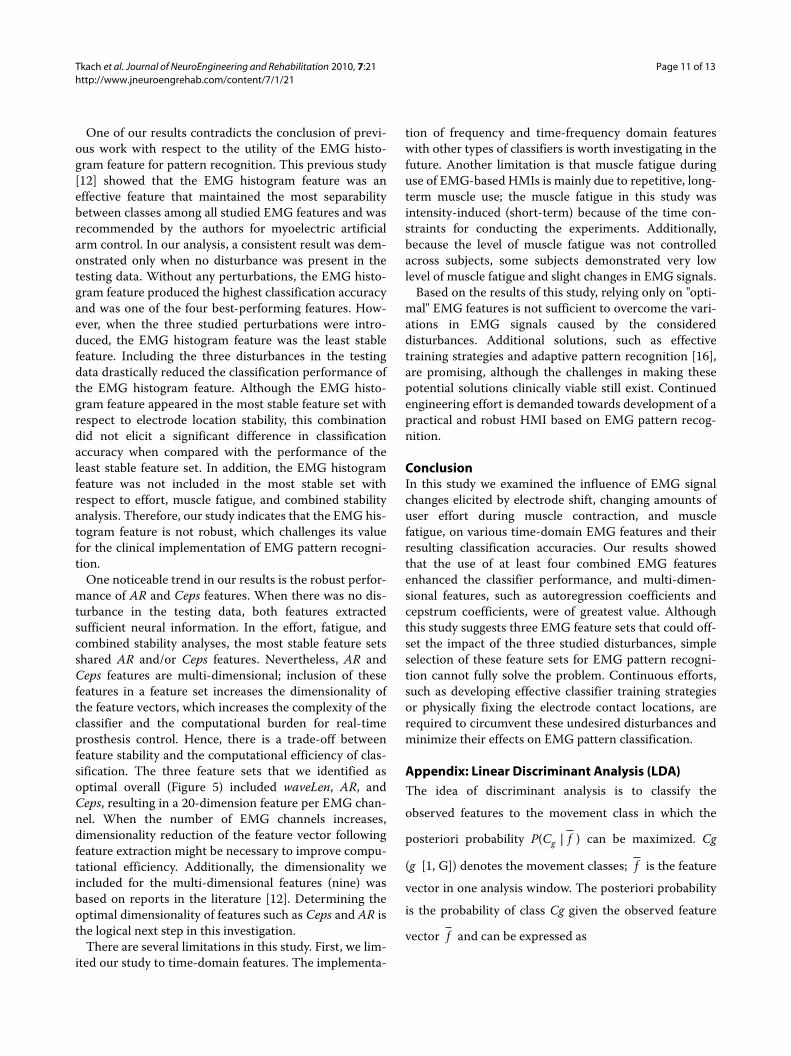
One of our results contradicts the conclusion of previ-ous work with respect to the utility of the EMG histo-gram feature for pattern recognition. This previous study[12] showed that the EMG histogram feature was aneffective feature that maintained the most separabilitybetween classes among all studied EMG features and wasrecommended by the authors for myoelectric artificialarm control. In our analysis, a consistent result was dem-onstrated only when no disturbance was present in thetesting data. Without any perturbations, the EMG histo-gram feature produced the highest classification accuracyand was one of the four best-performing features. How-ever, when the three studied perturbations were intro-duced, the EMG histogram feature was the least stablefeature. Including the three disturbances in the testingdata drastically reduced the classification performance ofthe EMG histogram feature. Although the EMG histo-gram feature appeared in the most stable feature set withrespect to electrode location stability, this combinationdid not elicit a significant difference in classificationaccuracy when compared with the performance of theleast stable feature set. In addition, the EMG histogramfeature was not included in the most stable set withrespect to effort, muscle fatigue, and combined stabilityanalysis. Therefore, our study indicates that the EMG his-togram feature is not robust, which challenges its valuefor the clinical implementation of EMG pattern recogni-tion.
One noticeable trend in our results is the robust perfor-mance of AR and Ceps features. When there was no dis-turbance in the testing data, both features extractedsufficient neural information. In the effort, fatigue, andcombined stability analyses, the most stable feature setsshared AR and/or Ceps features. Nevertheless, AR andCeps features are multi-dimensional; inclusion of thesefeatures in a feature set increases the dimensionality ofthe feature vectors, which increases the complexity of theclassifier and the computational burden for real-timeprosthesis control. Hence, there is a trade-off betweenfeature stability and the computational efficiency of clas-sification. The three feature sets that we identified asoptimal overall (Figure 5) included waveLen, AR, andCeps, resulting in a 20-dimension feature per EMG chan-nel. When the number of EMG channels increases,dimensionality reduction of the feature vector followingfeature extraction might be necessary to improve compu-tational efficiency. Additionally, the dimensionality weincluded for the multi-dimensional features (nine) wasbased on reports in the literature [12]. Determining theoptimal dimensionality of features such as Ceps and AR isthe logical next step in this investigation.
There are several limitations in this study. First, we lim-ited our study to time-domain features. The implementa-
tion of frequency and time-frequency domain featureswith other types of classifiers is worth investigating in thefuture. Another limitation is that muscle fatigue duringuse of EMG-based HMIs is mainly due to repetitive, long-term muscle use; the muscle fatigue in this study wasintensity-induced (short-term) because of the time con-straints for conducting the experiments. Additionally,because the level of muscle fatigue was not controlledacross subjects, some subjects demonstrated very lowlevel of muscle fatigue and slight changes in EMG signals.
Based on the results of this study, relying only on "opti-mal" EMG features is not sufficient to overcome the vari-ations in EMG signals caused by the considereddisturbances. Additional solutions, such as effectivetraining strategies and adaptive pattern recognition [16],are promising, although the challenges in making thesepotential solutions clinically viable still exist. Continuedengineering effort is demanded towards development of apractical and robust HMI based on EMG pattern recog-nition.
ConclusionIn this study we examined the influence of EMG signalchanges elicited by electrode shift, changing amounts ofuser effort during muscle contraction, and musclefatigue, on various time-domain EMG features and theirresulting classification accuracies. Our results showedthat the use of at least four combined EMG featuresenhanced the classifier performance, and multi-dimen-sional features, such as autoregression coefficients andcepstrum coefficients, were of greatest value. Althoughthis study suggests three EMG feature sets that could off-set the impact of the three studied disturbances, simpleselection of these feature sets for EMG pattern recogni-tion cannot fully solve the problem. Continuous efforts,such as developing effective classifier training strategiesor physically fixing the electrode contact locations, arerequired to circumvent these undesired disturbances andminimize their effects on EMG pattern classification.
Appendix: Linear Discriminant Analysis (LDA)The idea of discriminant analysis is to classify the
observed features to the movement class in which the
posteriori probability P(Cg | ) can be maximized. Cg
(g�[1, G]) denotes the movement classes; is the feature
vector in one analysis window. The posteriori probability
is the probability of class Cg given the observed feature
vector and can be expressed as
f
f
f

Tkach et al. Journal of NeuroEngineering and Rehabilitation 2010, 7:21http://www.jneuroengrehab.com/content/7/1/21
Page 12 of 13
where P(Cg) is the priori possibility, P( |Cg) is the like-
lihood, and P( ) is the possibility of observed feature
vector . Therefore, the discriminant analysis-based
classifiers can be mathematically described as
Given movement class Cg, the observed feature vectors
have a multivariate normal (MVN) distribution, i.e.
P( |Cg) ~ MVN(μg, Σg), where μg is the mean vector and
Σg is the covariance matrix of the class Cg. Additionally,
assume that the priori possibility P(Cg) is equivalent for
each movement class, and every class shared a common
covariance, i.e. Σg = Σ. Hence, the maximization of poste-
riori possibility in (13) becomes
is defined as the linear discriminant function.In the offline training μg and Σ were estimated by fea-
ture vectors calculated from a large amount of trainingdata and were stored in the flash memory.
where Kg is the number of observations in class Cg;
is the k observed feature vector in class Cg; Fg is
the feature matrix
; Mg is the mean
matrix that has the same number
of columns as in Fg. Therefore, the parameters in the lin-
ear discriminant function (15) were known, i.e.
In the testing phase, the observed feature derived
from each analysis window was fed to the classifier to cal-
culate in (16) for each movement class and was clas-
sified into a specific class that satisfied
Competing interestsThe authors declare that they have no competing interests.
Authors' contributionsDK designed experiments, conducted data collection and analysis, and draftedthe manuscript. HH and TK supervised the study and revised the manuscript.All authors read and approved the final manuscript.
AcknowledgementsThe authors sincerely thank Dr. Kevin Englehart of the University of New Bruns-wick, and Dr. Ping Zhou and Dr. Guanglin Li at the Rehabilitation Institute of Chicago for their assistance with this project. This work was supported by the NIH National Institute of Child and Human Development (Grants # R01 HD043137-01, #R01 HD044798 and # NO1-HD-5-3402), the Defense Advanced Research Projects Agency, and the National Institute on Disability and Rehabili-tation Research, U.S. Department of Education (Grant #H133F080006).
Author Details1Neural Engineering Center for Artificial Limbs, Rehabilitation Institute of Chicago, 345 E. Superior Street, Suite 1309, Chicago, IL, 60611, USA, 2Committee on Computational Neuroscience, University of Chicago, 1027 E 57th Street, Room 202, Chicago IL, 60637, USA, 3Department of Electrical, Computer, and Biomedical Engineering, University of Rhode Island, 4 E. Alumni Ave Kelly A-116, Kingston, RI, 02881, USA and 4Department of Physical Medicine and Rehabilitation, Northwestern University, Chicago, IL, 60611, USA
References1. Basmajian JV, De Luca CJ: Muscles alive: their functions revealed by
electromyography 5th edition. Baltimore: Williams & Wilkins; 1985. 2. Kuiken TA, Li G, Lock BA, Lipschutz RD, Miller LA, Stubblefield KA,
Englehart KB: Targeted muscle reinnervation for real-time myoelectric control of multifunction artificial arms. Jama 2009, 301:619-628.
3. Stein J, Narendran K, McBean J, Krebs K, Hughes R: Electromyography-controlled exoskeletal upper-limb-powered orthosis for exercise training after stroke. Am J Phys Med Rehabil 2007, 86:255-261.
4. Rosen J, Brand M, Fuchs MB, Arcan M: A myosignal-based powered exoskeleton system. IEEE Transation on System, Man, and Cybernetics 2001, 31:210-222.
5. Dipietro L, Ferraro M, Palazzolo JJ, Krebs HI, Volpe BT, Hogan N: Customized interactive robotic treatment for stroke: EMG-triggered therapy. IEEE Trans Neural Syst Rehabil Eng 2005, 13:325-334.
6. Kiguchi K, Imada Y, Liyanage M: EMG-based neuro-fuzzy control of a 4DOF upper-limb power-assist exoskeleton. Conf Proc IEEE Eng Med Biol Soc 2007, 2007:3040-3043.
7. Ferreira A, Celeste WC, Cheein FA, Bastos-Filho TF, Sarcinelli-Filho M, Carelli R: Human-machine interfaces based on EMG and EEG applied to robotic systems. J Neuroeng Rehabil 2008, 5:10.
8. Choi C, Micera S, Carpaneto J, Kim J: Development and quantitative performance evaluation of a noninvasive EMG computer interface. IEEE Trans Biomed Eng 2009, 56:188-191.
P C fP f Cg P Cg
P fg( | )
( | ) ( )
( )= (12)
f
f
f
C P C f P C f
P
g C g C g
C
g g
g
� � �= =
=
arg max { ( | )} arg max { ( | )}
arg max { (
ln
ln ff C P C P fg g� �| ) ( ) ( )}
.+ −ln ln
(13)
f
C P f C fg C g CT
g gT
gg g� � �= = ∑ − ∑− −arg max {ln ( | )} arg max { }1 11
2m m m
(14)
d f gT
CT
g gg= ∑ − ∑− −� 1 11
2m m m (15)
� �m g C kk
K
g g g gT
g
G
K g G K gf F M F M
g
g
= ∑ = − −= =
∑ ∑ −1 1 1
11 1
, ( )( )and
f C kg ,
F f f f f Kg C C C k C gg g g g= [ , , , , , ], , ,1 2 … …
Mg g g g= [ , , ]m m m� � … �
Received: 4 December 2009 Accepted: 21 May 2010 Published: 21 May 2010This article is available from: http://www.jneuroengrehab.com/content/7/1/21© 2010 Tkach et al; licensee BioMed Central Ltd. This is an Open Access article distributed under the terms of the Creative Commons Attribution License (http://creativecommons.org/licenses/by/2.0), which permits unrestricted use, distribution, and reproduction in any medium, provided the original work is properly cited.Journal of NeuroEngineering and Rehabilitation 2010, 7:21
d fCT
g gT
gg� � � � � � �= ∑ − ∑
− −1 112
m m m (16)
f
dC g�
C g�
C d C C C Cg C C g Gg g� � …= ∈arg max { }, { , , , }.1 2

Tkach et al. Journal of NeuroEngineering and Rehabilitation 2010, 7:21http://www.jneuroengrehab.com/content/7/1/21
Page 13 of 13
9. Williams TW: Practical methods for controlling powered upper-extremity prostheses. Assist Technol 1990, 2:3-18.
10. Englehart K, Hudgins B: A robust, real-time control scheme for multifunction myoelectric control. IEEE Trans Biomed Eng 2003, 50:848-854.
11. Englehart K, Hudgins B, Parker PA, Stevenson M: Classification of the myoelectric signal using time-frequency based representations. Med Eng Phys 1999, 21:431-438.
12. Zardoshti-Kermani M, Wheeler B, Badie K, Hashemi R: EMG feature evaulation for movement control of upper extrmity prostheses. IEEE Trans Rehabil Eng 1995, 3:324-333.
13. Hargrove L, Englehart K, Hudgins B: A Comparison of Surface and Intramuscular Myoelectric Signal Classification. IEEE Transactions on Biomedical Engineering 2007, 54:847-853.
14. Micera S, Sabatini AM, Dario P: On automatic identification of upper-limb movements using small-sized training sets of EMG signals. Med Eng Phys 2000, 22:527-533.
15. Zecca M, Micera S, Carrozza MC, Dario P: Control of multifunctional prosthetic hands by processing the electromyographic signal. Crit Rev Biomed Eng 2002, 30:459-485.
16. Sensinger JW, Lock BA, Kuiken TA: Adaptive pattern recognition of myoelectric signals: exploration of conceptual framework and practical algorithms. IEEE Trans Neural Syst Rehabil Eng 2009, 17:270-278.
17. Vigreux B, Cnockaert JC, Pertuzon E: Factors influencing quantified surface EMGs. Eur J Appl Physiol Occup Physiol 1979, 41:119-129.
18. Broman H, Bilotto G, De Luca CJ: Myoelectric signal conduction velocity and spectral parameters: influence of force and time. J Appl Physiol 1985, 58:1428-1437.
19. Sahlin K, Tonkonogi M, Soderlund K: Energy supply and muscle fatigue in humans. Acta Physiol Scand 1998, 162:261-266.
20. West W, Hicks A, Clements L, Dowling J: The relationship between voluntary electromyogram, endurance time and intensity of effort in isometric handgrip exercise. Eur J Appl Physiol Occup Physiol 1995, 71:301-305.
21. Delagi EF, Perotto A: Anatomic guide for the electromyographer--the limbs 2nd edition. Springfield, Ill.: Thomas; 1980.
22. Yoon T, Schlinder Delap B, Griffith EE, Hunter SK: Mechanisms of fatigue differ after low- and high-force fatiguing contractions in men and women. Muscle Nerve 2007, 36:515-524.
23. Sahlin K, Ren JM: Relationship of contraction capacity to metabolic changes during recovery from a fatiguing contraction. J Appl Physiol 1989, 67:648-654.
24. Smith IC, Newham DJ: Fatigue and functional performance of human biceps muscle following concentric or eccentric contractions. J Appl Physiol 2007, 102:207-213.
25. Zhou P, Lowery MM, Englehart KB, Huang H, Li G, Hargrove L, Dewald JP, Kuiken TA: Decoding a New Neural-Machine Interface for Control of Artificial Limbs. J Neurophysiol 2007.
26. Graupe D, Salahi J, Kohn KH: Multifunctional prosthesis and orthosis control via microcomputer identification of temporal pattern differences in single-site myoelectric signals. J Biomed Eng 1982, 4:17-22.
27. Hudgins B, Parker P, Scott RN: A new strategy for multifunction myoelectric control. IEEE Trans Biomed Eng 1993, 40:82-94.
28. Khezri M, Jahed M: Real-time intelligent pattern recognition algorithm for surface EMG signals. Biomed Eng Online 2007, 6:45.
29. Hargrove L, Englehart K, Hudgins B: The effect of electrode displacements on pattern recognition based myoelectric contro. the 28th IEEE EMG Annual International Conference; New York City 2006.
30. Huang H, Kuiken TA, Lipschutz RD: A strategy for identifying locomotion modes using surface electromyography. IEEE Trans Biomed Eng 2009, 56:65-73.
doi: 10.1186/1743-0003-7-21Cite this article as: Tkach et al., Study of stability of time-domain features for electromyographic pattern recognition Journal of NeuroEngineering and Rehabilitation 2010, 7:21


![Journal of NeuroEngineering and Rehabilitation BioMed Central · 2017. 4. 11. · and carryover of learned function to the home [3,5-22]. For example, research performed by Trombly,](https://img.pdfslide.us/doc/110x75/5fc34ac06ed1273d012cba37/journal-of-neuroengineering-and-rehabilitation-biomed-central-2017-4-11-and.jpg)


![Journal of NeuroEngineering and Rehabilitation BioMed Central...netorheological variable dampers [13], linear hydraulic actuators [14], electric actuators [15-18] and variable stiff-ness](https://img.pdfslide.us/doc/110x75/60cf54a6a10ddc313d3ffa49/journal-of-neuroengineering-and-rehabilitation-biomed-central-netorheological.jpg)
![Journal of NeuroEngineering and Rehabilitation BioMed Central...nized Gait Trainer [17,18], Lokomat® [19,20] and PAM, POGO and ARTHuR [21] are all examples of robotic devices that](https://img.pdfslide.us/doc/110x75/61085ab8b97ec57e51016c07/journal-of-neuroengineering-and-rehabilitation-biomed-central-nized-gait-trainer.jpg)


![Journal of NeuroEngineering and Rehabilitation BioMed Central...methods based on this approach, proved to be effective, e.g. treadmill training [4], but they require great physical](https://img.pdfslide.us/doc/110x75/60b96288caed344af334d893/journal-of-neuroengineering-and-rehabilitation-biomed-central-methods-based.jpg)