Embed Size (px)
Citation preview

21-1
Chapter 21Somatic Senses

21-2
Sensory Modalities
• Different types of sensations– touch, pain, temperature, vibration, hearing, vision
• Each type of sensory neuron can respond to only one type of stimuli
• Two classes of sensory modalities– general senses
• somatic are sensations from body walls
• visceral are sensations from internal organs
– special senses • smell, taste, hearing, vision, and balance

21-3
Process of Sensation
• Sensory receptors demonstrate selectivity– respond to only one type of stimuli
• Events occurring within a sensation– stimulation of the receptor– transduction (conversion) of stimulus into a
graded potential• vary in amplitude and are not propagated
– generation of impulses when graded potential reaches threshold
– integration of sensory input by the CNS

21-4
Sensory Receptors• Selectively respond to only one kind of stimuli
• Have simple or complex structures– General Sensory Receptors (Somatic Receptors)
• no structural specializations in free nerve endings that provide us with pain, tickle, itch, temperatures
• some structural specializations in receptors for touch, pressure & vibration
– Special Sensory Receptors (Special Sense Receptors)
• very complex structures---vision, hearing, taste, & smell

21-5
Classification of Sensory Receptors
• Structural classification
• Type of response to a stimulus
• Location of receptors & origin of stimuli
• Type of stimuli they detect

21-6
Structural Classification of Receptors
• Free nerve endings– bare dendrites– pain, temperature, tickle, itch & light touch
• Encapsulated nerve endings– dendrites enclosed in connective tissue capsule– pressure, vibration & deep touch
• Separate sensory cells– specialized cells that respond to stimuli– vision, taste, hearing, balance

21-7
Structural Classification
• Compare free nerve ending, encapsulated nerve ending and sensory receptor cell
21-8
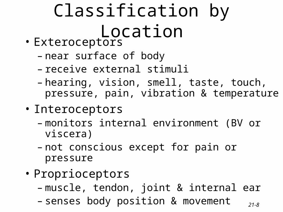
Classification by Location• Exteroceptors
– near surface of body– receive external stimuli– hearing, vision, smell, taste, touch, pressure, pain,
vibration & temperature
• Interoceptors– monitors internal environment (BV or viscera)– not conscious except for pain or pressure
• Proprioceptors– muscle, tendon, joint & internal ear– senses body position & movement

21-9
Classification by Stimuli Detected
• Mechanoreceptors– detect pressure or stretch– touch, pressure, vibration, hearing,
proprioception, equilibrium & blood pressure
• Thermoreceptors detect temperature
• Nociceptors detect damage to tissues
• Photoreceptors detect light
• Chemoreceptors detect molecules– taste, smell & changes in body fluid chemistry

21-10
Adaptation of Sensory Receptors
• Change in sensitivity to long-lasting stimuli – decrease in responsiveness of a receptor
• bad smells disappear
• very hot water starts to feel only warm
– potential amplitudes decrease during a maintained, constant stimulus
• Receptors vary in their ability to adapt– Rapidly adapting receptors (smell, pressure, touch)
• adapt quickly; specialized for signaling stimulus changes
– Slowly adapting receptors (pain, body position)• continuation of nerve impulses as long as stimulus persists

21-11
Somatic Tactile Sensations• Touch
– crude touch is ability to perceive something has touched the skin
– discriminative touch provides location and texture of source
• Pressure is sustained sensation over a large area
• Vibration is rapidly repetitive sensory signals
• Itching is chemical stimulation of free nerve endings
• Tickle is stimulation of free nerve endings only by someone else

21-12
Meissner’s Corpuscle
• Dendrites enclosed in CT in dermal papillae of hairless skin
• Discriminative touch & vibration-- rapidly adapting
• Generate impulses mainly at onset of a touch

21-13
•Free nerve endings found around follicles, detects movement of hair
Hair Root Plexus
21-14
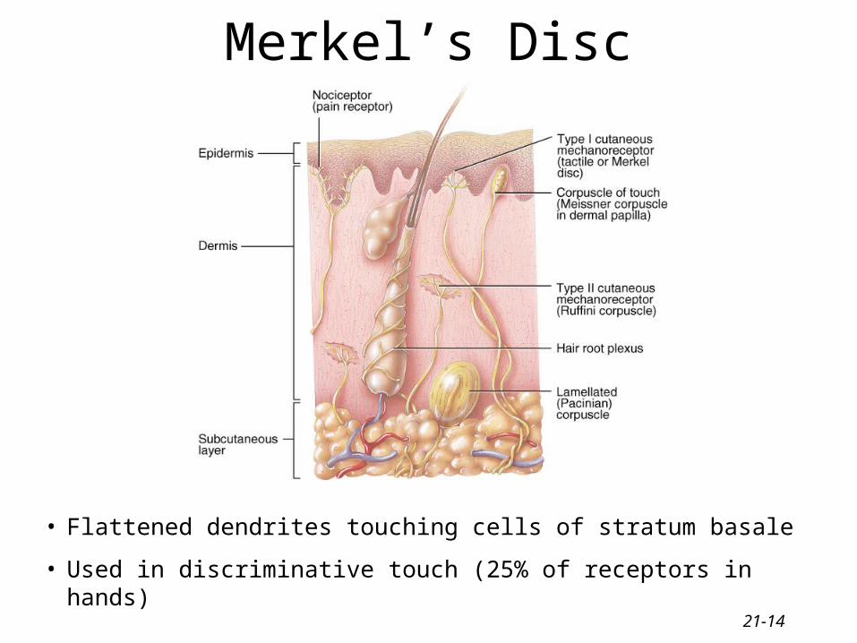
Merkel’s Disc
• Flattened dendrites touching cells of stratum basale
• Used in discriminative touch (25% of receptors in hands)

21-15
Ruffini Corpuscle
• Found deep in dermis of skin• Detect heavy touch, continuous touch, & pressure
21-16
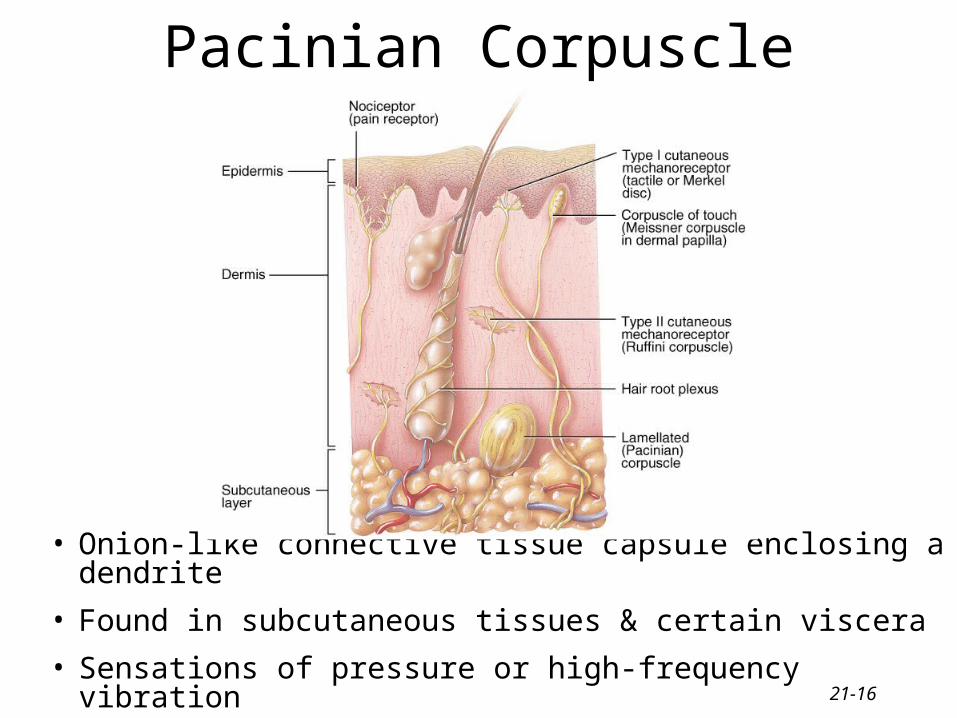
Pacinian Corpuscle
• Onion-like connective tissue capsule enclosing a dendrite
• Found in subcutaneous tissues & certain viscera
• Sensations of pressure or high-frequency vibration

21-17
Thermal Sensations• Free nerve endings with 1mm diameter receptive
fields on the skin surface
• Cold receptors in the stratum basale respond to temperatures between 50-105 degrees F
• Warm receptors in the dermis respond to temperatures between 90-118 degrees F
• Both adapt rapidly at first, but continue to generate impulses at a low frequency
• Pain is produced below 50 and over 118 degrees F.

21-18
Pain Sensations
• Nociceptors = pain receptors
• Free nerve endings found in every tissue of body except the brain
• Stimulated by excessive distension, muscle spasm, & inadequate blood flow
• Tissue injury releases chemicals such as K+, kinins or prostaglandins that stimulate nociceptors
• Little adaptation occurs

21-19
Types of Pain
• Fast pain (acute)– occurs rapidly after stimuli (.1 second)– sharp pain like needle puncture or cut– not felt in deeper tissues– larger A nerve fibers
• Slow pain (chronic)– begins more slowly & increases in intensity– aching or throbbing pain of toothache– in both superficial and deeper tissues– smaller C nerve fibers

21-20
Localization of Pain
• Superficial Somatic Pain arises from skin areas
• Deep Somatic Pain arises from muscle, joints, tendons & fascia
• Visceral Pain arises from receptors in visceral organs– localized damage (cutting) intestines causes no pain– diffuse visceral stimulation can be severe
• distension of a bile duct from a gallstone
• distension of the ureter from a kidney stone
• Phantom limb sensations -- cells in cortex still

21-21
Referred Pain
• Visceral pain that is felt just deep to the skin overlying the stimulated organ or in a surface area far from the organ.
• Skin area & organ are served by the same segment of the spinal cord.– Heart attack is felt in skin along left arm since both are supplied
by spinal cord segment T1-T5

21-22
Pain Relief
• Aspirin and ibuprofen block formation of prostaglandins that stimulate nociceptors
• Novocaine blocks conduction of nerve impulses along pain fibers
• Morphine lessen the perception of pain in the brain.

21-23
Proprioceptive or Kinesthetic Sense
• Awareness of body position & movement– walk or type without looking– estimate weight of objects
• Proprioceptors adapt only slightly
• Sensory information is sent to cerebellum & cerebral cortex– from muscle, tendon, joint capsules & hair cells
in the vestibular apparatus

21-24
Muscle Spindles
• Specialized intrafusal muscle fibers enclosed in a CT capsule and innervated by gamma motor neurons
• Stretching of the muscle stretches the muscle spindles sending sensory information back to the CNS
• Spindle sensory fiber monitor changes in muscle length
• Brain regulates muscle tone by controlling gamma fibers

21-25
Golgi Tendon Organs
• Found at junction of tendon & muscle• Consists of an encapsulated bundle of collagen fibers
laced with sensory fibers• When the tendon is overly stretched, sensory signals
head for the CNS & resulting in the muscle’s relaxation

21-26
Somatic Sensory Pathways
• First-order neuron conduct impulses to brainstem or spinal cord– either spinal or cranial nerves
• Second-order neurons conducts impulses from spinal cord or brainstem to thalamus--cross over to opposite side before reaching thalamus
• Third-order neuron conducts impulses from thalamus to primary somatosensory cortex (postcentral gyrus of parietal lobe)

21-27
Posterior Column-Medial Lemniscus Pathway of CNS
• Proprioception, vibration, discriminative touch, weight discrimination & stereognosis
• Signals travel up spinal cord in posterior column
• Fibers cross-over in medulla to become the medial lemniscus pathway ending in thalamus
• Thalamic fibers reach cortex

21-28
Spinothalamic Pathways• Lateral spinothalamic tract
carries pain & temperature• Anterior tract carries tickle,
itch, crude touch & pressure• First cell body in DRG with
synapses in cord• 2nd cell body in gray matter of
cord, sends fibers to other side of cord & up through white matter to synapse in thalamus
• 3rd cell body in thalamus projects to cerebral cortex

21-29
Somatosensory Map of Postcentral Gyrus
• Relative sizes of cortical areas
– proportional to number of sensory receptors
– proportional to the sensitivity of each part of the body
• Can be modified with learning
– learn to read Braille & will have larger area representing fingertips
21-30
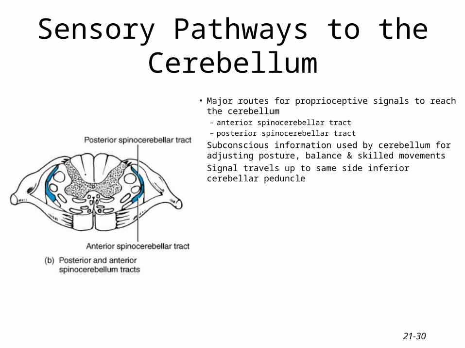
Sensory Pathways to the Cerebellum
• Major routes for proprioceptive signals to reach the cerebellum– anterior spinocerebellar tract
– posterior spinocerebellar tract
• Subconscious information used by cerebellum for adjusting posture, balance & skilled movements
• Signal travels up to same side inferior cerebellar peduncle

21-31
Somatic Motor Pathways• Control of body movement
– motor portions of cerebral cortex• initiate & control precise movements
– basal ganglia help establish muscle tone & integrate semivoluntary automatic movements
– cerebellum helps make movements smooth & helps maintain posture & balance
• Somatic motor pathways– direct pathway from cerebral cortex to spinal cord & out
to muscles– indirect pathway includes synapses in basal ganglia,
thalamus, reticular formation & cerebellum

21-32
Primary Motor Cortex
• Precentral gyrus initiates voluntary movement
• Cells are called upper motor neurons• Muscles represented unequally
(according to the number of motor units)
• More cortical area is needed if number of motor units in a muscle is high– vocal cords, tongue, lips, fingers &
thumb

21-33
Direct Pathway (Pyramidal Pathway)• 1 million upper motor neurons in cerebral cortex
– 60% in precentral gyrus & 40% in postcentral gyrus
• Axons form internal capsule in cerebrum and pyramids in the medulla oblongata
• 90% of fibers decussate(cross over) in the medulla– right side of brain controls left side muscles
• Terminate on interneurons which synapse on lower motor neurons in either:– nuclei of cranial nerves or anterior horns of spinal cord
• Integrate excitatory & inhibitory input to become final common pathway

21-34
Details of Motor Pathways• Lateral corticospinal tracts
– cortex, cerebral peduncles, 90% decussation of axons in medulla, tract formed in lateral column.
– skilled movements hands & feet• Anterior corticospinal tracts
– the 10% of axons that do not cross
– controls neck & trunk muscles• Corticobulbar tracts
– cortex to nuclei of CNs ---III, IV, V, VI, VII, IX, X, XI & XII
– movements of eyes, tongue, chewing, expressions & speech

21-35
Location of Direct Pathways
• Lateral corticospinal tract
• Anterior corticospinal tract
• Corticobulbar tract

21-36
Paralysis
• Flaccid paralysis = damage lower motor neurons– no voluntary movement on same side as damage– no reflex actions– muscle limp & flaccid– decreased muscle tone
• Spastic paralysis = damage upper motor neurons– paralysis on opposite side from injury– increased muscle tone– exaggerated reflexes

21-37
Final Common Pathway
• Lower motor neurons receive signals from both direct & indirect upper motor neurons
• Sum total of all inhibitory & excitatory signals determines the final response of the lower motor neuron & the skeletal muscles
21-38
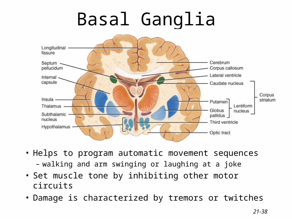
Basal Ganglia
• Helps to program automatic movement sequences– walking and arm swinging or laughing at a joke
• Set muscle tone by inhibiting other motor circuits• Damage is characterized by tremors or twitches

21-39
Basal Ganglia Connections
• Circuit of connections– cortex to basal ganglia
to thalamus to cortex
– planning movements
• Output from basal ganglia to reticular formation– reduces muscle tone
– damage produces rigidity of Parkinson’s disease

21-40
Cerebellar FunctionAspects of Function• learning• coordinated &
skilled movements• posture &
equilibrium
1. Monitors intentions for movements -- input from cerebral cortex
2. Monitors actual movements with feedback from proprioceptors
3. Compares intentions with actual movements
4. Sends out corrective signals to motor cortex
21-41
Spinal Cord Injury• Damaged by tumor, herniated disc, clot or trauma• Complete transection is cord severed resulting loss
of both sensation & movement below the injury• Paralysis
– monoplegia is paralysis of one limb only– diplegia is paralysis of both upper or both lower– hemiplegia is paralysis of one side– quadriplegia is paralysis of all four limbs
• Spinal shock is loss of reflex function (areflexia)– slow heart rate, low blood pressure, bladder problem– reflexes gradually return