Embed Size (px)
Citation preview
Motricidade © Edições Desafio Singular
2017, vol. 13, n. 4, pp. 62-73 http://dx.doi.org/10.6063/motricidade.9495
Manuscript received at June10th
2016; Accepted at June25th
2017
1 Universidade Federal do Rio Grande do Sul, UFRGS, Porto Alegre, RS, Brasil
*Corresponding author: Av. Paulo Gama, 110 - Farroupilha, Porto Alegre - RS, 90040-060, Brasil E-mail:
Feet positioning in the semi-static postural evaluation through photogrammetry: a systematic review
Arthur Antoniolli1*, Liliane Martini Araújo Ducati1, Emanuelle Francine Detogni Schmit1, Cláudia Tarragô Candotti1
REVIEW ARTICLE
ABSTRACT Photogrammetry is a relatively simple and objective instrument of evaluation that provides accurate and
reproducible quantitative results, if applied rigorously, existing nowadays several protocols. The main
purpose was to systematically review the utilized feet positioning to conduct a semi-static postural
evaluation through photogrammetry. It was performed a systematic review of observational studies and
clinic trials using the keywords “photogrammetry” and “spinal postural evaluation” to conduct searches on
scientific databases. The eligibility criteria adopted were: utilize the photogrammetry to evaluate children,
teenagers, adults or elders with or without pathologies; explicit the utilized feet positioning to conduct the
evaluation; and be written in English, Portuguese or Spanish. The methodological quality was assessed by
Downs and Black scale and the strength of evidence by the best evidence synthesis. It was found 1.786
articles and 40 were included in this review. The main feet positioning found were: self-referred, separated
and in parallel, united and in parallel and standard self-referred. The review exhibited strong strength of
evidence in the methodological quality assessment. Considering the main feet positioning found, it is
considered primordial that the selection of evaluation methodology respects the specific instructions of
each analysis protocol or software.
Keywords: Photogrammetry, Posture, Review.
INTRODUCTION
The body posture is a complex and hard to
measure phenomenon, its evaluation is the first
step to any physical or physiotherapy
intervention (Iunes et al., 2005). One option to
this type of evaluation is the computerized
photogrammetry, an objective method that is
reasonably simple and provides accurate and
reproducible quantitative results of easy
understanding (Belli, Chaves, de Oliveira, &
Grossi, 2009; Fortin et al., 2012; Moradi et al.,
2014).
The photogrammetry protocols exhibit some
discrepancies in between, as the feet positioning,
for example. On some protocols it is self-referred
by the evaluated individual, in which the
individual stands in the most comfortable
position (Ferreira et al., 2010). In others, the
position is previously determined. There is also
diversity in the supporting base shape, being
more acute (Furlanetto, Candotti, Comerlato, &
Loss, 2012) or obtuse (Kendall, McCreary, &
Provance, 1995). Considering the feet as the body
supporting base, its position might influence the
variables of the posture and consequently the
result. Therefore, the comparison of results of
different studies, epidemiological or
experimental, that utilize photogrammetry is
difficult. The solution to this problem initially
rests in the clever use of many feet positioning
possibilities proposed by different protocols and
software of evaluation. In this context, the
objective of this study was to identify, from a
systematic review, the feet positioning to conduct
a semi-static postural evaluation through
photogrammetry, pointing out positive and
negative aspects of each position, as well as
identifying the existence or not of an ideal
position of feet for the postural evaluation. It is
expected that the results of this systematic review
assist to support the selection of an evaluation
protocol and consequently feet positioning to be
Review of feet position in photogrammetry| 63
used by health professionals in the clinical and
scientific practice according to their reality.
METHOD
Research design
This study comprehended a systematic review
of literature (Galvão & Pereira, 2014) directed by
PRISMA Statement (Moher et al., 2009), based
on the recommendations of Cochrane (Higgins &
Green, 2011) collaboration, which has been
registered in PROSPERO
(http://www.crd.york.ac.uk/PROSPERO/display
_record.asp?ID=CRD42015026298) under the
code CRD42015026298.
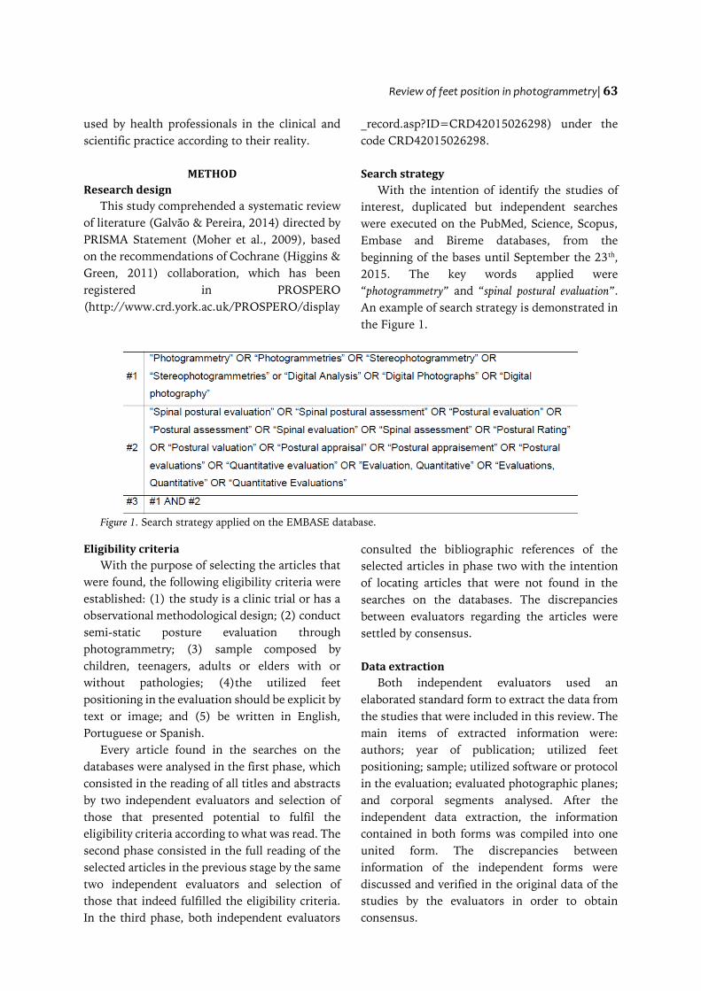
Search strategy
With the intention of identify the studies of
interest, duplicated but independent searches
were executed on the PubMed, Science, Scopus,
Embase and Bireme databases, from the
beginning of the bases until September the 23th
,
2015. The key words applied were
“photogrammetry” and “spinal postural evaluation”.
An example of search strategy is demonstrated in
the Figure 1.
Figure 1. Search strategy applied on the EMBASE database.
Eligibility criteria
With the purpose of selecting the articles that
were found, the following eligibility criteria were
established: (1) the study is a clinic trial or has a
observational methodological design; (2) conduct
semi-static posture evaluation through
photogrammetry; (3) sample composed by
children, teenagers, adults or elders with or
without pathologies; (4)the utilized feet
positioning in the evaluation should be explicit by
text or image; and (5) be written in English,
Portuguese or Spanish.
Every article found in the searches on the
databases were analysed in the first phase, which
consisted in the reading of all titles and abstracts
by two independent evaluators and selection of
those that presented potential to fulfil the
eligibility criteria according to what was read. The
second phase consisted in the full reading of the
selected articles in the previous stage by the same
two independent evaluators and selection of
those that indeed fulfilled the eligibility criteria.
In the third phase, both independent evaluators
consulted the bibliographic references of the
selected articles in phase two with the intention
of locating articles that were not found in the
searches on the databases. The discrepancies
between evaluators regarding the articles were
settled by consensus.
Data extraction
Both independent evaluators used an
elaborated standard form to extract the data from
the studies that were included in this review. The
main items of extracted information were:
authors; year of publication; utilized feet
positioning; sample; utilized software or protocol
in the evaluation; evaluated photographic planes;
and corporal segments analysed. After the
independent data extraction, the information
contained in both forms was compiled into one
united form. The discrepancies between
information of the independent forms were
discussed and verified in the original data of the
studies by the evaluators in order to obtain
consensus.
64 |A Antoniolli, LMA Ducati, EFD Schmit, CT Candotti
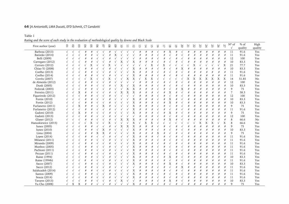
Table 1
Rating and the score of each study in the evaluation of methodological quality by downs and Black Scale
First author (year)
01
02
03
04
05
06
07
08
09
10
11
12
13
14
15
16
17
18
19
20
21
22
23
24
25
26
27
Nº of
√
% of
quality
High
quality
Barbosa (2013)
√ √ √ # # √ √ # √ √ √ √ # # # √ # X # √ # # # # # # # 11 91.6 Yes
Batistão (2014)
√ √ √ # # √ √ # X √ √ √ # # # √ # √ # √ # # # # # # # 11 91.6 Yes
Belli (2009)
√ √ √ # # √ √ # √ √ √ √ # # # √ # √ # √ # # # # # # # 12 100 Yes
Carregaro (2012)
√ √ √ # # √ √ # √ X √ X # # # √ # √ # √ # # # # # # # 10 83.3 Yes
Cerruto (2012) √ √ √ √ X √ √ X √ √ √ √ √ √ X √ X √ √ √ √ X √ √ √ √ X 21 77.7 Yes
Chiao Yi (2008)
√ √ √ # # √ √ # √ √ √ X # # # √ # √ # X # # # # # # # 10 83.3 Yes
Coelho (2013)
√ √ √ # # √ √ # √ √ √ X # # # √ # √ # √ # # # # # # # 11 91.6 Yes
Coelho (2014)
√ √ √ # # √ √ # √ √ √ X # # # √ # √ # √ # # # # # # # 11 91.6 Yes
Corrêa (2007) √ √ √ √ X √ √ X √ √ X X √ X X √ √ √ √ √ X X X X X X X 14 51.85 No
de Almeida (2012)
√ √ √ # # √ √ # √ √ √ √ # # # √ # √ # √ # # # # # # # 12 100 Yes
Dunk (2005)
√ √ √ # # √ √ # √ X √ X # # # √ # √ # √ # # # # # # # 10 83.3 Yes
Fedorak (2003)
√ √ √ # # √ √ # √ √ X X # # # √ # √ # X # # # # # # # 9 75 Yes
Ferreira (2011) √ √ X # # √ √ # √ X X X # # # √ # X # √ # # # # # # # 7 58.3 Yes
Figueiredo (2012)
√ √ √ # # √ √ # √ √ √ √ # # # √ # √ # √ # # # # # # # 12 100 Yes
Fortin (2010)
√ √ √ # # √ √ # √ √ √ X # # # √ # X # √ # # # # # # # 10 83.3 Yes
Fortin (2012)
√ √ √ # # √ √ # √ √ √ X # # # √ # X # √ # # # # # # # 10 83.3 Yes
Furlanetto (2011)
√ √ X # # √ X # √ √ √ X # # # √ # √ # √ # # # # # # # 9 75 Yes
Furlanetto (2012) √ √ √ # # √ √ # √ √ √ X # # # √ # √ # √ # # # # # # # 11 91.6 Yes
Gadotti (2010)
√ √ X # # √ √ # √ √ √ X # # # √ # X # √ # # # # # # # 9 75 Yes
Gadotti (2013) √ √ √ # # √ √ # √ √ √ √ # # # √ # √ # √ # # # # # # # 12 100 Yes
Glaner (2012)
√ √ √ # # √ √ # √ X X X # # # √ # X # √ # # # # # # # 8 66.6 No
Hanuskiewicz (2015)
√ √ √ √ X √ √ X √ √ √ X √ X X √ √ √ √ √ √ √ X X X √ X 8 66.6 No
Iunes (2005)
√ √ √ # # √ √ # √ X X X # # # √ # √ # √ # # # # # # # 9 75 Yes
Iunes (2010)
√ √ √ # # √ X # √ √ √ X # # # √ # √ # √ # # # # # # # 10 83.3 Yes
Lima (2004)
√ √ √ # # X X # √ √ √ X # # # X # √ # √ # # # # # # # 9 75 Yes
Lopes (2014)
√ √ √ # # √ √ # √ √ √ X # # # √ # √ # √ # # # # # # # 11 91.6 Yes
Milanesi (2011)
√ √ √ # # √ √ # √ √ √ X # # # √ # √ # √ # # # # # # # 11 91.6 Yes
Miranda (2009)
√ √ √ # # √ √ # √ √ √ X # # # √ # √ # √ # # # # # # # 11 91.6 Yes
Munhoz (2005)
√ √ √ # # √ √ # √ √ √ X # # # √ # √ # √ # # # # # # # 11 91.6 Yes
Pachioni (2011)
√ √ √ # # √ √ # √ √ √ X # # # √ # √ # √ # # # # # # # 11 91.6 Yes
Pezzan (2011)
√ √ √ # # √ √ # √ √ √ X # # # √ # √ # √ # # # # # # # 11 91.6 Yes
Raine (1994)
√ √ √ # # √ √ # √ √ √ X # # # √ # X # √ # # # # # # # 10 83.3 Yes
Raine (1994b)
√ √ √ # # √ √ # √ √ √ X # # # √ # √ # √ # # # # # # # 11 91.6 Yes
Sacco (2007)
√ √ √ # # √ √ # √ √ √ X # # # √ # X # √ # # # # # # # 10 83.3 Yes
Sacco (2012)
√ √ √ # # √ √ # √ √ √ √ # # # √ # √ # X # # # # # # # 11 91.6 Yes
Salahzadeh (2014)
√ √ √ # # √ √ # √ √ √ X # # # √ # √ # √ # # # # # # # 11 91.6 Yes
Santos (2009) √ √ √ # # √ √ # √ √ √ X # # # √ # √ # √ # # # # # # # 11 91.6 Yes
Souza (2014)
√ √ √ # # √ √ # √ √ √ X # # # √ # √ # √ # # # # # # # 11 91.6 Yes
Tavares (2013)
√ √ √ # # √ √ # √ X √ √ # # # √ # X # √ # # # # # # # 10 83.3 Yes
Yu Cho (2008)
√ X X # # √ √ # √ √ √ X # # # √ # √ # √ # # # # # # # 9 75 Yes
Review of feet position in photogrammetry| 65
Studies evaluation
To assess the methodological quality of
studies included in this review, the Downs and
Black scale (1998) (Downs & Black, 1998) was
used, which consists of a 27-item checklist that is
answered with yes when the information is
contemplated on the study (1 point) and no when
the information is not (0 points).The option to
use the Downs and Black scale was made because
it is a flexible instrument that evaluates
observational studies and clinic trials, in addition
to its internal consistency (Downs & Black, 1998)
and reproducibility. Every item was applied to the
clinic trials studies, however to the observational
studies only 12 items of the scale were considered
(Table 1) because the others referred to elements
that can only be applied to clinic trials. The
studies were classified with high methodological
quality when checked at least 70% of the
evaluation items (Silva & Carvalho, 2015). As
well as in the other stages, the methodological
quality assessment was conducted independently
by the same two evaluators and discrepancies
were settled by consensus.
Strength of evidence
The strength of evidence in this review was
analysed by the method of Best Evidence
Synthesis, which has been used by many groups
of researchers, including the Cochrane Back Review
Group(Trinh, 2009) and consists of an alternative
to the meta-analysis that determines the strength
of evidence through the number and quality of
included studies (Trinh, 2009).The following
criteria were adopted to analyse the strength of
evidence: strong evidence when many studies are
classified as high methodological quality;
moderate evidence when one study is classified as
high methodological quality and one or more
studies are classified as low methodological
quality; limited evidence when one study is
classified as high methodological quality and
many studies are classified as low methodological
quality; and no evidence when many studies are
classified as low methodological quality (Van
Tulder, Koes, & Bouter, 1997).
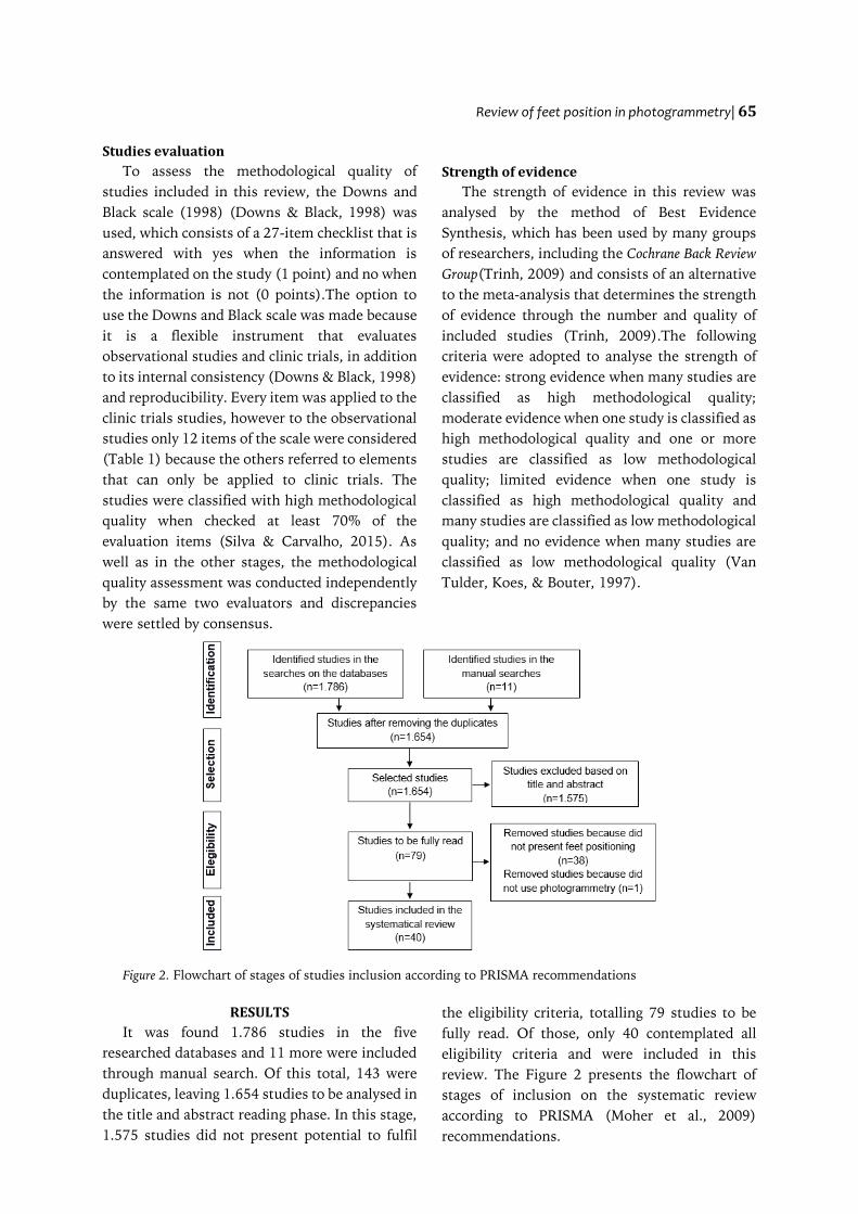
Figure 2. Flowchart of stages of studies inclusion according to PRISMA recommendations
RESULTS
It was found 1.786 studies in the five
researched databases and 11 more were included
through manual search. Of this total, 143 were
duplicates, leaving 1.654 studies to be analysed in
the title and abstract reading phase. In this stage,
1.575 studies did not present potential to fulfil
the eligibility criteria, totalling 79 studies to be
fully read. Of those, only 40 contemplated all
eligibility criteria and were included in this
review. The Figure 2 presents the flowchart of
stages of inclusion on the systematic review
according to PRISMA (Moher et al., 2009)
recommendations.
66 |A Antoniolli, LMA Ducati, EFD Schmit, CT Candotti
In the methodological quality assessment,
only three studies had percentage ≤ 70%, i.e.,
were classified as low methodological quality.
The percentage and score of each study are
presented in Table 1. Based on the Best Evidence
Synthesis and number of articles classified as
high methodological quality (37) and low
methodological quality (3), this systematic
review exhibits strong strength of evidence.
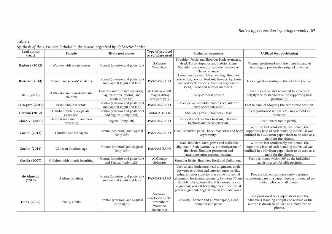
Table 2 presents the main methodological
information of included studies. Although the
methodology practically differs in all studies, the
feet positioning was assembled in groups and the
most common were: with inferior members in
parallel and separated by a previously determined
distance (Barbosa, Amorin, Zandonade, &
Delaprane, 2013; Belli, et al., 2009;Iunes, Cecílio,
Dozza, & Almeida, 2010; Iunes et al., 2005;
Milanesi et al., 2011; Pezzan, João, Ribeiro, &
Manfio, 2011; Sacco et al., 2007; Sacco et al.,
2012; Tavares et al., 2013); with the inferior
members positioned together (Chiao Yi, Jardim,
Inoue, & Pignatari, 2008; Futlanetto, Chaise,
Candotti, & Loss, 2011; Furlanetto et al., 2012;
Munhoz, Marquez, & Siqueira, 2005); with the
individual standing in self-referred or usual
posture (Fedorak, Ashworth, Marshall, & Paul,
2003; Gadotti & Biasotto-Gonzalez, 2010;
Gadotti, Armijo-Olivo, Silveira, & Magee, 2013;
Raine & Towmey, 1994a; Raine & Towmey,
1994b; Salazadeh et al., 2014; Yu Cho, 2007); and
standardizing the individual usual posture by
using the strategy of outlining the supporting
base of each person to utilize as a mold for the
photos (Coelho et al., 2013; Coelho et al., 2014;
de Almeida et al., 2013; Dunk, Lalonde, &
Callaghan, 2005; Ferreira, Duarte, Maldonado,
Bersanett, & Marques, 2011; Figueiredo, Amaral,
& Shimano, 2012;Glaner, Mota, Viana, & Santos,
2012; Lopes et al., 2014; Miranda, Schor, & Girão,
2009; Pachioni et al., 2011; Souza, Pasinato,
Corrêa, & da Silva, 2014).
The studies included in this review evaluated
children, teenagers and adults, some of them
from specific populations such as mouth
breathers (Chiao Yi et al., 2008; Corrêa & Bérzin,
2007; Lima, Baraúna, Sologurem, Canto, &
Gastaldi, 2004; Milanesi et al., 2011) and people
with temporomandibular joint disfunction
(Munhoz et al., 2005; Souza et al., 2014) (Table
2). Beyond that, most studies evaluated the
frontal and sagittal planes and corporal posture
almost completely, that is, evaluated parts of all
corporal segments, using several software or
protocols to obtain information about the
individual posture, highlighting the software
PAS/SAPO that was used in 16 studies
(Carregaro et al., 2012; Chiao Yi et al., 2008;
Coelho et al., 2013; Coelho et al., 2014; de
Almeida et al., 2013; Ferreira et al., 2011;
Figueiredo et al., 2012; Glaner et al., 2012; Lopes
et al., 2014; Milanesi et al., 2011; Pachioni et al.,
2011; Pezzan et al., 2011; Santos, Silva, Sanada,
& Alves, 2009; Souza et al., 2014; Tavares et al.,
2013) (Table 2).
DISCUSSION
According to the results of this review, there
is a huge variety of feet positioning utilized to
conduct a semi-static postural evaluation through
photogrammetry, however it does not derail the
execution of methodological comparisons, as
most of the studies determine that the individual
stands in a natural or self-referred posture for
photos (Fedorak et al., 2003; Gadotti & Biasotto-
Gonzalez, 2010; Gadotti et al., 2013; Raine &
Towmey, 1994a; Raine & Towmey, 1994b;
Salazadeh et al., 2014; Yu Cho, 2007). According
to Smith, Weiss, and Lehmkuhl (1997), the
aligned posture is not natural because requires a
conscious effort and an increase in muscular
activity, so they suggest that the standard feet
positioning prioritize body comfort and
relaxation instead of a previously determined
body alignment.
Although Bullock-Saxton (1993) and Lapierre
(1982) sustain that only the comfortable upright
posture adopted by the individual in the moment
of evaluation is the true representative of body
alignment, the use of a previously determined
supporting base before any evaluation starts is
repeatedly utilized. The reason is based on the
fact that any other protocol that does not utilize
a previously determined position derails the
comparison between another evaluation by the
same individual (Ferreira et al., 2010; Watson,
1998).
Review of feet position in photogrammetry| 67
Table 2
Synthesis of the 40 studies included in the review, organized by alphabetical order
Lead author
(year) Sample Evaluated planes
Type of protocol
or software used Evaluated segments Utilized feet positioning
Barbosa (2013) Women with breast cancer Frontal (anterior and posterior) Software
CorelDraw
Shoulder, Pelvis and Shoulder blade evenness,
Head, Torso, Superior and Inferior Spine,
Shoulder blade rotation and the distance of
Thales’ triangle.
Women positioned with their feet in parallel
standing on previously designed drawings.
Batistão (2014) Elementary schools’ students Frontal (anterior and posterior)
and Sagittal (right and left) PAS/PAS/SAPO
Lateral and forward Head leaning, Shoulder
protraction, cervical lordosis, thoracic kyphosis
and low back lordosis. Another segment of
Head, Torso and inferior members.
Feet aligned according to the width of the hip.
Belli (2009) Asthmatic and non-Asthmatic
children
Frontal (anterior and posterior),
Sagittal (three photos) and
zoom in the face
ALCimage-2000
Image-Editing
Software v1.5
Every corporal posture
Feet in parallel and separated by a piece of
polystyrene to standardize the supporting base
positioning.
Carregaro (2012) Brazil Public servants Frontal (anterior and posterior)
and Sagittal (right and left) PAS/PAS/SAPO
Head, pelvis, shoulder blade, knee, inferior
members/ankles/feet Feet in parallel adopting the orthostatic position.
Cerruto (2012) Children with quick palatal
expansion
Frontal (anterior and posterior)
and Sagittal (only right) AutoCAD2008 Shoulder girdle, Shoulders, Head
Feet positioned within 30º using a mold as
reference.
Chiao Yi (2008) Children with mouth and nose
breathing Sagittal (only left) PAS/PAS/SAPO
Cervical and Low back lordosis, Thoracic
kyphosis and pelvis position. Feet united and in parallel.
Coelho (2013) Children and teenagers Frontal posterior) and Sagittal
(only left) PAS/PAS/SAPO
Head, shoulder, pelvis, knee, malleolus and body
asymmetry.
With the feet comfortably positioned, the
supporting base of each standing individual was
outlined in a 30x40cm paper sheet to be used as a
mold for the photos.
Coelho (2014) Children in school age Frontal (anterior) and Sagittal
(only left) PAS/PAS/SAPO
Head, shoulder, knee, pelvis and malleolus
alignment. Body symmetry, anteriorization of
the Head, Shoulder protrusion and
anteroposterior corporal leaning.
With the feet comfortably positioned, the
supporting base of each standing individual was
outlined in a 30x40cm paper sheet to be used as a
mold for the photos.
Corrêa (2007) Children with mouth breathing Frontal (anterior and posterior)
and Sagittal (only right)
ALCimage
Software Shoulder blade, Shoulder, Head and Collarbone.
Feet positioned within 30º as the individual
stands in a comfortable position.
de Almeida
(2013) Asthmatic adults
Frontal (anterior and posterior)
and Sagittal (right and left) PAS/PAS/SAPO
Vertical and horizontal head alignment, angle
between acromion and anterior superior iliac
spine, anterior superior iliac spine horizontal
alignment, horizontal symmetry between T3 and
shoulder blade, vertical and horizontal torso
alignment, vertical body alignment, horizontal
pelvis alignment, angle between knee and ankle
Feet positioned on a previously designed
supporting base in a paper sheet as its rotated to
obtain photos of all planes.
Dunk (2005) Young adults Frontal (anterior) and Sagittal
(only right)
Software
developed by the
university of
Waterloo
(nameless)
Cervical, Thoracic and Lumbar spine, Head,
Shoulder and pelvis
Feet positioned on a paper sheet with the
individuals standing upright and relaxed as the
outline is drawn to be used as a mold for the
photos.
68 |A Antoniolli, LMA Ducati, EFD Schmit, CT Candotti
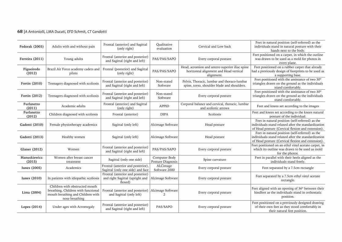
Fedorak (2003) Adults with and without pain Frontal (anterior) and Sagittal
(only right)
Qualitative
evaluation Cervical and Low back
Feet in natural position (self-referred) as the
individuals stand in natural posture with their
hands next to the body.
Ferreira (2011) Young adults Frontal (anterior and posterior)
and Sagittal (right and left) PAS/PAS/SAPO Every corporal posture
Feet positioned on a carpet, in which the outline
was drawn to be used as a mold for photos in
every plane.
Figueiredo
(2012)
Brazil Air Force academy cadets and
pilots
Frontal (posterior) and Sagittal
(only right) PAS/PAS/SAPO
Head, acromion and antero superior iliac spine
horizontal alignment and Head vertical
alignment.
Feet positioned on a rubber carpet that already
had a previously design of footprints to be used as
a supporting base.
Fortin (2010) Teenagers diagnosed with scoliosis Frontal (anterior and posterior)
and Sagittal (right and left)
Non-stated
Software
Pelvis, Thoracic, lumbar and thoraco-lumbar
spine, torso, shoulder blade and shoulders.
Feet positioned with the assistance of two 30º
triangles drawn on the ground as the individuals
stand comfortably.
Fortin (2012) Teenagers diagnosed with scoliosis Frontal (anterior and posterior)
and Sagittal (right and left)
Non-stated
Software Every corporal posture
Feet positioned with the assistance of two 30º
triangles drawn on the ground as the individuals
stand comfortably.
Furlanetto
(2011) Academic adults
Frontal (anterior) and Sagittal
(only right) APPID
Corporal balance and cervical, thoracic, lumbar
and scoliotic arrows Feet and knees set according to the images
Furlanetto
(2012) Children diagnosed with scoliosis Frontal (anterior) DIPA Scoliosis
Feet and knees set according to the knees natural
posture of the individual.
Gadotti (2010) Female physiotherapy academics Sagittal (only left) Alcimage Software Head posture
Feet in natural position (self-referred) as the
individuals stand relaxed after the standardization
of Head posture (Cervical flexion and extension).
Gadotti (2013) Healthy women Sagittal (only left) Alcimage Software Head posture
Feet in natural position (self-referred) as the
individuals stand relaxed after the standardization
of Head posture (Cervical flexion and extension).
Glaner (2012) Women Frontal (anterior and posterior)
and Sagittal (right and left) PAS/PAS/SAPO Every corporal posture
Feet positioned on an ethyl vinyl acetate carpet, in
which its outline was drawn to be used as mold
for the photos.
Hanuskiewicz
(2015)
Women after breast cancer
treatment Sagittal (only one side)
Computer Body
Posture Diagnosis Spine curvature
Feet in parallel with their heels aligned as the
individuals stand freely.
Iunes (2005) Academics Frontal (anterior and posterior),
Sagittal (only one side) and face
ALCimage
Software 2000 Every corporal posture Feet separated by a 7.5cm rectangle
Iunes (2010) In patients with idiopathic scoliosis
Frontal (anterior and posterior)
and right Sagittal (upright and
flexed)
Alcimage Software Every corporal posture Feet separated by a 7.5cm ethyl vinyl acetate
rectangle.
Lima (2004)
Children with obstructed mouth
breathing, Children with functional
mouth breathing and Children with
nose breathing
Frontal (anterior and posterior)
and Sagittal (only left)
Alcimage Software
2 Every corporal posture
Feet aligned with an opening of 36º between their
hindfeet as the individuals stand in orthostatic
position.
Lopes (2014) Under ages with Acromegaly Frontal (anterior and posterior)
and Sagittal (right and left) PAS/SAPO Every corporal posture
Feet positioned on a previously designed drawing
of their own feet as they stood comfortably in
their natural feet position.
Review of feet position in photogrammetry| 69
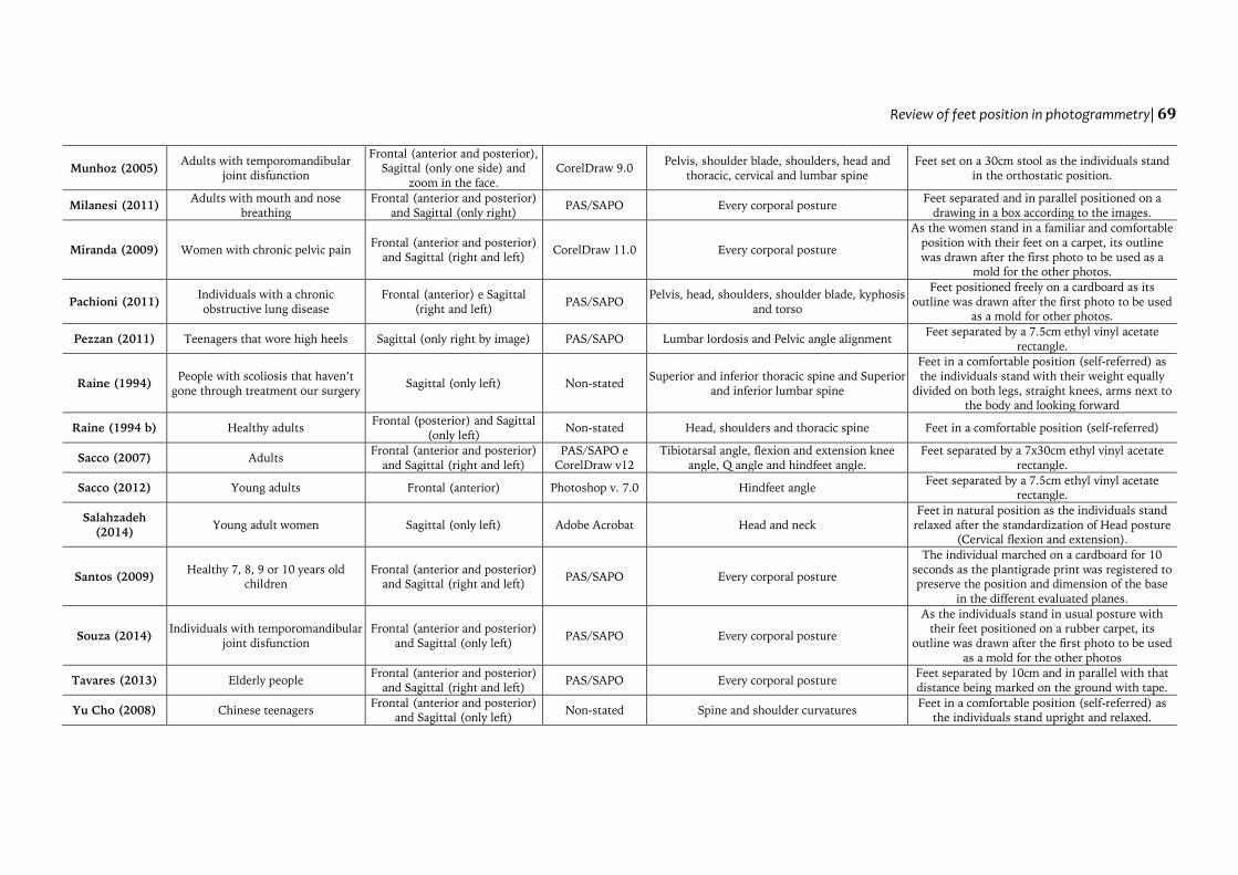
Munhoz (2005) Adults with temporomandibular
joint disfunction
Frontal (anterior and posterior),
Sagittal (only one side) and
zoom in the face.
CorelDraw 9.0 Pelvis, shoulder blade, shoulders, head and
thoracic, cervical and lumbar spine
Feet set on a 30cm stool as the individuals stand
in the orthostatic position.
Milanesi (2011) Adults with mouth and nose
breathing
Frontal (anterior and posterior)
and Sagittal (only right) PAS/SAPO Every corporal posture
Feet separated and in parallel positioned on a
drawing in a box according to the images.
Miranda (2009) Women with chronic pelvic pain Frontal (anterior and posterior)
and Sagittal (right and left) CorelDraw 11.0 Every corporal posture
As the women stand in a familiar and comfortable
position with their feet on a carpet, its outline
was drawn after the first photo to be used as a
mold for the other photos.
Pachioni (2011) Individuals with a chronic
obstructive lung disease
Frontal (anterior) e Sagittal
(right and left) PAS/SAPO
Pelvis, head, shoulders, shoulder blade, kyphosis
and torso
Feet positioned freely on a cardboard as its
outline was drawn after the first photo to be used
as a mold for other photos.
Pezzan (2011) Teenagers that wore high heels Sagittal (only right by image) PAS/SAPO Lumbar lordosis and Pelvic angle alignment Feet separated by a 7.5cm ethyl vinyl acetate
rectangle.
Raine (1994) People with scoliosis that haven’t
gone through treatment our surgery Sagittal (only left) Non-stated
Superior and inferior thoracic spine and Superior
and inferior lumbar spine
Feet in a comfortable position (self-referred) as
the individuals stand with their weight equally
divided on both legs, straight knees, arms next to
the body and looking forward
Raine (1994 b) Healthy adults Frontal (posterior) and Sagittal
(only left) Non-stated Head, shoulders and thoracic spine Feet in a comfortable position (self-referred)
Sacco (2007) Adults Frontal (anterior and posterior)
and Sagittal (right and left)
PAS/SAPO e
CorelDraw v12
Tibiotarsal angle, flexion and extension knee
angle, Q angle and hindfeet angle.
Feet separated by a 7x30cm ethyl vinyl acetate
rectangle.
Sacco (2012) Young adults Frontal (anterior) Photoshop v. 7.0 Hindfeet angle Feet separated by a 7.5cm ethyl vinyl acetate
rectangle.
Salahzadeh
(2014) Young adult women Sagittal (only left) Adobe Acrobat Head and neck
Feet in natural position as the individuals stand
relaxed after the standardization of Head posture
(Cervical flexion and extension).
Santos (2009) Healthy 7, 8, 9 or 10 years old
children
Frontal (anterior and posterior)
and Sagittal (right and left) PAS/SAPO Every corporal posture
The individual marched on a cardboard for 10
seconds as the plantigrade print was registered to
preserve the position and dimension of the base
in the different evaluated planes.
Souza (2014) Individuals with temporomandibular
joint disfunction
Frontal (anterior and posterior)
and Sagittal (only left) PAS/SAPO Every corporal posture
As the individuals stand in usual posture with
their feet positioned on a rubber carpet, its
outline was drawn after the first photo to be used
as a mold for the other photos
Tavares (2013) Elderly people Frontal (anterior and posterior)
and Sagittal (right and left) PAS/SAPO Every corporal posture
Feet separated by 10cm and in parallel with that
distance being marked on the ground with tape.
Yu Cho (2008) Chinese teenagers Frontal (anterior and posterior)
and Sagittal (only left) Non-stated Spine and shoulder curvatures
Feet in a comfortable position (self-referred) as
the individuals stand upright and relaxed.
70 |A Antoniolli, LMA Ducati, EFD Schmit, CT Candotti
However, this standardization is still fairly
vague, and the literature does not answer this
question with certainty as it is proven by the
many feet positioning found in the studies. The
most mentioned standardization is the strategy of
standardize the self-referred position of each
individual. To do that, the evaluators request that
before the first photograph the individuals stand
comfortably on a paper sheet, cardboard or
carpet. After that procedure, the evaluator
outlines their feet to obtain a mold for position in
the evaluation. Every time that there is a change
in the photo plane, the mold is rotated to the
desired position and the individual is relocated to
step on the previously determined
drawing.(Coelho et al., 2013; Coelho et al., 2014;
de Almeida et al., 2013; Dunk, Lalonde, &
Callaghan, 2005; Ferreira, Duarte, Maldonado,
Bersanett, & Marques, 2011; Figueiredo, Amaral,
& Shimano, 2012; Glaner, Mota, Viana, & Santos,
2012; Lopes et al., 2014; Miranda, Schor, & Girão,
2009; Pachioni et al., 2011; Souza, Pasinato,
Corrêa, & da Silva, 2014). This manoeuvre might
be efficient in reproducibility studies,
nevertheless when adopted to only one
evaluation, its standardization may be questioned
because the usual intention is to evaluate the
individual in its natural posture.
Another well-established pattern of foot
positioning utilizes the lower limbs positioned in
parallel and apart (Barbosa et al., 2013; Belli et
al., 2009; Iunes et al., 2005; Iunes et al., 2010;
Milanesi et al., 2011; Pezzan et al., 2011; Sacco et
al., 2007; Sacco et al., 2012; Tavares et al., 2013).
One example of protocol that utilizes that type of
position for photogrammetry is the classical
study by Kendall et al. (1995) which believes that
postural evaluation should be conducted with a
supporting base with the individual’s feet in
parallel, their heels separated by a 7.5cm distance
and the hind feet abducted 8º to 10º from the
median line. For Ferreira et al., (2010), the
distances between the anatomical references of
the feet, such as heels and medial malleolus, are
measures that do not correctly manifest a
supporting base because they do not quantify the
possible rotation in the feet positioning, being
only valid to quantify valgus knee.
It is common to find studies that do not
denote the position of evaluated inferior
members clearly, such as the study of Batistão et
al. (2014), which requires the individuals to align
their feet according to the width of their hip and
the study of Hanuskiewicz et al. (2015), that
requires the individuals to stand freely with their
feet in parallel and heels aligned. Both studies do
not determine angles or distances between feet,
which might be considered a lack of rigor in the
protocol’s methodology and produce misleading
results in the evaluation.
The positioning of inferior members as a set,
considering knees and feet, provides a more
obtuse supporting base and is also utilized for
postural evaluation through photogrammetry
(Chiao Yi et al., 2008; Furlanetto et al., 2011;
Furlanetto et al., 2012; Munhoz et al., 2005). In
that position of inferior members, it is important
that knees are aligned. For example, if the
individual has a space between the knees when
its malleolus are touching, the individual should
stay with the malleolus united and the rest of
inferior members in the most comfortable
position; if the individual has a space between the
malleolus when its knees are touching, the
individual should stay with the knees united and
preserve its physiological space between
malleolus; and if the individual succeed in
aligning knees and malleolus simultaneously,
that position should be adopted (Watson, 1998).
The option for this standardization protocol of
feet positioning ensures the maintenance of
valgus knee standards. In case the evaluator does
not observe the individual’s position, it might
mask the results of some evaluation segments,
even because of the discomfort that may be
generated in the individual.
Only one study of all found used a dynamic
standardization to determine the feet positioning
for the semi-static postural evaluation. Santos et
al., (2009) requires the individual to march, i.e.,
to execute some type of static walk on a
cardboard, in which the plantigrade print of the
feet positioning is registered to be utilized as a
mold for the photos. For Santos (2011), the feet
position of a step should be utilized as it is more
functional and unique for each individual and
because when feet are in parallel, the hip joints
Review of feet position in photogrammetry| 71
rotates internally, the great trochanters go
forward and the muscles within this space are
tensioned, as this tension pulls its near insertions
in the femur direction and provokes a small
retraction of the pelvis and the internal rotation
of femoral condyles, patella and malleolus, it is
impossible to know the position that those
segments really adopt in the orthostatic
physiological posture.
In summary, though many of feet positioning
to conduct a semi-static postural evaluation
through photogrammetry were found, none of
the studies compared their results in different
feet positioning. Therefore, it is considered that
is not possible yet to affirm which is the best feet
positioning to be adopted in a postural
evaluation, as the decision still depends on the
professional and its experience and objectives.
The diversity of protocols of postural evaluation,
as well as the lack of detailed description for an
ideal understanding of data collection procedure,
limits a complex discussion about the topic.
CONCLUSION
The positioning of feet using the strategy of
standardizing the self-reported position of each
individual has been the most used in the studies.
However, based on the results, it is still not
possible to state that this is the most adequate
foot positioning for use in the semi-static
postural evaluation through photogrammetry.
Considering that this systematic review presents
strong evidence and that there is currently no
single foot position considered to be ideal, it is
considered primordial that the selection of
evaluation methodology respects the specific
instructions of each analysis protocol or software
used to conduct a semi-static postural evaluation
through photogrammetry, such as its procedures
and particularities. Experimental studies are still
necessary to obtain accurate information about
the best positioning of feet for this type of
evaluation.
Acknowledgments:
Nothing to declare.
Conflict of interests:
Nothing to declare.
Funding:
Nothing to declare.
REFERENCES
Barbosa, J. A. N., Amorin, M. H. C., Zandonade, E., &
Delaprane, M. L. (2013). Avaliação da postura
corporal em mulheres com câncer de mama.
Revista Brasileira de Ginecologia e Obstetetricia,
35(5), 215-20.
Batistão, M. V., Carnaz, L., Barbosas L. F., da Motta, G.
C., & Sato, T. O. (2014). Posture and
musculoskeletal pain in eutrophic, overweighed,
and obese student: A cross-sectional study.
Motriz, 20(2), 192-199.
Belli, J. F., Chaves, T. C., de Oliveira, A. S., & Grossi,
D. B. (2009). Analysis of body posture in children
with mild to moderate asthma. European Journal of
Pediatrics, 168(10), 1207–16.
Bullock-Saxton, J. (1993). Postural alignment in
standing: a repeatability study. Australian
Physiotherapy, 39(1)25-29.
Carregaro, R., Falcão, J., Massuda, K., Masunaga, D.,
Sinzato, C., de Oliveira, A. B., & Padula, R. S.
(2012). Postural analysis and psychosocial
measurements of federal civil servants of an
institution of higher education. Work, 41(Suppl
1), 4795-4800.
Cerruto, C., Di Vecce, L., Doldo, T., Giovanetti, A.,
Polimeni, A., & Goracci, C. (2012). A
computerized photographic method to evaluate
changes in head posture and scapular position
following rapid palatal expansion: A pilot study.
Journal of Clinical Pediatric Dentistry, 37(2), 213-
218.
Chiao Yi, L., Jardim, J. R., Inoue, D. P., & Pignatari, S.
S. N. (2008). Relationship between excursion of
the diaphragm and curvatures of the spinal
column in mouth breathing children. Journal of
Pediatrics, 84(2), 171-177.
Coelho, J. J., Graciosa, M. D., De Medeiros, D. L., Da
Costa, L. M. R., Martinello, M., & Ries, L. G. K.
(2013). Influence of nutritional status and
physical activity on the posture of children and
adolescents. Fisioterapia e Pesquisa, 20(2), 136-142.
Coelho, J. J., Graciosa, M. D., de Medeiros, D. L.,
Pacheco, S. C. S., da Costa, L. M. R., & Ries, L. G.
K. (2014). Influence of flexibility and gender on
the posture of school children. Revista Paulista de
Pediatria, 32(3), 233-228.
Corrêa, E. C. R, & Bérzin, F. (2007). Efficacy of
physical therapy on cervical muscle activity and
on body posture in school-age mouth breathing
children. International Journal of Pediatric
Otorhinolaryngology, 71(10), 1527-1535.
de Almeida, V. P., Guimarães, F. S., Moço, V. J. R.,
Ferreira, A. S., Menezes, S. L., & Lopes, A. J.
(2013). Is there an association between postural
balance and pulmonary function with asthma?
Clinics, 68(11), 1421-1427.
72 |A Antoniolli, LMA Ducati, EFD Schmit, CT Candotti
Downs, S. H., Black, N. (1998). The feasibility of
creating a checklist for the assessment of the
methodological quality both of randomised and
non-randomised studies of health care
interventions. Journal of Epidemiology and
Community Health, 52(6),377-84.
Dunk, N. M., Lalonde, J., & Callaghan, J. P. (2005).
Implications for the use of postural analysis as a
clinical diagnostic tool: reliability of quantifying
upright standing spinal postures from
photographic images. Journal of Manipulative and
Physiological Therapeutics, 28(6), 386-92.
Fedorak, C., Ashworth, N., Marshall, J., & Paull, H.
(2003). Reliability of the visual assessment of
cervical and lumbar lordosis: How good are we?
Spine, 28(16), 1857-9.
Ferreira, E. A., Duarte, M., Maldonado, E. P.,
Bersanett, A. A., & Marques, A. P. (2011).
Quantitative assessment of postural alignment in
young adults based on photographs of anterior,
posterior, and lateral views. Journal Manipulative
and Physiological Therapeutics, 34(6), 371-80.
Ferreira, E. A. G., Duarte, M., Maldonado, E. P., Burke,
T. N., & Marques, A. P. (2010). Postural
assessment software (PAS/SAPO): validation and
reliability. Clinics, 65(7), 675–681.
Figueiredo, R. V., Amaral, A. C., & Shimano, A. C.
(2012). Photogrammetry on the identification of
postural asymmetries in cadets and pilots of the
Brazilian air force academy. Revista Brasileira de
Fisioterapia, 16(1), 54-60.
Fortin, C., Feldman, D. E., Chariet, F., Gravel, D.,
Gauthier, F., & Labelle, H. (2012). Reliability of
a quantitative clinical posture assessment tool
among persons with idiopathic scoliosis.
Physiotherapy, 98(1), 64-75.
Fortin, C., Feldman, D. E., Chariet, F., & Labelle, H.
(2010). Validity of a quantitative clinical
measurement tool of trunk posture in idiopathic
scoliosis. Spine, 35(19), E988-E994.
Furlanetto, T. S., Candotti, C. T., Comerlato, T., &
Loss, J. F. (2012). Validating a posture evaluation
method developed using a Digital Image-based
Postural Assessment (DIPA) software. Computer
Methods and Programs in Biomed, 108(1), 203-212.
Furlanetto, T. S., Chaise, F. O., Candotti, C. T., & Loss,
J. F. (2011). Fidedignidade de um protocolo de
avaliação postural. Revista de Educação Física/UEM,
22(3), 411-419.
Gadotti, I. C., Armijo-Olivo, S., Silveira, A., & Magee,
D. (2013). Reliability of the craniocervical
posture assessment: visual and angular
measurements using photographs and
radiographs. Journal of Manipulative and
Physiological Therapeutics, 36(9), 619-625.
Gadotti, I. C., & Biasotto-Gonzalez, D. A. (2010).
Sensitivity of clinical assessments of sagittal head
posture. Journal of Evaluation in Clinical Practice,
16(1), 141–144.
Galvão, T. F., & Pereira, M. G. (2014). Revisões
sistemáticas da literatura: passos para sua
elaboração. Epidemiologia e Serviços de Saúde, 23(1),
183-184.
Glaner, M. F., Mota, Y. L., Viana, A. C. R., & Santos,
M. C. (2012). Fotogrametria: Fidedignidade e
falta de objetividade na avaliação postural.
Motricidade, 8(1), 78-85.
Hanuskiewicz, J., Malicka, I., Barczyk-Pawelee, K., &
Wozniewski, M. (2015). Effects of selected forms
of physical activity on body posture in the sagittal
plane in women post breast cancer treatment.
Journal of Back and Musculoskeletal Rehabilitation,
28(1), 35-42.
Higgins, J., & Green, S. (2011). Cochrane handbook for
systematic reviews of interventions (5th
ed.).
Chichester: John Wiley & Sons.
Iunes, D. H., Castro, F. A., Salgado, H. S., Moura, I.C.,
Oliveira, A, S., & Bevilaqua-Grossi, D. (2005).
Intra and interexaminer reliability and method
repeatability of postural evaluation via
photogrammetry. Revista Brasileira de Fisioterapia,
9(3), 327–334.
Iunes, D. H., Cecílio, M. B. B, Dozza, M. A., &
Almeidas P. R. (2010). Quantitative
photogrammetric of the Klapp method for
treating idiopathic scoliosis. Revista Brasileira de
Fisioterapia, 14(2), 133-40.
Kendall, F. P., McCreary, K. E., & Provence, P. G.
(1995). Músculos: provas e funções. São Paulo:
Manole.
Lapierre, A. (1992). A reeducação física (6th
Ed., Vol. 1).
São Paulo: Manole.
Lima, L. C. O., Baraúna, M. A., Sologurem, M. J. J.,
Canto, R. S. T., & Gastaldi, A. C. Postural
alterations in children with mouth breathing
assessed by computerized biophotogrammetry.
Journal of Applied Oral Science, 12(3), 232-7.
Lopes, A. J., da Silva, D. P. G., Kasuki, L., Gadelha, M,
R., Camilo, G. B., & Guimarães, F. S. (2014).
Posture and balance control in patients with
acromegaly: Results of a cross-sectional study.
Gait Posture, 40(1), 154–159.
Milanesi, J. M., Borin, G., Corrêa, E. C. R., da Silva, A.
M. T, Bortoluzzi, D. C., & Souza, J. A. (2011).
Impact of the mouth breathing occurred during
childhood in the adult age: Biophotogrammetric
postural analysis. International Journal of Pediatric
Otorhinolaryngology, 75(8), 999-1004.
Miranda, R., Schor, E., & Girão, M. J. B. C. (2009).
Avaliação postural em mulheres com dor pélvica
crônica. Revista Brasileira de Ginecologia e
Obstetricia, 31(7), 353-60.
Moher, D., Liberati, A., Tetzlaff, J., Altman, D. G., &
PRISMA Group. (2009). Preferred reporting
items for systematic reviews and meta-analyses:
the PRISMA statement. PLoS Medicine, 6(7),
e1000097.
https://doi.org/10.1371/journal.pmed.1000097
Moradi, N., Maroufi, N., Bijankhan, et al., (2014).
Intrarater and Interrater Reliability of Sagittal
Head Posture: A Novel Technique Performed by a
Physiotherapist and a Speech and Language
Pathologist. Journal of Voice, 28(6), 842-847.
Review of feet position in photogrammetry| 73
Munhoz, W. C., Marques, A. P., & Siqueira, J. T. T.
(2005). Evaluation of body posture in individuals
with internal temporomandibular joint
derangement. Cranio, 23(4), 269-278.
Pachioni, C. A. S., Ferrante, J. A., Panissa, T. S. D.,
Ferreira, D. M. A., Ramos, D., Moreira, G. L., &
Ramos, E. M. C. (2011). Avaliação postural em
pacientes com doença pulmonar obstrutiva
crônica. Fisioterapia e Pesquisa, 18(4), 341-5.
Pezzan, P. A. O., João, S. M. A., Ribeiro, A. P, & Manfio,
E. F. (2011). Postural assessment of lumbar
lordosis and pelvic alignment angles on
adolescent users and nonusers of high-heeled
shoes. Journal of Manipulative and Physiological
Therapeutics, 34(9), 614-621.
Raine, S., & Twomey, L. T. (1994a). Validation of a
non-invasive method of measuring the surface
curvature of the erect spine. Journal of Manipulative
and Physiological Therapeutics, 2(1), 11-21.
Raine, S., & Twomey, L. T. (1994b). Posture of head,
shoulfers and thoracic spine in comfortable erect
standing. Australian Physiotherapy, 40(1), 25-32.
Sacco, I. C. N., Alibert, S., Queiroz, B. W. C., Pripas,
D., Kieling, I., Kimura, A. A., … Sera, M. T.
(2007). Reliability of photogrammetry in relation
to goniometry for postural lower limb
assessment. Brazilian Journal of Physical Therapy,
11(5), 411–417. https://doi.org/10.1590/S1413-
35552007000500013
Sacco, I. C. N., Picon, A. P., Ribeiro, A. P., Sartor, C.
D., Camargo-Junior, F., Macedo, D. O., …
Aliberti, S. (2012). Effect of image resolution
manipulation in rearfoot angle measurements
obtained with photogrammetry. Brazilian Journal
of Medical and Biological Research, 45(9), 806–810.
Salahzadeh, Z., Maroufi, N., Ahmadi, A., Behtash, H.,
Razmjoo, A., Gohari, M., & Parnianpour, M.
(2014). Assessment of forward head posture in
females: Observational and photogrammetry
methods. Journal of Back and Musculoskeletal
Rehabilitation , 27(2), 131-139.
Santos, A. (2011). Diagnóstico clínico postural: um guia
prático. São Paulo: Summus.
Santos, M. M., Silva, M. P. C., Sanada, L. S, & Alves, C.
R. J. (2009). Photogrammetric postural analysis
on healthy seven to ten-year-old children:
interrater reliability. Revista Brasileira de
Fisioterapia, 13(4), 350-355.
Silva, F. F., & Carvalho, J. F. (2015). Intensity of
anticoagulation in the treatment of thrombosis in
the antiphospholipid syndrome: a meta-analysis.
Revista Brasileira de Reumatologia, 55(2), 159-166.
Smith, L. K., Weiss, E. L., & Lehmkuhl, L. D. (1997)
Cinesiologia Clínica de Brunnstrom (5th
ed.). São
Paulo: Manole.
Souza, J. A., Pasinato, F., Corrêa, E. C. R., & da Silva,
A. M. T. (2014). Global body posture and plantar
pressure distribution in individuals with and
without temporomandibular disorder: a
preliminary study. Journal of Manipulative and
Physiological Therapeutics, 37(6), 407-14.
Tavares, G. M. S, da Rocha, T. R., do Espírito Santo, C.
C., Piazza, L., Sperandio, F. F., Mazo, G. Z., &
Santos, G. M. (2013). Características posturais de
idosos praticantes de atividade física. Scientia
Medica, 23(4), 244-250.
Trinh, K. (2009). Summaries and recommendations of
the global impression method. Journal of
Acupucture and Tuina Science, 7(5), 296-302.
van Tulder, M. W., Koes, B. W., & Bouter, L. M.
(1997). Conservative treatment of acute and
chronic nonspecific low back pain. A systematic
review of randomized controlled trials of the most
common interventions. Spine, 22(18), 2128–
2156.
Watson, A. W. S. (1998). Procedure for the
production of high quality 13. photographs
suitable for the recording and evaluation of
posture. Fisioterapia e Pesquisa, 5(1), 20-26.
Yu Cho, C. (2007). Survey of faulty posture and
associated factors among Chinese adolescents.
Journal of Manipulative and Physiological Therapeutics,
31(3), 224-229.
All content of Journal Motricidade is licensed under Creative Commons, except when
otherwise specified and in content retrieved from other bibliographic sources.