Embed Size (px)
Citation preview
ESC GUIDELINES
2015 ESC Guidelines for the diagnosis andmanagement of pericardial diseasesThe Task Force for the Diagnosis and Management of PericardialDiseases of the European Society of Cardiology (ESC)
Endorsed by: The European Association for Cardio-Thoracic Surgery(EACTS)
Authors/Task Force Members: Yehuda Adler* (Chairperson) (Israel),Philippe Charron* (Chairperson) (France), Massimo Imazio† (Italy), Luigi Badano(Italy), Gonzalo Baron-Esquivias (Spain), Jan Bogaert (Belgium), Antonio Brucato(Italy), Pascal Gueret (France), Karin Klingel (Germany), Christos Lionis (Greece),Bernhard Maisch (Germany), Bongani Mayosi (South Africa), Alain Pavie (France),Arsen D. Ristic (Serbia), Manel Sabate Tenas (Spain), Petar Seferovic (Serbia),Karl Swedberg (Sweden), and Witold Tomkowski (Poland)
Document Reviewers: Stephan Achenbach (CPG Review Coordinator) (Germany), Stefan Agewall(CPG Review Coordinator) (Norway), Nawwar Al-Attar (UK), Juan Angel Ferrer (Spain), Michael Arad (Israel),Riccardo Asteggiano (Italy), Hector Bueno (Spain), Alida L. P. Caforio (Italy), Scipione Carerj (Italy), Claudio Ceconi(Italy), Arturo Evangelista (Spain), Frank Flachskampf (Sweden), George Giannakoulas (Greece), Stephan Gielen(Germany), Gilbert Habib (France), Philippe Kolh (Belgium), Ekaterini Lambrinou (Cyprus), Patrizio Lancellotti(Belgium), George Lazaros (Greece), Ales Linhart (Czech Republic), Philippe Meurin (France), Koen Nieman(The Netherlands), Massimo F. Piepoli (Italy), Susanna Price (UK), Jolien Roos-Hesselink (The Netherlands),
* Corresponding authors: Yehuda Adler, Management, Sheba Medical Center, Tel Hashomer Hospital, City of Ramat-Gan, 5265601, Israel. Affiliated with Sackler Medical School,Tel Aviv University, Tel Aviv, Israel, Tel: +972 03 530 44 67, Fax: +972 03 530 5118, Email: [email protected].
Philippe Charron, Service de Cardiologie, Chu Ambroise Pare, 9 av Charles de Gaulle, 92104 Boulogne Billancourt, France, Tel: +33 1 49 09 55 43, Fax: +33 1 42 16 13 64,Email: [email protected].†Massimo Imazio: Coordinator, affiliation listed in the Appendix.
ESC Committee for Practice Guidelines (CPG) and National Cardiac Societies document reviewers: listed in Appendix.a Representing the European Association for Cardio-Thoracic Surgery (EACTS).
ESC entities having participated in the development of this document.
ESC Associations: Acute Cardiovascular Care Association (ACCA), European Association for Cardiovascular Prevention and Rehabilitation (EACPR), European Association of Car-diovascular Imaging (EACVI), European Association of Percutaneous Cardiovascular Interventions (EAPCI), Heart Failure Association (HFA).
ESC Councils: Council for Cardiology Practice (CCP), Council on Cardiovascular Nursing and Allied Professions (CCNAP), Council on Cardiovascular Primary Care (CCPC).
ESC Working Groups: Cardiovascular Pharmacotherapy, Cardiovascular Surgery, Grown-up Congenital Heart Disease, Myocardial and Pericardial Diseases, Pulmonary Circulationand Right Ventricular Function, Valvular Heart Disease.The content of these European Society of Cardiology (ESC) Guidelines has been published for personal and educational use only. No commercial use is authorized. No part of theESC Guidelines may be translated or reproduced in any form without written permission from the ESC. Permission can be obtained upon submission of a written request to OxfordUniversity Press, the publisher of the European Heart Journal and the party authorized to handle such permissions on behalf of the ESC.
Disclaimer: The ESC Guidelines represent the views of the ESC and were produced after careful consideration of the scientific and medical knowledge and the evidence available atthe time of their publication. The ESC is not responsible in the event of any contradiction, discrepancy and/or ambiguity between the ESC Guidelines and any other official recom-mendations or guidelines issued by the relevant public health authorities, in particular in relation to good use of healthcare or therapeutic strategies. Health professionals are encour-aged to take the ESC Guidelines fully into account when exercising their clinical judgment, as well as in the determination and the implementation of preventive, diagnostic ortherapeutic medical strategies; however, the ESC Guidelines do not override, in any way whatsoever, the individual responsibility of health professionals to make appropriate andaccurate decisions in consideration of each patient’s health condition and in consultation with that patient and, where appropriate and/or necessary, the patient’s caregiver. Nordo the ESC Guidelines exempt health professionals from taking into full and careful consideration the relevant official updated recommendations or guidelines issued by the competentpublic health authorities, in order to manage each patient’s case in light of the scientifically accepted data pursuant to their respective ethical and professional obligations. It is also thehealth professional’s responsibility to verify the applicable rules and regulations relating to drugs and medical devices at the time of prescription.
& The European Society of Cardiology 2015. All rights reserved. For permissions please email: [email protected].
European Heart Journaldoi:10.1093/eurheartj/ehv318
European Heart Journal Advance Access published August 29, 2015 by guest on Septem
ber 23, 2015http://eurheartj.oxfordjournals.org/
Dow
nloaded from
Francois Roubille (France), Frank Ruschitzka (Switzerland), Jaume Sagrista Sauleda (Spain), Miguel Sousa-Uvaa
(Portugal), Jens Uwe Voigt (Belgium), and Jose Luis Zamorano (Spain)
The disclosure forms of all experts involved in the development of these guidelines are available on the ESC websitehttp://www.escardio.org/guidelines.
- - - - - - - - - - - - - - - - - - - - - - - - - - - - - - - - - - - - - - - - - - - - - - - - - - - - - - - - - - - - - - - - - - - - - - - - - - -- - - - - - - - - - - - - - - - - - - - - - - - - - - - - - - - - - - - - - - - - - - - - - - - - - - - - - - - - - - - - - - - - - - - - - - - - - -Keywords Guidelines † Aetiology † Constrictive pericarditis † Diagnosis † Myopericarditis † Pericardial effusion †
Pericardiocentesis † Pericarditis † Pericardium † Prognosis † Tamponade † Therapy
Table of ContentsAbbreviations and acronyms . . . . . . . . . . . . . . . . . . . . . . . . 3
Preamble . . . . . . . . . . . . . . . . . . . . . . . . . . . . . . . . . . . . . 3
1. Introduction . . . . . . . . . . . . . . . . . . . . . . . . . . . . . . . . . 4
1.1 What is new in pericardial diseases? . . . . . . . . . . . . . . 5
2. Epidemiology, aetiology and classification of pericardial
diseases . . . . . . . . . . . . . . . . . . . . . . . . . . . . . . . . . . . . . . 5
2.1 Epidemiology . . . . . . . . . . . . . . . . . . . . . . . . . . . . . 5
2.2 Aetiology . . . . . . . . . . . . . . . . . . . . . . . . . . . . . . . 5
3. Pericardial syndromes . . . . . . . . . . . . . . . . . . . . . . . . . . . 5
3.1 Acute pericarditis . . . . . . . . . . . . . . . . . . . . . . . . . . 5
3.1.1 Clinical management and therapy . . . . . . . . . . . . . 7
3.1.2 Prognosis . . . . . . . . . . . . . . . . . . . . . . . . . . . . 9
3.2 Incessant and chronic pericarditis . . . . . . . . . . . . . . . . 9
3.3 Recurrent pericarditis . . . . . . . . . . . . . . . . . . . . . . . 9
3.3.1 Therapy . . . . . . . . . . . . . . . . . . . . . . . . . . . . . 9
3.3.2 Prognosis . . . . . . . . . . . . . . . . . . . . . . . . . . . . 12
3.4 Pericarditis associated with myocardial involvement
(myopericarditis) . . . . . . . . . . . . . . . . . . . . . . . . . . . . . 12
3.4.1 Definition and diagnosis . . . . . . . . . . . . . . . . . . . 12
3.4.2 Management . . . . . . . . . . . . . . . . . . . . . . . . . . 12
3.4.3 Prognosis . . . . . . . . . . . . . . . . . . . . . . . . . . . . 13
3.5 Pericardial effusion . . . . . . . . . . . . . . . . . . . . . . . . . 13
3.5.1 Clinical presentation and diagnosis . . . . . . . . . . . . 13
3.5.2 Triage and management . . . . . . . . . . . . . . . . . . . 14
3.5.3 Therapy . . . . . . . . . . . . . . . . . . . . . . . . . . . . . 14
3.5.4 Prognosis and follow-up . . . . . . . . . . . . . . . . . . . 15
3.6 Cardiac tamponade . . . . . . . . . . . . . . . . . . . . . . . . . 16
3.7 Constrictive pericarditis . . . . . . . . . . . . . . . . . . . . . . 17
3.7.1 Clinical presentation . . . . . . . . . . . . . . . . . . . . . 17
3.7.2 Diagnosis . . . . . . . . . . . . . . . . . . . . . . . . . . . . 17
3.7.3 Therapy . . . . . . . . . . . . . . . . . . . . . . . . . . . . . 17
3.7.4 Specific forms . . . . . . . . . . . . . . . . . . . . . . . . . 18
3.7.4.1 Transient constrictive pericarditis . . . . . . . . . 18
3.7.4.2 Effusive-constrictive pericarditis . . . . . . . . . . 19
3.7.4.3 Chronic constrictive pericarditis . . . . . . . . . . 19
4. Multimodality cardiovascular imaging and diagnostic work-up . 20
4.1 Multimodality imaging . . . . . . . . . . . . . . . . . . . . . . . 20
4.1.1 Chest X-ray . . . . . . . . . . . . . . . . . . . . . . . . . . . 20
4.1.2 Echocardiography . . . . . . . . . . . . . . . . . . . . . . . 20
4.1.3 Computed tomography . . . . . . . . . . . . . . . . . . . 20
4.1.4 Cardiac magnetic resonance . . . . . . . . . . . . . . . . 20
4.1.5 Nuclear medicine . . . . . . . . . . . . . . . . . . . . . . . 22
4.1.6 Cardiac catheterization . . . . . . . . . . . . . . . . . . 22
4.1.7 Multimodality imaging . . . . . . . . . . . . . . . . . . . 22
4.2 Proposal for a general diagnostic workup . . . . . . . . . . 23
5. Specific aetiologies of pericardial syndromes . . . . . . . . . . . 24
5.1 Viral pericarditis . . . . . . . . . . . . . . . . . . . . . . . . . . 24
5.1.2 Definition and clinical spectrum . . . . . . . . . . . . . 24
5.1.3 Pathogenesis . . . . . . . . . . . . . . . . . . . . . . . . . 25
5.1.4 Diagnosis . . . . . . . . . . . . . . . . . . . . . . . . . . . 25
5.1.5 Identification of viral nucleic acids . . . . . . . . . . . . 26
5.1.6 Therapy . . . . . . . . . . . . . . . . . . . . . . . . . . . . 26
5.2 Bacterial pericarditis . . . . . . . . . . . . . . . . . . . . . . . 26
5.2.1 Tuberculous pericarditis . . . . . . . . . . . . . . . . . . 26
5.2.1.1 Diagnosis . . . . . . . . . . . . . . . . . . . . . . . . 27
5.2.1.2 Management . . . . . . . . . . . . . . . . . . . . . . 27
5.2.2 Purulent pericarditis . . . . . . . . . . . . . . . . . . . . 28
5.2.2.1 Epidemiology . . . . . . . . . . . . . . . . . . . . . 28
5.2.2.2 Diagnosis . . . . . . . . . . . . . . . . . . . . . . . . 28
5.2.2.3 Management . . . . . . . . . . . . . . . . . . . . . . 28
5.3 Pericarditis in renal failure . . . . . . . . . . . . . . . . . . . 29
5.4 Pericardial involvement in systemic autoimmune and
autoinflammatory diseases . . . . . . . . . . . . . . . . . . . . . . 29
5.5 Post-cardiac injury syndromes . . . . . . . . . . . . . . . . . 30
5.5.1 Definition and diagnosis . . . . . . . . . . . . . . . . . . 30
5.5.2 Management . . . . . . . . . . . . . . . . . . . . . . . . . 30
5.5.3 Prevention . . . . . . . . . . . . . . . . . . . . . . . . . . . 30
5.5.4 Prognosis . . . . . . . . . . . . . . . . . . . . . . . . . . . 30
5.5.4.1 Post-myocardial infarction pericarditis . . . . . 30
5.5.4.2 Postoperative effusions . . . . . . . . . . . . . . . 31
5.6 Traumatic pericardial effusion and haemopericardium . 31
5.7 Pericardial involvement in neoplastic disease . . . . . . . 32
5.8 Other forms of pericardial disease . . . . . . . . . . . . . . 33
5.8.1 Radiation pericarditis . . . . . . . . . . . . . . . . . . . . 33
5.8.2 Chylopericardium . . . . . . . . . . . . . . . . . . . . . . 34
5.8.3 Drug-related pericarditis and pericardial effusion . . 34
5.8.4 Pericardial effusion in metabolic and endocrine
disorders . . . . . . . . . . . . . . . . . . . . . . . . . . . . . . . 34
5.8.5 Pericardial involvement in pulmonary arterial
hypertension . . . . . . . . . . . . . . . . . . . . . . . . . . . . . 34
5.8.6 Pericardial cysts . . . . . . . . . . . . . . . . . . . . . . . 35
6. Age and gender issues in pericardial diseases . . . . . . . . . . . 35
6.1 Paediatric setting . . . . . . . . . . . . . . . . . . . . . . . . . 35
6.2 Pregnancy, lactation and reproductive issues . . . . . . . . 35
ESC GuidelinesPage 2 of 44
by guest on September 23, 2015
http://eurheartj.oxfordjournals.org/D
ownloaded from
6.3 The elderly . . . . . . . . . . . . . . . . . . . . . . . . . . . . . . 36
7. Interventional techniques and surgery . . . . . . . . . . . . . . . . 36
7.1 Pericardiocentesis and pericardial drainage . . . . . . . . . 36
7.2 Pericardioscopy . . . . . . . . . . . . . . . . . . . . . . . . . . . 37
7.3 Pericardial fluid analysis, pericardial and epicardial biopsy 37
7.4 Intrapericardial treatment . . . . . . . . . . . . . . . . . . . . . 37
7.5 Pericardial access for electrophysiology . . . . . . . . . . . . 37
7.6 Surgery for pericardial diseases . . . . . . . . . . . . . . . . . 37
7.6.1 Pericardial window . . . . . . . . . . . . . . . . . . . . . . 37
7.6.2 Pericardiectomy . . . . . . . . . . . . . . . . . . . . . . . . 37
8. Perspective and unmet needs . . . . . . . . . . . . . . . . . . . . . . 38
9. To do and not to do messages from the pericardium guidelines 38
10. Web addenda . . . . . . . . . . . . . . . . . . . . . . . . . . . . . . . 39
11. Appendix . . . . . . . . . . . . . . . . . . . . . . . . . . . . . . . . . . 39
12. References . . . . . . . . . . . . . . . . . . . . . . . . . . . . . . . . . 40
Abbreviations and acronyms
ADA adenosine deaminaseAMI acute myocardial infarctionANA anti-nuclear antibodybFGF basic fibroblast growth factorCK creatine kinaseCMR cardiac magnetic resonanceCMV cytomegalovirusCP Child–PughCRP C-reactive proteinCT computed tomographyEBV Epstein–Barr virusECG electrocardiogramESR erythrocyte sedimentation rateESRD end-stage renal diseaseFDG fluorodeoxyglucoseFMF familial Mediterranean feverGM-CSF granulocyte-macrophage colony-stimulating factorHHV human herpesvirusHIV human immunodeficiency virusHR hazard ratioIL interleukinIVIG intravenous immunoglobulinsLCE late contrast-enhancedNSAIDs non-steroidal anti-inflammatory drugsOR odds ratioPAH pulmonary arterial hypertensionPCIS post-cardiac injury syndromesPCR polymerase chain reactionPET positron emission tomographyPPS post-pericardiotomy syndromeRCT randomized controlled trialspp. speciesSSFP steady-state free-precessionSTIR short-tau inversion-recoveryTB tuberculosisTNF tumour necrosis factor
TRAPS tumour necrosis factor receptor-associated periodicsyndrome
TSH thyroid stimulating hormoneTx treatmentuIFN-g unstimulated interferon-gammaVEGF vascular endothelial growth factor
PreambleGuidelines summarize and evaluate all available evidence on a par-ticular issue at the time of the writing process, with the aim of assist-ing health professionals in selecting the best management strategiesfor an individual patient with a given condition, taking into accountthe impact on outcome, as well as the risk–benefit ratio of particu-lar diagnostic or therapeutic means. Guidelines and recommenda-tions should help health professionals to make decisions in theirdaily practice. However, the final decisions concerning an individualpatient must be made by the responsible health professional(s) inconsultation with the patient and caregiver as appropriate.
A great number of Guidelines have been issued in recent years bythe European Society of Cardiology (ESC) as well as by other soci-eties and organisations. Because of the impact on clinical practice,quality criteria for the development of guidelines have been estab-lished in order to make all decisions transparent to the user. The re-commendations for formulating and issuing ESC Guidelines can befound on the ESC Web Site (http://www.escardio.org/Guidelines-&-Education/Clinical-Practice-Guidelines/Guidelines-development/Writing-ESC-Guidelines). ESC Guidelines represent the official pos-ition of the ESC on a given topic and are regularly updated.
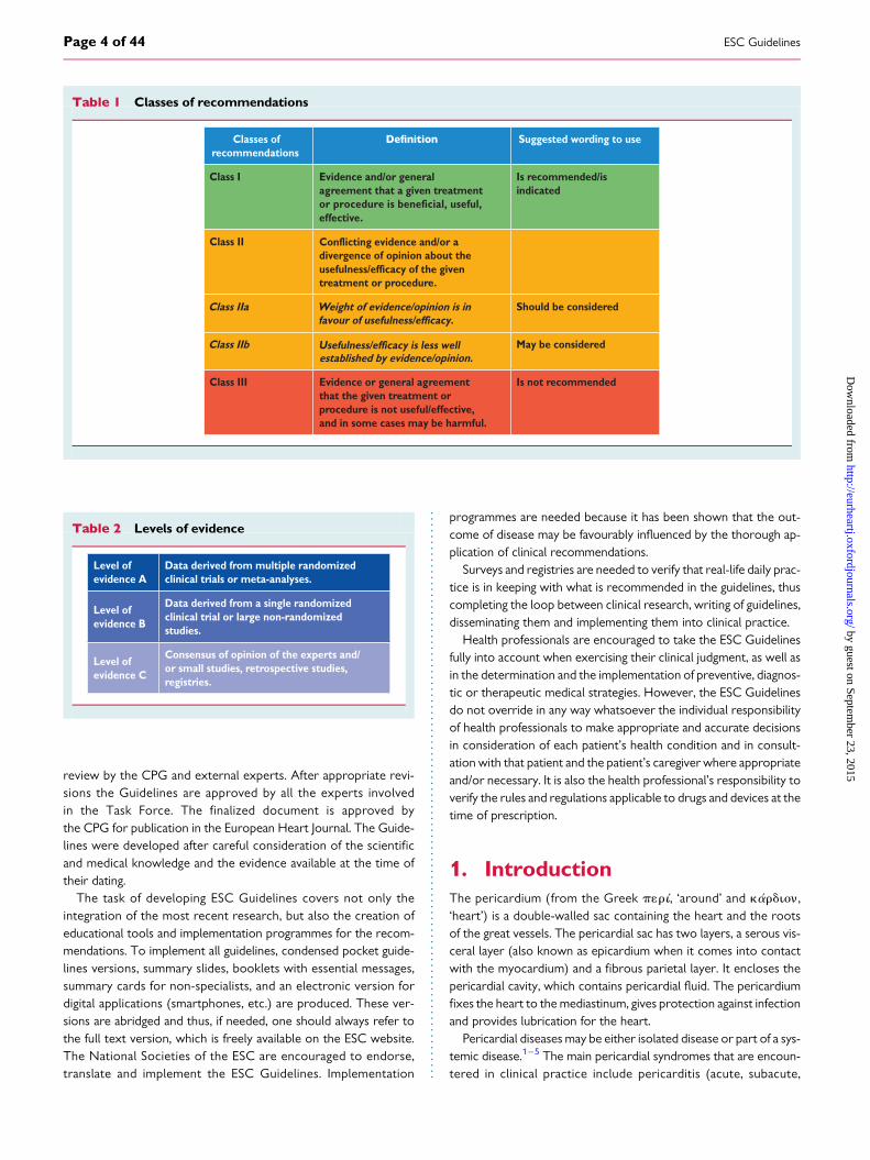
Members of this Task Force were selected by the ESC torepresent professionals involved with the medical care of patientswith this pathology. Selected experts in the field undertook acomprehensive review of the published evidence for management(including diagnosis, treatment, prevention and rehabilitation) ofa given condition according to ESC Committee for PracticeGuidelines (CPG) policy. A critical evaluation of diagnostic andtherapeutic procedures was performed, including assessment ofthe risk–benefit ratio. Estimates of expected health outcomes forlarger populations were included, where data exist. The level ofevidence and the strength of the recommendation of particularmanagement options were weighed and graded according to prede-fined scales, as outlined in Tables 1 and 2.
The experts of the writing and reviewing panels provided declara-tions of interest forms for all relationships that might be perceived asreal or potential sources of conflicts of interest. These forms werecompiled into one file and can be found on the ESC website (http://www.escardio.org/guidelines). Any changes in declarations of inter-est that arise during the writing period must be notified to the ESCand updated. The Task Force received its entire financial supportfrom the ESC without any involvement from the healthcareindustry.
The ESC CPG supervises and coordinates the preparation of newGuidelines produced by task forces, expert groups or consensus pa-nels. The Committee is also responsible for the endorsement pro-cess of these Guidelines. The ESC Guidelines undergo extensive
ESC Guidelines Page 3 of 44
by guest on September 23, 2015
http://eurheartj.oxfordjournals.org/D
ownloaded from
review by the CPG and external experts. After appropriate revi-sions the Guidelines are approved by all the experts involvedin the Task Force. The finalized document is approved bythe CPG for publication in the European Heart Journal. The Guide-lines were developed after careful consideration of the scientificand medical knowledge and the evidence available at the time oftheir dating.
The task of developing ESC Guidelines covers not only theintegration of the most recent research, but also the creation ofeducational tools and implementation programmes for the recom-mendations. To implement all guidelines, condensed pocket guide-lines versions, summary slides, booklets with essential messages,summary cards for non-specialists, and an electronic version fordigital applications (smartphones, etc.) are produced. These ver-sions are abridged and thus, if needed, one should always refer tothe full text version, which is freely available on the ESC website.The National Societies of the ESC are encouraged to endorse,translate and implement the ESC Guidelines. Implementation
programmes are needed because it has been shown that the out-come of disease may be favourably influenced by the thorough ap-plication of clinical recommendations.
Surveys and registries are needed to verify that real-life daily prac-tice is in keeping with what is recommended in the guidelines, thuscompleting the loop between clinical research, writing of guidelines,disseminating them and implementing them into clinical practice.
Health professionals are encouraged to take the ESC Guidelinesfully into account when exercising their clinical judgment, as well asin the determination and the implementation of preventive, diagnos-tic or therapeutic medical strategies. However, the ESC Guidelinesdo not override in any way whatsoever the individual responsibilityof health professionals to make appropriate and accurate decisionsin consideration of each patient’s health condition and in consult-ation with that patient and the patient’s caregiver where appropriateand/or necessary. It is also the health professional’s responsibility toverify the rules and regulations applicable to drugs and devices at thetime of prescription.
1. IntroductionThe pericardium (from the Greek p1ri, ‘around’ and kardion,‘heart’) is a double-walled sac containing the heart and the rootsof the great vessels. The pericardial sac has two layers, a serous vis-ceral layer (also known as epicardium when it comes into contactwith the myocardium) and a fibrous parietal layer. It encloses thepericardial cavity, which contains pericardial fluid. The pericardiumfixes the heart to the mediastinum, gives protection against infectionand provides lubrication for the heart.
Pericardial diseases may be either isolated disease or part of a sys-temic disease.1 –5 The main pericardial syndromes that are encoun-tered in clinical practice include pericarditis (acute, subacute,
Table 2 Levels of evidence
Level of evidence A
Data derived from multiple randomized clinical trials or meta-analyses.
Level of evidence B
Data derived from a single randomized clinical trial or large non-randomized studies.
Level of evidence C
Consensus of opinion of the experts and/or small studies, retrospective studies, registries.
Table 1 Classes of recommendations
Classes of recommendations
Suggested wording to use
Class I Evidence and/or general agreement that a given treatment or procedure is beneficial, useful,effective.
Is recommended/is indicated
Class II divergence of opinion about the Conflicting evidence and/or a
usefulness/efficacy of the given
favour of usefulness/efficacy.
Usefulness/efficacy is less well
treatment or procedure.
Class IIa Weight of evidence/opinion is in Should be considered
Class IIbestablished by evidence/opinion.
May be considered
Class III Evidence or general agreement that the given treatment or procedure is not useful/effective, and in some cases may be harmful.
Is not recommended
ESC GuidelinesPage 4 of 44
by guest on September 23, 2015
http://eurheartj.oxfordjournals.org/D
ownloaded from
chronic and recurrent), pericardial effusion, cardiac tamponade,constrictive pericarditis and pericardial masses.1,4,5 All medical ther-apies for pericardial diseases are off-label, since no drug has been re-gistered until now for a specific pericardial indication.
1.1 What is new in pericardial diseases?Pericardial diseases are relatively common in clinical practice andnew data have been published since the publication of the 2004ESC Guidelines on pericardial diseases.1
New diagnostic strategies have been proposed for the triage ofpatients with pericarditis and pericardial effusion and allow the se-lection of high-risk patients to be admitted as well as when and howadditional diagnostic investigations are to be performed.4– 9 More-over, specific diagnostic criteria have been proposed for acute andrecurrent pericarditis in clinical practice.2,4– 15
Multimodality imaging for pericardial diseases has become an es-sential approach for a modern and comprehensive diagnostic evalu-ation. Both the American Society of Echocardiography and theEuropean Association of Cardiovascular Imaging have provided rec-ommendation documents in recent years.2,3
The aetiology and pathophysiology of pericardial diseases remainto be better characterized, but new data supporting the immune-mediated pathogenesis of recurrences and new forms related toautoinflammatory diseases have been documented, especially inpaediatric patients.4,6 The first epidemiological data have becomeavailable.7,16
Age and gender issues are now more evident and clear, includingspecific recommendations for patients during pregnancy.17– 27
Major advances have occurred in therapy with the first multi-centre randomized clinical trials.10,11,13 – 15 Colchicine has beendemonstrated as a first-line drug to be added to conventional anti-inflammatory therapies in patients with a first episode of pericarditisor recurrences in order to improve the response to therapy, in-crease remission rates and reduce recurrences.10,11,13 – 15 Specifictherapeutic dosing without a loading dose and weight-adjusteddoses have been proposed to improve patient compliance.11,15
New therapeutic choices have become available for refractoryrecurrent pericarditis, including alternative immunosuppressivetherapies (e.g. azathioprine), intravenous immunoglobulins (IVIGs)and interleukin-1 (IL-1) antagonists (e.g. anakinra).20 – 23,28 – 32
Pericardiectomy has been demonstrated as a possible valuablealternative to additional medical therapies in patients with re-fractory recurrent pericarditis.33 The first large prospective andretrospective studies (.100 patients) have investigated the prog-nosis and complication risk in patients with acute and recurrentpericarditis.7,9,34 – 38
Imaging techniques for the detection of pericardial inflammation[e.g. cardiac magnetic resonance (CMR)] may identify forms of initialreversible constrictive pericarditis, allowing a trial of medical anti-inflammatory therapy that may reduce the need for surgery.2,39– 41
In conclusion, significant new data have become available since2004, and a new version of guidelines has become mandatory forclinical practice. Nevertheless, in the field of pericardial diseasesthere are a limited number of randomized controlled trials(RCTs). Therefore the number of class I level A indications arelimited.
2. Epidemiology, aetiology andclassification of pericardial diseases
2.1 EpidemiologyDespite the relative high frequency of pericardial diseases, there arefew epidemiological data, especially from primary care. Pericarditisis the most common disease of the pericardium encountered in clin-ical practice. The incidence of acute pericarditis has been reportedas 27.7 cases per 100,000 population per year in an Italian urbanarea.7 Pericarditis is responsible for 0.1% of all hospital admissionsand 5% of emergency room admissions for chest pain.4,5,42 Data col-lected from a Finnish national registry (2000–9) showed a standar-dized incidence rate of hospitalizations for acute pericarditis of 3.32per 100,000 person-years.16 These data were limited to hospitalizedpatients and therefore may account for only a minority of cases, asmany patients with pericarditis are commonly not admitted to hos-pital.8,9,42,43 Men ages 16–65 years were at higher risk for pericar-ditis (relative risk 2.02) than women in the general admittedpopulation, with the highest risk difference among young adultscompared with the overall population. Acute pericarditis caused0.20% of all cardiovascular admissions. The proportion of caused ad-missions declined by an estimated 51% per 10-year increase in age.The in-hospital mortality rate for acute pericarditis was 1.1% andwas increased with age and severe co-infections (pneumonia orsepticaemia).16 However, this is a study based on hospital admis-sions only. Recurrences affect about 30% of patients within18 months after a first episode of acute pericarditis.10,11
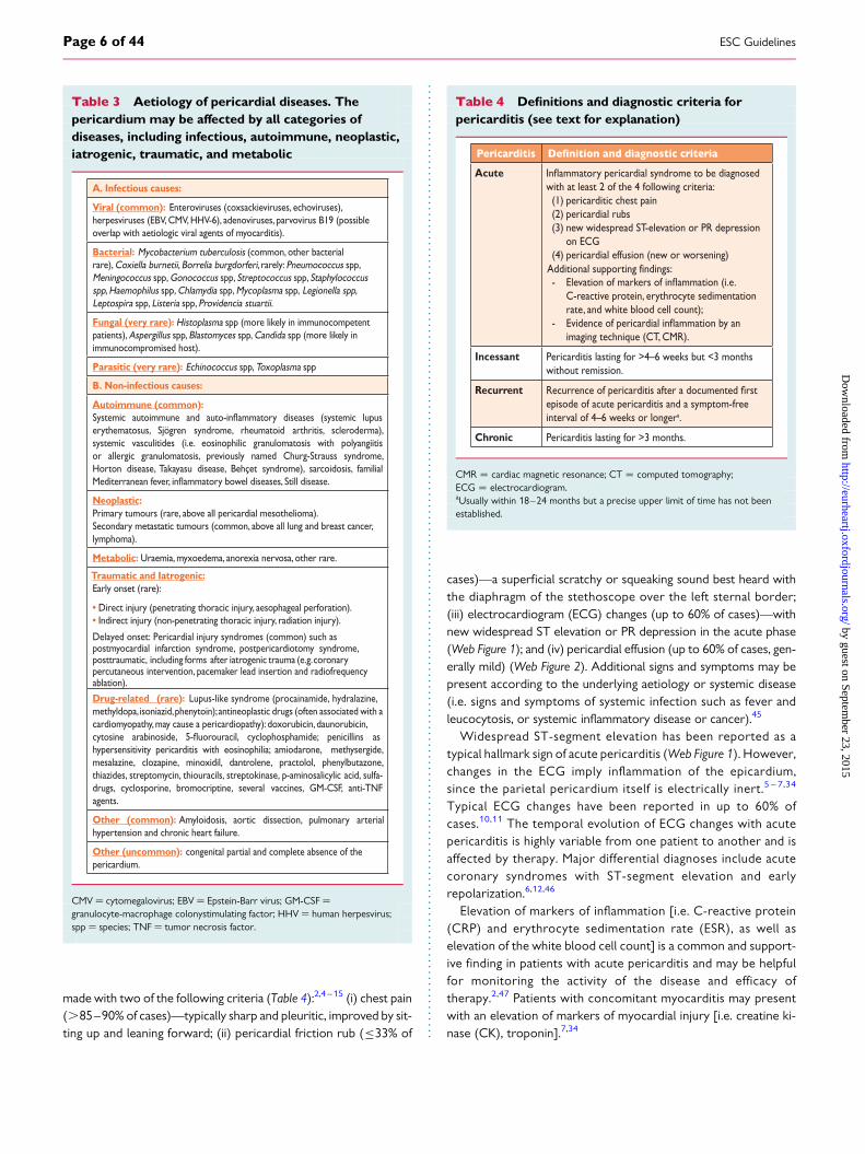
2.2 AetiologyA simple aetiological classification for pericardial diseases is toconsider infectious and non-infectious causes (Table 3).4,6,12,44 Theaetiology is varied and depends on the epidemiological background,patient population and clinical setting. In developed countries,viruses are usually the most common aetiological agents of pericar-ditis,6 whereas tuberculosis (TB) is the most frequent cause ofpericardial diseases in the world and developing countries, whereTB is endemic. In this setting, TB is often associated with humanimmunodeficiency virus (HIV) infection, especially in sub-SaharanAfrica.44
3. Pericardial syndromesPericardial syndromes include different clinical presentations ofpericardial diseases with distinctive signs and symptoms that canbe grouped in specific ‘syndromes’. The classical pericardial syn-dromes include pericarditis, pericardial effusion, cardiac tamponadeand constrictive pericarditis. Pericardial effusion and cardiac tam-ponade may occur without pericarditis and will be considered inseparate chapters. Specific considerations apply to cases with peri-carditis and concomitant myocardial inflammatory involvement,usually referred to in the literature as ‘myopericarditis’.
3.1 Acute pericarditisAcute pericarditis is an inflammatory pericardial syndrome with orwithout pericardial effusion.1 – 11,42 The clinical diagnosis can be
ESC Guidelines Page 5 of 44
by guest on September 23, 2015
http://eurheartj.oxfordjournals.org/D
ownloaded from
made with two of the following criteria (Table 4):2,4 –15 (i) chest pain(.85–90% of cases)—typically sharp and pleuritic, improved by sit-ting up and leaning forward; (ii) pericardial friction rub (≤33% of
cases)—a superficial scratchy or squeaking sound best heard withthe diaphragm of the stethoscope over the left sternal border;(iii) electrocardiogram (ECG) changes (up to 60% of cases)—withnew widespread ST elevation or PR depression in the acute phase(Web Figure 1); and (iv) pericardial effusion (up to 60% of cases, gen-erally mild) (Web Figure 2). Additional signs and symptoms may bepresent according to the underlying aetiology or systemic disease(i.e. signs and symptoms of systemic infection such as fever andleucocytosis, or systemic inflammatory disease or cancer).45
Widespread ST-segment elevation has been reported as atypical hallmark sign of acute pericarditis (Web Figure 1). However,changes in the ECG imply inflammation of the epicardium,since the parietal pericardium itself is electrically inert.5 – 7,34
Typical ECG changes have been reported in up to 60% ofcases.10,11 The temporal evolution of ECG changes with acutepericarditis is highly variable from one patient to another and isaffected by therapy. Major differential diagnoses include acutecoronary syndromes with ST-segment elevation and earlyrepolarization.6,12,46
Elevation of markers of inflammation [i.e. C-reactive protein(CRP) and erythrocyte sedimentation rate (ESR), as well aselevation of the white blood cell count] is a common and support-ive finding in patients with acute pericarditis and may be helpfulfor monitoring the activity of the disease and efficacy oftherapy.2,47 Patients with concomitant myocarditis may presentwith an elevation of markers of myocardial injury [i.e. creatine ki-nase (CK), troponin].7,34
Table 3 Aetiology of pericardial diseases. Thepericardium may be affected by all categories ofdiseases, including infectious, autoimmune, neoplastic,iatrogenic, traumatic, and metabolic
A. Infectious causes:
Viral (common): Enteroviruses (coxsackieviruses, echoviruses),herpesviruses (EBV, CMV, HHV-6), adenoviruses, parvovirus B19 (possible overlap with aetiologic viral agents of myocarditis).
Bacterial: Mycobacterium tuberculosis (common, other bacterial rare), Coxiella burnetii, Borrelia burgdorferi, rarely: Pneumococcus spp, Meningococcus spp, Gonococcus spp, Streptococcus spp, Staphylococcus spp, Haemophilus spp, Chlamydia spp, Mycoplasma spp, Legionella spp, Leptospira spp, Listeria spp, Providencia stuartii.
Fungal (very rare): Histoplasma spp (more likely in immunocompetent patients), Aspergillus spp, Blastomyces spp, Candida spp (more likely in immunocompromised host).
Parasitic (very rare): Echinococcus spp, Toxoplasma spp
B. Non-infectious causes:
Autoimmune (common):
erythematosus, Sjögren syndrome, rheumatoid arthritis, scleroderma), systemic vasculitides (i.e. eosinophilic granulomatosis with polyangiitis or allergic granulomatosis, previously named Churg-Strauss syndrome, Horton disease, Takayasu disease, Behçet syndrome), sarcoidosis, familial
Neoplastic:Primary tumours (rare, above all pericardial mesothelioma).Secondary metastatic tumours (common, above all lung and breast cancer, lymphoma).
Metabolic: Uraemia, myxoedema, anorexia nervosa, other rare.
Early onset (rare):
• Direct injury (penetrating thoracic injury, aesophageal perforation).• Indirect injury (non-penetrating thoracic injury, radiation injury).
Delayed onset: Pericardial injury syndromes (common) such aspostmyocardial infarction syndrome, postpericardiotomy syndrome,posttraumatic, including forms after iatrogenic trauma (e.g. coronarypercutaneous intervention, pacemaker lead insertion and radiofrequencyablation).
Drug-related (rare): Lupus-like syndrome (procainamide, hydralazine,methyldopa, isoniazid, phenytoin); antineoplastic drugs (often associated with a cardiomyopathy, may cause a pericardiopathy): doxorubicin, daunorubicin,
hypersensitivity pericarditis with eosinophilia; amiodarone, methysergide, mesalazine, clozapine, minoxidil, dantrolene, practolol, phenylbutazone, thiazides, streptomycin, thiouracils, streptokinase, p-aminosalicylic acid, sulfa-drugs, cyclosporine, bromocriptine, several vaccines, GM-CSF, anti-TNF agents.
Other (common): Amyloidosis, aortic dissection, pulmonary arterial hypertension and chronic heart failure.
Other (uncommon): congenital partial and complete absence of the pericardium.
Traumatic and Iatrogenic:
CMV ¼ cytomegalovirus; EBV ¼ Epstein-Barr virus; GM-CSF ¼granulocyte-macrophage colonystimulating factor; HHV ¼ human herpesvirus;spp ¼ species; TNF ¼ tumor necrosis factor.
Table 4 Definitions and diagnostic criteria forpericarditis (see text for explanation)
Acutewith at least 2 of the 4 following criteria: (1) pericarditic chest pain (2) pericardial rubs (3) new widespread ST-elevation or PR depression
Additional supporting findings:
on ECG (4) pericardial effusion (new or worsening)
- Ele C-reactive protein, erythrocyte sedimentation rate, and white blood cell count); - Evidence of pericar imaging technique (CT, CMR).
Incessant Pericarditis lasting for >4–6 weeks but <3 months without remission.
Recurrentepisode of acute pericarditis and a symptom-free interval of 4–6 weeks or longera.
Chronic Pericarditis lasting for >3 months.
CMR ¼ cardiac magnetic resonance; CT ¼ computed tomography;ECG ¼ electrocardiogram.aUsually within 18–24 months but a precise upper limit of time has not beenestablished.
ESC GuidelinesPage 6 of 44
by guest on September 23, 2015
http://eurheartj.oxfordjournals.org/D
ownloaded from
A chest X-ray is generally normal in patients with acutepericarditis since an increased cardiothoracic ratio only occurswith pericardial effusions exceeding 300 ml.48 In the case of pleur-opulmonary diseases, signs of pleuropericardial involvement may befound in patients with pericarditis.2,3
Recommendations for diagnosis of acute pericarditis
Recommendations Classa Levelb Ref.c
ECG is recommended in all patients withsuspected acute pericarditis
I C
Transthoracic echocardiography isrecommended in all patients withsuspected acute pericarditis
I C
Chest X-ray is recommended in allpatients with suspected acutepericarditis
I C
Assessment of markers ofinflammation (i.e. CRP) and myocardialinjury (i.e. CK, troponin) isrecommended in patients withsuspected acute pericarditis
I C
CK ¼ creatine kinase; CRP ¼ C-reactive protein; ECG ¼ electrocardiogram.aClass of recommendation.bLevel of evidence.cReference(s) supporting recommendations.
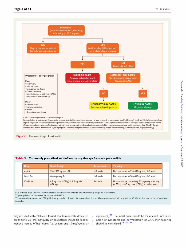
3.1.1 Clinical management and therapyIt is not mandatory to search for the aetiology in all patients, espe-cially in countries with a low prevalence of TB, because of the rela-tively benign course associated with the common causes ofpericarditis and the relatively low yield of diagnostic inves-tigations.6,8,12,49 Specific final identifiable causes (non-viral –non-idiopathic) as well as high-risk features in the context of acutepericarditis have been identified as being associated with an in-creased risk of complications during follow-up (tamponade, recur-rences and constriction).9,12,43,50 The major risk factors associatedwith poor prognosis after multivariate analysis include high fever[.388C (.100.48F)], subacute course (symptoms over severaldays without a clear-cut acute onset), evidence of large pericardialeffusion (i.e. diastolic echo-free space .20 mm), cardiac tampon-ade and failure to respond within 7 days to non-steroidal anti-inflammatory drugs (NSAIDs).9,43,50 Other risk factors should alsobe considered (i.e. ‘minor risk factors’); these are based on expertopinion and literature review, including pericarditis associatedwith myocarditis (myopericarditis), immunodepression, traumaand oral anticoagulant therapy.
On this basis a triage for acute pericarditis is proposed (Figure 1,Web Table 6).5,6,43 Any clinical presentation that may suggest anunderlying aetiology (e.g. a systemic inflammatory disease) orwith at least one predictor of poor prognosis (major or minorrisk factors) warrants hospital admission and an aetiologysearch.9,43,49 – 51 On the other hand, patients without these
features can be managed as outpatients with empiric anti-inflammatories and short-term follow-up after 1 week to assessthe response to treatment.9
Recommendations for the management of acutepericarditis
Recommendations Classa Levelb Ref.c
Hospital admission is recommended forhigh-risk patients with acute pericarditis(at least one risk factord)
I B 8,9
Outpatient management isrecommended for low-risk patients withacute pericarditis
I B 8,9
Evaluation of response toanti-inflammatory therapy isrecommended after 1 week
I B 8,9
aClass of recommendation.bLevel of evidence.cReference(s) supporting recommendations.dSee Figure 1 (both major and minor predictors of poor prognosis).
In patients identified with a cause other than viral infection, spe-cific therapy appropriate to the underlying disorder is indi-cated49,51 and the epidemiological background (high vs. lowprevalence of TB) should be considered.8,12,52 The first non-pharmacological recommendation is to restrict physical activitybeyond ordinary sedentary life until resolution of symptoms andnormalization of CRP for patients not involved in competitivesports.53 Athletes are recommended to return to competitivesports only after symptoms have resolved and diagnostic tests(i.e. CRP, ECG and echocardiogram) have been normalized.53,54
A minimal restriction of 3 months (after the initial onset of the at-tack) has been arbitrarily defined according to expert consensus.54
We suggest applying this restriction only to athletes, while ashorter period (until remission) may be suitable for non-athletes.Aspirin or NSAIDs are mainstays of therapy for acute peri-carditis.5,6,55,56 Different anti-inflammatory drugs have been pro-posed (Table 5).
The choice of drug should be based on the history of the patient(contraindications, previous efficacy or side effects), the presence ofconcomitant diseases (favouring aspirin over other NSAIDs whenaspirin is already needed as antiplatelet treatment) and physicianexpertise.56
Colchicine is recommended at low, weight-adjusted doses toimprove the response to medical therapy and prevent recur-rences.10,11,57 – 59 Tapering of colchicine is not mandatory butmay be considered to prevent persistence of symptoms and re-currence.5,6,56 Corticosteroids should be considered as a secondoption in patients with contraindications and failure of aspirin orNSAIDs because of the risk of favouring the chronic evolution ofthe disease and promoting drug dependence; in this case
ESC Guidelines Page 7 of 44
by guest on September 23, 2015
http://eurheartj.oxfordjournals.org/D
ownloaded from
they are used with colchicine. If used, low to moderate doses (i.e.prednisone 0.2–0.5 mg/kg/day or equivalent) should be recom-mended instead of high doses (i.e. prednisone 1.0 mg/kg/day or
equivalent).35 The initial dose should be maintained until reso-lution of symptoms and normalization of CRP, then taperingshould be considered.5,6,35,47,56
Pericarditis?(physical examination, ECG, chest x-ray,
echacardiogram, CRP, troponin)
NODiagnostic criteria not satisfied.Search for alternative diagnoses
YESSpecific aetiology highly suspected or
any predictor of poor prognosis
YES NOEmpiric trial with NSAID
NO YES
MODERATE RISK CASESAdmission and aetiology search
LOW RISK CASESOutpatient follow-up
HIGH RISK CASESAdmission and aetiology search
(major or minor prognostic predictor)
NON-HIGH RISK CASESNo admission and etiology search.
Response to NSAID?
Predictors of poor prognosis:
Major• Fever >38°C• Subacute onset• Large pericardial effusion• Cardiac tamponade• Lack of response to aspirin or NSAIDs after at least 1 week of therapy
Minor• Myopericarditis• Immunosuppression• Trauma• Oral anticoagulant therapy
CRP = C-reactive protein; ECG = electrocardiogram.
Figure 1 Proposed triage of pericarditis.
Table 5 Commonly prescribed anti-inflammatory therapy for acute pericarditis
Drug Usual dosinga Tx durationb Taperinga
Aspirin 750–1000 mg every 8h 1–2 weeks Decrease doses by 250–500 mg every 1–2 weeks
Ibuprofen 600 mg every 8h 1–2 weeks Decrease doses by 200–400 mg every 1–2 weeks
Colchicine 0.5 mg once (<70 kg) or 0.5 mg b.i.d. (≥70 kg)
3 months Not mandatory, alternatively 0.5 mg every other day (< 70 kg) or 0.5 mg once (≥70 kg) in the last weeks
b.i.d. ¼ twice daily; CRP ¼ C-reactive protein; NSAIDs ¼ non-steroidal anti-inflammatory drugs; Tx ¼ treatment.aTapering should be considered for aspirin and NSAIDs.bTx duration is symptoms and CRP guided but generally 1–2 weeks for uncomplicated cases. Gastroprotection should be provided. Colchicine is added on top of aspirin oribuprofen.
ESC GuidelinesPage 8 of 44
by guest on September 23, 2015
http://eurheartj.oxfordjournals.org/D
ownloaded from
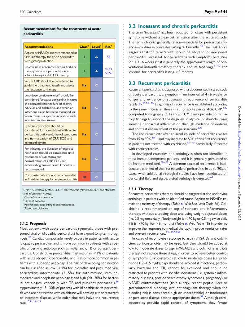
Recommendations for the treatment of acutepericarditis
Recommendations Classa Levelb Ref.c
Aspirin or NSAIDs are recommended asfirst-line therapy for acute pericarditiswith gastroprotection
I A 55
Colchicine is recommended as first-linetherapy for acute pericarditis as anadjunct to aspirin/NSAID therapy
I A10,11,58,59
Serum CRP should be considered toguide the treatment length and assessthe response to therapy
IIa C
Low-dose corticosteroidsd should beconsidered for acute pericarditis in casesof contraindication/failure of aspirin/NSAIDs and colchicine, and when aninfectious cause has been excluded, orwhen there is a specific indication suchas autoimmune disease
IIa C
Exercise restriction should beconsidered for non-athletes with acutepericarditis until resolution of symptomsand normalization of CRP, ECG andechocardiogram
IIa C
For athletes, the duration of exerciserestriction should be considered untilresolution of symptoms andnormalization of CRP, ECG andechocardiogram—at least 3 months isrecommended
IIa C
Corticosteroids are not recommendedas first-line therapy for acute pericarditis
III C
CRP ¼ C-reactive protein; ECG ¼ electrocardiogram; NSAIDs ¼ non-steroidalanti-inflammatory drugs.aClass of recommendation.bLevel of evidence.cReference(s) supporting recommendations.dAdded to colchicine.
3.1.2 PrognosisMost patients with acute pericarditis (generally those with pre-sumed viral or idiopathic pericarditis) have a good long-term prog-nosis.36 Cardiac tamponade rarely occurs in patients with acuteidiopathic pericarditis, and is more common in patients with a spe-cific underlying aetiology such as malignancy, TB or purulent peri-carditis. Constrictive pericarditis may occur in ,1% of patientswith acute idiopathic pericarditis, and is also more common in pa-tients with a specific aetiology. The risk of developing constrictioncan be classified as low (,1%) for idiopathic and presumed viralpericarditis; intermediate (2–5%) for autoimmune, immune-mediated and neoplastic aetiologies; and high (20–30%) for bacter-ial aetiologies, especially with TB and purulent pericarditis.36
Approximately 15–30% of patients with idiopathic acute pericardi-tis who are not treated with colchicine will develop either recurrentor incessant disease, while colchicine may halve the recurrencerate.10,11,13– 15
3.2 Incessant and chronic pericarditisThe term ‘incessant’ has been adopted for cases with persistentsymptoms without a clear-cut remission after the acute episode.The term ‘chronic’ generally refers—especially for pericardial effu-sions—to disease processes lasting .3 months.48 The Task Forcesuggests that the term ‘acute’ should be adopted for new-onsetpericarditis, ‘incessant’ for pericarditis with symptoms persistingfor .4–6 weeks (that is generally the approximate length of con-ventional anti-inflammatory therapy and its tapering),11,60 and‘chronic’ for pericarditis lasting .3 months.
3.3 Recurrent pericarditisRecurrent pericarditis is diagnosed with a documented first episodeof acute pericarditis, a symptom-free interval of 4–6 weeks orlonger and evidence of subsequent recurrence of pericarditis(Table 4).11,13 – 15 Diagnosis of recurrence is established accordingto the same criteria as those used for acute pericarditis. CRP,2,47
computed tomography (CT) and/or CMR may provide confirma-tory findings to support the diagnosis in atypical or doubtful casesshowing pericardial inflammation through evidence of oedemaand contrast enhancement of the pericardium.2,39
The recurrence rate after an initial episode of pericarditis rangesfrom 15 to 30%,10,11 and may increase to 50% after a first recurrencein patients not treated with colchicine,13 – 15 particularly if treatedwith corticosteroids.
In developed countries, the aetiology is often not identified inmost immunocompetent patients, and it is generally presumed tobe immune-mediated.60– 62 A common cause of recurrence is inad-equate treatment of the first episode of pericarditis. In up to 20% ofcases, when additional virological studies have been conducted onpericardial fluid and tissue, a viral aetiology is detected.63
3.3.1 TherapyRecurrent pericarditis therapy should be targeted at the underlyingaetiology in patients with an identified cause. Aspirin or NSAIDs re-main the mainstay of therapy (Table 6, Web Box, Web Table 1A). Col-chicine is recommended on top of standard anti-inflammatorytherapy, without a loading dose and using weight-adjusted doses(i.e. 0.5 mg once daily if body weight is ,70 kg or 0.5 mg twice dailyif it is ≥70 kg, for ≥6 months) (Table 6, Web Table 1B) in order toimprove the response to medical therapy, improve remission ratesand prevent recurrences.13–15,58,59
In cases of incomplete response to aspirin/NSAIDs and colchi-cine, corticosteroids may be used, but they should be added atlow to moderate doses to aspirin/NSAIDs and colchicine as tripletherapy, not replace these drugs, in order to achieve better controlof symptoms. Corticosteroids at low to moderate doses (i.e. pred-nisone 0.2–0.5 mg/kg/day) should be avoided if infections, particu-larly bacterial and TB, cannot be excluded and should berestricted to patients with specific indications (i.e. systemic inflam-matory diseases, post-pericardiotomy syndromes, pregnancy) orNSAID contraindications (true allergy, recent peptic ulcer orgastrointestinal bleeding, oral anticoagulant therapy when thebleeding risk is considered high or unacceptable) or intoleranceor persistent disease despite appropriate doses.58 Although corti-costeroids provide rapid control of symptoms, they favour
ESC Guidelines Page 9 of 44
by guest on September 23, 2015
http://eurheartj.oxfordjournals.org/D
ownloaded from
chronicity, more recurrences and side effects.35,55,61 If corticoster-oids are used, their tapering should be particularly slow. A criticalthreshold for recurrences is a 10–15 mg/day dose of prednisoneor equivalent. At this threshold, very slow decrements as small as1.0–2.5 mg at intervals of 2–6 weeks are useful. In cases of recur-rence, every effort should be made not to increase the dose or toreinstate corticosteroids (Tables 6 and 7).5,6,35,61
After obtaining a complete response, tapering should be donewith a single class of drug at a time before colchicine is gradually dis-continued (over several months in the most difficult cases). Recur-rences are possible after discontinuation of each drug. Each taperingshould be attempted only if symptoms are absent and CRP isnormal.5,6,47,56 The Task Force does not recommend influenza vac-cine as a preventive measure for pericarditis in patients with recur-rent pericarditis, since the influenza virus is not a usual cause ofpericarditis. The influenza vaccine should be administered accordingto specific indications beyond pericarditis; moreover, recurrencesare generally immune mediated, and inappropriate or unwantedstimulation of the immune system may trigger or worsen an episodeof pericarditis.
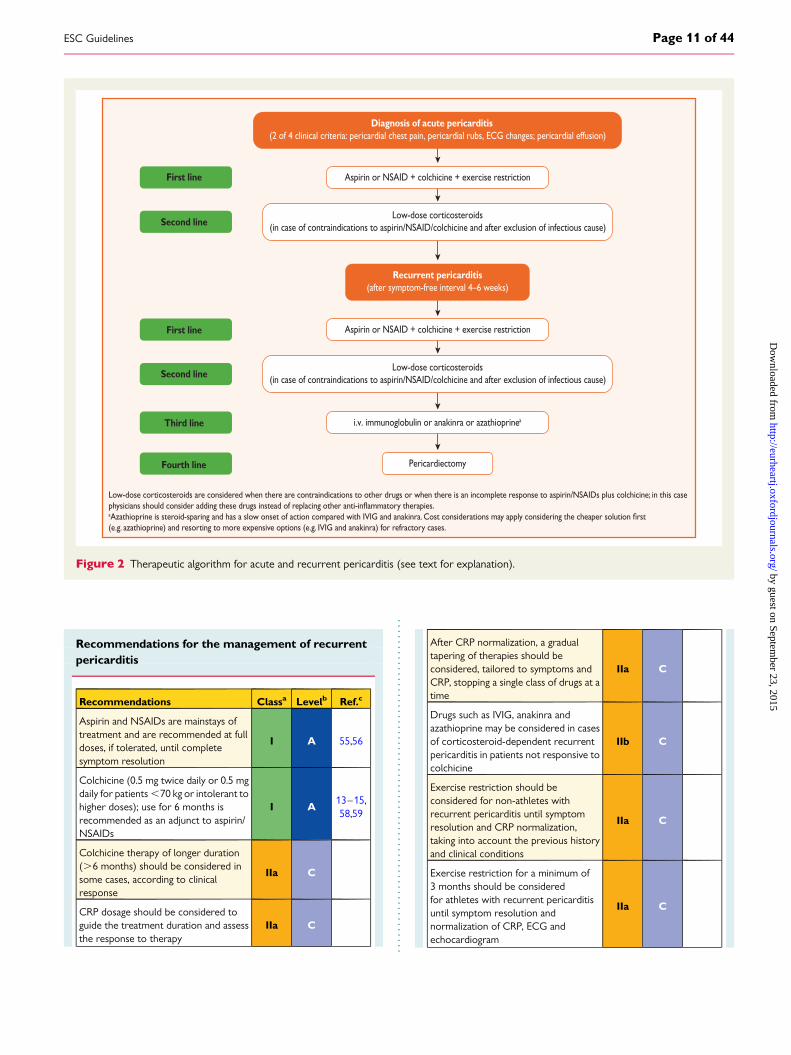
An alternative effective approach to minimize systemic side ef-fects related to corticosteroids may be intrapericardial administra-tion of non-absorbable corticosteroids,64,65 but this techniquerequires further investigation. For those patients who require un-acceptably high long-term doses of corticosteroids (e.g. prednisone15–25 mg/day) or who do not respond to anti-inflammatory ther-apies, several drugs have been used, including azathioprine,28 IVIG(immunomodulatory but also anti-viral)29,30 and anakinra, a recom-binant IL-1b receptor antagonist,31,32 but strong evidence-baseddata are lacking (Web Table 2). Other immunosuppressive drugs[i.e. cyclophosphamide, cyclosporine, methotrexate, hydroxychlor-oquine, anti-tumour necrosis factor (TNF) agents] have been onlyanecdotally reported. Less toxic agents might be preferred, andeventually combined, with the therapy being tailored to the individ-ual patient and physician experience (Figure 2). Azathioprine is main-ly a slow-acting corticosteroid-sparing agent, useful to control thedisease for a long-term follow-up, while anakinra and IVIG are
effective during the acute phase, though recurrences may occurafter discontinuation.29 – 32 Drugs such as IVIG, anakinra andazathioprine may be considered in cases of proven infection-negative, corticosteroid-dependent, recurrent pericarditis not re-sponsive to colchicine after careful assessment of the costs, risksand eventually consultation by multidisciplinary experts, includingimmunologists and/or rheumatologists, in the absence of a specificexpertise. It is also mandatory to educate the patient and his/hercaregivers about the clinical risks related to immunomodulatory/im-munosuppressive drugs and the safety measures to adopt during thetreatment. As a last resort, pericardiectomy may be considered, butonly after a thorough trial of unsuccessful medical therapy, and withreferral of the patient to a centre with specific expertise in this sur-gery.33 The physical activity restrictions in acute pericarditis applyalso to recurrences.53,54
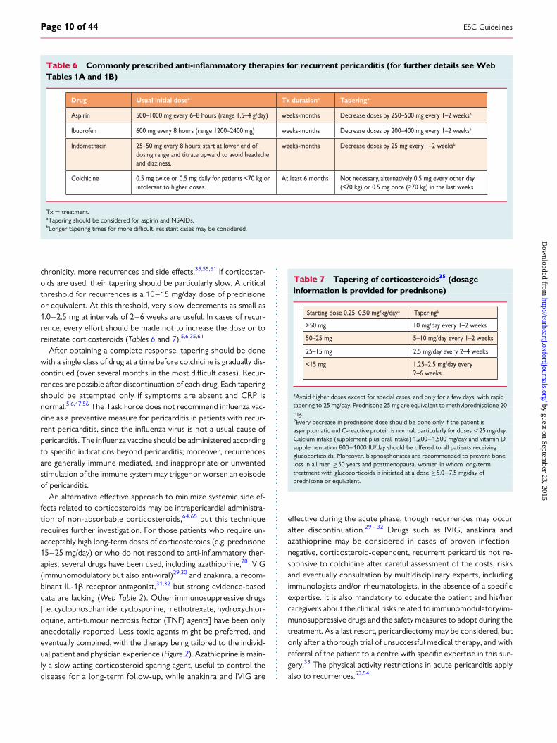
Table 6 Commonly prescribed anti-inflammatory therapies for recurrent pericarditis (for further details see WebTables 1A and 1B)
Drug Usual initial dosea Tx durationb Taperinga
Aspirin 500–1000 mg every 6–8 hours (range 1,5–4 g/day) weeks-months Decrease doses by 250–500 mg every 1–2 weeksb
Ibuprofen 600 mg every 8 hours (range 1200–2400 mg) weeks-months Decrease doses by 200–400 mg every 1–2 weeksb
Indomethacin 25–50 mg every 8 hours: start at lower end of dosing range and titrate upward to avoid headache and dizziness.
weeks-months Decrease doses by 25 mg every 1–2 weeksb
Colchicine 0.5 mg twice or 0.5 mg daily for patients <70 kg or intolerant to higher doses.
At least 6 months Not necessary, alternatively 0.5 mg every other day (<70 kg) or 0.5 mg once (≥70 kg) in the last weeks
Tx ¼ treatment.aTapering should be considered for aspirin and NSAIDs.bLonger tapering times for more difficult, resistant cases may be considered.
Table 7 Tapering of corticosteroids35 (dosageinformation is provided for prednisone)
Starting dose 0.25–0.50 mg/kg/daya Taperingb
>50 mg 10 mg/day every 1–2 weeks
50–25 mg 5–10 mg/day every 1–2 weeks
25–15 mg 2.5 mg/day every 2–4 weeks
<15 mg 1.25–2.5 mg/day every2–6 weeks
aAvoid higher doses except for special cases, and only for a few days, with rapidtapering to 25 mg/day. Prednisone 25 mg are equivalent to methylprednisolone 20mg.bEvery decrease in prednisone dose should be done only if the patient isasymptomatic and C-reactive protein is normal, particularly for doses ,25 mg/day.Calcium intake (supplement plus oral intake) 1,200–1,500 mg/day and vitamin Dsupplementation 800–1000 IU/day should be offered to all patients receivingglucocorticoids. Moreover, bisphosphonates are recommended to prevent boneloss in all men ≥50 years and postmenopausal women in whom long-termtreatment with glucocorticoids is initiated at a dose ≥5.0–7.5 mg/day ofprednisone or equivalent.
ESC GuidelinesPage 10 of 44
by guest on September 23, 2015
http://eurheartj.oxfordjournals.org/D
ownloaded from
Recommendations for the management of recurrentpericarditis
Recommendations Classa Levelb Ref.c
Aspirin and NSAIDs are mainstays oftreatment and are recommended at fulldoses, if tolerated, until completesymptom resolution
I A 55,56
Colchicine (0.5 mg twice daily or 0.5 mgdaily for patients ,70 kg or intolerant tohigher doses); use for 6 months isrecommended as an adjunct to aspirin/NSAIDs
I A13–15,58,59
Colchicine therapy of longer duration(.6 months) should be considered insome cases, according to clinicalresponse
IIa C
CRP dosage should be considered toguide the treatment duration and assessthe response to therapy
IIa C
After CRP normalization, a gradualtapering of therapies should beconsidered, tailored to symptoms andCRP, stopping a single class of drugs at atime
IIa C
Drugs such as IVIG, anakinra andazathioprine may be considered in casesof corticosteroid-dependent recurrentpericarditis in patients not responsive tocolchicine
IIb C
Exercise restriction should beconsidered for non-athletes withrecurrent pericarditis until symptomresolution and CRP normalization,taking into account the previous historyand clinical conditions
IIa C
Exercise restriction for a minimum of3 months should be consideredfor athletes with recurrent pericarditisuntil symptom resolution andnormalization of CRP, ECG andechocardiogram
IIa C
Diagnosis of acute pericarditis(2 of 4 clinical criteria: pericardial chest pain, pericardial rubs, ECG changes; pericardial effusion)
Recurrent pericarditis(after symptom-free interval 4–6 weeks)
Aspirin or NSAID + colchicine + exercise restriction
Low-dose corticosteroids(in case of contraindications to aspirin/NSAID/colchicine and after exclusion of infectious cause)
Aspirin or NSAID + colchicine + exercise restriction
i.v. immunoglobulin or anakinra or azathioprinea
Pericardiectomy
Low-dose corticosteroids(in case of contraindications to aspirin/NSAID/colchicine and after exclusion of infectious cause)
First line
Second line
First line
Second line
Third line
Fourth line
Low-dose corticosteroids are considered when there are contraindications to other drugs or when there is an incomplete response to aspirin/NSAIDs plus colchicine; in this case
a
(e.g. azathioprine) and resorting to more expensive options (e.g. IVIG and anakinra) for refractory cases.
Figure 2 Therapeutic algorithm for acute and recurrent pericarditis (see text for explanation).
ESC Guidelines Page 11 of 44
by guest on September 23, 2015
http://eurheartj.oxfordjournals.org/D
ownloaded from
If ischaemic heart disease is a concern orantiplatelet therapy is required, aspirinshould be considered, at medium highdoses (1–2.4 g/day)* (Web box)
IIa C
If symptoms recur during therapytapering, the management shouldconsider not increasing the dose ofcorticosteroids to control symptoms,but increasing to the maximum dose ofaspirin or NSAIDs, well distributed,generally every 8 hours, andintravenously if necessary, addingcolchicine and adding analgesics for paincontrol
IIa C
Corticosteroid therapy is notrecommended as a first line-approach III B
13–15,35,37,
55
CRP ¼ C-reactive protein; ECG ¼ electrocardiogram; IVIG ¼ intravenousimmunoglobulin; NSAIDs ¼ non-steroidal anti-inflammatory drugs.aClass of recommendation.bLevel of evidence.cReference(s) supporting recommendations.
3.3.2 PrognosisSevere complications are uncommon in idiopathic recurrent peri-carditis.37,60,61 Cardiac tamponade is rare and generally occurs atthe beginning of the disease. Constrictive pericarditis has neverbeen reported in these patients, despite numerous recurrences,and the overall risk is lower than that recorded after a first episodeof acute pericarditis (,1%).36,37,61 Thus it is important to reassurepatients about their prognosis, explaining the nature of the diseaseand its likely course. The complication rates are related to the aeti-ology and not to the number of recurrences. Drug treatment shouldtake into account this favourable outcome to avoid more toxicagents. However, quality of life can be severely affected in patientswith repeated recurrences, subacute or incessant pericarditis andglucocorticoid dependence.
3.4 Pericarditis associated withmyocardial involvement(myopericarditis)Pericarditis and myocarditis share common aetiologies, and overlap-ping forms may be encountered in clinical practice.34,66 Pericarditiswith known or clinically suspected concomitant myocardial involve-ment should be referred to as ‘myopericarditis’, while predominantmyocarditis with pericardial involvement should be referred to as‘perimyocarditis’, according to Task Force consensus. The classicalpresentation is chest pain associated with other signs of pericarditis(pericardial rubs, ST-segment elevation and pericardial effusion)plus the elevation of markers of myocardial damage (i.e. troponins).Limited clinical data on the causes of myopericarditis suggest thatviral infections are among the most common causes in developedcountries, while other infectious causes are more common in devel-oping countries (especially TB). Cardiotropic viruses can cause peri-cardial and myocardial inflammation via direct cytolytic or cytotoxiceffects and/or subsequent immune-mediated mechanisms. Such
mechanisms are especially involved in cases associated with con-nective tissue diseases, inflammatory bowel diseases andradiation-induced, drug-induced or vaccinia-associated myopericar-dial involvement. Many cases of myopericarditis are subclinical. Inother patients, cardiac symptoms and signs are masked by pro-nounced systemic manifestations of infection or inflammation.66 Inmany cases, myopericarditis manifestations are preceded by orare sometimes concomitant with an acute respiratory illness (espe-cially acute tonsillitis, pneumonia) or gastroenteritis. The increasedsensitivity of troponin assays and contemporary widespread useof troponins has greatly increased the reported number ofcases.7,34,66–68
3.4.1 Definition and diagnosisThe diagnosis of predominant pericarditis with myocardial involve-ment, or ‘myopericarditis’, can be clinically established if patientswith definite criteria for acute pericarditis show elevated biomar-kers of myocardial injury (troponin I or T, CK-MB fraction) withoutnewly developed focal or diffuse impairment of left ventricular func-tion in echocardiography or CMR.34 The term myopericarditis indi-cates a primarily pericarditic syndrome with minor myocardialinvolvement, which describes the majority of combined pericarditisand myocarditis cases encountered in clinical practice.7,9,34,68
On the other hand, evidence of new-onset focal or diffuse reduc-tion of left ventricular function in patients with elevated myocardialbiomarkers and clinical criteria for acute pericarditis suggests pre-dominant myocarditis with pericardial involvement (‘perimyocardi-tis’).34,66 Definite confirmation of the presence of myocarditis willrequire endomyocardial biopsy according to the Myocardial andPericardial Diseases Working Group position statement.69 How-ever, the benign prognosis of patients with suspected concomitantmyocardial involvement in predominant pericarditis (myopericardi-tis), with absent or mild left ventricular dysfunction, and no symp-toms of heart failure does not clinically require endomyocardialbiopsy.6,34,66–68,70,71
In cases of pericarditis with suspected associated myocarditis,coronary angiography (according to clinical presentation and riskfactor assessment) is recommended in order to rule out acute cor-onary syndromes. CMR is recommended for the confirmation ofmyocardial involvement and to rule out ischaemic myocardial ne-crosis in the absence of significant coronary disease; this has clinicaland therapeutic implications.34,66
3.4.2 ManagementHospitalization is recommended for diagnosis and monitoring ofpatients with myocardial involvement and differential diagnosis,especially with acute coronary syndromes. In the setting of myoper-icarditis, management is similar to that recommended for pericardi-tis. Empirical anti-inflammatory therapies (i.e. aspirin 1500–3000mg/day) or NSAIDs (ibuprofen 1200–2400 mg/day or indometh-acin 75–150 mg/day) are usually prescribed to control chest pain,while corticosteroids are prescribed as a second choice in casesof contraindication, intolerance or failure of aspirin/NSAIDs.66 Inthe setting of myopericarditis, some authors recommend reducingdosages, as compared with pure pericarditis, because in animal mod-els of myocarditis, NSAIDs have been shown to be non-efficaciousand may enhance inflammation, increasing mortality.69,70,72,73
ESC GuidelinesPage 12 of 44
by guest on September 23, 2015
http://eurheartj.oxfordjournals.org/D
ownloaded from
However, the application of these findings from animal models tohumans may be questionable.66 In addition, there are insufficientdata to recommend the use of colchicine, which is a well-establishedadjunctive treatment for acute and recurrent pericarditis.58 Despitethe lack of specific therapies for most cases, several non-specific re-commendations are important. Rest and avoidance of physical activ-ity beyond normal sedentary activities is recommended in allpatients with myopericarditis.53,54,66
Sudden cardiac death cases have been reported in military per-sonnel after strenuous exertion and also in male athletes withoutprodromic symptoms [football (soccer) players, swimming].53,54,66
While in isolated pericarditis, return to exercise is permissiblewhen there is no further evidence of active disease in non-athletes,or after 3 months in athletes, the presence or suspicion of myocar-dial involvement leads to contraindication of physical exercise for atleast 6 months from the onset of the illness according to expertopinion and previous recommendations for participation in com-petitive sports.53,54,66
3.4.3 PrognosisMyocardial involvement in pericarditis has a good prognosis,and several observational series have demonstrated noevolution to heart failure or mortality in patients with myo-pericarditis.34,66– 68,70,71
Recommendations for the diagnosis and managementof pericarditis associated with myocarditis
Recommendations Classa Levelb Ref.c
In cases of pericarditis with suspectedassociated myocarditis, coronaryangiography (according to clinicalpresentation and risk factor assessment)is recommended in order to rule outacute coronary syndromes
I C
Cardiac magnetic resonance isrecommended for the confirmation ofmyocardial involvement
I C
Hospitalization is recommended fordiagnosis and monitoring in patients withmyocardial involvement
I C
Rest and avoidance of physical activitybeyond normal sedentary activities isrecommended in non-athletes andathletes with myopericarditis for aperiod of 6 months
I C
Empirical anti-inflammatory therapies(lowest efficacious doses) should beconsidered to control chest pain
IIa C
aClass of recommendation.bLevel of evidence.cReference(s) supporting recommendations.
3.5 Pericardial effusionThe normal pericardial sac contains 10–50 ml of pericardial fluid asa plasma ultrafiltrate that acts as a lubricant between the pericardial
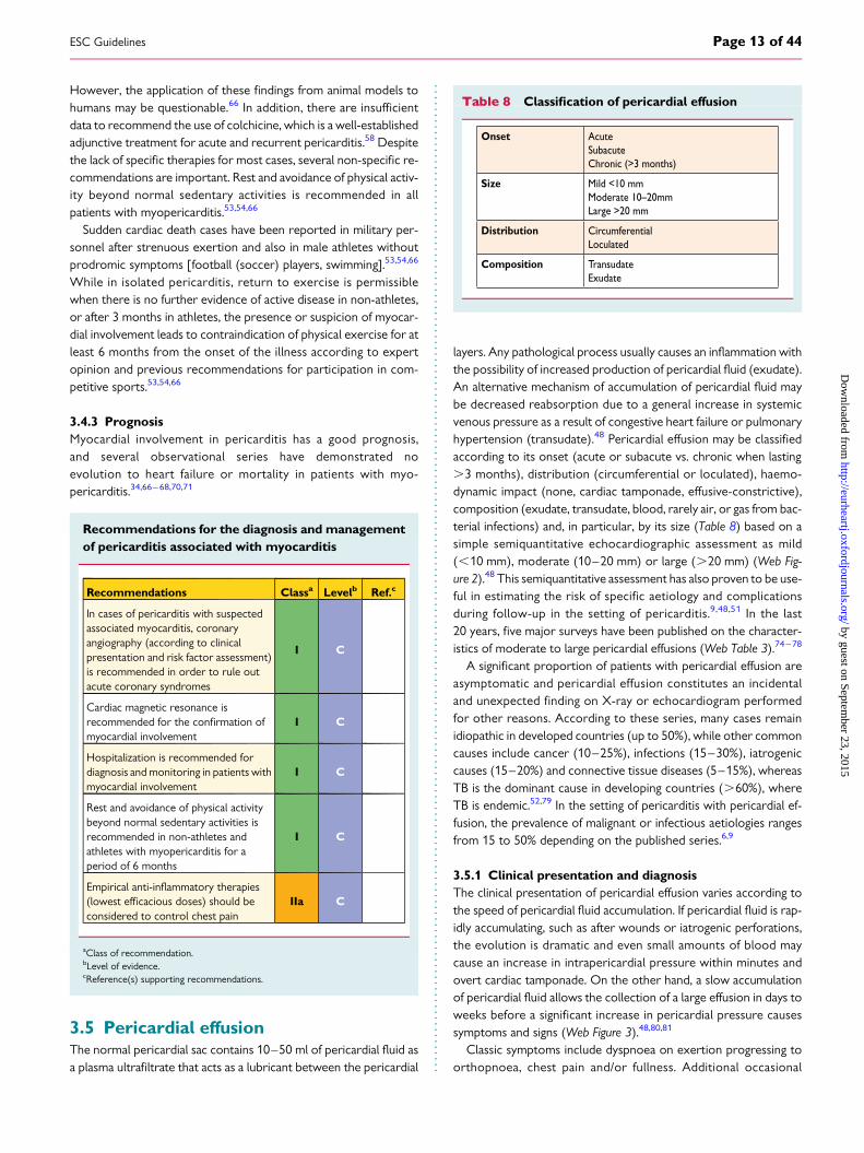
layers. Any pathological process usually causes an inflammation withthe possibility of increased production of pericardial fluid (exudate).An alternative mechanism of accumulation of pericardial fluid maybe decreased reabsorption due to a general increase in systemicvenous pressure as a result of congestive heart failure or pulmonaryhypertension (transudate).48 Pericardial effusion may be classifiedaccording to its onset (acute or subacute vs. chronic when lasting.3 months), distribution (circumferential or loculated), haemo-dynamic impact (none, cardiac tamponade, effusive-constrictive),composition (exudate, transudate, blood, rarely air, or gas from bac-terial infections) and, in particular, by its size (Table 8) based on asimple semiquantitative echocardiographic assessment as mild(,10 mm), moderate (10–20 mm) or large (.20 mm) (Web Fig-ure 2).48 This semiquantitative assessment has also proven to be use-ful in estimating the risk of specific aetiology and complicationsduring follow-up in the setting of pericarditis.9,48,51 In the last20 years, five major surveys have been published on the character-istics of moderate to large pericardial effusions (Web Table 3).74–78
A significant proportion of patients with pericardial effusion areasymptomatic and pericardial effusion constitutes an incidentaland unexpected finding on X-ray or echocardiogram performedfor other reasons. According to these series, many cases remainidiopathic in developed countries (up to 50%), while other commoncauses include cancer (10–25%), infections (15–30%), iatrogeniccauses (15–20%) and connective tissue diseases (5–15%), whereasTB is the dominant cause in developing countries (.60%), whereTB is endemic.52,79 In the setting of pericarditis with pericardial ef-fusion, the prevalence of malignant or infectious aetiologies rangesfrom 15 to 50% depending on the published series.6,9
3.5.1 Clinical presentation and diagnosisThe clinical presentation of pericardial effusion varies according tothe speed of pericardial fluid accumulation. If pericardial fluid is rap-idly accumulating, such as after wounds or iatrogenic perforations,the evolution is dramatic and even small amounts of blood maycause an increase in intrapericardial pressure within minutes andovert cardiac tamponade. On the other hand, a slow accumulationof pericardial fluid allows the collection of a large effusion in days toweeks before a significant increase in pericardial pressure causessymptoms and signs (Web Figure 3).48,80,81
Classic symptoms include dyspnoea on exertion progressing toorthopnoea, chest pain and/or fullness. Additional occasional
Table 8 Classification of pericardial effusion
Onset AcuteSubacuteChronic (>3 months)
Size Mild <10 mmModerate 10–20mmLarge >20 mm
Distribution CircumferentialLoculated
Composition TransudateExudate
ESC Guidelines Page 13 of 44
by guest on September 23, 2015
http://eurheartj.oxfordjournals.org/D
ownloaded from
symptoms due to local compression may include nausea (dia-phragm), dysphagia (oesophagus), hoarseness (recurrent laryngealnerve) and hiccups (phrenic nerve). Non-specific symptoms includecough, weakness, fatigue, anorexia and palpitations, and reflectthe compressive effect of the pericardial fluid on contiguousanatomic structures or reduced blood pressure and secondary sinustachycardia.82–84 Fever is a non-specific sign that may be associatedwith pericarditis, either infectious or immune mediated (i.e. systemicinflammatory diseases).45
Physical examination may be absolutely normal in patients with-out haemodynamic compromise. When tamponade develops, clas-sic signs include neck vein distension with elevated jugular venouspressure at bedside examination, pulsus paradoxus and diminishedheart sounds on cardiac auscultation in cases of moderate to largeeffusions.82 – 84 Pericardial friction rubs are rarely heard; they canusually be detected in patients with concomitant pericarditis.8
The diagnosis of pericardial effusion is generally performed byechocardiography, which also enables semiquantitative assessmentof the pericardial effusion size and its haemodynamic effects. Al-though echocardiography remains the primary diagnostic tool forthe study of pericardial diseases because of its widespread availabil-ity, portability and limited costs, CT and CMR provide a larger fieldof view, allowing the detection of loculated pericardial effusion andpericardial thickening and masses, as well as associated chestabnormalities.2,3,84
Recommendations for the diagnosis of pericardialeffusion
Recommendations Classa Levelb Ref.c
Transthoracic echocardiography isrecommended in all patients withsuspected pericardial effusion
I C
Chest X-ray is recommended inpatients with a suspicion of pericardialeffusion or pleuropulmonaryinvolvement
I C
Assessment of markers ofinflammation (i.e. CRP) arerecommended in patients withpericardial effusion
I C
CT or CMR should be considered insuspected cases of loculated pericardialeffusion, pericardial thickening andmasses, as well as associated chestabnormalities
IIa C
CMR ¼ cardiac magnetic resonance; CRP ¼ C-reactive protein; CT ¼computed tomography.aClass of recommendation.bLevel of evidence.cReference(s) supporting recommendations.
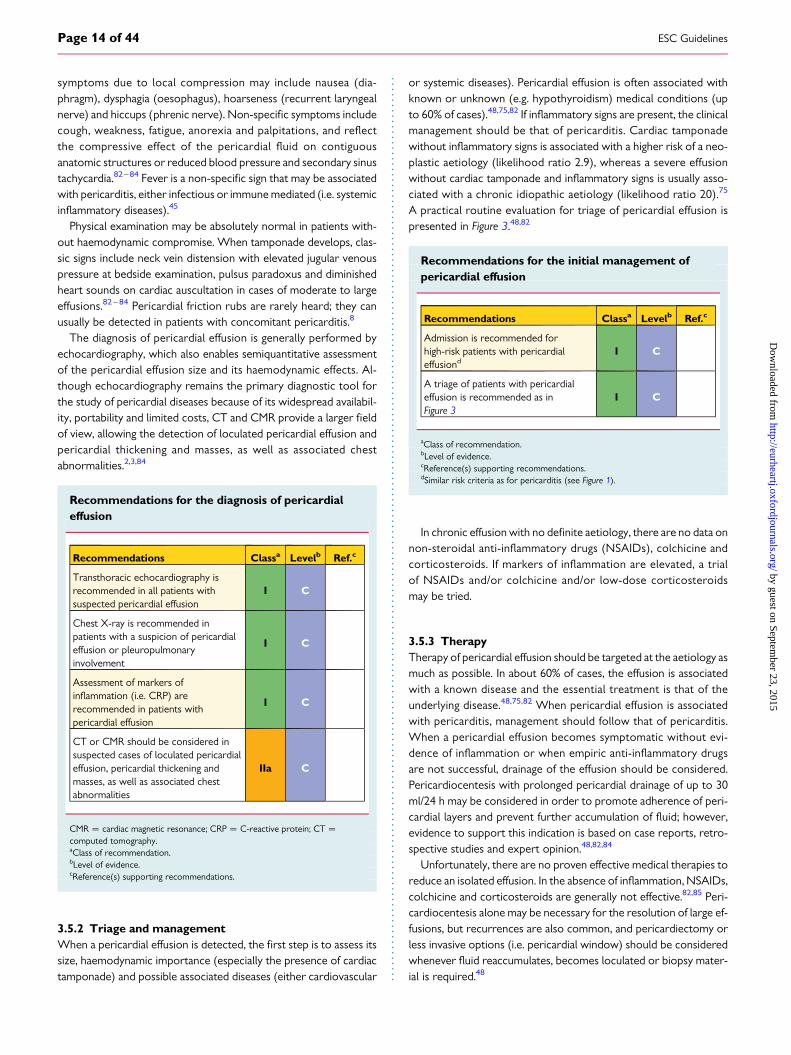
3.5.2 Triage and managementWhen a pericardial effusion is detected, the first step is to assess itssize, haemodynamic importance (especially the presence of cardiactamponade) and possible associated diseases (either cardiovascular
or systemic diseases). Pericardial effusion is often associated withknown or unknown (e.g. hypothyroidism) medical conditions (upto 60% of cases).48,75,82 If inflammatory signs are present, the clinicalmanagement should be that of pericarditis. Cardiac tamponadewithout inflammatory signs is associated with a higher risk of a neo-plastic aetiology (likelihood ratio 2.9), whereas a severe effusionwithout cardiac tamponade and inflammatory signs is usually asso-ciated with a chronic idiopathic aetiology (likelihood ratio 20).75
A practical routine evaluation for triage of pericardial effusion ispresented in Figure 3.48,82
Recommendations for the initial management ofpericardial effusion
Recommendations Classa Levelb Ref.c
Admission is recommended forhigh-risk patients with pericardialeffusiond
I C
A triage of patients with pericardialeffusion is recommended as inFigure 3
I C
aClass of recommendation.bLevel of evidence.cReference(s) supporting recommendations.dSimilar risk criteria as for pericarditis (see Figure 1).
In chronic effusion with no definite aetiology, there are no data onnon-steroidal anti-inflammatory drugs (NSAIDs), colchicine andcorticosteroids. If markers of inflammation are elevated, a trialof NSAIDs and/or colchicine and/or low-dose corticosteroidsmay be tried.
3.5.3 TherapyTherapy of pericardial effusion should be targeted at the aetiology asmuch as possible. In about 60% of cases, the effusion is associatedwith a known disease and the essential treatment is that of theunderlying disease.48,75,82 When pericardial effusion is associatedwith pericarditis, management should follow that of pericarditis.When a pericardial effusion becomes symptomatic without evi-dence of inflammation or when empiric anti-inflammatory drugsare not successful, drainage of the effusion should be considered.Pericardiocentesis with prolonged pericardial drainage of up to 30ml/24 h may be considered in order to promote adherence of peri-cardial layers and prevent further accumulation of fluid; however,evidence to support this indication is based on case reports, retro-spective studies and expert opinion.48,82,84
Unfortunately, there are no proven effective medical therapies toreduce an isolated effusion. In the absence of inflammation, NSAIDs,colchicine and corticosteroids are generally not effective.82,85 Peri-cardiocentesis alone may be necessary for the resolution of large ef-fusions, but recurrences are also common, and pericardiectomy orless invasive options (i.e. pericardial window) should be consideredwhenever fluid reaccumulates, becomes loculated or biopsy mater-ial is required.48
ESC GuidelinesPage 14 of 44
by guest on September 23, 2015
http://eurheartj.oxfordjournals.org/D
ownloaded from
Recommendations for the therapy of pericardialeffusion
Recommendations Classa Levelb Ref.c
It is recommended to target thetherapy of pericardial effusion at theaetiology
I C
Aspirin/NSAIDs/colchicine andtreatment of pericarditis isrecommended when pericardial effusionis associated with systemic inflammation
I C
Pericardiocentesis or cardiac surgery isindicated for cardiac tamponade or forsymptomatic moderate to largepericardial effusions not responsive tomedical therapy, and for suspicion ofunknown bacterial or neoplasticaetiology
I C
NSAIDs ¼ non-steroidal anti-inflammatory drugs.aClass of recommendation.bLevel of evidence.cReference(s) supporting recommendations.
3.5.4 Prognosis and follow-upThe prognosis of pericardial effusion is essentially related to the aeti-ology.48,82,86 The size of the effusion is correlated with the progno-sis, as moderate to large effusions are more common for specificaetiologies such as bacterial and neoplastic conditions.9,48 Idiopathicpericardial effusion and pericarditis have an overall good prognosiswith a very low risk of complications, especially if the effusion is mildto moderate. In contrast with these observations, a recently pub-lished prospective study has shown that even with mild pericardialeffusion the overall prognosis may be worse than in age- and sex-matched controls.87
Large idiopathic chronic effusions (.3 months) have a 30–35%risk of progression to cardiac tamponade.88 Also, subacute (4–6weeks) large effusions not responsive to conventional therapy andwith echocardiographic signs of collapse of the right chambersmay have an increased risk of progression according to someauthors, who recommend preventive drainage in such cases.89
Documented idiopathic pericarditis has a very low risk of constrict-ive pericarditis despite several recurrences: here the risk is relatedto the aetiology and not the number of recurrences.36 The follow-up of pericardial effusion is mainly based on the evaluation of symp-toms and the echocardiographic size of the effusion, as well asadditional features such as inflammatory markers (i.e. CRP).48
Cardiac tamponade orsuspected bacterial orneoplastic aetiology?
Pericardiocentesis andaetiology search
Elevated inflammatorymarkers?
Empiric ant-inflammatorytherapy (treat as pericarditis)
Known associateddisease?
Pericardial effusionprobably related.Treat the disease.
Large (>20 mm)pericardial effusion?
Follow-up
Consider pericardiocentesisand drainage
if chronic (>3 months)
Yes No
Yes No
Yes No
Yes
No
Figure 3 A simplified algorithm for pericardial effusion triage and management.
ESC Guidelines Page 15 of 44
by guest on September 23, 2015
http://eurheartj.oxfordjournals.org/D
ownloaded from
A mild idiopathic effusion (,10 mm) is usually asymptomatic,generally has a good prognosis and does not require specific mon-itoring.48 Moderate to large effusions (.10 mm) may worsen, andespecially severe effusions may evolve towards cardiac tamponadein up to one-third of cases. For idiopathic moderate effusions, anappropriate timing for echocardiographic follow-up may be anechocardiogram every 6 months. For a severe effusion, an echocar-diographic follow-up may be every 3–6 months. A tailored follow-up is also warranted considering the relative stability or evolution ofthe size.48 Specific considerations on pericardial effusion in the post-operative setting are discussed in the section on post-cardiac injurysyndromes (section 5.5).
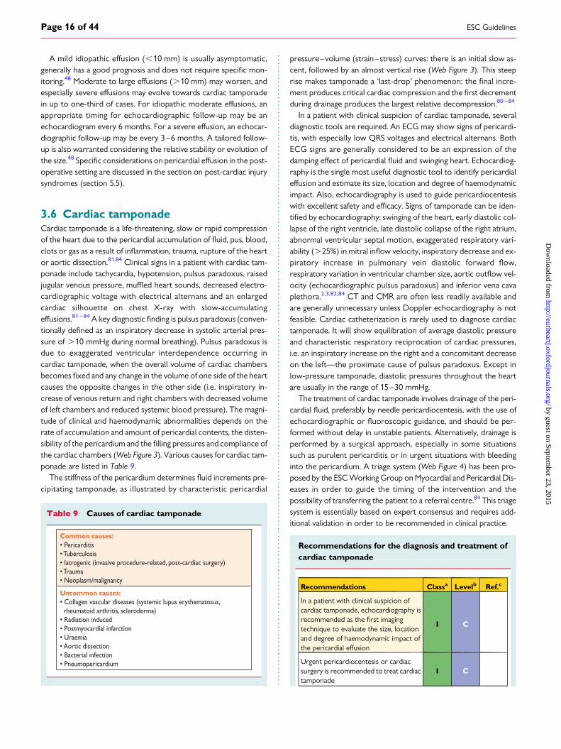
3.6 Cardiac tamponadeCardiac tamponade is a life-threatening, slow or rapid compressionof the heart due to the pericardial accumulation of fluid, pus, blood,clots or gas as a result of inflammation, trauma, rupture of the heartor aortic dissection.81,84 Clinical signs in a patient with cardiac tam-ponade include tachycardia, hypotension, pulsus paradoxus, raisedjugular venous pressure, muffled heart sounds, decreased electro-cardiographic voltage with electrical alternans and an enlargedcardiac silhouette on chest X-ray with slow-accumulatingeffusions.81– 84 A key diagnostic finding is pulsus paradoxus (conven-tionally defined as an inspiratory decrease in systolic arterial pres-sure of .10 mmHg during normal breathing). Pulsus paradoxus isdue to exaggerated ventricular interdependence occurring incardiac tamponade, when the overall volume of cardiac chambersbecomes fixed and any change in the volume of one side of the heartcauses the opposite changes in the other side (i.e. inspiratory in-crease of venous return and right chambers with decreased volumeof left chambers and reduced systemic blood pressure). The magni-tude of clinical and haemodynamic abnormalities depends on therate of accumulation and amount of pericardial contents, the disten-sibility of the pericardium and the filling pressures and compliance ofthe cardiac chambers (Web Figure 3). Various causes for cardiac tam-ponade are listed in Table 9.
The stiffness of the pericardium determines fluid increments pre-cipitating tamponade, as illustrated by characteristic pericardial
pressure–volume (strain–stress) curves: there is an initial slow as-cent, followed by an almost vertical rise (Web Figure 3). This steeprise makes tamponade a ‘last-drop’ phenomenon: the final incre-ment produces critical cardiac compression and the first decrementduring drainage produces the largest relative decompression.80– 84
In a patient with clinical suspicion of cardiac tamponade, severaldiagnostic tools are required. An ECG may show signs of pericardi-tis, with especially low QRS voltages and electrical alternans. BothECG signs are generally considered to be an expression of thedamping effect of pericardial fluid and swinging heart. Echocardiog-raphy is the single most useful diagnostic tool to identify pericardialeffusion and estimate its size, location and degree of haemodynamicimpact. Also, echocardiography is used to guide pericardiocentesiswith excellent safety and efficacy. Signs of tamponade can be iden-tified by echocardiography: swinging of the heart, early diastolic col-lapse of the right ventricle, late diastolic collapse of the right atrium,abnormal ventricular septal motion, exaggerated respiratory vari-ability (.25%) in mitral inflow velocity, inspiratory decrease and ex-piratory increase in pulmonary vein diastolic forward flow,respiratory variation in ventricular chamber size, aortic outflow vel-ocity (echocardiographic pulsus paradoxus) and inferior vena cavaplethora.2,3,82,84 CT and CMR are often less readily available andare generally unnecessary unless Doppler echocardiography is notfeasible. Cardiac catheterization is rarely used to diagnose cardiactamponade. It will show equilibration of average diastolic pressureand characteristic respiratory reciprocation of cardiac pressures,i.e. an inspiratory increase on the right and a concomitant decreaseon the left—the proximate cause of pulsus paradoxus. Except inlow-pressure tamponade, diastolic pressures throughout the heartare usually in the range of 15–30 mmHg.
The treatment of cardiac tamponade involves drainage of the peri-cardial fluid, preferably by needle pericardiocentesis, with the use ofechocardiographic or fluoroscopic guidance, and should be per-formed without delay in unstable patients. Alternatively, drainage isperformed by a surgical approach, especially in some situationssuch as purulent pericarditis or in urgent situations with bleedinginto the pericardium. A triage system (Web Figure 4) has been pro-posed by the ESC Working Group on Myocardial and Pericardial Dis-eases in order to guide the timing of the intervention and thepossibility of transferring the patient to a referral centre.84 This triagesystem is essentially based on expert consensus and requires add-itional validation in order to be recommended in clinical practice.
Recommendations for the diagnosis and treatment ofcardiac tamponade
Recommendations Classa Levelb Ref.c
In a patient with clinical suspicion ofcardiac tamponade, echocardiography isrecommended as the first imagingtechnique to evaluate the size, locationand degree of haemodynamic impact ofthe pericardial effusion
I C
Urgent pericardiocentesis or cardiacsurgery is recommended to treat cardiactamponade
I C
Table 9 Causes of cardiac tamponade
Common causes: • Pericarditis• Tuberculosis• Iatrogenic (invasive procedure-related, post-cardiac surgery)• Trauma• Neoplasm/malignancy
Uncommon causes: • Collagen vascular diseases (systemic lupus erythematosus, rheumatoid arthritis, scleroderma)• Radiation induced• Postmyocardial infarction• Uraemia• Aortic dissection• Bacterial infection• Pneumopericardium
ESC GuidelinesPage 16 of 44
by guest on September 23, 2015
http://eurheartj.oxfordjournals.org/D
ownloaded from
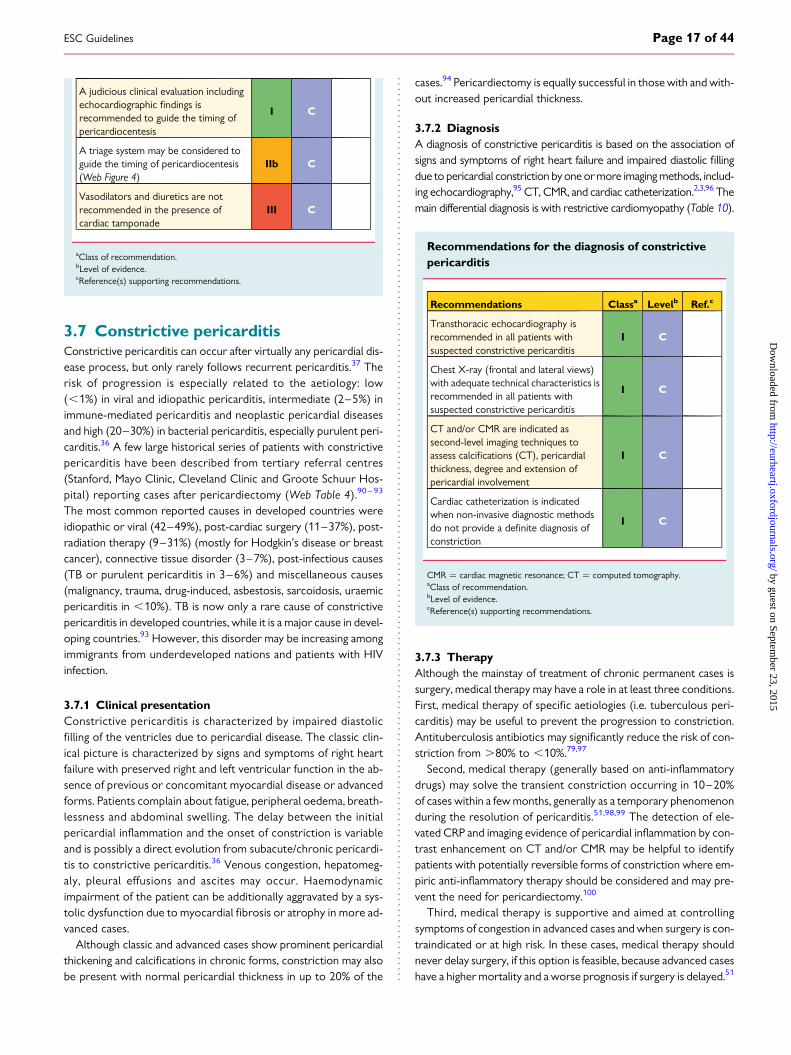
A judicious clinical evaluation includingechocardiographic findings isrecommended to guide the timing ofpericardiocentesis
I C
A triage system may be considered toguide the timing of pericardiocentesis(Web Figure 4)
IIb C
Vasodilators and diuretics are notrecommended in the presence ofcardiac tamponade
III C
aClass of recommendation.bLevel of evidence.cReference(s) supporting recommendations.
3.7 Constrictive pericarditisConstrictive pericarditis can occur after virtually any pericardial dis-ease process, but only rarely follows recurrent pericarditis.37 Therisk of progression is especially related to the aetiology: low(,1%) in viral and idiopathic pericarditis, intermediate (2–5%) inimmune-mediated pericarditis and neoplastic pericardial diseasesand high (20–30%) in bacterial pericarditis, especially purulent peri-carditis.36 A few large historical series of patients with constrictivepericarditis have been described from tertiary referral centres(Stanford, Mayo Clinic, Cleveland Clinic and Groote Schuur Hos-pital) reporting cases after pericardiectomy (Web Table 4).90 – 93
The most common reported causes in developed countries wereidiopathic or viral (42–49%), post-cardiac surgery (11–37%), post-radiation therapy (9–31%) (mostly for Hodgkin’s disease or breastcancer), connective tissue disorder (3–7%), post-infectious causes(TB or purulent pericarditis in 3–6%) and miscellaneous causes(malignancy, trauma, drug-induced, asbestosis, sarcoidosis, uraemicpericarditis in ,10%). TB is now only a rare cause of constrictivepericarditis in developed countries, while it is a major cause in devel-oping countries.93 However, this disorder may be increasing amongimmigrants from underdeveloped nations and patients with HIVinfection.
3.7.1 Clinical presentationConstrictive pericarditis is characterized by impaired diastolicfilling of the ventricles due to pericardial disease. The classic clin-ical picture is characterized by signs and symptoms of right heartfailure with preserved right and left ventricular function in the ab-sence of previous or concomitant myocardial disease or advancedforms. Patients complain about fatigue, peripheral oedema, breath-lessness and abdominal swelling. The delay between the initialpericardial inflammation and the onset of constriction is variableand is possibly a direct evolution from subacute/chronic pericardi-tis to constrictive pericarditis.36 Venous congestion, hepatomeg-aly, pleural effusions and ascites may occur. Haemodynamicimpairment of the patient can be additionally aggravated by a sys-tolic dysfunction due to myocardial fibrosis or atrophy in more ad-vanced cases.
Although classic and advanced cases show prominent pericardialthickening and calcifications in chronic forms, constriction may alsobe present with normal pericardial thickness in up to 20% of the
cases.94 Pericardiectomy is equally successful in those with and with-out increased pericardial thickness.
3.7.2 DiagnosisA diagnosis of constrictive pericarditis is based on the association ofsigns and symptoms of right heart failure and impaired diastolic fillingdue to pericardial constriction by one or more imaging methods, includ-ing echocardiography,95 CT, CMR, and cardiac catheterization.2,3,96 Themain differential diagnosis is with restrictive cardiomyopathy (Table 10).
Recommendations for the diagnosis of constrictivepericarditis
Recommendations Classa Levelb Ref.c
Transthoracic echocardiography isrecommended in all patients withsuspected constrictive pericarditis
I C
Chest X-ray (frontal and lateral views)with adequate technical characteristics isrecommended in all patients withsuspected constrictive pericarditis
I C
CT and/or CMR are indicated assecond-level imaging techniques toassess calcifications (CT), pericardialthickness, degree and extension ofpericardial involvement
I C
Cardiac catheterization is indicatedwhen non-invasive diagnostic methodsdo not provide a definite diagnosis ofconstriction
I C
CMR ¼ cardiac magnetic resonance; CT ¼ computed tomography.aClass of recommendation.bLevel of evidence.cReference(s) supporting recommendations.
3.7.3 TherapyAlthough the mainstay of treatment of chronic permanent cases issurgery, medical therapy may have a role in at least three conditions.First, medical therapy of specific aetiologies (i.e. tuberculous peri-carditis) may be useful to prevent the progression to constriction.Antituberculosis antibiotics may significantly reduce the risk of con-striction from .80% to ,10%.79,97
Second, medical therapy (generally based on anti-inflammatorydrugs) may solve the transient constriction occurring in 10–20%of cases within a few months, generally as a temporary phenomenonduring the resolution of pericarditis.51,98,99 The detection of ele-vated CRP and imaging evidence of pericardial inflammation by con-trast enhancement on CT and/or CMR may be helpful to identifypatients with potentially reversible forms of constriction where em-piric anti-inflammatory therapy should be considered and may pre-vent the need for pericardiectomy.100
Third, medical therapy is supportive and aimed at controllingsymptoms of congestion in advanced cases and when surgery is con-traindicated or at high risk. In these cases, medical therapy shouldnever delay surgery, if this option is feasible, because advanced caseshave a higher mortality and a worse prognosis if surgery is delayed.51
ESC Guidelines Page 17 of 44
by guest on September 23, 2015
http://eurheartj.oxfordjournals.org/D
ownloaded from

3.7.4 Specific formsThe classic description of chronic permanent constrictive pericardi-tis has been challenged by specific forms of constrictive syndromes(i.e. transient constriction, effusive-constrictive forms). Definitions,main differential diagnoses and treatment of the main constrictivepericardial syndromes are summarized in Table 11.51
3.7.4.1 Transient constrictive pericarditisA temporary form of constriction usually develops with pericar-ditis and mild effusion and resolves with anti-inflammatory
therapy within several weeks.98,99 The typical clinical course im-plies the presence of acute inflammatory pericarditis with con-striction due to inflammation, which resolves once theinflammatory process is treated.98,99 Thus, in the absence of evi-dence that the condition is chronic (e.g. cachexia, atrial fibrillation,hepatic dysfunction or pericardial calcification), patients withnewly diagnosed constrictive pericarditis who are haemodynam-ically stable may be given a trial of conservative managementfor 2–3 months before recommending pericardiectomy. Sincethe inflamed pericardium is enhanced on CT and/or CMR,
Table 11 Definitions and therapy of main constrictive pericardial syndromes (adapted from Imazio et al.51)
Syndrome Therapy
Transient constriction (d.d. permanent constrictive pericarditis, restrictive CMP).
Reversible pattern of constriction following spontaneous recovery or medical therapy. medical therapy.
Effusive-constrictive pericarditis (d.d. cardiac tamponade, constrictive pericarditis).
Failure of the right atrial pressure to fall by 50% or to a level below 10 mmHg after pericardiocentesis. May be diagnosed also by non-invasive imaging.
Pericardiocentesis followed by medical therapy. Surgery for persistent cases.
Chronic constriction (d.d. transient constriction, restrictive CMP).
Persistent constriction after 3–6 months. Pericardiectomy, medical therapy for advanced cases or high risk of surgery or mixed forms with myocardial involvement.
CMP ¼ cardiomyopathy; d.d. ¼ differential diagnosis.
Table 10 Constrictive pericarditis vs. restrictive cardiomyopathy: a brief overview of features for the differentialdiagnosis (Modified from Imazio et al.51)
Diagnostic evaluation
Constrictive pericarditis Restrictive cardiomyopathy
Regurgitant murmur, Kussmaul sign may be present, S3 (advanced).
ECG Low voltages, pseudoinfarction, possible widening of QRS,
Echocardiography • Septal bounce. • P• Respiratory variation of the mitral peak E velocity of >25% and variation in the pulmonar• w propagation velocity (Vp) >45 cm/sec.• Tissue Doppler: peak e' >8.0 cm/s.
• Small left ventricle with large atria, possible increased wall thickness.• E/A ratio >2, short DT.• • w propagation velocity (Vp) <45 cm/sec.• Tissue Doppler: peak e' <8.0 cm/s.
Cardiac Catheterization
‘Dip and plateau’ or ‘square root’ sign, right ventricular diastolic, and left ventricular diastolic pressures usually equal, ventricular interdependence (i.e. assessed by the systolic area index >1.1).a
Marked right ventricular systolic hypertension (>50 mmHg) and left ventricular diastolic pressure exceeds right ventricular diastolic pressure (LVEDP >RVEDP) at rest or during exercise by 5 mmHg or more (RVEDP <1/3 RVSP).
CT/CMRinterdependence (real-time cine CMR).
Normal pericardial thickness (<3.0 mm), myocardial involvement by morphology and functional study (CMR).
CMR ¼ cardiac magnetic resonance; CT ¼ computed tomography; DT¼ deceleration time; ECG ¼ electrocardiogram; LVEDP ¼ left ventricular end-diastolic pressure;RVEDP¼ right ventricular end-diastolic pressure; RVSP ¼ right ventricular systolic pressure; S3 ¼ third sound. Kussmaul sign is a paradoxical rise in jugular venous pressure oninspiration.aThe systolic area index was defined as the ratio of the RV area (mmHg x s) to the LV area (mmHg x s) in inspiration versus expiration.96
Specific diagnostic echocardiographic criteria for the diagnosis of constrictive pericarditis has been recently proposed by the Mayo Clinic and include: septal bounce or ventricularseptal shift with either medial e′ .8 cm/s or hepatic vein expiratory diastolic reversal ratio .0.78 (sensitivity 87%, specificity 91%; specificity may increase to 97% if all criteria arepresent with a correspondent decrease of sensitivity to 64%.95
ESC GuidelinesPage 18 of 44
by guest on September 23, 2015
http://eurheartj.oxfordjournals.org/D
ownloaded from
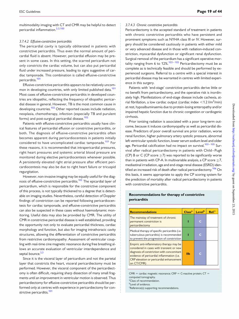
multimodality imaging with CT and CMR may be helpful to detectpericardial inflammation.2,3,100
3.7.4.2 Effusive-constrictive pericarditisThe pericardial cavity is typically obliterated in patients withconstrictive pericarditis. Thus even the normal amount of peri-cardial fluid is absent. However, pericardial effusion may be pre-sent in some cases. In this setting, the scarred pericardium notonly constricts the cardiac volume, but can also put pericardialfluid under increased pressure, leading to signs suggestive of car-diac tamponade. This combination is called effusive-constrictivepericarditis.101
Effusive-constrictive pericarditis appears to be relatively uncom-mon in developing countries, with only limited published data.101
Most cases of effusive-constrictive pericarditis in developed coun-tries are idiopathic, reflecting the frequency of idiopathic pericar-dial disease in general. However, TB is the most common cause indeveloping countries.102 Other reported causes include radiation,neoplasia, chemotherapy, infection (especially TB and purulentforms) and post-surgical pericardial disease.102
Patients with effusive-constrictive pericarditis usually have clin-ical features of pericardial effusion or constrictive pericarditis, orboth. The diagnosis of effusive-constrictive pericarditis oftenbecomes apparent during pericardiocentesis in patients initiallyconsidered to have uncomplicated cardiac tamponade.101 Forthese reasons, it is recommended that intrapericardial pressures,right heart pressures and systemic arterial blood pressure aremonitored during elective pericardiocentesis whenever possible.A persistently elevated right atrial pressure after efficient peri-cardiocentesis may also be due to right heart failure or tricuspidregurgitation.
However, non-invasive imaging may be equally useful for the diag-nosis of effusive-constrictive pericarditis.102 The epicardial layer ofpericardium, which is responsible for the constrictive componentof this process, is not typically thickened to a degree that is detect-able on imaging studies. Nevertheless, careful detection of Dopplerfindings of constriction can be reported following pericardiocen-tesis for cardiac tamponade, and effusive-constrictive pericarditiscan also be suspected in these cases without haemodynamic mon-itoring. Useful data may also be provided by CMR. The utility ofCMR in constrictive pericardial disease is well established, providingthe opportunity not only to evaluate pericardial thickness, cardiacmorphology and function, but also for imaging intrathoracic cavitystructures, allowing the differentiation of constrictive pericarditisfrom restrictive cardiomyopathy. Assessment of ventricular coup-ling with real-time cine magnetic resonance during free breathing al-lows an accurate evaluation of ventricular interdependence andseptal bounce.2,3
Since it is the visceral layer of pericardium and not the parietallayer that constricts the heart, visceral pericardiectomy must beperformed. However, the visceral component of the pericardiect-omy is often difficult, requiring sharp dissection of many small frag-ments until an improvement in ventricular motion is observed. Thuspericardiectomy for effusive-constrictive pericarditis should be per-formed only at centres with experience in pericardiectomy for con-strictive pericarditis.101
3.7.4.3 Chronic constrictive pericarditisPericardiectomy is the accepted standard of treatment in patientswith chronic constrictive pericarditis who have persistent andprominent symptoms such as NYHA class III or IV. However, sur-gery should be considered cautiously in patients with either mildor very advanced disease and in those with radiation-induced con-striction, myocardial dysfunction or significant renal dysfunction.Surgical removal of the pericardium has a significant operative mor-tality ranging from 6 to 12%.103 – 105 Pericardiectomy must be ascomplete as is technically feasible and should be performed by ex-perienced surgeons. Referral to a centre with a special interest inpericardial disease may be warranted in centres with limited experi-ence in this surgery.
Patients with ‘end-stage’ constrictive pericarditis derive little orno benefit from pericardiectomy, and the operative risk is inordin-ately high. Manifestations of end-stage disease include cachexia, at-rial fibrillation, a low cardiac output (cardiac index ,1.2 l/m2/min)at rest, hypoalbuminaemia due to protein-losing enteropathy and/orimpaired hepatic function due to chronic congestion or cardiogeniccirrhosis.
Prior ionizing radiation is associated with a poor long-term out-come, because it induces cardiomyopathy as well as pericardial dis-ease. Predictors of poor overall survival are prior radiation, worserenal function, higher pulmonary artery systolic pressure, abnormalleft ventricular systolic function, lower serum sodium level and olderage. Pericardial calcification had no impact on survival.103 – 105 Sur-vival after radical pericardiectomy in patients with Child–Pugh(CP) B or C (CP score ≥7) was reported to be significantly worsethan in patients with CP-A. In multivariable analysis, a CP score ≥7,mediastinal irradiation, age and end-stage renal disease (ESRD) iden-tified an increased risk of death after radical pericardiectomy.106 Onthis basis, it seems appropriate to apply the CP scoring system forthe prediction of mortality after radical pericardiectomy in patientswith constrictive pericarditis.
Recommendations for therapy of constrictivepericarditis
Recommendations Classa Levelb Ref.c
The mainstay of treatment of chronicpermanent constriction ispericardiectomy
I C
Medical therapy of specific pericarditis (i.e.tuberculous pericarditis) is recommendedto prevent the progression of constriction
I C
Empiric anti-inflammatory therapy may beconsidered in cases with transient or newdiagnosis of constriction with concomitantevidence of pericardial inflammation (i.e.CRP elevation or pericardial enhancementon CT/CMR)
IIb C
CMR ¼ cardiac magnetic resonance; CRP ¼ C-reactive protein; CT ¼computed tomography.aClass of recommendation.bLevel of evidence.cReference(s) supporting recommendations.
ESC Guidelines Page 19 of 44
by guest on September 23, 2015
http://eurheartj.oxfordjournals.org/D
ownloaded from
4. Multimodality cardiovascularimaging and diagnostic work-up
4.1 Multimodality imaging4.1.1 Chest X-rayAlthough chest X-ray can detect pericardial calcifications, present-ing as a curvilinear density at the extreme margin of the silhouette,particularly on the lateral view,107 other techniques (i.e. echocardi-ography, CT) yield much greater accuracy in assessing the heart andlungs, providing information with regard to cardiac size and the pres-ence of pulmonary pathology (e.g., pulmonary congestion, pneumo-nia, TB, lung cancer), pleural effusion and hilar and mediastinalenlargement.
4.1.2 EchocardiographyTransthoracic echocardiography is the first-line imaging test in pa-tients with suspected pericardial disease, because it accurately de-tects pericardial effusion and cardiac tamponade, as well asventricular dysfunction due to myocardial involvement.2,3 Althoughpatients with purely fibrinous acute pericarditis may have a normalechocardiogram, the presence of a pericardial effusion is consistentwith acute pericarditis and is one of the criteria for its diagno-sis.2,5,6,10,11 Echocardiography may help to differentiate acute peri-carditis from myocardial ischaemia by excluding wall motionabnormalities consistent with coronary flow distribution in the set-ting of patients with acute chest pain. However, �5% of patientswith acute pericarditis and myocardial involvement may demon-strate wall motion abnormalities.
Clinically, two-dimensional echocardiography with Doppler pro-vides the most cost-effective way of diagnosing pericardial effusionand assessing its haemodynamic significance.48 The size of pericar-dial effusion on two-dimensional echocardiography is qualitativelyassessed by the end-diastolic distance of the echo-free space be-tween the epicardium and parietal pericardium: small (,10 mm),moderate (10–20 mm), large (.20 mm) (Web Figure 2).48
In order to allow follow-up studies, it is recommended that theimages be documented digitally and the effusion size described ina detailed way in the echocardiographic report, including not onlythe extent, but also the location of each measurement. However,the haemodynamic tolerance is more related to the rapidity of ap-pearance of the effusion than to its total volume.48,80
Loculated pericardial effusions or pericardial effusions that con-tain clots (e.g. after cardiac surgery) may be difficult to diagnoseusing a transthoracic approach and may require transoesophagealechocardiography.3,4 Specific findings in pericardial syndromes arediscussed in the pertinent paragraphs.
4.1.3 Computed tomographyCT should be regarded as a valuable complementary imaging modal-ity to echocardiography.3,4,41,108,109 CT is the most accurate tech-nique to image calcified tissue.2,3 Current multidetector CTscanners combine acquisition speed, high contrast and spatial reso-lution with volumetric scanning to provide excellent anatomical de-tail of the heart and pericardium. The anatomical region of interestcovered by CT can be limited to the heart and pericardium (‘cardiac
CT’), although in patients with neoplastic, inflammatory or aorticdisease it may encompass the chest entirely and possibly also includethe abdomen and pelvis.108,109 Low-radiation cardiac CT is feasibleusing prospective electrocardiographic triggering.108,109 Althoughthe functional consequences of pericardial disease on the heartcan be evaluated by CT—at the expense of significantly higher radi-ation doses—echocardiography and CMR are more appropriate forassessing this feature. Intravenous administration of iodinated con-trast material is recommended to increase the density of bloodand to depict pericardial inflammation. The normal pericardium isvisible as a thin curvilinear structure surrounded by the hypodensemediastinal and epicardial fat, and has a thickness ranging from 0.7 to2.0 mm. The pericardial sinuses and their respective recesses arevisible, in particular when they contain small amounts of pericardialfluid. The main CT findings in pericardial effusion and pericarditis aresummarized in Table 12.41,108,109
In patients with neoplastic disease, pericardial involvement mayoccur by direct tumour invasion or metastatic spread. CT is import-ant in treatment planning and patient follow-up. The diagnosis of(congenital) pericardial cysts—presenting as well-defined, fluid-dense structures along the left or right heart border—as well asthe differential diagnosis with other cystic structures, such as bron-chogenic or duplication cysts, is usually straightforward. Finally, CTcan be helpful to establish the diagnosis in congenital absence of thepericardium by showing displacement of cardiac structures throughthe pericardial defect. CT is also essential in the preoperative work-up of some patients with constrictive pericarditis, especially to de-pict the extension of calcifications and for those with a history ofprior cardiothoracic surgery.109
4.1.4 Cardiac magnetic resonanceOver the years, CMR has shifted from a morphologic imaging mo-dality towards a comprehensive one, allowing visualization and tis-sue characterization of the pericardium (and heart) in patientswith pericardial disease and appraisal of the consequences of peri-cardial abnormalities on cardiac function and filling patterns.110,111
As such, it is probably the preferred imaging modality to optimallyassess pericardial disease.112,113 Cardiac and pericardial morph-ology are evaluated by dark-blood T1-weighted fast spin-echo andbright-blood cine steady-state free-precession (SSFP) imaging.Cine SSFP imaging has become the reference sequence to assessand quantify cardiac volumes, myocardial mass and ventricular func-tion. When acquired in real-time, this sequence can be used to as-sess ventricular coupling by assessing the changes in ventricularseptal shape and motion over the respiratory cycle.109,110 Tissuecharacterization of the heart and pericardium is achieved by dark-blood T1-weighted and dark-blood T2-weighted, short-tauinversion-recovery (STIR) spin-echo imaging, cine SSFP imagingand T1-weighted contrast-enhanced and/or late contrast-enhanced(LCE) imaging following intravenous administration of paramagneticgadolinium chelates.3,4,114 The LCE sequence uses an inversion-recovery pre-pulse to increase image contrast and is well suitedto visualize pericardial inflammation.114,115
Ventricular inflow and venous flow patterns can be evaluatedusing phase contrast imaging.111 Similar to CT, the normal
ESC GuidelinesPage 20 of 44
by guest on September 23, 2015
http://eurheartj.oxfordjournals.org/D
ownloaded from

Table 12 Diagnostic contribution of the different imaging modalities in various pericardial diseases
Echocardiography Computerized tomography Cardiac magnetic resonance
Acutepericarditis
- veral patients- thick- variable amount of pericar
- wall motion abnormalities in myo-pericarditis
- thickened pericardial layers enhancing after contrast administration- abnormalities involving entire pericardium- variable amount of pericar
- thickened pericardial layers- strong pericardial LGE following contrast administration- variable amount of pericar
- (subepicardial/mid-wall) myocardial LGE in cases of myopericarditis.- inspirator on real-time cine CMR, due to decreased pericardial compliance
Recurrent pericarditis
- ditis - ditis- possibl adhesions- ir
- ditis- possibly heterogeneous distribution due otic adhesions.- ir deformation)
Constrictive pericarditis
- thick
- ± ascites- dilated atria- inspiratory ventricular septal motion toward left ventricle (septal bounce) best documented with M-mode- marked dilation and absent or diminished collapse of the IVC and hepatic veins - premature opening of the pulmonary valve- r - w velocity and >40 % incr beat after inspiration;- opposite changes during expiration; - normal or increased propagation velocity of earl M-mode- decreased expiratory diastolic hepatic vein velocities with large reversals- normal or increased mitral annular velocity (>7 cm/sec) at tissue Doppler- annulus reversus (e’ septal >e’ lateral)
- thickened pericardial layers ± pericardial - thickening may be mild to moderate- abnormalities usually most pronounced at the base of the ventricles (RV>LV), atrioventricular grooves and atria- adjacent myocardium- compression of cardiac contents by rigid, deformed pericardium- abnormal shape of ventricular septum- dilated atria, caval/hepatic veins hepatic congestion- contrast reversal in caval/hepatic veins
- ± ascites- atypical presentations *focal constrictive forms *effusive-constrictive forms
- thickened pericardial layers - pericar- thickening may be mild to moderate- abnormalities usually most pronounced at the base of the ventricles (RV>LV), atrioventricular grooves and atria- pericar - possibl adjacent myocardium- compression of cardiac contents by rigid, deformed pericardium- dilated atria, caval/hepatic veins
- ± ascites- increased ventricular coupling assessed by real-time cine CMR and/or real-time phase-contrast imaging- otic adhesion of pericardial layers on CMR tagging- atypical presentations *focal constrictive forms *effusive-constrictive forms
Pericardialeffusion
- ulation in pericardial sac and/or pericardial sinuses- pericardial echolucent space throughout cardiac cycle- - semi-quantitative assessment of effusion severity
- ulation in pericardial sac and/or pericardial sinuses- pericardial width >4 mm regarded as abnormal - advantageous to depict focal effusions and to pr- atten yield inf *simple effusion: 0–20 HU *proteinaceous/haemorrhagic: >20 HU *if very high HU, suspect intrapericardial leakage of contrast (e.g. ruptured aortic dissection) *chylopericardium: negative HU values *pneumopericar center settings)- pericardial layers have normal thickness, *if thickened and enhancing: suspect *if thick pericarditis- may be associated with pericardial tamponade- CT of the heart may be part of a more extended examination including the remainder of the chest ± abdomen
- ulation in pericardial sac and/ or pericardial sinuses- pericardial width >4 mm regarded as - advantageous to depict focal effusions and to precisely quantitate the amount - combination of sequences with different ‘weighting’ yield information with regard to the nature of the effusion- pericardial layers have normal thickness, *if thickened and enhancing suspect - advantageous to evaluate the remainder of the heart: *myocardial tissue characterisation (oedema, osis) *myocardial/valvular function w patterns- may be associated with pericardial tamponade
Cardiac tamponade
- semi-quantitative assessment of effusion severity- - assessment of its haemodynamic impact- guide and monitoring pericardiocentesis- re-evaluation for timing catheter removal
CMR ¼ cardiac magnetic resonance; CT ¼ computed tomography; HU ¼ Hounsfield units; IVC ¼ inferior vena cava; LGE¼ late gadolinium enhancement; LV ¼ left ventricle;RV ¼ right ventricle.
ESC Guidelines Page 21 of 44
by guest on September 23, 2015
http://eurheartj.oxfordjournals.org/D
ownloaded from
pericardium appears on T1-weighted imaging as a thin hypointense(‘dark’) curvilinear structure surrounded by hyperintense (‘bright’)mediastinal and epicardial fat. Normal pericardial thickness rangesfrom 1.2 to 1.7 mm. The imaging characteristics of pericardial effu-sion and pericarditis at CMR are shown in Table 12. It should be em-phasized that CMR can accurately distinguish between mixedmyopericardial diseases such as mixed inflammatory forms (e.g.myopericarditis or perimyocarditis) and post-myocardial infarctionpericardial injury.116,117 In patients with constrictive pericarditis,CMR is particularly important in the diagnosis of atypical presenta-tions, such as those with minimally thickened pericardium oreffusive-constrictive pericarditis, and those with potentially revers-ible or transient forms of constrictive pericarditis, showing enhance-ment of the pericardial layers at LCE imaging.115,118,119 Comparedwith CT, CMR has the advantage of providing information with re-gard to the haemodynamic consequences of the non-compliantpericardium on cardiac filling,109 – 111 and has the potential of show-ing fibrotic fusion of pericardial layers.120
In patients with congenital pericardial pathology and pericardialmalignancy, CMR shares the advantages of CT, but allows better tis-sue characterization and the possibility of evaluating the functionalconsequences.121 Moreover, novel techniques, such as diffusion-weighted and dynamic contrast-enhanced magnetic resonanceimaging, open perspectives for improved tissue characterization inpatients with pericardial tumours.122
4.1.5 Nuclear medicineIn selected cases, positron emission tomography (PET) alone, orpreferably in combination with CT (PET/CT), can be indicated todepict the metabolic activity of pericardial disease. Pericardial up-take of 18F-fluorodeoxyglucose (FDG) tracer in patients with solidcancers and lymphoma is indicative of (malignant) pericardial in-volvement, thus providing essential information on the diagnosis,staging and assessment of the therapeutic response.123 The uptakeis usually intense and often associated with a focal soft tissuemass.124 PET/CT is also of value in identifying the nature of inflam-matory pericarditis. In particular, tuberculous pericarditis yieldshigher FDG uptakes than idiopathic forms.125 However, differenti-ation between benign and malignant pericardial disease, as well asdifferentiation between physiological and pathological cardiacFDG uptake by PET/CT, remains challenging.123
4.1.6 Cardiac catheterizationCardiac catheterization is not routinely used for the diagnosis ofpericardial disease, as current non-invasive techniques are usuallyable to solve the differential diagnosis of a patient with the suspicionof heart disease involving the pericardium. However, right heartcatheterization may be useful in certain circumstances. Early recog-nition of abnormal haemodynamics related to cardiac tamponadeduring invasive procedures (i.e. epicardial ablation, percutaneousaortic valve implantation, complex angioplasty or complex proce-dures involving trans-septal punctures, among others) may helpavoid serious consequences for the patient. In addition, the differen-tiation between constrictive pericarditis and restrictive cardiomyop-athy is sometimes difficult and may require an invasive test.
In cardiac tamponade, the right atrial pressure waveform has anattenuated or an absent Y-descent. Absent Y-descent is secondaryto diastolic equalization of pressures in the right atrium and rightventricle and lack of effective flow across the tricuspid valve in earlyventricular diastole. Also, equalization of mean right atrial, right ven-tricular and pulmonary artery diastolic pressures and mean pulmon-ary capillary wedge pressures can be present. Other haemodynamicabnormalities include elevation of filling pressures in all four cardiacchambers, right ventricle and left ventricle peak systolic pressuresout of phase, peak aortic pressure varying more than 10–12mmHg and a decrease in cardiac output.126,127
The differentiation of constrictive pericarditis from restrictivecardiomyopathy remains difficult. Visualization of the pericardiumby CT or CMR may help in detecting an abnormal pericardium.But these tests provide anatomical information and do not necessar-ily reflect the pathophysiological abnormality present. Also, patientswith surgically proven constrictive pericarditis may have a normal-appearing pericardium on imaging studies. Alternatively patientsmay have abnormal pericardial thickness in the absence of constric-tion, especially after radiation therapy or prior cardiac surgery. Clas-sically, direct measurements of pressures show M- or W-shapedatrial pressure waveforms and ‘square root’ or ‘dip-and-plateau’right ventricular pressure waveforms, reflecting impaired ventricularfilling. End-diastolic pressure equalization (typically within 5 mmHg)occurs between these cardiac chambers in constrictive pericarditisbecause of the fixed and limited space within the thickened and stiffpericardium. Pulmonary artery systolic pressures are usually normalin pericardial constriction; higher pulmonary pressures suggest a re-strictive cardiomyopathy.126
Recently a novel haemodynamic parameter has been tested todifferentiate both entities.96 Specifically, the ratio of the right ven-tricular to left ventricular systolic pressure–time area during inspir-ation versus expiration (systolic area index) was used as ameasurement of enhanced ventricular interaction. In patients withsurgically documented constrictive pericarditis, during inspirationthere is an increase in the area of the right ventricular pressure curvecompared with expiration. The area of the left ventricular pressurecurve decreases during inspiration as compared with expiration. Incontrast, patients with restrictive myocardial disease documentedby endomyocardial biopsy usually present a decrease in the areaof the right ventricular pressure curve during inspiration as com-pared with expiration. The area of the left ventricular pressure curveis unchanged during inspiration as compared with expiration. Thissystolic area index presented a 97% sensitivity and 100% predictiveaccuracy for identifying patients with surgically proven constrictivepericarditis.96
4.1.7 Multimodality imagingEchocardiography, cardiac CT and CMR are often used as comple-mentary imaging modalities (Table 13). The choice of one or mul-tiple imaging modalities is driven by the clinical context orcondition of the patient. A modern approach for the managementof pericardial diseases should include the integration of different im-aging modalities in order to improve the diagnostic accuracy andclinical management of patients.2,3
ESC GuidelinesPage 22 of 44
by guest on September 23, 2015
http://eurheartj.oxfordjournals.org/D
ownloaded from
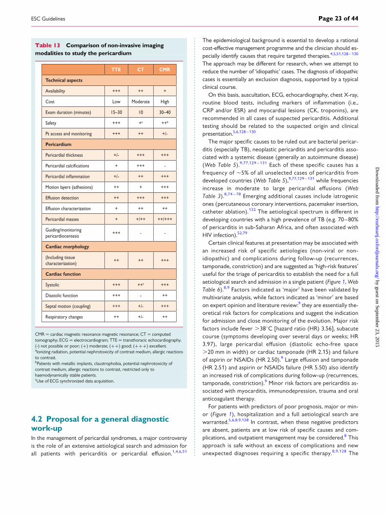
4.2 Proposal for a general diagnosticwork-upIn the management of pericardial syndromes, a major controversyis the role of an extensive aetiological search and admission forall patients with pericarditis or pericardial effusion.1,4,6,51
The epidemiological background is essential to develop a rationalcost-effective management programme and the clinician should es-pecially identify causes that require targeted therapies.4,5,51,128 –130
The approach may be different for research, when we attempt toreduce the number of ‘idiopathic’ cases. The diagnosis of idiopathiccases is essentially an exclusion diagnosis, supported by a typicalclinical course.
On this basis, auscultation, ECG, echocardiography, chest X-ray,routine blood tests, including markers of inflammation (i.e.,CRP and/or ESR) and myocardial lesions (CK, troponins), arerecommended in all cases of suspected pericarditis. Additionaltesting should be related to the suspected origin and clinicalpresentation.5,6,128 – 130
The major specific causes to be ruled out are bacterial pericar-ditis (especially TB), neoplastic pericarditis and pericarditis asso-ciated with a systemic disease (generally an autoimmune disease)(Web Table 5).9,77,129 – 131 Each of these specific causes has afrequency of �5% of all unselected cases of pericarditis fromdeveloped countries (Web Table 5),9,77,129 – 131 while frequenciesincrease in moderate to large pericardial effusions (WebTable 3).8,74 – 78 Emerging additional causes include iatrogenicones (percutaneous coronary interventions, pacemaker insertion,catheter ablation).132 The aetiological spectrum is different indeveloping countries with a high prevalence of TB (e.g. 70–80%of pericarditis in sub-Saharan Africa, and often associated withHIV infection).52,79
Certain clinical features at presentation may be associated withan increased risk of specific aetiologies (non-viral or non-idiopathic) and complications during follow-up (recurrences,tamponade, constriction) and are suggested as ‘high-risk features’useful for the triage of pericarditis to establish the need for a fullaetiological search and admission in a single patient (Figure 1, WebTable 6).8,9 Factors indicated as ‘major’ have been validated bymultivariate analysis, while factors indicated as ‘minor’ are basedon expert opinion and literature review:9 they are essentially the-oretical risk factors for complications and suggest the indicationfor admission and close monitoring of the evolution. Major riskfactors include fever .388C [hazard ratio (HR) 3.56], subacutecourse (symptoms developing over several days or weeks; HR3.97), large pericardial effusion (diastolic echo-free space.20 mm in width) or cardiac tamponade (HR 2.15) and failureof aspirin or NSAIDs (HR 2.50).9 Large effusion and tamponade(HR 2.51) and aspirin or NSAIDs failure (HR 5.50) also identifyan increased risk of complications during follow-up (recurrences,tamponade, constriction).9 Minor risk factors are pericarditis as-sociated with myocarditis, immunodepression, trauma and oralanticoagulant therapy.
For patients with predictors of poor prognosis, major or min-or (Figure 1), hospitalization and a full aetiological search arewarranted.5,6,8,9,128 In contrast, when these negative predictorsare absent, patients are at low risk of specific causes and com-plications, and outpatient management may be considered.8 Thisapproach is safe without an excess of complications and newunexpected diagnoses requiring a specific therapy.8,9,128 The
Table 13 Comparison of non-invasive imagingmodalities to study the pericardium
TTE CT CMR
Technical aspects
Availability +++ ++ +
Cost Low Moderate High
Exam duration (minutes) 15–30 10 30–40
Safety +++ +a ++b
Pt access and monitoring +++ ++ +/-
Pericardium
Pericardial thickness +/- +++ +++
+ +++ -
+/- ++ +++
Motion layers (adhesions) ++ + +++
Effusion detection ++ +++ +++
Effusion characterization + ++ ++
Pericardial masses + +/++ ++/+++
Guiding/monitoring pericardiocentesis
+++ - -
Cardiac morphology
(Including tissue characterization)
++ ++ +++
Cardiac function
Systolic +++ ++c +++
Diastolic function +++ - ++
Septal motion (coupling) +++ +/- +++
Respiratory changes ++ +/- ++
CMR ¼ cardiac magnetic resonance magnetic resonance; CT ¼ computedtomography; ECG ¼ electrocardiogram; TTE ¼ transthoracic echocardiography.(-) not possible or poor; (+) moderate; (++) good; (+++) excellent.aIonizing radiation, potential nephrotoxicity of contrast medium, allergic reactionsto contrast.bPatients with metallic implants, claustrophobia, potential nephrotoxicity ofcontrast medium, allergic reactions to contrast, restricted only tohaemodynamically stable patients.cUse of ECG synchronized data acquisition.
ESC Guidelines Page 23 of 44
by guest on September 23, 2015
http://eurheartj.oxfordjournals.org/D
ownloaded from
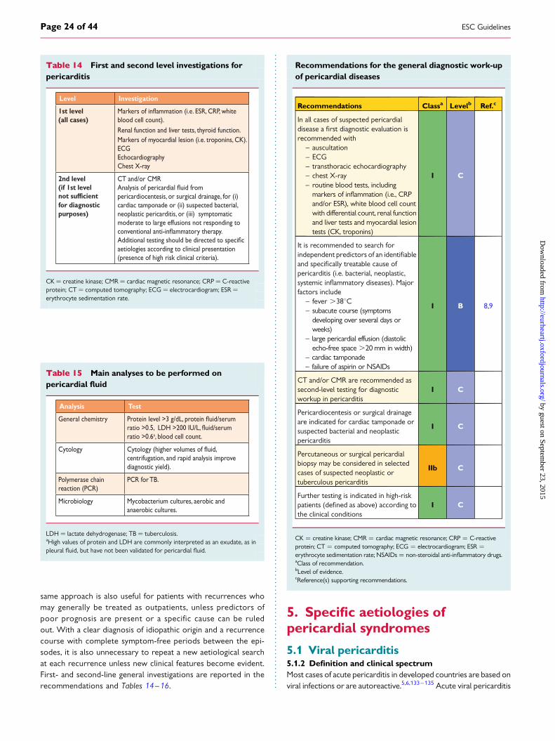
same approach is also useful for patients with recurrences whomay generally be treated as outpatients, unless predictors ofpoor prognosis are present or a specific cause can be ruledout. With a clear diagnosis of idiopathic origin and a recurrencecourse with complete symptom-free periods between the epi-sodes, it is also unnecessary to repeat a new aetiological searchat each recurrence unless new clinical features become evident.First- and second-line general investigations are reported in therecommendations and Tables 14–16.
Recommendations for the general diagnostic work-upof pericardial diseases
Recommendations Classa Levelb Ref.c
In all cases of suspected pericardialdisease a first diagnostic evaluation isrecommended with
– auscultation– ECG– transthoracic echocardiography– chest X-ray– routine blood tests, including
markers of inflammation (i.e., CRPand/or ESR), white blood cell countwith differential count, renal functionand liver tests and myocardial lesiontests (CK, troponins)
I C
It is recommended to search forindependent predictors of an identifiableand specifically treatable cause ofpericarditis (i.e. bacterial, neoplastic,systemic inflammatory diseases). Majorfactors include
– fever .388C– subacute course (symptoms
developing over several days orweeks)
– large pericardial effusion (diastolicecho-free space .20 mm in width)
– cardiac tamponade– failure of aspirin or NSAIDs
I B 8,9
CT and/or CMR are recommended assecond-level testing for diagnosticworkup in pericarditis
I C
Pericardiocentesis or surgical drainageare indicated for cardiac tamponade orsuspected bacterial and neoplasticpericarditis
I C
Percutaneous or surgical pericardialbiopsy may be considered in selectedcases of suspected neoplastic ortuberculous pericarditis
IIb C
Further testing is indicated in high-riskpatients (defined as above) according tothe clinical conditions
I C
CK ¼ creatine kinase; CMR ¼ cardiac magnetic resonance; CRP ¼ C-reactiveprotein; CT ¼ computed tomography; ECG ¼ electrocardiogram; ESR ¼erythrocyte sedimentation rate; NSAIDs ¼ non-steroidal anti-inflammatory drugs.aClass of recommendation.bLevel of evidence.cReference(s) supporting recommendations.
5. Specific aetiologies ofpericardial syndromes
5.1 Viral pericarditis5.1.2 Definition and clinical spectrumMost cases of acute pericarditis in developed countries are based onviral infections or are autoreactive.5,6,133 – 135 Acute viral pericarditis
Table 14 First and second level investigations forpericarditis
Level Investigation
1st level(all cases) blood cell count).
Renal function and liver tests, thyroid function.Markers of myocardial lesion (i.e. troponins, CK).ECGEchocardiographyChest X-ray
2nd level(if 1st level not sufficientfor diagnostic purposes)
CT and/or CMR
pericardiocentesis, or surgical drainage, for (i) cardiac tamponade or (ii) suspected bacterial, neoplastic pericarditis, or (iii) symptomatic moderate to large effusions not responding to
aetiologies according to clinical presentation (presence of high risk clinical criteria).
CK ¼ creatine kinase; CMR ¼ cardiac magnetic resonance; CRP ¼ C-reactiveprotein; CT ¼ computed tomography; ECG ¼ electrocardiogram; ESR ¼erythrocyte sedimentation rate.
Table 15 Main analyses to be performed onpericardial fluid
Analysis Test
ratio >0.6a, blood cell count.
Cytologycentrifugation, and rapid analysis improve diagnostic yield).
Polymerase chain reaction (PCR)
PCR for TB.
Microbiology Mycobacterium cultures, aerobic and anaerobic cultures.
LDH ¼ lactate dehydrogenase; TB ¼ tuberculosis.aHigh values of protein and LDH are commonly interpreted as an exudate, as inpleural fluid, but have not been validated for pericardial fluid.
ESC GuidelinesPage 24 of 44
by guest on September 23, 2015
http://eurheartj.oxfordjournals.org/D
ownloaded from
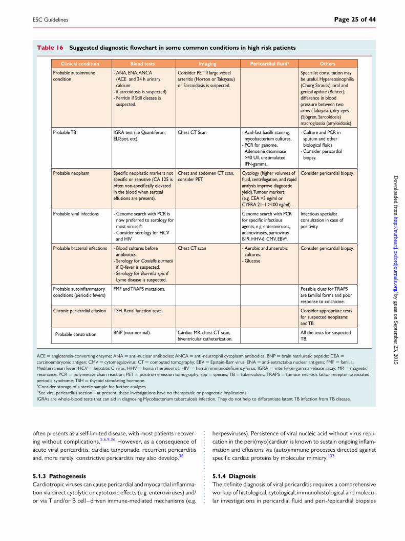
often presents as a self-limited disease, with most patients recover-ing without complications.5,6,9,36 However, as a consequence ofacute viral pericarditis, cardiac tamponade, recurrent pericarditisand, more rarely, constrictive pericarditis may also develop.36
5.1.3 PathogenesisCardiotropic viruses can cause pericardial and myocardial inflamma-tion via direct cytolytic or cytotoxic effects (e.g. enteroviruses) and/or via T and/or B cell–driven immune-mediated mechanisms (e.g.
herpesviruses). Persistence of viral nucleic acid without virus repli-cation in the peri(myo)cardium is known to sustain ongoing inflam-mation and effusions via (auto)immune processes directed againstspecific cardiac proteins by molecular mimicry.133
5.1.4 DiagnosisThe definite diagnosis of viral pericarditis requires a comprehensiveworkup of histological, cytological, immunohistological and molecu-lar investigations in pericardial fluid and peri-/epicardial biopsies
Table 16 Suggested diagnostic flowchart in some common conditions in high risk patients
Clinical condition Blood tests Imaging a Others
Probable autoimmune condition
- ANA, ENA, ANCA (ACE and 24 h urinary calcium - if sarcoidosis is suspected)- Ferritin if Still disease is suspected.
Consider PET if large vessel arteritis (Horton or Takayasu) or Sarcoidosis is suspected.
Specialist consultation may be useful. Hypereosinophilia (Churg Strauss), oral and genital apthae (Behcet); difference in blood pressure between two arms (Takayasu), dry eyes (Sjögren, Sarcoidosis) macroglossia (amyloidosis).
Probable TB IGRA test (i.e Quantiferon, ELISpot, etc).
Chest CT Scan - Acid-fast bacilli staining, mycobacterium cultures,- PCR for genome. Adenosine deaminase >40 U/l, unstimulated IFN-gamma.
- Culture and PCR in sputum and other - Consider pericardial biopsy.
Probable neoplasm
in the blood when serosal effusions are present).
Chest and abdomen CT scan, consider PET.
Cytology (higher volumes of
analysis improve diagnostic yield). Tumour markers (e.g. CEA >5 ng/ml or CYFRA 21–1 >100 ng/ml).
Consider pericardial biopsy.
Probable viral infections - Genome search with PCR is now preferred to serology for most virusesb.- Consider serology for HCV and HIV
Genome search with PCR
agents, e.g. enteroviruses, adenoviruses, parvovirus B19, HHV-6, CMV, EBVb.
Infectious specialist consultation in case of positivity.
Probable bacterial infections - Blood cultures before antibiotics.- Serology for Coxiella burnetii if Q-fever is suspected.- Serology for Borrelia spp. if Lyme disease is suspected.
Chest CT scan - Aerobic and anaerobic cultures.- Glucose
Consider pericardial biopsy.
conditions (periodic fevers) FMF and TRAPS mutations. Possible clues for TRAPS
are familial forms and poor response to colchicine.
Chronic pericardial effusion TSH. Renal function tests. Consider appropriate tests for suspected neoplasms and TB.
Probable constriction BNP (near-normal). Cardiac MR, chest CT scan, biventricular catheterization.
All the tests for suspected TB.
ACE ¼ angiotensin-converting enzyme; ANA ¼ anti-nuclear antibodies; ANCA ¼ anti-neutrophil cytoplasm antibodies; BNP ¼ brain natriuretic peptide; CEA ¼carcinoembryonic antigen; CMV ¼ cytomegalovirus; CT ¼ computed tomography; EBV ¼ Epstein-Barr virus; ENA ¼ anti-extractable nuclear antigens; FMF ¼ familialMediterranean fever; HCV ¼ hepatitis C virus; HHV ¼ human herpesvirus; HIV ¼ human immunodeficiency virus; IGRA ¼ interferon-gamma release assay; MR ¼ magneticresonance; PCR ¼ polymerase chain reaction; PET ¼ positron emission tomography; spp ¼ species; TB ¼ tuberculosis; TRAPS ¼ tumour necrosis factor receptor-associatedperiodic syndrome; TSH ¼ thyroid stimulating hormone.aConsider storage of a sterile sample for further analyses.bSee viral pericarditis section—at present, these investigations have no therapeutic or prognostic implications.IGRAs are whole-blood tests that can aid in diagnosing Mycobacterium tuberculosis infection. They do not help to differentiate latent TB infection from TB disease.
ESC Guidelines Page 25 of 44
by guest on September 23, 2015
http://eurheartj.oxfordjournals.org/D
ownloaded from
obtained in conjunction with pericardioscopy, permitting the evalu-ation of possible algorithms for a causative therapy.133 In contrast,serological tests were found to be futile in the diagnosis of viral peri-carditis. Whereas no up-regulation of pro-inflammatory cytokineexpression is noted in the serum, TNF-a, vascular endothelialgrowth factor (VEGF), basic fibroblast growth factor (bFGF), IL-6,IL-8 and interferon-gamma (IFN-g) are increased in the pericardialeffusions of patients with pericarditis, indicating the presence of lo-cal inflammatory reactions.134,135 Accordingly, there is no correl-ation of antiviral antibodies in the serum or virus isolation fromthroat or rectal swabs with positive molecular polymerase chain re-action (PCR)/in situ hybridization analyses for the detection of car-diotropic viruses in pericardial tissue and fluid.136
5.1.5 Identification of viral nucleic acidsMainly by quantitative PCR techniques, nucleic acids of differentcardiotropic RNA and DNA viruses have been detected inepicardial and pericardial biopsies and the pericardial fluid of chil-dren and adults with acute pericarditis, but also in patients with re-curring and constrictive pericarditis.133,137 Regarding RNAviruses, various subtypes of enteroviruses including echovirusesand coxsackieviruses of groups A (A4, A16) and B (CVB2,CVB3, CVB4) were identified in patients with acute and constrict-ive pericarditis.137,138 Among the RNA viruses, influenza A viruses(e.g. H1N1, H5N1, H3N2) and occasionally chikungunya virus, hu-man coronavirus NL-63, respiratory syncytial virus and denguevirus infections were suspected as aetiopathogenic agents inpericarditis.
Compared with RNA viruses, nucleic acids of DNA viruses, in-cluding parvovirus B19 and herpesviruses [Epstein–Barr virus(EBV) and human herpesvirus 6 (HHV-6)], are present in pericardialbiopsies and pericardial fluid at greater frequencies and higher viralDNA copy numbers.133 Whereas parvovirus B19, with up to 7 ×10⁶ GE/lg DNA was predominantly detected in epicardial tissue,EBV was most frequently found in pericardial fluid.133 DNA ofvaricella zoster virus, herpes simplex virus and adenoviruses isonly rarely detected in pericarditis patients. Cytomegalovirus(CMV)-associated pericarditis is mainly found in immunocomprom-ised and HIV patients.1 In developing countries with a delayed roll-out of antiretroviral therapy, HIV-associated inflammatory reactions(often related to TB) of the pericardium and myocardium are com-mon complications.139 However, at present these investigations areusually not performed because of their complexity, cost, invasive na-ture and low availability.
5.1.6 TherapyAcute viral pericarditis often presents as a self-limiting diseasethat responds well to a short course of treatment with NSAIDs,with the adjunct of colchicine, especially for prevention ofrecurrences.4– 6,50,58,59 The identification of specific viral signaturesaids in understanding the pathogenetic mechanisms in pericarditis,and might enable an individualized aetiologically driven specifictreatment approach to be established by distinguishing a viral aeti-ology from autoreactive inflammation.133 Some experts suggestantiviral treatment similar to that for myocarditis (IVIG therapy inacute systemic enteroviral, CMV, EBV and parvovirus B19 infection,
oral valganciclovir in HHV-6 perimyocarditis, IFN-a for enteroviralpericarditis).133 However, these treatments are still under evalu-ation and rarely used. Involvement of infectious disease specialistsis recommended. So far, no therapy is available to solve the problemof virus persistence and consecutive inflammation, particularly wheninduced by herpesviruses and parvovirus B19 infections.133 Import-antly, corticosteroids are generally not indicated in viral pericarditis,as they are known to reactivate many virus infections and thus leadto ongoing inflammation.133
Recommendations for the diagnosis and therapy ofviral pericarditis
Recommendations Classa Levelb Ref.c
For the definited diagnosis of viralpericarditis, a comprehensive workup ofhistological, cytological,immunohistological and molecularinvestigations in pericardial fluid andperi-/epicardial biopsies should beconsidered
IIa C
Routine viral serology is notrecommended, with the possibleexception of HIV and HCV
III C
Corticosteroid therapy is notrecommended in viral pericarditis
III C
HCV ¼ hepatitis C virus; HIV ¼ human immunodeficiency virus.aClass of recommendation.bLevel of evidence.cReference(s) supporting recommendations.dIn the absence of such an argument, the term ‘presumed viral pericarditis’should be used.
5.2 Bacterial pericarditisBacterial pericarditis is relatively uncommon in clinical practice indeveloped countries with a low prevalence of TB. Tuberculous peri-carditis is the most common form all over the world and the mostcommon cause of pericardial diseases in developing countries. Wewill discuss this form and also purulent pericarditis, which is lesscommon.
5.2.1 Tuberculous pericarditisTuberculous pericarditis accounts for ≤4% of pericardial disease inthe developed world.5,6,52. In contrast, TB is the cause of clinicallysignificant pericardial effusion in .90% of HIV-infected and 50–70% of non-HIV-infected individuals who live in developing coun-tries where TB is endemic.77 The disease can occur at any age,and men are affected more frequently than women.140 Clinical evi-dence of chronic cardiac compression mimicking congestive heartfailure is the most common presentation.79,93 Clinical presentationsare pericardial effusion, effusive-constrictive pericarditis and con-strictive pericarditis.79 Tuberculous pericarditis has a mortalityrate of 17–40% at 6 months after diagnosis.141 It should be empha-sized that the majority of the information on tuberculous
ESC GuidelinesPage 26 of 44
by guest on September 23, 2015
http://eurheartj.oxfordjournals.org/D
ownloaded from
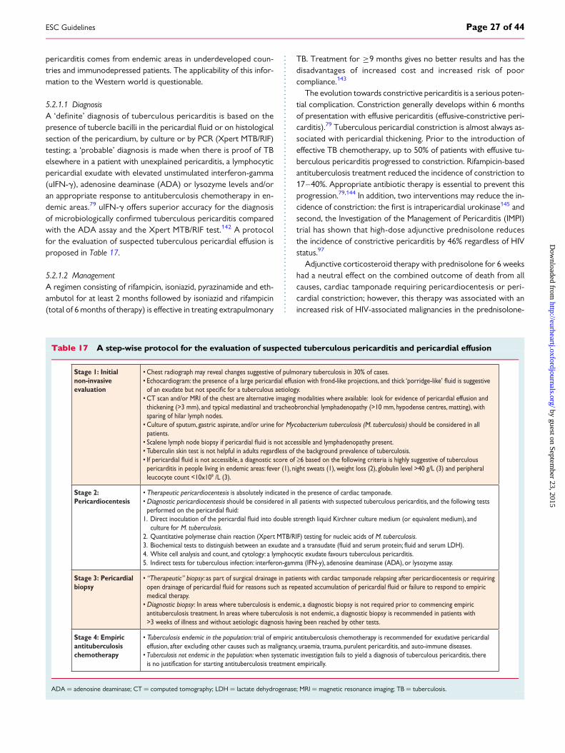
pericarditis comes from endemic areas in underdeveloped coun-tries and immunodepressed patients. The applicability of this infor-mation to the Western world is questionable.
5.2.1.1 DiagnosisA ‘definite’ diagnosis of tuberculous pericarditis is based on thepresence of tubercle bacilli in the pericardial fluid or on histologicalsection of the pericardium, by culture or by PCR (Xpert MTB/RIF)testing; a ‘probable’ diagnosis is made when there is proof of TBelsewhere in a patient with unexplained pericarditis, a lymphocyticpericardial exudate with elevated unstimulated interferon-gamma(uIFN-g), adenosine deaminase (ADA) or lysozyme levels and/oran appropriate response to antituberculosis chemotherapy in en-demic areas.79 uIFN-g offers superior accuracy for the diagnosisof microbiologically confirmed tuberculous pericarditis comparedwith the ADA assay and the Xpert MTB/RIF test.142 A protocolfor the evaluation of suspected tuberculous pericardial effusion isproposed in Table 17.
5.2.1.2 ManagementA regimen consisting of rifampicin, isoniazid, pyrazinamide and eth-ambutol for at least 2 months followed by isoniazid and rifampicin(total of 6 months of therapy) is effective in treating extrapulmonary
TB. Treatment for ≥9 months gives no better results and has thedisadvantages of increased cost and increased risk of poorcompliance.143
The evolution towards constrictive pericarditis is a serious poten-tial complication. Constriction generally develops within 6 monthsof presentation with effusive pericarditis (effusive-constrictive peri-carditis).79 Tuberculous pericardial constriction is almost always as-sociated with pericardial thickening. Prior to the introduction ofeffective TB chemotherapy, up to 50% of patients with effusive tu-berculous pericarditis progressed to constriction. Rifampicin-basedantituberculosis treatment reduced the incidence of constriction to17–40%. Appropriate antibiotic therapy is essential to prevent thisprogression.79,144 In addition, two interventions may reduce the in-cidence of constriction: the first is intrapericardial urokinase145 andsecond, the Investigation of the Management of Pericarditis (IMPI)trial has shown that high-dose adjunctive prednisolone reducesthe incidence of constrictive pericarditis by 46% regardless of HIVstatus.97
Adjunctive corticosteroid therapy with prednisolone for 6 weekshad a neutral effect on the combined outcome of death from allcauses, cardiac tamponade requiring pericardiocentesis or peri-cardial constriction; however, this therapy was associated with anincreased risk of HIV-associated malignancies in the prednisolone-
Table 17 A step-wise protocol for the evaluation of suspected tuberculous pericarditis and pericardial effusion
Stage 1: Initial non-invasive evaluation
• Chest radiograph may reveal changes suggestive of pulmonary tuberculosis in 30% of cases. • Echocar or a tuberculous aetiology.• CT scan and/or MRI of the chest are alternative imaging modalities where available: look for evidence of pericardial effusion and thickening (>3 mm), and typical mediastinal and tracheobronchial lymphadenopathy (>10 mm, hypodense centres, matting), with sparing of hilar lymph nodes. • Culture of sputum, gastric aspirate, and/or urine for Mycobacterium tuberculosis (M. tuberculosis) should be considered in all patients.• Scalene l• Tuberculin skin test is not helpful in adults regardless of the background prevalence of tuberculosis.• If pericar ≥6 based on the following criteria is highly suggestive of tuberculous pericarditis in people living in endemic areas: fever (1), night sweats (1), weight loss (2), globulin level >40 g/L (3) and peripheral leucocyte count <10x109 /L (3).
Stage 2: Pericardiocentesis
• Therapeutic pericardiocentesis is absolutely indicated in the presence of cardiac tamponade.• Diagnostic pericardiocentesis should be considered in all patients with suspected tuberculous pericarditis, and the following tests perf1. Dir culture for M. tuberculosis.2. Quantitative polymerase chain reaction (Xpert MTB/RIF) testing for nucleic acids of M. tuberculosis.3. Biochemical tests to distinguish betw4. White cell analysis and count, and cytology: a lymphocytic exudate favours tuberculous pericarditis.5. Indirect tests for tuberculous infection: interferon-gamma (IFN-y), adenosine deaminase (ADA), or lysozyme assay.
Stage 3: Pericardial biopsy
• “Therapeutic” biopsy: as part of surgical drainage in patients with cardiac tamponade relapsing after pericardiocentesis or requiring open drainage of pericar medical therapy.• Diagnostic biopsy: In areas where tuberculosis is endemic, a diagnostic biopsy is not required prior to commencing empiric antituberculosis treatment. In areas where tuberculosis is not endemic, a diagnostic biopsy is recommended in patients with >3 weeks of illness and without aetiologic diagnosis having been reached by other tests.
Stage 4: Empiric antituberculosis chemotherapy
• Tuberculosis endemic in the population: trial of empiric antituberculosis chemotherapy is recommended for exudative pericardial effusion, after excluding other causes such as malignancy, uraemia, trauma, purulent pericarditis, and auto-immune diseases.• Tuberculosis not endemic in the population: when systematic investigation fails to yield a diagnosis of tuberculous pericarditis, there or starting antituberculosis treatment empirically.
ADA ¼ adenosine deaminase; CT ¼ computed tomography; LDH ¼ lactate dehydrogenase; MRI ¼ magnetic resonance imaging; TB ¼ tuberculosis.
ESC Guidelines Page 27 of 44
by guest on September 23, 2015
http://eurheartj.oxfordjournals.org/D
ownloaded from
treated group.97 Adjunctive steroid therapy was associated with areduced incidence of pericardial constriction and hospitalization.The beneficial effects of prednisolone on constriction and hospital-ization were similar in HIV-positive and HIV-negative patients. Onthis basis, it may be reasonable to use adjunctive corticosteroidsin patients with tuberculous pericarditis without HIV infection andto avoid them in HIV-infected individuals because of the increasedrisk of malignancy.97
Recommendations for the diagnosis and treatment oftuberculous pericarditis and effusion
Recommendations Classa Levelb Ref.c
Diagnostic pericardiocentesis should beconsidered in all patients with suspectedtuberculous pericarditis
IIa C
Intrapericardial urokinase may beconsidered to reduce the risk ofconstriction in tuberculous effusivepericarditis
IIb C
In patients living in non-endemic areas,empiric antituberculosis treatment isnot recommended when systematicinvestigation fails to yield a diagnosisof tuberculous pericarditis
III C
In patients living in endemic areas,empiric antituberculosis chemotherapyis recommended for exudativepericardial effusion, after excludingother causes
I C
Adjunctive steroids may be consideredin HIV-negative cases of TB pericarditisand avoided in HIV-associated TBpericarditis
IIb C
HIV ¼ human immunodeficiency virus; TB ¼ tuberculosis.aClass of recommendation.bLevel of evidence.cReference(s) supporting recommendations.
Recommendations for the general management ofconstrictive tuberculous pericarditis
Recommendations Classa Levelb Ref.c
Standard antituberculosis drugs for6 months is recommended for theprevention of tuberculous pericardialconstriction
I C
Pericardiectomy is recommended if thepatient’s condition is not improving or isdeteriorating after 4–8 weeks ofantituberculosis therapy
I C
aClass of recommendation.bLevel of evidence.cReference(s) supporting recommendations.
5.2.2 Purulent pericarditis5.2.2.1 EpidemiologyPurulent pericarditis is rare, accounting for ,1% of cases.5,6 In West-ern series, the most common organisms have been staphylococci,streptococci and pneumococci, while the predominant associated le-sions were empyema (50%) or pneumonia (33%).146 In immunosup-pressed patients or following thoracic surgery, Staphylococcus aureus(30%) and fungi (20%) are more common.147 Also, anaerobes origin-ating from the oropharynx have been reported.148 Seeding may behaematogenous or by contiguous spread from the retropharyngealspace, cardiac valves and below the diaphragm.149 Neisseria meningiti-dis may involve the pericardium either through initiating an immune-mediated sterile effusion or by direct infection and purulent reaction.The modern era of iatrogenic and HIV-associated immunosuppres-sion has witnessed more unusual organisms.
5.2.2.2 DiagnosisPurulent pericarditis is rare and generally manifested as a seriousfebrile disease. The underlying sepsis may predominate theillness.146 – 149 Suspicion of purulent pericarditis is an indication forurgent pericardiocentesis,1,5,12 which is diagnostic. The fluid maybe frankly purulent. A low pericardial:serum glucose ratio (mean0.3) and elevated pericardial fluid white cell count with a high pro-portion of neutrophils (mean cell count 2.8/ml, 92% neutrophils)differentiate purulent from tuberculous (glucose ratio 0.7, count1.7/ml, 50% neutrophils) and neoplastic (glucose ratio 0.8, count3.3/ml, 55% neutrophils) pericarditis.150 Fluid should be sent for bac-terial, fungal and tuberculous studies, with blood for cultures andother samples being taken as guided by the clinical presentation.12
5.2.2.3 ManagementPurulent pericarditis should be managed aggressively, as death is inev-itable if untreated, whereas with comprehensive therapy 85% of caseshave been reported to survive the episode and have a good long-termoutcome.50,146 Intravenous antimicrobial therapy should be startedempirically until microbiological results are available. Drainage is crucial.Purulent effusions are often heavily loculated and likely to rapidly re-accumulate. Intrapericardial thrombolysis is a possible treatment forcases with loculated effusions in order to achieve adequate drainage be-fore resorting to surgery.151 Subxiphoid pericardiostomy and rinsing ofthe pericardial cavity should be considered.1 This allows more com-plete drainage of the effusion, as loculations can be manually lysed.
Recommendations for the diagnosis of purulentpericarditis
Recommendations Classa Levelb Ref.c
Urgent pericardiocentesis isrecommended for the diagnosis ofpurulent pericarditis
I C
It is recommended that pericardial fluidbe sent for bacterial, fungal andtuberculous studies and blood drawn forcultures
I C
aClass of recommendation.bLevel of evidence.cReference(s) supporting recommendations.
ESC GuidelinesPage 28 of 44
by guest on September 23, 2015
http://eurheartj.oxfordjournals.org/D
ownloaded from
Recommendations for the therapy of purulentpericarditis
Recommendations Classa Levelb Ref.c
Effective pericardial drainage isrecommended for purulent pericarditis
I C
Administration of intravenousantibiotics is indicated to treat purulentpericarditis
I C
Subxiphoid pericardiotomy and rinsingof the pericardial cavity should beconsidered
IIa C
Intrapericardial thrombolysis should beconsidered
IIa C
Pericardiectomy for dense adhesions,loculated or thick purulent effusion,recurrence of tamponade, persistentinfection and progression to constrictionshould be considered
IIa C
aClass of recommendation.bLevel of evidence.cReference(s) supporting recommendations.
5.3 Pericarditis in renal failureRenal disease and overall ESRD are associated with possiblepericardial involvement.152 Three different pathologies are foundin uraemic patients: uraemic pericarditis—before renal replace-ment therapy or within 8 weeks of its initiation; dialysis pericardi-tis—after being stabilized on dialysis (usually ≥8 weeks after itsinitiation)153 and, very rarely, constrictive pericarditis. The globalincidence of pericarditis in ESDR patients has declined to �5%in those patients starting dialysis.152 The reported frequency ofdialysis pericarditis ranges from 2 to 21%, but recent data arelacking.
Pericardial involvement in ESRD is manifested most commonly asacute pericarditis and chronic pericardial effusion and infrequentlyas chronic constrictive pericarditis. Typical features of this form ofpericarditis include a lower rate of pleuritic chest pain (up to 30%of patients are asymptomatic) and the absence of ECG abnormal-ities in most cases, probably due to the lack of myocardial inflamma-tion.152 – 154 Patients with ESRD are more likely to develop chronicpericardial effusion due to continuous volume overload.152 Not allpericardial effusions result from inflammation, and the normal vol-ume of pericardial fluid is larger in stable haemodialysis patientsthan in normal controls.155 With the advent of advanced renal re-placement therapy, the incidence of haemodynamically significanteffusions has decreased.152,156,157 The most probable cause of ur-aemic pericarditis is the retention of toxic metabolites.152,157 Sincepericardial effusion is often bloody in uraemic patients, anticoagula-tion should be carefully considered or avoided in patients startingdialysis.152,157
Recommendations for the management of pericarditisin renal failure
Recommendations Classa Levelb Ref.c
Dialysis should be considered in uraemicpericarditis
IIa C
When patients with adequate dialysisdevelop pericarditis, intensifying dialysisshould be considered
IIa C
Pericardial aspiration and/or drainagemay be considered in non-responsivepatients with dialysis
IIb C
NSAIDs and corticosteroids(systemic or intrapericardial) may beconsidered when intensive dialysis isineffective
IIb C
Colchicine is contraindicated inpatients with pericarditis and severerenal impairment (see Web Table 1B)
III C
NSAIDs ¼ non-steroidal anti-inflammatory drugs.aClass of recommendation.bLevel of evidence.cReference(s) supporting recommendations.
5.4 Pericardial involvement in systemicautoimmune and autoinflammatorydiseasesPericardial involvement in systemic autoimmune diseases may besymptomatic (pericarditis and symptomatic pericardial effusion)or asymptomatic (usually pericardial effusion) and generally re-flects the degree of activity of the underlying disease.45 Approxi-mately 5–15% of patients with acute or recurrent pericarditismay have a systemic autoimmune disease, either overt or under-lying (Table 1, Web Table 5).9,77,129 – 131 Pericardial involvement iscommon in systemic lupus erythematosus, Sjogren’s syndrome,rheumatoid arthritis and scleroderma, but may also be present insystemic vasculitides, Behcet’s syndrome, sarcoidosis and inflam-matory bowel diseases. Pericardial involvement rarely occurs asthe first manifestation of these diseases. In most patients theunderlying disease has already been diagnosed by classical symp-toms and signs. Concomitant myocardial inflammatory involve-ment may complicate the presentation and should be ruled out.If clinical features suggest a possible systemic autoimmune disease,a targeted aetiological search is warranted in cooperation withspecialist consultation. The treatment is especially targeted at con-trol of the systemic underlying disease.45
A specific subset of patients includes those with periodic fevers.These are genetic disorders characterized by mutations of genes in-volved in the regulation of the inflammatory response, without in-volvement of specific T cells or autoantibodies.158 – 161 Thesedisorders are usually detected in the paediatric population, althoughsome patients experience disease onset during adulthood. The most
ESC Guidelines Page 29 of 44
by guest on September 23, 2015
http://eurheartj.oxfordjournals.org/D
ownloaded from
common autoinflammatory syndromes include familial Mediterra-nean fever (FMF), in which serositis episodes last only 1–3 days,and tumour necrosis factor receptor-associated periodic syndrome(TRAPS), in which the episodes last for weeks. Mutations associatedwith these disorders are rare in recurrent pericarditis.158 –161 Theseconditions may be characterized by an exaggerated expression ofIL-1.161 A 10% rate of familial occurrence of pericarditis has been re-ported among the relatives of these patients.162 –164 These data sug-gest a genetic predisposition in at least a subset of patients;counselling may be warranted in these cases. A positive family his-tory for pericarditis or periodic fevers, a poor response to colchi-cine, as well as the need for immunosuppressive agents are cluesto the possible presence of an auto-inflammatory disease;160 par-ticularly in these conditions, anti-IL-1 or anti-TNF agents may beconsidered.31,32,160
5.5 Post-cardiac injury syndromesThe term post-cardiac injury syndromes (PCIS) is an umbrella termindicating a group of inflammatory pericardial syndromes includingpost-myocardial infarction pericarditis, post-pericardiotomy syn-drome (PPS) and post-traumatic pericarditis (either iatrogenic ornot).132 Such syndromes are presumed to have an autoimmunepathogenesis triggered by initial damage to pericardial and/or pleuraltissues caused by either myocardial necrosis (late post-myocardialinfarction pericarditis or Dressler syndrome), surgical trauma(PPS), accidental thoracic trauma (traumatic pericarditis) or iatro-genic trauma with or without bleeding (pericarditis after invasivecardiac interventions).131 The immune-mediated pathogenesis issupported by a latent period generally of a few weeks until the ap-pearance of the first manifestations and the response to anti-inflammatory drugs (NSAIDs, corticosteroids, colchicine) with thepossibility of recurrences. Pericardial bleeding and pleura incisionare triggers for the syndrome.165,166 Some forms, such as Dresslersyndrome, have become rare with early reperfusion therapy of myo-cardial infarction; however, it may occur especially in cases of evenminor bleeding into the pericardium.167
5.5.1 Definition and diagnosisAccording to proposed diagnostic criteria for PPS,168 – 170 the diag-nosis of PCIS may be reached after a cardiac injury following clinicalcriteria: (i) fever without alternative causes, (ii) pericarditic or pleur-itic chest pain, (iii) pericardial or pleural rubs, (iv) evidence of peri-cardial effusion and/or (v) pleural effusion with elevated CRP. Atleast two of five criteria should be fulfilled. The rationale for propos-ing specific criteria instead of adopting the same for pericarditis isthat these syndromes may have concomitant pleuropericardial in-volvement and possible pulmonary infiltrates, and are not simplypericarditis.170 Moreover, it is sometimes difficult to differentiatePCIS from the simple mechanical consequences of surgery (suchas pericardial or pleural effusion). The demonstration of inflamma-tory activity should be essential to establish the diagnosis. Basic diag-nostic evaluation of a patient with a suspected PCIS includes physicalexamination, ECG, echocardiogram, thoracic echography and/orchest X-ray.132,165 On this basis, echocardiography is recommendedwhen an iatrogenic complication is suspected after a cardiovascularintervention.2,3,132
5.5.2 ManagementTreatment of PCIS is essentially based on empiric anti-inflammatorytherapy, and may improve remission rates and reduce the risk ofrecurrences.171 The same therapeutic scheme adopted for peri-carditis is efficacious for all these forms, including post-myocardialinfarction pericarditis (Table 3). Colchicine is not recommendedfor postoperative effusions in the absence of systemic inflamma-tion.172– 174 Similarly NSAIDs are generally not indicated in asymp-tomatic post-surgical effusions, and this therapy may be associatedwith an increased risk of side effects related to NSAIDs.173,174
5.5.3 PreventionDifferent preventive strategies have been examined in a few studiesregarding aspirin,175 methylprednisone,176 dexamethasone177 andcolchicine.168,169,172 Four controlled clinical trials for primary pre-vention of PPS were included in a systematic review on 894 patients;three studies were double-blind RCTs. Treatment comparisonswere colchicine vs. placebo (two RCTs enrolling 471 patients),methylprednisolone vs. placebo (one RCT involving 246 paediatricpatients) and aspirin vs. historical controls (one non-randomizedstudy involving 177 paediatric patients). Meta-analytic poolingshowed that only colchicine was associated with decreased risk ofPPS [odds ratio (OR) 0.38]. Data on methylprednisolone (OR1.13) and aspirin (OR 1.00) were negative.178 The Colchicine forPrevention of the Post-pericardiotomy Syndrome and Post-operative Atrial Fibrillation (COPPS-2) trial confirmed the overallefficacy of perioperative use of colchicine, but it was also found tobe associated with an increased risk of gastrointestinal side effects172
compared with postoperative use of colchicine.169 Colchicine is notrecommended for the perioperative treatment and prevention ofpostoperative effusions in the absence of systemic inflammation.172
In another trial,177 high-dose dexamethasone (1 mg/kg as a single in-traoperative dose) was not efficacious in preventing PPS or compli-cated PPS.
5.5.4 PrognosisDespite limited published data, the prognosis of PPS is generallygood.178 There are very few available data on other forms of post-pericardial injury syndromes. In the largest published series on PPSpatients after cardiac surgery,166 complication rates were low: ,4%for recurrences, ,2% for cardiac tamponade and no cases of con-striction, although hospital stay may be prolonged in these patients.However, the development of constrictive pericarditis has been re-ported in �3% of cases.36
5.5.4.1 Post-myocardial infarction pericarditisFollowing an acute myocardial infarction (AMI), three major pericar-dial complications may occur: (i) pericardial effusion, (ii) earlyinfarct-associated pericarditis (often called early post-infarctionpericarditis, typically a few days after AMI) and (iii) late pericarditisor post-cardiac injury (Dressler) syndrome (typically 1–2 weeksafter AMI).
Early post-infarction pericarditis usually occurs soon after theAMI and is transient. This complication is rare in the primary percu-taneous coronary intervention era and is especially related to latereperfusion or failed coronary reperfusion.167 Diagnostic criteriado not differ from those for acute pericarditis. ECG changes are
ESC GuidelinesPage 30 of 44
by guest on September 23, 2015
http://eurheartj.oxfordjournals.org/D
ownloaded from
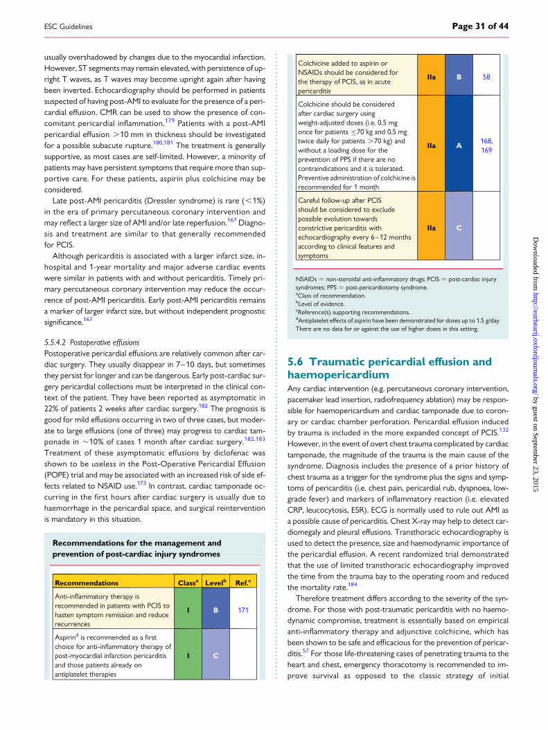
usually overshadowed by changes due to the myocardial infarction.However, ST segments may remain elevated, with persistence of up-right T waves, as T waves may become upright again after havingbeen inverted. Echocardiography should be performed in patientssuspected of having post-AMI to evaluate for the presence of a peri-cardial effusion. CMR can be used to show the presence of con-comitant pericardial inflammation.179 Patients with a post-AMIpericardial effusion .10 mm in thickness should be investigatedfor a possible subacute rupture.180,181 The treatment is generallysupportive, as most cases are self-limited. However, a minority ofpatients may have persistent symptoms that require more than sup-portive care. For these patients, aspirin plus colchicine may beconsidered.
Late post-AMI pericarditis (Dressler syndrome) is rare (,1%)in the era of primary percutaneous coronary intervention andmay reflect a larger size of AMI and/or late reperfusion.167 Diagno-sis and treatment are similar to that generally recommendedfor PCIS.
Although pericarditis is associated with a larger infarct size, in-hospital and 1-year mortality and major adverse cardiac eventswere similar in patients with and without pericarditis. Timely pri-mary percutaneous coronary intervention may reduce the occur-rence of post-AMI pericarditis. Early post-AMI pericarditis remainsa marker of larger infarct size, but without independent prognosticsignificance.167
5.5.4.2 Postoperative effusionsPostoperative pericardial effusions are relatively common after car-diac surgery. They usually disappear in 7–10 days, but sometimesthey persist for longer and can be dangerous. Early post-cardiac sur-gery pericardial collections must be interpreted in the clinical con-text of the patient. They have been reported as asymptomatic in22% of patients 2 weeks after cardiac surgery.182 The prognosis isgood for mild effusions occurring in two of three cases, but moder-ate to large effusions (one of three) may progress to cardiac tam-ponade in �10% of cases 1 month after cardiac surgery.182,183
Treatment of these asymptomatic effusions by diclofenac wasshown to be useless in the Post-Operative Pericardial Effusion(POPE) trial and may be associated with an increased risk of side ef-fects related to NSAID use.173 In contrast, cardiac tamponade oc-curring in the first hours after cardiac surgery is usually due tohaemorrhage in the pericardial space, and surgical reinterventionis mandatory in this situation.
Recommendations for the management andprevention of post-cardiac injury syndromes
Recommendations Classa Levelb Ref.c
Anti-inflammatory therapy isrecommended in patients with PCIS tohasten symptom remission and reducerecurrences
I B 171
Aspirind is recommended as a firstchoice for anti-inflammatory therapy ofpost-myocardial infarction pericarditisand those patients already onantiplatelet therapies
I C
Colchicine added to aspirin orNSAIDs should be considered forthe therapy of PCIS, as in acutepericarditis
IIa B 58
Colchicine should be consideredafter cardiac surgery usingweight-adjusted doses (i.e. 0.5 mgonce for patients ≤70 kg and 0.5 mgtwice daily for patients .70 kg) andwithout a loading dose for theprevention of PPS if there are nocontraindications and it is tolerated.Preventive administration of colchicine isrecommended for 1 month
IIa A168,169
Careful follow-up after PCISshould be considered to excludepossible evolution towardsconstrictive pericarditis withechocardiography every 6–12 monthsaccording to clinical features andsymptoms
IIa C
NSAIDs ¼ non-steroidal anti-inflammatory drugs; PCIS ¼ post-cardiac injurysyndromes; PPS ¼ post-pericardiotomy syndrome.aClass of recommendation.bLevel of evidence.cReference(s) supporting recommendations.dAntiplatelet effects of aspirin have been demonstrated for doses up to 1.5 g/day.There are no data for or against the use of higher doses in this setting.
5.6 Traumatic pericardial effusion andhaemopericardiumAny cardiac intervention (e.g. percutaneous coronary intervention,pacemaker lead insertion, radiofrequency ablation) may be respon-sible for haemopericardium and cardiac tamponade due to coron-ary or cardiac chamber perforation. Pericardial effusion inducedby trauma is included in the more expanded concept of PCIS.132
However, in the event of overt chest trauma complicated by cardiactamponade, the magnitude of the trauma is the main cause of thesyndrome. Diagnosis includes the presence of a prior history ofchest trauma as a trigger for the syndrome plus the signs and symp-toms of pericarditis (i.e. chest pain, pericardial rub, dyspnoea, low-grade fever) and markers of inflammatory reaction (i.e. elevatedCRP, leucocytosis, ESR). ECG is normally used to rule out AMI asa possible cause of pericarditis. Chest X-ray may help to detect car-diomegaly and pleural effusions. Transthoracic echocardiography isused to detect the presence, size and haemodynamic importance ofthe pericardial effusion. A recent randomized trial demonstratedthat the use of limited transthoracic echocardiography improvedthe time from the trauma bay to the operating room and reducedthe mortality rate.184
Therefore treatment differs according to the severity of the syn-drome. For those with post-traumatic pericarditis with no haemo-dynamic compromise, treatment is essentially based on empiricalanti-inflammatory therapy and adjunctive colchicine, which hasbeen shown to be safe and efficacious for the prevention of pericar-ditis.57 For those life-threatening cases of penetrating trauma to theheart and chest, emergency thoracotomy is recommended to im-prove survival as opposed to the classic strategy of initial
ESC Guidelines Page 31 of 44
by guest on September 23, 2015
http://eurheartj.oxfordjournals.org/D
ownloaded from
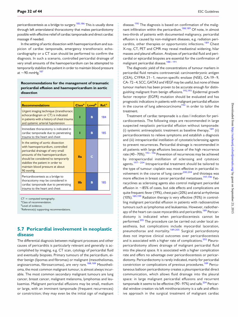
pericardiocentesis as a bridge to surgery.185,186 This is usually donethrough left anterolateral thoracotomy that makes pericardiotomypossible with effective relief of cardiac tamponade and direct cardiacmassage if needed.
In the setting of aortic dissection with haemopericardium and sus-picion of cardiac tamponade, emergency transthoracic echo-cardiography or a CT scan should be performed to confirm thediagnosis. In such a scenario, controlled pericardial drainage ofvery small amounts of the haemopericardium can be attempted totemporarily stabilize the patient in order to maintain blood pressureat �90 mmHg.187
Recommendations for the management of traumaticpericardial effusion and haemopericardium in aorticdissection
Recommendations Classa Levelb Ref.c
Urgent imaging technique (transthoracicechocardiogram or CT) is indicatedin patients with a history of chest traumaand systemic arterial hypotension
I B 184
Immediate thoracotomy is indicated incardiac tamponade due to penetratingtrauma to the heart and chest
I B 185
In the setting of aortic dissectionwith haemopericardium, controlledpericardial drainage of very smallamounts of the haemopericardiumshould be considered to temporarilystabilize the patient in order tomaintain blood pressure at about90 mmHg
IIa C
Pericardiocentesis as a bridge tothoracotomy may be considered incardiac tamponade due to penetratingtrauma to the heart and chest
IIb B 185
CT ¼ computed tomography.aClass of recommendation.bLevel of evidence.cReference(s) supporting recommendations.
5.7 Pericardial involvement in neoplasticdiseaseThe differential diagnosis between malignant processes and othercauses of pericarditis is particularly relevant and generally is ac-complished by imaging, e.g. CT scan, cytology of pericardial fluidand eventually biopsies. Primary tumours of the pericardium, ei-ther benign (lipomas and fibromas) or malignant (mesotheliomas,angiosarcomas, fibrosarcomas), are very rare.188,189 Mesotheli-oma, the most common malignant tumour, is almost always incur-able. The most common secondary malignant tumours are lungcancer, breast cancer, malignant melanoma, lymphomas and leu-kaemias. Malignant pericardial effusions may be small, mediumor large, with an imminent tamponade (frequent recurrences)or constriction; they may even be the initial sign of malignant
disease.190 The diagnosis is based on confirmation of the malig-nant infiltration within the pericardium.188,189 Of note, in almosttwo-thirds of patients with documented malignancy, pericardialeffusion is caused by non-malignant diseases, e.g. radiation peri-carditis, other therapies or opportunistic infections.189 ChestX-ray, CT, PET and CMR may reveal mediastinal widening, hilarmasses and pleural effusion. Analyses of pericardial fluid and peri-cardial or epicardial biopsies are essential for the confirmation ofmalignant pericardial disease.188 – 191
The diagnostic yield of the concentrations of tumour markers inpericardial fluid remains controversial: carcinoembryonic antigen(CEA), CYFRA 21–1, neuron-specific enolase (NSE), CA-19–9,CA- 72–4, SCC, GATA3 and VEGF may be useful, but none of thesetumour markers has been proven to be accurate enough for distin-guishing malignant from benign effusions.192,193 Epidermal growthfactor receptor (EGFR) mutation should be evaluated and hasprognostic indications in patients with malignant pericardial effusionin the course of lung adenocarcinoma194 in order to tailor thetreatment.
Treatment of cardiac tamponade is a class I indication for peri-cardiocentesis. The following steps are recommended in largesuspected neoplastic pericardial effusion without tamponade:(i) systemic antineoplastic treatment as baseline therapy,189 (ii)pericardiocentesis to relieve symptoms and establish a diagnosisand (iii) intrapericardial instillation of cytostatic/sclerosing agentsto prevent recurrences. Pericardial drainage is recommended inall patients with large effusions because of the high recurrencerate (40–70%).193 – 196 Prevention of recurrences may be achievedby intrapericardial instillation of sclerosing and cytotoxicagents.197 – 204 Intrapericardial treatment should be tailored tothe type of tumour: cisplatin was most effective in pericardial in-volvement in the course of lung cancer200,204 and thiotepa wasmore effective in breast cancer pericardial metastases.197,198 Tet-racyclines as sclerosing agents also control malignant pericardialeffusion in �85% of cases, but side effects and complications arequite frequent: fever (19%), chest pain (20%) and atrial arrhythmias(10%).189,199 Radiation therapy is very effective (93%) in control-ling malignant pericardial effusion in patients with radiosensitivetumours such as lymphomas and leukaemias. However, radiother-apy of the heart can cause myocarditis and pericarditis.189 Pericar-diotomy is indicated when pericardiocentesis cannot beperformed.205 The procedure can be carried out under local an-aesthesia, but complications include myocardial laceration,pneumothorax and mortality.189,205 Surgical pericardiotomydoes not improve clinical outcomes over pericardiocentesisand is associated with a higher rate of complications.202 Pleuro-pericardiotomy allows drainage of malignant pericardial fluidinto the pleural space. It is associated with a higher complicationrate and offers no advantage over pericardiocentesis or pericar-diotomy. Pericardiectomy is rarely indicated, mainly for pericardialconstriction or complications of previous procedures.189 Percu-taneous balloon pericardiotomy creates a pleuropericardial directcommunication, which allows fluid drainage into the pleuralspace: in large malignant pericardial effusions and recurrenttamponade it seems to be effective (90–97%) and safe.204 Pericar-dial window creation via left minithoracotomy is a safe and effect-ive approach in the surgical treatment of malignant cardiac
ESC GuidelinesPage 32 of 44
by guest on September 23, 2015
http://eurheartj.oxfordjournals.org/D
ownloaded from
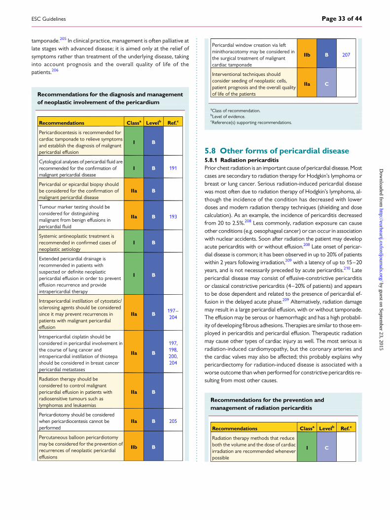
tamponade.205 In clinical practice, management is often palliative atlate stages with advanced disease; it is aimed only at the relief ofsymptoms rather than treatment of the underlying disease, takinginto account prognosis and the overall quality of life of thepatients.206
Recommendations for the diagnosis and managementof neoplastic involvement of the pericardium
Recommendations Classa Levelb Ref.c
Pericardiocentesis is recommended forcardiac tamponade to relieve symptomsand establish the diagnosis of malignantpericardial effusion
I B
Cytological analyses of pericardial fluid arerecommended for the confirmation ofmalignant pericardial disease
I B 191
Pericardial or epicardial biopsy shouldbe considered for the confirmation ofmalignant pericardial disease
IIa B
Tumour marker testing should beconsidered for distinguishingmalignant from benign effusions inpericardial fluid
IIa B 193
Systemic antineoplastic treatment isrecommended in confirmed cases ofneoplastic aetiology
I B
Extended pericardial drainage isrecommended in patients withsuspected or definite neoplasticpericardial effusion in order to preventeffusion recurrence and provideintrapericardial therapy
I B
Intrapericardial instillation of cytostatic/sclerosing agents should be consideredsince it may prevent recurrences inpatients with malignant pericardialeffusion
IIa B197–204
Intrapericardial cisplatin should beconsidered in pericardial involvement inthe course of lung cancer andintrapericardial instillation of thiotepashould be considered in breast cancerpericardial metastases
IIa B
197,198,200,204
Radiation therapy should beconsidered to control malignantpericardial effusion in patients withradiosensitive tumours such aslymphomas and leukaemias
IIa B
Pericardiotomy should be consideredwhen pericardiocentesis cannot beperformed
IIa B 205
Percutaneous balloon pericardiotomymay be considered for the prevention ofrecurrences of neoplastic pericardialeffusions
IIb B
Pericardial window creation via leftminithoracotomy may be considered inthe surgical treatment of malignantcardiac tamponade
IIb B 207
Interventional techniques shouldconsider seeding of neoplastic cells,patient prognosis and the overall qualityof life of the patients
IIa C
aClass of recommendation.bLevel of evidence.cReference(s) supporting recommendations.
5.8 Other forms of pericardial disease5.8.1 Radiation pericarditisPrior chest radiation is an important cause of pericardial disease. Mostcases are secondary to radiation therapy for Hodgkin’s lymphoma orbreast or lung cancer. Serious radiation-induced pericardial diseasewas most often due to radiation therapy of Hodgkin’s lymphoma, al-though the incidence of the condition has decreased with lowerdoses and modern radiation therapy techniques (shielding and dosecalculation). As an example, the incidence of pericarditis decreasedfrom 20 to 2.5%.208 Less commonly, radiation exposure can causeother conditions (e.g. oesophageal cancer) or can occur in associationwith nuclear accidents. Soon after radiation the patient may developacute pericarditis with or without effusion.208 Late onset of pericar-dial disease is common; it has been observed in up to 20% of patientswithin 2 years following irradiation,209 with a latency of up to 15–20years, and is not necessarily preceded by acute pericarditis.210 Latepericardial disease may consist of effusive-constrictive pericarditisor classical constrictive pericarditis (4–20% of patients) and appearsto be dose dependent and related to the presence of pericardial ef-fusion in the delayed acute phase.209 Alternatively, radiation damagemay result in a large pericardial effusion, with or without tamponade.The effusion may be serous or haemorrhagic and has a high probabil-ity of developing fibrous adhesions. Therapies are similar to those em-ployed in pericarditis and pericardial effusion. Therapeutic radiationmay cause other types of cardiac injury as well. The most serious isradiation-induced cardiomyopathy, but the coronary arteries andthe cardiac valves may also be affected; this probably explains whypericardiectomy for radiation-induced disease is associated with aworse outcome than when performed for constrictive pericarditis re-sulting from most other causes.
Recommendations for the prevention andmanagement of radiation pericarditis
Recommendations Classa Levelb Ref.c
Radiation therapy methods that reduceboth the volume and the dose of cardiacirradiation are recommended wheneverpossible
I C
ESC Guidelines Page 33 of 44
by guest on September 23, 2015
http://eurheartj.oxfordjournals.org/D
ownloaded from
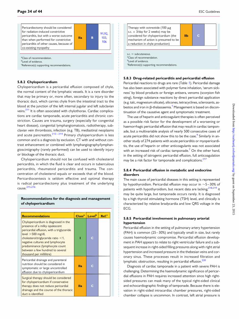
Pericardiectomy should be consideredfor radiation-induced constrictivepericarditis, but with a worse outcomethan when performed for constrictivepericarditis of other causes, because ofco-existing myopathy
IIa B91,92,103,106
aClass of recommendation.bLevel of evidence.cReference(s) supporting recommendations.
5.8.2 ChylopericardiumChylopericardium is a pericardial effusion composed of chyle,the normal content of the lymphatic vessels. It is a rare disorderthat may be primary or, more often, secondary to injury to thethoracic duct, which carries chyle from the intestinal tract to theblood at the junction of the left internal jugular and left subclavianveins.211 It is often associated with chylothorax. Cardiac complica-tions are cardiac tamponade, acute pericarditis and chronic con-striction. Causes are trauma, surgery (especially for congenitalheart disease), congenital lymphangiomatosis, radiotherapy, sub-clavian vein thrombosis, infection (e.g. TB), mediastinal neoplasmsand acute pancreatitis.212 – 214 Primary chylopericardium is lesscommon and is a diagnosis by exclusion. CT with and without con-trast enhancement or combined with lymphangiography/lymphan-gioscintigraphy (rarely performed) can be used to identify injuryor blockage of the thoracic duct.
Chylopericardium should not be confused with cholesterolpericarditis, in which the fluid is clear and occurs in tuberculouspericarditis, rheumatoid pericarditis and trauma. The con-centration of cholesterol equals or exceeds that of the blood.Pericardiocentesis is seldom effective and optimal therapyis radical pericardiectomy plus treatment of the underlyingcause.215,216
Recommendations for the diagnosis and managementof chylopericardium
Recommendations Classa Levelb Ref.c
Chylopericardium is diagnosed in thepresence of a milky opalescentpericardial effusion, with a triglyceridelevel .500 mg/dl,cholesterol:triglyceride ratio ,1,negative cultures and lymphocytepredominance (lymphocyte countbetween a few hundred to severalthousand per millilitre)
I C
Pericardial drainage and parenteralnutrition should be considered insymptomatic or large uncontrolledeffusion due to chylopericardium
IIa C
Surgical therapy should be consideredfor chylopericardium if conservativetherapy does not reduce pericardialdrainage and the course of the thoracicduct is identified
IIa C
Therapy with octreotide (100 mgs.c. × 3/day for 2 weeks) may beconsidered for chylopericardium (themechanism of action is presumed to bea reduction in chyle production)
IIb C
s.c. ¼ subcutaneous.aClass of recommendation.bLevel of evidence.cReference(s) supporting recommendations.
5.8.3 Drug-related pericarditis and pericardial effusionPericardial reactions to drugs are rare (Table 1). Pericardial damagehas also been associated with polymer fume inhalation, ‘serum sick-ness’ by blood products or foreign antisera, venoms (scorpion fishsting), foreign substance reactions by direct pericardial application(e.g. talc, magnesium silicate), silicones, tetracyclines, sclerosants, as-bestos and iron in b-thalassaemia.1 Management is based on discon-tinuation of the causative agent and symptomatic treatment.
The use of heparin and anticoagulant therapies is often perceivedas a possible risk factor for the development of a worsening orhaemorrhagic pericardial effusion that may result in cardiac tampon-ade, but a multivariable analysis of nearly 500 consecutive cases ofacute pericarditis did not show this to be the case.9 Similarly in an-other study of 274 patients with acute pericarditis or myopericardi-tis, the use of heparin or other anticoagulants was not associatedwith an increased risk of cardiac tamponade.7 On the other hand,in the setting of iatrogenic pericardial effusion, full anticoagulationmay be a risk factor for tamponade and complications.217
5.8.4 Pericardial effusion in metabolic and endocrinedisordersThe main cause of pericardial diseases in this setting is representedby hypothyroidism. Pericardial effusion may occur in �5–30% ofpatients with hypothyroidism, but recent data are lacking;218,219 itmay be quite large, but tamponade occurs rarely. It is diagnosedby a high thyroid stimulating hormone (TSH) level, and clinically ischaracterized by relative bradycardia and low QRS voltage in theECG.
5.8.5 Pericardial involvement in pulmonary arterialhypertensionPericardial effusion in the setting of pulmonary artery hypertension(PAH) is common (25–30%) and typically small in size, but rarelycauses haemodynamic compromise. Pericardial effusion develop-ment in PAH appears to relate to right ventricular failure and a sub-sequent increase in right-sided filling pressures along with right atrialhypertension and increased pressure in the thebesian veins and cor-onary sinus. These processes result in increased filtration andlymphatic obstruction, resulting in pericardial effusion.220
Diagnosis of cardiac tamponade in a patient with severe PAH ischallenging. Determining the haemodynamic significance of pericar-dial effusions in PAH requires increased attention since high right-sided pressures can mask many of the typical right-sided clinicaland echocardiographic findings of tamponade. Because there is ele-vation in right-sided intracardiac chamber pressures, right-sidedchamber collapse is uncommon. In contrast, left atrial pressure is
ESC GuidelinesPage 34 of 44
by guest on September 23, 2015
http://eurheartj.oxfordjournals.org/D
ownloaded from
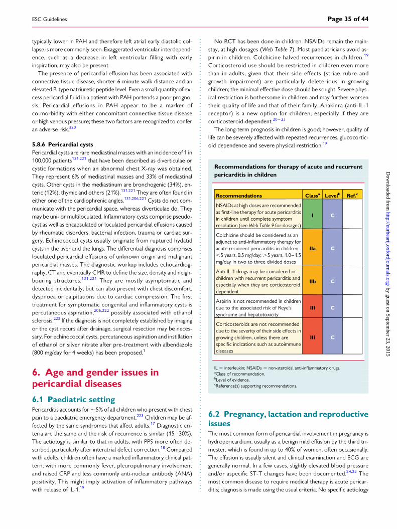
typically lower in PAH and therefore left atrial early diastolic col-lapse is more commonly seen. Exaggerated ventricular interdepend-ence, such as a decrease in left ventricular filling with earlyinspiration, may also be present.
The presence of pericardial effusion has been associated withconnective tissue disease, shorter 6-minute walk distance and anelevated B-type natriuretic peptide level. Even a small quantity of ex-cess pericardial fluid in a patient with PAH portends a poor progno-sis. Pericardial effusions in PAH appear to be a marker ofco-morbidity with either concomitant connective tissue diseaseor high venous pressure; these two factors are recognized to conferan adverse risk.220
5.8.6 Pericardial cystsPericardial cysts are rare mediastinal masses with an incidence of 1 in100,000 patients131,221 that have been described as diverticulae orcystic formations when an abnormal chest X-ray was obtained.They represent 6% of mediastinal masses and 33% of mediastinalcysts. Other cysts in the mediastinum are bronchogenic (34%), en-teric (12%), thymic and others (21%).131,221 They are often found ineither one of the cardiophrenic angles.131,206,221 Cysts do not com-municate with the pericardial space, whereas diverticulae do. Theymay be uni- or multiloculated. Inflammatory cysts comprise pseudo-cyst as well as encapsulated or loculated pericardial effusions causedby rheumatic disorders, bacterial infection, trauma or cardiac sur-gery. Echinococcal cysts usually originate from ruptured hydatidcysts in the liver and the lungs. The differential diagnosis comprisesloculated pericardial effusions of unknown origin and malignantpericardial masses. The diagnostic workup includes echocardiog-raphy, CT and eventually CMR to define the size, density and neigh-bouring structures.131,221 They are mostly asymptomatic anddetected incidentally, but can also present with chest discomfort,dyspnoea or palpitations due to cardiac compression. The firsttreatment for symptomatic congenital and inflammatory cysts ispercutaneous aspiration,206,222 possibly associated with ethanolsclerosis.222 If the diagnosis is not completely established by imagingor the cyst recurs after drainage, surgical resection may be neces-sary. For echinococcal cysts, percutaneous aspiration and instillationof ethanol or silver nitrate after pre-treatment with albendazole(800 mg/day for 4 weeks) has been proposed.1
6. Age and gender issues inpericardial diseases
6.1 Paediatric settingPericarditis accounts for �5% of all children who present with chestpain to a paediatric emergency department.223 Children may be af-fected by the same syndromes that affect adults.17 Diagnostic cri-teria are the same and the risk of recurrence is similar (15–30%).The aetiology is similar to that in adults, with PPS more often de-scribed, particularly after interatrial defect correction.18 Comparedwith adults, children often have a marked inflammatory clinical pat-tern, with more commonly fever, pleuropulmonary involvementand raised CRP and less commonly anti-nuclear antibody (ANA)positivity. This might imply activation of inflammatory pathwayswith release of IL-1.19
No RCT has been done in children. NSAIDs remain the main-stay, at high dosages (Web Table 7). Most paediatricians avoid as-pirin in children. Colchicine halved recurrences in children.19
Corticosteroid use should be restricted in children even morethan in adults, given that their side effects (striae rubre andgrowth impairment) are particularly deleterious in growingchildren; the minimal effective dose should be sought. Severe phys-ical restriction is bothersome in children and may further worsentheir quality of life and that of their family. Anakinra (anti-IL-1receptor) is a new option for children, especially if they arecorticosteroid-dependent.20 – 23
The long-term prognosis in children is good; however, quality oflife can be severely affected with repeated recurrences, glucocortic-oid dependence and severe physical restriction.19
Recommendations for therapy of acute and recurrentpericarditis in children
Recommendations Classa Levelb Ref.c
NSAIDs at high doses are recommendedas first-line therapy for acute pericarditisin children until complete symptomresolution (see Web Table 9 for dosages)
I C
Colchicine should be considered as anadjunct to anti-inflammatory therapy foracute recurrent pericarditis in children:,5 years, 0.5 mg/day; .5 years, 1.0–1.5mg/day in two to three divided doses
IIa C
Anti-IL-1 drugs may be considered inchildren with recurrent pericarditis andespecially when they are corticosteroiddependent
IIb C
Aspirin is not recommended in childrendue to the associated risk of Reye’ssyndrome and hepatotoxicity
III C
Corticosteroids are not recommendeddue to the severity of their side effects ingrowing children, unless there arespecific indications such as autoimmunediseases
III C
IL ¼ interleukin; NSAIDs ¼ non-steroidal anti-inflammatory drugs.aClass of recommendation.bLevel of evidence.cReference(s) supporting recommendations.
6.2 Pregnancy, lactation and reproductiveissuesThe most common form of pericardial involvement in pregnancy ishydropericardium, usually as a benign mild effusion by the third tri-mester, which is found in up to 40% of women, often occasionally.The effusion is usually silent and clinical examination and ECG aregenerally normal. In a few cases, slightly elevated blood pressureand/or aspecific ST-T changes have been documented.24,25 Themost common disease to require medical therapy is acute pericar-ditis; diagnosis is made using the usual criteria. No specific aetiology
ESC Guidelines Page 35 of 44
by guest on September 23, 2015
http://eurheartj.oxfordjournals.org/D
ownloaded from
is usually identified. Nowadays the general outcomes of pregnanciesin these women when followed by dedicated multidisciplinary teamsare similar to those expected in the general population.25
A proposed treatment scheme for pericarditis during pregnancyis described in Web Table 8.25 –27 Pregnancy in women with recur-rent pericarditis should be planned during a phase of disease quies-cence.25 – 27 Classic NSAIDs (ibuprofen, indomethacin) may beconsidered during the first and second trimesters;25 – 27 most ex-perts prefer high-dose aspirin, since it is regularly used in anti-phospholipid syndrome in pregnancy and is moderately effectivein the prevention of pre-eclampsia in high-risk obstetric pa-tients.224,225 After gestational week 20, all NSAIDs (except aspirin≤100 mg/day) can cause constriction of the ductus arteriosus andimpair foetal renal function, and they should be withdrawn in anycase at gestational week 32.224,225 The lowest effective doses ofprednisone may be used throughout pregnancy and breastfeeding(with supplementation with calcium and vitamin D).25– 27 Paraceta-mol is allowed throughout pregnancy and breastfeeding, as areanti-histamine H2 blockers or proton pump inhibitors.226 Duringpregnancy, tapering of therapies should be extremely cautious. Nor-mal vaginal delivery is possible and should be considered in theabsence of contraindications.25 – 27 Ibuprofen, indomethacin, na-proxen and prednisone may be considered in women who are breast-feeding. After discontinuation of breastfeeding, gradual tapering ofprednisone should be considered, eventually resuming colchicine.Colchicine is considered contraindicated during pregnancy andbreastfeeding, even though no adverse events during pregnancy andfoetal or child development have been reported in women with FMFtreated with colchicine during pregnancy and breastfeeding.227–229
6.3 The elderlyMost guidelines have not discussed the applicability of their recom-mendations to older patients with multiple co-morbidities.230 Nopaper has specifically addressed pericardial diseases in the elderly,so only expert opinion exists. Therapy adherence and compliancemay be problematic in the elderly because of cognitive impairment,poor vision or hearing and cost, but the strongest predictor of non-adherence is the number of medications.230 Indomethacin is not in-dicated, the colchicine dose should be halved and particular careshould be taken to evaluate renal impairment and drug interactions.
7. Interventional techniques andsurgeryThe aetiology of pericardial diseases remains unresolved in manycases because the full spectrum of diagnostic methods is not usedin many institutions. The gold standard remains surgical by the sub-xiphoid approach, allowing collection of fluid samples and perform-ing pericardial biopsy and pericardial drainage. Interventionaltechniques206 include the combined use of imaging by pericardio-scopy first described in combination with diagnostic molecular,histological and immunohistological methods to assess the aetiologyand pathogenesis of pericardial and epicardial disease manifesta-tions133 and the option to intervene therapeutically by instillationof drugs into the pericardial sac.63,204
7.1 Pericardiocentesis and pericardialdrainageFor pericardial drainage and biopsy, the surgical approach remainsthe gold standard. The classical approach is by subxiphoid incision,through which it is easy to take fluid samples and perform a pericar-dial biopsy. The operation is completed by leaving a small drain inplace to evacuate remaining effusion. This technique using a subxi-phoid approach is straightforward for a thoracic or cardiovascularsurgeon, if such a team is close to the cardiology team. In clinicalpractice, pericardial fluid is aspirated by pericardiocentesis.
State-of-the-art pericardiocentesis must be guided either byfluoroscopy or echocardiography206 under local anaesthesia. Blindprocedures must be not be used to avoid the risk of laceration ofthe heart or other organs, except in very rare situations that are im-mediately life threatening. An experienced operator and staff shouldperform pericardiocentesis in a facility equipped for radiographic,echocardiographic, haemodynamic and ECG monitoring.
For echo-guided pericardiocentesis, the ideal entry site is thepoint on the body surface where the effusion is closest to the trans-ducer and the fluid collection is maximal. The needle trajectory isdefined by the angulation of the handheld transducer and shouldavoid vital structures such as the liver, myocardium, lung, internalmammary artery (3–5 cm away from the parasternal border) andthe vascular bundle at the inferior margin of each rib. The intendedpoint of entry is marked on the skin and the direction of the ultra-sound beam is carefully noted (see Web supplemental material). Anadditional technique is the echo-guided approach followed by echo-monitoring of the procedure.
For fluoroscopic-guided pericardiocentesis, a polytef-sheathedneedle with an attached saline-filled syringe is advanced under mod-erate suction until the pericardial sac is reached.206 When using themore common subxiphoid route for pericardiocentesis, a Tuohy-17,blunt-tip introducer needle is advanced to the left shoulder at a30-degree angle to the skin, thus avoiding coronary, pericardial andinternal mammary arteries. The lateral angiographic view providesthe best visualization of the puncturing needle and its relation tothe diaphragm and pericardium. The needle is slowly advanced to-wards the heart shadow and the epicardial halo phenomenon, undermoderate suction and with injection of small amounts of diluted con-trast medium, until pericardial fluid is aspirated. If haemorrhagic fluidis freely aspirated, a few millilitres of contrast medium may be injectedunder fluoroscopic control to verify the position of the needle. A softJ-tip guidewire is then introduced and after dilatation is exchanged fora multihole pigtail catheter, through which the fluid is evacuated un-der the control of intrapericardial pressure.206
Pericardiocentesis should be performed by experienced opera-tors and carries a risk of complications ranging from 4 to 10% de-pending on the type of monitoring, the skill of the operator andthe setting (emergency vs. urgent vs. elective).183,206 Most commoncomplications include arrhythmias, coronary artery or cardiacchamber puncture, haemothorax, pneumothorax, pneumopericar-dium and hepatic injury (Web Table 9).
Pericardiocentesis may have additional limitations/dangers whenpericardial fluid is not free and when located in a lateral or posteriorposition or ,10 mm. In these cases a surgical approach might besafer, depending on local expertise and availability.
ESC GuidelinesPage 36 of 44
by guest on September 23, 2015
http://eurheartj.oxfordjournals.org/D
ownloaded from
7.2 PericardioscopyPericardioscopy permits visualization of the pericardial sac with itsepicardial and pericardial layers. Macroscopic views show a cluster-ing of protrusions, haemorrhagic areas and neovascularization inmalignant pericardial effusion, which are often haemorrhagic, in con-trast to radiogenic or viral and autoimmune effusions.133,206
Pericardioscopy enables the performing physician to take targetedbiopsy specimens from epicardial and pericardial layers, avoiding epi-cardial vessels and increasing the probability of obtaining disease-specific results. For safety reasons it is important to have a secondwire in place. The safety wire allows a quick exchange with a pigtailcatheter and allows autotransfusion in the case of relevant bleeding.By selecting the biopsy site, less informative white areas of fibrin canbe avoided and dark spots of inflammation, malignancy or haemor-rhagic imbibitions can be selected, which can be identified best inthe blue-light mode. Pericardial biopsy can even be taken underradiologic control alone. The open jaws of the bioptome are ad-vanced gently until the silhouette of the pericardial sac is reached.Then the jaws are closed and the biopsy sample is taken. Seven to10 samples should be taken to reduce sampling error. The mostmeaningful diagnostic yield from pericardial biopsies can be obtainedby multiple pericardioscopically guided tissue acquisitions.
This technique is quite demanding and can be performed in only alimited number of experienced tertiary referral centres. Pericardio-scopy may be considered as a diagnostic method for inspection ofthe pericardium and epicardium in experienced centres. It permitssafe tissue acquisition in pericardial diseases of unknown origin.
7.3 Pericardial fluid analysis, pericardialand epicardial biopsySerosanguinous or haemorrhagic fluid can be found in malignant aswell as post-pericardiotomy, rheumatologic and traumatic effusionsor can be caused by iatrogenic lesions during pericardiocentesis, butalso in idiopathic and viral forms. In cases of sepsis, TB or in HIV-positive patients, bacterial cultures can be diagnostic. Fluid cytologycan separate malignant from non-malignant effusions, which is im-portant for effusions in tumour patients after radiotherapy of themediastinum. Discriminative signatures between malignant andautoreactive effusions are higher levels of tumour markers in neo-plastic pericardial effusion.133,206
Cytology and assessment of bacterial cultures of the fluid, histo-logical/immunohistological evaluation of biopsy specimens and mo-lecular analysis (PCR for microbial agents of fluid and tissue) allow adefinite aetiological diagnosis in many cases, which permits furthertreatment.133
7.4 Intrapericardial treatmentIn patients with a larger effusion of unknown origin, prolonged peri-cardial drainage may allow subsequent intrapericardial treatment.
In neoplastic pericardial effusion, most frequently due to bron-chus carcinoma or breast cancer, intrapericardial cisplatin or thiote-pa therapy have been proposed in combination with systemicantineoplastic treatment, which should be tailored in collaborationwith the oncologist.204
In autoreactive and lymphocytic pericardial effusion disease-specific intrapericardial crystalloid triamcinolone (300 mg/m2
body surface) may be considered.64 Viral pericarditis may be ex-cluded by PCR in fluid and tissue specimens, but such investigationsare not usually performed in uncomplicated cases in clinical practice.
In cases of uraemic pericardial effusion, intrapericardial therapywith triamcinolone may be considered, apart from intensifiedhaemo- or peritoneal dialysis and fluid evacuation.64,65
In rare cases of recurrent effusion, balloon pericardiotomy is anoption that allows a (transient) pericardio(-pleural-)abdominal win-dow for drainage. This approach should be avoided in neoplastic orpurulent effusions.
7.5 Pericardial access forelectrophysiologyFirst reported in 1996,231pericardial access has been used for themapping and ablation of epicardial substrates of ventricular tachyar-rhythmias with improved success rates and avoidance of a surgicalprocedure232,233 (see supplemental Web material and Web Table 9for complications of the procedure).
7.6 Surgery for pericardial diseases7.6.1 Pericardial windowA pericardial window is a cardiac surgical procedure to create acommunication, or ‘window’, from the pericardial space to thepleural cavity. The purpose of the window is to allow a pericardialeffusion (usually malignant) to drain from the space surrounding theheart into the chest cavity in order to prevent a large pericardial ef-fusion and cardiac tamponade.
The window is usually performed by a cardiac surgeon, but a peri-cardial window may be created by video-assisted thoracoscopy orballoon pericardiotomy by a percutaneous intervention. The mainindication is represented by recurrent large effusions or cardiactamponade when a more complex operation such pericardiectomyis a high risk or the life expectancy of the patient is reduced (e.g.neoplastic pericardial disease) and the intervention is palliative.The results of a pericardial window are less definitive since the com-munication may close and recurrent effusions, especially loculated,are common and may require additional interventions comparedwith pericardiectomy, which is a more complex but completeoperation.105
7.6.2 PericardiectomyIn constrictive pericarditis the treatment is pericardectomy. Thedecortications should remove as much of the pericardium as pos-sible with all constricting parietal and epicardial layers,103 – 105 buttaking care of perserving the phrenic nerves bilaterally. Only byusing sternotomy can all the constricting pericardial layers beremoved. Therefore, left anterolateral thoracotomy should beavoided since it permits only a partial resection.
It is also necessary to liberate all of the right atrium, the superiorvena cava and especially the inferior vena cava and the inferior partof the right ventricle adjacent to the diaphragm as far as pos-sible.103 – 105 Only when the constricting peel is adherent andcalcified is it necessary to leave behind a few islands of the pericar-dium. To avoid bleeding, cardiopulmonary bypass should beemployed only in cases of co-existent cardiac surgical lesions, butcardiopulmonary bypass may be needed in stand-by, in case of theoccurrence of haemorrhagic lesions during the procedure. By
ESC Guidelines Page 37 of 44
by guest on September 23, 2015
http://eurheartj.oxfordjournals.org/D
ownloaded from
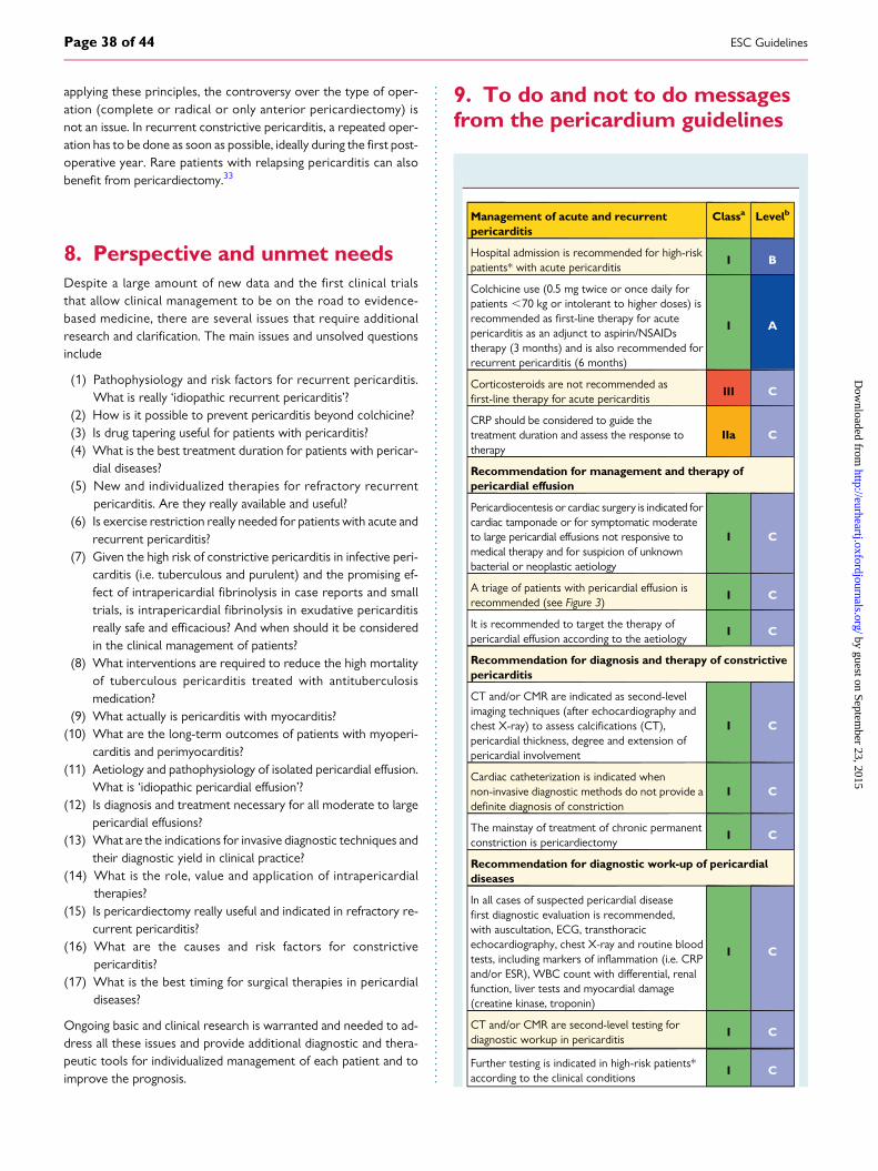
applying these principles, the controversy over the type of oper-ation (complete or radical or only anterior pericardiectomy) isnot an issue. In recurrent constrictive pericarditis, a repeated oper-ation has to be done as soon as possible, ideally during the first post-operative year. Rare patients with relapsing pericarditis can alsobenefit from pericardiectomy.33
8. Perspective and unmet needsDespite a large amount of new data and the first clinical trialsthat allow clinical management to be on the road to evidence-based medicine, there are several issues that require additionalresearch and clarification. The main issues and unsolved questionsinclude
(1) Pathophysiology and risk factors for recurrent pericarditis.What is really ‘idiopathic recurrent pericarditis’?
(2) How is it possible to prevent pericarditis beyond colchicine?(3) Is drug tapering useful for patients with pericarditis?(4) What is the best treatment duration for patients with pericar-
dial diseases?(5) New and individualized therapies for refractory recurrent
pericarditis. Are they really available and useful?(6) Is exercise restriction really needed for patients with acute and
recurrent pericarditis?(7) Given the high risk of constrictive pericarditis in infective peri-
carditis (i.e. tuberculous and purulent) and the promising ef-fect of intrapericardial fibrinolysis in case reports and smalltrials, is intrapericardial fibrinolysis in exudative pericarditisreally safe and efficacious? And when should it be consideredin the clinical management of patients?
(8) What interventions are required to reduce the high mortalityof tuberculous pericarditis treated with antituberculosismedication?
(9) What actually is pericarditis with myocarditis?(10) What are the long-term outcomes of patients with myoperi-
carditis and perimyocarditis?(11) Aetiology and pathophysiology of isolated pericardial effusion.
What is ‘idiopathic pericardial effusion’?(12) Is diagnosis and treatment necessary for all moderate to large
pericardial effusions?(13) What are the indications for invasive diagnostic techniques and
their diagnostic yield in clinical practice?(14) What is the role, value and application of intrapericardial
therapies?(15) Is pericardiectomy really useful and indicated in refractory re-
current pericarditis?(16) What are the causes and risk factors for constrictive
pericarditis?(17) What is the best timing for surgical therapies in pericardial
diseases?
Ongoing basic and clinical research is warranted and needed to ad-dress all these issues and provide additional diagnostic and thera-peutic tools for individualized management of each patient and toimprove the prognosis.
9. To do and not to do messagesfrom the pericardium guidelines
Management of acute and recurrentpericarditis
Classa Levelb
Hospital admission is recommended for high-riskpatients* with acute pericarditis
I B
Colchicine use (0.5 mg twice or once daily forpatients ,70 kg or intolerant to higher doses) isrecommended as first-line therapy for acutepericarditis as an adjunct to aspirin/NSAIDstherapy (3 months) and is also recommended forrecurrent pericarditis (6 months)
I A
Corticosteroids are not recommended asfirst-line therapy for acute pericarditis
III C
CRP should be considered to guide thetreatment duration and assess the response totherapy
IIa C
Recommendation for management and therapy ofpericardial effusion
Pericardiocentesis or cardiac surgery is indicated forcardiac tamponade or for symptomatic moderateto large pericardial effusions not responsive tomedical therapy and for suspicion of unknownbacterial or neoplastic aetiology
I C
A triage of patients with pericardial effusion isrecommended (see Figure 3)
I C
It is recommended to target the therapy ofpericardial effusion according to the aetiology
I C
Recommendation for diagnosis and therapy of constrictivepericarditis
CT and/or CMR are indicated as second-levelimaging techniques (after echocardiography andchest X-ray) to assess calcifications (CT),pericardial thickness, degree and extension ofpericardial involvement
I C
Cardiac catheterization is indicated whennon-invasive diagnostic methods do not provide adefinite diagnosis of constriction
I C
The mainstay of treatment of chronic permanentconstriction is pericardiectomy
I C
Recommendation for diagnostic work-up of pericardialdiseases
In all cases of suspected pericardial diseasefirst diagnostic evaluation is recommended,with auscultation, ECG, transthoracicechocardiography, chest X-ray and routine bloodtests, including markers of inflammation (i.e. CRPand/or ESR), WBC count with differential, renalfunction, liver tests and myocardial damage(creatine kinase, troponin)
I C
CT and/or CMR are second-level testing fordiagnostic workup in pericarditis
I C
Further testing is indicated in high-risk patients*according to the clinical conditions
I C
ESC GuidelinesPage 38 of 44
by guest on September 23, 2015
http://eurheartj.oxfordjournals.org/D
ownloaded from
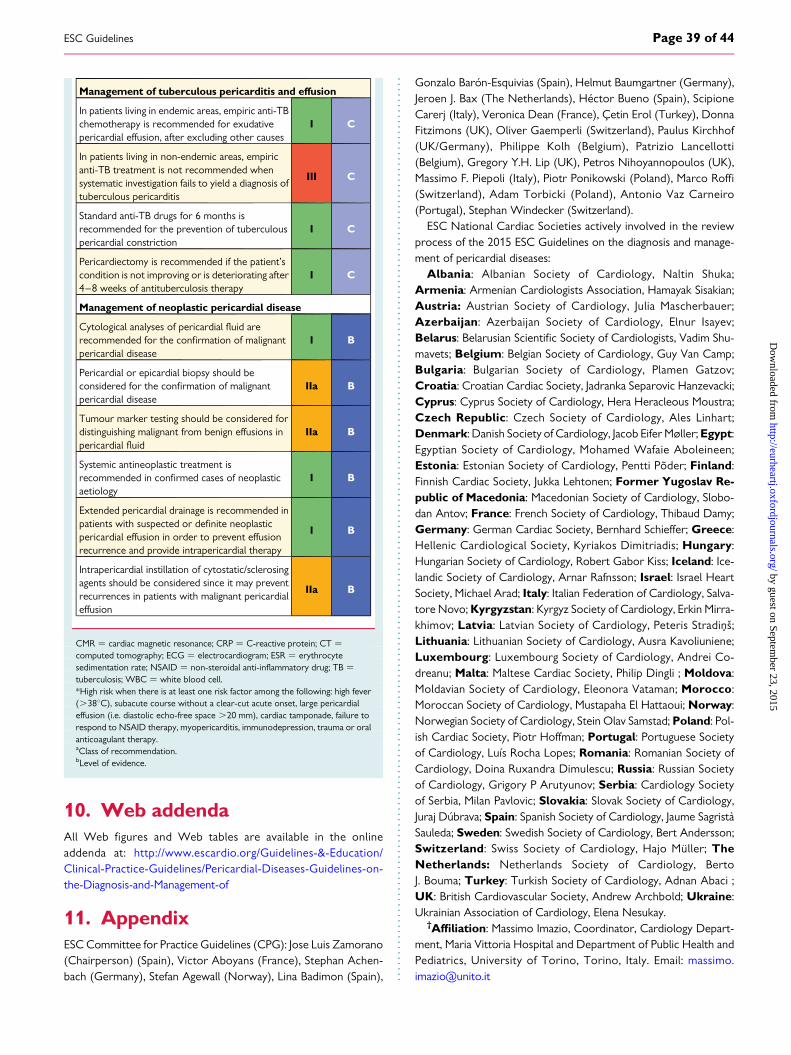
Management of tuberculous pericarditis and effusion
In patients living in endemic areas, empiric anti-TBchemotherapy is recommended for exudativepericardial effusion, after excluding other causes
I C
In patients living in non-endemic areas, empiricanti-TB treatment is not recommended whensystematic investigation fails to yield a diagnosis oftuberculous pericarditis
III C
Standard anti-TB drugs for 6 months isrecommended for the prevention of tuberculouspericardial constriction
I C
Pericardiectomy is recommended if the patient’scondition is not improving or is deteriorating after4–8 weeks of antituberculosis therapy
I C
Management of neoplastic pericardial disease
Cytological analyses of pericardial fluid arerecommended for the confirmation of malignantpericardial disease
I B
Pericardial or epicardial biopsy should beconsidered for the confirmation of malignantpericardial disease
IIa B
Tumour marker testing should be considered fordistinguishing malignant from benign effusions inpericardial fluid
IIa B
Systemic antineoplastic treatment isrecommended in confirmed cases of neoplasticaetiology
I B
Extended pericardial drainage is recommended inpatients with suspected or definite neoplasticpericardial effusion in order to prevent effusionrecurrence and provide intrapericardial therapy
I B
Intrapericardial instillation of cytostatic/sclerosingagents should be considered since it may preventrecurrences in patients with malignant pericardialeffusion
IIa B
CMR ¼ cardiac magnetic resonance; CRP ¼ C-reactive protein; CT ¼computed tomography; ECG ¼ electrocardiogram; ESR ¼ erythrocytesedimentation rate; NSAID ¼ non-steroidal anti-inflammatory drug; TB ¼tuberculosis; WBC ¼ white blood cell.*High risk when there is at least one risk factor among the following: high fever(.388C), subacute course without a clear-cut acute onset, large pericardialeffusion (i.e. diastolic echo-free space .20 mm), cardiac tamponade, failure torespond to NSAID therapy, myopericarditis, immunodepression, trauma or oralanticoagulant therapy.aClass of recommendation.bLevel of evidence.
10. Web addendaAll Web figures and Web tables are available in the onlineaddenda at: http://www.escardio.org/Guidelines-&-Education/Clinical-Practice-Guidelines/Pericardial-Diseases-Guidelines-on-the-Diagnosis-and-Management-of
11. AppendixESC Committee for Practice Guidelines (CPG): Jose Luis Zamorano(Chairperson) (Spain), Victor Aboyans (France), Stephan Achen-bach (Germany), Stefan Agewall (Norway), Lina Badimon (Spain),
Gonzalo Baron-Esquivias (Spain), Helmut Baumgartner (Germany),Jeroen J. Bax (The Netherlands), Hector Bueno (Spain), ScipioneCarerj (Italy), Veronica Dean (France), Çetin Erol (Turkey), DonnaFitzimons (UK), Oliver Gaemperli (Switzerland), Paulus Kirchhof(UK/Germany), Philippe Kolh (Belgium), Patrizio Lancellotti(Belgium), Gregory Y.H. Lip (UK), Petros Nihoyannopoulos (UK),Massimo F. Piepoli (Italy), Piotr Ponikowski (Poland), Marco Roffi(Switzerland), Adam Torbicki (Poland), Antonio Vaz Carneiro(Portugal), Stephan Windecker (Switzerland).
ESC National Cardiac Societies actively involved in the reviewprocess of the 2015 ESC Guidelines on the diagnosis and manage-ment of pericardial diseases:
Albania: Albanian Society of Cardiology, Naltin Shuka;Armenia: Armenian Cardiologists Association, Hamayak Sisakian;Austria: Austrian Society of Cardiology, Julia Mascherbauer;Azerbaijan: Azerbaijan Society of Cardiology, Elnur Isayev;Belarus: Belarusian Scientific Society of Cardiologists, Vadim Shu-mavets; Belgium: Belgian Society of Cardiology, Guy Van Camp;Bulgaria: Bulgarian Society of Cardiology, Plamen Gatzov;Croatia: Croatian Cardiac Society, Jadranka Separovic Hanzevacki;Cyprus: Cyprus Society of Cardiology, Hera Heracleous Moustra;Czech Republic: Czech Society of Cardiology, Ales Linhart;Denmark: Danish Society of Cardiology, Jacob Eifer Møller; Egypt:Egyptian Society of Cardiology, Mohamed Wafaie Aboleineen;Estonia: Estonian Society of Cardiology, Pentti Poder; Finland:Finnish Cardiac Society, Jukka Lehtonen; Former Yugoslav Re-public of Macedonia: Macedonian Society of Cardiology, Slobo-dan Antov; France: French Society of Cardiology, Thibaud Damy;Germany: German Cardiac Society, Bernhard Schieffer; Greece:Hellenic Cardiological Society, Kyriakos Dimitriadis; Hungary:Hungarian Society of Cardiology, Robert Gabor Kiss; Iceland: Ice-landic Society of Cardiology, Arnar Rafnsson; Israel: Israel HeartSociety, Michael Arad; Italy: Italian Federation of Cardiology, Salva-tore Novo; Kyrgyzstan: Kyrgyz Society of Cardiology, Erkin Mirra-khimov; Latvia: Latvian Society of Cardiology, Peteris Stradins;Lithuania: Lithuanian Society of Cardiology, Ausra Kavoliuniene;Luxembourg: Luxembourg Society of Cardiology, Andrei Co-dreanu; Malta: Maltese Cardiac Society, Philip Dingli ; Moldova:Moldavian Society of Cardiology, Eleonora Vataman; Morocco:Moroccan Society of Cardiology, Mustapaha El Hattaoui; Norway:Norwegian Society of Cardiology, Stein Olav Samstad; Poland: Pol-ish Cardiac Society, Piotr Hoffman; Portugal: Portuguese Societyof Cardiology, Luıs Rocha Lopes; Romania: Romanian Society ofCardiology, Doina Ruxandra Dimulescu; Russia: Russian Societyof Cardiology, Grigory P Arutyunov; Serbia: Cardiology Societyof Serbia, Milan Pavlovic; Slovakia: Slovak Society of Cardiology,Juraj Dubrava; Spain: Spanish Society of Cardiology, Jaume SagristaSauleda; Sweden: Swedish Society of Cardiology, Bert Andersson;Switzerland: Swiss Society of Cardiology, Hajo Muller; TheNetherlands: Netherlands Society of Cardiology, BertoJ. Bouma; Turkey: Turkish Society of Cardiology, Adnan Abaci ;UK: British Cardiovascular Society, Andrew Archbold; Ukraine:Ukrainian Association of Cardiology, Elena Nesukay.
†Affiliation: Massimo Imazio, Coordinator, Cardiology Depart-ment, Maria Vittoria Hospital and Department of Public Health andPediatrics, University of Torino, Torino, Italy. Email: [email protected]
ESC Guidelines Page 39 of 44
by guest on September 23, 2015
http://eurheartj.oxfordjournals.org/D
ownloaded from
12. References1. Maisch B, Seferovic PM, Ristic AD, Erbel R, Rienmuller R, Adler Y,
Tomkowski WZ, Thiene G, Yacoub MH; Task Force on the Diagnosis and Man-agement of Pericardial Diseases of the European Society of Cardiology. Guide-lines on the diagnosis and management of pericardial diseases executivesummary. Eur Heart J 2004;25:587–610.
2. Klein AL, Abbara S, Agler DA, Appleton CP, Asher CR, Hoit B, Hung J, Garcia MJ,Kronzon I, Oh JK, Rodriguez ER, Schaff HV, Schoenhagen P, Tan CD, White RD.American Society of Echocardiography clinical recommendations for multimodal-ity cardiovascular imaging of patients with pericardial disease: endorsed by the So-ciety for Cardiovascular Magnetic Resonance and Society of CardiovascularComputed Tomography. J Am Soc Echocardiogr 2013;26:965–1012.e15.
3. Cosyns B, Plein S, Nihoyanopoulos P, Smiseth O, Achenbach S, Andrade MJ,Pepi M, Ristic A, Imazio M, Paelinck B, Lancellotti P; on behalf of the European As-sociation of Cardiovascular Imaging (EACVI) and European Society of CardiologyWorking Group (ESC WG) on Myocardial and Pericardial diseases. European As-sociation of Cardiovascular Imaging (EACVI) position paper: multimodality im-aging in pericardial disease. Eur Heart J Cardiovasc Imaging 2014;16:12–31.
4. Imazio M. Contemporary management of pericardial diseases. Curr Opin Cardiol2012;27:308–317.
5. Imazio M, Gaita F. Diagnosis and treatment of pericarditis. Heart 2015;101:1159–1168.
6. Imazio M, Spodick DH, Brucato A, Trinchero R, Adler Y. Controversial issues inthe management of pericardial diseases. Circulation 2010;121:916–928.
7. Imazio M, Cecchi E, Demichelis B, Chinaglia A, Ierna S, Demarie D, Ghisio A,Pomari F, Belli R, Trinchero R. Myopericarditis versus viral or idiopathic acutepericarditis. Heart 2008;94:498–501.
8. Imazio M, Demichelis B, Parrini I, Giuggia M, Cecchi E, Gaschino G, Demarie D,Ghisio A, Trinchero R. Day-hospital treatment of acute pericarditis: a manage-ment program for outpatient therapy. J Am Coll Cardiol 2004;43:1042–1046.
9. Imazio M, Cecchi E, Demichelis B, Ierna S, Demarie D, Ghisio A, Pomari F, Coda L,Belli R, Trinchero R. Indicators of poor prognosis of acute pericarditis. Circulation2007;115:2739–2744.
10. Imazio M, Bobbio M, Cecchi E, Demarie D, Demichelis B, Pomari F, Moratti M,Gaschino G, Giammaria M, Ghisio A, Belli R, Trinchero R. Colchicine in additionto conventional therapy for acute pericarditis: results of the COlchicine for acutePEricarditis (COPE) trial. Circulation 2005;112:2012–2016.
11. Imazio M, Brucato A, Cemin R, Ferrua S, Maggiolini S, Beqaraj F, Demarie D,Forno D, Ferro S, Maestroni S, Belli R, Trinchero R, Spodick DH, Adler Y;ICAP Investigators. A randomized trial of colchicine for acute pericarditis. NEngl J Med 2013;369:1522–1528.
12. Imazio M, Brucato A, Derosa FG, Lestuzzi C, Bombana E, Scipione F, Leuzzi S,Cecchi E, Trinchero R, Adler Y. Aetiological diagnosis in acute and recurrent peri-carditis: when and how. J Cardiovasc Med (Hagerstown) 2009;10:217–230.
13. Imazio M, Bobbio M, Cecchi E, Demarie D, Pomari F, Moratti M, Ghisio A, Belli R,Trinchero R. Colchicine as first-choice therapy for recurrent pericarditis: resultsof the CORE (COlchicine for REcurrent pericarditis) trial. Arch Intern Med 2005;165:1987–1991.
14. Imazio M, Brucato A, Cemin R, Ferrua S, Belli R, Maestroni S, Trinchero R,Spodick DH, Adler Y; CORP (COlchicine for Recurrent Pericarditis) Investiga-tors. Colchicine for recurrent pericarditis (CORP): a randomized trial. Ann InternMed 2011;155:409–414.
15. Imazio M, Belli R, Brucato A, Cemin R, Ferrua S, Beqaraj F, Demarie D, Ferro S,Forno D, Maestroni S, Cumetti D, Varbella F, Trinchero R, Spodick DH, Adler Y.Efficacy and safety of colchicine for treatment of multiple recurrences of pericar-ditis (CORP-2): a multicentre, double-blind, placebo-controlled, randomised trial.Lancet 2014;383:2232–2237.
16. Kyto V, Sipila J, Rautava P. Clinical profile and influences on outcomes in patientshospitalized for acute pericarditis. Circulation 2014;130:1601–1606.
17. Shakti D, Hehn R, Gauvreau K, Sundel RP, Newburger JW. Idiopathic pericarditisand pericardial effusion in children: contemporary epidemiology and manage-ment. J Am Heart Assoc 2014;3:e001483.
18. Raatikka M, Pelkonem PM, Karjalainen J, Jokinen E. Recurrent pericarditis in chil-dren and adolescents. J Am Coll Cardiol 2003;42:759–764.
19. Imazio M, Brucato A, Pluymaekers N, Breda L, Calabri G, Cantarini L, Cimaz R,Colimodio F, Corona F, Cumetti D, Di Blasi Lo Cuccio C, Gattorno M,Insalaco A, Limongelli G, Russo MG, Valenti A, Finkelstein Y, Martini A. Recurrentpericarditis in children and adolescents: etiology, presentation, therapies, and out-comes. A multicenter cohort study. J Cardiovasc Med (in press)
20. Picco P, Brisca G, Traverso F, Loy A, Gattorno M, Martini A. Successful treatmentof idiopathic recurrent pericarditis in children with interleukin-1b receptor antag-onist (anakinra): an unrecognized autoinflammatory disease? Arthritis Rheum 2009;60:264–268.
21. Finetti M, Insalaco A, Cantarini L, Meini A, Breda L, Alessio M, D’Alessandro M,Picco P, Martini A, Gattorno M. Long term efficacy of interleukin-1 receptor an-tagonist (anakinra) in steroid dependent and colchicine-resistant recurrent peri-carditis. J Pediatr 2014;164:1425–1431.
22. Scardapane A, Brucato A, Chiarelli F, Breda L. Efficacy of interleukin-1beta recep-tor antagonist (anakinra) in idiopathic recurrent pericarditis. Pediatr Cardiol 2013;34:1989–1991.
23. Gaspari S, Marsili M, Imazio M, Brucato A. New insights in the pathogenesis andtherapy of idiopathic recurrent pericarditis in children. Clin Exp Rheumatol 2013;31:788–794.
24. Ristic AD, Seferovic PM, Ljubic A, Jovanovic I, Ristic G, Pankuweit S, Ostojic M,Maisch B. Pericardial disease in pregnancy. Herz 2003;28:209–215.
25. Brucato A, Imazio M, Curri S, Palmieri G, Trinchero R. Medical treatment of peri-carditis during pregnancy. Int J Cardiol 2010;144:413–414.
26. Imazio M, Brucato A. Management of pericarditis in women. Womens Health (LondEngl) 2012;8:341–348.
27. Imazio M, Brucato A, Rampello S, Armellino F, Trinchero R, Spodick DH, Adler Y.Management of pericardial diseases during pregnancy. J Cardiovasc Med (Hagers-town) 2010;11:557–562.
28. Vianello F, Cinetto F, Cavraro M, Battisti A, Castelli M, Imbergamo S,Marcolongo R. Azathioprine in isolated recurrent pericarditis: a single centre ex-perience. Int J Cardiol 2011;147:477–478.
29. Moretti M, Buiatti A, Merlo M, Massa L, Fabris E, Pinamonti B, Sinagra G. Useful-ness of high-dose intravenous human immunoglobulins treatment for refractoryrecurrent pericarditis. Am J Cardiol 2013;112:1493–1498.
30. Imazio M, Lazaros G, Brucato A, Picardi E, Vasileiou P, Carraro M, Tousoulis D,Belli R, Gaita F. Intravenous human immunoglobulin for refractory recurrent peri-carditis. A systematic review of all published cases. J Cardiovasc Med 2015 Jun 18[Epub ahead of print].
31. Lazaros G, Vasileiou P, Koutsianas C, Antonatou K, Stefanadis C, Pectasides D,Vassilopoulos D. Anakinra for the management of resistant idiopathic recurrentpericarditis. Initial experience in 10 adult cases. Ann Rheum Dis 2014 Aug 27. pii:annrheumdis-2014–205990.
32. Lazaros G, Imazio M, Brucato A, Picardi E, Vassilopoulos D, Vasileiou P,Tousouis D, Gaita F. Anakinra: an emerging option for refractory idiopathic re-current pericarditis. A systematic review of published evidence. J CardiovascMed (Hagerstown) 2015 Jun 18 [Epub ahead of print].
33. Khandaker MH, Schaff HV, Greason KL, Anavekar NS, Espinosa RE, Hayes SN,Nishimura RA, Oh JK. Pericardiectomy vs medical management in patients withrelapsing pericarditis. Mayo Clin Proc 2012;87:1062–1070.
34. Imazio M, Brucato A, Barbieri A, Ferroni F, Maestroni S, Ligabue G, Chinaglia A,Cumetti D, Della Casa G, Bonomi F, Mantovani F, Di Corato P, Lugli R, Faletti R,Leuzzi S, Bonamini R, Modena MG, Belli R. Good prognosis for pericarditis withand without myocardial involvement: results from a multicenter, prospective co-hort study. Circulation 2013;128:42–49.
35. Imazio M, Brucato A, Cumetti D, Brambilla G, Demichelis B, Ferro S, Maestroni S,Cecchi E, Belli R, Palmieri G, Trinchero R. Corticosteroids for recurrent pericarditis:high versus low doses: a nonrandomized observation. Circulation 2008;118:667–671.
36. Imazio M, Brucato A, Maestroni S, Cumetti D, Belli R, Trinchero R, Adler Y. Risk ofconstrictive pericarditis after acute pericarditis. Circulation 2011;124:1270–1275.
37. Imazio M, Brucato A, Adler Y, Brambilla G, Artom G, Cecchi E, Palmieri G,Trinchero R. Prognosis of idiopathic recurrent pericarditis as determined frompreviously published reports. Am J Cardiol 2007;100:1026–1028.
The CME text ‘2015 ESC Guidelines on the diagnosis and management of pericardial diseases’ is accredited by the European Board for Accreditation in Cardiology (EBAC). EBACworks according to the quality standards of the European Accreditation Council for Continuing Medical Education (EACCME), which is an institution of the European Union of MedicalSpecialists (UEMS). In compliance with EBAC/EACCME Guidelines, all authors participating in this programme have disclosed any potential conflicts of interest that might cause a bias inthe article. The Organizing Committee is responsible for ensuring that all potential conflicts of interest relevant to the programme are declared to the participants prior to the CMEactivities.
CME questions for this article are available at: European Heart Journal http://www.oxforde-learning.com/eurheartj and European Society of Cardiology http://www.escardio.org/guidelines
ESC GuidelinesPage 40 of 44
by guest on September 23, 2015
http://eurheartj.oxfordjournals.org/D
ownloaded from
38. Imazio M, Lazaros G, Picardi E, Vasileiou P, Orlando F, Carraro M, Tsiachris D,Vlachopoulos C, Georgiopoulos G, Tousoulis D, Belli R, Gaita F. Incidence andprognostic significance of new onset atrial fibrillation/flutter in acute pericarditis.Heart 2015 Apr 29. pii: heartjnl-2014–307398. [Epub ahead of print].
39. Alraies MC, AlJaroudi W, Yarmohammadi H, Yingchoncharoen T, Schuster A,Senapati A, Tariq M, Kwon D, Griffin BP, Klein AL. Usefulness of cardiac magneticresonance-guided management in patients with recurrent pericarditis. Am J Cardiol2015;115:542–547.
40. Feng D, Glockner J, Kim K, Martinez M, Syed IS, Araoz P, Breen J, Espinosa RE,Sundt T, Schaff HV, Oh JK. Cardiac magnetic resonance imaging pericardial lategadolinium enhancement and elevated inflammatory markers can predict the re-versibility of constrictive pericarditis after antiinflammatory medical therapy: a pi-lot study. Circulation 2011;124:1830–1837.
41. Yared K, Baggish AL, Picard MH, Hoffmann U, Hung J. Multimodality imaging ofpericardial disease. JACC Cardiovasc Imaging 2010;3:650–660.
42. LeWinter MM. Clinical practice. Acute pericarditis. N Engl J Med 2014;371:2410–2416.
43. Lilly LS. Treatment of acute and recurrent idiopathic pericarditis. Circulation 2013;127:1723–1726.
44. Sliwa K, Mocumbi AO. Forgotten cardiovascular diseases in Africa. Clin Res Cardiol2010;99:65–74.
45. Imazio M. Pericardial involvement in systemic inflammatory diseases. Heart 2011;97:1882–1892.
46. Bhardwaj R, Berzingi C, Miller C, Hobbs G, Gharib W, Beto RJ, Warden BE,Jain AC. Differential diagnosis of acute pericarditis from normal variant early re-polarization and left ventricular hypertrophy with early repolarization: an electro-cardiographic study. Am J Med Sci 2013;345:28–32.
47. Imazio M, Brucato A, Maestroni S, Cumetti D, Dominelli A, Natale G, Trinchero R.Prevalence of C-reactive protein elevation and time course of normalization inacute pericarditis: implications for the diagnosis, therapy, and prognosis of peri-carditis. Circulation 2011;123:1092–1097.
48. Imazio M, Adler Y. Management of pericardial effusion. Eur Heart J 2013;34:1186–1197.
49. Permanyer-Miralda G. Acute pericardial disease: approach to the aetiologic diag-nosis. Heart 2004;90:252–254.
50. Imazio M, Brucato A, Mayosi BM, Derosa FG, Lestuzzi C, Macor A, Trinchero R,Spodick DH, Adler Y. Medical therapy of pericardial diseases: part I: idiopathic andinfectious pericarditis. J Cardiovasc Med (Hagerstown) 2010;11:712–722.
51. Imazio M, Brucato A, Mayosi BM, Derosa FG, Lestuzzi C, Macor A, Trinchero R,Spodick DH, Adler Y. Medical therapy of pericardial diseases: part II: noninfectiouspericarditis, pericardial effusion and constrictive pericarditis. J Cardiovasc Med(Hagerstown) 2010;11:785–794.
52. Mayosi BM. Contemporary trends in the epidemiology and management of car-diomyopathy and pericarditis in sub-Saharan Africa. Heart 2007;93:1176–1183.
53. Seidenberg PH, Haynes J. Pericarditis: diagnosis, management, and return to play.Curr Sports Med Rep 2006;5:74–79.
54. Pelliccia A, Corrado D, Bjørnstad HH, Panhuyzen-Goedkoop N, Urhausen A,Carre F, Anastasakis A, Vanhees L, Arbustini E, Priori S. Recommendations forparticipation in competitive sport and leisure-time physical activity in individualswith cardiomyopathies, myocarditis and pericarditis. Eur J Cardiovasc Prev Rehabil2006;13:876–885.
55. Lotrionte M, Biondi-Zoccai G, Imazio M, Castagno D, Moretti C, Abbate A,Agostoni P, Brucato AL, Di Pasquale P, Raatikka M, Sangiorgi G, Laudito A,Sheiban I, Gaita F. International collaborative systematic review of controlled clin-ical trials on pharmacologic treatments for acute pericarditis and its recurrences.Am Heart J 2010;160:662–670.
56. Imazio M, Brucato A, Trinchero R, Spodick D, Adler Y. Individualized therapy forpericarditis. Expert Rev Cardiovasc Ther 2009;7:965–975.
57. Imazio M, Brucato A, Trinchero R, Spodick DH, Adler Y. Colchicine for pericar-ditis: hype or hope? Eur Heart J 2009;30:532–539.
58. Imazio M, Brucato A, Belli R, Forno D, Ferro S, Trinchero R, Adler Y. Colchicinefor the prevention of pericarditis: what we know and what we do not know in2014—systematic review and meta-analysis. J Cardiovasc Med (Hagerstown)2014;15:840–846.
59. Alabed S, Cabello JB, Irving GJ, Qintar M, Burls A. Colchicine for pericarditis. Co-chrane Database Syst Rev 2014 Aug 28;8:CD010652.
60. Soler-Soler J, Sagrista-Sauleda J, Permanyer-Miralda G. Relapsing pericarditis.Heart 2004;90:1364–1368.
61. Brucato A, Brambilla G, Moreo A, Alberti A, Munforti C, Ghirardello A, Doria A,Shynar Y, Livneh A, Adler Y, Shoenfeld Y, Mauri F, Palmieri G, Spodick DH. Long-term outcomes in difficult-to-treat patients with recurrent pericarditis. Am J Car-diol 2006;98:267–271.
62. Caforio AL, Brucato A, Doria A, Brambilla G, Angelini A, Ghirardello A, Bottaro S,Tona F, Betterle C, Daliento L, Thiene G, Iliceto S. Anti-heart and anti-intercalated
disk autoantibodies: evidence for autoimmunity in idiopathic recurrent acute peri-carditis. Heart 2010;96:779–784.
63. Pankuweit S, Stein A, Karatolios K, Richter A, Ruppert V, Maisch B. Viral genomesin the pericardial fluid and in peri- and epicardial biopsies from a German cohortof patients with large to moderate pericardial effusions. Heart Fail Rev 2013;18:329–336.
64. Maisch B, Ristic AD, Pankuweit S. Intrapericardial treatment of autoreactive peri-cardial effusion with triamcinolone: the way to avoid side effects of systemic cor-ticosteroid therapy. Eur Heart J 2002;23:1503–1508.
65. Frasiolas JA, Cahoon WD. Intrapericardial triamcinolone administration for auto-reactive pericarditis. Ann Pharmacother 2010;44:1641–1646.
66. Imazio M, Cooper LT. Management of myopericarditis. Expert Rev Cardiovasc Ther2013;11:193–201.
67. Abu Fanne R, Banai S, Chorin U, Rogowski O, Keren G, Roth A. Diagnostic yield ofextensive infectious panel testing in acute pericarditis. Cardiology 2011;119:134–139.
68. Buiatti A, Merlo M, Pinamonti B, De Biasio M, Bussani R, Sinagra G. Clinical pres-entation and long-term follow-up of perimyocarditis. J Cardiovasc Med (Hagers-town) 2013;14:235–241.
69. Caforio AL, Pankuweit S, Arbustini E, Basso C, Gimeno-Blanes J, Felix SB, Fu M,Helio T, Heymans S, Jahns R, Klingel K, Linhart A, Maisch B, McKenna W,Mogensen J, Pinto YM, Ristic A, Schultheiss HP, Seggewiss H, Tavazzi L,Thiene G, Yilmaz A, Charron P, Elliott PM; European Society of Cardiology Work-ing Group on Myocardial and Pericardial Diseases. Current state of knowledge onaetiology, diagnosis, management, and therapy of myocarditis: a position state-ment of the European Society of Cardiology Working Group on Myocardialand Pericardial Diseases. Eur Heart J 2013;34:2636–2648.
70. Imazio M. Pericarditis with troponin elevation: is it true pericarditis and a reasonfor concern? J Cardiovasc Med (Hagerstown) 2014;15:73–77.
71. Imazio M, Brucato A, Spodick DH, Adler Y. Prognosis of myopericarditis as deter-mined from previously published reports. J Cardiovasc Med (Hagerstown) 2014;15:835–839.
72. Khatib R, Reyes MP, Smith F, Khatib G, Rezkalla S. Enhancement of coxsackievirusB4 virulence by indomethacin. J Lab Clin Med 1990;116:116–120.
73. Imazio M, Trinchero R. Myopericarditis: etiology, management, and prognosis. Int JCardiol 2008;127:17–26.
74. Corey GR, Campbell PT, Van Trigt P, Kenney RT, O’Connor CM, Sheikh KH,Kisslo JA, Wall TC. Etiology of large pericardial effusions. Am J Med 1993;95:209–213.
75. Sagrista-Sauleda J, Merce J, Permanyer-Miralda G, Soler-Soler J. Clinical clues tothe causes of large pericardial effusions. Am J Med 2000;109:95–101.
76. Levy PY, Corey R, Berger P, Habib G, Bonnet JL, Levy S, Messana T, Djiane P,Frances Y, Botta C, DeMicco P, Dumon H, Mundler O, Chomel JJ, Raoult D. Etio-logic diagnosis of 204 pericardial effusions. Medicine (Baltimore) 2003;82:385–391.
77. Reuter H, Burgess LJ, Doubell AF. Epidemiology of pericardial effusions at a largeacademic hospital in South Africa. Epidemiol Infect 2005;133:393–399.
78. Ma W, Liu J, Zeng Y, Chen S, Zheng Y, Ye S, Lan L, Liu Q, Weig HJ, Liu Q. Causes ofmoderate to large pericardial effusion requiring pericardiocentesis in 140 HanChinese patients. Herz 2012;37:183–187.
79. Mayosi BM, Burgess LJ, Doubell AF. Tuberculous pericarditis. Circulation 2005;112:3608–3616.
80. Shabetai R. Pericardial effusion: haemodynamic spectrum. Heart 2004;90:255–156.
81. Spodick DH. Acute cardiac tamponade. N Engl J Med 2003;349:684–690.82. Imazio M, Mayosi BM, Brucato A, Markel G, Trinchero R, Spodick DH, Adler Y.
Triage and management of pericardial effusion. J Cardiovasc Med (Hagerstown)2010;11:928–935.
83. Roy CL, Minor MA, Brookhart MA, Choudhry NK. Does this patient with a peri-cardial effusion have cardiac tamponade? JAMA 2007;297:1810–1818.
84. Ristic AD, Imazio M, Adler Y, Anastasakis A, Badano LP, Brucato A, Caforio AL,Dubourg O, Elliott P, Gimeno J, Helio T, Klingel K, Linhart A, Maisch B, Mayosi B,Mogensen J, Pinto Y, Seggewiss H, Seferovic PM, Tavazzi L, Tomkowski W,Charron P. Triage strategy for urgent management of cardiac tamponade: a pos-ition statement of the European Society of Cardiology Working Group on Myo-cardial and Pericardial Diseases. Eur Heart J 2014;35:2279–2284.
85. Imazio M, Brucato A, Trinchero R, Spodick DH, Adler Y. Colchicine for pericar-ditis: hype or hope? Eur Heart J 2009;30:532–539.
86. Frohlich GM, Keller P, Schmid F, Wolfrum M, Osranek M, Falk C, Noll G,Enseleit F, Reinthaler M, Meier P, Luscher TF, Ruschitzka F, Tanner FC. Haemo-dynamically irrelevant pericardial effusion is associated with increased mortality inpatients with chronic heart failure. Eur Heart J 2013;34:1414–1423.
87. Mitiku TY, Heidenreich PA. A small pericardial effusion is a marker of increasedmortality. Am Heart J 2011;161:152–157.
88. Sagrista-Sauleda J, Angel J, Permanyer-Miralda G, Soler-Soler J. Long-term follow-up of idiopathic chronic pericardial effusion. N Engl J Med 1999;341:2054–2059.
ESC Guidelines Page 41 of 44
by guest on September 23, 2015
http://eurheartj.oxfordjournals.org/D
ownloaded from
89. Little WC, Freeman GL. Pericardial disease. Circulation 2006;113:1622–1632.90. Cameron J, Oesterle SN, Baldwin JC, Hancock EW. The etiologic spectrum of
constrictive pericarditis. Am Heart J 1987;113(2 Pt 1):354–380.91. Ling LH, Oh JK, Schaff HV, Danielson GK, Mahoney DW, Seward JB, Tajik AJ. Con-
strictive pericarditis in the modern era: evolving clinical spectrum and impact onoutcome after pericardiectomy. Circulation 1999;100:1380–1386.
92. Bertog SC, Thambidorai SK, Parakh K, Schoenhagen P, Ozduran V,Houghtaling PL, Lytle BW, Blackstone EH, Lauer MS, Klein AL. Constrictive peri-carditis: etiology and cause-specific survival after pericardiectomy. J Am Coll Car-diol 2004;43:1445–1452.
93. Mutyaba AK, Balkaran S, Cloete R, du Plessis N, Badri M, Brink J, Mayosi BM. Con-strictive pericarditis requiring pericardiectomy at Groote Schuur Hospital, CapeTown, South Africa: causes and perioperative outcomes in the HIV era (1990–2012). J Thorac Cardiovasc Surg 2014;148:3058–3065.e1.
94. Talreja DR, Edwards WD, Danielson GK, Schaff HV, Tajik AJ, Tazelaar HD,Breen JF, Oh JK. Constrictive pericarditis in 26 patients with histologically normalpericardial thickness. Circulation 2003;108:1852–1857.
95. Welch TD, Ling LH, Espinosa RE, Anavekar NS, Wiste HJ, Lahr BD, Schaff HV,Oh JK. Echocardiographic diagnosis of constrictive pericarditis: Mayo Clinic cri-teria. Circ Cardiovasc Imaging 2014;7:526–534.
96. Talreja DR, Nishimura RA, Oh JK, Holmes DR. Constrictive pericarditis in themodern era: novel criteria for diagnosis in the cardiac catheterization laboratory.J Am Coll Cardiol 2008;51:315–319.
97. Mayosi BM, Ntsekhe M, Bosch J, Pandie S, Jung H, Gumedze F, Pogue J, Thabane L,Smieja M, Francis V, Joldersma L, Thomas KM, Thomas B, Awotedu AA,Magula NP, Naidoo DP, Damasceno A, Chitsa Banda A, Brown B, Manga P,Kirenga B, Mondo C, Mntla P, Tsitsi JM, Peters F, Essop MR, Russell JBW,Hakim J, Matenga J, Barasa AF, Sani MU, Olunuga T, Ogah O, Ansa V, Aje A,Danbauchi S, Ojji D, Yusuf S. Prednisolone and Mycobacterium indicus pranii in tu-berculous pericarditis. N Engl J Med 2014;371:1121–1130.
98. Sagrista-Sauleda J, Permanyer-Miralda G, Candell-Riera J, Angel J, Soler-Soler J.Transient cardiac constriction: an unrecognized pattern of evolution in effusiveacute idiopathic pericarditis. Am J Cardiol 1987;59:961–966.
99. Haley JH, Tajik AJ, Danielson GK, Schaff HV, Mulvagh SL, Oh JK. Transient con-strictive pericarditis: causes and natural history. J Am Coll Cardiol 2004;43:271–275.
100. Syed FF, Schaff HV, Oh JK. Constrictive pericarditis—a curable diastolic heart fail-ure. Nat Rev Cardiol 2014;11:530–544.
101. Sagrista-Sauleda J, Angel J, Sanchez A, Permanyer-Miralda G, Soler-Soler J.Effusive-constrictive pericarditis. N Engl J Med 2004;350:469–475.
102. Ntsekhe M, Wiysonge CS, Commerford PJ, Mayosi BM. The prevalence and out-come of effusive constrictive pericarditis: a systematic review of the literature.Cardiovasc J Afr 2012;23:281–25.
103. DeValeria PA, Baumgartner WA, Casale AS, Greene PS, Cameron DE,Gardner TJ, Gott VL, Watkins L, Reitz BA. Current indications, risks, and out-come after pericardiectomy. Ann Thorac Surg 1991;52:219–224.
104. Chowdhury UK, Subramaniam GK, Kumar AS, Airan B, Singh R, Talwar S, Seth S,Mishra PK, Pradeep KK, Sathia S, Venugopal P. Pericardiectomy for constrictivepericarditis: a clinical, echocardiographic, and hemodynamic evaluation of twosurgical techniques. Ann Thorac Surg 2006;81:522–529.
105. Cho YH, Schaff HV, Dearani JA, Daly RC, Park SJ, Li Z, Oh JK. Completion peri-cardiectomy for recurrent constrictive pericarditis: importance of timing of recur-rence on late clinical outcome of operation. Ann Thorac Surg 2012;93:1236–1241.
106. Komoda T, Frumkin A, Knosalla C, Hetzer R. Child-Pugh score predicts survivalafter radical pericardiectomy for constrictive pericarditis. Ann Thorac Surg 2013;96:1679–1685.
107. Ferguson EC, Berkowitz EA. Cardiac and pericardial calcifications on chest radio-graphs. Clin Cardiol 2010;65:685–694.
108. Verhaert D, Gabriel RS, Johnston D, Lytle BW, Desai MY, Klein AL. The role ofmultimodality imaging in the management of pericardial disease. Circ CardiovascImaging 2010;3:333–343.
109. Bogaert J, Francone M. Pericardial disease: value of CT and MR imaging. Radiology2013;267:340–356.
110. Frank H, Globits S. Magnetic resonance imaging evaluation of myocardial and peri-cardial disease. J Magn Reson Imaging 1999;10:617–626.
111. Francone M, Dymarkowski S, Kalantzi M, Rademakers FE, Bogaert J. Assessmentof ventricular coupling with real-time cine MRI and it value to differentiate con-strictive pericarditis from restrictive cardiomyopathy. Eur Radiol 2006;16:944–951.
112. Bogaert J, Francone M. Cardiovascular magnetic resonance in pericardial diseases.J Cardiovasc Magn Reson 2009;11:14.
113. Misselt AJ, Harris SR, Glockner J, Feng D, Syed IS, Araoz PA. MR imaging of thepericardium. Magn Reson Imaging Clin N Am 2008;16:185–199.
114. Alter P, Figiel JH, Rupp TP, Bachmann GF, Maisch B, Rominger MB. MR, CT, andPET imaging in pericardial disease. Heart Fail Rev 2013;18:289–306.
115. Taylor AM, Dymarkowski S, Verbeken EK, Bogaert J. Detection of pericardial in-flammation with late-enhancement cardiac magnetic resonance imaging: initial re-sults. Eur Radiol 2006;16:569–574.
116. Dawson D, Rubens M, Mohiaddin R. Contemporary imaging of the pericardium.JACC Cardiovasc Imaging 2011;4:680–684.
117. Yelgec NS, Dymarkowski S, Ganame J, Bogaert J. Value of MRI in patients with aclinical suspicion of acute myocarditis. Eur Radiol 2007;17:2211–2217.
118. Feng DL, Glockner J, Kim K, Martinez M, Syed IS, Araoz P, Breen J, Espinosa RE,Sundt T, Schaff HV, Oh JK. Cardiac magnetic resonance imaging pericardial lategadolinium enhancement and elevated inflammatory markers can predict thereversibility of constrictive pericarditis after antiinflammatory medical therapy.A pilot study. Circulation 2011;124:1830–1837.
119. Zurick AO, Bolen MA, Kwon DH, Tan CD, Popovic ZB, Rajeswaran J,Rodriguez ER, Flamm SD, Klein AL. Pericardial delayed hyperenhancement withCMR imaging in patients with constrictive pericarditis undergoing surgical pericar-diectomy. A case series with histopathological correlation. JACC Cardiovasc Imaging2011;4:1180–91.
120. Kojima S, Yamada N, Goto Y. Diagnosis of constrictive pericarditis by tagged cinemagnetic resonance imaging. N Engl J Med 1999;341:373–374.
121. Psychidis-Papakyritsis P, de Roos A, Kroft LJM. Functional MRI of congenital ab-sence of the pericardium. AJR Am J Roentgenol 2007;189:W312–W314.
122. Coolen J, De Keyzer F, Nafteux P, De Wever W, Dooms C, Van steenkiste J,Roebben I, Verbeken E, De Leyn P, Van Raemdonck D, Nackaerts K,Dymarkowski S, Verschakelen J. Malignant pleural disease: diagnosis by usingdiffusion-weighted and dynamic contrast-enhanced MR imaging—initial experi-ence. Radiology 2012;263:884–892.
123. Lobert P, Brown RK, Dvorak RA, Corbett JR, Kazerooni EA, Wong KK. Spectrumof physiological and pathological cardiac and pericardial uptake of FDG in oncol-ogy PET-CT. Clin Radiol 2013;68:e59–e71.
124. James OG, Christensen JD, Wong T, Borges-Neto S, Koweek LM. Utility of FDGPET/CT in inflammatory cardiovascular disease. RadioGraphics 2011;31:1271–1286.
125. Dong A, Dong H, Wang Y, Cheng C, Zuo C, Lu J. (18)F-FDG PET/CT in differen-tiating acute tuberculous from idiopathic pericarditis: preliminary study. Clin NuclMed 2013;38:e160–e165.
126. Crossman AW, Sasseen BM. Right heart catheterization and hemodynamic pro-files. In: Kay IP, Sabate M, Costa MA, eds. Cardiac catheterization and percutaneousintervention. London: Taylor & Francis, 2004:93–119.
127. Meltser H, Kalaria VG. Cardiac tamponade. Catheter Cardiovasc Interv 2005;64:245–255.
128. Imazio M, Trinchero R. Triage and management of acute pericarditis. Int J Cardiol2007;118:286–294
129. Permanyer-Miralda G, Sagrista-Sauleda J, Soler-Soler J. Primary acute pericardialdisease: a prospective series of 231 consecutive patients. Am J Cardiol 1985;56:623–630.
130. Zayas R, Anguita M, Torres F, Gimenez D, Bergillos F, Ruiz M, Ciudad M,Gallardo A, Valles F. Incidence of specific etiology and role of methods for specificetiologic diagnosis of primary acute pericarditis. Am J Cardiol 1995;75:378–382.
131. Gouriet F, Levy PY, Casalta JP, Zandotti C, Collart F, Lepidi H, Cautela J, Bonnet JL,Thuny F, Habib G, Raoult D. Etiology of pericarditis in a prospective cohort of1162 cases. Am J Med 2015;128:784.e1–784.e8.
132. Imazio M, Hoit BD. Post-cardiac injury syndromes. An emerging cause of pericar-dial diseases. Int J Cardiol 2013;168:648–652.
133. Maisch B, Rupp H, Ristic A, Pankuweit S. Pericardioscopy and epi- and pericardialbiopsy—a new window to the heart improving etiological diagnoses and permit-ting targeted intrapericardial therapy. Heart Fail Rev 2013;18:317–328.
134. Pankuweit S, Wadlich A, Meyer E, Portig I, Hufnagel G, Maisch B. Cytokine acti-vation in pericardial fluids in different forms of pericarditis. Herz 2000;25:748–754.
135. Ristic AD, Pankuweit S, Maksimovic R, Moosdorf R, Maisch B. Pericardial cyto-kines in neoplastic, autoreactive, and viral pericarditis. Heart Fail Rev 2013;18:345–353.
136. Mahfoud F, Gartner B, Kindermann M, Ukena C, Gadomski K, Klingel K, Kandolf R,Bohm M, Kindermann I. Virus serology in patients with suspected myocarditis:utility or futility? Eur Heart J 2011;32:897–903.
137. Levy PY, Fournier PE, Charrel R, Metras D, Habib G, Raoult D. Molecular analysisof pericardial fluid: a 7-year experience. Eur Heart J 2006;27:1942–1946.
138. Wessely R, Vorpahl M, Schomig A, Klingel K. Late constrictive involvement of thepericardium in a case of previous myocarditis. Cardiovasc Pathol 2004;13:327–329.
139. Thienemann F, Sliwa K, Rockstroh JK. HIV and the heart: the impact of antiretro-viral therapy: a global perspective. Eur Heart J 2013;34:3538–3546.
140. Mayosi BM, Wiysonge CS, Ntsekhe M, Volmink JA, Gumedze F, Maartens G,Aje A, Thomas BM, Thomas KM, Awotedu AA, Thembela B, Mntla P, Maritz F,Ngu Blackett K, Nkouonlack DC, Burch VC, Rebe K, Parish A, Sliwa K,Vezi BZ, Alam N, Brown BG, Gould T, Visser T, Shey MS, Magula NP,Commerford PJ. Clinical characteristics and initial management of patients withtuberculous pericarditis in the HIV era: the Investigation of the Management ofPericarditis in Africa (IMPI Africa) registry. BMC Infect Dis 2006;6:2.
ESC GuidelinesPage 42 of 44
by guest on September 23, 2015
http://eurheartj.oxfordjournals.org/D
ownloaded from
141. Mayosi BM, Wiysonge CS, Ntsekhe M, Volmink JA, Gumedze F, Maartens G,Aje A, Thomas BM, Thomas KM, Awotedu AA, Thembela B, Mntla P, Maritz F,Ngu Blackett K, Nkouonlack DC, Burch VC, Rebe K, Parish A, Sliwa K,Vezi BZ, Alam N, Brown BG, Gould T, Visser T, Shey MS, Magula NP,Commerford PJ. Mortality in patients treated for tuberculous pericarditis in sub-Saharan Africa. S Afr Med J 2008;98:36–40.
142. Pandie S, Peter JG, Kerbelker ZS, Meldau R, Theron G, Govender U, Ntsekhe M,Dheda K, Mayosi BM. Diagnostic accuracy of quantitative PCR (Xpert MTB/RIF)for tuberculous pericarditis compared to adenosine deaminase and unstimulatedinterferon-g in a high burden setting: a prospective study. BMC Medicine 2014;12:101
143. Mayosi BM, Ntsekhe M, Volmink JA, Commerford PJ. Interventions for treatingtuberculous pericarditis. Cochrane Database Syst Rev 2002;(4):CD000526.
144. Reuter H, Burgess LJ, Louw VJ, Doubell AF. The management of tuberculous peri-cardial effusion: experience in 233 consecutive patients. Cardiovasc J S Afr 2007;18:20–25.
145. Cui HB, Chen XY, Cui CC, Shou XL, Liu XH, Yao XW, Wang JK, Guan GC. Pre-vention of pericardial constriction by transcatheter intrapericardial fibrinolysiswith urokinase. Chin Med Sci J 2005;20:5–10.
146. Sagrista Sauleda J, Barrabes JA, Permanyer Miralda G, Soler Soler J. Purulent peri-carditis: review of a 20-year experience in a general hospital. J Am Coll Cardiol 1993;22:1661–1665.
147. Rubin RH, Moellering RC Jr. Clinical, microbiologic and therapeutic aspects ofpurulent pericarditis. Am J Med Sci 1975;59:68–78.
148. Brook I, Frazier EH. Microbiology of acute purulent pericarditis. A 12-year experi-ence in a military hospital. Arch Intern Med 1996;156:1857–1860.
149. Goodman LJ. Purulent pericarditis. Curr Treat Options Cardiovasc Med 2000;2:343–350.
150. Ben-Horin S, Bank I, Shinfeld A, Kachel E, Guetta V, Livneh A. Diagnostic value ofthe biochemical composition of pericardial effusions in patients undergoing peri-cardiocentesis. Am J Cardiol 2007;99:1294–1297.
151. Augustin P, Desmard M, Mordant P, Lasocki S, Maury JM, Heming N, Montravers P.Clinical review: intrapericardial fibrinolysis in management of purulent pericardi-tis. Crit Care 2011;15:220.
152. Alpert MA, Ravenscraft MD. Pericardial involvement in end-stage renal disease.Am J Med Sci 2003;325:228–236.
153. Renfrew R, Buselmeier TJ, Kjellstrand CM. Pericarditis and renal failure. Annu RevMed 1980;31:345–360.
154. Gunukula SR, Spodick DH. Pericardial disease in renal patients. Semin Nephrol2001;21:52–56.
155. Banerjee A, Davenport A. Changing patterns of pericardial disease in patients withend-stage renal disease. Hemodial Int 2006;10:249–255.
156. Kabukcu M, Demircioglu F, Yanik E, Basarici I, Ersel F. Pericardial tamponade andlarge pericardial effusions: causal factors and efficacy of percutaneous catheterdrainage in 50 patients. Tex Heart Inst J 2004;31:398–403.
157. Wood JE, Mahnensmith RL. Pericarditis associated with renal failure. Evolution andmanagement. Semin Dial 2001;14:61–66.
158. Imazio M. Idiopathic recurrent pericarditis as an immune-mediated disease: cur-rent insights into pathogenesis and emerging treatment options. Expert Rev ClinImmunol 2014;10:1487–1492.
159. Brucato A, Shinar Y, Brambilla G, Robbiolo L, Ferrioli G, Patrosso MC, Zanni D,Benco S, Boiani E, Ghirardello A, Caforio AL, Bergantin A, Tombini V, Moreo A,Ashtamkar L, Doria A, Shoenfeld Y, Livneh A. Idiopathic recurrent acute pericar-ditis: familial Mediterranean fever mutations and disease evolution in a large co-hort of Caucasian patients. Lupus 2005;14:670–674.
160. Cantarini L, Lucherini OM, Brucato A, Barone L, Cumetti D, Iacoponi F, Rigante D,Brambilla G, Penco S, Brizi MG, Patrosso MC, Valesini G, Frediani B, Galeazzi M,Cimaz R, Paolazzi G, Vitale A, Imazio M. Clues to detect tumor necrosis factorreceptor-associated periodic syndrome (TRAPS) among patients with idiopathicrecurrent acute pericarditis: results of a multicentre study. Clin Res Cardiol 2012;101:525–531.
161. Cantarini L, Imazio M, Brizi MG, Lucherini OM, Brucato A, Cimaz R, Galeazzi M.Role of autoimmunity and autoinflammation in the pathogenesis of idiopathic re-current pericarditis. Clin Rev Allergy Immunol 2013;44:6–13.
162. DeLine JM, Cable DG. Clustering of recurrent pericarditis with effusion and con-striction in a family. Mayo Clin Proc 2002;77:39–43.
163. Maggiolini S, Tiberti G, Cantarini L, Carbone C, Mariani S, Achilli F, Maestroni S,Brucato A. Large pericardial effusion in a family with recurrent pericarditis: a re-port of probable X-linked transmission. Exp Clin Cardiol 2011;16:54–56.
164. Brucato A, Brambilla G. Recurrent idiopathic pericarditis: familial occurrence. Int JCardiol 2005;102:529.
165. Imazio M. The post-pericardiotomy syndrome. Curr Opin Pulm Med 2012;18:366–374.
166. Imazio M, Brucato A, Rovere ME, Gandino A, Cemin R, Ferrua S, Maestroni S,Barosi A, Simon C, Ferrazzi P, Belli R, Trinchero R, Spodick D, Adler Y. Contem-porary features, risk factors, and prognosis of the postpericardiotomy syndrome.Am J Cardiol 2011;108:1183–1187.
167. Imazio M, Negro A, Belli R, Beqaraj F, Forno D, Giammaria M, Trinchero R,Adler Y, Spodick D. Frequency and prognostic significance of pericarditis follow-ing acute myocardial infarction treated by primary percutaneous coronary inter-vention. Am J Cardiol 2009;103:1525–1529.
168. Finkelstein Y, Shemesh J, Mahlab K, Abramov D, Bar-El Y, Sagie A, Sharoni E,Sahar G, Smolinsky AK, Schechter T, Vidne BA, Adler Y. Colchicine for the pre-vention of postpericardiotomy syndrome. Herz 2002;27:791–794.
169. Imazio M, Trinchero R, Brucato A, Rovere ME, Gandino A, Cemin R, Ferrua S,Maestroni S, Zingarelli E, Barosi A, Simon C, Sansone F, Patrini D, Vitali E,Ferrazzi P, Spodick DH, Adler Y; COPPS Investigators. COlchicine for the Preven-tion of the Postpericardiotomy Syndrome (COPPS): a multicentre, randomized,double-blind, placebo-controlled trial. Eur Heart J 2010;31:2749–2754.
170. Imazio M, Brucato A, Ferrazzi P, Spodick DH, Alder Y. Postpericardiotomy syn-drome: a proposal for diagnostic criteria. J Cardiovasc Med (Hagerstown) 2013;14:351–353.
171. Horneffer PJ, Miller RH, Pearson TA, Rykiel MF, Reitz BA, Gardner TJ. The effect-ive treatment of postpericardiotomy syndrome after cardiac operations. A rando-mized placebo-controlled trial. J Thorac Cardiovasc Surg 1990;100:292–296.
172. Imazio M, Brucato A, Ferrazzi P, Pullara A, Adler Y, Barosi A, Caforio AL, Cemin R,Chirillo F, Comoglio C, Cugola D, Cumetti D, Dyrda O, Ferrua S, Finkelstein Y,Flocco R, Gandino A, Hoit B, Innocente F, Maestroni S, Musumeci F, Oh J,Pergolini A, Polizzi V, Ristic A, Simon C, Spodick DH, Tarzia V, Trimboli S,Valenti A, Belli R, Gaita F, for the COPPS-2 Investigators. Colchicine for Preven-tion of Postpericardiotomy Syndrome and Postoperative Atrial Fibrillation.The COPPS-2 Randomized Clinical Trial. JAMA 2014;312:1016–1023.
173. Meurin P, Tabet JY, Thabut G, Cristofini P, Farrokhi T, Fischbach M, Pierre B,Driss AB, Renaud N, Iliou MC, Weber H; French Society of Cardiology. Nonster-oidal anti-inflammatory drug treatment for postoperative pericardial effusion: amulticenter randomized, double-blind trial. Ann Intern Med 2010;152:137–143.
174. Meurin P, Lelay-Kubas S, Pierre B, Pereira H, Pavy B, Iliou MC, Bussiere JL,Weber H, Beugin JP, Farrokhi T, Bellemain-Appaix A, Briota L, Tabet JY; FrenchSociety of Cardiology. Colchicine for postoperative pericardial effusion: a multi-centre, double-blind, randomised controlled trial. Heart 2015 Jun 15. pii:heartjnl-2015–307827. doi: 10.1136/heartjnl-2015-307827 [Epub ahead of print].
175. Gill PJ, Forbes K, Coe JY. The effect of short-term prophylactic acetylsalicylic acidon the incidence of postpericardiotomy syndrome after surgical closure of atrialseptal defects. Pediatr Cardiol 2009;30:1061–1067.
176. Mott AR, Fraser CD Jr, Kusnoor AV, Giesecke NM, Reul GJ Jr, Drescher KL,Watrin CH, Smith EO, Feltes TF. The effect of short-term prophylactic methyl-prednisolone on the incidence and severity of postpericardiotomy syndrome inchildren undergoing cardiac surgery with cardiopulmonary bypass. J Am Coll Car-diol 2001;37:1700–1706.
177. Bunge JJ, van Osch D, Dieleman JM, Jacob KA, Kluin J, van Dijk D, Nathoe HM;Dexamethasone for Cardiac Surgery (DECS) Study Group. Dexamethasonefor the prevention of postpericardiotomy syndrome: a DExamethasone for Car-diac Surgery substudy. Am Heart J 2014;168:126–131.e1.
178. Imazio M, Brucato A, Markel G, Cemin R, Trinchero R, Spodick DH, Adler Y.Meta-analysis of randomized trials focusing on prevention of the postpericardiot-omy syndrome. Am J Cardiol 2011;108:575–579.
179. Doulaptsis C, Goetschalckx K, Masci PG, Florian A, Janssens S, Bogaert J. Assess-ment of early post-infarction pericardial injury using cardiac magnetic resonance(CMR). JACC Cardiovasc Imaging 2013;6:411–413.
180. Figueras J, Juncal A, Carballo J, Cortadellas J, Soler JS. Nature and progression ofpericardial effusion in patients with a first myocardial infarction: relationship toage and free wall rupture. Am Heart J 2002;144:251–258.
181. Figueras J, Barrabes JA, Serra V, Cortadellas J, Lidon RM, Carrizo A,Garcia-Dorado D. Hospital outcome of moderate to severe pericardial effusioncomplicating ST-elevation acute myocardial infarction. Circulation 2010;122:1902–1909.
182. Meurin P, Weber H, Renaud N, Larrazet F, Tabet JY, Demolis P, Ben Driss A. Evo-lution of the postoperative pericardial effusion after day 15: the problem of thelate tamponade. Chest 2004;125:2182–2187.
183. Tsang TS, Enriquez-Sarano M, Freeman WK, Barnes ME, Sinak LJ, Gersh BJ,Bailey KR, Seward JB. Consecutive 1127 therapeutic echocardiographically guidedpericardiocenteses: clinical profile, practice patterns, and outcomes spanning 21years. Mayo Clin Proc 2002;77:429–436.
184. Ferrada P, Evans D, Wolfe L, Anand RJ, Vanguri P, Mayglothling J, Whelan J,Malhotra A, Goldberg S, Duane T, Aboutanos M, Ivatury RR. Findings of a rando-mized controlled trial using limited transthoracic echocardiogram (LTTE) as ahemodynamic monitoring tool in the trauma bay. J Trauma Acute Care Surg2014;76:31–37.
185. Lee TH, Ouellet JF, Cook M, Schreiber MA, Kortbeek B. Pericardiocentesis intrauma: a systematic review. J Trauma Acute Care Surg 2013;75:543–549.
186. ATLS Subcommittee, American College of Surgeons’ Committee on Trauma,International ATLS Working Group. Advanced trauma life support (ATLSw):the ninth edition. J Trauma Acute Care Surg 2013;74:1363–1366.
ESC Guidelines Page 43 of 44
by guest on September 23, 2015
http://eurheartj.oxfordjournals.org/D
ownloaded from
187. Hayashi T, Tsukube T, Yamashita T, Haraguchi T, Matsukawa R, Kozawa S,Ogawa K, Okita Y. Impact of controlled pericardial drainage on critical cardiactamponade with acute type A aortic dissection. Circulation 2012;126(11 Suppl1):S97–S101.
188. Maisch B, Ristic A, Pankuweit. Evaluation and management of pericardial effusionin patients with neoplastic disease. Prog Cardiovasc Dis 2010;53:157–163.
189. Vaitkus PT, Herrmann HC, LeWinter MM. Treatment of malignant pericardial ef-fusion. JAMA 1994;272:59–64.
190. Imazio M, Demichelis B, Parrini I, Favro E, Beqaraj F, Cecchi E, Pomari F,Demarie D, Ghisio A, Belli R, Bobbio M, Trinchero R. Relation of acute pericardialdisease to malignancy. Am J Cardiol 2005;95:1393–1394.
191. Meyers DG, Bouska DJ. Diagnostic usefulness of pericardial fluid cytology. Chest1989;95:1142–1143.
192. Karatolios K, Pankuweit S, Maisch B. Diagnostic value of biochemical biomarkersin malignant and non-malignant pericardial effusion. Heart Fail Rev 2013;18:337–344
193. Pawlak Cieslik A, Szturmowicz M, Fijałkowska A, Gatarek J, Gralec R,Błasinska-Przerwa K, Szczepulska-Wojcik E, Skoczylas A, Bilska A,Tomkowski W. Diagnosis of malignant pericarditis: a single centre experience.Kardiol Pol 2012;70:1147–1153.
194. Smits AJ, Kummer JA, Hinrichs JW, Herder GJ, Scheidel-Jacobse KC, Jiwa NM,Ruijter TE, Nooijen PT, Looijen-Salamon MG, Ligtenberg MJ, Thunnissen FB,Heideman DA, de Weger RA, Vink A. EGFR and KRAS mutations in lung carcin-omas in the Dutch population: increased EGFR mutation frequency in malignantpleural effusion of lung adenocarcinoma. Cell Oncol 2012;35:189–196.
195. Tomkowski W, Szturmowicz M, Fijalkowska A, Burakowski J, Filipecki S. New ap-proaches to the management and treatment of malignant pericardial effusion. Sup-port Care Cancer 1997;5:64–66.
196. Tsang TSM, Seward JB, Barnes ME. Outcomes of primary and secondary treat-ment of pericardial effusion in patients with malignancy. Mayo Clin Proc 2000;75:248–253.
197. Bishiniotis TS, Antoniadou S, Katseas G, Mouratidou D, Litos AG, Balamoutsos N.Malignant cardiac tamponade in women with breast cancer treated by pericardio-centesis and intrapericardial administration of triethylenethiophosphoramide(thiotepa). Am J Cardiol 2000;86:362–364.
198. Colleoni M, Martinelli G, Beretta F, Marone C, Gallino A, Fontana M, Graffeo R,Zampino G, De Pas T, Cipolla G, Martinoni C, Goldhirsch A. Intracavitary chemo-therapy with thiotepa in malignant pericardial effusion: an active and well toler-ated regimen. J Clin Oncol 1998;16:2371–2376.
199. Girardi LN, Ginsberg RJ, Burt ME. Pericardiocentesis and intrapericardial scler-osis: effective therapy for malignant pericardial effusion. Ann Thorac Surg 1997;64:1422–1428.
200. Lestuzzi C, Bearz A, Lafaras C, Gralec R, Cervesato E, Tomkowski W, DeBiasio M,Viel E, Bishiniotis T, Platogiannis DN, Buonadonna A, Tartuferi L, Piazza R,Tumolo S, Berretta M, Santini F, Imazio M. Neoplastic pericardial disease inlung cancer: impact on outcomes of different treatment strategies. A multicenterstudy. Lung Cancer 2011;72:340–347.
201. Dempke W, Firusian N. Treatment of malignant pericardial effusion with32 P-colloid. Br J Cancer 1999;80:1955–1957.
202. Maruyama R, Yokoyama H, Seto T, Nagashima S, Kashiwabara K, Araki J, Semba H,Ichinose Y. Catheter drainage followed by the instillation of bleomycin to managemalignant pericardial effusion in non-small cell lung cancer: a multi-institutionalphase II trial. J Thorac Oncol 2007;2:65–8.
203. Kunitoh H, Tamura T, Shibata T, Imai M, Nishiwaki Y, Nishio M, Yokoyama A,Watanabe K, Noda K, Saijo N; JCOG Lung Cancer Study Group, Tokyo, Japan.A randomised trial of intrapericardial bleomycin for malignant pericardial effusionwith lung cancer (JCOG9811). Br J Cancer 2009;100:464–469.
204. Maisch B, Ristic AD, Pankuweit S, Neubauer A, Moll R. Neoplastic pericardial ef-fusion: efficacy and safety of intrapericardial treatment with cisplatin. Eur Heart J2002;23:1625–1631.
205. Patel N, Rafique AM, Eshaghian S, Mendoza F, Biner S, Cercek B, Siegel RJ. Retro-spective comparison of outcomes, diagnostic value, and complications of percu-taneous prolonged drainage versus surgical pericardiotomy of pericardial effusionassociated with malignancy. Am J Cardiol 2013;112:1235–1239.
206. Maisch B, Ristic AD, Seferovic M, Tsang SMT. Interventional pericardiology: peri-cardiocentesis, pericardioscopy, pericardial biopsy, balloon pericardiotomy, andintrapericardial therapy. Heidelberg: Springer, 2011.
207. Celik S, Celik M, Aydemir B, Tanrıkulu H, Okay T, Tanrikulu N. Surgical propertiesand survival of a pericardial window via left minithoracotomy for benign and ma-lignant pericardial tamponade in cancer patients. World J Surg Oncol 2012;10:123–131.
208. Stewart JR, Fajardo LF, Gillette SM, Constine LS. Radiation injury to the heart. Int JRadiat Oncol Biol Phys 1995;31:1205–1211.
209. Lancellotti P, Nkomo VT, Badano LP, Bergler-Klein J, Bogaert J, Davin L, Cosyns B,Coucke P, Dulgheru R, Edvardsen T, Gaemperli O, Galderisi M, Griffin B,
Heidenreich PA, Nieman K, Plana JC, Port SC, Scherrer-Crosbie M,Schwartz RG, Sebag IA, Voigt JU, Wann S, Yang PC; European Society of Cardi-ology Working Groups on Nuclear Cardiology and Cardiac Computed Tomog-raphy and Cardiovascular Magnetic Resonance, American Society of NuclearCardiology, Society for Cardiovascular Magnetic Resonance, and Society of Car-diovascular Computed Tomography. Expert consensus for multi-modality im-aging evaluation of cardiovascular complications of radiotherapy in adults: areport from the European Association of Cardiovascular Imaging and the Ameri-can Society of Echocardiography. J Am Soc Echocardiogr 2013;26:1013–1032.
210. Applefeld MM, Wiernik PH. Cardiac disease after radiation therapy for Hodgkin’sdisease: analysis of 48 patients. Am J Cardiol 1983;51:1679–1681.
211. Wurnig PN, Hollaus PH, Ohtsuka T, Flege JB, Wolf RK. Thoracoscopic direct clip-ping of the thoracic duct for chylopericardium and chylothorax. Ann Thorac Surg2000;70:1662.
212. Andrade Santiago J, Robles L, Casimiro C, Casado V, Ageitos A, Domine M,Estevez L, Vicente J, Lobo F. Chylopericardium of neoplastic aetiology. Ann Oncol1998;9:1339–1342.
213. Tchervenkov CI, Dobell AR. Chylopericardium following cardiac surgery. Can JSurg 1985;28:542–543.
214. Dib C, Tajik AJ, Park S, Kheir ME, Khandieria B, Mookadam F. Chylopericardium inadults: a literature review over the past decade (1996–2006). J Thorac CardiovascSurg 2008;136:650–656.
215. Nguyen DM, Shum-Tim D, Dobell AR, Tchervenkov CI. The management of chy-lothorax/chylopericardium following pediatric cardiac surgery: a 10-year experi-ence. J Card Surg 1995;10:302–308.
216. Szabados E, Toth K, Mezosi E. Use of octreotide in the treatment of chylopericar-dium. Heart Lung 2011;40:574–575.
217. Holmes DR Jr, Nishimura R, Fountain R, Turi ZG. Iatrogenic pericardial effusionand tamponade in the percutaneous intracardiac intervention era. JACC CardiovascInterv 2009;2:705–717.
218. Klein I, Danzi S. Thyroid disease and the heart. Circulation 2007;116:1725–1735.219. Klein I, Ojamaa K. Thyroid hormone and the cardiovascular system. N Engl J Med
2001;344:501–509.220. Fenstad ER, Le RJ, Sinak LJ, Maradit-Kremers H, Ammash NM, Ayalew AM,
Villarraga HR, Oh JK, Frantz RP, McCully RB, McGoon MD, Kane GC. Pericardialeffusions in pulmonary arterial hypertension: characteristics, prognosis, and roleof drainage. Chest 2013;144:1530–158.
221. Berry MF. Evaluation of mediastinal masses. In: UptoDate. Wellesley, MA: Upto-Date Online, http://www.uptodate.com; accessed 10 September 2014.
222. Maisch B. Alcohol ablation of pericardial cysts under pericardioscopical control.Heart Fail Rev 2013;18:361–365.
223. Geggel RL. Conditions leading to pediatric cardiology consultation in a tertiaryacademic hospital pediatrics. Pediatrics 2004;114:409–417.
224. Østensen M, Khamashta M, Lockshin M, Parke A, Brucato A, Carp H, Doria A,Rai R, Meroni P, Cetin I, Derksen R, Branch W, Motta M, Gordon C,Ruiz-Irastorza G, Spinillo A, Friedman D, Cimaz R, Czeizel A, Piette JC,Cervera R, Levy RA, Clementi M, De Carolis S, Petri M, Shoenfeld Y, Faden D,Valesini G, Tincani A. Anti-inflammatory and immunosuppressive drugs and re-production. Arthritis Res Ther 2006;8:209.
225. Henderson JT, Whitlock EP, O’Connor E, Senger CA, Thompson JH,Rowland MG. Low-dose aspirin for prevention of morbidity and mortality frompreeclampsia: a systematic evidence review for the U.S. Preventive Services TaskForce. Ann Intern Med 2014;160:695–703.
226. Gill SK, O’Brien L, Einarson TR, Koren G. The safety of proton pump inhibitors(PPIs) in pregnancy: a meta-analysis. Am J Gastroenterol 2009;104:1541–1545.
227. Ben-Chetrit E, Levy M. Reproductive system in familial Mediterranean fever: anoverview. Ann Rheum Dis 2003;62:916–919.
228. Ben-Chetrit E, Scherrmann JM, Levy M. Colchicine in breast milk of patients withfamilial Mediterranean fever. Arthritis Rheum 1996;39:1213–1217.
229. Ehrenfeld M, Brzezinski A, Levy M, Eliakim M. Fertility and obstetric history in pa-tients with familial Mediterranean fever on long-term colchicine therapy. Br J Ob-stet Gynaecol 1987;94:1186–1191.
230. Pasina L, Brucato AL, Falcone C, Cucchi E, Bresciani A, Sottocorno M, Taddei GC,Casati M, Franchi C, Djade CD, Nobili A. Medication non-adherence among eld-erly patients newly discharged and receiving polypharmacy. Drugs Aging 2014;31:283–289.
231. Sosa E, Scanavacca M, D’Avila A, Pilleggi F. A new technique to perform epicardialmapping in the electrophysiology laboratory. J Cardiovasc Electrophysiol 1996;7:531–536.
232. Yamada T. Transthoracic epicardial catheter ablation. Indications, techniques andcomplications. Circ J 2013;77:1672–1680.
233. Sacher F, Roberts-Thomson K, Maury P, Tedrow U, Nault I, Steven D, Hocini M,Koplan B, Leroux L, Derval N, Seiler J, Wright MJ, Epstein L, Haissaguerre M, Jais P,Stevenson WG. Epicardial ventricular tachycardia ablation a multicenter safetystudy. J Am Coll Cardiol 2010;55:2366–2372.
ESC GuidelinesPage 44 of 44
by guest on September 23, 2015
http://eurheartj.oxfordjournals.org/D
ownloaded from