Embed Size (px)
Citation preview
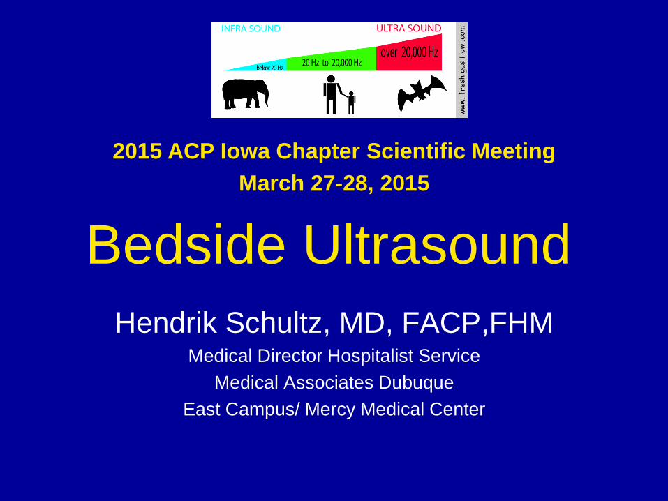
Bedside Ultrasound Hendrik Schultz, MD, FACP,FHM
Medical Director Hospitalist Service Medical Associates Dubuque
East Campus/ Mercy Medical Center
2015 ACP Iowa Chapter Scientific Meeting March 27-28, 2015
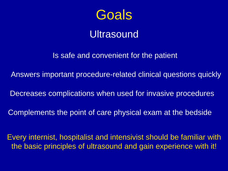
Goals Ultrasound
Complements the point of care physical exam at the bedside
Answers important procedure-related clinical questions quickly
Decreases complications when used for invasive procedures
Is safe and convenient for the patient
Every internist, hospitalist and intensivist should be familiar with the basic principles of ultrasound and gain experience with it!
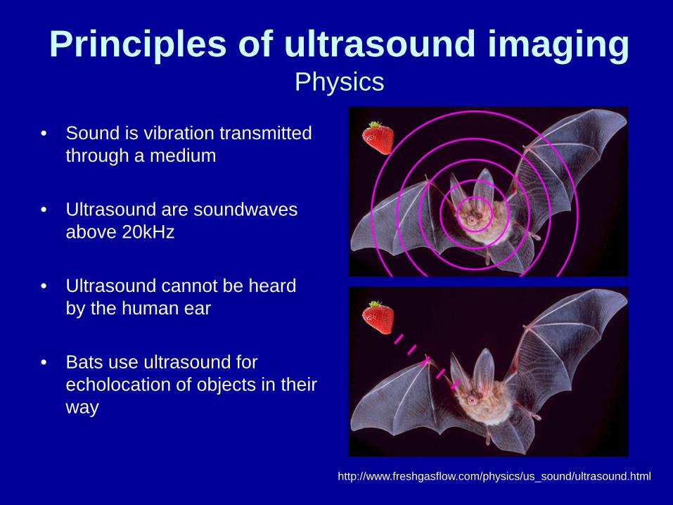
Principles of ultrasound imaging Physics
• Sound is vibration transmitted
through a medium
• Ultrasound are soundwaves above 20kHz
• Ultrasound cannot be heard by the human ear
• Bats use ultrasound for echolocation of objects in their way
http://www.freshgasflow.com/physics/us_sound/ultrasound.html
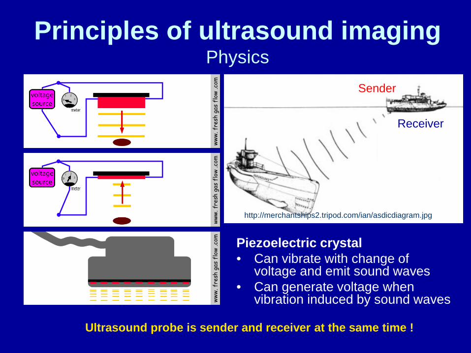
Principles of ultrasound imaging Physics
Piezoelectric crystal • Can vibrate with change of
voltage and emit sound waves • Can generate voltage when
vibration induced by sound waves
http://merchantships2.tripod.com/ian/asdicdiagram.jpg
Sender
Receiver
Ultrasound probe is sender and receiver at the same time !
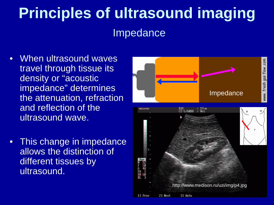
Principles of ultrasound imaging Impedance
• When ultrasound waves travel through tissue its density or “acoustic impedance” determines the attenuation, refraction and reflection of the ultrasound wave.
• This change in impedance allows the distinction of different tissues by ultrasound.
Impedance
http://www.medison.ru/uzi/img/p4.jpg
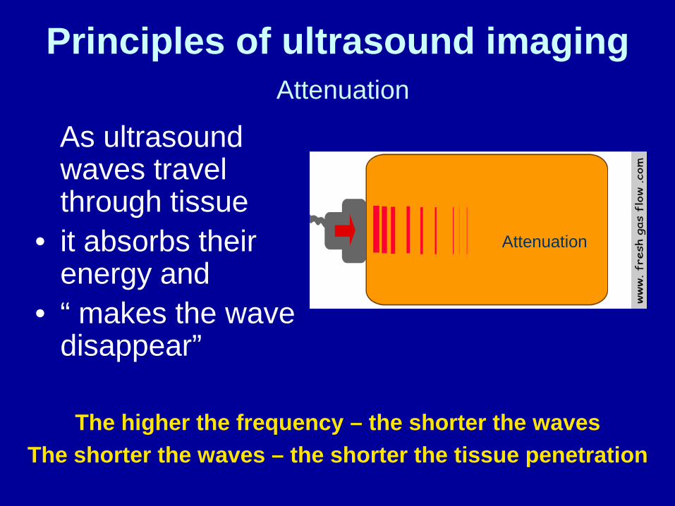
Principles of ultrasound imaging Attenuation
As ultrasound waves travel through tissue
• it absorbs their energy and
• “ makes the wave disappear”
Attenuation
The higher the frequency – the shorter the waves The shorter the waves – the shorter the tissue penetration
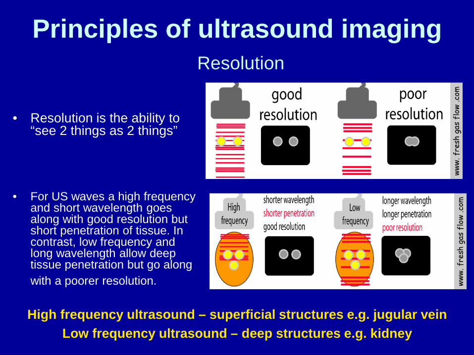
Principles of ultrasound imaging Resolution
• For US waves a high frequency and short wavelength goes along with good resolution but short penetration of tissue. In contrast, low frequency and long wavelength allow deep tissue penetration but go along with a poorer resolution.
High frequency ultrasound – superficial structures e.g. jugular vein Low frequency ultrasound – deep structures e.g. kidney
• Resolution is the ability to “see 2 things as 2 things”
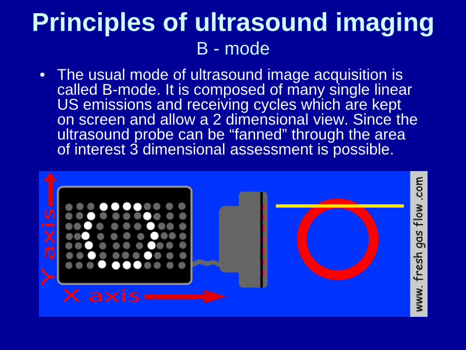
Principles of ultrasound imaging B - mode
• The usual mode of ultrasound image acquisition is called B-mode. It is composed of many single linear US emissions and receiving cycles which are kept on screen and allow a 2 dimensional view. Since the ultrasound probe can be “fanned” through the area of interest 3 dimensional assessment is possible.
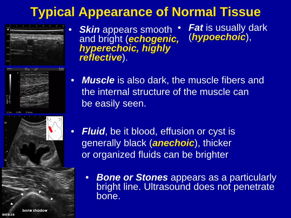
Typical Appearance of Normal Tissue • Skin appears smooth
and bright (echogenic, hyperechoic, highly reflective).
• Fat is usually dark (hypoechoic),
• Muscle is also dark, the muscle fibers and the internal structure of the muscle can be easily seen.
http://xray.net.au/ultrasoundpicture.jpg
• Fluid, be it blood, effusion or cyst is generally black (anechoic), thicker or organized fluids can be brighter
• Bone or Stones appears as a particularly bright line. Ultrasound does not penetrate bone.
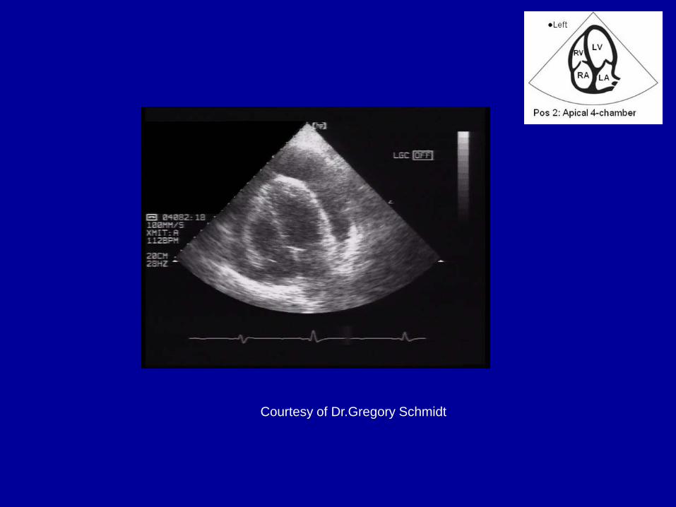
Let’s do some ultrasound!

QUIZ
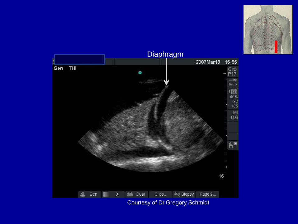
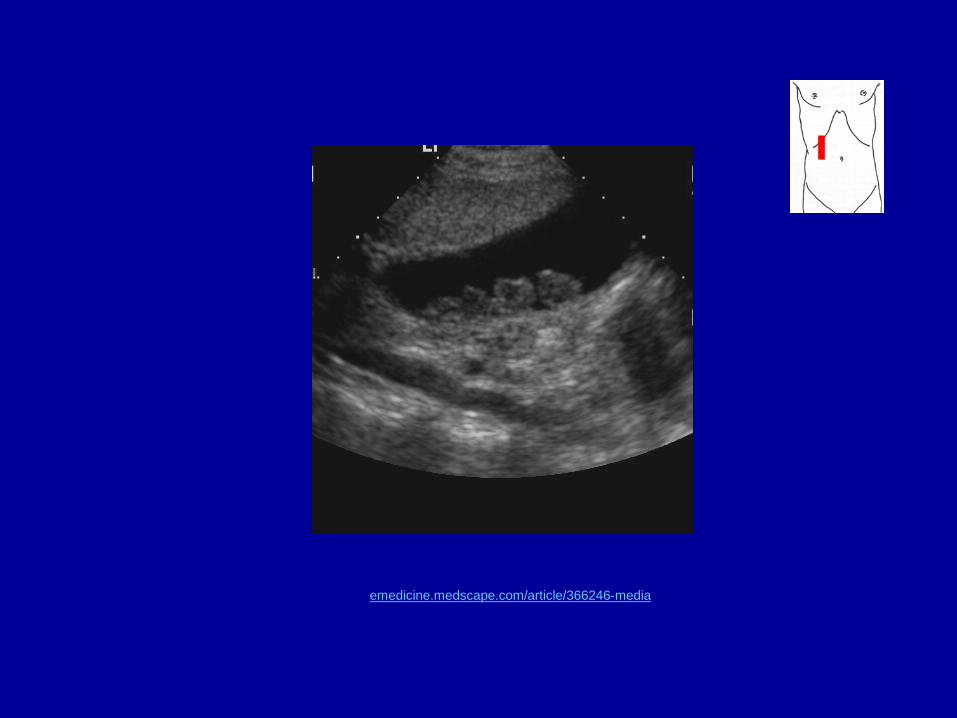
Courtesy of Dr.Gregory Schmidt
Diaphragm
Courtesy of Dr.Gregory Schmidt
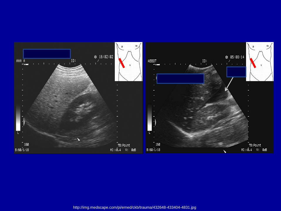
http://img.medscape.com/pi/emed/ckb/trauma/432648-433404-4831.jpg
Fluid
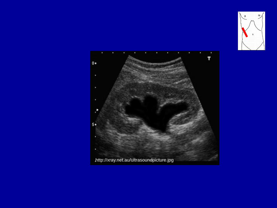
http://xray.net.au/ultrasoundpicture.jpg
References and suggested literature
• K.S.Cosby and J.L. Kendall: Practical guide to emergency ultrasound. Lippincott Williams and Wilkins 2006
• Mayo, PH, et al: Chest 2009;135:1050-1060 • American College of Emergency Physicians: Emergency
Ultrasound guidelines. ACEP 2008 • Martin LD, et.al: Am J Med 122:35, 2009 • Lucas BP, et.al: J Hosp Med 4:340, 2009 • Alpert JS, et.al: Am J Med 122: 1-3, 2009 • Brook OR, et.al: J Ultrasound Med 28:749, 2009 • Leung J, et.al: Ann Emerg Med 48:540, 2006 • Brennan JM, et.al: Am J Cardiol 99:1614, 2007 • Karakitsos D, et.al: Crit Care 10:175, 2006

Appendix
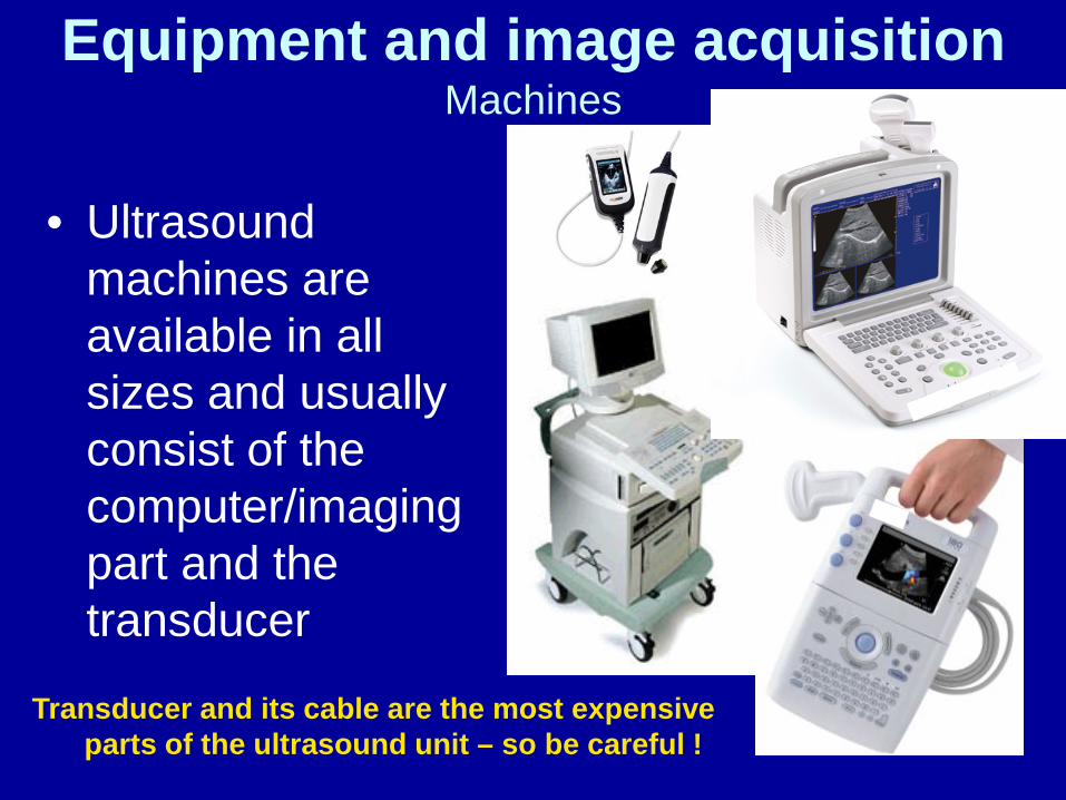
Equipment and image acquisition Machines
• Ultrasound
machines are available in all sizes and usually consist of the computer/imaging part and the transducer
Transducer and its cable are the most expensive parts of the ultrasound unit – so be careful !
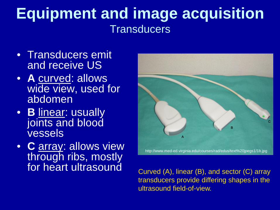
Equipment and image acquisition Transducers
• Transducers emit
and receive US • A curved: allows
wide view, used for abdomen
• B linear: usually joints and blood vessels
• C array: allows view through ribs, mostly for heart ultrasound
http://www.med-ed.virginia.edu/courses/rad/edus/text%20jpegs1/1b.jpg
Curved (A), linear (B), and sector (C) array transducers provide differing shapes in the ultrasound field-of-view.
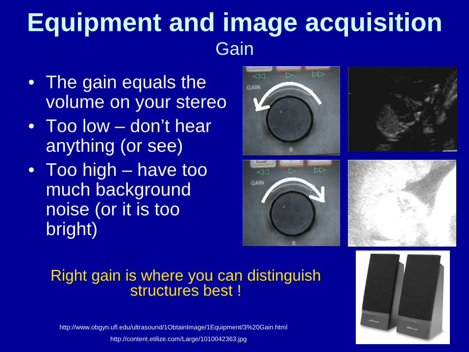
Equipment and image acquisition Gain
• The gain equals the volume on your stereo
• Too low – don’t hear anything (or see)
• Too high – have too much background noise (or it is too bright)
http://www.obgyn.ufl.edu/ultrasound/1ObtainImage/1Equipment/3%20Gain.html
http://content.etilize.com/Large/1010042363.jpg
Right gain is where you can distinguish structures best !
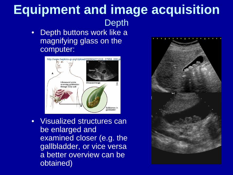
Equipment and image acquisition Depth
• Depth buttons work like a magnifying glass on the computer:
• Visualized structures can be enlarged and examined closer (e.g. the gallbladder, or vice versa a better overview can be obtained)
http://www.hopkins-gi.org/Upload/200904271319_27959_000.jpg
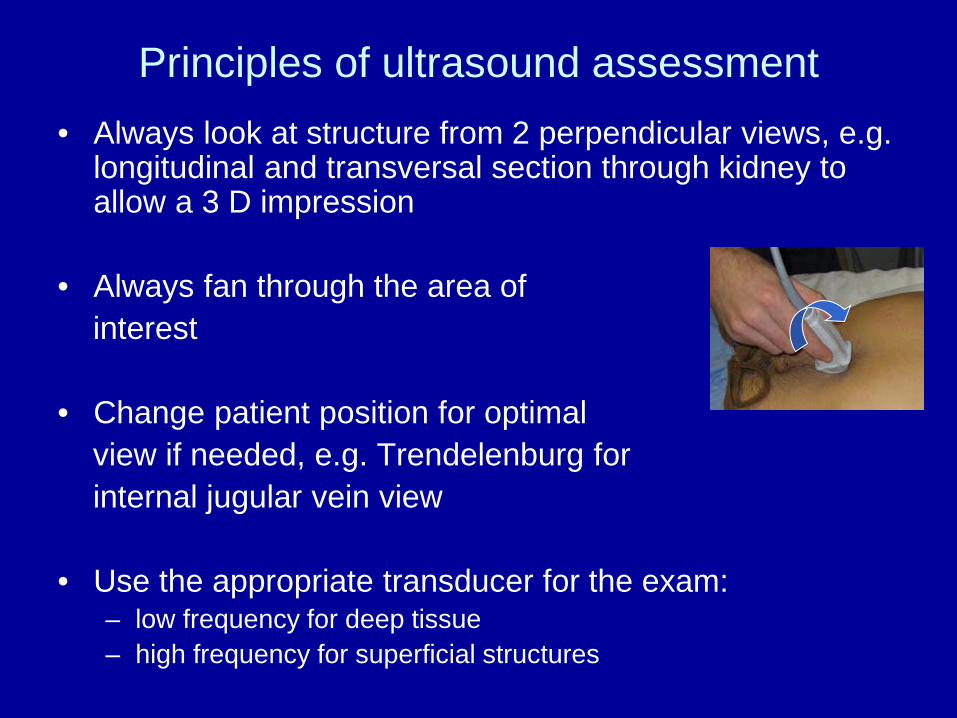
Principles of ultrasound assessment • Always look at structure from 2 perpendicular views, e.g.
longitudinal and transversal section through kidney to allow a 3 D impression
• Always fan through the area of interest • Change patient position for optimal view if needed, e.g. Trendelenburg for internal jugular vein view • Use the appropriate transducer for the exam:
– low frequency for deep tissue – high frequency for superficial structures
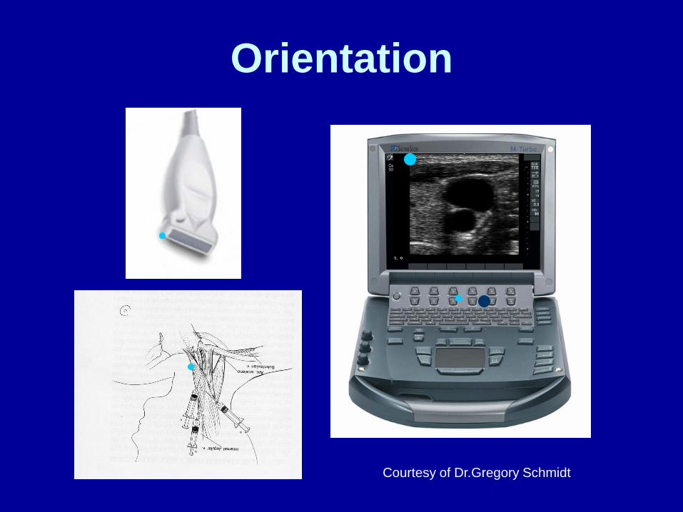
Orientation
Courtesy of Dr.Gregory Schmidt
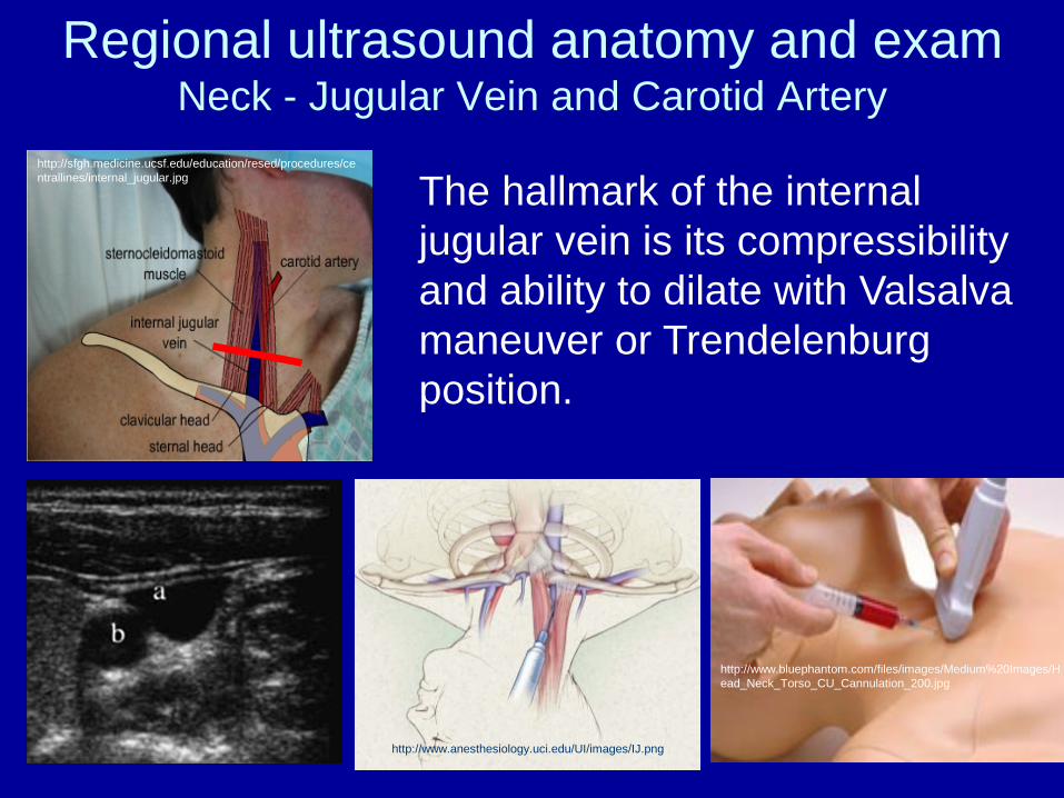
Regional ultrasound anatomy and exam Neck - Jugular Vein and Carotid Artery
http://www.bluephantom.com/files/images/Medium%20Images/Head_Neck_Torso_CU_Cannulation_200.jpg
http://www.anesthesiology.uci.edu/UI/images/IJ.png
http://sfgh.medicine.ucsf.edu/education/resed/procedures/centrallines/internal_jugular.jpg The hallmark of the internal
jugular vein is its compressibility and ability to dilate with Valsalva maneuver or Trendelenburg position.
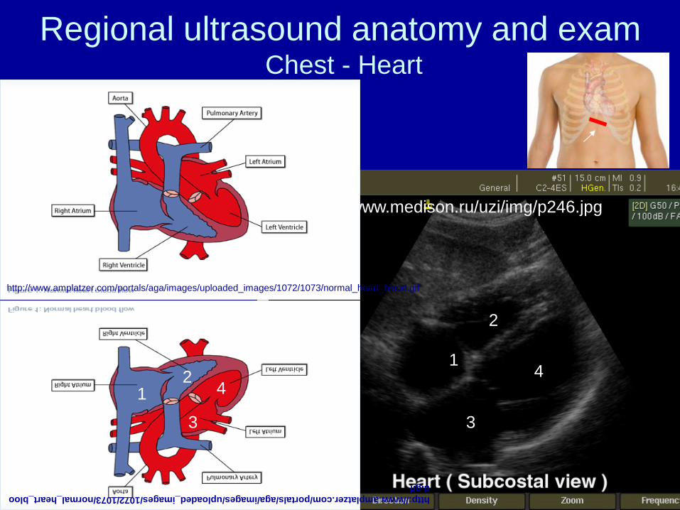
Regional ultrasound anatomy and exam Chest - Heart
http://www.medison.ru/uzi/img/p246.jpg
2
3
4 1
http://www.amplatzer.com/portals/aga/images/uploaded_images/1072/1073/normal_heart_blood.gif
1 2
3
4
http://www.amplatzer.com/portals/aga/images/uploaded_images/1072/1073/normal_heart_blood.gif
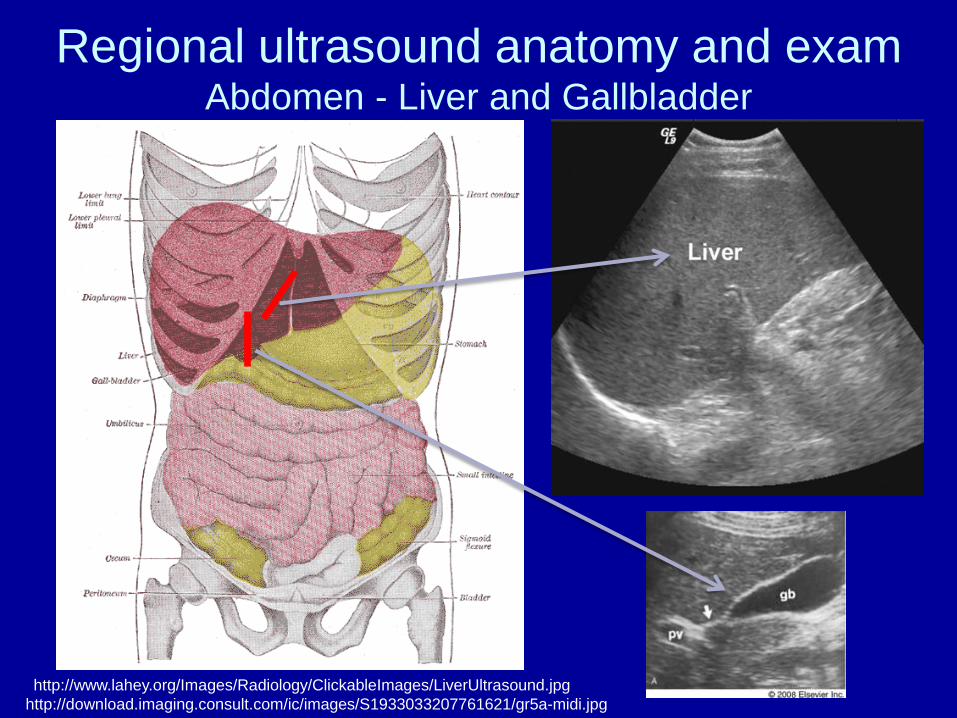
Regional ultrasound anatomy and exam Abdomen - Liver and Gallbladder
http://www.lahey.org/Images/Radiology/ClickableImages/LiverUltrasound.jpg http://download.imaging.consult.com/ic/images/S1933033207761621/gr5a-midi.jpg
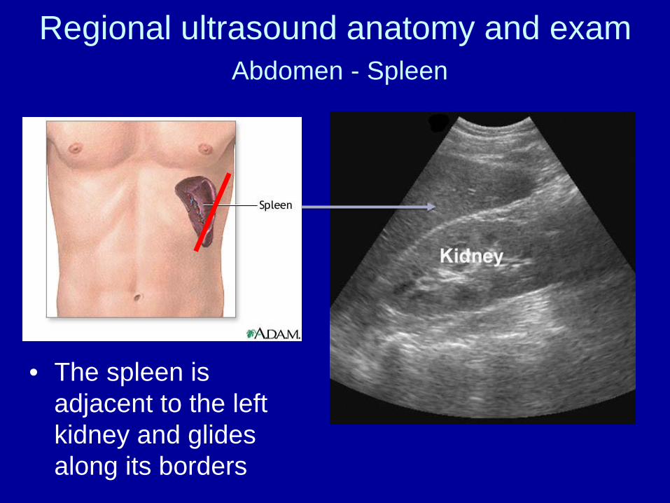
Regional ultrasound anatomy and exam Abdomen - Spleen
• The spleen is adjacent to the left kidney and glides along its borders
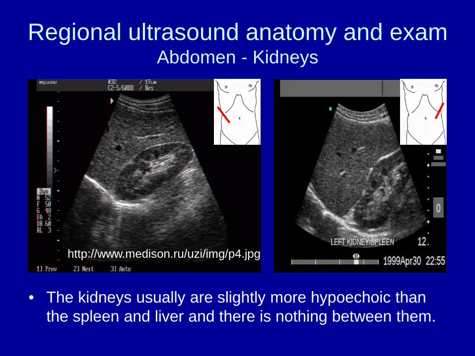
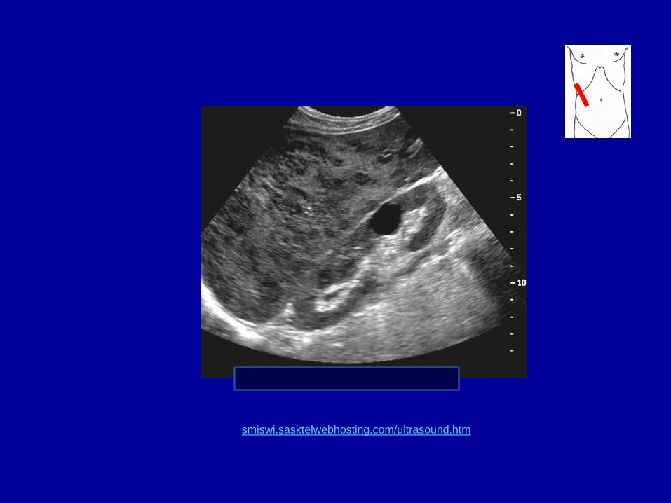
Regional ultrasound anatomy and exam Abdomen - Kidneys
• The kidneys usually are slightly more hypoechoic than the spleen and liver and there is nothing between them.
http://www.medison.ru/uzi/img/p4.jpg
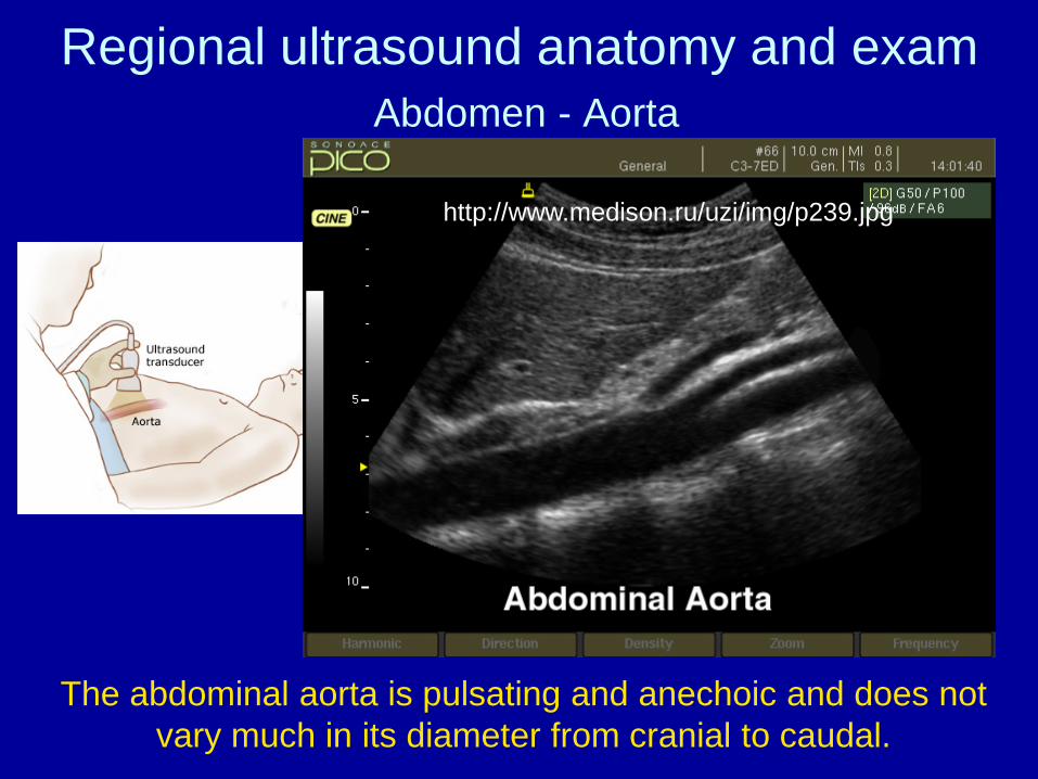
Regional ultrasound anatomy and exam Abdomen - Aorta
http://www.medison.ru/uzi/img/p239.jpg
The abdominal aorta is pulsating and anechoic and does not vary much in its diameter from cranial to caudal.