Embed Size (px)
Citation preview

1© 2014 Wiley-VCH Verlag GmbH & Co. KGaA, Weinheim wileyonlinelibrary.com
Enhanced Two-Photon Fluorescence Imaging and Therapy of Cancer Cells via Gold@Bridged Silsesquioxane Nanoparticles
Jonas Croissant , Marie Maynadier , Olivier Mongin , Vincent Hugues , Mireille Blanchard-Desce , * Arnaud Chaix , Xavier Cattoën , Michel Wong Chi Man , Audrey Gallud , Magali Gary-Bobo , * Marcel Garcia , Laurence Raehm , and Jean-Olivier Durand *
Two-photon excited photodynamic therapy (TPE-PDT)
offers promising perspective for cancer treatment [ 1 ] thanks
to the deep penetration of the two-photon light in the near-
infrared region and the unique spatial resolution it provides.
In particular, the use of two-photon excitable nanoparticles
(NPs) for cancer diagnosis and therapy would offer new
opportunities in nanomedicine. TPE-PDT combined with
NPs is indeed a new area of investigation which is still in
its infancy. [ 2,3 ] In most of the studies, either a porphyrin or
a chlorin derivative was indirectly activated by FRET from
an antenna designed for two-photon absorption. [ 4–11 ] Alter-
natively, a photosensitizer designed for its high two-photon
absorption cross-section can be incorporated within the
framework of silica-based nanoparticles. [ 12,13 ] However, this
approach is much less exploited due to the diffi culties in
Photodynamic Therapy
DOI: 10.1002/smll.201401759
J. Croissant, A. Chaix, Dr. X. Cattoën, Dr. M. Wong Chi Man, Dr. L. Raehm, Dr. J.-O. Durand Institut Charles Gerhardt Montpellier, UMR-5253 CNRS-UM2-ENSCM-UM1 cc 1701, Place Eugène Bataillon F-34095 , Montpellier cedex 05 , France E-mail: [email protected]
Dr. O. Mongin Institut Des Sciences Chimiques de Rennes CNRS UMR 6226 Université Rennes 1 Campus Beaulieu F-35042 , Rennes Cedex , France
V. Hugues, Dr. M. Blanchard-Desce Univ. Bordeaux 1 Institut des Sciences Moléculaires UMR CNRS 5255, 351 Cours de la Libération F-33405 , Talence Cedex , France E-mail: [email protected]
Dr. M. Maynadier, A. Gallud, Dr. M. Gary-Bobo, Dr. M. Garcia Institut des Biomolécules Max Mousseron, UMR 5247 CNRS UM 1; UM 2 – Faculté de Pharmacie 15 Avenue Charles Flahault 34093 , Montpellier Cedex 05 , France E-mail: [email protected]
designing and synthesizing the two-photon photosensitizers.
The low probability associated with TPE requires using
molecules with very high two-photon effi ciencies, and maxi-
mizing the density of the two-photon absorbing fragments in
the NPs. To this aim, we focused our effort on bridged silses-
quioxanes NPs (BSNPs) which allow the highest loading rate
of regularly and covalently linked organic functionalities [ 14 ]
and which are directly prepared by the sol-gel process from
a single molecular organosilane possessing two or more tri-
alkoxysilyl groups. To date, very few reports deal with the
preparation of BSNPs due to the diffi culties of precisely
controlling the growth of the NPs during the sol-gel hydrol-
ysis-condensation of the activated organosilanes. Shea and
co-workers’ pioneering work led to BSNPs with photore-
sponsive, [ 15,16 ] electrochromic [ 17 ] and electrochemical [ 18 ] prop-
erties and quite recently, Hammers and co-workers prepared
BSNPs from perylene diimide for photovoltaïc applications
using a modifi ed Stöber method. [ 19 ] To our knowledge, only a
single study has been reported concerning cancer therapy, [ 20 ]
with targeted BSNPs for platinum-based chemotherapy.
In the context of imaging, biodegradable BSNPs with high
Gd(III) payloads were recently designed for MRI of cancer
cells in vitro. [ 21 ] BSNPs thus constitute a promising platform
for cancer imaging and therapy.
In this work we therefore prepared novel BSNPs based
on the design of a two-photon photosensitizer possessing four
triethoxysilyl groups for TPE imaging and PDT of cancer
cells. Moreover, gold core BS shell NPs (Au@BSNPs) and
BSNPs decorated by gold nanospheres (BS@AuNPs) were
designed to increase the two-photon properties of BSNPs.
The precursor and the nanomaterials physico-chemical and
photophysical properties were fully characterized via ver-
satile techniques. Furthermore, TPE-PDT and fl uorescence
imaging on cancer cells revealed a remarkable exaltation of
the two-photon properties of the NPs, which correlated with
the photophysical studies.
The diaminodiphenylbutadiene core structure of the
two-photon photosensitizer (2PS) precursor depicted in
Scheme 1 a was known to selectively photo-activate electron
transfer in bio processes under TPE, [ 22–24 ] but this property
small 2014, DOI: 10.1002/smll.201401759

J. Croissant et al.
2 www.small-journal.com
communications
© 2014 Wiley-VCH Verlag GmbH & Co. KGaA, Weinheim
has never been exploited for TPE-PDT. This 2PS molecule,
which exhibits four triethoxysilyl groups, was elaborated at
the gram scale in fi ve steps (see Figure S1), the last being the
[CuBr(PPh 3 ) 3 ]-catalyzed CuAAC click reaction with (3-azi-
dopropyl)triethoxysilane under mild conditions. [ 25 ] The 2PS
precursor was fully characterized via proton and carbon
nuclear magnetic resonance (NMR), Fourier-transform
infrared (FTIR), UV-visible, and mass spectroscopies (see SI,
and Figure S2). Besides, a non-alkoxysilylated reference of
the 2PS molecule (2PS Ref, see Figure 2 a and S3) was devel-
oped and fully characterized (Figure S4 and S5), in order to
study the photophysical properties of the 2PS fragment.
The BSNPs were prepared by a careful control of the
hydrolysis and polycondensation of the 2PS precursor in a
cetyltrimethylammonium bromide / sodium hydroxide / water
/ ethanol mixture at 60 °C for ten minutes. The presence of
EtOH in the procedure was critical to solubilize the 2PS and
to slow down the reaction kinetic in order to obtain and iso-
late the nanoparticles with the desired size and morphology.
The time and temperature of the reaction were also care-
fully optimized for this purpose. Notably, BSNPs could also
be obtained with a triethylamine catalyst in similar conditions
(ESI). Original BS@AuNPs were obtained by mixing gold
NPs [ 26 ] with these BSNPs, without the need of thiol linkers to
immobilize gold NPs on the surface of BSNPs. For the prep-
aration of Au@BSNPs, a one-pot procedure was developed.
First gold nanospheres were prepared in-situ [ 26 ] and stabilized
with a thin silica layer. The 2PS precursor was subsequently
added under the same conditions as for the BSNPs affording
the Au@BSNPs.
The BS, Au@BS and BS@Au nanostructures, compositions,
and properties were then characterized. Transmission elec-
tron microscopy (TEM) analysis on the three types of nano-
material revealed 150 nm nearly monodisperse spherical NPs
( Figure 1 a–c), as confi rmed by the scanning electron micros-
copy (SEM) images (Figure 1 d–e) and by dynamic light scat-
tering (DLS) size distributions (Figure S6). Au@BSNPs tended
to be slightly smaller and aggregated. The gold nanospheres
were readily visible on the surface of BS@AuNPs (Figure 1 b),
or within the BS nanoshells (Figure 1 c). Moreover, 29 Si and
13 C CP-MAS NMR spectra confi rmed both the formation of
the siloxane inorganic network with a high condensation of
the 2PS precursor and the preservation of the organic moiety,
as shown by the major proportion of T 3 silicon environ-
ments (Figure 1 g–h) and the carbon environments of the 2PS
(Figure S7). This is further supported by the FTIR spectra of
the BSNPs and Au@BSNPs nanomaterials with the red-shift of
the ν Si-O band from 1080 cm −1 in the 2PS precursor to 1137 cm −1
in the materials (Figure S8). The gold content of Au@BSNPs
and BS@AuNPs were assessed by induced-coupled plasma
and energy dispersive spectroscopy respectively and were
found to be of 24 and 25% in weight (Table S1).
small 2014, DOI: 10.1002/smll.201401759
Scheme 1. A novel two-photon sensitizer (2PS) (a) condensing via sol-gel chemistry in BSNPs, and in Au@BSNPs or BS@AuNPs with the combination of gold nanospheres (b). The presence of gold exalts the nanomaterials TPE-PDT and fl uorescence imaging (c).
Figure 1. Representation of BSNPs, BS@AuNPs, and Au@BSNPs along with their TEM (a–c) and SEM images (d–f). Solid state 29 Si CPMAS NMR spectra of BSNPs (g) and Au@BSNPs (h).
Enhanced Two-Photon Fluorescence Imaging and Therapy of Cancer Cells
3www.small-journal.com© 2014 Wiley-VCH Verlag GmbH & Co. KGaA, Weinheim
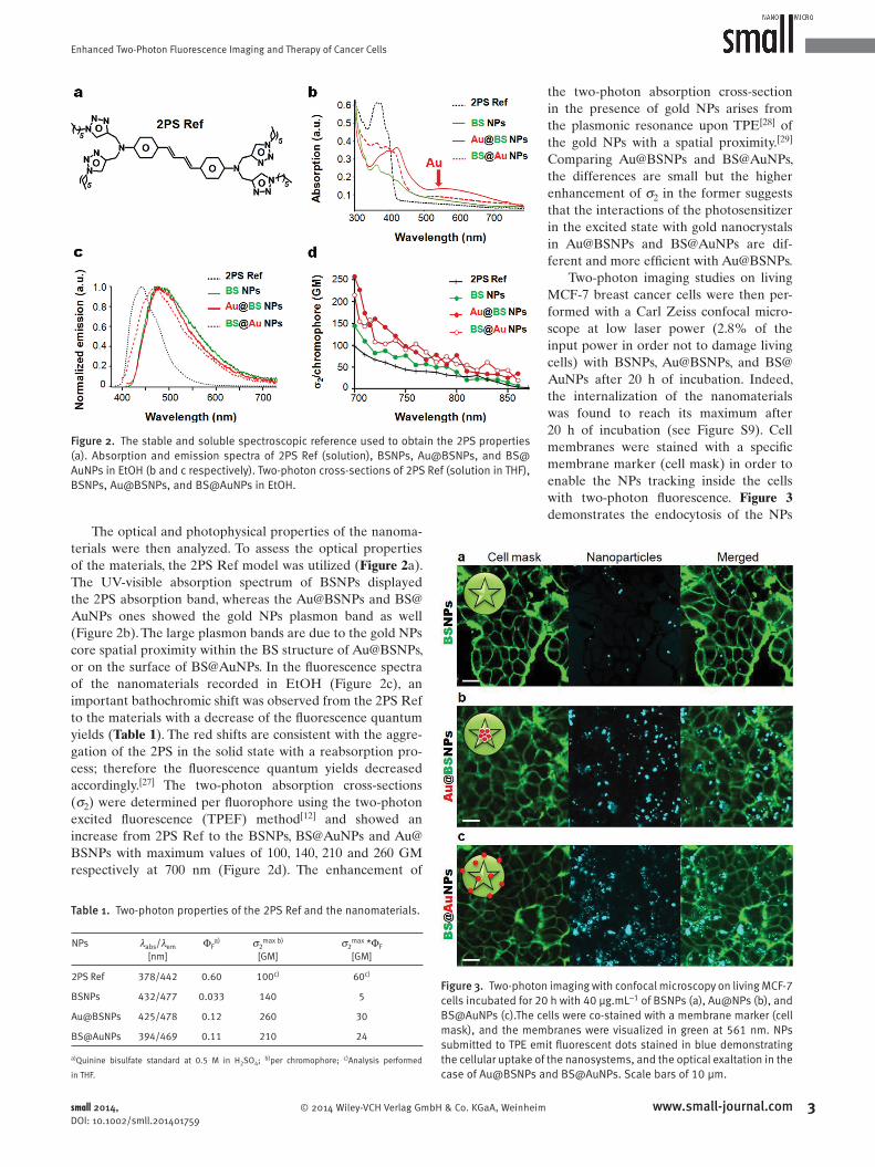
The optical and photophysical properties of the nanoma-
terials were then analyzed. To assess the optical properties
of the materials, the 2PS Ref model was utilized ( Figure 2 a).
The UV-visible absorption spectrum of BSNPs displayed
the 2PS absorption band, whereas the Au@BSNPs and BS@
AuNPs ones showed the gold NPs plasmon band as well
(Figure 2 b). The large plasmon bands are due to the gold NPs
core spatial proximity within the BS structure of Au@BSNPs,
or on the surface of BS@AuNPs. In the fl uorescence spectra
of the nanomaterials recorded in EtOH (Figure 2 c), an
important bathochromic shift was observed from the 2PS Ref
to the materials with a decrease of the fl uorescence quantum
yields ( Table 1 ). The red shifts are consistent with the aggre-
gation of the 2PS in the solid state with a reabsorption pro-
cess; therefore the fl uorescence quantum yields decreased
accordingly. [ 27 ] The two-photon absorption cross-sections
(σ 2 ) were determined per fl uorophore using the two-photon
excited fl uorescence (TPEF) method [ 12 ] and showed an
increase from 2PS Ref to the BSNPs, BS@AuNPs and Au@
BSNPs with maximum values of 100, 140, 210 and 260 GM
respectively at 700 nm (Figure 2 d). The enhancement of
the two-photon absorption cross-section
in the presence of gold NPs arises from
the plasmonic resonance upon TPE [ 28 ] of
the gold NPs with a spatial proximity. [ 29 ]
Comparing Au@BSNPs and BS@AuNPs,
the differences are small but the higher
enhancement of σ 2 in the former suggests
that the interactions of the photosensitizer
in the excited state with gold nanocrystals
in Au@BSNPs and BS@AuNPs are dif-
ferent and more effi cient with Au@BSNPs.
Two-photon imaging studies on living
MCF-7 breast cancer cells were then per-
formed with a Carl Zeiss confocal micro-
scope at low laser power (2.8% of the
input power in order not to damage living
cells) with BSNPs, Au@BSNPs, and BS@
AuNPs after 20 h of incubation. Indeed,
the internalization of the nanomaterials
was found to reach its maximum after
20 h of incubation (see Figure S9). Cell
membranes were stained with a specifi c
membrane marker (cell mask) in order to
enable the NPs tracking inside the cells
with two-photon fl uorescence. Figure 3
demonstrates the endocytosis of the NPs
small 2014, DOI: 10.1002/smll.201401759
Figure 2. The stable and soluble spectroscopic reference used to obtain the 2PS properties (a). Absorption and emission spectra of 2PS Ref (solution), BSNPs, Au@BSNPs, and BS@AuNPs in EtOH (b and c respectively). Two-photon cross-sections of 2PS Ref (solution in THF), BSNPs, Au@BSNPs, and BS@AuNPs in EtOH.
Table 1. Two-photon properties of the 2PS Ref and the nanomaterials.
NPs λ abs /λ em [nm]
Φ F a) σ 2 max b)
[GM]σ 2 max *Φ F
[GM]
2PS Ref 378/442 0.60 100 c) 60 c)
BSNPs 432/477 0.033 140 5
Au@BSNPs 425/478 0.12 260 30
BS@AuNPs 394/469 0.11 210 24
a) Quinine bisulfate standard at 0.5 M in H 2 SO 4 ; b) per chromophore; c) Analysis performed
in THF.
Figure 3. Two-photon imaging with confocal microscopy on living MCF-7 cells incubated for 20 h with 40 µg.mL −1 of BSNPs (a), Au@NPs (b), and BS@AuNPs (c).The cells were co-stained with a membrane marker (cell mask), and the membranes were visualized in green at 561 nm. NPs submitted to TPE emit fl uorescent dots stained in blue demonstrating the cellular uptake of the nanosystems, and the optical exaltation in the case of Au@BSNPs and BS@AuNPs. Scale bars of 10 µm.

J. Croissant et al.
4 www.small-journal.com
communications
© 2014 Wiley-VCH Verlag GmbH & Co. KGaA, Weinheim
by cells. Comparing BSNPs, Au@BSNPs, and BS@AuNPs at
the same laser power, an enhancement of the TPE-excited
fl uorescence was observed with both gold-doped nanosys-
tems (Figure 3 b–c, see Figure S10). Indeed the quantum
yields and two-photon absorption cross-sections were higher
with the Au@BSNPs, and BS@AuNPs thus enhancing the
fl uorescence signal (see Table 1 ). Note that the lumines-
cence signal corresponds to the emission of the fl uorophore
(Figure S10), but a contribution of the gold NPs to the lumi-
nescence cannot be excluded.
Eventually, in vitro TPE therapy was performed on breast
cancer cells. MCF-7 cells were incubated for 20 h in the pres-
ence of BSNPs, Au@BSNPs or BS@AuNPs. No cytotoxicity
was observed without irradiation up to a concentration of
40 µg.mL −1 ( Figure 4 ) showing the biocompatibility of the pre-
pared nanomaterials. Additionally, we examined the infl uence
of the laser on epithelial normal cells (ARPE-19 cell line) which
confi rmed the harmlessness of the irradiation (see Figure S11).
On the contrary, when the MCF-7 cells were incubated with the
NPs and irradiated at 760 nm with 3 scans of 1.57 s at the max-
imum power of the laser (input power 3 W) and with a focused
laser beam, an important cancer cell death was observed at a
concentration of 40 µg.mL −1 where only 24%, 6% and 0% of
cancer cells survived respectively with BSNPs, Au@BSNPs,
and BS@AuNPs. The presence of gold again enhanced the effi -
ciency of the Au@BS and BS@AuNPs compared to BSNPs due
to a synergy between the 2PS and gold NPs. The cell survival
difference between Au@BSNPs and BS@AuNPs is not statisti-
cally signifi cant to assess the impact of the gold spatial position
in BSNPs on the PDT effect.
Note that 2PS Ref and the BSNPs were not able to pro-
duce singlet oxygen under irradiation (data not shown).
Therefore, we believe that a mechanism based on electron
transfer is involved in cancer cell killing with TPE-PDT. [ 30 ]
Photothermal effects have been shown to be negligible with
gold nanorods upon TPE-PDT, [ 2 ] but a contribution of pho-
tothermal therapy cannot be excluded with our systems.
In conclusion, a novel two-photon photosensitizer was
reported, and the subsequent sol-gel process with or without
gold NPs led to BSNPs and gold-doped BSNPs. The phys-
ico-chemical properties of the precursor and the resulting
nanomaterials were fully characterized via complemen-
tary techniques. The two-photon fl uorescence quantum
yields of the nanomaterials were measured and two-photon
absorption cross-sections were higher with all the synthe-
sized NPs than with the precursor. Furthermore, such NPs
were applied on cancer cells as powerful multifunctional
nanosystems combining both TPE-PDT and ultra-bright
fl uorescence imaging. The addition of gold nanospheres to
the BSNPs matrix induced a remarkable exaltation of the
in vitro fl uorescence correlating the photophysical studies.
Finally, a clear gold core mediated-enhancement of 20–25%
in TPE-PDT cell killing was obtained with gold-doped BS
nanosystems when compared to BSNPs. Consequently,
BSNPs and gold-doped BSNPs are very promising cancer
theranostics and work is in progress to further exploit these
nanomaterials.
Experimental Section
BSNPs (with NaOH Catalyst) : A mixture composed of water (25 mL), ethanol (10 mL), and cetyltrimethylammonium bromide (0.160 g, 4.40.10 −1 mmol) was stirred at 80 °C in a 50 mL round-bottom fl ask. Then a solution of the 2PS precursor in anhydrous ethanol (2PS: 44.4 mg, 2.7.10 −2 mmol; EtOH: 500 µL) was quickly injected. After 1 minute, sodium hydroxide (NaOH, 150 µL (2 M)) was added to trigger the sol-gel process. The reaction mixture was stirred for 2 minutes at 80 °C, and the solution was then neutral-ized by addition of hydrochloric acid (HCl (0.2 M), 1.38 mL). Then, the mixture was cooled down to 40 °C, and the nanomaterial was collected through centrifugation at 21000 rpm for 15 minutes. Each fraction was washed with ethanol (40 mL per fraction) and centrifuged for 10 minutes. This operation was repeated twice, and the compound was dried under vacuum for few hours.
Two-Photon Photodynamic Therapy : Human breast cancer cells MCF-7 (purchased from ATCC) were cultured in DMEM supple-mented with 10% fetal bovine serum and 50 µg.mL −1 gentamycin. All cells were allowed to grow in humidifi ed atmosphere at 37 °C under 5% CO 2 . For in vitro phototoxicity, MCF-7 cells were seeded into a 384 multiwell glass-bottomed plate (thickness 0.17 mm), with a black polystyrene frame, 2000 cells per well in 50 µL of cul-ture medium, and allowed to grow for 24 h. BSNPs were then dis-persed under ultrasounds in PBS at a concentration of 1 mg.mL −1 and cells were then incubated for 20 h with or without BSNPs at a fi nal concentration of 40 µg.mL −1 in DMEM. After incubation with BSNPs, cells were washed twice, maintained in fresh culture medium, and then submitted (or not) to laser irradiation; with the Carl Zeiss Microscope (laser power input 3W). Half of the well was irradiated at 760 nm by three scans of 1.57 s duration in 4 dif-ferent areas of the well. The laser beam was focused by a micro-scope objective lens (Carl Zeiss 10x/0.3 EC Plan-Neofl uar). The scan size does not allow irradiating more areas without overlap-ping. After 2 days, the MTS assay was performed (as previously described [ 12 ] and was corrected according to the following formula Abs control −2 × (Abs control - Abs BS ).
small 2014, DOI: 10.1002/smll.201401759
Figure 4. TPE-PDT on MCF-7 cancer cells. The cells were incubated 20 h with 40 µg.mL −1 of BSNPs, Au@BSNPs, and BS@AuNPs. * n = 3, *p < 0.05 for treatment with Au@BSNPs or BS@AuNPs and irradiation compared to treatment with BSNPs using Student's T test.

Enhanced Two-Photon Fluorescence Imaging and Therapy of Cancer Cells
5www.small-journal.com© 2014 Wiley-VCH Verlag GmbH & Co. KGaA, Weinheimsmall 2014, DOI: 10.1002/smll.201401759
Supporting Information
Supporting Information is available from the Wiley Online Library or from the author.
Acknowledgment
ANR P2N Mechanano is gratefully acknowledged. We gratefully thank Montpellier RIO imaging platform.
[1] J. R. Starkey , E. M. Pascucci , M. A. Drobizhev , A. Elliott , A. K. Rebane , Biochem. Biophys. Acta Gen. Subj. 2013 , 1830 , 4594 – 4603 .
[2] T. Zhao , X. Shen , L. Li , Z. Guan , N. Gao , P. Yuan , S. Q. Yao , Q.-H. Xu , G. Q. Xu , Nanoscale 2012 , 4 , 7712 – 7719 .
[3] J. Qian , D. Wang , F. Cai , Q. Zhan , Y. Wang , S. He , Biomaterials 2012 , 33 , 4851 – 4860 .
[4] K.-L. Chou , N. Won , J. Kwag , S. Kim , J.-Y. Chen , J. Mater. Chem. B 2013 , 1 , 4584 – 4592 .
[5] C. Fowley , N. Nomikou , A. P. McHale , B. McCaughan , J. F. Callan , Chem. Commun. 2013 , 49 , 8934 – 8936 .
[6] C. Fowley , N. Nomikou , A. P. McHale , P. A. McCarron , B. McCaughan , J. F. Callan , J. Mater. Chem. 2012 , 22 , 6456 – 6462 .
[7] X. Shen , L. Li , H. Wu , S. Q. Yao , Q.-H. Xu , Nanoscale 2011 , 3 , 5140 – 5146 .
[8] Z.-D. Qi , D.-W. Li , P. Jiang , F.-L. Jiang , Y.-S. Li , Y. Liu , W.-K. Wong , K.-W. Cheah , J. Mater. Chem. 2011 , 21 , 2455 – 2458 .
[9] J. L. Grimland , C. Wu , R. R. Ramoutar , J. L. Brumaghim , J. McNeill , Nanoscale 2011 , 3 , 1451 – 1455 .
[10] S.-H. Cheng , C.-C. Hsieh , N.-T. Chen , C.-H. Chu , C.-M. Huang , P.-T. Chou , F.-G. Tseng , C.-S. Yang , C.-Y. Mou , L.-W. Lo , Nano Today 2011 , 6 , 552 – 563 .
[11] S. Kim , T. Y. Ohulchanskyy , H. E. Pudavar , R. K. Pandey , P. N. Prasad , J. Am. Chem. Soc. 2007 , 129 , 2669 – 2675 .
[12] M. Gary-Bobo , Y. Mir , C. Rouxel , D. Brevet , I. Basile , M. Maynadier , O. Vaillant , O. Mongin , M. Blanchard-Desce , A. Morere , M. Garcia ,
J.-O. Durand , L. Raehm , Angew. Chem. Int. Ed. 2011 , 50 , 11425 – 11429 .
[13] M. Velusamy , J. Y. Shen , J. T. Lin , Y. C. Lin , C. C. Hsieh , C. H. Lai , C. W. Lai , M. L. Ho , Y. C. Chen , P. T. Chou , J. K. Hsiao , Adv. Funct. Mater. 2009 , 19 , 2388 – 2397 .
[14] L.-C. Hu , K. J. Shea , Chem. Soc. Rev. 2011 , 40 , 688 – 695 . [15] L.-C. Hu , Y. Yonamine , S.-H. Lee , W. E. van der Veer , K. J. Shea ,
J. Am. Chem. Soc. 2012 , 134 , 11072 – 11075 . [16] L. Zhao , D. A. Loy , K. J. Shea , J. Am. Chem. Soc. 2006 , 128 ,
14250 – 14251 . [17] V. Jain , M. Khiterer , R. Montazami , H. M. Yochum , K. J. Shea ,
J. R. Hefl in , ACS Appl. Mater. Interfaces. 2009 , 1 , 83 – 89 . [18] M. Khiterer , K. J. Shea , Nano Lett. 2007 , 7 , 2684 – 2687 . [19] H. Rathnayake , J. Binion , A. McKee , D. J. Scardino , N. I. Hammer ,
Nanoscale 2012 , 4 , 4631 – 4640 . [20] J. Della Rocca , R. C. Huxford , E. Comstock-Duggan , W. Lin , Angew.
Chem. Int. Ed. 2011 , 50 , 10330 – 10334 . [21] J. L. Vivero-Escoto , W. J. Rieter , H. Lau , R. C. Huxford-Phillips ,
W. Lin , Small 2013 , 9 , 3523 – 3531 . [22] E. Beaumont , J. C. Lambry , M. Blanchard-Desce , P. Martasek ,
S. P. Panda , E. E. H. van Faassen , J. C. Brochon , E. Deprez , A. Slama-Schwok , ChemBioChem 2009 , 10 , 690 – 701 .
[23] E. Beaumont , J. C. Lambry , A. C. Robin , P. Martasek , M. Blanchard-Desce , A. Slama-Schwok , ChemPhysChem 2008 , 9 , 2325 – 2331 .
[24] A. C. Robin , S. Gmouh , O. Mongin , V. Jouikov , M. H. V. Werts , C. Gautier , A. Slama-Schwok , M. Blanchard-Desce , Chem. Commun. 2007 , 1334 – 1336 .
[25] N. Moitra , J. J. E. Moreau , X. Cattoën , M. Wong Chi Man , Chem. Commun. 2010 , 46 , 8416 – 8418 .
[26] J. Croissant , J. I. Zink , J. Am. Chem. Soc. 2012 , 134 , 7628 – 7631 . [27] K. Natte , T. Behnke , G. Orts-Gil , C. Wuerth , J. F. Friedrich ,
W. Oesterle , U. Resch-Genger , J. Nanopart. Res. 2012 , 14 , 680 . [28] S. T. Sivapalan , J. H. Vella , T. K. Yang , M. J. Dalton , R. N. Swiger ,
J. E. Haley , T. M. Cooper , A. M. Urbas , L.-S. Tan , C. J. Murphy , Lang-muir 2012 , 28 , 9147 – 9154 .
[29] C. Jiang , T. Zhao , P. Yuan , N. Gao , Y. Pan , Z. Guan , N. Zhou , Q.-H. Xu , ACS Appl. Mater. Interfaces. 2013 , 5 , 4972 – 4977 .
[30] Y. Mir , J. E. van Lier , B. Paquette , D. Houde , Photochem. Photobiol. 2008 , 84 , 1182 – 1186 .
Received: June 17, 2014 Revised: July 28, 2014 Published online: