Embed Size (px)
Citation preview
Nishimura, RA et al. 2014 AHA/ACC Valvular Heart Disease Guideline
Page 1 of 96
2014 AHA/ACC Guideline for the Management of Patients With Valvular Heart Disease: Executive Summary
A Report of the American College of Cardiology/American Heart Association Task Force on Practice Guidelines
Developed in Collaboration With the American Association for Thoracic Surgery, American Society of
Echocardiography, Society for Cardiovascular Angiography and Interventions, Society of Cardiovascular Anesthesiologists, and Society of Thoracic Surgeons
WRITING COMMITTEE MEMBERS*
Rick A. Nishimura, MD, MACC, FAHA, Co-Chair† Catherine M. Otto, MD, FACC, FAHA, Co-Chair†
Robert O. Bonow, MD, MACC, FAHA† Carlos E. Ruiz, MD, PhD, FACC† Blase A. Carabello, MD, FACC*† Nikolaos J. Skubas, MD, FASE¶ John P. Erwin III, MD, FACC, FAHA‡ Paul Sorajja, MD, FACC, FAHA# Robert A. Guyton, MD, FACC*§ Thoralf M. Sundt III, MD* **†† Patrick T. O’Gara, MD, FACC, FAHA† James D. Thomas, MD, FASE, FACC, FAHA‡‡
ACC/AHA TASK FORCE MEMBERS
Jeffrey L. Anderson, MD, FACC, FAHA, Chair
Jonathan L. Halperin, MD, FACC, FAHA, Chair-Elect Nancy M. Albert, PhD, CCNS, CCRN, FAHA Judith S. Hochman, MD, FACC, FAHA Biykem Bozkurt, MD, PhD, FACC, FAHA Richard J. Kovacs, MD, FACC, FAHA Ralph G. Brindis, MD, MPH, MACC E. Magnus Ohman, MD, FACC Mark A. Creager, MD, FACC, FAHA§§ Susan J. Pressler, PhD, RN, FAHA Lesley H. Curtis, PhD, FAHA Frank W. Sellke, MD, FACC, FAHA David DeMets, PhD Win-Kuang Shen, MD, FACC, FAHA Robert A. Guyton, MD, FACC§§ William G. Stevenson, MD, FACC, FAHA§§
Clyde W. Yancy, MD, FACC, FAHA§§ *Writing committee members are required to recuse themselves from voting on sections to which their specific relationships with industry and other entities may apply; see Appendix 1 for recusal information. †ACC/AHA representative. ‡ACC/AHA Task Force on Performance Measures liaison. §ACC/AHA Task Force on Practice Guidelines liaison. ¶Society of Cardiovascular Anesthesiologists representative. #Society for Cardiovascular Angiography and Interventions representative. **American Association for Thoracic Surgery representative. ††Society of Thoracic Surgeons representative. ‡‡American Society of Echocardiography representative. §§Former Task Force member during the writing effort.
by guest on November 25, 2015http://circ.ahajournals.org/Downloaded from by guest on November 25, 2015http://circ.ahajournals.org/Downloaded from by guest on November 25, 2015http://circ.ahajournals.org/Downloaded from by guest on November 25, 2015http://circ.ahajournals.org/Downloaded from by guest on November 25, 2015http://circ.ahajournals.org/Downloaded from by guest on November 25, 2015http://circ.ahajournals.org/Downloaded from by guest on November 25, 2015http://circ.ahajournals.org/Downloaded from by guest on November 25, 2015http://circ.ahajournals.org/Downloaded from by guest on November 25, 2015http://circ.ahajournals.org/Downloaded from by guest on November 25, 2015http://circ.ahajournals.org/Downloaded from by guest on November 25, 2015http://circ.ahajournals.org/Downloaded from by guest on November 25, 2015http://circ.ahajournals.org/Downloaded from by guest on November 25, 2015http://circ.ahajournals.org/Downloaded from by guest on November 25, 2015http://circ.ahajournals.org/Downloaded from by guest on November 25, 2015http://circ.ahajournals.org/Downloaded from by guest on November 25, 2015http://circ.ahajournals.org/Downloaded from by guest on November 25, 2015http://circ.ahajournals.org/Downloaded from by guest on November 25, 2015http://circ.ahajournals.org/Downloaded from by guest on November 25, 2015http://circ.ahajournals.org/Downloaded from by guest on November 25, 2015http://circ.ahajournals.org/Downloaded from by guest on November 25, 2015http://circ.ahajournals.org/Downloaded from by guest on November 25, 2015http://circ.ahajournals.org/Downloaded from by guest on November 25, 2015http://circ.ahajournals.org/Downloaded from by guest on November 25, 2015http://circ.ahajournals.org/Downloaded from by guest on November 25, 2015http://circ.ahajournals.org/Downloaded from by guest on November 25, 2015http://circ.ahajournals.org/Downloaded from by guest on November 25, 2015http://circ.ahajournals.org/Downloaded from by guest on November 25, 2015http://circ.ahajournals.org/Downloaded from by guest on November 25, 2015http://circ.ahajournals.org/Downloaded from by guest on November 25, 2015http://circ.ahajournals.org/Downloaded from by guest on November 25, 2015http://circ.ahajournals.org/Downloaded from by guest on November 25, 2015http://circ.ahajournals.org/Downloaded from by guest on November 25, 2015http://circ.ahajournals.org/Downloaded from by guest on November 25, 2015http://circ.ahajournals.org/Downloaded from by guest on November 25, 2015http://circ.ahajournals.org/Downloaded from by guest on November 25, 2015http://circ.ahajournals.org/Downloaded from by guest on November 25, 2015http://circ.ahajournals.org/Downloaded from by guest on November 25, 2015http://circ.ahajournals.org/Downloaded from by guest on November 25, 2015http://circ.ahajournals.org/Downloaded from by guest on November 25, 2015http://circ.ahajournals.org/Downloaded from by guest on November 25, 2015http://circ.ahajournals.org/Downloaded from by guest on November 25, 2015http://circ.ahajournals.org/Downloaded from by guest on November 25, 2015http://circ.ahajournals.org/Downloaded from by guest on November 25, 2015http://circ.ahajournals.org/Downloaded from by guest on November 25, 2015http://circ.ahajournals.org/Downloaded from by guest on November 25, 2015http://circ.ahajournals.org/Downloaded from by guest on November 25, 2015http://circ.ahajournals.org/Downloaded from by guest on November 25, 2015http://circ.ahajournals.org/Downloaded from by guest on November 25, 2015http://circ.ahajournals.org/Downloaded from by guest on November 25, 2015http://circ.ahajournals.org/Downloaded from by guest on November 25, 2015http://circ.ahajournals.org/Downloaded from by guest on November 25, 2015http://circ.ahajournals.org/Downloaded from by guest on November 25, 2015http://circ.ahajournals.org/Downloaded from by guest on November 25, 2015http://circ.ahajournals.org/Downloaded from by guest on November 25, 2015http://circ.ahajournals.org/Downloaded from by guest on November 25, 2015http://circ.ahajournals.org/Downloaded from by guest on November 25, 2015http://circ.ahajournals.org/Downloaded from by guest on November 25, 2015http://circ.ahajournals.org/Downloaded from by guest on November 25, 2015http://circ.ahajournals.org/Downloaded from by guest on November 25, 2015http://circ.ahajournals.org/Downloaded from by guest on November 25, 2015http://circ.ahajournals.org/Downloaded from by guest on November 25, 2015http://circ.ahajournals.org/Downloaded from by guest on November 25, 2015http://circ.ahajournals.org/Downloaded from by guest on November 25, 2015http://circ.ahajournals.org/Downloaded from by guest on November 25, 2015http://circ.ahajournals.org/Downloaded from by guest on November 25, 2015http://circ.ahajournals.org/Downloaded from by guest on November 25, 2015http://circ.ahajournals.org/Downloaded from by guest on November 25, 2015http://circ.ahajournals.org/Downloaded from by guest on November 25, 2015http://circ.ahajournals.org/Downloaded from by guest on November 25, 2015http://circ.ahajournals.org/Downloaded from by guest on November 25, 2015http://circ.ahajournals.org/Downloaded from by guest on November 25, 2015http://circ.ahajournals.org/Downloaded from by guest on November 25, 2015http://circ.ahajournals.org/Downloaded from by guest on November 25, 2015http://circ.ahajournals.org/Downloaded from by guest on November 25, 2015http://circ.ahajournals.org/Downloaded from
Nishimura, RA et al. 2014 AHA/ACC Valvular Heart Disease Guideline
Page 2 of 96
Full-text guideline available at: Circulation. 2014;129:xxx-xxx. This document was approved by the American College of Cardiology Board of Trustees and the American Heart Association Science Advisory and Coordinating Committee in January 2014. The online-only Data Supplement is available with this article at
http://circ.ahajournals.org/lookup/suppl/doi:10.1161/CIR.0000000000000029/-/DC1.
The online-only Comprehensive Relationships With Industry table is available with this article at
http://circ.ahajournals.org/lookup/suppl/doi:10.1161/CIR.0000000000000029/-/DC2.
The American Heart Association requests that this document be cited as follows: Nishimura RA, Otto CM, Bonow RO, Carabello BA, Erwin JP III, Guyton RA, O’Gara PT, Ruiz CE, Skubas NJ, Sorajja P, Sundt TM III, Thomas JD. 2014 AHA/ACC guideline for the management of patients with valvular heart disease: executive summary: a report of the American College of Cardiology/American Heart Association Task Force on Practice Guidelines. Circulation. 2014; 129:–. This article has been copublished in the Journal of the American College of Cardiology. Copies: This document is available on the World Wide Web sites of the American College of Cardiology (www.cardiosource.org) and the American Heart Association (my.americanheart.org). A copy of the document is available at http://my.americanheart.org/statements by selecting either the “By Topic” link or the “By Publication Date” link. To purchase additional reprints, call 843-216-2533 or e-mail [email protected]. Expert peer review of AHA Scientific Statements is conducted by the AHA Office of Science Operations. For more on AHA statements and guidelines development, visit http://my.americanheart.org/statements and select the “Policies and Development” link. Permissions: Multiple copies, modification, alteration, enhancement, and/or distribution of this document are not permitted without the express permission of the American Heart Association. Instructions for obtaining permission are located at http://www.heart.org/HEARTORG/General/Copyright-Permission-Guidelines_UCM_300404_Article.jsp. A link to the “Copyright Permissions Request Form” appears on the right side of the page. (Circulation. 2014;129:000–000.) © 2014 by the American Heart Association, Inc., and the American College of Cardiology Foundation.
by guest on November 25, 2015http://circ.ahajournals.org/Downloaded from
Nishimura, RA et al. 2014 AHA/ACC Valvular Heart Disease Guideline
Page 3 of 96
Table of Contents Preamble ................................................................................................................................................................................... 5 1. Introduction .......................................................................................................................................................................... 9
1.1. Methodology and Evidence Review ............................................................................................................................. 9 1.2. Organization of the Writing Committee ....................................................................................................................... 9 1.3. Document Review and Approval .................................................................................................................................. 9 1.4. Scope of the Guideline ............................................................................................................................................... 10
2. General Principles .............................................................................................................................................................. 11 2.1. Evaluation of the Patient With Suspected VHD ......................................................................................................... 11 2.2. Definitions of Severity of Valve Disease ................................................................................................................... 11 2.3. Diagnostic TestingDiagnosis and Follow-Up: Recommendations ......................................................................... 12 2.4. Basic Principles of Medical Therapy: Recommendations .......................................................................................... 13 2.5. Evaluation of Surgical and Interventional Risk .......................................................................................................... 14 2.6. The Heart Valve Team and Heart Valve Centers of Excellence: Recommendations ................................................. 14
3. Aortic Stenosis: Recommendations .................................................................................................................................... 16 3.1. Stages of Valvular AS ................................................................................................................................................ 16 3.2. Diagnosis and Follow-Up ........................................................................................................................................... 19 3.3. Medical Therapy ......................................................................................................................................................... 19 3.4. Timing of Intervention ................................................................................................................................................ 19 3.5. Choice of Intervention ................................................................................................................................................ 22
4. Aortic Regurgitation: Recommendations ........................................................................................................................... 23 4.1. Stages of Chronic Aortic Regurgitation ...................................................................................................................... 23 4.2. Diagnosis and Follow-Up ........................................................................................................................................... 27 4.3. Medical Therapy ......................................................................................................................................................... 27 4.4. Timing of Intervention ................................................................................................................................................ 27
5. Bicuspid Aortic Valve and Aortopathy: Recommendations .............................................................................................. 29 5.1. Diagnosis and Follow-Up ........................................................................................................................................... 29 5.2. Intervention ................................................................................................................................................................. 29
6. Mitral Stenosis: Recommendations .................................................................................................................................... 29 6.1. Stages of MS ............................................................................................................................................................... 29 6.2. Diagnosis and Follow-Up ........................................................................................................................................... 32 6.3. Medical Therapy ......................................................................................................................................................... 32 6.4. Intervention ................................................................................................................................................................. 32
7. Mitral Regurgitation: Recommendations ........................................................................................................................... 34 7.1. Stages of Chronic MR ................................................................................................................................................ 34 7.2. Chronic Primary MR .................................................................................................................................................. 38
7.2.1. Diagnosis and Follow-Up ................................................................................................................................... 38 7.2.2. Medical Therapy ................................................................................................................................................. 38 7.2.3. Intervention ......................................................................................................................................................... 38
7.3. Chronic Secondary MR .............................................................................................................................................. 40 7.3.1. Diagnosis and Follow-Up ................................................................................................................................... 40 7.3.2. Medical Therapy ................................................................................................................................................. 40 7.3.3. Intervention ......................................................................................................................................................... 41
8. Tricuspid Valve Disease: Recommendations ..................................................................................................................... 42 8.1. Stages of TR ............................................................................................................................................................... 42 8.2. Tricuspid Regurgitation .............................................................................................................................................. 46
8.2.1. Diagnosis and Follow-Up ................................................................................................................................... 46 8.2.2. Medical Therapy ................................................................................................................................................. 46 8.2.3. Intervention ......................................................................................................................................................... 46
8.3. Stages of Tricuspid Stenosis ....................................................................................................................................... 47 8.4. Tricuspid Stenosis....................................................................................................................................................... 48
8.4.1. Diagnosis and Follow-Up ................................................................................................................................... 48 8.4.2. Intervention ......................................................................................................................................................... 48
9. Stages of Pulmonic Valve Disease ..................................................................................................................................... 48 10. Prosthetic Valves: Recommendations .............................................................................................................................. 49
10.1. Evaluation and Selection of Prosthetic Valves ......................................................................................................... 49 10.1.1. Diagnosis and Follow-Up ................................................................................................................................. 49
by guest on November 25, 2015http://circ.ahajournals.org/Downloaded from
Nishimura, RA et al. 2014 AHA/ACC Valvular Heart Disease Guideline
Page 4 of 96
10.1.2. Intervention ....................................................................................................................................................... 49 10.2. Antithrombotic Therapy for Prosthetic Valves ......................................................................................................... 50 10.3. Bridging Therapy for Prosthetic Valves ................................................................................................................... 51 10.4. Excessive Anticoagulation and Serious Bleeding With Prosthetic Valves ............................................................... 51 10.5. Prosthetic Valve Thrombosis .................................................................................................................................... 52
10.5.1. Diagnosis and Follow-Up ................................................................................................................................. 52 10.5.2. Medical Therapy ............................................................................................................................................... 52 10.5.3. Intervention ....................................................................................................................................................... 53
10.6. Prosthetic Valve Stenosis ......................................................................................................................................... 54 10.7. Prosthetic Valve Regurgitation ................................................................................................................................. 54
11. Infective Endocarditis: Recommendations ....................................................................................................................... 54 11.1. Diagnosis and Follow-Up ......................................................................................................................................... 54 11.2. Medical Therapy ....................................................................................................................................................... 55 11.3. Intervention ............................................................................................................................................................... 56
12. Pregnancy and VHD: Recommendations ......................................................................................................................... 57 12.1. Native Valve Stenosis ............................................................................................................................................... 57
12.1.1. Diagnosis and Follow-Up ................................................................................................................................. 58 12.1.2. Medical Therapy ............................................................................................................................................... 58 12.1.3. Intervention ....................................................................................................................................................... 58
12.2. Native Valve Regurgitation ...................................................................................................................................... 59 12.2.1. Diagnosis and Follow-Up ................................................................................................................................. 59 12.2.2. Medical Therapy ............................................................................................................................................... 59 12.2.3. Intervention ....................................................................................................................................................... 59
12.3. Prosthetic Valves in Pregnancy ................................................................................................................................ 60 12.3.1. Diagnosis and Follow-Up ................................................................................................................................. 60 12.3.2. Medical Therapy ............................................................................................................................................... 60
13. Surgical Considerations: Recommendations .................................................................................................................... 63 13.1. Evaluation of Coronary Anatomy ............................................................................................................................. 63 13.2. Concomitant Procedures ........................................................................................................................................... 63
13.2.1. Intervention for CAD ........................................................................................................................................ 63 13.2.2. Intervention for AF ........................................................................................................................................... 64
14. Noncardiac Surgery in Patients With VHD: Recommendations ...................................................................................... 65 Appendix 1. Author Relationships With Industry and Other Entities (Relevant) .................................................................. 66 Appendix 2. Reviewer Relationships With Industry and Other Entities (Relevant) .............................................................. 69 References .............................................................................................................................................................................. 80
by guest on November 25, 2015http://circ.ahajournals.org/Downloaded from
Nishimura, RA et al. 2014 AHA/ACC Valvular Heart Disease Guideline
Page 14 of 96
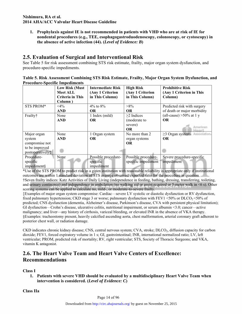
1. Prophylaxis against IE is not recommended in patients with VHD who are at risk of IE for nondental procedures (e.g., TEE, esophagogastroduodenoscopy, colonoscopy, or cystoscopy) in the absence of active infection (44). (Level of Evidence: B)
2.5. Evaluation of Surgical and Interventional Risk See Table 5 for risk assessment combining STS risk estimate, frailty, major organ system dysfunction, and procedure-specific impediments. Table 5. Risk Assessment Combining STS Risk Estimate, Frailty, Major Organ System Dysfunction, and Procedure-Specific Impediments
Low Risk (Must Meet ALL Criteria in This Column )
Intermediate Risk (Any 1 Criterion in This Column)
High Risk (Any 1 Criterion in This Column)
Prohibitive Risk (Any 1 Criterion in This Column)
STS PROM* <4% AND
4% to 8% OR
>8% OR
Predicted risk with surgery of death or major morbidity (all-cause) >50% at 1 y OR
Frailty† None AND
1 Index (mild) OR
≥2 Indices (moderate to severe) OR
Major organ system compromise not to be improved postoperatively‡
None AND
1 Organ system OR
No more than 2 organ systems OR
≥3 Organ systems OR
Procedure-specific impediment§
None Possible procedure-specific impediment
Possible procedure-specific impediment
Severe procedure-specific impediment
*Use of the STS PROM to predict risk in a given institution with reasonable reliability is appropriate only if institutional outcomes are within 1 standard deviation of STS average observed/expected ratio for the procedure in question. †Seven frailty indices: Katz Activities of Daily Living (independence in feeding, bathing, dressing, transferring, toileting, and urinary continence) and independence in ambulation (no walking aid or assist required or 5-meter walk in <6 s). Other scoring systems can be applied to calculate no, mild-, or moderate-to-severe frailty. ‡Examples of major organ system compromise: Cardiac—severe LV systolic or diastolic dysfunction or RV dysfunction, fixed pulmonary hypertension; CKD stage 3 or worse; pulmonary dysfunction with FEV1 <50% or DLCO2 <50% of predicted; CNS dysfunction (dementia, Alzheimer’s disease, Parkinson’s disease, CVA with persistent physical limitation); GI dysfunction—Crohn’s disease, ulcerative colitis, nutritional impairment, or serum albumin <3.0; cancer—active malignancy; and liver—any history of cirrhosis, variceal bleeding, or elevated INR in the absence of VKA therapy. §Examples: tracheostomy present, heavily calcified ascending aorta, chest malformation, arterial coronary graft adherent to posterior chest wall, or radiation damage. CKD indicates chronic kidney disease; CNS, central nervous system; CVA, stroke; DLCO2, diffusion capacity for carbon dioxide; FEV1, forced expiratory volume in 1 s; GI, gastrointestinal; INR, international normalized ratio; LV, left ventricular; PROM, predicted risk of mortality; RV, right ventricular; STS, Society of Thoracic Surgeons; and VKA, vitamin K antagonist.
2.6. The Heart Valve Team and Heart Valve Centers of Excellence: Recommendations Class I
1. Patients with severe VHD should be evaluated by a multidisciplinary Heart Valve Team when intervention is considered. (Level of Evidence: C)
Class IIa
by guest on November 25, 2015http://circ.ahajournals.org/Downloaded from
Nishimura, RA et al. 2014 AHA/ACC Valvular Heart Disease Guideline
Page 15 of 96
1. Consultation with or referral to a Heart Valve Center of Excellence is reasonable when discussing treatment options for 1) asymptomatic patients with severe VHD, 2) patients who may benefit from valve repair versus valve replacement, or 3) patients with multiple comorbidities for whom valve intervention is considered. (Level of Evidence: C)
A competent, practicing cardiologist should have the ability to diagnose and direct the treatment of most patients
with VHD. For instance, otherwise healthy patients with severe VHD who become symptomatic should nearly
always be considered for intervention. However, more complex decision-making processes may be required in
select patient populations, such as those who have asymptomatic severe VHD, those who are at high risk for
intervention, or those who could benefit from specialized therapies such as valve repair or transcatheter valve
intervention.
The management of patients with complex severe VHD is best achieved by a Heart Valve Team
composed primarily of a cardiologist and surgeon (including a structural valve interventionist if a catheter-based
therapy is being considered). In selected cases, there may be a multidisciplinary, collaborative group of
caregivers, including cardiologists, structural valve interventionalists, cardiovascular imaging specialists,
cardiovascular surgeons, anesthesiologists, and nurses, all of whom have expertise in the management and
outcomes of patients with complex VHD. The Heart Valve Team should optimize patient selection for available
procedures through a comprehensive understanding of the risk–benefit ratio of different treatment strategies.
This is particularly beneficial in patients in whom there are several options for treatment, such as the elderly
high-risk patient with severe symptomatic aortic stenosis (AS) being considered for transcatheter aortic valve
replacement (TAVR) or surgical aortic valve replacement (AVR). The patient and family should be sufficiently
educated by the Heart Valve Team about all alternatives for treatment so that their expectations can be met as
fully as possible using a shared decision-making approach.
The optimal care of the patient with complex heart disease is best performed in centers that can provide
all available options for diagnosis and management, including the expertise for complex aortic or mitral valve
repair, aortic surgery, and transcatheter therapies. This has led to the development of Heart Valve Centers of
Excellence. Heart Valve Centers of Excellence 1) are composed of experienced healthcare providers with
expertise from multiple disciplines; 2) offer all available options for diagnosis and management, including
complex valve repair, aortic surgery, and transcatheter therapies; 3) participate in regional or national outcome
registries; 4) demonstrate adherence to national guidelines; 5) participate in continued evaluation and quality
improvement processes to enhance patient outcomes; and 6) publicly report their available mortality and success
rates. Decisions about intervention at the Heart Valve Centers of Excellence should be dependent on the centers’
publicly available mortality rates and operative outcomes. It is recognized that some Heart Valve Centers of
Excellence may have expertise in select valve problems.
by guest on November 25, 2015http://circ.ahajournals.org/Downloaded from
Nishimura, RA et al. 2014 AHA/ACC Valvular Heart Disease Guideline
Page 16 of 96
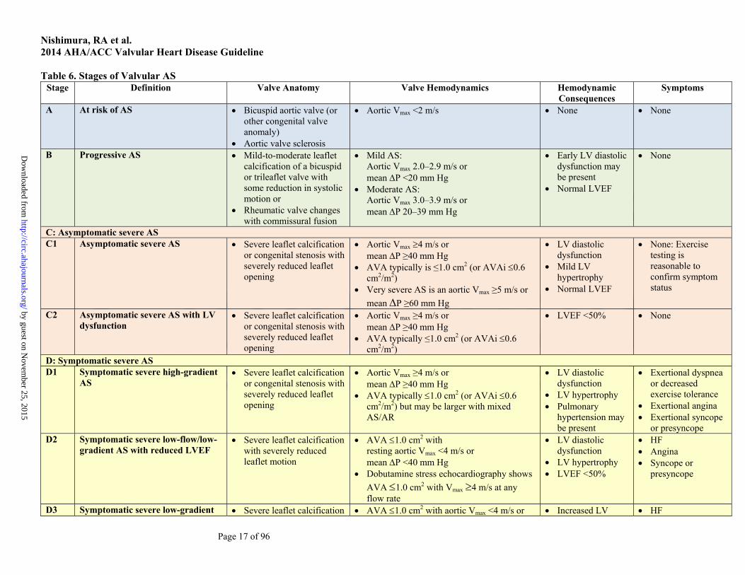
3. Aortic Stenosis: Recommendations See Table 6 for the stages of valvular AS; Tables 7 and 8 for a summary of recommendations for choice and timing of intervention; and Figure 1 for indications for AVR in patients with AS.
3.1. Stages of Valvular AS Medical and interventional approaches to the management of patients with valvular AS depend on accurate
diagnosis of the cause and stage of the disease process. Table 6 shows the stages of AS ranging from patients at
risk of AS (stage A) or with progressive hemodynamic obstruction (stage B) to severe asymptomatic (stage C)
and symptomatic AS (stage D). Each of these stages is defined by valve anatomy, valve hemodynamics, the
consequences of valve obstruction on the left ventricle and vasculature, as well as by patient symptoms.
Hemodynamic severity is best characterized by the transaortic maximum velocity (or mean pressure gradient)
when the transaortic volume flow rate is normal. However, some patients with AS have a low transaortic
volume flow rate due to either left ventricular (LV) systolic dysfunction with a low left ventricular ejection
fraction (LVEF) or due to a small hypertrophied left ventricle with a low stroke volume. These categories of
severe AS pose a diagnostic and management challenge distinctly different from the majority of patients with
AS who have a high gradient and velocity when AS is severe. These special subgroups with low-flow AS are
designated D2 (with a low LVEF) and D3 (with a normal LVEF).
The definition of severe AS is based on natural history studies of patients with unoperated AS, which
show that the prognosis is poor once there is a peak aortic valve velocity of >4.0 m per second, corresponding to
a mean aortic valve gradient >40 mm Hg. In patients with low forward flow, severe AS can be present with
lower aortic valve velocities and lower aortic valve gradients. Thus, an aortic valve area should be calculated in
these patients. The prognosis of patients with AS is poorer when the aortic valve area is <1.0 cm2. At normal
flow rates, an aortic valve area of <0.8 cm2 correlates with a mean aortic valve gradient >40 mm Hg. However,
symptomatic patients with a calcified aortic valve with reduced opening and an aortic valve area between 0.8
cm2 and 1.0 cm2 should be closely evaluated to determine whether they would benefit from valve intervention.
Meticulous attention to detail is required when assessing aortic valve hemodynamics, either with Doppler
echocardiography or cardiac catheterization, and the inherent variability of the measurements and calculations
should always be considered in clinical-decision making.
by guest on November 25, 2015http://circ.ahajournals.org/Downloaded from
Nishimura, RA et al. 2014 AHA/ACC Valvular Heart Disease Guideline
Page 17 of 96
Table 6. Stages of Valvular AS Stage Definition Valve Anatomy Valve Hemodynamics Hemodynamic
Consequences Symptoms
A At risk of AS Bicuspid aortic valve (or other congenital valve anomaly)
Aortic valve sclerosis
Aortic Vmax <2 m/s None None
B Progressive AS Mild-to-moderate leaflet calcification of a bicuspid or trileaflet valve with some reduction in systolic motion or
Rheumatic valve changes with commissural fusion
Mild AS: Aortic Vmax 2.0–2.9 m/s or mean P <20 mm Hg
Moderate AS: Aortic Vmax 3.0–3.9 m/s or mean P 20–39 mm Hg
Early LV diastolic dysfunction may be present
Normal LVEF
None
C: Asymptomatic severe AS C1 Asymptomatic severe AS Severe leaflet calcification
or congenital stenosis with severely reduced leaflet opening
Aortic Vmax 4 m/s or mean P ≥40 mm Hg
AVA typically is ≤1.0 cm2 (or AVAi 0.6 cm2/m2)
Very severe AS is an aortic Vmax ≥5 m/s or
mean P ≥60 mm Hg
LV diastolic dysfunction
Mild LV hypertrophy
Normal LVEF
None: Exercise testing is reasonable to confirm symptom status
C2 Asymptomatic severe AS with LV dysfunction
Severe leaflet calcification or congenital stenosis with severely reduced leaflet opening
Aortic Vmax ≥4 m/s or mean P ≥40 mm Hg
AVA typically ≤1.0 cm2 (or AVAi 0.6 cm2/m2)
LVEF <50% None
D: Symptomatic severe AS D1 Symptomatic severe high-gradient
AS Severe leaflet calcification
or congenital stenosis with severely reduced leaflet opening
Aortic Vmax ≥4 m/s or mean P ≥40 mm Hg
AVA typically 1.0 cm2 (or AVAi 0.6 cm2/m2) but may be larger with mixed AS/AR
LV diastolic dysfunction
LV hypertrophy Pulmonary
hypertension may be present
Exertional dyspnea or decreased exercise tolerance
Exertional angina Exertional syncope
or presyncope D2 Symptomatic severe low-flow/low-
gradient AS with reduced LVEF Severe leaflet calcification
with severely reduced leaflet motion
AVA 1.0 cm2 with resting aortic Vmax <4 m/s or mean P <40 mm Hg
Dobutamine stress echocardiography shows
AVA 1.0 cm2 with Vmax 4 m/s at any flow rate
LV diastolic dysfunction
LV hypertrophy LVEF <50%
HF Angina Syncope or
presyncope
D3 Symptomatic severe low-gradient Severe leaflet calcification AVA 1.0 cm2 with aortic Vmax <4 m/s or Increased LV HF
by guest on Novem
ber 25, 2015http://circ.ahajournals.org/
Dow
nloaded from
Nishimura, RA et al. 2014 AHA/ACC Valvular Heart Disease Guideline
Page 18 of 96
AS with normal LVEF or paradoxical low-flow severe AS
with severely reduced leaflet motion
mean P <40 mm Hg Indexed AVA 0.6 cm2/m2 and Stroke volume index <35 mL/m2 Measured when patient is normotensive
(systolic BP <140 mm Hg)
relative wall thickness
Small LV chamber with low stroke volume
Restrictive diastolic filling
LVEF ≥50%
Angina Syncope or
presyncope
AR indicates aortic regurgitation; AS, aortic stenosis; AVA, aortic valve area; AVAi, aortic valve area indexed to body surface area; BP, blood pressure; HF, heart failure; LV, left ventricular; LVEF, left ventricular ejection fraction; P, pressure gradient; and Vmax, maximum aortic velocity.
by guest on Novem
ber 25, 2015http://circ.ahajournals.org/
Dow
nloaded from
Nishimura, RA et al. 2014 AHA/ACC Valvular Heart Disease Guideline
Page 19 of 96
3.2. Diagnosis and Follow-Up The overall approach to the initial diagnosis of VHD is discussed in Section 2.3, and additional considerations
specific to patients with AS are addressed here.
Class I
1. TTE is indicated in patients with signs or symptoms of AS or a bicuspid aortic valve for accurate diagnosis of the cause of AS, hemodynamic severity, LV size and systolic function, and for determining prognosis and timing of valve intervention (26, 27, 45). (Level of Evidence: B)
Class IIa
1. Low-dose dobutamine stress testing using echocardiographic or invasive hemodynamic measurements is reasonable in patients with stage D2 AS with all of the following (46-48), (Level of Evidence: B):
a. Calcified aortic valve with reduced systolic opening; b. LVEF less than 50%; c. Calculated valve area 1.0 cm2 or less; and d. Aortic velocity less than 4.0 m per second or mean pressure gradient less than 40 mm Hg.
2. Exercise testing is reasonable to assess physiological changes with exercise and to confirm the absence of symptoms in asymptomatic patients with a calcified aortic valve and an aortic velocity 4.0 m per second or greater or mean pressure gradient 40 mm Hg or higher (stage C) (27, 37, 38, 49). (Level of Evidence: B)
Class III: Harm
1. Exercise testing should not be performed in symptomatic patients with AS when the aortic velocity is 4.0 m per second or greater or mean pressure gradient is 40 mm Hg or higher (stage D) (50). (Level of Evidence: B)
3.3. Medical Therapy Class I
1. Hypertension in patients at risk for developing AS (stage A) and in patients with asymptomatic AS (stages B and C) should be treated according to standard GDMT, started at a low dose, and gradually titrated upward as needed with frequent clinical monitoring (51-53). (Level of Evidence: B)
Class IIb
1. Vasodilator therapy may be reasonable if used with invasive hemodynamic monitoring in the acute management of patients with severe decompensated AS (stage D) with New York Heart Association (NYHA) class IV heart failure (HF) symptoms. (Level of Evidence: C)
Class III: No Benefit
1. Statin therapy is not indicated for prevention of hemodynamic progression of AS in patients with mild-to-moderate calcific valve disease (stages B to D) (54-56). (Level of Evidence: A)
3.4. Timing of Intervention See Table 7 for a summary of recommendations from this section. Class I
by guest on November 25, 2015http://circ.ahajournals.org/Downloaded from
Nishimura, RA et al. 2014 AHA/ACC Valvular Heart Disease Guideline
Page 20 of 96
1. AVR is recommended in symptomatic patients with severe AS (stage D1) with (57-60), (Level of Evidence: B):
a. Decreased systolic opening of a calcified or congenitally stenotic aortic valve; and b. An aortic velocity 4.0 m per second or greater or mean pressure gradient 40 mm Hg or
higher; and c. Symptoms of HF, syncope, exertional dyspnea, angina, or presyncope by history or on
exercise testing. 2. AVR is recommended for asymptomatic patients with severe AS (stage C2) and an LVEF less
than 50% with decreased systolic opening of a calcified aortic valve with an aortic velocity 4.0 m per second or greater or mean pressure gradient 40 mm Hg or higher (61, 62). (Level of Evidence: B)
3. AVR is indicated for patients with severe AS (stage C or D) when undergoing cardiac surgery for other indications when there is decreased systolic opening of a calcified aortic valve and an aortic velocity 4.0 m per second or greater or mean pressure gradient 40 mm Hg or higher (63, 64). (Level of Evidence: B)
Class IIa
1. AVR is reasonable for asymptomatic patients with very severe AS (stage C1) with (65, 66), (Level of Evidence: B):
a. Decreased systolic opening of a calcified valve; b. An aortic velocity 5.0 m per second or greater or mean pressure gradient 60 mm Hg or
higher; and c. A low surgical risk.
2. AVR is reasonable in apparently asymptomatic patients with severe AS (stage C1) with (27, 38), (Level of Evidence: B):
a. A calcified aortic valve; b. An aortic velocity of 4.0 m per second to 4.9 m per second or mean pressure gradient of 40
mm Hg to 59 mm Hg; and c. An exercise test demonstrating decreased exercise tolerance or a fall in systolic blood
pressure (BP). 3. AVR is reasonable in symptomatic patients with low-flow/low-gradient severe AS with reduced
LVEF (stage D2) with a (67-69), (Level of Evidence: B): a. Calcified aortic valve with reduced systolic opening; b. Resting valve area 1.0 cm2 or less; c. Aortic velocity less than 4.0 m per second or mean pressure gradient less than 40 mm Hg; d. LVEF less than 50%; and e. A low-dose dobutamine stress study that shows an aortic velocity 4.0 m per second or
greater or mean pressure gradient 40 mm Hg or higher with a valve area 1.0 cm2 or less at any dobutamine dose.
4. AVR is reasonable in symptomatic patients with low-flow/low-gradient severe AS (stage D3) with an LVEF 50% or greater, a calcified aortic valve with significantly reduced leaflet motion, and a valve area 1.0 cm2 or less only if clinical, hemodynamic, and anatomic data support valve obstruction as the most likely cause of symptoms and data recorded when the patient is normotensive (systolic BP <140 mm Hg) indicate (Level of Evidence: C):
a. An aortic velocity less than 4.0 m per second or mean pressure gradient less than 40 mm Hg; and
b. A stroke volume index less than 35 mL/m2; and c. An indexed valve area 0.6 cm2/m2 or less.
5. AVR is reasonable for patients with moderate AS (stage B) with an aortic velocity between 3.0 m per second and 3.9 m per second or mean pressure gradient between 20 mm Hg and 39 mm Hg who are undergoing cardiac surgery for other indications. (Level of Evidence: C)
by guest on November 25, 2015http://circ.ahajournals.org/Downloaded from
Nishimura, RA et al. 2014 AHA/ACC Valvular Heart Disease Guideline
Page 21 of 96
Class IIb 1. AVR may be considered for asymptomatic patients with severe AS (stage C1) with an aortic
velocity 4.0 m per second or greater or mean pressure gradient 40 mm Hg or higher if the patient is at low surgical risk and serial testing shows an increase in aortic velocity 0.3 m/s or greater per year. (Level of Evidence: C)
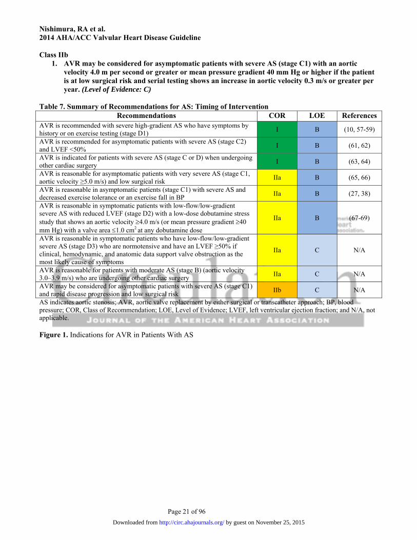
Table 7. Summary of Recommendations for AS: Timing of Intervention
Recommendations COR LOE References AVR is recommended with severe high-gradient AS who have symptoms by history or on exercise testing (stage D1)
I B (10, 57-59)
AVR is recommended for asymptomatic patients with severe AS (stage C2) and LVEF <50%
I B (61, 62)
AVR is indicated for patients with severe AS (stage C or D) when undergoing other cardiac surgery
I B (63, 64)
AVR is reasonable for asymptomatic patients with very severe AS (stage C1, aortic velocity ≥5.0 m/s) and low surgical risk
IIa B (65, 66)
AVR is reasonable in asymptomatic patients (stage C1) with severe AS and decreased exercise tolerance or an exercise fall in BP
IIa B (27, 38)
AVR is reasonable in symptomatic patients with low-flow/low-gradient severe AS with reduced LVEF (stage D2) with a low-dose dobutamine stress study that shows an aortic velocity 4.0 m/s (or mean pressure gradient 40 mm Hg) with a valve area 1.0 cm2 at any dobutamine dose
IIa B (67-69)
AVR is reasonable in symptomatic patients who have low-flow/low-gradient severe AS (stage D3) who are normotensive and have an LVEF ≥50% if clinical, hemodynamic, and anatomic data support valve obstruction as the most likely cause of symptoms
IIa C N/A
AVR is reasonable for patients with moderate AS (stage B) (aortic velocity 3.0–3.9 m/s) who are undergoing other cardiac surgery
IIa C N/A
AVR may be considered for asymptomatic patients with severe AS (stage C1) and rapid disease progression and low surgical risk
IIb C N/A
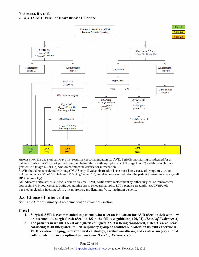
AS indicates aortic stenosis; AVR, aortic valve replacement by either surgical or transcatheter approach; BP, blood pressure; COR, Class of Recommendation; LOE, Level of Evidence; LVEF, left ventricular ejection fraction; and N/A, not applicable. Figure 1. Indications for AVR in Patients With AS
by guest on November 25, 2015http://circ.ahajournals.org/Downloaded from
Nishimura, RA et al. 2014 AHA/ACC Valvular Heart Disease Guideline
Page 22 of 96
Arrows show the decision pathways that result in a recommendation for AVR. Periodic monitoring is indicated for all patients in whom AVR is not yet indicated, including those with asymptomatic AS (stage D or C) and those with low-gradient AS (stage D2 or D3) who do not meet the criteria for intervention. *AVR should be considered with stage D3 AS only if valve obstruction is the most likely cause of symptoms, stroke volume index is <35 mL/m2, indexed AVA is ≤0.6 cm2/m2, and data are recorded when the patient is normotensive (systolic BP <140 mm Hg). AS indicates aortic stenosis; AVA; aortic valve area; AVR, aortic valve replacement by either surgical or transcatheter approach; BP, blood pressure; DSE, dobutamine stress echocardiography; ETT, exercise treadmill test; LVEF, left ventricular ejection fraction; Pmean, mean pressure gradient; and Vmax, maximum velocity.
3.5. Choice of Intervention See Table 8 for a summary of recommendations from this section. Class I
1. Surgical AVR is recommended in patients who meet an indication for AVR (Section 3.4) with low or intermediate surgical risk (Section 2.5 in the full-text guideline) (70, 71). (Level of Evidence: A)
2. For patients in whom TAVR or high-risk surgical AVR is being considered, a Heart Valve Team consisting of an integrated, multidisciplinary group of healthcare professionals with expertise in VHD, cardiac imaging, interventional cardiology, cardiac anesthesia, and cardiac surgery should collaborate to provide optimal patient care. (Level of Evidence: C)
by guest on November 25, 2015http://circ.ahajournals.org/Downloaded from
Nishimura, RA et al. 2014 AHA/ACC Valvular Heart Disease Guideline
Page 23 of 96
3. TAVR is recommended in patients who meet an indication for AVR (Section 3.4) who have a prohibitive risk for surgical AVR (Section 2.5 in the full-text guideline) and a predicted post-TAVR survival greater than 12 months (72, 73). (Level of Evidence: B)
Class IIa
1. TAVR is a reasonable alternative to surgical AVR in patients who meet an indication for AVR (Section 3.4) and who have high surgical risk for surgical AVR (Section 2.5 in the full-text guideline) (74, 75). (Level of Evidence: B)
Class IIb
1. Percutaneous aortic balloon dilation may be considered as a bridge to surgical AVR or TAVR in patients with severe symptomatic AS. (Level of Evidence: C)
Class III: No Benefit 1. TAVR is not recommended in patients in whom existing comorbidities would preclude the
expected benefit from correction of AS (72). (Level of Evidence: B)
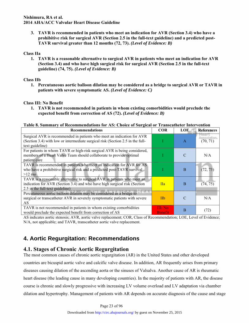
Table 8. Summary of Recommendations for AS: Choice of Surgical or Transcatheter Intervention
Recommendations COR LOE References
Surgical AVR is recommended in patients who meet an indication for AVR (Section 3.4) with low or intermediate surgical risk (Section 2.5 in the full-text guideline)
I A (70, 71)
For patients in whom TAVR or high-risk surgical AVR is being considered, members of a Heart Valve Team should collaborate to provide optimal patient care
I C N/A
TAVR is recommended in patients who meet an indication for AVR for AS who have a prohibitive surgical risk and a predicted post-TAVR survival >12 mo
I B (72, 73)
TAVR is a reasonable alternative to surgical AVR in patients who meet an indication for AVR (Section 3.4) and who have high surgical risk (Section 2.5 in the full-text guideline)
IIa B (74, 75)
Percutaneous aortic balloon dilation may be considered as a bridge to surgical or transcatheter AVR in severely symptomatic patients with severe AS
IIb C N/A
TAVR is not recommended in patients in whom existing comorbidities would preclude the expected benefit from correction of AS
III: No Benefit
B (72)
AS indicates aortic stenosis; AVR, aortic valve replacement; COR, Class of Recommendation; LOE, Level of Evidence; N/A, not applicable; and TAVR, transcatheter aortic valve replacement. 4. Aortic Regurgitation: Recommendations
4.1. Stages of Chronic Aortic Regurgitation The most common causes of chronic aortic regurgitation (AR) in the United States and other developed
countries are bicuspid aortic valve and calcific valve disease. In addition, AR frequently arises from primary
diseases causing dilation of the ascending aorta or the sinuses of Valsalva. Another cause of AR is rheumatic
heart disease (the leading cause in many developing countries). In the majority of patients with AR, the disease
course is chronic and slowly progressive with increasing LV volume overload and LV adaptation via chamber
dilation and hypertrophy. Management of patients with AR depends on accurate diagnosis of the cause and stage
by guest on November 25, 2015http://circ.ahajournals.org/Downloaded from
Nishimura, RA et al. 2014 AHA/ACC Valvular Heart Disease Guideline
Page 34 of 96
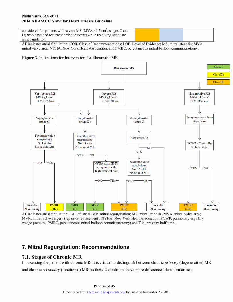
considered for patients with severe MS (MVA ≤1.5 cm2, stages C and D) who have had recurrent embolic events while receiving adequate anticoagulation AF indicates atrial fibrillation; COR, Class of Recommendations; LOE, Level of Evidence; MS, mitral stenosis; MVA, mitral valve area; NYHA, New York Heart Association; and PMBC, percutaneous mitral balloon commissurotomy. Figure 3. Indications for Intervention for Rheumatic MS
AF indicates atrial fibrillation; LA, left atrial; MR, mitral regurgitation; MS, mitral stenosis; MVA, mitral valve area; MVR, mitral valve surgery (repair or replacement); NYHA, New York Heart Association; PCWP, pulmonary capillary wedge pressure; PMBC, percutaneous mitral balloon commissurotomy; and T ½, pressure half-time.
7. Mitral Regurgitation: Recommendations
7.1. Stages of Chronic MR In assessing the patient with chronic MR, it is critical to distinguish between chronic primary (degenerative) MR
and chronic secondary (functional) MR, as these 2 conditions have more differences than similarities.
by guest on November 25, 2015http://circ.ahajournals.org/Downloaded from
Nishimura, RA et al. 2014 AHA/ACC Valvular Heart Disease Guideline
Page 35 of 96
In chronic primary MR, the pathology of ≥1 of the components of the valve (leaflets, chordae tendineae,
papillary muscles, annulus) causes valve incompetence with systolic regurgitation of blood from the left
ventricle to the LA (Table 13). The most common cause of chronic primary MR in developed countries is mitral
valve prolapse, which has a wide spectrum of etiology and presentation. Younger populations present with
severe myxomatous degeneration with gross redundancy of both anterior and posterior leaflets and the chordal
apparatus (Barlow’s valve). Alternatively, older populations present with fibroelastic deficiency disease, in
which lack of connective tissue leads to chordal rupture. The differentiation between these 2 etiologies has
important implications for operative intervention. Other less common causes of chronic primary MR include IE,
connective tissue disorders, rheumatic heart disease, cleft mitral valve, and radiation heart disease. If the
subsequent volume overload of chronic primary MR is prolonged and severe, it causes myocardial damage, HF,
and eventual death. Correction of the MR is curative. Thus, MR is “the disease.”
In chronic secondary MR, the mitral valve is usually normal (Table 14). Instead, severe LV dysfunction
is caused either by CAD, related myocardial infarction (ischemic chronic secondary MR), or idiopathic
myocardial disease (nonischemic chronic secondary MR). The abnormal and dilated left ventricle causes
papillary muscle displacement, which in turn results in leaflet tethering with associated annular dilation that
prevents coaptation. Because MR is only 1 component of the disease (severe LV dysfunction, coronary disease,
or idiopathic myocardial disease are the others), restoration of mitral valve competence is not by itself curative;
thus, the best therapy for chronic secondary MR is much less clear than it is for chronic primary MR. The data
are limited, and there is greater difficulty in defining the severity of MR in patients with secondary MR than in
those with primary MR. In patients with secondary MR, adverse outcomes are associated with a smaller
calculated effective regurgitant orifice compared to primary MR due to multiple reasons. The MR will likely
progress due to the associated progressive LV systolic dysfunction and adverse remodeling. In addition, there is
an underestimation of effective regurgitant orifice area by the 2-dimensional echocardiographyderived flow
convergence method due to the crescenticshape of the regurgitant orifice. There are the additional clinical
effects of a smaller amount of regurgitation in the presence of compromised LV systolic function and baseline
elevated filling pressures.
by guest on November 25, 2015http://circ.ahajournals.org/Downloaded from
Nishimura, RA et al. 2014 AHA/ACC Valvular Heart Disease Guideline
Page 36 of 96
Table 13. Stages of Primary MR Grade Definition Valve Anatomy Valve Hemodynamics* Hemodynamic
Consequences Symptoms
A At risk of MR Mild mitral valve prolapse with normal coaptation
Mild valve thickening and leaflet restriction
No MR jet or small central jet area <20% LA on Doppler
Small vena contracta <0.3 cm
None None
B Progressive MR Severe mitral valve prolapse with normal coaptation
Rheumatic valve changes with leaflet restriction and loss of central coaptation
Prior IE
Central jet MR 20%–40% LA or late systolic eccentric jet MR
Vena contracta <0.7 cm Regurgitant volume <60 mL Regurgitant fraction <50% ERO <0.40 cm2 Angiographic grade 1–2+
Mild LA enlargement No LV enlargement Normal pulmonary
pressure
None
C Asymptomatic severe MR
Severe mitral valve prolapse with loss of coaptation or flail leaflet
Rheumatic valve changes with leaflet restriction and loss of central coaptation
Prior IE Thickening of leaflets with
radiation heart disease
Central jet MR >40% LA or holosystolic eccentric jet MR
Vena contracta ≥0.7 cm Regurgitant volume ≥60 mL Regurgitant fraction ≥50% ERO ≥0.40 cm2 Angiographic grade 3–4+
Moderate or severe LA enlargement
LV enlargement Pulmonary hypertension
may be present at rest or with exercise
C1: LVEF >60% and LVESD <40 mm
C2: LVEF ≤60% and LVESD ≥40 mm
None
D Symptomatic severe MR
Severe mitral valve prolapse with loss of coaptation or flail leaflet
Rheumatic valve changes with leaflet restriction and loss of central coaptation
Prior IE Thickening of leaflets with
radiation heart disease
Central jet MR >40% LA or holosystolic eccentric jet MR
Vena contracta ≥0.7 cm Regurgitant volume ≥60 mL Regurgitant fraction ≥50% ERO ≥0.40 cm2 Angiographic grade 3–4+
Moderate or severe LA enlargement
LV enlargement Pulmonary hypertension
present
Decreased exercise tolerance
Exertional dyspnea
*Several valve hemodynamic criteria are provided for assessment of MR severity, but not all criteria for each category will be present in each patient. Categorization of MR severity as mild, moderate, or severe depends on data quality and integration of these parameters in conjunction with other clinical evidence. ERO indicates effective regurgitant orifice; IE, infective endocarditis; LA, left atrium/atrial; LV, left ventricular; LVEF, left ventricular ejection fraction; LVESD; left ventricular end-systolic dimension; and MR, mitral regurgitation
by guest on Novem
ber 25, 2015http://circ.ahajournals.org/
Dow
nloaded from
Nishimura, RA et al. 2014 AHA/ACC Valvular Heart Disease Guideline
Page 37 of 96
Table 14. Stages of Secondary MR Grade Definition Valve Anatomy Valve Hemodynamics* Associated Cardiac Findings Symptoms A At risk of MR Normal valve leaflets, chords,
and annulus in a patient with coronary disease or cardiomyopathy
No MR jet or small central jet area <20% LA on Doppler
Small vena contracta <0.30 cm
Normal or mildly dilated LV size with fixed (infarction) or inducible (ischemia) regional wall motion abnormalities
Primary myocardial disease with LV dilation and systolic dysfunction
Symptoms due to coronary ischemia or HF may be present that respond to revascularization and appropriate medical therapy
B Progressive MR Regional wall motion abnormalities with mild tethering of mitral leaflet
Annular dilation with mild loss of central coaptation of the mitral leaflets
ERO <0.20 cm2† Regurgitant volume <30 mL Regurgitant fraction <50%
Regional wall motion abnormalities with reduced LV systolic function
LV dilation and systolic dysfunction due to primary myocardial disease
Symptoms due to coronary ischemia or HF may be present that respond to revascularization and appropriate medical therapy
C Asymptomatic severe MR
Regional wall motion abnormalities and/or LV dilation with severe tethering of mitral leaflet
Annular dilation with severe loss of central coaptation of the mitral leaflets
ERO ≥0.20 cm2 † Regurgitant volume ≥30 mL Regurgitant fraction ≥50%
Regional wall motion abnormalities with reduced LV systolic function
LV dilation and systolic dysfunction due to primary myocardial disease
Symptoms due to coronary ischemia or HF may be present that respond to revascularization and appropriate medical therapy
D Symptomatic severe MR
Regional wall motion abnormalities and/or LV dilation with severe tethering of mitral leaflet
Annular dilation with severe loss of central coaptation of the mitral leaflets
ERO ≥0.20 cm2† Regurgitant volume ≥30 mL Regurgitant fraction ≥50%
Regional wall motion abnormalities with reduced LV systolic function
LV dilation and systolic dysfunction due to primary myocardial disease
HF symptoms due to MR persist even after revascularization and optimization of medical therapy
Decreased exercise tolerance
Exertional dyspnea *Several valve hemodynamic criteria are provided for assessment of MR severity, but not all criteria for each category will be present in each patient. Categorization of MR severity as mild, moderate, or severe depends on data quality and integration of these parameters in conjunction with other clinical evidence. †The measurement of the proximal isovelocity surface area by 2D TTE in patients with secondary MR underestimates the true ERO due to the crescentic shape of the proximal convergence. 2D indicates 2-dimensional; ERO, effective regurgitant orifice; HF, heart failure; LA, left atrium; LV, left ventricular; MR, mitral regurgitation; and TTE, transthoracic echocardiogram.
by guest on Novem
ber 25, 2015http://circ.ahajournals.org/
Dow
nloaded from
Nishimura, RA et al. 2014 AHA/ACC Valvular Heart Disease Guideline
Page 38 of 96
7.2. Chronic Primary MR
7.2.1. Diagnosis and Follow-Up Class I
1. TTE is indicated for baseline evaluation of LV size and function, right ventricular (RV) function and left atrial size, pulmonary artery pressure, and mechanism and severity of primary MR (stages A to D) in any patient suspected of having chronic primary MR (6, 23, 146-162). (Level of Evidence: B)
2. CMR is indicated in patients with chronic primary MR to assess LV and RV volumes, function, or MR severity and when these issues are not satisfactorily addressed by TTE (157, 163, 164). (Level of Evidence: B)
3. Intraoperative TEE is indicated to establish the anatomic basis for chronic primary MR (stages C and D) and to guide repair (165, 166). (Level of Evidence: B)
4. TEE is indicated for evaluation of patients with chronic primary MR (stages B to D) in whom noninvasive imaging provides nondiagnostic information about severity of MR, mechanism of MR, and/or status of LV function. (Level of Evidence: C)
Class IIa
1. Exercise hemodynamics with either Doppler echocardiography or cardiac catheterization is reasonable in symptomatic patients with chronic primary MR where there is a discrepancy between symptoms and the severity of MR at rest (stages B and C) (167, 168). (Level of Evidence: B)
2. Exercise treadmill testing can be useful in patients with chronic primary MR to establish symptom status and exercise tolerance (stages B and C). (Level of Evidence: C)
7.2.2. Medical Therapy Class IIa
1. Medical therapy for systolic dysfunction is reasonable in symptomatic patients with chronic primary MR (stage D) and LVEF less than 60% in whom surgery is not contemplated (169-173). (Level of Evidence: B)
Class III: No Benefit
1. Vasodilator therapy is not indicated for normotensive asymptomatic patients with chronic primary MR (stages B and C1) and normal systolic LV function (173-178). (Level of Evidence: B)
7.2.3. Intervention See Table 15 for a summary of recommendations from this section. Class I
1. Mitral valve surgery is recommended for symptomatic patients with chronic severe primary MR (stage D) and LVEF greater than 30% (156, 179). (Level of Evidence: B)
2. Mitral valve surgery is recommended for asymptomatic patients with chronic severe primary MR and LV dysfunction (LVEF 30% to 60% and/or LVESD ≥40 mm, stage C2) (150-153, 180-182). (Level of Evidence: B)
3. Mitral valve repair is recommended in preference to mitral valve replacement (MVR) when surgical treatment is indicated for patients with chronic severe primary MR limited to the posterior leaflet (155, 183-198). (Level of Evidence: B)
4. Mitral valve repair is recommended in preference to MVR when surgical treatment is indicated for patients with chronic severe primary MR involving the anterior leaflet or both leaflets when a successful and durable repair can be accomplished (195-197, 199-203). (Level of Evidence: B)
by guest on November 25, 2015http://circ.ahajournals.org/Downloaded from
Nishimura, RA et al. 2014 AHA/ACC Valvular Heart Disease Guideline
Page 39 of 96
5. Concomitant mitral valve repair or MVR is indicated in patients with chronic severe primary MR undergoing cardiac surgery for other indications (204). (Level of Evidence: B)
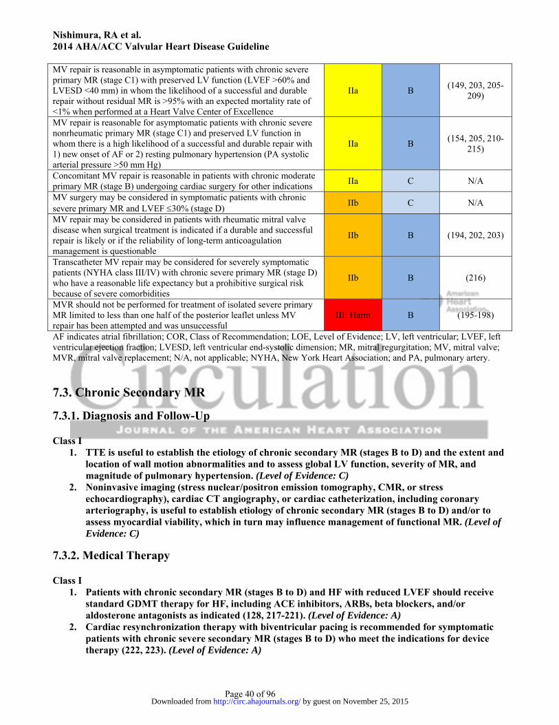
Class IIa
1. Mitral valve repair is reasonable in asymptomatic patients with chronic severe primary MR (stage C1) with preserved LV function (LVEF >60% and LVESD <40 mm) in whom the likelihood of a successful and durable repair without residual MR is greater than 95% with an expected mortality rate of less than 1% when performed at a Heart Valve Center of Excellence (149, 203, 205-209). (Level of Evidence: B)
2. Mitral valve repair is reasonable for asymptomatic patients with chronic severe nonrheumatic primary MR (stage C1) and preserved LV function (LVEF >60% and LVESD <40 mm) in whom there is a high likelihood of a successful and durable repair with 1) new onset of AF or 2) resting pulmonary hypertension (pulmonary artery systolic arterial pressure >50 mm Hg) (154, 205, 210-215). (Level of Evidence: B)
3. Concomitant mitral valve repair is reasonable in patients with chronic moderate primary MR (stage B) when undergoing cardiac surgery for other indications. (Level of Evidence: C)
Class IIb
1. Mitral valve surgery may be considered in symptomatic patients with chronic severe primary MR and LVEF less than or equal to 30% (stage D). (Level of Evidence: C)
2. Mitral valve repair may be considered in patients with rheumatic mitral valve disease when surgical treatment is indicated if a durable and successful repair is likely or when the reliability of long-term anticoagulation management is questionable (194, 202, 203). (Level of Evidence: B)
3. Transcatheter mitral valve repair may be considered for severely symptomatic patients (NYHA class III to IV) with chronic severe primary MR (stage D) who have favorable anatomy for the repair procedure and a reasonable life expectancy but who have a prohibitive surgical risk because of severe comorbidities and remain severely symptomatic despite optimal GDMT for HF (216). (Level of Evidence: B)
Class III: Harm 1. MVR should not be performed for the treatment of isolated severe primary MR limited to less
than one half of the posterior leaflet unless mitral valve repair has been attempted and was unsuccessful (195-198). (Level of Evidence: B)
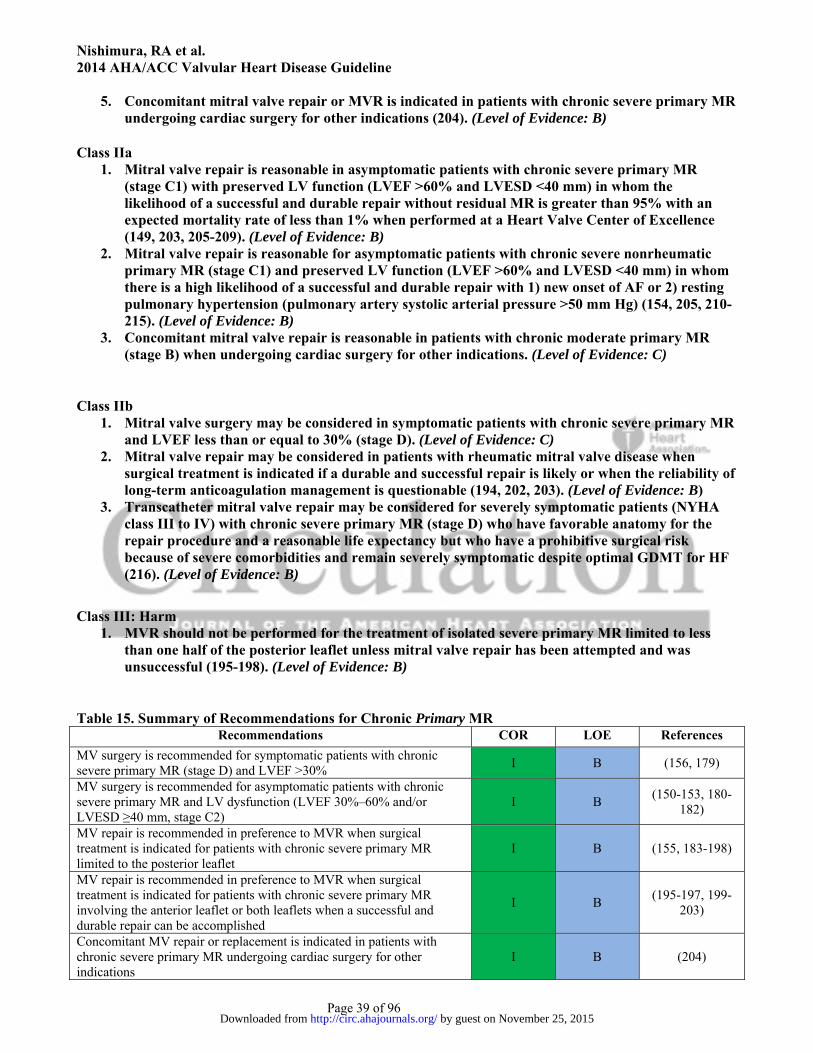
Table 15. Summary of Recommendations for Chronic Primary MR
Recommendations COR LOE References
MV surgery is recommended for symptomatic patients with chronic severe primary MR (stage D) and LVEF >30%
I B (156, 179)
MV surgery is recommended for asymptomatic patients with chronic severe primary MR and LV dysfunction (LVEF 30%–60% and/or LVESD ≥40 mm, stage C2)
I B (150-153, 180-
182)
MV repair is recommended in preference to MVR when surgical treatment is indicated for patients with chronic severe primary MR limited to the posterior leaflet
I B (155, 183-198)
MV repair is recommended in preference to MVR when surgical treatment is indicated for patients with chronic severe primary MR involving the anterior leaflet or both leaflets when a successful and durable repair can be accomplished
I B (195-197, 199-
203)
Concomitant MV repair or replacement is indicated in patients with chronic severe primary MR undergoing cardiac surgery for other indications
I B (204)
by guest on November 25, 2015http://circ.ahajournals.org/Downloaded from
Nishimura, RA et al. 2014 AHA/ACC Valvular Heart Disease Guideline
Page 40 of 96
MV repair is reasonable in asymptomatic patients with chronic severe primary MR (stage C1) with preserved LV function (LVEF >60% and LVESD <40 mm) in whom the likelihood of a successful and durable repair without residual MR is >95% with an expected mortality rate of <1% when performed at a Heart Valve Center of Excellence
IIa B (149, 203, 205-
209)
MV repair is reasonable for asymptomatic patients with chronic severe nonrheumatic primary MR (stage C1) and preserved LV function in whom there is a high likelihood of a successful and durable repair with 1) new onset of AF or 2) resting pulmonary hypertension (PA systolic arterial pressure >50 mm Hg)
IIa B (154, 205, 210-
215)
Concomitant MV repair is reasonable in patients with chronic moderate primary MR (stage B) undergoing cardiac surgery for other indications
IIa C N/A
MV surgery may be considered in symptomatic patients with chronic severe primary MR and LVEF 30% (stage D)
IIb C N/A
MV repair may be considered in patients with rheumatic mitral valve disease when surgical treatment is indicated if a durable and successful repair is likely or if the reliability of long-term anticoagulation management is questionable
IIb B (194, 202, 203)
Transcatheter MV repair may be considered for severely symptomatic patients (NYHA class III/IV) with chronic severe primary MR (stage D) who have a reasonable life expectancy but a prohibitive surgical risk because of severe comorbidities
IIb B (216)
MVR should not be performed for treatment of isolated severe primary MR limited to less than one half of the posterior leaflet unless MV repair has been attempted and was unsuccessful
III: Harm B (195-198)
AF indicates atrial fibrillation; COR, Class of Recommendation; LOE, Level of Evidence; LV, left ventricular; LVEF, left ventricular ejection fraction; LVESD, left ventricular end-systolic dimension; MR, mitral regurgitation; MV, mitral valve; MVR, mitral valve replacement; N/A, not applicable; NYHA, New York Heart Association; and PA, pulmonary artery.
7.3. Chronic Secondary MR
7.3.1. Diagnosis and Follow-Up Class I
1. TTE is useful to establish the etiology of chronic secondary MR (stages B to D) and the extent and location of wall motion abnormalities and to assess global LV function, severity of MR, and magnitude of pulmonary hypertension. (Level of Evidence: C)
2. Noninvasive imaging (stress nuclear/positron emission tomography, CMR, or stress echocardiography), cardiac CT angiography, or cardiac catheterization, including coronary arteriography, is useful to establish etiology of chronic secondary MR (stages B to D) and/or to assess myocardial viability, which in turn may influence management of functional MR. (Level of Evidence: C)
7.3.2. Medical Therapy Class I
1. Patients with chronic secondary MR (stages B to D) and HF with reduced LVEF should receive standard GDMT therapy for HF, including ACE inhibitors, ARBs, beta blockers, and/or aldosterone antagonists as indicated (128, 217-221). (Level of Evidence: A)
2. Cardiac resynchronization therapy with biventricular pacing is recommended for symptomatic patients with chronic severe secondary MR (stages B to D) who meet the indications for device therapy (222, 223). (Level of Evidence: A)
by guest on November 25, 2015http://circ.ahajournals.org/Downloaded from
Nishimura, RA et al. 2014 AHA/ACC Valvular Heart Disease Guideline
Page 41 of 96
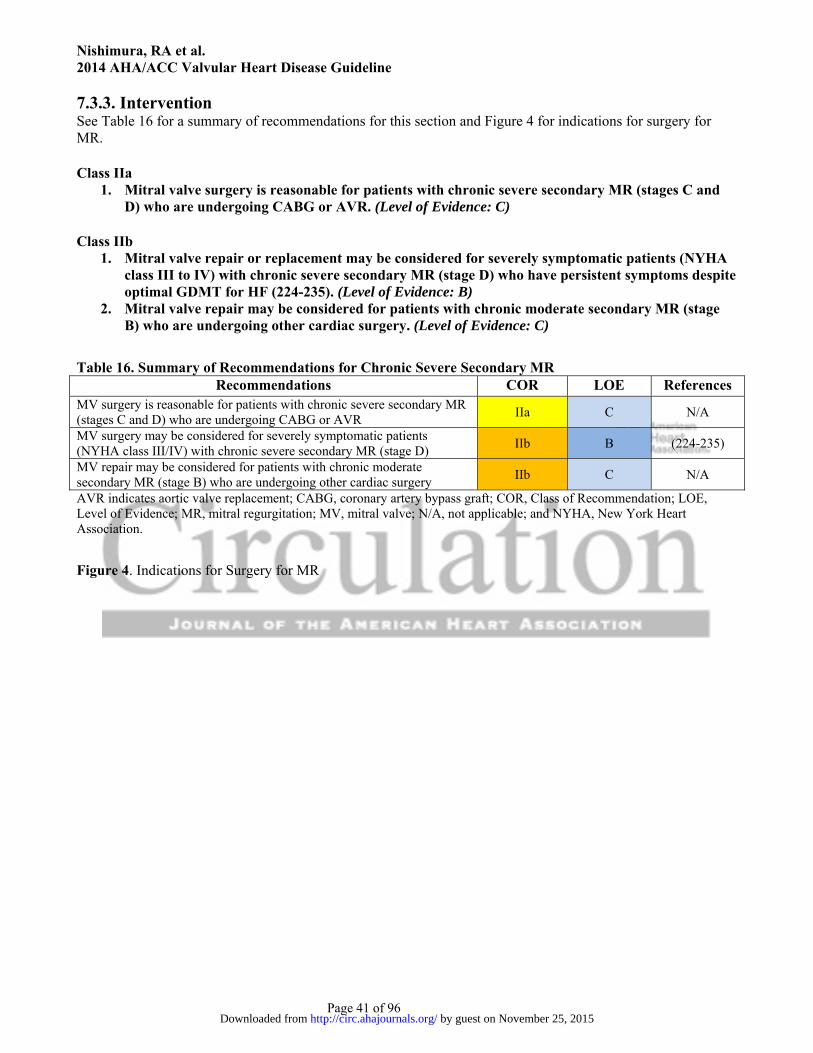
7.3.3. Intervention See Table 16 for a summary of recommendations for this section and Figure 4 for indications for surgery for MR. Class IIa
1. Mitral valve surgery is reasonable for patients with chronic severe secondary MR (stages C and D) who are undergoing CABG or AVR. (Level of Evidence: C)
Class IIb
1. Mitral valve repair or replacement may be considered for severely symptomatic patients (NYHA class III to IV) with chronic severe secondary MR (stage D) who have persistent symptoms despite optimal GDMT for HF (224-235). (Level of Evidence: B)
2. Mitral valve repair may be considered for patients with chronic moderate secondary MR (stage B) who are undergoing other cardiac surgery. (Level of Evidence: C)
Table 16. Summary of Recommendations for Chronic Severe Secondary MR Recommendations COR LOE References
MV surgery is reasonable for patients with chronic severe secondary MR (stages C and D) who are undergoing CABG or AVR
IIa C N/A
MV surgery may be considered for severely symptomatic patients (NYHA class III/IV) with chronic severe secondary MR (stage D)
IIb B (224-235)
MV repair may be considered for patients with chronic moderate secondary MR (stage B) who are undergoing other cardiac surgery
IIb C N/A
AVR indicates aortic valve replacement; CABG, coronary artery bypass graft; COR, Class of Recommendation; LOE, Level of Evidence; MR, mitral regurgitation; MV, mitral valve; N/A, not applicable; and NYHA, New York Heart Association.
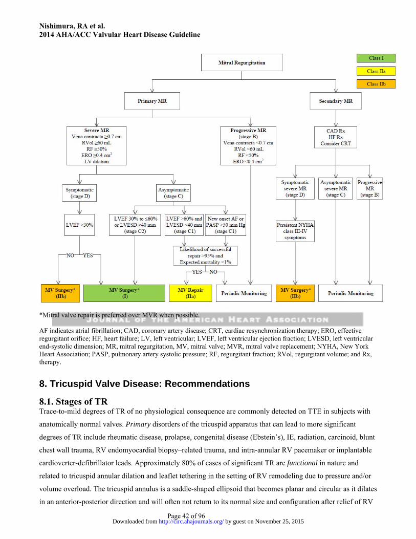
Figure 4. Indications for Surgery for MR
by guest on November 25, 2015http://circ.ahajournals.org/Downloaded from
Nishimura, RA et al. 2014 AHA/ACC Valvular Heart Disease Guideline
Page 42 of 96
*Mitral valve repair is preferred over MVR when possible. AF indicates atrial fibrillation; CAD, coronary artery disease; CRT, cardiac resynchronization therapy; ERO, effective regurgitant orifice; HF, heart failure; LV, left ventricular; LVEF, left ventricular ejection fraction; LVESD, left ventricular end-systolic dimension; MR, mitral regurgitation, MV, mitral valve; MVR, mitral valve replacement; NYHA, New York Heart Association; PASP, pulmonary artery systolic pressure; RF, regurgitant fraction; RVol, regurgitant volume; and Rx, therapy.
8. Tricuspid Valve Disease: Recommendations
8.1. Stages of TR Trace-to-mild degrees of TR of no physiological consequence are commonly detected on TTE in subjects with
anatomically normal valves. Primary disorders of the tricuspid apparatus that can lead to more significant
degrees of TR include rheumatic disease, prolapse, congenital disease (Ebstein’s), IE, radiation, carcinoid, blunt
chest wall trauma, RV endomyocardial biopsy–related trauma, and intra-annular RV pacemaker or implantable
cardioverter-defibrillator leads. Approximately 80% of cases of significant TR are functional in nature and
related to tricuspid annular dilation and leaflet tethering in the setting of RV remodeling due to pressure and/or
volume overload. The tricuspid annulus is a saddle-shaped ellipsoid that becomes planar and circular as it dilates
in an anterior-posterior direction and will often not return to its normal size and configuration after relief of RV
by guest on November 25, 2015http://circ.ahajournals.org/Downloaded from