Embed Size (px)
Citation preview
PP-232
2013 Florida Plant Disease Management Guide: Guava (Psidium guajava)1
Michael Merida and Aaron J. Palmateer2
1. This document is PP-232, one of a series of the Plant Pathology Department, UF/IFAS Extension. Original publication date June 2006. Revised April 2014. Reviewed December 2017. Visit the EDIS website at http://edis.ifas.ufl.edu.
2. Michael A. Merida, graduate assistant-research, Plant Pathology Department; Aaron J. Palmateer, assistant Extension scientist, Ph.D., Tropical Research and Education Center--Homestead, FL; UF/IFAS Extension, Gainesville, FL.
The use of trade names in this publication is solely for the purpose of providing specific information. UF/IFAS does not guarantee or warranty the products named, and references to them in this publication does not signify our approval to the exclusion of other products of suitable composition. Use pesticides safely. Read and follow directions on the manufacturer’s label.
The Institute of Food and Agricultural Sciences (IFAS) is an Equal Opportunity Institution authorized to provide research, educational information and other services only to individuals and institutions that function with non-discrimination with respect to race, creed, color, religion, age, disability, sex, sexual orientation, marital status, national origin, political opinions or affiliations. For more information on obtaining other UF/IFAS Extension publications, contact your county’s UF/IFAS Extension office.
U.S. Department of Agriculture, UF/IFAS Extension Service, University of Florida, IFAS, Florida A & M University Cooperative Extension Program, and Boards of County Commissioners Cooperating. Nick T. Place, dean for UF/IFAS Extension.
Guava Diseases Caused by Fungi and StramenopilesAnthracnoseAnthracnose is the most commonly observed disease that affects both pre- and post-harvest management of guava. This disease can cause considerable postharvest losses and can affect young developing flowers and fruit. It has been reported in all guava-growing areas around the world where high rainfall and humidity are present.
SYMPTOMSSymptoms of this disease are observed on mature fruits on the tree. The characteristic symptoms consist of sunken, dark colored, necrotic lesions. Under humid conditions, the necrotic lesions become covered with pinkish spore masses. As the disease progresses, the small sunken lesions coalesce to form large necrotic patches affecting the flesh of the fruit (Figure 1).
CAUSAL ORGANISMColletotrichum gloeosporioides (teleomorph: Glomerella cingulata), is the pathogen responsible for causing anthrac-nose. The teleomorph stage may or may not play a role in the disease cycle.
Colonies of C. gloeosporioides on potato-dextrose agar are grayish white to dark gray. Production of aerial mycelia
Figure 1. Symptoms of anthracnose on fruit.
22013 Florida Plant Disease Management Guide: Guava (Psidium guajava)
by strains varies, ranging from a thick mat to sparse tufts associated with fructifications.
Conidia are hyaline, unicellular, and either cylindrical with obscure ends or ellipsoidal with a rounded apex and a nar-row, truncate base. They form on hyaline to faintly brown conidiophores in acervuli that are irregular in shape and approximately 500 µm in diameter. Setae are one to four septate, brown, slightly swollen at the base, and tapered at the apex.
DISEASE CYCLE AND EPIDEMIOLOGYConidia (asexual spores) are the fungal structures respon-sible for anthracnose infection. Conidia are produced on dead twigs, necrotic fruit lesions, inflorescences, and leaves. Inflorescences and young fruit are extremely susceptible and, if infected, may cause abortion and abscission. Conidia spread via rain splash and can cause infection; symptoms may develop shortly thereafter on any above ground host tissue. Latent infections are common with this disease and may remain quiescent for months.
MANAGEMENTControl measures are needed in commercial guava production. The use of resistant cultivars provides the most efficient tactic in disease management. Additional cultural control tactics used to aide in disease management include disease monitoring and the use of micro-irrigation. Chemical control can be quite effective and several systemic and non-systemic fungicides are available for use on guava in Florida (Table 1). Timely applications shortly before and during flowering and fruit development are crucial for disease management; subsequent applications may alleviate pre-harvest and post-harvest disease on fruit.
Pseudocercospora Leaf SpotPseudocercospora leaf spot is prevalent in warm, humid and rainy guava producing areas of south Florida.
SYMPTOMSSymptoms may occur on leaves, stems and fruit. Small lesions (2–8 mm) appear as irregular to sub-circular, dark smokey brown on the upper leaf surface, with a darker brown, diffuse border. Under high humidity, sporulation of the causal fungus may be seen in lesion centers as greenish-gray, felty tufts of mycelium. Individual lesions may coalesce to form large areas of necrotic tissue. The fungi can infect fruit and cause lesions and fruit cracking which may lead to secondary infections of other opportunistic fungi and bacteria.
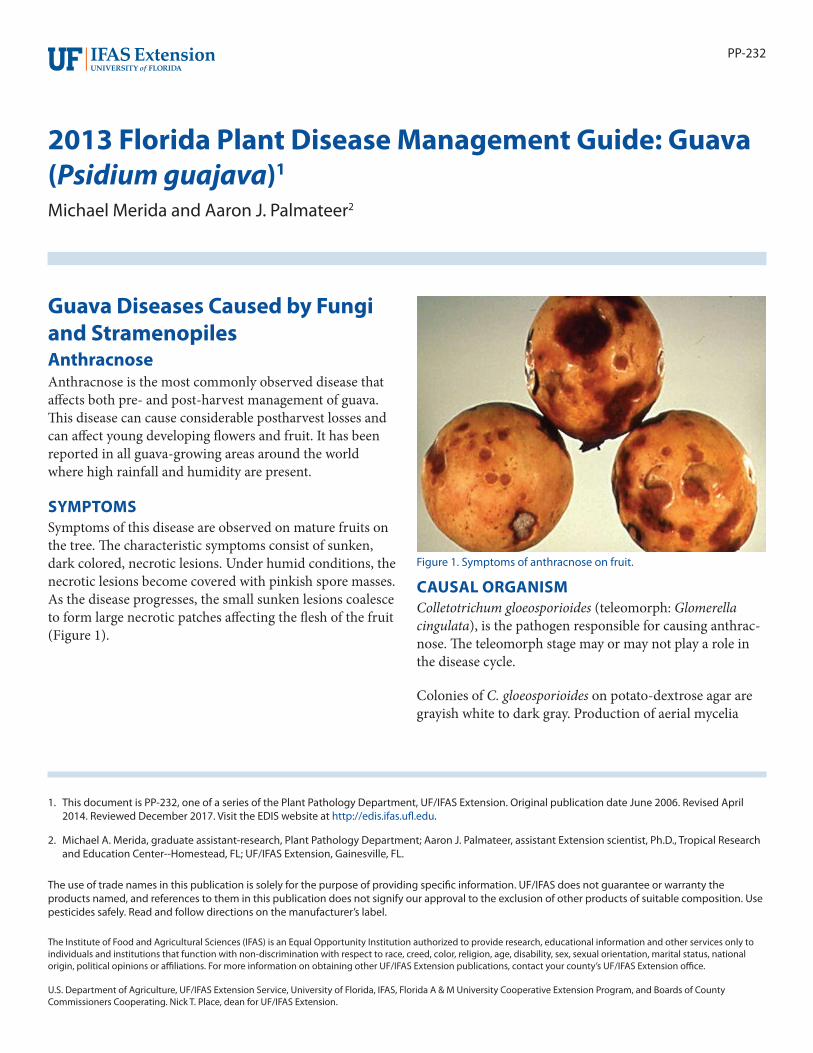
CAUSAL ORGANISMThe mycelium of Pseudocercospora psidii is internal, oliva-ceous, consisting of septate, branched, smooth hyphae, 3–4 µm wide. Conidiophores are aggregated in dense fascicles arising from the upper cells of a light brown stroma up to 50 µm wide; conidiophores light brown, smooth, 1–3-septate, subcylindrical, straight to variously curved, and unbranched. Conidia solitary, olivaceous, smooth, non-guttulate subcylindrical to narrowly obclavate, apex subobtuse, base narrowly obconically truncate, straight to curved, 1–5-septate (Figure 2).
DISEASE CYCLE AND EPIDEMIOLOGYThe source of inoculum most likely originates from infected leaves. The pathogens are capable of directly penetrating host tissues. Once infected, the fungus reproduces abun-dantly from the lower leaf surfaces.
Sporulation is greatest during warm, wet weather and most abundant from May thru September. Spores are dissemi-nated via wind, splashing rain, insects and irrigation.
Very small fruit and those at or near maturity are less susceptible than those fruit which are one-fourth to three-fourth of full size.
MANAGEMENTDue to the favorable environment for disease development in south Florida, strategic chemical control is deemed necessary for successful guava production. Copper based fungicides are labeled for the use on guava in Florida (Table 1).
Figure 2. Conidia of Psuedocercospora psidii.

32013 Florida Plant Disease Management Guide: Guava (Psidium guajava)
Guava RustThe rust disease can be very destructive to guava producing regions around the world. The pathogen has been reported to occur in Central and South America, the Caribbean and in Florida. Aside from guava, the disease has been reported to affect other members of the Myrtaceae family including allspice (Pimenta dioica), Eucalyptus (Eucalyptus spp.) and Melaleuca (Melaleuca quinquenervia) (Alfieri et al. 1994). Recently in Florida, the rust has been reported to occur in other hosts such as Myrcianthes fragrans, Myrtus communis, Callistemon viminalis, Syzygium jambos, S. paniculata, S. camini (Leahy 2004) and Myrtales in the family Heteropyxi-daceae on the genus Heteropyxis (Alfenas et al. 2005).
SYMPTOMSThe pathogen can affect foliage, young shoots, inflores-cences and fruit of guava. Typical symptoms associated with this disease include distortion, defoliation, reduced growth and if severe, mortality. On fully expanded leaves, dark bordered, roughly circular brown lesions with yellow halos develop (Burnett and Schubert 1985).
CAUSAL ORGANISMThe rust pathogen, Puccinia psidii G. Wint., is an obligate parasite belonging to the Basidiomycete class of the order Uredinales. It produces pale yellow, amphigenous uredinia, sub-epidermal then becoming erumpent, in groups on brownish or blackish spots (up to 5 mm in diameter) (Laundon and Waterson 1965). It produces urediniospores and teliospores, but teliospores have not been observed in Florida (Leahy 2004). Urediniospores are globose, ellipsoid to obovoid, with finely echinulate cell walls. If observed, teliospores are ellipsoid to oblong, rounded above and nar-row below, slightly constricted at the septum, pale yellow in color and smooth.
DISEASE CYCLE AND EPIDEMIOLOGYInfection of upper and lower leaves occurs in wet to moist environments within temperature ranges of 55–77°F (Burnett and Schubert 1985). Urediospores germinate at temperatures in the range of 18–22°C (64–71°F). Disease usually begins to occur at the onset and development of young shoots; leaves that are 40 days or more old have been observed to be more resistant to infection (Holliday 1980). Urediospore dissemination occurs via rainsplash.
MANAGEMENTControl of guava rust is based on the use of fungicides. Scouting fields for onset of disease or during the times of year when environmental conditions are favorable for
pathogen infection are recommended so that proper and timely fungicide applications can be made. In addition, proper cultural tactics such as proper fertilization, irriga-tion, pruning and sanitation aide in achieving a healthy, vigorously growing tree less vulnerable to disease pressures.
Mushroom Root RotMushroom root rot is a common and widespread disease that affects conifers and hardwoods in Florida. Aside from affecting Guava, the disease has been reported on over 200 species of trees and shrubs.
SYMPTOMSInfected trees will usually not show any symptoms until the disease has debilitated a significant portion of the root system. Diseased trees may exhibit a variety of symptoms, including: thinning of the crown, yellowing of foliage, premature defoliation, branch dieback, decaying roots, and lesions at the root collar. On some occasions, a tree may show symptoms of decline for several years before dying where as in other instances, trees may die rapidly without any visible symptoms.
CAUSAL ORGANISMMushroom root rot, caused by the fungus Armillaria tabescens, can cause severe problems for guava producing areas if the pathogen is present in the field. The most notable sign of disease, if present, is the characteristic mushrooms which develop near the base of infected trees. The mushrooms usually appear in fall, but they can occur at other times as well. The mushrooms, when fresh, are tan to brown, fleshy, with gills beneath the cap, and lacking an an-nulus around the stem (Ash and Barnard 1994). The disease may not always produce fruiting bodies when guava trees are infected. When a tree is in decline, and the mushrooms are absent, the fungus can be identified by the characteristic cream – colored mycelium just beneath the bark of infected roots and tree bases (Ash and Barnard 1994).
DISEASE CYCLE AND EPIDEMIOLOGYThe fungus usually spreads vegatatively via threadlike fungal strands from infected roots to healthy roots through root to root contact. Initial infections of the pathogen have been presumed to be via airborne basidiospores released from the gills on the undersides of the mushrooms, but this method has never been demonstrated. The removal of infected plants does not eradicate the mushroom root rot disease. The fungus survives saprophytically in stumps and dead roots and may cause infections for several years.
42013 Florida Plant Disease Management Guide: Guava (Psidium guajava)
Infected plants may not show any visible symptoms until a major portion of the root system is affected. The fungus can remain on the root system feeding without obvious injury to the plant. If plants are grown under optimum conditions with little stress, the plant can withstand the damage to the roots caused by Armillaria by continuously producing new, uninfected roots. However, if environmental conditions are not optimal and the plant is stressed, the pathogen can cause extensive damage leading to the rapid decline and eventual death of the plant without exhibiting obvious symptoms. In some instances where the environment is optimal for the fungus, rapid colonization and spread onto new and existing roots will cause a quick decline of a plant even though growing conditions are optimal for plant growth and development.
MANAGEMENTPreventative measures to manage mushroom root rot such as the removal of diseased trees and their root system before re-planting should be taken in order to reduce the possibility of future infections. Cultural practices to maintain vigorously growing plants help to reduce the colonization of the fungus. These practices include keeping root damage to a minimum when planting or transplanting, planting at proper depths so that the root collar is not buried in the soil, keeping mulch away from the root collar, and maintaining an optimum growing environment (i.e., soil fertility, pH, irrigation) to alleviate any stresses to the plant. Currently, there are no known effective fungicides registered for the control of mushroom root rot on guava.
Thread BlightThread blight occurs on a wide range of host plants. The disease develops primarily in the spring and summer months when warm, humid conditions are present.
SYMPTOMSTypical symptoms consist of circular to sub-circular leaf spots, tan to brown in color with alternating light and dark concentric rings dispersed throughout the canopy leaf spots eventually coalesce, resulting in a blight appearance. Leaves may be stuck together with tan to brown mycelium originating from the leaf margin.
Leaves, either on a new flush or an older flush, may appear water-soaked, necrotic, and the disease may eventually defoliate the plant. Leaves are held onto twigs by coarse brown threads of the causal pathogen. The fungus can invade leaves and twigs causing a dieback. It has been observed under warm, humid conditions, that the interior of the plant canopy is affected by the web blight disease
(Benson and Jones 2001). Distribution of the disease is likely when plants are in close proximity to each other under ideal conditions.
CAUSAL ORGANISMRhizoctonia solani Kuen (syn. Thanatephorus cucumeris [A. B. Frank]) Donk, teleomorph, is a hyphomycete with sterile mycelia. Hyphae of mycelium are brown with long cells and septa of branch set off from main hypha (Barnett and Hunter 1998). R. solani can be isolated from infected leaves cultured on growing media. Media used for mass hyphal transfers include Flowers media (FM), Acidified Potato Dextrose Agar (APDA), Quarter Strength Acidified Potato Dextrose Agar (QAPDA) and Water Agar (WA). The morphological characteristics of the pathogen grown on the media were indicative of a Rhizoctonia species. In order to speciate the Rhizoctonia pathogen, a nuclear staining technique was utilized to determine if the particular species was binucleate or multinucleate (Tu and Kimbrough 1973). Following the technique of Tu and Kimbrough (1973), it was determined that the pathogen was multinucleate, and confirming it as R. solani.
DISEASE CYCLE AND EPIDEMIOLOGYRhizoctonia spp. are among the most diverse of plant pathogenic fungi, causing root, stem, and foliar diseases of many subtropical and tropical hosts. Typically, Rhizoctonia spp. attack plants at the soil line causing root and stem rots. This pathogen can attack leaves as well, and is especially severe when trees are planted close together and the foliage remains moist for prolonged periods.
On guava, disease development can occur in less than one week. When humidity is high, the web-like brown myce-lium of the fungus can cover portions of the foliage that are infected. The pathogen may invade the canopy from the ground level by means of mycelial webbing (Schubert and El-Gholl 1995). Under favorable conditions, the pathogen may produce a thin grayish or tannish-white hymenial layer consisting of mycelium, basidia and basidiospores of the telemorph, Thanatephorus cucumeris, on the zonate lesions (Schubert and El-Gholl 1995). The lesions produced may appear water-soaked and discolored (Benson and Jones 2001). The characteristic feature of R. solani is the tan to brown colored mycelium produced on the margins of in-fected leaves that will cause necrotic and abscised leaves to web or stick together. If the infection is severe, the center of the plant may become defoliated. Rhizoctonia may survive as hyphae and sclerotia associated with infected crop debris (Benson and Jones 2001). Once the pathogen is established in a plant canopy, it may spread to adjacent plants if plants
52013 Florida Plant Disease Management Guide: Guava (Psidium guajava)
are spaced in close proximity to each other. It has been observed that under moist conditions leaf infection occurs within 48 hours of inoculation (Benson 1993).
MANAGEMENTCultural practices such as increased plant to plant spacing to promote air circulation, avoidance of overhead irrigation and irrigation regimens that extend leaf wetness periods, good sanitation practices (removal of infected plant debris, management of weeds that may be potential hosts, standing water, etc.) and proper nutrient management plans to maintain vigorous plants.
Fungicides may be used in a preventive or curative manner if infection is likely or present. Available fungicides for the use on Guava in Florida are found in Table 1.
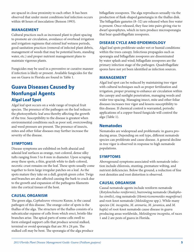
Guava Diseases Caused by Nonfungal AgentsAlgal Leaf SpotAlgal leaf spot occurs on a wide range of tropical fruit species. The presence of the pathogen on the leaf reduces the photosynthetic leaf area thereby affecting the growth of the tree. Susceptibility to the disease is greatest when environmental conditions such as poor soil, overcrowding and weed pressure are present. The presence of insects, mites and other foliar diseases may further increase the severity of the disease.
SYMPTOMSDisease symptoms are exhibited on both abaxial and adaxial leaf surfaces as orange, rust-colored, dense silky tufts ranging from 5 to 8 mm in diameter. Upon scraping away these spots, a thin, grayish white to dark-colored, necrotic crust remains on the leaf. These spots usually come together to form large irregular patches on a leaf. As the spots mature they take on a dull, grayish green color. Twigs and branches are also affected causing the bark to crack due to the growth and expansion of the pathogens filaments into the cortical tissues of the host.
CAUSAL ORGANISMThe green alga, Cephaleuros virescens Kunze, is the causal pathogen of this disease. The orange color of spots is the thallus of the alga. The structure of the thallus consists of a subcuticular expanse of cells from which erect, bristle-like branches arise. The apical parts of some cells swell to form enlarged support cells that produce several stalked, terminal or ovoid sporangia that are 30 x 24 µm. The stalked cell may be bent. The sporangia of the alga produce
biflagellate zoospores. The alga reproduces sexually via the production of flask-shaped gametangia in the thallus disk. The biflagellate gametes (8–32) are released when free water is present. Once released, they fuse into pairs giving rise to dwarf sporophytes, which in turn produce microsporangia that bear quadriflagellate zoospores.
DISEASE CYCLE AND EPIDEMIOLOGYAlgal leaf spots proliferate under wet or humid conditions within the trees canopy. Infectious propagules such as sporangia and biflagellate zoospores are disseminated by water splash and wind; biflagellate zoospores are the primary infection stage of the pathogen. Quadriflagellate spores have not yet been identified as infection sources.
MANAGEMENTAlgal leaf spot can be reduced by maintaining tree vigor with cultural techniques such as proper fertilization and irrigation, proper pruning to enhance air circulation within the canopy and sunlight penetration, managing weeds and wider tree spacing. Managing insect, mite and other foliar diseases increases tree vigor and lessens susceptibility to this disease. If chemical control is warranted, periodic applications of a copper-based fungicide will control the alga (Table 1).
NematodesNematodes are widespread and problematic in guava pro-ducing areas. Depending on soil type, different nematode species can proliferate and cause disease. A general decline in tree vigor is observed in response to high nematode populations.
SYMPTOMSAboveground symptoms associated with nematode infec-tion include chlorosis, stunting, premature wilting, and nutrient deficiencies. Below the ground, a reduction of fine root densities and root distortion is observed.
CAUSAL ORGANISMCausal nematode agents include reniform nematode (Rotylenchulus reniformis), burrowing nematode (Radopho-lus similis), ring nematode (Hemicriconemoides magniferae) and root-knot nematode (Meloidogyne spp.). While many species (M. incognita, M. arenaria, M. javanica, and M. hapla) of root-knot nematode cause disease in guava producing areas worldwide, Meloidogyne incognita, of races 1 and 2 are pests of guava in Florida.
62013 Florida Plant Disease Management Guide: Guava (Psidium guajava)
MANAGEMENTThe use of nematode free planting stock is the best control method. In Florida, there are no currently registered nematicides for the use on guava.
References CitedAlfieri, S.A., Jr., K.R. Langdon, J.W. Kimbrough, N.E. El-Gholl, and C. Wehlburg. 1994. Diseases and disorders of plants in Florida. Florida Department of Agriculture & Consumer Services, Division of Plant Industry, Gainesville, FL. Bulletin 14. 653 p.
Alfenas, A.C., Zauza, E.A.V., Wingfeld, M.J., Roux, J., and Glen, M. 2005. Heteropyxis natalensis, a new host of Puc-cinia psidii rust. Australas. Pl. Pathol. 34: 285 – 286.
Ash III, E.C. and Barnard, E.L. (1994). Mushroom Root Rot. Forest and Shade Tree Pests. Division of Forestry. Leaflet number 11 (February). Florida Department of Agriculture & Consumer Services.
Barnett, H.L. and B.B. Hunter. 1998. Illustrated Genera of Imperfect Fungi, 4th edition. APS Press, St. Paul, MN.
Benson, M.D. 1993. Rhizoctonia Web Blight. P. 20-21. Compendium of Rhododendron Diseases. APS Press, St. Paul, MN.
Benson, M.D. and R.K. Jones. 2001. Rhizoctonia Web Blight. P. 63-64. Diseases of Woody Ornamentals and Trees in Nurseries. APS Press, St. Paul, MN.
Burnett H.C. and Schubert, T.S. 1985. Puccinia psidii on Allspice and Related Plants. Plant Pathology Circular No. 271 (May). Florida Department of Agriculture & Consumer Services, Division of Plant Industry, Gainesville, FL.
Chemsearch. 2006. Labels & MSDS. http://www.cdms.net/LabelsMsds/LMDefault.aspx
Hernández, J.R.. 27 February 2006. Invasive Fungi. Puccinia psidii. Systematic Botany & Mycology Laboratory, ARS, USDA. Retrieved May 3, 2006, from http://nt.ars-grin.gov/sbmlweb/OnlineResources/FungiOnline.cfm .
Holliday, P. (1980). Fungus Diseases of Tropical Crops. Cambridge University Press, Cambridge. p. 401.
Laundon, G.F. and Waterson, J.M. (1965) Puccinia psidii . CMI Descriptions of Pathogenic Fungi and Bacteria No. 56. Commonwealth Mycological Institute, Kew, UK.
Leahy, R. (2004). Recent History of Puccinia psidii on Myrtaceae in Florida. UF/IFAS Pest Alert (January). Florida Department of Agriculture & Consumer Services, Division of Plant Industry, Gainesville, FL.
Schubert, T.S. and N.E. El-Gholl. 1995. Rhizoctonia Leaf Spot of Rhaphiolepis. Plant Pathology Circular No. 374 (Nov./Dec.). Florida Department of Agriculture & Con-sumer Services, Division of Plant Industry, Gainesville, FL.
Tu, C.C., and J.W. Kimbrough. 1973. A Rapid Staining Technique for Rhizoctonia Solani and Related Fungi. Mycologia, Vol. LXV, No. 4, pp. 941- 944, July- Aug.
72013 Florida Plant Disease Management Guide: Guava (Psidium guajava)
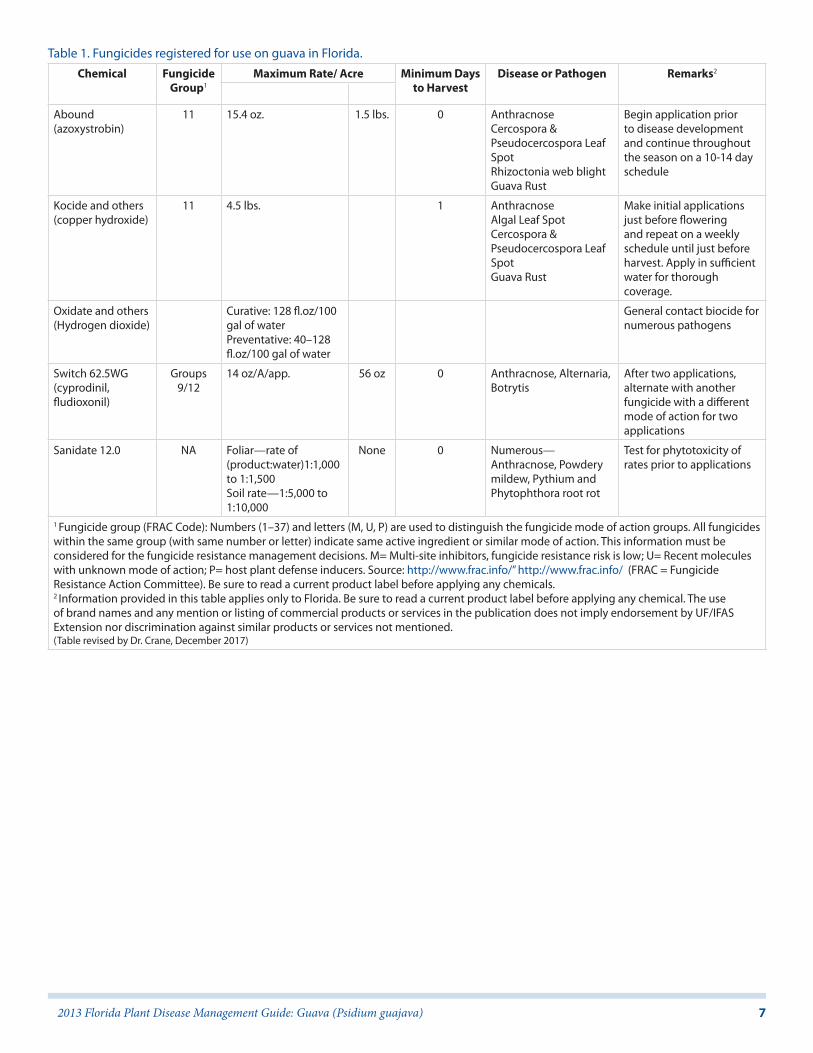
Table 1. Fungicides registered for use on guava in Florida.Chemical Fungicide
Group1Maximum Rate/ Acre Minimum Days
to HarvestDisease or Pathogen Remarks2
Abound (azoxystrobin)
11 15.4 oz. 1.5 lbs. 0 Anthracnose Cercospora & Pseudocercospora Leaf Spot Rhizoctonia web blight Guava Rust
Begin application prior to disease development and continue throughout the season on a 10-14 day schedule
Kocide and others (copper hydroxide)
11 4.5 lbs. 1 Anthracnose Algal Leaf Spot Cercospora & Pseudocercospora Leaf Spot Guava Rust
Make initial applications just before flowering and repeat on a weekly schedule until just before harvest. Apply in sufficient water for thorough coverage.
Oxidate and others (Hydrogen dioxide)
Curative: 128 fl.oz/100 gal of water Preventative: 40–128 fl.oz/100 gal of water
General contact biocide for numerous pathogens
Switch 62.5WG (cyprodinil, fludioxonil)
Groups 9/12
14 oz/A/app. 56 oz 0 Anthracnose, Alternaria, Botrytis
After two applications, alternate with another fungicide with a different mode of action for two applications
Sanidate 12.0 NA Foliar—rate of (product:water)1:1,000 to 1:1,500Soil rate—1:5,000 to 1:10,000
None 0 Numerous—Anthracnose, Powdery mildew, Pythium and Phytophthora root rot
Test for phytotoxicity of rates prior to applications
1 Fungicide group (FRAC Code): Numbers (1–37) and letters (M, U, P) are used to distinguish the fungicide mode of action groups. All fungicides within the same group (with same number or letter) indicate same active ingredient or similar mode of action. This information must be considered for the fungicide resistance management decisions. M= Multi-site inhibitors, fungicide resistance risk is low; U= Recent molecules with unknown mode of action; P= host plant defense inducers. Source: http://www.frac.info/” http://www.frac.info/ (FRAC = Fungicide Resistance Action Committee). Be sure to read a current product label before applying any chemicals.2 Information provided in this table applies only to Florida. Be sure to read a current product label before applying any chemical. The use of brand names and any mention or listing of commercial products or services in the publication does not imply endorsement by UF/IFAS Extension nor discrimination against similar products or services not mentioned.(Table revised by Dr. Crane, December 2017)