Embed Size (px)
Citation preview

BIO.FAS.VER.O2-12March2012int
Fastin
Elastin Assay
biocolor life science assays
Internet Manual
Downloaded from www.biocolor.co.uk
TM
2012
EDITION

2
Fastin Elastin Assay
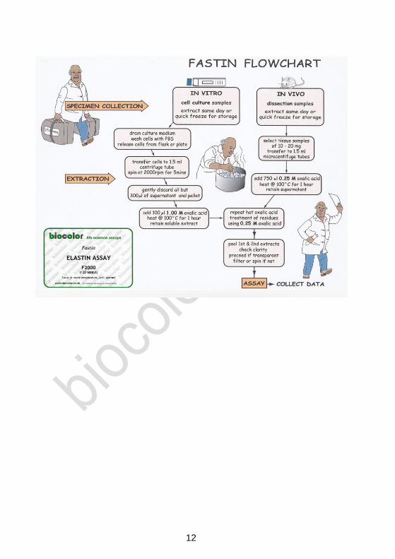
General Protocol Detection Limit: 5μg All test samples require the conversion of the native hydrophobic
elastin into a water soluble derivative, (α-elastin). To extract as α-elastin the sample is heated at 100
0C for two one
hour periods with 0.25M oxalic acid. For detailed information on extraction treatment of in-vivo and in-vitro samples see pages 4 to 8.
_________________________________________________ Label a set of 1.5 ml microcentrifuge tubes. If sufficient test material
is available run duplicate samples.
[1] Prepare Reagent Blanks: 100μl of test solution solvent, (buffer /
PBS / water / 0.25M oxalic acid).
[2] -elastin standard: suggested volumes - 12.5, 25.0 and 50.0μl
duplicate aliquots.
[3] Test samples: in vivo derived tissue extracts or in vitro cell culture
samples. Select volumes between 50μl and 500μl ofelastin.
______________________________________________________
[4] To each tube add an equal volume of Elastin Precipitating
Reagent (stored at 40C).
[5] Cap tubes and briefly vortex to mix contents; leave for 15 minutes
to complete precipitation of elastin.
[6] Centrifuge tubes @ >10,000 x g for 10 minutes, (Fig. 1a).
[7] Drain tube's liquid contents into a beaker. While the tube is still
inverted remove most of the remaining fluid from the tube by tapping
the inverted tube onto a single thickness absorbent paper towel.
ASSAY PROTOCOL CONTINUED ON INSIDE BACK COVER OF MANUAL
0
mins
Extraction
Isolation
Assay

3
PROTOCOL CONTINUED FROM INSIDE FRONT COVER
Formation of Elastin - Dye Complex
[8] To all tubes add 1.00ml of Dye Reagent.
Cap tubes and mix contents by inverting the tubes. Then disperse the the elastin precipitate using a vortex mixer.
Place the rack of tubes on a mechanical shaker and allow reaction
between the elastin and the dye to proceed for 90 minutes.
[9] Centrifuge the tubes @ >10,000 x g for 10 minutes.
Recovery of Elastin-Dye Complex
[10] Drain the tubes of unbound dye. While the tube is still inverted
remove most of the remaining fluid from the tube by FIRMLY tapping
the lip of the tube onto a single thickness absorbent paper towel.
A 'cotton bud', (or Q-tip), can be useful for removing any fluid droplets
from the rim of the tube. On returning the tube to the upright position
not more than 25μl of fluid should be found in the bottom of the tube.
[11] The elastin-dye complex can be observed as a reddish-brown
deposit in the bottom and inside lower wall of the tube, (Fig. 1b).
Release, and Recovery, of the Elastin Bound Dye
[12] To each tube add 250μl of Dye Dissociation Reagent. Cap tubes
and release the dye into solution with the aid of a vortex mixer.
Repeat the vortex mixing after 10 minutes to ensure that all bound
dye has passed into solution.
[13] Transfer the contents of each tube to a well in a ninety-six well
flat bottom microwell plate. Prepare a map in the lab notebook to
record which tube contents went into which well.
Elastin Measurement; (dye recovered)
[14] Place microwell plate into the Microplate Reader. Select
wavelength or colour filter nearest to 513 nm, (blue-green colour).
Plot Reference Standards and use this graph to determine the elastin
content of the Test Samples.
PLEASE READ MANUAL BEFORE USING THE ASSAY
Dye
Binding
Dye Bound Elastin
Dye
Release
Dye
Recovery
4
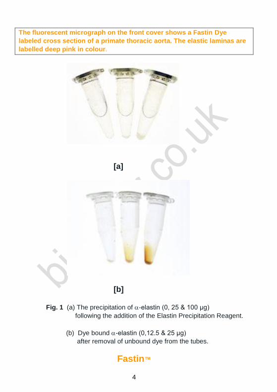
The fluorescent micrograph on the front cover shows a Fastin Dye
labeled cross section of a primate thoracic aorta. The elastic laminas are
labelled deep pink in colour.
[a]
[b]
Fig. 1 (a) The precipitation of -elastin (0, 25 & 100 μg)
following the addition of the Elastin Precipitation Reagent.
(b) Dye bound -elastin (0,12.5 & 25 μg)
after removal of unbound dye from the tubes.
Fastin™

5
Elastin Assay
The Fastin Assay has been designed for research work only.
Handle the Fastin Assay using Good Laboratory Practice.
TECHNICAL INFORMATION
ASSAY PROTOCOL Inside front and back page cover
Intended Applications 1
Assay Components 2
Mode of Action 3
Extraction of Elastin from Animal Tissue 4
Test Results: Mouse Tissue Elastin Contents 5
Recovery of Elastin from Cells during In-Vitro Culture 6
Test Results: Mammalian Cell Elastin during In-Vitro Culture 7
Assay; Supplementary Notes 9
Elastin Biography 11
© Biocolor Ltd., 2011
Fastin is a Trademark of Biocolor Ltd.
Published by
Biocolor
8 Meadowbank Road, Carrickfergus,
BT38 8YF, Northern Ireland, U.K.
www.biocolor.co.uk
1
Assay Manual Intended Applications
The Fastin Elastin Assay is a quantitative dye-binding method for the analysis of
elastins extracted from mammalian sources.
The dye label employed is 5,10,15,20-tetraphenyl-21H,23H-porphine tetra-sulfonate
(TPPS). For the structural form of the dye see Fig.2 (page 3).
Test Sample Material
Tissue extracts and cells during in-vitro culture.
Elastin forms that can be measured by the Fastin Assay as α-elastin:
(i) soluble tropoelastins
(ii) lathyrogenic elastins
(iii) insoluble elastins, following solubilization to elastin polypeptides, [α-elastin; κ-
elastin]
The dye reagent binds to the 'basic' and 'non-polar' amino acid sequences found in
mammalian elastins.
Test sample quantities
A sample volume of between 50 and 500μl is required, containing not less than 5µg
and not more than 70µg elastin.
Test sample composition
For analysis of soluble elastin, samples should be free of any particulate material (cell
debris, insoluble extracellular matrix material). The presence of other soluble proteins
or of complex carbohydrates does not interfere with the Fastin Assay.
Fastin Kit Pack Size and Storage Conditions
Standard Assay Kit Product code: F2000 (120 assays)
Economy Pack Product Code: F4000 (475 assays)
Storage Unopened - All of the reagents have long term stability (one year), when
stored at room temperature. Do not freeze as complete solubility may not occur on
thawing.
Storage after Opening - Reference Standard: When stored at +4ºC the α-elastin
standard is a clear transparent solution. On holding at room temperature the solution
may be observed to become opalescent. This is due to the characteristic coacervation
property of soluble elastin. On cooling, the process is reversible and the elastin solution
again becomes transparent.

2
Fastin Assay Kit Components
1. Fastin Dye Reagent contains 5,10,15,20-tetraphenyl-21H,23H-porphine
tetrasulfonate (TPPS) in a citrate-phosphate buffer.
The pH of this reagent is pH 7.5 and, to limit microbial growth, inhibitors have
been added to the reagent. These agents, bromopol and sorbic acid, are
compatible with the Fastin Assay but are not 'universal' microbial inhibitors.
Storing an opened bottle of Fastin Dye Reagent at 4ºC can extend the shelf
life of the reagent.
2. Elastin Precipitating Reagent contains trichloroacetic and hydrochloric
acids.
3. Elastin Standard is a high molecular weight fraction of α-elastin prepared
from bovine neck ligament elastin. The α-elastin standard is supplied as a
sterile solution; concentration 1.0 mg/ml, in 0.25M oxalic acid.
The full metal seal should not be removed from the vial.
(a) Remove the centre metal disc only from the vial top.
(b) Obtain aliquots from the vial by using a syringe fitted with a sterile
hypodermic needle. The butyl rubber seal on the vial has a thin centre disc.
(c) Do not return any unused aliquots to the vial.
(d) The α-elastin standard should be discarded if the solution becomes turbid.
4. Dye Dissociation Reagent contains guanidine HCl and propan-1-ol.
5. Oxalic acid, 1.0M. Dilute to 0.25M for tissue samples (see page 4).
6. Assay Manual. Additional free copies can be downloaded from Biocolor’s
website.
Equipment Required
Mechanical shaker to provide gentle mixing of the elastin and the Dye Reagent.
A centrifuge, fitted with a 1.5 ml microcentrifuge tube rotor head; capable of 10,000 x g.
A Microplate Reader, with a suitable colour filter (absorbance peak of dye occurs at 513 nm). A metal heating block with the thermostat set between 95 and 100
0C is convenient for
extracting elastin from tissue samples. Otherwise a boiling water resistant glass beaker
on a hotplate with thermostat set between 95 and 1000C, can be used.

3
Mode of Action of the Fastin Dye Reagent with Elastin
The Fastin Dye Reagent contains a synthetic porphyrin, 5,10,15,20-tetraphenyl-21H,23H-porphine, that is water soluble in the sulfonate form. The TPPS molecules contain four sulfate groups.
Fig. 2 The visible absorbance spectrum and structural form of
5,10,15,20-tetraphenyl-21H,23H-porphine,tetra-sulfonate.
‘The Winkelman Reaction’
The affinity of TPPS for elastin was first observed when used as a 'vital stain' on live animals. Most tissues took up the dye initially but only elastin retained the TPPS molecules over time. [Winkelman, J. (1962), Cancer Res. 22, 589-596; Winkelman, J & Spicer, S. (1962), Stain Technol. 37, 303-305].
SAMPLE PREPARATION PRIOR TO ASSAY
The Fastin Assay protocol is found on the front and rear inside covers of this manual
and supplementary notes for the protocol on pages 9 and 10.
However, before the assay can be performed cell and tissue elastin must be converted
into a water soluble derivative; α-elastin. Extraction of the elastin component of the
elastic matrix is obtained by heating the test samples in hot (95 - 1000C) oxalic acid for
one hour, converting the hydrophobic elastin into water soluble α-elastin.
The recommended extraction procedures are described in the following pages.
OS
OO
-
OS
O
O-
O
N
NH
N
NH
SO
O-
OS
OO
-
OS
OO
-
OS
O
O-
O
N
NH
N
NH
SO
O-
OS
OO
-
4
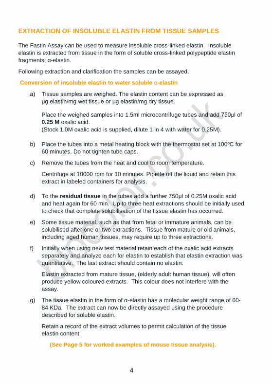
EXTRACTION OF INSOLUBLE ELASTIN FROM TISSUE SAMPLES The Fastin Assay can be used to measure insoluble cross-linked elastin. Insoluble
elastin is extracted from tissue in the form of soluble cross-linked polypeptide elastin
fragments; α-elastin.
Following extraction and clarification the samples can be assayed.
Conversion of insoluble elastin to water soluble α-elastin
a) Tissue samples are weighed. The elastin content can be expressed as
μg elastin/mg wet tissue or μg elastin/mg dry tissue.
Place the weighed samples into 1.5ml microcentrifuge tubes and add 750μl of
0.25 M oxalic acid.
(Stock 1.0M oxalic acid is supplied, dilute 1 in 4 with water for 0.25M).
b) Place the tubes into a metal heating block with the thermostat set at 100ºC for
60 minutes. Do not tighten tube caps.
c) Remove the tubes from the heat and cool to room temperature.
Centrifuge at 10000 rpm for 10 minutes. Pipette off the liquid and retain this
extract in labeled containers for analysis.
d) To the residual tissue in the tubes add a further 750μl of 0.25M oxalic acid
and heat again for 60 min. Up to three heat extractions should be initially used
to check that complete solubilisation of the tissue elastin has occurred.
e) Some tissue material, such as that from fetal or immature animals, can be
solubilised after one or two extractions. Tissue from mature or old animals,
including aged human tissues, may require up to three extractions.
f) Initially when using new test material retain each of the oxalic acid extracts
separately and analyze each for elastin to establish that elastin extraction was
quantitative. The last extract should contain no elastin.
Elastin extracted from mature tissue, (elderly adult human tissue), will often
produce yellow coloured extracts. This colour does not interfere with the
assay.
g) The tissue elastin in the form of α-elastin has a molecular weight range of 60-
84 KDa. The extract can now be directly assayed using the procedure
described for soluble elastin.
Retain a record of the extract volumes to permit calculation of the tissue
elastin content.
(See Page 5 for worked examples of mouse tissue analysis).
5
Skin
Heart
Liv
er
Lu
ng
0
10
20
30
40
50
60Skin
Heart
Liver
Lung
ela
sti
n c
on
ten
t
ug
/10 m
g w
et
tissu
e
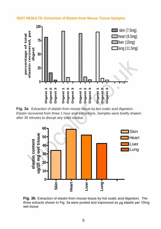
TEST RESULTS: Extraction of Elastin from Mouse Tissue Samples
Dig
est 1
Dig
est 2
Dig
est 3
Dig
est 1
Dig
est 2
Dig
est 3
Dig
est 1
Dig
est 2
Dig
est 3
Dig
est 1
Dig
est 2
Dig
est 3
0
25
50
75
100 skin (7.5mg)
heart (8.5mg)
liver (15mg)
lung (11.5mg)
percen
tag
e o
f t
otal
ela
stin
reco
vered
, p
er
dig
est
Fig. 3a Extraction of elastin from mouse tissue by hot oxalic acid digestion.
Elastin recovered from three 1 hour acid extractions. Samples were briefly shaken
after 30 minutes to disrupt any solid residue.
Fig. 3b Extraction of elastin from mouse tissue by hot oxalic acid digestion. The
three extracts shown in Fig. 3a were pooled and expressed as μg elastin per 10mg wet tissue
6
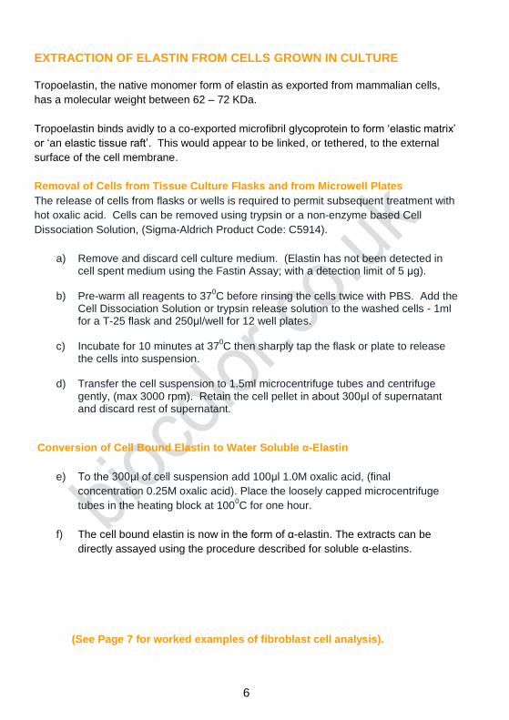
EXTRACTION OF ELASTIN FROM CELLS GROWN IN CULTURE Tropoelastin, the native monomer form of elastin as exported from mammalian cells,
has a molecular weight between 62 – 72 KDa.
Tropoelastin binds avidly to a co-exported microfibril glycoprotein to form ‘elastic matrix’
or ‘an elastic tissue raft’. This would appear to be linked, or tethered, to the external
surface of the cell membrane.
Removal of Cells from Tissue Culture Flasks and from Microwell Plates
The release of cells from flasks or wells is required to permit subsequent treatment with
hot oxalic acid. Cells can be removed using trypsin or a non-enzyme based Cell
Dissociation Solution, (Sigma-Aldrich Product Code: C5914).
a) Remove and discard cell culture medium. (Elastin has not been detected in cell spent medium using the Fastin Assay; with a detection limit of 5 μg).
b) Pre-warm all reagents to 370C before rinsing the cells twice with PBS. Add the
Cell Dissociation Solution or trypsin release solution to the washed cells - 1ml for a T-25 flask and 250μl/well for 12 well plates.
c) Incubate for 10 minutes at 370C then sharply tap the flask or plate to release
the cells into suspension.
d) Transfer the cell suspension to 1.5ml microcentrifuge tubes and centrifuge gently, (max 3000 rpm). Retain the cell pellet in about 300μl of supernatant and discard rest of supernatant.
Conversion of Cell Bound Elastin to Water Soluble α-Elastin
e) To the 300μl of cell suspension add 100μl 1.0M oxalic acid, (final
concentration 0.25M oxalic acid). Place the loosely capped microcentrifuge
tubes in the heating block at 1000C for one hour.
f) The cell bound elastin is now in the form of α-elastin. The extracts can be
directly assayed using the procedure described for soluble α-elastins.
(See Page 7 for worked examples of fibroblast cell analysis).

7
TEST RESULTS: Extraction of Elastin from In Vitro Cultured Fibroblasts
dig
esti
on
1
dig
esti
on
2
dig
esti
on
3
flask r
esu
du
e
0
100
200
300
400
500
600
ela
sti
n r
eco
vere
d / u
g
Fig. 4 Extraction of elastin from a CHO cell line using hot oxalic acid.
T25 cell culture flasks containing CHO cells were permitted to come to confluency.
The flasks were then drained of cell medium and washed twice with PBS. Two ml of
0.25M oxalic acid was added to each flask. The flasks were placed in an oven,
thermostat controlled, at 1000 C for one hour. The digest extract was then removed
and retained. To the residue 1.0ml of fresh oxalic acid was added and the extraction
collected, and the process repeated to give a total of three extractions.
The flask was then washed with PBS and 2 ml of dye added for 90 mins. The
unbound dye was removed and bound dye released. The finding indicated some
accumulation of cells and elastin on the plastic surface of the flasks, (Fig. 4).
Elastin was not detected in the spent medium, (detection limit; 5μg).
Each extract (200μl) was mixed with the Elastin Precipitating Reagent (200μl). After
10 minutes the samples were centrifuged. The drained elastin pellets were then
mixed with 2ml of the Fastin Dye for 90 mins. Unbound dye was removed. Bound
dye was released from the elastin-dye pellet using 1 ml of the Dye Dissociation
Reagent.
The mean data from six T25 flasks with CHO cells is shown in Fig.4. The findings
displayed an abundance of elastin produced by CHO cells after 48 hours in culture.
A 100μl aliquot of the pooled extracts produced 30μg of elastin. This was well within
the range of the Fastin Standard Curve.

8
Measurement of Cell Generated Elastin in T25 Flasks
24h
48h
72h
96h
0
50
100
150
200
250
300
350
400
45024h
48h
72h
96h
po
ole
d d
igests
ela
sti
n
(ug
)
Fig. 5 Rate and quantity of elastin produced by CHO cells.
In this series of experiments using CHO cells, (seeding density 5 x 10⁵), in T25
flasks the cells were removed from the flasks using 1 ml of a detergent based cell
releasing agent, (Sigma-Aldrich Cell Dissociation Solution, Product Code: C1419).
This permitted the acid digestion to be carried out in 1.5 ml microcentrifuge tubes.
1.0 M oxalic acid (100μl) was added to the cell suspensions (300μl) giving a final
concentration of 0.25 M oxalic acid. This was digested at 1000C for 1 hour, with
occasional vortexing. The transparent extract was treated with an equal volume of
the Acid Precipitating Reagent and the elastin pellet recovered then mixed with the
Fastin Dye (Fig. 5). Unlike the previous experiment where digestion and extraction
was carried out in the T25 flask these T25 flasks were stripped of elastin and cells
before acid digestion.
Measurement of Cell Generated Elastin in 12 Well Microplates.
The seeding density was 1 x 10⁵ and grown to confluence by 48 hours. The cell
medium was removed and cells washed twice with PBS. 300μl of the detergent
based cell releasing agent was added and the plate incubated at 370C for 10 mins.
The wells were tapped and scraped and the cell suspension transferred to
microcentrifuge tubes where 100μl of 1.0M oxalic acid was added, and extraction
carried out as described above.
Elastin Recovered from CHO Cells after 48 Hours of In-Vitro Culture.
___________________________________________________________________
Format Seeding density Growth surface Elastin Recovered
T25 5 x 10⁵ 25 cm
2 204 μg
Flask
12 well 1 x 10
5 3.8 cm
2 42 μg
plate
__________________________________________________________________

9
SUPPLEMENTARY NOTES FOR THE MEASUREMENT OF SOLUBLE ELASTIN
(i) Test Samples
Try single 50µl aliquots of test sample, for the first trial, where the elastin concentration
range is as yet unknown
If the absorbance readings are found to be more than 1.0 (after subtraction of the
reagent blank value) repeat assay using a smaller test sample aliquot, or dilute.
If the absorbance readings are less than 0.05 the test sample contains less than 5µg
elastin and will require a larger sample aliquot or concentration before being re-
assayed.
For reliable and accurate results all test samples should have their absorbance
readings within the range of the Elastin Standards that were plotted on the calibration
curve. Fig.6 displays a typical standard curve.
Fig. 6 Elastin Standard Curve. The curve was generated from samples on a 96
well microplate, 250μl/well.

10
(ii) Elastin Isolation (inside manual cover, step 4) The Precipitating Reagent has been developed for elastin recovery. The reagent
should not be diluted with sample volumes greater than in a ratio of 1:1. The reagent
can be pre-cooled to <5ºC (store this reagent in the refrigerator so that it is ready for
use). To tubes, containing Standards or Test Samples, add an equal volume of the
Reagent.
Following the precipitation of the elastin, the microcentrifuge tubes are centrifuged for
10,000 x g for 10 minutes, to pack the precipitated α-elastin. Remove tubes from the
centrifuge, uncap and carefully invert, to drain the liquid contents into a waste beaker.
While inverted remove any remaining fluid from the top of the tubes by tapping the tube
onto an absorbent paper towel. A 'cotton bud', (or Q-tip), can be useful to remove any
fluid droplets from the rim of the tube. On returning the tube to the upright position not
more than 25μl of fluid should be found in the bottom of the tube.
Low concentrations of α-elastin can be difficult to 'see' as it occurs as a translucent gel.
On the outside back cover of this manual the top photograph required 100μg of
α-elastin to visually display the protein pellet (Fig. 1a).
(iii) Recovery of the Elastin-Dye Complex (inside manual cover, step 8)
Following the dye binding step the elastin-dye complex formed becomes insoluble in
the presence of ammonium sulfate within the Fastin Dye Reagent.
The elastin-dye complex is separated from the remaining soluble unbound dye by
centrifuging the tubes (>10,000 x g for 10 minutes).
Visual inspection should reveal a red residue within the elastin standard tubes and,
hopefully, also in the test sample tubes, (Fig. 1b).
(iv) Release of the Elastin Bound Dye (inside manual cover, step 12)
To each tube add 250μl of Dye Dissociation Reagent. Cap the tubes and bring
the elastin-bound dye into solution using a vortex mixer. Two brief mixing periods are usually more effective than one long mixing period. Tubes should not be uncapped until transfer to the wells of a 96-well microplate for absorbance measurement. The dye extract is stable for several hours, but if readings are to be delayed store the tube rack containing the microcentrifuge tubes in a light-proof container or cupboard.
(iv) Elastin Measurement (inside manual cover, step 14)
The elastin content of the assayed samples is determined by the amount of bound dye released from the α- elastin.
Transfer the complete 250μl contents of the labeled microcentrifuge tubes to wells of a 96 well microplate, (with flat-bottom wells to reduce light scatter). The absorbance peak of TPPS in the Dye Dissociation Reagent occurs at 513 nm. Although the instrument can be set to zero using the reagent blank, it is usually better to determine the absorbance of the blank as a quality control check of the assay. Check the colour filter options that are available for the Microplate Reader; a blue green filter will probably be found to be suitable.

11
ELASTIN SOURCE REFERENCES
Biochemistry, biophysics; preparation, extraction & analysis
[2008]: Mecham, R.P. ‘Methods in Elastic Tissue Biology: Elastin Isolation and
Purification ’ Methods, 45, 32-41.
[2005]: Parry, D.A.D & Squire, xx. Eds; ‘Fibrous Proteins’, Advances in Protein
Chemistry, Vol. 70, Elsevier Academic Press, San Diago.
[2003]: Shewey, P.R., Tatham, A.S. & Bailey, A.J. Eds; ‘Elastomeric Proteins’
Cambridge University Press, Cambridge.
[1993]: Kreis, T & Vale, R. Eds; 'Guidebook to the Extracellular Matrix and Adhesion
Proteins'. Oxford University Press, Oxford.
[1993]: Royce, P.M. & Steinmann, B.U. Eds of 'Connective tissue and its heritable
disorders; molecular, genetic and medical aspects'. Wiley-Liss, New York.
[1989]: Robert, L. & Hornebeck, W. Eds of 'Elastin and Elastases', in two volumes.
CRC Press Inc. Boca Raton, Florida.
[1987]: Cunningham, L.W. Ed; ‘Structural and Contractile Proteins’. Methods of
Enzymology, Vol.144, Academic Press, Florida.
[1987]: Uitto, J., & Perejda, A. J. [1987], Eds 'Connective Tissue Disease. Molecular
Pathology of the Extracellular Matrix'. Marcel Dekker, New York.
[1986]: Mecham, R.P. Ed 'Regulation of Matrix Accumulation'. Academic Press,
New York.
[1982[: Cunningham, L.W. & Frederiksen, D.W. Eds of 'Elastin Structure and
Biosynthesis'. Methods of Enzymology, Vol. 82. Academic Press, New York.
[1980]: Robert, A.M. & Robert, L. Eds; ‘Biology and Pathology of Elastic Tissues’
Frontiers of Matrix Biology, Vol. 8. S. Karger, Basel.
[1977]: Sandberg, L.B., Gray, W.R. & Franzblau, C. Eds of 'Elastin and Elastic Tissue',
Advances Exper. Med. Biol. Series, Vol. 79, Plenum Press, New York.
Biocolor’s website has a list of research papers
that have cited the Fastin Assay.
BIO.FAS.VER.O1-05July2011int
12


![[Slideshare]fardhu'ain lesson#14-arkaan-ul-islam(5)fastin gin-ramadhan-(3-feb-l2012)](https://img.pdfslide.us/doc/110x75/54bbc85f4a795929048b459a/slidesharefardhuain-lesson14-arkaan-ul-islam5fastin-gin-ramadhan-3-feb-l2012.jpg)