Embed Size (px)
DESCRIPTION
Sindrome
Citation preview

Rev esp cir ortop traumatol. 2011;55(2):140-150
1888-4415/$ - see front matter © 2010 SECOT. Published by Elsevier España, S.L. All rights reserved.
www.elsevier.es/rot
Revista Española de Cirugía Ortopédica y Traumatología
☆ This study was financed by CIBERNED and the ISCIII (PI07/132E) Health Research Fund.*Corresponding author.E-mail: [email protected] (J. Berciano).
REVIEW ARTICLE
Charcot-Marie-Tooth disease: a review with emphasis on the pathophysiology of pes cavus ☆
J. Berciano, a,d,* E. Gallardo, b,d A. García,c,d A.L. Pelayo-Negro, a,d J. Infante, a,d O. Combarros a,d
a Servicio de Neurología, Hospital Universitario Marqués de Valdecilla (IFIMAV), Universidad de Cantabria (UC), Santander, Spainb Servicio de Radiodiagnóstico, Hospital Universitario Marqués de Valdecilla (IFIMAV), Universidad de Cantabria (UC), Santander, Spainc Servicio de Neurofisiología Clínica, Hospital Universitario Marqués de Valdecilla (IFIMAV), Universidad de Cantabria (UC), Santander, Spaind Centro de Investigación Biomédica en Red de Enfermedades Neurodegenerativas (CIBERNED), Santander, Spain
Received September 23, 2010; accepted September 30, 2010
KEYWORDSAxon;Charcot-Marie-Tooth disease;Dejerine-Sottas disease;Gene mutation;Genetic neuropathy;Pes cavus;Magnetic resonance;Myelin;Nerve conduction velocity;Vitamin C
Abstract Charcot-Marie-Tooth disease is the most frequent inherited neuropathy with a prevalence ratio in Spain of 28.2 cases / 100,000 inhabitants. It is a sensory-motor polyneuropathic syndrome, either demyelinating or axonal, which might be transmitted with autosomal dominant, autosomal recessive or X-linked pattern. Despite presenting with a stereotyped semiology, this a genetically complex syndrome comprising 36 localized loci with 30 cloned mutated genes. Here we briefly review the pathogenic mechanisms of these gene mutations. We address the pathophysiology of pes cavus, which is a cardinal manifestation of the disease. In the early clinical stages, forefoot pes cavus is most probably due to selective denervation of foot musculature, and particularly of the lumbricals, which causes an imbalance between intrinsic and extrinsic foot muscles leading to toe clawing, retraction of plantar fascia, approximation of the pillars of the longitudinal arch, and shortening of the Achilles tendon. We review the disease diagnosis and treatment.© 2010 SECOT. Published by Elsevier España, S.L. All rights reserved.
PALABRAS CLAVEAxón;Enfermedad de Charcot-Marie-Tooth;
Enfermedad de Charcot-Marie-Tooth: revisión con énfasis en la fisiopatología del pie cavo
Resumen La enfermedad de Charcot-Marie-Tooth es la neuropatía hereditaria más fre-cuente con una prevalencia en España de 28,2 casos/100.000 habitantes. Se trata de un síndrome polineuropático sensitivo-motor, desmielinizante o axonal, que puede transmi-

Charcot-Marie-Tooth disease: a review with emphasis on the pathophysiology of pes cavus 141
Introduction: brief historical note
Charcot-Marie-Tooth (CMT) is the most common hereditary neuropathy with a prevalence in Spain of 28.2 cases per 100,000 population.1 In 1886, the disease was independently described by Charcot and Marie France2 and by Tooth in England.3 A few years later, Dejerine and Sottas4 reported a more severe and premature variant of the disease. These descriptions are original masterpieces of clinical semiology, which is why we summarise them below.
Tooth described the case of 5 patients with CMT whose ages ranged between 7 and 49 years, with a symptom onset between 6 and 35 years.3 Three patients were sporadic, one patient had a brother who was affected by it and the remaining patient’s mother was affected. The basic semiology was muscular atrophy that began in the leg muscles, often in the peroneal muscles, but also affected the anterior tibial muscles, long finger extensors or calves; such topographic distribution of amyotrophy led the author to propose that the disease be designated as the peroneal type of progressive muscular atrophy. The leg amyotrophy was asymmetric in 2 patients (fig. 1A), a finding that occurs in about 20% of CMT cases.6 Hand atrophy and quadriceps areflexia were indicated in 2 other cases. One patient had pes cavus. Sensitivity was preserved.
The Charcot and Marie series consisted of 5 patients aged between 7 and 25 years with symptom onset at 3-15 years. Three of them were sporadic and the remaining were 2 brothers (Cases 2 and 3).2 The authors illustrated the article with 6 excellent photographs. Although the clinical picture was similar to that reported by Tooth, photographs of Case 2, taken at age 11, illustrate a marked atrophy of hands and knees with valgus deformity of the left foot and varus deformity of the right foot. Unlike the Tooth series, areflexia of the lower extremities was detected in all cases, although there was hypoesthesia in only one of them.
Dejerine and Sottas4 reported the case of a brother and sister with severe semiology of sensory-motor polyneuropathy that started during their childhood (fig. 1B and C), palpable thickening of the nerves of the limbs, kyphoscoliosis and Argyll Robertson pupil. Their parents were not affected, suggesting an autosomal recessive (AR) transmission. The macroscopic examination of both patients showed hypertrophic neuropathy.4,7
In short, these original descriptions summarise many of the characteristics of CMT, namely: 1) It can be sporadic or familial, 2) in familial cases, there is autosomal dominant (AD) or AR transmission and 3) semiology varies from one patient to another, although generally with a predominance of motor over sensory.
In the following decades, clinical and neurophysiological studies helped to determine that CMT can be transmitted through AD, AR or X chromosome-linked inheritance, and that there are demyelinating nerve (median motor nerve conduction velocity [NCV] <38 m/s), axonal (median NCV >38 m/s) and intermediate (median NCV 30-40 m/s) forms depending on the range of nerve conduction.8,10 In good correlation with neuropsychological descriptions, histological studies of the peripheral nervous system (PNS) showed a dual pattern, as demyelinating or axonal (fig. 2). In the decade of the 1970s, Dyck13 proposed a simple,
Enfermedad de Dejerine-Sottas;Mielina; Mutación génica;Neuropatía genética;Pie cavo;Resonancia magnética;Velocidad de conducción nerviosa;Vitamina C
tirse con herencia autosómica dominante, autosómica recesiva, o ligada al cromosoma X. Pese a su semiología estereotipada, es un síndrome genéticamente complejo, dado que se han localizado 36 loci con una treintena de genes mutantes clonados. Analizamos los mecanismos patogénicos de estas mutaciones génicas. Abordamos la fisiopatología del pie cavo, que es manifestación cardinal de la enfermedad. En estadios clínicos iniciales, el pie cavo probablemente sea desencadenado por una desnervación selectiva de la mus-culatura intrínseca del pie, que causa un desequilibrio entre sus músculos intrínsecos y extrínsecos con dedos en garra, retracción de la fascia plantar, elevación del arco plan-tar, y acortamiento del tendón de Aquiles. Revisamos el diagnóstico y tratamiento de la enfermedad.© 2010 SECOT. Publicado por Elsevier España, S.L. Todos los derechos reservados.
Figure 1 (A) Pencil copy of figure 3 by Tooth,3 demonstrating asymmetric peroneal muscular atrophy, which the author describes as follows: Right leg. Peronei, tibialis anticus and extensor longus digitorum are very flabby, but no so far gone as calf muscles. (B, C) Copy of figures 2 and 3 by Dejerine and Sottas4 corresponding to the Hug (Fanny) case. (B) Note the massive atrophy of the muscles of both legs, and the marked deformity of both feet in cavus-varus with clawed toes. (C) Atrophy of the hand muscles, described by the authors as Mains simiennes. Atrophie des thénars et des interosseux sans griffe cubitale. Note also the flattening of the muscles of the forearm. Taken from Berciano et al5 with permission.

142 J. Berciano et al
universally accepted classification, which includes the following types: 1) type I (CMT1, hypertrophic or demyelinating) with AD or AR inheritance, 2) type II (CMT2, neuronal or axonal) with AD or AR inheritance, 3) type III (usually with AR inheritance), reserved for the Dejerine-Sottas disease or patients with severe forms of hypomyelinating CMT , 4) X chromosome-linked forms, and 5) complex forms (e.g., optic atrophy, deafness or pigmentary degeneration of the retina). Although the disease has also been designated as hereditary motor-sensory neuropathy (HMSN) in medical literature, the acronym CMT is currently preferred. It is worth pointing out that the only clinical sign difference between CMT1 and CMT2 is the presence of thickening, visible or palpable nerve trunks in CMT1 (see below).
CMT in the molecular age
Before beginning this paragraph, it should be noted that CMT has a very close relationship with another two nosological entities of hereditary neuropathy: distal hereditary motor neuronopathy (dHMN is the Anglo-Saxon acronym used here because it is used in the OMIM and PubMed) and hereditary sensory and autonomic neuropathy (HSAN). There is not only phenotypic overlap among these three syndromes, but there is the phenomenon of allelic heterogeneity (identical phenotype caused by different mutations in the gene and chromosomal locus) and locus heterogeneity (mutations in genes that are produced at different chromosomal loci resulting in the same phenotype). For the sake of brevity, we will deal with CMT with only timely reference to HSAN and dHMN wherever appropriate.
With the advent of molecular genetics two decades ago, CMT nosology has been in constant change. Using genetic linkage analysis, 36 loci with 30 cloned genes (for recent reviews, see the references9,10,14-16) have been found. Twelve additional loci with 9 cloned genes have been described in HMN/HSAN. It is estimated that, generally, the molecular basis of one third of CMT cases has yet to be discovered, which is a challenge that may be easier with new techniques of whole-genome sequencing.17,18 These genes and their proteins form a microarray of molecules that are necessary for normal SNP functioning. It is ironic that CMT, despite the apparent simplicity of the semiotic repertoire, has emerged
as one of the more genetically complex neurological clinical cases.
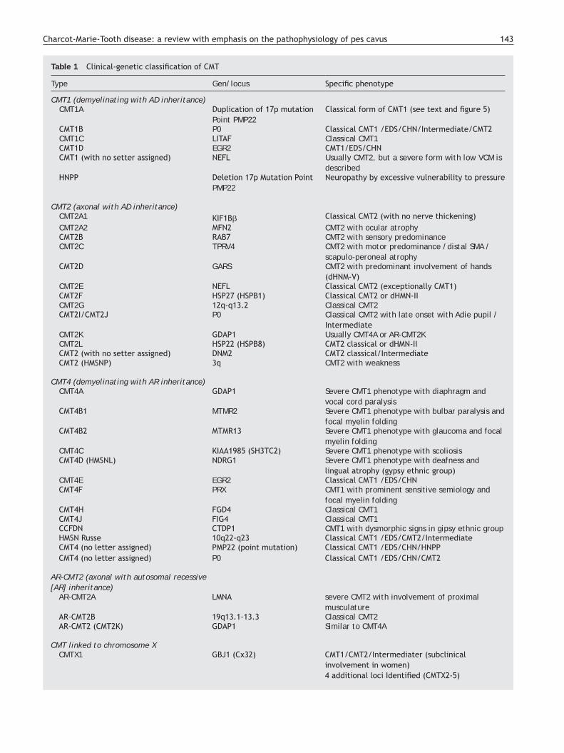
Adapted from references 10, 14 and 16, Table 1 shows an updated clinical-genetic classification of CMT, which is provisional as there is no unanimous view on the use of its types and subtypes. There is universal agreement to accept CMT1 as the header for demyelinating phenotypes with AD inheritance. Some authors include axonal forms with AD or AR inheritance under CMT2, while others include only the AD forms, creating the acronym AR-CMT2 for axonal forms with AR transmission; we have followed this approach. The acronym CMT3 in the classification of Dyck,13 applied to syndromes similar to those described by Dejerine and Sottas,4 disappears; it is replaced by CMT4, which encompasses all demyelinating syndromes with AR inheritance. The DI-CMT acronym is introduced for the intermediate forms with AD transmission.
Figure 3 illustrates the location of the mutated proteins, which was predictable for the known SNP components, such as PMP22 and P0 proteins of compact myelin. However, in other situations, the discovery of the pathogenic mutant protein proved to be unexpected, such as illustrated by the case of GDAP1 in the SNP whose function was unknown until the identification of CMT4A.
From an educational point of view, the specific mechanisms of the mutated proteins are summarised as follows14: 1) by altering myelin development and maintenance, 2) by altering protein biosynthesis and degradation, 3) through alternating endocytosis and membrane dynamics including those of the mitochondria, 4) by altering the axonal cytoskeleton, 5) seipinopathy and 6) channelopathy through TRPV4 mutation. We shall briefly review these six sections.
In CMT1/CMT4 forms by mutation of certain components of myelin, it is assumed that the defect in Schwann cells causes demyelination with secondary axonal axonopathy, which is ultimately responsible for clinical symptomatology. The most common syndrome in this section is CMT1A, which represents 70% of all cases of CMT1 and is usually caused by a 1.5 Mb allelic 17p11.2 trisomy that contains the PMP22 protein gene. Such trisomy causes an excess gene dosage, which implies an overproduction of PMP22 and its accumulation in the Schwann cells, inducing stress of the endoplasmic reticulum, resulting in programmed cell death. The deletion works by reducing the expression of PMP22,
Figure 2 (A) Semi-fine section of sural nerve from a patient with CMT1A. There is a marked loss of myelinated fibres, several images of Schwann cell proliferation with onion bulb formation (arrow), and remyelinated fibres (arrowhead). (B) Semi-fine section of external popliteal sciatic nerve from a patient who died from CMT2G.11,12 There is marked loss of thick myelinated fibres and clusters of regenerating fibres (arrow); also note the absence of hypertrophic phenomena.
Charcot-Marie-Tooth disease: a review with emphasis on the pathophysiology of pes cavus 143
Table 1 Clinical-genetic classification of CMT
Type Gen/locus Specific phenotype
CMT1 (demyelinating with AD inheritance)CMT1A Duplication of 17p mutation
Point PMP22Classical form of CMT1 (see text and figure 5)
CMT1B P0 Classical CMT1 /EDS/CHN/Intermediate/CMT2CMT1C LITAF Classical CMT1 CMT1D EGR2 CMT1/EDS/CHNCMT1 (with no setter assigned) NEFL Usually CMT2, but a severe form with low VCM is
describedHNPP Deletion 17p Mutation Point
PMP22Neuropathy by excessive vulnerability to pressure
CMT2 (axonal with AD inheritance)CMT2A1 KIF1Bβ Classical CMT2 (with no nerve thickening)CMT2A2 MFN2 CMT2 with ocular atrophyCMT2B RAB7 CMT2 with sensory predominanceCMT2C TPRV4 CMT2 with motor predominance /distal SMA /
scapulo-peroneal atrophyCMT2D GARS CMT2 with predominant involvement of hands
(dHNM-V)CMT2E NEFL Classical CMT2 (exceptionally CMT1)CMT2F HSP27 (HSPB1) Classical CMT2 or dHMN-IICMT2G 12q-q13.2 Classical CMT2 CMT2I/CMT2J P0 Classical CMT2 with late onset with Adie pupil /
IntermediateCMT2K GDAP1 Usually CMT4A or AR-CMT2KCMT2L HSP22 (HSPB8) CMT2 classical or dHMN-IICMT2 (with no setter assigned) DNM2 CMT2 classical/IntermediateCMT2 (HMSNP) 3q CMT2 with weakness
CMT4 (demyelinating with AR inheritance)CMT4A GDAP1 Severe CMT1 phenotype with diaphragm and
vocal cord paralysisCMT4B1 MTMR2 Severe CMT1 phenotype with bulbar paralysis and
focal myelin folding CMT4B2 MTMR13 Severe CMT1 phenotype with glaucoma and focal
myelin foldingCMT4C KIAA1985 (SH3TC2) Severe CMT1 phenotype with scoliosisCMT4D (HMSNL) NDRG1 Severe CMT1 phenotype with deafness and
lingual atrophy (gypsy ethnic group)CMT4E EGR2 Classical CMT1 /EDS/CHNCMT4F PRX CMT1 with prominent sensitive semiology and
focal myelin foldingCMT4H FGD4 Classical CMT1 CMT4J FIG4 Classical CMT1CCFDN CTDP1 CMT1 with dysmorphic signs in gipsy ethnic group HMSN Russe 10q22-q23 Classical CMT1 /EDS/CMT2/IntermediateCMT4 (no letter assigned) PMP22 (point mutation) Classical CMT1 /EDS/CHN/HNPPCMT4 (no letter assigned) P0 Classical CMT1 /EDS/CHN/CMT2
AR-CMT2 (axonal with autosomal recessive [AR] inheritance)
AR-CMT2A LMNA severe CMT2 with involvement of proximal musculature
AR-CMT2B 19q13.1-13.3 Classical CMT2 AR-CMT2 (CMT2K) GDAP1 Similar to CMT4A
CMT linked to chromosome XCMTX1 GBJ1 (Cx32) CMT1/CMT2/Intermediater (subclinical
involvement in women)4 additional loci Identified (CMTX2-5)

144 J. Berciano et al
which creates unstable myelin manifested by a syndrome of excessive vulnerability to pressure (HNPP is the Anglo-Saxon acronym; see Table 1). In a small percentage of cases, duplication / deletion can occur as a de novo phenomenon. The point mutations of the PMP22 gene are rare and cause severe phenotypes, and AD (probably by a mechanism of gain of function) or AR (loss of function due to failure in the synthesis of PMP22). The P0 protein is quantitatively the
most abundant of compact myelin, and an essential element for compaction. In 10% of the cases, CMT is caused by point mutations of P0 that are either an early-onset AD demyelinating phenotype (CMT1B) and exceptionally AR, or late-onset axonal phenotypes (CMT2I and CMT2J). Consequently, the molecular pathology of PMP22/P0 has revealed that their mutations can be inherited through transmission of both AD and AR and that, in the case of P0,
Table 1 (Continued)
Type Gen/locus Specific phenotype
Dominant Intermediate (DI) CMT with autosomal dominant (AD) inheritance
DI-CMTA 1q24.1-25.1 Classical CMT1 (with no nerve thickening)DI-CMTB DNM2 Classical CMT1 with cataracts and neutropeniaDI-CMTC YARS Classical CMT1 DI-CMTD P0 Classical CMT1
CHN=congenital hypomyelinating neuropathy; CMT=Charcot-Marie-Tooth disease; CTDP1=carboxy-terminal domain (CTD) phosphatase subunit 1; DI-CMTA=dominant intermediate CMT DNM=dynamin 2; DSD=Dejerine-Sottas disease; EGR2=Early Growth response 2; FDG4=RhoGEF; FIG4=phosphatidylinositol (Ptdlns) (3,5)P2 5-phosphatase; GARS=glycyl tRNA synthetase; GBJ1=gap junction protein beta 1; GDAP1=ganglioside-induced differentiation-associated protein 1; HMSNL=hereditary motor and sensory neuropathy-Lom; HNPP=hereditary neuropathy with susceptibility to pressure palsy; HSP22=heat shock protein 22 kDa; HSP27=heat shock protein 27 kDa; KIF1Bβ=kinesin family member 1-Bβ; LITAF=lipopolysaccharide-induced tumour necrosis factor-alpha factor; LMNA=lamin A/C; MFN2=mitofusin 2; MTMR2=myotubularin related protein 2; MTMR13=myotubularin-related protein 13; NDRG1=N-myc downstream-regulated gene 1; NEFL=neurofilament light polypeptide 68 kDa; PMP22=peripheral myelin protein 22; P0=myelin protein zero; PRX=periaxin; RAB7=RAB7, member RAS oncogene family; SH3TC2=SH3 domain and tetratricopeptide repeats 2; SMA=spinal muscular atrophy; TRPV4=transient receptor potential vanilloid 4; YARS=tyrosyl tRNA synthetase.
Figure 3 Schematic drawing of a myelinated nerve fibre adapted from Niemann et al.14 The mutated proteins, causing CMT, HMN or HSAN, identified up to 2006 are in black, while those described subsequently are displayed in red. The meaning of acronyms is given at the foot of Table 1. Note that mutations of SPTLC1 (serine palmitoyltransferase long chain base subunit 1), HSN2 (hereditary sensory neuropathy type 2), NTRK1 (neurotrophic tyrosine kinase receptor type 1), IKBKAP (inhibitor of kappa light polypeptide gene enhancer in B-cells, kinase complex-associated protein) and NGF1 (nerve growth factor beta polypeptide) are involved in the pathogenesis of hereditary sensory and autonomic neuropathies, not reviewed in this article (see text).
Charcot-Marie-Tooth disease: a review with emphasis on the pathophysiology of pes cavus 145
their mutations cause both demyelinating and axonal phenotype. This only underscores that in SNP the dialogue between Schwann cells and axon companions is continuous.14 Such phenomena are applicable to CMT-causing mutations in other genes (Table 1). GBJ1 (Cx32) is a gap type protein of paranodal myelin, whose gene is located on chromosome X. The second most frequent cause of CMT, point mutations in gene GBJ1 originate a radial transit of small molecules dysfunction between Schwann cells and axon. Probably by a mechanism of haploinsufficiency, such mutations cause a more severe phenotype in males than in females, which can be neurophysiologically demyelinating, intermediate or axonal. Other rare causes include CMT1/CMT4 EGR2 mutation (a transcription factor involved in myelin gene regulation) and PRX mutation (an anchor protein of the Schwann cell cytoskeleton).
The correct composition and maintenance of the membranous compartments of Schwann cells and PNS neurons are dependent on a perfect balance between the synthesis of structural and signalling components, and on their degradation processes.14 Among the point mutations of proteins involved in endocytosis processes are the following (Table 1): 1) phosphatase (MTMR2, MTMR13 and figure 4), which cause severe AR phenotypes (CMT4B1, CMT4B2 and CMT4J) with focal folds of myelin (CMT4B1 and CMT4B2); 2) GTPase, DNM2 AD phenotype that can be both intermediate (DI-CMTB) and axonal,19,20 Rab7 (which causes CMT2B, a phenotype similar to HSNA1) and Frabin, is associated with CMT4H; and 3) NDRG1, a poorly known gene that regulates function, whose mutation causes a severe syndrome (CMT4D) in subjects of gypsy ethnicity. In regards to components involved in protein synthesis, sorting and degradation, mutations affect the following components: 1) SIMPLE/LITAF, a ubiquitin ligase that causes CMT1C and 2) GARS and YARS, proteins involved in loading the tRNA with glycine and
tyrosine, which cause CMT2D/dHMN-V and DI-CMTC, respectively (Table 1).
Both sensory and motor CNS neurons have to move proteins, vesicles and organelles through the long axon tracts ranging from the soma to its terminals; this requires a complex and efficient transportation system. The growing number of axonal forms of CMT caused by mutations in proteins associated with cytoskeletons, and transport of proteins, vesicles and organelles cannot be a surprise to anyone (Table 1). Mutations in the neurofilament light chain (NEFL) cause CMT2E, and exceptionally CMT1F. Heat shock proteins (HSP) are ubiquitous macromolecules that control the assembly of neurofilaments in SNP. Mutations in HSP27 cause CMT2F/dHMN-II, whereas mutations associated with HSP22 cause CMT2L/dHMN-II. Recently, in a line of CMT associated with HSP27 R127W mutation, with 10 patients examined clinically and neurophysiologically, there were cases with CMT2 phenotype and others with HMN phenotype; this only underscores that both syndromes may be a unique nosological entity.21 The kinesin are a family of motor proteins that mediate the axonal anterograde transport of microtubules, whereas dynein mediate retrograde transports. Mutations in KIF1Bβ are associated with CMT2A1 and mutations of Rab7 (GTPase that regulates the function of dynein) causes CMT2B. Mitochondrial morphology is determined by a balance between fusion and fission processes of the organelle. MNF2 is a GTPase of the outer wall of the mitochondria, where it acts as a regulator of mitochondrial fusion. Point mutations of MFN2 cause CMT2A2, presently the most common cause of CMT2 (20%), with a fifth of the cases presenting as de novo mutations. Similar to HMSN-VI, in CMT2A2 there may be optic atrophy, especially in severe forms of early onset. GDAP1 is the counterpart of MFN2 processes involved in mitochondrial fission. Homozygous mutations in GDAP1 cause either CMT4A
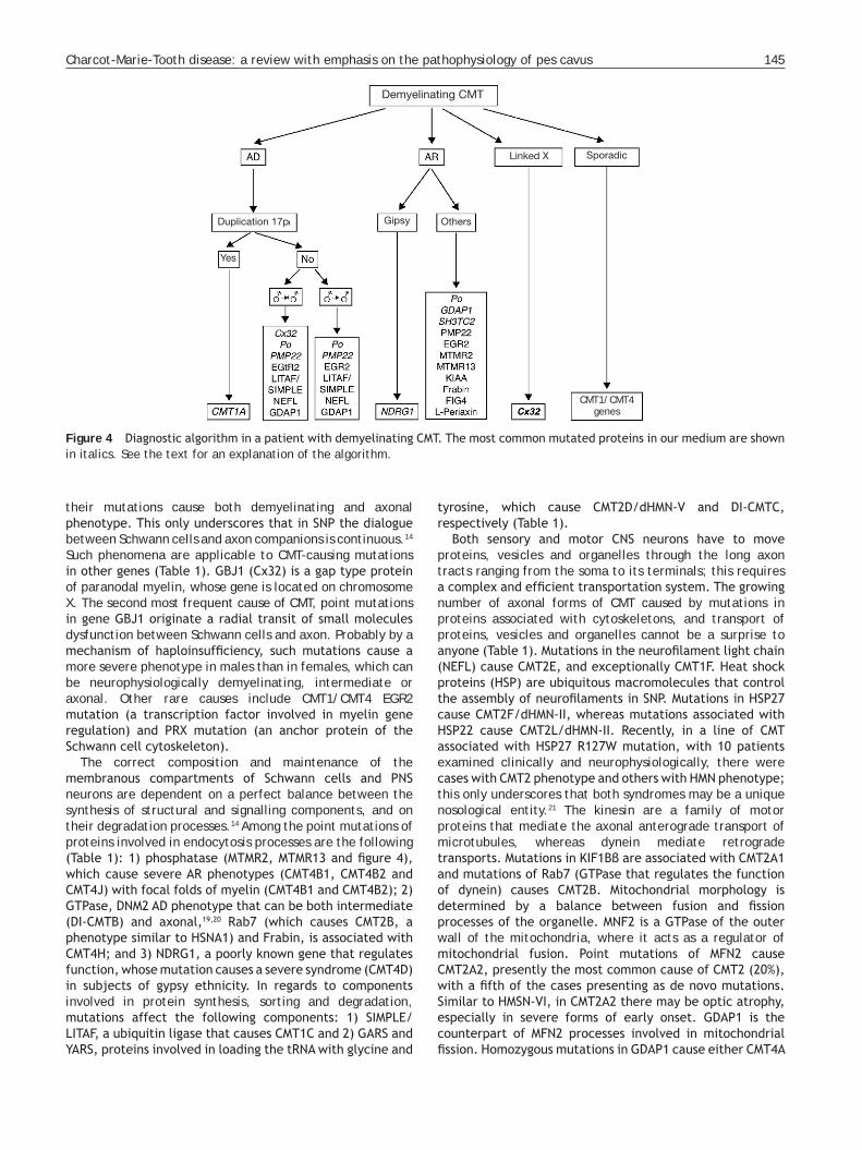
Figure 4 Diagnostic algorithm in a patient with demyelinating CMT. The most common mutated proteins in our medium are shown in italics. See the text for an explanation of the algorithm.
Demyelinating CMT
Linked X Sporadic
Duplication 17p Gipsy Others
Yes
CMT1/CMT4 genes

146 J. Berciano et al
or AR-CMT2; certain mutations in this gene infrequently cause disease in heterozygous state (CMT2K). In Spain, GDAP1 mutations are more frequent in CMT with AR inheritance; this is a severe phenotype, normally accompanied by vocal cord and diaphragm paralysis.22 LMNA is a nuclear membrane protein whose mutation is associated with AR-CMT2A; it is interesting to note that mutations in the same gene can cause Emery-Dreifuss myopathy. An adapter protein, KIAA1985/SH3TC2 and its mutations cause a severe phenotype (CMT4C) 17 (Table 1).
The acronym BSCL2 derives from Berardinelli-Seip Congenital lipodystrophy 2, a syndrome originally described in strains with lipoatrophy, insulin resistance, hypertriglyceridaemia, mental retardation and AD inheritance. Seipin, or BSCL2, is a glycosylated protein of the endoplasmic reticulum, which means point mutations activate unfolded protein response (UPR) by inducing endoplasmic reticulum stress and programmed cell death.23 The seipinopathies are considered a new model of disease due to protein conformation alterations. Point mutations cause neurodegenerative syndromes with AD transmission, including dHMN-V, Silver syndrome (spastic paraparesis and hand amyotrophy), CMT2 and hereditary spastic paraparesis; the mutation has incomplete penetrance24 in an important percentage of cases.
The TRPV4 gene is a member of nonselective cationic channels involved in the detection of physical and chemical stimuli and multiple physiological functions.25 Heterozygous TRPV4 mutations were associated with bone dysplasia. It was known through genetic linkage analysis that CMT2C, the scapular-peroneal form of spinal muscular atrophy (SMA) and the congenital distal form of SMA could be allelic syndromes (12q21-q24). Four recent studies have shown that, in fact, these syndromes, often with incomplete penetrance, are associated with different heterozygous mutations in the ankyrin domain of TRPV4.26-29 The mechanism by which these mutations cause degeneration of the SNP is unknown. In any case, the disease is a prototypical example of inter-and intrafamilial variable expressivity.
Diagnosis of CMT
The first step is to establish whether the patient has hereditary neuropathy. The answer may be evident when the household survey shows that individuals in ancestral lineage are affected, suggesting a sex-linked or AD inheritance (with no male-male transmission). Occurrence of disease among siblings joined to parental consanguinity suggests an AR inheritance. However, the household survey is sometimes negative, in which case there are a number of factors that orient towards genetic neuropathy, namely: 1) presentation in childhood, 2) prolonged and slowly progressive clinical course, 3) presence of pes cavus (see below), and 4) in contrast to acquired neuropathies, absence of positive sensory symptoms (paresthesia or dysesthesia) although there is clear semiology of sensory deficit.16 Given that the affected subjects often have subtle symptoms or are asymptomatic, it is important to explore the maximum possible number of subjects at risk for the strain (secondary cases) besides the subjects themselves. This allows minimal
signs of disease (e.g., pes cavus and areflexia) to be detected in subclinical cases, and consequently makes it possible to profile the pattern of inheritance better.
The next step is the neurophysiological examination, which should include determination of the NCV and VCS in at least three nerves. When interpreting the degree of slowness of NCV, the amplitude of compound motor action potential (CMAP) should be taken into account, because a sharp fall in distal CMAP amplitude involves loss of coarse distance-dependent fibres, which may well lead to a proportional NCV reduction. To distinguish between an NCV drop from myelinopathy or axonopathy, it is recommended that you should study proximal nerve segments, where the conduct is similarly slowed in cases of demyelinating CMT and less slowed and even preserved in cases of axonal CMT. In CMT1/CMT4, NCV/VCS slowing is diffuse and uniform and the morphology of the CMAP and the terminal latency index tend to be preserved, which is in contrast to what happens in acquired inflammatory neuropathies.
Currently, nerve biopsy is reserved for cases where there are problems with the differential diagnosis with other hereditary neuropathies (e.g., amyloidosis) or acquired neuropathies.
Of the thirty or pathogenic genes so far identified, only a dozen of them are available for diagnostic purposes in clinical practice. Molecular tests are also expensive. Consequently, the selection of genetic testing is of paramount importance and must be based on clinical data as well as the frequency of various genotypes in the country or region under study.
Figure 4 shows the diagnostic algorithm in a patient with demyelinating CMT. If inheritance is AD and considering that CMT1A is the most common form of CMT1, an analysis of the 17p duplication should be carried out because its detection is diagnostic of CMT1A. The usual clinical setting is that of a peroneal muscular atrophy syndrome with generalised areflexia, mild distal sensory loss in a stocking-glove pattern, and other signs illustrated in figure 5. The symptomatic onset usually occurs in the first two decades of life, although the existence of subclinical cases is not rare.1-10,30,31 The NCV are usually around 20 m/s.30,31 If there is no duplication and no evidence of male-male transmission, screening for mutations in Cx32 should be performed, especially if THE conduction speed is in the midrange. The next most common mutation is P0, especially if the NCV is around 10 m/s.32 When molecular analysis is negative, point mutations can be searched for in PMP22, SIMPLE/LITAF, NEFL and GDAP1. If there is evidence of male-male transmission, the strategy is the same, but omitting the Cx32 analysis. The AR forms (CMT4) are especially prevalent in countries or regions with strong inbreeding. As in the original description by Dejerine and Sottas,4 these are considered severe forms with childhood or congenital onset. The nerve conduction velocities, when obtained, are often below 10 m/s. As this is a genetically complex syndromic group, it is essential to be guided by data from genetic epidemiology. Consequently, the first step in dealing with subjects of gypsy ethnicity is to eliminate mutations in NDRG4. In other ethnic groups of the Spanish population, P0, GDAP1 and SH3TC2 have to be considered first, and then the remaining eight mutations mentioned in the algorithm

Charcot-Marie-Tooth disease: a review with emphasis on the pathophysiology of pes cavus 147
(fig. 4). In X-chromosome linked forms and sporadic cases, one proceeds as indicated by the algorithm.
Figure 6 shows the diagnostic algorithm in a patient with axonal CMT. If the inheritance is AD and there is no evidence of male-male transmission, the molecular study begins with
Cx32, to continue with MFN2 mutations, P0 and DNM2, then nine other less common gene mutations (see algorithm). If there is evidence of male-male transmission, the molecular screening is identical, but omitting Cx32. In the forms with AR transmission, mutation of GDAP1 is the most frequent;
Figure 5 Composition of clinical signs in CMT1A in patients studied by the authors. (A) Thickening of the auricular nerve in an 8-year-old patient. (B, C) Peroneal muscular atrophy in a 17-year-old patient; note the presence of clawed toes, calcaneal inversion with forefoot adduction and varus deviation of the ankle. (D-G) Approximation photographs illustrating pes cavus in lateral and plantar views, clawing of the toes and atrophy of the extensor digitorum brevis (EDB). (H, I) Atrophy of the hands in a 75-year-old patient; this sign is usually characteristic of advanced stages of the disease.
Figure 6 Diagnostic algorithm in a patient with axonal CMT. The most frequently mutated proteins in our environment are shown in italics. See the text for an explanation of the algorithm.
Axonal CMT
Linked X Sporadic
CMT1 Genes

148 J. Berciano et al
after that one should consider LMNA and NEFL. For X-chromosome inheritance and sporadic cases, one should proceed as indicated in the algorithm.
Pes cavus in CMT
Pes cavus is a primary manifestation of the disease.8,9,13,30,31 As indicated in figures 1 and 5, it is a cavus foot and forefoot valgus (forefoot cavus), usually accompanied by claw toes and hindfoot varus.33,34 The presence of pes cavus indicates that the process of foot muscle denervation began before completing their growth.35,36 The pathophysiology of pes cavus in CMT is a controversial issue. There is general agreement that pes cavus is caused by an imbalance between agonist and antagonist muscles of the foot or leg. In orthopaedic literature, there is agreement in stating that
claw toes are the result of a paresis of the intrinsic musculature of the foot with preservation of the extrinsic muscles.34 Regarding the mechanism of pes cavus in CMT there are two differing hypotheses.33-37 Under the first (proposed by most authors), forefoot cavus is the result of an imbalance between the strength preserved in the peroneus longus muscle and paresis of the peroneus brevis and/or tibialis anterior. The decompensated action of the peroneus longus could cause excessive plantar flexion of the first metatarsal with increased arch height and subtalar varus deviation. The second hypothesis is that pes cavus is a denervation of the intrinsic foot muscles, particularly of the lumbricals, with relative preservation of the extrinsic muscles; that is, both claw toes and pes cavus would be the expression of an imbalance in the action of the intrinsic muscles (denervated) and extrinsic (preserved) of the foot.38
Figure 7 From images taken from Sabir and Lyttle38 and from MRI findings in foot musculature,41 pathophysiological interpretation of pes cavus in early CMT1A stages. (A) The disease begins with denervation of lumbrical and other intrinsic muscles of the foot. (B) Lumbrical paresis causes clawed toes, flattening of the plantar arch and contracture of the short flexor muscles, bringing the pillars of the longitudinal arch of the foot closer together. (C) When walking, before the toes are lifted, the plantar fascia wraps around the metatarsal heads (capstan effect) by the extension of the metatarsophalangeal joints, bringing the pillars of the longitudinal arch of the foot closer together and shortening the Achilles tendon, which limits dorsiflexion of the foot.

Charcot-Marie-Tooth disease: a review with emphasis on the pathophysiology of pes cavus 149
The main limitation in the pathophysiological interpretation of pes cavus in CMT is that it was based on cross-sectional studies of symptomatic cases, and especially of index cases in which the presence of atrophy and weakness of the legs was the rule. We lacked longitudinal clinical-neurophysiological studies of secondary cases (subclinical) to establish the evolution of foot semiology. We addressed this issue by exploring prospectively, over two decades, 20 children at risk of CMT1A with duplication (affected mother or father), out of which 12 were found to be affected.30,31,39,40 The inclusion period was the first 5 years of life (12 patients between 0 and 4 years; mean age 2 years), when maturation occurs in nerve conduction. At the end of the study, age ranged between 4 and 19 years (mean age, 8). In the inclusion period, only 4 of 12 patients had cavus; between 5 and 10 years, 5 of 10 patients who had reached this age group had pes cavus; and from age 11, all of them (7 of 7) had cavus foot. Only peroneal muscular atrophy was detected in the third age group (4 of them). We also note that the onset and progression of atrophy in the extensor digitorum brevis (EDB) muscle correlates not with the degree of slowness of the peroneal nerve NCV, but with the fall of the CMAP, i.e., the distance-dependent secondary axonopathy. Our studies thus showed that pes cavus will appear in childhood or adolescence and that it is unrelated to the presence of tibio-peroneal paresis. This strengthens the pathophysiological role of denervation of the intrinsic muscles of the foot in the development of pes cavus.38
Subsequently, to verify the clinical and neurophysiological findings, we conducted a magnetic resonance imaging (MRI) study of the muscles of legs and feet in 11 CMT1A patients, 6 with mild phenotype including cavus but no paresis of the leg muscles, and 5 with more advanced phenotype including cavus and tibio-peroneal paresis.41 In agreement with our previous neurophysiological clinical findings, the MRI image showed that patients with slight phenotype showed that fat atrophy remained confined to toe muscle, while moderate phenotype patients had a combination of massive fat atrophy of foot musculature and (to a lesser extent) principally distal atrophy of the muscles of the leg. Almost simultaneously and in an unprecedented muscular balance study, Vinci et al showed that, in cases of CMT1A phenotype, mild paresis may be restricted to the lumbricals and the short extensor muscle of the big toe, with total preservation of the tibio-peroneal musculature.42
In summary, in CMT1A cavus initially depends on selective denervation of foot muscles, which causes an imbalance between the intrinsic and extrinsic muscles (fig. 7). The imbalance in the action of the peroneal-tibial muscles may have a pathophysiological role as the disease progresses, or in severe cases with early denervation of the muscles of the anterolateral compartment of the leg. This notion is probably applicable to other CMT syndromes different from CMT1A with duplication.
Treatment of CMT
The treatment of the disease is multidisciplinary, involving paediatricians, neurologists and orthopaedic rehabilitation. The orthopaedic approach to the disease is discussed by
Fernandez-Retana and Poggio in an article published in this issue of the journal. For our part, we would like to stress that CMT in children is often an expression of a mere alteration of foot architecture, with no real loss of leg muscle strength; this calls for a conservative physical therapy, to mitigate claw toe development and Achilles tendon retraction that limits the dorsiflexion of the foot as much as possible. Patients should be encouraged to observe as active a life as possible, controlling their weight, avoiding alcohol abuse and the administration of neurotoxic drugs.
In 2004, Passage et al reported that ascorbic acid, a promoter of myelination, improved phenotype in a model of CMT1A in mice that over-expressed PMP22.43 This experimental finding led to the 136th ENMC International Workshop on Charcot-Marie-Tooth disease type 1A being held. A conclusion of the workshop was that undertaking clinical trials of vitamin C in patients with CMT1A was justified.44 Unfortunately, the three clinical trials reported so far have been negative,45-47 indicating that the animal models of CMT1A do not necessarily recapitulate the human phenotype. For other therapeutic approaches, we refer to the recent revision by Reilly and Shy.16
Level of evidence
Update topic with level of evidence V.
Conflict of interest
The authors declare no conflict of interest.
References
1. Combarros O, Calleja J, Polo JM, Berciano J. Prevalence of hereditary motor and sensory neuropathy in Cantabria. Acta Neurol Scand. 1987;75:9—12.
2. Charcot JM, Marie P. Sur une forme particulière d’atrophie musculaire progressive, souvent familiale, débutant par les pieds et les jambes et atteignant plus tard les mains. Rev Méd Paris. 1886;6:97—138.
3. Tooth HH. The peroneal type of progressive muscular atrophy. London: HK Lewis; 1886.
4. Dejerine J, Sottas J. Sur la névrite interstitielle, hypertrophique et progressive de l’enfance. Affection souvent familiale et à début infantile, caractérisée par une atrophie musculaire des extrémités, avec troubles marqués de la sensibilité et ataxie des mouvements et relevant d’une névrite interstitielle hypertrophique á marche ascendante, avec lésions médullaires consécutives. C R Soc Biol (Paris). 1893;45:63—96.
5. Berciano J, Berciano MT, Combarros O. Original descriptions of peroneal muscular atrophy. Muscle Nerve. 2003;28:251—2.
6. Buchthal F, Behse F. Peroneal muscular atrophy (PMA) and related disorders. I. Clinical manifestations as related to biopsy findings, nerve conduction and electromyography. Brain. 1977;100:41—66.
7. Dejerine J, André-Thomas. Sur la névrite interstitielle hypertrophique et progressive de l’enfance (2e observation suivie d’autopsie). Nouv Iconogr Salpêt. 1906;19:477—509.
8. Harding AE, Thomas PK. The clinical features of hereditary motor and sensory neuropathy types I and II. Brain. 1980;103:259—80.

150 J. Berciano et al
9. Shy ME, Lupski JR, Chance PH, Klein CJ, Dyck PJ. Hereditary motor and sensory neuropathies. In: Dyck PJ, Thomas PK, editors. Peripheral neuropathy. Philadelphia: Elsevier Saunders; 2005. p. 1623—58.
10. Pareyson D, Marchesi C. Diagnosis, natural history, and management of Charcot-Marie-Tooth disease. Lancet Neurol. 2009;8:654—67.
11. Berciano J, Combarros O, Figols J, Calleja J, Cabello A, Silos I, et al. Hereditary motor and sensory neuropathy type II. Clinicopathological study of a family. Brain. 1986;109:897—914.
12. Nelis E, Berciano J, Verpoorten N, Coen K, Dierick I, Van Gerwen V, et al. Autosomal dominant axonal Charcot-Marie-Tooth disease type 2 (CMT2G) maps to chromosome 12q12-q13.3. J Med Genet. 2004;41:193—7.
13. Dyck PJ. Inherited neuronal degeneration and atrophy affecting peripheral motor, sensory, and autonomic neurons. In: Dyck PJ, et al, editors. Peripheral neuropathy. Philadelphia: Elsevier Saunders; 1975. p. 825—67.
14. Niemann A, Berger P, Suter U. Pathomechanisms of mutant proteins in Charcot-Marie-Tooth disease. Neuromolecular Med. 2006;8:217—42.
15. Szigeti K, Lupski JR. Charcot-Marie-Tooth disease. Eur J Hum Genet. 2009;17:703—10.
16. Reilly MM, Shy ME. Diagnosis and new treatments in genetic neuropathies. J Neurol Neurosurg Psychiatry. 2009;80:1304—14.
17. Lupski JR, Reid JG, Gonzaga-Jauregui C, Rio Deiros D, Chen DC, Nazareth L, et al. Whole-genome sequencing in a patient with Charcot-Marie-Tooth neuropathy. N Engl J Med. 2010;362:1181—91.
18. Züchner S. Peripheral neuropathies: whole genome sequencing identifies causal variants in CMT. Nat Rev Neurol. 2010;6:424—5.
19. Claeys KG, Züchner S, Kennerson M, Berciano J, García A, Verhoeven K, et al. Phenotypic spectrum of dynamin 2 mutations in Charcot-Marie-Tooth neuropathy. Brain. 2009;132:1741—52.
20. Gallardo E, Claeys KG, Nelis E, García A, Canga A, Combarros O, et al. Magnetic resonance imaging findings of leg musculature in Charcot-Marie-Tooth disease type 2 due to dynamin 2 mutation. J Neurol. 2008;255:986—92.
21. Solla P, Vannelli A, Bolino A, Marrosu G, Coviello S, Murru MR, et al. Heat shock protein 27 R127W mutation: evidence of a continuum between axonal Charcot-Marie-Tooth and distal hereditary motor neuropathy. J Neurol Neurosurg Psychiatry. 2010;81:958—62.
22. Sevilla T, Jaijo T, Nauffal D, Collado D, Chumillas MJ, Vilchez JJ, et al. Vocal cord paresis and diaphragmatic dysfunction are severe and frequent symptoms of GDAP1-associated neuropathy. Brain. 2008;131:3051—61.
23. Ito D, Suzuki N. Seipinopathy: a novel endoplasmic reticulum stress-associated disease. Brain. 2009;132:8—15.
24. Auer-Grumbach M, Schlotter-Weigel B, Lochmüller H, Strobl-Wildemann G, Auer-Grumbach P, Fischer R, et al. Phenotypes of the N88S Berardinelli-Seip congenital lipodystrophy 2 mutation. Ann Neurol. 2005;57:415—24.
25. Nilius B, Owsianik G. Channelopathies converge on TRPV4. Nat Genet. 2010;42:98—100.
26. Auer-Grumbach M, Olschewski A, Papi´c L, Kremer H, McEntagart ME, Uhrig S, et al. Alterations in the ankyrin domain of TRPV4 cause congenital distal SMA, scapuloperoneal SMA and HMSN2C. Nat Genet. 2010;42:160—4.
27. Deng HX, Klein CJ, Yan J, Shi Y, Wu Y, Fecto F, et al. Scapuloperoneal spinal muscular atrophy and CMT2C are allelic disorders caused by alterations in TRPV4. Nat Genet. 2010;42:165—9.
28. Landouré G, Zdebik AA, Martinez TL, Burnett BG, Stanescu HC, Inada H, et al. Mutations in TRPV4 cause Charcot-Marie-Tooth
disease type 2C. Nat Genet. 2010;42:170—4.29. Zimo´n M, Baets J, Auer-Grumbach M, Berciano J, García A,
López-Laso E, et al. Dominant mutations in the cation channel gene transient receptor potential vanilloid 4 cause an unusual spectrum of neuropathies. Brain. 2010;133:1798—809.
30. Berciano J, Combarros O, Calleja J, Polo JM, Leno C. The application of nerve conduction and clinical studies to genetic counseling in hereditary motor and sensory neuropathy type I. Muscle Nerve. 1989;12:302—6.
31. García A, Combarros O, Calleja J, Berciano J. Charcot-Marie-Tooth disease type 1A with 17p duplication in infancy and early childhood: a longitudinal clinical and electrophysiologic study. Neurology. 1998;50:1061—7.
32. Shy ME, Jáni A, Krajewski K, Grandis M, Lewis RA, Li J, et al. Phenotypic clustering in MPZ mutations. Brain. 2004;127:371—84.
33. Mann RA, Missirian J. Pathophysiology of Charcot-Marie-Tooth disease. Clin Orthop Relat Res. 1988;234:221—8.
34. Guyton GP. Current concepts review: orthopaedic aspects of Charcot-Marie-Tooth disease. Foot Ankle Int. 2006;27:1003—10.
35. Alexander IJ, Johnson KA. Assessment and management of pes cavus in Charcot-Marie-Tooth disease. Clin Orthop Relat Res. 1989;246:273—81.
36. Thompson PD, Thomas PK. Clinical patterns of peripheral neuropathy. In: Dyck PJ, Thomas PK, editors. Peripheral neuropathy. Philadelphia: Elsevier Saunders; 2005. p1137—161.
37. Tynan MC, Klenerman L, Helliwell TR, Edwards RH, Hayward M. Investigation of muscle imbalance in the leg in symptomatic forefoot pes cavus: a multidisciplinary study. Foot Ankle. 1992;13:489—501.
38. Sabir M, Lyttle D. Pathogenesis of pes cavus in Charcot-Marie-Tooth disease. Clin Orthop Relat Res. 1983;175:173—8.
39. Berciano J, García A, Calleja J, Combarros O. Clinicoelectrophysiological correlation of extensor digitorum brevis muscle atrophy in children with Charcot-Marie-Tooth disease 1A duplication. Neuromuscul Disord. 2000;10:419—24.
40. Berciano J, García A, Combarros O. Initial semeiology in children with Charcot-Marie-Tooth disease. Muscle Nerve. 2003;27:34—9.
41. Gallardo E, García A, Combarros O, Berciano J. Charcot-Marie-Tooth disease type 1A duplication: spectrum of clinical and magnetic resonance imaging features in leg and foot muscles. Brain. 2006;129:426—37.
42. Vinci P, Serrao M, Pierelli F, Sandrini G, Santilli V. Lower limb manual muscle testing in the early stages of Charcot-Marie-Tooth disease type 1A. Funct Neurol. 2006;21:159—63.
43. Passage E, Norreel JC, Noack-Fraissignes P, Sanguedolce V, Pizant J, Thirion X, et al. Ascorbic acid treatment corrects the phenotype of a mouse model of Charcot-Marie-Tooth disease. Nat Med. 2004;10:396—401.
44. Reilly MM, de Jonghe P, Pareyson D. 136th ENMC International Workshop: Charcot-Marie-Tooth disease type 1A (CMT1A), 8-10 April 2005, Naarden, The Netherlands. Neuromuscul Disord. 2006;16:396—402.
45. Burns J, Ouvrier RA, Yiu EM, Joseph PD, Kornberg AJ, Fahey MC, et al. Ascorbic acid for Charcot-Marie-Tooth disease type 1A in children: a randomised, double-blind, placebo-controlled, safety and efficacy trial. Lancet Neurol. 2009;8:537—44.
46. Micallef J, Attarian S, Dubourg O, Gonnaud PM, Hogrel JY, Stojkovic T, et al. Effect of ascorbic acid in patients with Charcot-Marie-Tooth disease type 1A: a multicentre, randomised, double-blind, placebo-controlled trial. Lancet Neurol. 2009;8:1103—10.
47. Verhamme C, de Haan RJ, Vermeulen M, Baas F, de Visser M, van Schaik IN. Oral high dose ascorbic acid treatment for one year in young CMT1A patients: a randomised, double-blind, placebocontrolled phase II trial. BMC Med. 2009;7:70.