Embed Size (px)
Citation preview

Mini-implants are often placed in the non- tooth-bearing area of the palate because of
the superior quality of the bone, the reduced risk of damage to dental roots, and the lack of interfer-ence with tooth movement during treatment.1 The Onplant System* was the first temporary anchor-age device developed for palatal placement; unfor-tunately, its invasive surgical design requires considerable healing time after implantation.2-4
Furthermore, while various tooth movements are possible with palatal anchorage, they often involve complicated procedures and bulky structures, including transpalatal arches, sheaths, and bonded composite.5-7
In an attempt to simplify the insertion and application of palatal anchorage, Chung and col-leagues designed the C-palatal plate, which is placed in the midpalatal suture for retraction of the maxillary anterior teeth in adults.8 Our modified
C-palatal plate (MCPP) further simplifies the placement procedure, reduces patient discomfort, and increases the efficiency of tooth movement. The MCPP is highly effective in distalization of the maxillary dentition in adolescent patients as well as adults.
Appliance Design and Placement
The MCPP** contains three holes for 6mm-long, 1.8mm-diameter screws,** two posterior and one offset anterior, located so as to avoid the mid-palatal suture in adolescent patients (Fig. 1). Two extended lever arms have three notches each.
A 1mm-diameter stainless steel palatal wire
© 2010 JCO, Inc.
A Modified Palatal Anchorage Plate for Simple and Efficient DistalizationYOON-AH KOOK, DDS, MSD, PHDSEONG-HUN KIM, DMD, MSD, PHDKYU-RHIM CHUNG, DMD, MSD, PHD
VOLUME XLIV NUMBER 12 719
Dr. ChungDr. KimDr. Kook
*Nobel Biocare Services AG, Balsberg, Balz Zimmermann-Strasse 7, CH-8302 Kloten, Switzerland; www.nobelbiocare.com.
**Jeil Medical Corporation, #702, Kolon Science Valley 2nd 811, Guro-Dong, Guro-Ku, Seoul, Korea; www.jeilmed.co.kr.
Dr. Kook is a Professor and Chair, Department of Orthodontics, Catholic University of Korea, Seoul St. Mary’s Hospital, #505 Banpo-dong, Seocho-gu, Seoul, South Korea; e-mail: [email protected]. Dr. Kim is an Associate Professor, Department of Orthodontics, Kyung Hee University, and Dr. Chung is President of the Korean Society of Speedy Orthodontics, Seoul, Korea.
©2010 JCO, Inc. May not be distributed without permission. www.jco-online.com

is soldered to the upper first molar bands, and closed-coil springs or power chains are attached between hooks on the palatal wire and the notches on the lever arms (Fig. 2). The direction of force can be changed by varying the hook positions and using different plate notches.
The miniscrews for the MCPP are usually placed in the posterior portion of the palate, about
2mm on either side of the midpalatal suture. Before delivering the appliance, bend the bases of the extension arms 1-2mm away from the palate on the plaster cast, then adjust them to follow the contours of the palate. In the mouth, after admin-istration of local anesthesia, place the sterilized plate with self-drilling mini-implants, using a screwdriver at 30rpm with less than 30Ncm of force. Blanching of the palatal tissue during place-ment indicates excessive pressure. Immediately after placement of the palatal plate and palatal arch, distalization can begin with elastics or nick-el titanium closed-coil springs.
In cases of bimaxillary protrusion, the MCPP can be used with Class III elastics for simultaneous retraction of both arches. For unilat-eral distalization of posterior teeth, one lever arm and posterior hole are cut away from the MCPP, which is then placed on the appropriate side of the midpalatal suture.
Case 1
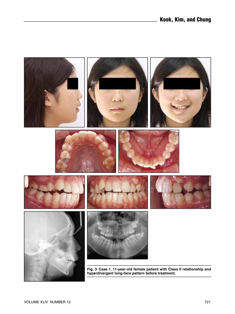
An 11-year-old female in the late mixed dentition presented with the chief complaint of a labially blocked-out maxillary left canine. She had a slightly protrusive upper lip, a long lower face, Class II canine and molar relationships, and an open bite. The maxillary dental midline was devi-ated 3mm to the left, and the lower dentition showed minor crowding (Fig. 3). Radiographic evaluation re vealed a retained maxillary right second deciduous molar, erupting second perma-nent molars, and third molar tooth germs.
720 JCO/DECEMBER 2010
A Modified Palatal Anchorage Plate for Simple and Efficient Distalization
Fig. 1 A. Modified C-palatal plate includes three short arms for miniscrew anchorage and two ex -tended lever arms, each with three notches. B. Two posterior holes and one asymmetrical anterior hole accommodate 6mm-long, 1.8mm-diameter screws.
Fig. 2 Attachment of various distalizing forces to MCPPs.
B
A
VOLUME XLIV NUMBER 12 721
Kook, Kim, and Chung
Fig. 3 Case 1. 11-year-old female patient with Class II relationship and hyperdivergent long-face pattern before treatment.
Cephalometric analysis indicated a hyperdivergent growth pattern (Table 1).
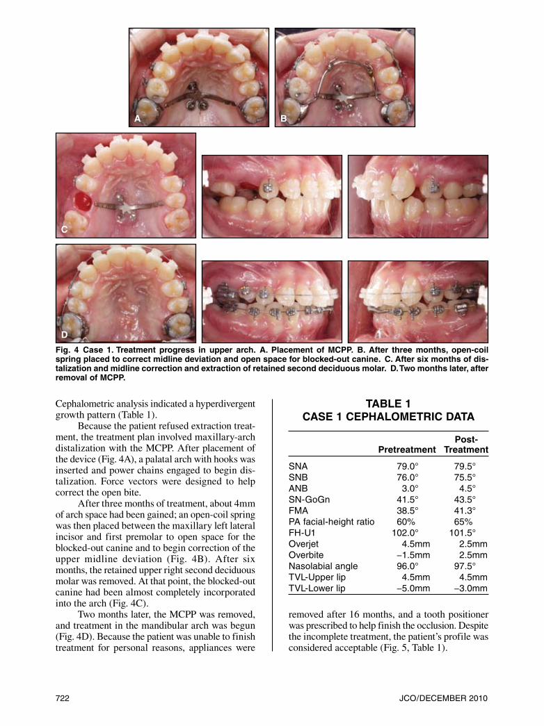
Because the patient refused extraction treat-ment, the treatment plan involved maxillary-arch distalization with the MCPP. After placement of the device (Fig. 4A), a palatal arch with hooks was inserted and power chains engaged to begin dis-talization. Force vectors were designed to help correct the open bite.
After three months of treatment, about 4mm of arch space had been gained; an open-coil spring was then placed between the maxillary left lateral incisor and first premolar to open space for the blocked-out canine and to begin correction of the upper midline deviation (Fig. 4B). After six months, the retained upper right second deciduous molar was removed. At that point, the blocked-out canine had been almost completely incorporated into the arch (Fig. 4C).
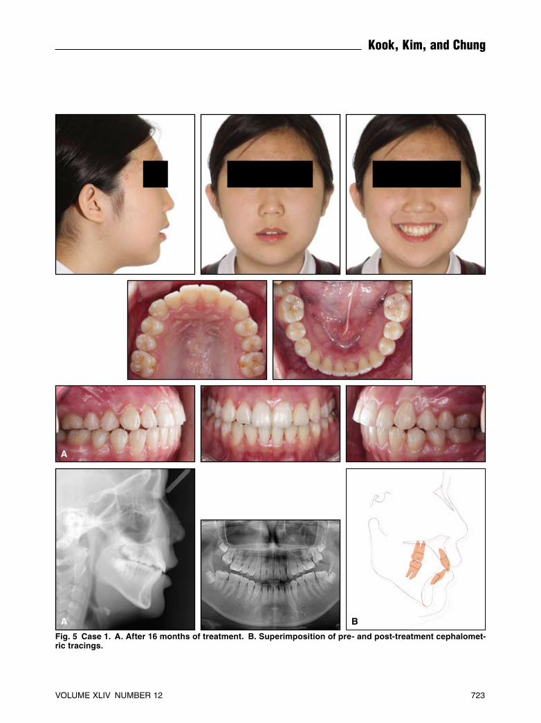
Two months later, the MCPP was removed, and treatment in the mandibular arch was begun (Fig. 4D). Because the patient was unable to finish treatment for personal reasons, appliances were
removed after 16 months, and a tooth positioner was prescribed to help finish the occlusion. Despite the incomplete treatment, the patient’s profile was considered acceptable (Fig. 5, Table 1).
722 JCO/DECEMBER 2010
Fig. 4 Case 1. Treatment progress in upper arch. A. Placement of MCPP. B. After three months, open-coil spring placed to correct midline deviation and open space for blocked-out canine. C. After six months of dis-talization and midline correction and extraction of retained second deciduous molar. D. Two months later, after removal of MCPP.
TABLE 1CASE 1 CEPHALOMETRIC DATA
Post- Pretreatment Treatment
SNA 79.0° 79.5°SNB 76.0° 75.5°ANB 3.0° 4.5°SN-GoGn 41.5° 43.5°FMA 38.5° 41.3°PA facial-height ratio 60% 65%FH-U1 102.0° 101.5°Overjet 4.5mm 2.5mmOverbite −1.5mm 2.5mmNasolabial angle 96.0° 97.5°TVL-Upper lip 4.5mm 4.5mmTVL-Lower lip −5.0mm −3.0mm
C
D
A B
VOLUME XLIV NUMBER 12 723
Kook, Kim, and Chung
A B
A
Fig. 5 Case 1. A. After 16 months of treatment. B. Superimposition of pre- and post-treatment cephalomet-ric tracings.

724 JCO/DECEMBER 2010
A Modified Palatal Anchorage Plate for Simple and Efficient Distalization
Fig. 6 Case 2. 13-year-old female patient with Class II canine relation-ship, mild posterior crossbite in left premolar area, and 5mm interlabial gap before treatment.

Case 2
A 13-year-old female presented with the chief complaint of crooked teeth. She had a Class II canine relationship, a mild posterior crossbite in the left premolar area, slightly protrusive lips, and a 5mm interlabial gap (Fig. 6). Erupting second molars and third-molar germs were evident in the radiographs. Skeletal parameters were within nor-mal limits (Table 2).
The initial treatment plan was to distalize the upper anterior teeth with anchorage from two
mini-implants placed buccally between the second premolars and first molars.9 The screws were also to be used as anchorage for Class III elastics to retract the lower dentition.10 After 14 months of leveling and alignment and retraction, however, the patient’s lip protrusion had worsened (Fig. 7A, Table 2). Her nasolabial angle had decreased, subnasale vertical to the upper and lower lips had increased, and FH-U1 had also increased. Further distalization was limited by the mini-implants’ location between the roots and the lack of inter-dental space.
VOLUME XLIV NUMBER 12 725
Fig. 7 Case 2. A. Increased lip protrusion after 14 months of leveling and alignment, despite use of buccal mini-implants for skeletal anchor-age. B. Placement of MCPP.
A
B

After the patient rejected any treatment plan involving extractions, the buccal mini-implants were removed, and an MCPP was placed for dis-talization of the entire arch. Power chains were engaged to hooks on the palatal wire, an .016" × .022" stainless steel maxillary archwire was
cinched back distal to the first molars, and 450-500g of distalizing force was applied (Fig. 7B). Class III elastics were attached between hooks on the upper first molar tubes and on the lower arch-wire between the lateral incisors and canines to simultaneously retract the mandibular dentition.
726 JCO/DECEMBER 2010
A B
A
Fig. 8 Case 2. A. Patient after 27 months of treatment (radiographs taken at end of MCPP phase). B. Super-imposition of pre- and post-treatment cephalometric tracings.

Following 11 months of whole-arch distalization, the MCPP was removed.
After 27 months of treatment, the patient’s soft-tissue profile was improved; she had no com-plaints involving the TMJs and was pleased with the treatment results (Fig. 8A, Table 2). Super -imposition of the pre- and post-treatment tracings showed distalization of both arches (Fig. 8B).
Case 3
A 24-year-old female presented with the chief complaint of dental protrusion. She had Class II molar and canine relationships, an interlabial gap of 7mm, a 9mm overjet, a high mandibular plane angle, and crowding of 5mm in the upper arch and 4mm in the lower (Fig. 9, Table 3). The patient rejected surgical correction, so an extrac-tion plan utilizing the MCPP for distalization was proposed.
After extraction of the maxillary first premo-lars, an MCPP was placed for combined maxillary anterior retraction and molar distalization (Fig. 10). Class III elastics were applied at the same time to retract the lower dentition. The MCPP was placed more to the right than usual to increase the range of action of its left arm, so that the right arm was angled slightly more than the left. When the plate was removed after 10 months, the mucosa was slightly inflamed around the screw sites. This inflammation resolved within a few days.
Total treatment time was 28 months. The patient was satisfied with the final occlusion and profile (Fig. 11A, Table 3). Cephalometric super-impositions demonstrated the amount of tooth movement accomplished after placement of the MCPP (Fig. 11B,C).
Discussion
Mini-implants in the buccal regions have been successfully used as anchorage for distaliza-tion of groups of teeth in adult patients.9 Buccal mini-implant placement in the late mixed dentition is more difficult, however, because of the nar-rower interradicular spaces and erupting perma-nent teeth. In cases requiring movement of teeth over long distances, buccal mini-implants must often be relocated in midtreatment to allow for root movements. The MCPP is easier to place and does not need to be moved during treatment. In extrac-tion cases, it provides absolute skeletal anchorage for retraction of anterior teeth, with the possibility of whole-arch distalization when the extraction space is insufficient to resolve the protrusion.
In adolescent patients, the distance between the two posterior holes of the MCPP is wide enough to avoid contact with the narrow midpala-tal suture.11,12 Still, because palatal bone tends to be thinner laterally and posteriorly,13 the holes should be placed as close as possible to the mid-palatal suture for stability.
In the cases presented here, distalization of the maxillary dentition required the application of 450-500g of force. We have used similarly high forces to retract an anterior corticotomized seg-ment using a C-plate with three palatal screws.8 A single mini-implant might not be able to withstand such forces,14 especially in adolescent bone,15 even though the thickness of the palatal bone (about 6mm) would be sufficient for stable retention.11
For better vertical control or when intrusion is desired during distalization, the palatal plate can be connected directly with power chains to buttons
VOLUME XLIV NUMBER 12 727
TABLE 3CASE 3 CEPHALOMETRIC DATA
Post- Pretreatment Progress Treatment
SNA 77.0° 76.0° 75.0°SNB 70.5° 68.5° 69.0°ANB 6.5° 7.5° 6.0°SN-GoGn 51.5° 52.5° 52.5°FH-U1 123.0° 102.5° 94.5°Overjet 5.0mm 3.5mm 2.5mmOverbite 3.5mm 3.0mm 2.0mmNasolabial angle 99.0° 100.5° 103.5°TVL-Upper lip 8.0mm 6.0mm 4.0mmTVL-Lower lip 3.5mm 3.5mm 2.0mmIMPA 94.0° 102.5° 96.5°U1-SN 109.0° 89.0° 81.0°
TABLE 2CASE 2 CEPHALOMETRIC DATA
Post- Pretreatment Progress Treatment
SNA 85.5° 85.5° 84.5°SNB 83.5° 83.5° 81.5°ANB 2.0° 2.0° 3.0°SN-GoGn 32.0° 32.0° 33.5°FH-U1 117.0° 120.5° 112.0°Overjet 3.5mm 3.5mm 3.5mmOverbite 3.5mm 3.5mm 2.5mmNasolabial angle 81.5° 78.5° 82.5°TVL-Upper lip 8.0mm 9.5mm 7.5mmTVL-Lower lip 7.5mm 12.0mm 4.5mm

728 JCO/DECEMBER 2010
Fig. 10 Case 3. MCPP placed after closure of maxillary first premolar extraction spaces.
Fig. 9 Case 3. 24-year-old female with Class II molar and canine rela-tionships, significant upper and lower crowding, and 9mm overjet before treatment.

VOLUME XLIV NUMBER 12 729
Kook, Kim, and Chung
A CB
A
Fig. 11 Case 3. A. Patient after 28 months of treatment. B. Superimposition of pre- and post-treatment trac-ings. C. Superimposition of progress and post-treatment cephalometric tracings.

on the lingual surfaces of the molars. If elastics are attached to the palatal plate notch closest to the root apex, the maxillary first molar will translate distally and be slightly intruded.16
In Case 2, superimposition of the cephalo-metric tracings showed bodily movement of the maxillary dentition with minimal tipping (Fig. 8B). This important advantage of the MCPP over buccal mini-implants may indicate that the plate system can better control the force vector in rela-tion to the center of resistance for each patient. Further study may be warranted to compare the application and treatment effects of the MCPP vs. buccal mini-implants.
SN-GoGn and FMA increased during treat-ment in all three cases shown here. Mandibular dental changes, such as extrusion of the mandibu-lar molars, seem to have contributed to this clock-wise rotation of the mandible. In each case, the mandibular first molars were extruded, and in Case 3, the maxillary first molars also tipped distally, resulting in an opening of the mandibular plane angle (Figs. 5B,8B,11B,C).
Conclusion
Advantages of the MCPP include ease of application, use of flapless surgical procedures, minimal risk of damage to neurovascular bundles, and lack of interference with growth. Unlike con-ventional mini-plates, it also offers the convenience of chairside insertion. Disadvantages can include gingival impingement and inflammation of the palatal mucosa. The mucosa of the hard palate has a high regeneration rate, and secondary healing17 should resolve any inflammation within a few days after plate removal. Still, the plate design may need refinement to minimize such occurrences.
The MCPP enables the clinician to distalize the entire maxillary dentition in a protrusive case without the need for extractions. We have found this technique to be simple and effective in both adolescents and adults.
ACKNOWLEDGMENTS: This study was partly supported by the Catholic University of Korea, the alumni fund of the Department of Dentistry, and the Graduate School of Clinical Dental Science. The authors thank Drs. Mohamed Bayome and Seong Ho Han for
their assistance in manuscript preparation. The authors are also grateful to Mr. Hyeung-Kuen Kook for his invaluable help in prepar-ing the graphics for this report.
REFERENCES
1. Jung, B.A.; Kunkel, M.; Göllner, P.; Liechti, T.; and Wehrbein, H.: Success rate of second-generation palatal implants, Angle Orthod. 79:85-90, 2009.
2. Block, M.S. and Hoffman, D.R.: A new device for absolute anchorage for orthodontics, Am. J. Orthod. 107:251-258, 1995.
3. Janssens, F.; Swennen, G.; Dujardin, T.; Glineur, R.; and Malevez, C.: Use of an onplant as orthodontic anchorage, Am. J. Orthod. 122:566-570, 2002.
4. Bernhart, T.; Vollgruber, A.; Gahleitner, A.; Dortbudak, O.; and Haas, R.: Alternative to median region of the palate for placement of an orthodontic implant, Clin. Oral Implants Res. 11:595-601, 2000.
5. Kyung, S.H.; Lee, J.Y.; Shin, J.W.; Hong, C.; Dietz, V.; and Gianelly, A.: Distalization of the entire maxillary arch in an adult, Am. J. Orthod. 135:S123-S132, 2009.
6. Papadopoulos, M.A.: Orthodontic treatment of Class II mal-occlusion with miniscrew implants, Am. J. Orthod. 134:604.e1-e16, 2008.
7. Polat-Ozsoy, O.; Kircelli, B.H.; Arman-Ozçirpici, A.; Pekta, Z.O.; and Uçkan, S.: Pendulum appliances with 2 anchorage designs: Conventional anchorage vs bone anchorage, Am. J. Orthod. 133:339.e9-e17, 2008.
8. Chung, K.R.; Kim, S.H.; and Kook, Y.A.: C-palatal plate, in OrthoTADs: The Clinical Guide and Atlas, ed. J.B. Cope, UnderDog Media, Dallas, 2007, pp. 431-438.
9. Park, H.S.; Lee, S.K.; and Kwon, O.W.: Group distal move-ment of teeth using microscrew implant anchorage, Angle Orthod. 75:602-609, 2005.
10. Chung, K.; Kim, S.H.; and Kook, Y.: C-orthodontic micro-implant for distalization of mandibular dentition in Class III correction, Angle Orthod. 75:119-128, 2005.
11. Gracco, A.; Lombardo, L.; Cozzani, M.; and Siciliani, G.: Quantitative evaluation with CBCT of palatal bone thickness in growing patients, Prog. Orthod. 7:164-174, 2006.
12. Stockmann, P.; Schlegel, K.A.; Srour, S.; Neukam, F.W.; Fenner, M.; and Felszeghy, E.: Which region of the median palate is a suitable location of temporary orthodontic anchor-age devices? A histomorphometric study on human cadavers aged 15-20 years, Clin. Oral Implants Res. 20:306-312, 2009.
13. Kang, S.; Lee, S.J.; Ahn, S.J.; Heo, M.S.; and Kim, T.W.: Bone thickness of the palate for orthodontic mini-implant anchor-age in adults, Am. J. Orthod. 131:S74-S81, 2007.
14. Florvaag, B.; Kneuertz, P.; Lazar, F.; Koebke, J.; Zöller, J.E.; Braumann, B.; and Mischkowski, R.A.: Biomechanical prop-erties of orthodontic miniscrews: An in-vitro study, J. Orofac. Orthop. 71:53-67, 2010.
15. Lee, J.W.: Comparison of bone density in para-palatal suture area between adolescent and adult ages, thesis, Catholic University of Korea, 2009.
16. Ryu, I.J.: Finite element analysis of tooth movement by maxil-lary molar distalization appliances, thesis, Catholic University of Korea, 2010.
17. Genden, E.M.; Lee, B.B.; and Urken, M.L.: The palatal island flap for reconstruction of palatal and retromolar trigone de -fects revisited, Arch. Otolaryngol. Head Neck Surg. 127:837-841, 2001.
730 JCO/DECEMBER 2010
A Modified Palatal Anchorage Plate for Simple and Efficient Distalization


![Jco.2010.33.2742.full[1] copy](https://img.pdfslide.us/doc/110x75/55506eaeb4c90524138b49ce/jco2010332742full1-copy.jpg)


![Bapi jco[1]](https://img.pdfslide.us/doc/110x75/55587609d8b42aaa7e8b5447/bapi-jco1.jpg)