Embed Size (px)
Citation preview

2008 Sunrise Free Radical SchoolPresentation by:
Pedro Cabrales, Ph.D.

What is the oxygen tensionWhat is the oxygen tension in vivo?
Pedro CabralesPedro Cabrales
La Jolla Bioengineering InstituteMicrohemodynamics Laboratory
University of California, San DiegoUniversity of California, San Diego
15th Annual Meeting of the SFRBM, Nov. 19-23, 2008

Circulatory system: delivery of nutrients and oxygen (O ) removal ofCirculatory system: delivery of nutrients and oxygen (O2), removal of waste, transport between organs, endocrine pathway, heat exchange, immunological and fluid balance
O2 is required by mammalian cells to support metabolism. It cannot be obtained directly from the environment in sufficient quantity (diffusion)
It has been resolved by two convective driven processes: air pump (the lungs) and a fluid pump (the heart)g ) p p ( )
As blood passes through the lung, O2 diffuses down into the bloodstream where it binds to the down into the bloodstream, where it binds to the hemoglobin in the red blood cells (RBCs) and is carried by convective transport through the heart and large and small arteries to the and large and small arteries to the microcirculatory vessels where the partial pressure gradient favors diffusion from the RBC to the tissue
15th Annual Meeting of the SFRBM, Nov. 19-23, 2008
to the tissue

OutlineOutline• Is there a consensus for tissue pO2?Is there a consensus for tissue pO2?• Methods to measure tissue O2 tension in vivo
H i O d li d?• How is O2 delivered?• Importance of intravascular - tissue O2 gradient• How do in vivo O2 tensions compare to in vitro
experiments? p
15th Annual Meeting of the SFRBM, Nov. 19-23, 2008

Consensus for tissue pO2?Consensus for tissue pO2?Until recently, it was assumed that offloading of O2 from the blood to y g 2
the tissue occurred mostly in the capillaries
“Capillaries are the sole suppliers of O to the tissue” is a cornerstone“Capillaries are the sole suppliers of O2 to the tissue” is a cornerstone of physiology--Krogh and Erlangen in 1918, who developed the “Krogh cylinder model”all oxygen exchange takes place at the capillary, with the entrance
pO2 being the large artery and the exit pO2 being the large veinunder reduced blood flow or low arterial oxygen level sites at theunder reduced blood flow or low arterial oxygen level, sites at the
greatest radial distance from the venous end of the capillary would lack the most O2
This model ignores heterogeneity of capillary network and hemodynamics, and assumes O2 exchange only at the capillary l l
15th Annual Meeting of the SFRBM, Nov. 19-23, 2008
level

Consensus for tissue pO2? (1)Consensus for tissue pO2? (1)
pO2s, different tissues and techniques
Tissue (species, reference) Technique
Cheek Pouch (hamster Duling BR Circ Res 31: 481 489
pO2 range, mmHg
Cheek Pouch (hamster, Duling BR Circ Res 31: 481–489, 1972) Microelectrode 18 - 12
Spinotrapezius Ms (rat, Boland EJ et al J Appl Physiol 62: 791–797, 1987) Microelectrode 26 - 13
Sartorius Ms (cat, Boegehold MA et al Am J Physiol Heart Circ Physiol 254: H929–H936, 1988) Microelectrode 40 - 22
Sartorius Ms (cat - low flow, Boegehold MA et al Am J Physiol Heart Circ Physiol 254: H929–H936, 1988) Spectrophotometric 14 - 9
Skinfold (hamster, Intaglietta M et al Cardiovasc Res 32: 632–643, 1996) Phosphorescence 34 - 29
Skinfold (hamster - perivascular, Intaglietta M et al Cardiovasc Res 32: 632–643, 1996) Phosphorescence 30 - 21Cardiovasc Res 32: 632 643, 1996)
Spinotrapezius Ms (rat, Shonat RD Am J Physiol Heart Circ Physiol 272: H2233–H2240, 1997) Phosphorescence 32 - 22
Brain (rat - cortex, Vovenko EP Pflügers Arch 437: 617–623, 1999) Microelectrode 57 - 31
15th Annual Meeting of the SFRBM, Nov. 19-23, 2008
1999) Microelectrode 57 31

Consensus for tissue pO2? (2)Consensus for tissue pO2? (2)
pO2s, different tissues and techniquespO2s, different tissues and techniques
Tissue (species, reference) Technique pO2 range, mmHg
Retractor Ms (hamster, Ellsworth ML et al Am J Physiol Heart Circ Physiol 252: H1031–H1040, 1987) Spectrophotometric 30 - 21
Retractor Ms (hamster, Kuo l Am J Physiol Heart Circ Physiol 254: H331 H338 1988) Spectrophotometric 30 - 22254: H331–H338, 1988) Spec op o o e c 30
Myocardium (dog, Honig CR et al Adv Exp Med Biol 248: 591–599, 1989) Cryoscopic 64 - 25
Retractor Ms (hamster, Swain DP et al Am J Physiol Heart Spectrophotometric 24 23Circ Physiol 256: H247–H255, 1989) Spectrophotometric 24 - 23
Gracillis Ms (dog - 4 Hz stimulation, Honig CR et al Am J Physiol Heart Circ Physiol 261: H2031–H2043, 1991) Cryoscopic 31 - 20
Intestinal Villus and Submucosa (rat Bohlen HG etIntestinal Villus and Submucosa (rat, Bohlen HG et al Am J Physiol Heart Circ Physiol 269: H1342–H1348, 1995) Spectrophotometric 48 - 30
Intestine Villus and Submucosa (rabbit, Bohlen HG et al Am J Physiol Heart Circ Physiol 269: H1342–H1348, 1995) Spectrophotometric 64 - 38
15th Annual Meeting of the SFRBM, Nov. 19-23, 2008

Measuring in vivo tissue pO2g p 2
Polarographic electrodePolarographic electrodeDavies PW and Brink F, Rev. Sci. Instrum. 1942
Fluorescence quenchingLongmuir IS and Knopp JA J Appl Physiol 1976Longmuir IS and Knopp JA, J Appl Physiol. 1976
Phosphorescence quenchingPhosphorescence quenching Vanderkooi JM et al, J Biol Chem. 1987
EPR oximetry Swartz HM et al Biochemistry 198915th Annual Meeting of the SFRBM, Nov. 19-23, 2008
Swartz HM et al, Biochemistry. 1989

Polarographic electrodePolarographic electrodeO2 molecules diffuse to the cathode and are immediately reduced by
applying polarization tension
pO2 on the surface of the electrode (platinum cathode) is zeropO2 on the surface of the electrode (platinum cathode) is zero
Reduction current is determined by O2 diffusion
Assuming constant diffusion, tissue pO2 is only determined by reduction current
Polarographic system consists of a tension generator and a current metere e
15th Annual Meeting of the SFRBM, Nov. 19-23, 2008

Polarography electrodePolarography electrode
Electrodes Cons Pros
Consume O2, requires a stable boundary layer noisy slow time Clark boundary layer, noisy, slow time response, perturbs tissue environment
Simple, easy to use, economic
WhalenMetal surface from the glass micropipette tip
Fragile, perturbs tissue environment
Low O2 consumptionLow drift, noise and variabilityFast time responsemicropipette tip Fast time response
SurfaceBoth anode and cathode sealed Slow time response, price, Low noise and variabilitywith a lipophilic membrane
p pperturbs tissue environment No motion artifacts
15th Annual Meeting of the SFRBM, Nov. 19-23, 2008

Hemoglobin SpectrophotometricHemoglobin SpectrophotometricBlood microvessels pO2 can be determined by evaluating O2 saturation p 2 y g 2
of hemoglobin (Hb), through measurements of Hb light absorption at different wavelengths
It has been implemented initially utilizing two and three wavelengthsIt has been implemented initially utilizing two and three wavelengths, and even full spectrum
Technique utilizes optical means that are easily implemented at the microscope
However, it depends on the Hb absorption spectrum at local conditions (pCO2, pH, temp, …), the tissue optical properties and light(pCO2, pH, temp, …), the tissue optical properties and light scattering
Does not provide information about tissue PO2
PO b i d i h h i h i i hPO2 obtained with spectrophotometric technique agree with periarteriolar microelectrode measurements
Pittman RN And Duling BR. Measurement of percent hemoglobin in the microvasculature. J Appl Physiol 38: 321–327, 1975
15th Annual Meeting of the SFRBM, Nov. 19-23, 2008
Steenbergen JM, Lash JM, And Bohlen HG. Role of lymphatic system in glucose absorption and the accompanying microvascular hyperemia. Am J Physiol Gastrointest Liver Physiol 267: G529–G535, 1994.

Cryoscopic Hb and MyoglobinCryoscopic Hb and MyoglobinEstimates O2 in the vascular lumen and parenchymal cells Hb and
m oglobin (Mb) sat rationsmyoglobin (Mb) saturations
Copper plate cooled with liquid nitrogen is rapidly applied to the surface of the tissue (cooling 500 µm below the surface in 50 ms)of the tissue (cooling 500 µm below the surface in 50 ms)
Isosbestic wavelengths for Hb and Mb are used to determine O2saturationsaturation
Measurements made for a variety of vascular and tissue sites at a fixed time pointtime point
Rate of cooling does not prevent water crystallization, limiting optical resolution and measurements accuracyresolution and measurements accuracy
Gayeski TEJ and Honig CR. Oxygen gradients from sarcolema to cell interior in a red muscle at maximal oxygen consumption. Am J Physiol Heart Circ Ph i l 251 H789 H799 1986
15th Annual Meeting of the SFRBM, Nov. 19-23, 2008
Physiol 251: H789–H799, 1986

EPR oximetryEPR oximetryElectron paramagnetic resonance (EPR) is the resonant absorption of
micro a e radiation b paramagnetic s stems in the presence of anmicrowave radiation by paramagnetic systems in the presence of an applied magnetic field
EPR is based on the fact that the spectra of paramagnetic species canEPR is based on the fact that the spectra of paramagnetic species can reflect interactions with other unpaired spins
Dissolved O cannot be observed directly by EPR but its presence canDissolved O2 cannot be observed directly by EPR, but its presence can be quantified by measuring the effects it produces in the spectra of the appropriate radical
Soluble and Solid probesΔBppΔBpp
id hOxygen
id hOxygen
id hOxygen
id hOxygen
litud
ewidthSplittin
g
litud
ewidthSplittin
g
litud
ewidthSplittin
g
litud
ewidthSplittin
g
15th Annual Meeting of the SFRBM, Nov. 19-23, 2008
Magnetic Field (B)Magnetic Field (B)
ampl
ampl
ampl
ampl

Fluorescence quenchingFluorescence quenchingO2 will quench fluorescence by colliding with the fluorescent molecule
when the latter is in the excited state
Number of collisions will be proportional to the amount of O2 presentNumber of collisions will be proportional to the amount of O2 present per unit volume
Advantages: low O consumption and spatial resolutionAdvantages: low O2 consumption and spatial resolution
Disadvantages: obtains a 2-D projection of 3-D events, affected by ffluorophore concentration
15th Annual Meeting of the SFRBM, Nov. 19-23, 2008

Phosphorescence quenchingPhosphorescence quenchingBased on the rate of decay of excited phosphorescence from Pd-
porph rin bo nd to alb min and the local pO (Stern Volmerporphyrin bound to albumin and the local pO2 (Stern-Volmer equation)
Phosphorescence emission results from transition into a triplet state byPhosphorescence emission results from transition into a triplet state by absorbing light (short flash) and then passing from this state to a singlet ground state
Pd-porphyrin releases the absorbed energy as light or transferred this energy to O2, which prevents light emission
Light emission is quenched, fewer photons are emitted, translates into a shorter time constant
ity ExcitationPalladium-mesotetra-(4-carboxyphenyl)porphyrin
Inten
s Excitation
EmissionHigh pO2
Rate of phosphorescence decay depends on O2 amount (dye concentration independent)
EV, extravascularPV, perivascularIV, intravascular
PV
IV
EVPV
IV
EV
15th Annual Meeting of the SFRBM, Nov. 19-23, 2008 time
Low pO2
50 m50 m

O2 consumption by phosphorescence quenching
Phosphorescence consumes OPhosphorescence consumes O2depending on the concentration of the dye and the total energy delivered by the light source
Emission and the phosphorescence decay obtained may be the summation y yof signals from adjoining areas, particularly in the neighborhood of a microvessel (no uniform where the oxygen field)
Golub AS et al. Am J Physiol Heart Circ Physiol 294: H2905-H2916 2008
Problems can be circumvented by using (i) repeated light excitation of
yg )
Problems can be circumvented by using (i) repeated light excitation of low intensity over a period that allows diffusion to replenish the consumed oxygen and (ii) averaging the signals
15th Annual Meeting of the SFRBM, Nov. 19-23, 2008
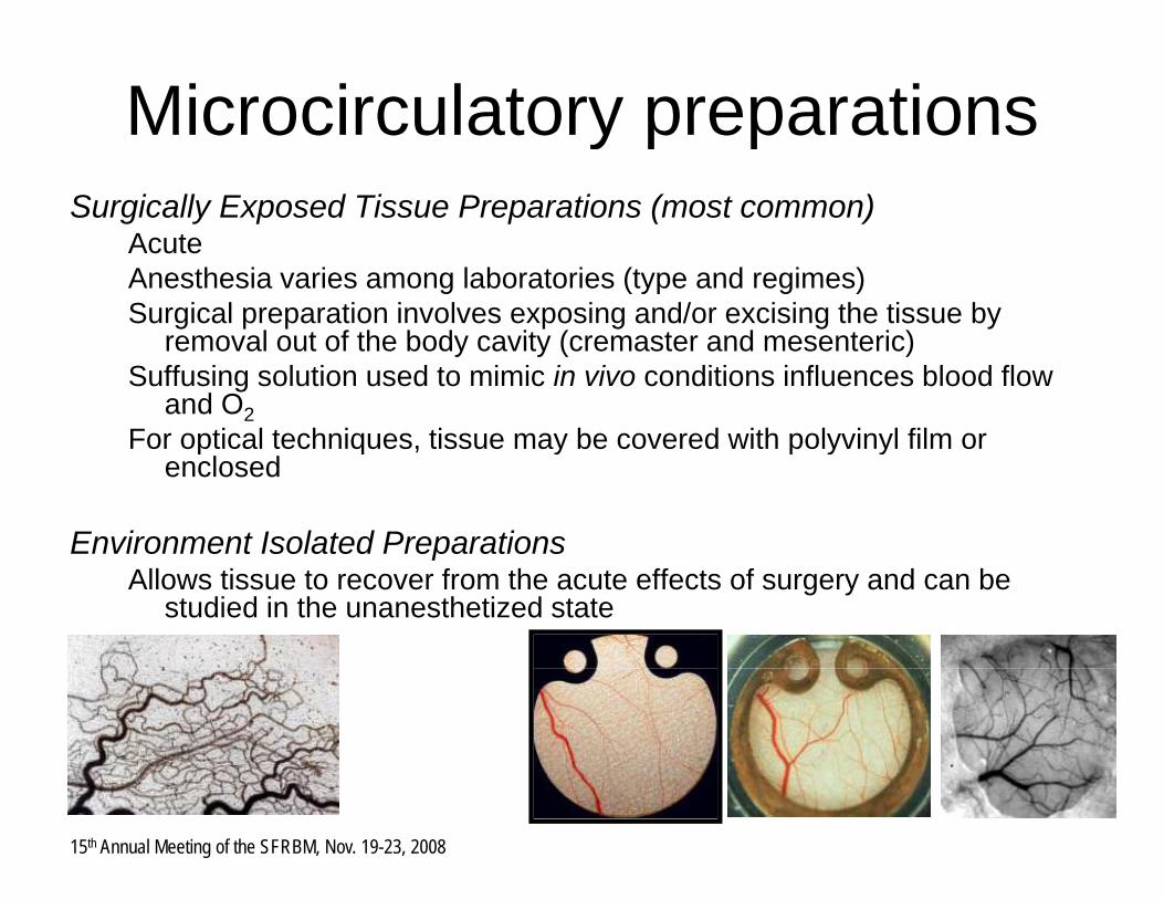
Microcirculatory preparationsMicrocirculatory preparations Surgically Exposed Tissue Preparations (most common)
AcuteAnesthesia varies among laboratories (type and regimes)Surgical preparation involves exposing and/or excising the tissue by
removal out of the body cavity (cremaster and mesenteric)removal out of the body cavity (cremaster and mesenteric)Suffusing solution used to mimic in vivo conditions influences blood flow
and O2For optical techniques, tissue may be covered with polyvinyl film or p q , y p y y
enclosed
Environment Isolated PreparationspAllows tissue to recover from the acute effects of surgery and can be
studied in the unanesthetized state
15th Annual Meeting of the SFRBM, Nov. 19-23, 2008

How is O2 delivered?How is O2 delivered?
Kerger et al., Systemic, subcutaneous microvascular oxygen tension in conscious Syrian golden hamsters. Am J Physiol 1995;268:H802-810.
Convective transport = Diffusion flux out of the vessel = O2 consumed
Convective transport, difference between O2 entering and exiting a segment
15th Annual Meeting of the SFRBM, Nov. 19-23, 2008
Diffusion flux out of the vessel, diffusion constant (D), O2 solubility (α), and pO2 radial gradient
O2 consumed, is defined by average consumption rate (Mavg)

Intravascular - O2 gradientIntravascular O2 gradientRadial gradient is steepest in the arteriolar network and
UPUP
Radial gradient is steepest in the arteriolar network and diminishes in the capillary and venular regions
Steepest radial gradients are in the immediate vicinity of the
DOWN
50 m
DOWN
50 mSteepest radial gradients are in the immediate vicinity of the vasculature, arteriolar vessels and can not be explained on the basis of diffusion alone PV
IVPV
IV
O2 loss during O2 loss during g
INTRAVASCULAR GRADIENT PERIVASCULAR
g
INTRAVASCULAR GRADIENT PERIVASCULAR
2 gconvection
is equal to
Diffusive O2 loss +
2 gconvection
is equal to
Diffusive O2 loss +pO
2, m
mH
gpO
2, m
mH
g
+ O2 consumption
+ O2 consumption
Vessel Order
p
Vessel Order
p
EV, extravascularPV, perivascular
15th Annual Meeting of the SFRBM, Nov. 19-23, 2008
IV, intravascularKerger et al., Systemic, subcutaneous microvascular oxygen tension in conscious Syrian golden hamsters. Am J Physiol 1995;268:H802-810.

How does critical pO2 in vivocompare to in vitro ? (1)
Critical pO2: pO2 required to support oxidative metabolism Skeletal muscle , in vitro
15th Annual Meeting of the SFRBM, Nov. 19-23, 2008 Richmond KN et a. Am J Physiol Cell Physiol 273: C1613-C1622 1997

How does critical pO2 in vivocompare to in vitro ? (2)
Critical pO2: pO2 required to support oxidative metabolism Skeletal muscle , in vivo
15th Annual Meeting of the SFRBM, Nov. 19-23, 2008 Richmond KN et al Am J Physiol Heart Circ Physiol 277: H1831-H1840 1999

Effects of mismatching in vivo and in vitro O2 tensions (1)
Effects of pO during shear exposure on BAEC respiration
Shear StressShear StressShear StressShear StressShear StressShear Stress 21%, 10% and 5% O2(5% CO2).
10 dyn/cm2
Effects of pO2 during shear exposure on BAEC respiration
ECECECECECEC
Exposing BAEC to steady laminar shear stress at higher oxygen tensions than physiological pO2s results in peroxynitrite formation and inactivation of the electron transport chain
15th Annual Meeting of the SFRBM, Nov. 19-23, 2008 Jones CI et al Am J Physiol Cell Physiol 295: C180-C191 2008

Summary• In vivo, the interstitial pO2 is not uniform
• Heterogeneity occurs on many levels: morphological, hemodynamics and metabolic
• Arterioles are as important as capillaries in oxygenating the tissue
• O exiting the circulation implies the existence of large blood/tissue• O2 exiting the circulation, implies the existence of large blood/tissue oxygen gradients
C ill /ti O di t i l i th l (50 H )• Capillary/tissue O2 gradients are maximal in the lung (50 mmHg) and minimal in the resting tissues (0.5 mmHg)
• The fundamental understating of how O2 is managed in vivoinfluences the translation of in vitro studies into physiological and pathophysiological mechanisms
15th Annual Meeting of the SFRBM, Nov. 19-23, 2008

AcknowledgementsAcknowledgements
UCSD team:Marcos Intaglietta, Ph.D.g ,Paul C. Johnson, Ph.D.Amy G. Tsai, Ph.D.y ,
Funding sources : NIH Heart and Lung Institute R24 HL64395, R01 HL62354, R01 HL62318, R01 HL76182, P01 HL71064 and US Army PR023085.
15th Annual Meeting of the SFRBM, Nov. 19-23, 2008

AcknowledgementsAcknowledgements
UCSD team:Marcos Intaglietta, Ph.D.Paul C. Johnson, Ph.D.Amy G. Tsai, Ph.D.
Funding sources : NIH Heart and Lung Institute R24 HL64395, R01 HL62354, R01 HL62318, R01HL76182, P01 HL71064 and US Army PR023085P01 HL71064 and US Army PR023085.
15th Annual Meeting of the SFRBM, Nov. 19-23, 2008

Effects of mismatching in vivo and in vitro O2 tensions (2)
Effects of pO during shear exposure on EC respirationEffects of pO2 during shear exposure on EC respiration
BAEC d t t d l i h t lt i it it f ti dBAEC exposed to steady laminar shear stress results in peroxynitrite formation and inactivation of the electron transport chain
Jones CI et al Am J Physiol Cell Physiol 295: C180-C191 2008
15th Annual Meeting of the SFRBM, Nov. 19-23, 2008

15th Annual Meeting of the SFRBM, Nov. 19-23, 2008

Polarography electrodePolarography electrodeClark electrode consumes oxygen, generating a current proportional yg g g p p
to the O2 concentration. Requires stable boundary/diffusion layer
Whalen electrode has a recess (metal surface from the glassWhalen electrode has a recess (metal surface from the glass micropipette tip), eliminates motion free layer. They have low drift and O2 consumption (10-6 µl/min) and fast time constant (1s). They are fragile and their presence introduces perturbations of the tissue, noisy when used in flowing blood
Surface electrodes have both cathode and anode sealed with a lipophilic membrane to prevents impurities and eliminates motion artifacts Their dimension (10 20 µm) increases catchment volumeartifacts. Their dimension (10–20 µm) increases catchment volume and the time to form a stable boundary layer. Often configured into an array and provided a histogram of O2 tensions
15th Annual Meeting of the SFRBM, Nov. 19-23, 2008

How is O2 delivered?How is O2 delivered?
15th Annual Meeting of the SFRBM, Nov. 19-23, 2008 TSAI, A. G. et al. Physiol. Rev. 83: 933-963 2003

How is O2 delivered?How is O2 delivered?In vascular beds with low metabolic tissue demand (resting skeletal
l ) th i ifi t l it di l di t f O i thmuscle), there are significant longitudinal gradients of pO2 in the arteriolar circulation
Ti ith hi h t b li d d (b i d i t ti ) h d lTissue with higher metabolic demand (brain and intestine) had lower gradients
L it di l t i l O di t fl t th ti f bl d fl tLongitudinal arteriolar pO2 gradient reflects the ratio of blood flow to metabolic O2 demand
O d li b ill i i l b d L tiO2 delivery by capillaries varies, among vascular beds. Low, resting skeletal muscle and high, brain and myocardium
Hi h l O l ti t ill d ti O l i d bHigher venular pO2 relative to capillary and tissue pO2 are explained by arterio-venous shunts, anatomic distribution and the Bohr effect
15th Annual Meeting of the SFRBM, Nov. 19-23, 2008

EPR oximetryEPR oximetryElectron paramagnetic resonance (EPR) is the resonant absorption of
micro a e radiation b paramagnetic s stems in the presence of anmicrowave radiation by paramagnetic systems in the presence of an applied magnetic field
EPR is based on the fact that the spectra of paramagnetic species canEPR is based on the fact that the spectra of paramagnetic species can reflect interactions with other unpaired spins
Dissolved O cannot be observed directly by EPR but its presence canDissolved O2 cannot be observed directly by EPR, but its presence can be quantified by measuring the effects it produces in the spectra of the appropriate radical
Spatial information can be obtained using EPR imaging (EPRI) ΔBppΔBpp
id hOxygen
id hOxygen
id hOxygen
id hOxygen
litud
ewidthSplittin
g
litud
ewidthSplittin
g
litud
ewidthSplittin
g
litud
ewidthSplittin
g
15th Annual Meeting of the SFRBM, Nov. 19-23, 2008
Magnetic Field (B)Magnetic Field (B)
ampl
ampl
ampl
ampl

EPR oximetry probesEPR oximetry, probesParticulate (Solid) probes( ) p
Lithium phthalocyanine (LiPc)Sugar charsFusiniteCoalIndia ink
Soluble probesSoluble probesNitroxidesTrityl radicals
which translates to EPR The collision frequency w according to the hard line-broadening as
w = 4pRp(DSL + DO2) [O2]
w, according to the hard sphere theory of Smoluchowski is
Dw = k DO2 [O2]
15th Annual Meeting of the SFRBM, Nov. 19-23, 2008
p p( SL O2) [ 2]