Embed Size (px)
Citation preview
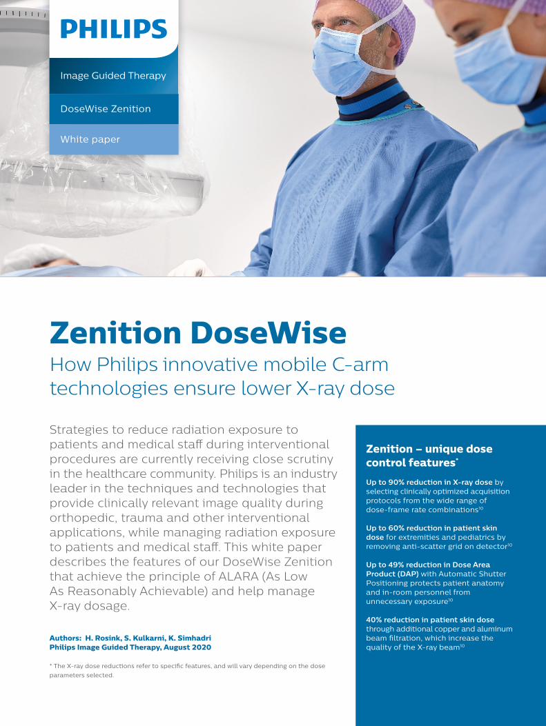
Zenition DoseWiseHow Philips innovative mobile C-arm technologies ensure lower X-ray dose
Strategies to reduce radiation exposure to patients and medical staff during interventional procedures are currently receiving close scrutiny in the healthcare community. Philips is an industry leader in the techniques and technologies that provide clinically relevant image quality during orthopedic, trauma and other interventional applications, while managing radiation exposure to patients and medical staff . This white paper describes the features of our DoseWise Zenition that achieve the principle of ALARA (As Low As Reasonably Achievable) and help manage X-ray dosage.
Authors: H. Rosink, S. Kulkarni, K. SimhadriPhilips Image Guided Therapy, August 2020
* The X-ray dose reductions refer to specifi c features, and will vary depending on the dose parameters selected.
Zenition – unique dose control features*
Up to 90% reduction in X-ray dose by selecting clinically optimized acquisition protocols from the wide range of dose-frame rate combinations10
Up to 60% reduction in patient skin dose for extremities and pediatrics by removing anti-scatter grid on detector10
Up to 49% reduction in Dose Area Product (DAP) with Automatic Shutter Positioning protects patient anatomy and in-room personnel from unnecessary exposure10
40% reduction in patient skin dosethrough additional copper and aluminum beam fi ltration, which increase the quality of the X-ray beam10
Image Guided Therapy
DoseWise Zenition
White paper

Introduction
Facts, perception and dose awareness
The detrimental effects of X-rays on human tissues had already become apparent just months after their discovery by Wilhem Röntgen in 1895.1 These effects have been classified as either deterministic (such as skin injuries) or stochastic (such as increased likelihood of developing cancer). The main concern for the patient is usually associated with the risk of skin injuries.2 Deterministic effects like skin injuries occur only when radiation dose thresholds are exceeded, and their probability and severity increase as the dose increases beyond the threshold.3 Stochastic effects may be induced at any dose.
Although it has been established that radiation dose per capita from medical imaging has increased six-fold in the USA during the last quarter century,4 it remains unclear how the medical community should address the associated risks. An indication of the growing interest from accreditation organizations such as the Joint Commission on Accreditation of Healthcare Organizations (JCAHO) is demonstrated by the fact that in 2006 the JCAHO added a cumulative fluoroscopic skin dose of > 15 Gy to their list of reviewable sentinel events.5
Besides the risks for the patient, scatter radiation from the patient may also be harmful for the medical staff present during procedures. There have been reports in the literature of an increased occurrence of brain tumors6 and cataracts among interventional cardiologists and their nurses and technicians.7
Although tissue reactions among patients and workers from fluoroscopy procedures have, to date, only been reported in interventional radiology and cardiology, the level of fluoroscopy use outside imaging departments creates the potential for such injuries.8
As a result of advances in technology, the growth in the number and complexity of interventional procedures has been significant,9 with the risk of exceeding dose thresholds. Therefore, efforts to minimize radiation dose are crucial.
Successful management of patient radiation dose can only be achieved through the optimization of medical imaging technology, combined with the best control of the imaging equipment by the operator.10 In this respect, it is the goal of manufacturers to provide medical professionals with the technology and features that facilitate the application of the ALARA principle to reduce the radiation dose delivered to both patients and medical staff.
Philips vigorously applies the ALARA principle to its technology to benefit the health and safety of patients and medical staff. Each Zenition mobile C-arm is equipped with several dose saving features and clinically relevant presets, which provide an optimal balance between dose and image quality. If required, adjustments can be made by Philips specialists to suit the particular needs of individual physicians.
2
3
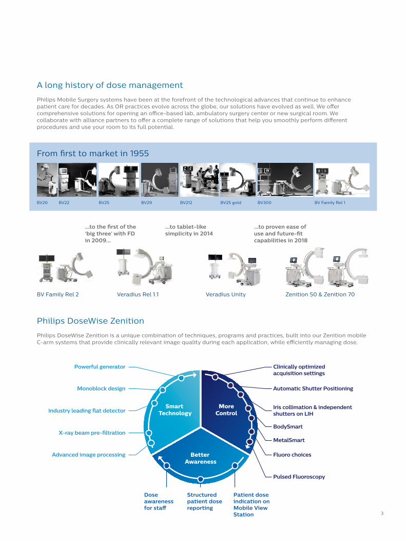
A long history of dose management
Philips Mobile Surgery systems have been at the forefront of the technological advances that continue to enhance patient care for decades. As OR practices evolve across the globe, our solutions have evolved as well. We off er comprehensive solutions for opening an offi ce-based lab, ambulatory surgery center or new surgical room. We collaborate with alliance partners to off er a complete range of solutions that help you smoothly perform diff erent procedures and use your room to its full potential.
Philips DoseWise Zenition
Philips DoseWise Zenition is a unique combination of techniques, programs and practices, built into our Zenition mobile C-arm systems that provide clinically relevant image quality during each application, while effi ciently managing dose.
Smart Technology
MoreControl
Better Awareness
DoseWise ZenitionClinically optimized acquisition settings
Automatic Shutter Positioning
Powerful generator
Monoblock design
Patient dose indication on Mobile View Station
Iris collimation & independent shutters on LIH Industry leading �at detector
BodySmart X-ray beam pre-ltration
Advanced image processing Fluoro choices
Pulsed Fluoroscopy
MetalSmart
Structured patient dose reporting
Dose awareness for sta�
BV20 BV22 BV25 BV29 BV212 BV25 gold BV300 BV Family Rel 1
From fi rst to market in 1955
…to the fi rst of the ‘big three’ with FD in 2009…
BV Family Rel 2
…to tablet-like simplicity in 2014
Veradius Rel 1.1
...to proven ease of use and future-fi t capabilities in 2018
Veradius Unity Zenition 50 & Zenition 70

4
X-ray dose – general terms and definitions
Image quality vs X-ray dose
Each procedure requires a different balance between image quality and X-ray dose, based on procedure type, patient size, projection angle and physician preferences. Furthermore, during a procedure, different tasks may require different image qualities and therefore different dose levels as is illustrated in figure1.
Finding the right balance between image quality and X-ray dose involves numerous parameters, such as frame rate, collimation, shutter and iris positions, image processing settings and many others. Therefore, the Zenition mobile X-ray system is delivered, by default, with dozens of clinically relevant presets, each with fine-tuned parameters that can be further adjusted to the particular needs of each physician by Philips specialists.
Measuring dose to patients
In order for clinicians to properly manage the magnitude of the dose given to a patient, several dose related parameters are displayed on the monitor which give clinicians insight in the amount of dose that is used. By changing other exposure techniques at their disposal, like pulse rate or selected dose level, while monitoring the displayed dose indicators, the clinician can influence the dose and resulting image quality. Patient dose can be estimated from these indirect measurements but cannot be determined precisely.11
Examination dose – An important parameter for dose management is the air kerma or cumulative examination dose (measured in mGy) at the patient entrance reference point (PERP) introduced by the IEC. The PERP is an approximation of the location of the patient’s skin. It is located in the central ray of the X-ray beam, 30 cm from the detector entrance plane. Depending on the patient’s size, the table height, and the angulation of the beam, the PERP may be outside the patient, may coincide with the skin surface, or may be inside the patient. The examination dose is displayed on the Zenition Mobile View Station and on the touchscreen at the C-arm stand.
Dose area product (DAP) – Dose area product (DAP), also called kerma area product (KAP) is the most widely applied parameter used to monitor patient dose and assess the radiation risk from diagnostic X-ray examinations and interventional procedures. The DAP is also commonly used to compare diagnostic reference levels or guidance levels of different procedures.12
The DAP is defined as the absorbed dose multiplied by the area irradiated, expressed in gray square centimeters (Gy*cm²).13 It reflects not only the dose within the radiation field, but also the area of tissue irradiated. Therefore, it may be a better indicator of the overall risk of inducing cancer than the examination dose. It also has the advantages that it can be easily calculated because all parameters (technique settings, calibrated dose levels, irradiated exposure area) are known to the system. Alternatively, the DAP could also be measured, by installing a DAP meter on the X-ray system.
The DAP can be influenced by changing the irradiated area, for example by using the collimator iris and/or shutters to reduce the field-of-view. This is beneficial in• Reducing the stochastic risk to the patient by reducing
the volume of tissue at risk• Reducing scatter radiation to the patient and
in-room personnel• Reducing potential overlap of fields when the X ray
beam is reoriented
Detector dose – Only a very small proportion of an X-ray beam that enters a patient’s body will reachthe image detector because a large portion of the photons engage in interactions within the body that divert photons in all directions (called ‘scatter radiation’).Consequently, a relationship between detector doseand patient dose is absent, and so detector doseshould never be considered as an indicator for patient or staff dose. Detector dose is a technical parameter used by vendors to control exposure settings. Another reason for not accepting detector dose as a relevant metric is that no norm exists on how it should be measured in a standardized way.
Figure1: Relative patient dose and image quality
Rel
ativ
e p
atie
nt
do
se
Localization Characterization
Required image quality

5
DoseWise Zenition dose management – personalized X-ray
At Philips, we divide factors that influence dose and image quality into three main categories:
1. Smart technology – These ‘intrinsic’ features are those components within the system that form the foundation of exceptional imaging, such as the X-ray generator with monoblock architecture, dynamic flat detector, and real-time image processing.
2. More control – Features such as clinically optimized X-ray acquisition protocols, low dose
protocols, fluoro choices and fluoro store provide a dose-optimized workflow.
3. Better awareness – DoseWise Zenition assists the user in creating dose awareness via examination dose display, structured patient dose reporting and real-time patient dose display.
Smart Technology
MoreControl
Better Awareness
DoseWise ZenitionClinically optimized acquisition settingsApply dedicated �uoroscopy settings to obtain superb image quality for the anatomy of interest without applying more X-ray dose than necessary
Automatic Shutter PositioningUp to 49% reduction in Dose Area Product (DAP) with Automatic Shutter Positioning protects patient anatomy and in-room personnel from unnecessary exposure10
Powerful generator High power 15 kW generator with rotating
anode X-ray tube and improved heat management increases �uoroscopy
time by 16%10,14
Monoblock designDelivers sharp pulse-shaped edges,
thereby reducing soft radiation that does not contribute to the image, but would be
absorbed by the patient
Patient dose indication on Mobile View StationReal-time patient dose indication
Iris collimation & independent shutters on LIH Adjust shutters and image orientation while on last image hold without using radiation
Industry leading �at detector Highly sensitive 4th generation dynamic
�at detector with small pixel pitch. Removable anti-scatter grid allows up
to 60% reduction in X-ray dose by tailoring settings to small objects and
pediatric patients10
BodySmart and MetalSmart algorithmsProvide excellent dose control by adjusting the image processing parameters based on the speci�c anatomyX-ray beam pre-ltration
40% reduction in patient skin dose through additional copper and aluminum
beam �ltration, which increase the quality of the X-ray beam10
Advanced image processing Image processing by leveraging Philips advanced image processing algorithms
from Philips �xed C-arm systems
Fluoro choices Up to 90% reduction in X-ray dose by selecting optimized protocols for the anatomy from the wide range of dose-frame rate combinations10
Pulsed Fluoroscopy Enhances imaging of dense and complex anatomy to support you in managing dose
Structured patient dose reporting Enhanced patient dose management
Dose awareness for sta�
Dose alert when exceeding a pre-de�ned procedure dose level

6
Smart Technology
Exceptional image quality and dose management capabilities begin with the components of the imaging chain.
X-raygenerator
X-raytube
Imageprocessing
Collimator Table top Flat detector
Acquisition control
GridShutterswedges
Monitors
Figure 2: Schematic showing the diff erent components in the imaging chain
• X-ray generator – The Philips microprocessor-controlled, 15 kW high frequency generator uses a monoblock architecture. It generates high voltage close to the X-ray tube, rather than farther away in the stand base, which would lead to more soft radiation. Hence, a monoblock design delivers sharp pulse- shaped edges, thereby reducing soft radiation that does not contribute to the image, but would be absorbed by the patient. In Philips Zenition systems, the housing of the monoblock has been redesigned to make it more compact. The detector can get closer to patient, which enhances the image quality.
• X-ray tube – The powerful 25 kW rotating anode X-ray tube is capable of sustaining high load capacities for extended fl uoroscopy times. Its improved heat management provides a 16% increase in fl uoroscopy time.10,14 This high performance X-ray tube has proven reliability and noiseless operation.
• Shutters – The two shutters of the collimator can be positioned independently and asymetrically from each other. In addition, the shutters and iris can be positioned on Last Image Hold (LIH) without radiation.
• X-ray beam pre-fi ltration – Additional copper and aluminum beam fi ltration increases the quality of the X-ray beam, allowing a 40% reduction in skin entrance dose rate compared to the minimum fi ltering required by international standards.10 Filtration in the Zenition systems has been optimized compared to legacy products, extending procedure times without overheating the system.
• Flat detector – The highly sensitive, 4th generation dynamic fl at detector uses an anti-scatter grid to help eliminate scattered X-rays. By removing the anti-scatter grid up to 60% reduction in patient skin dose for extremities and pediatrics can be achieved.10
• Advanced Image processing – Real-time image processing enhances the contrast to noise ratio, resulting in high image quality at low X-ray dose levels.
• Monitors – High brightness monitors ensure optimal image visualization.
Highlights
• Up to 60% reduction in patient skin dose for extremities and pediatrics by removing anti-scatter grid on detector10
• 40% reduction in patient skin dose through additional copper and aluminum beam fi ltration, which increase the quality of the X-ray beam10
Smart Technology
MoreControl
Better Awareness
Smart Technology More
Control
Better AwAwA areness
Zenition 70 removable grid Zenition 50 removable grid

7
Detector DQE
The latest 16-bit technology FD 26 x 26 and FD 21 x 21 detectors have an improved scintillator layer compared to the previous generation, which results in more effi cient X-ray detection
Detector DQE
The latest 16-bit technology FD 26 x 26 and FD 21 x 21 detectors have an improved scintillator layer compared to the previous generation, which results in more effi cient X-ray detection
8
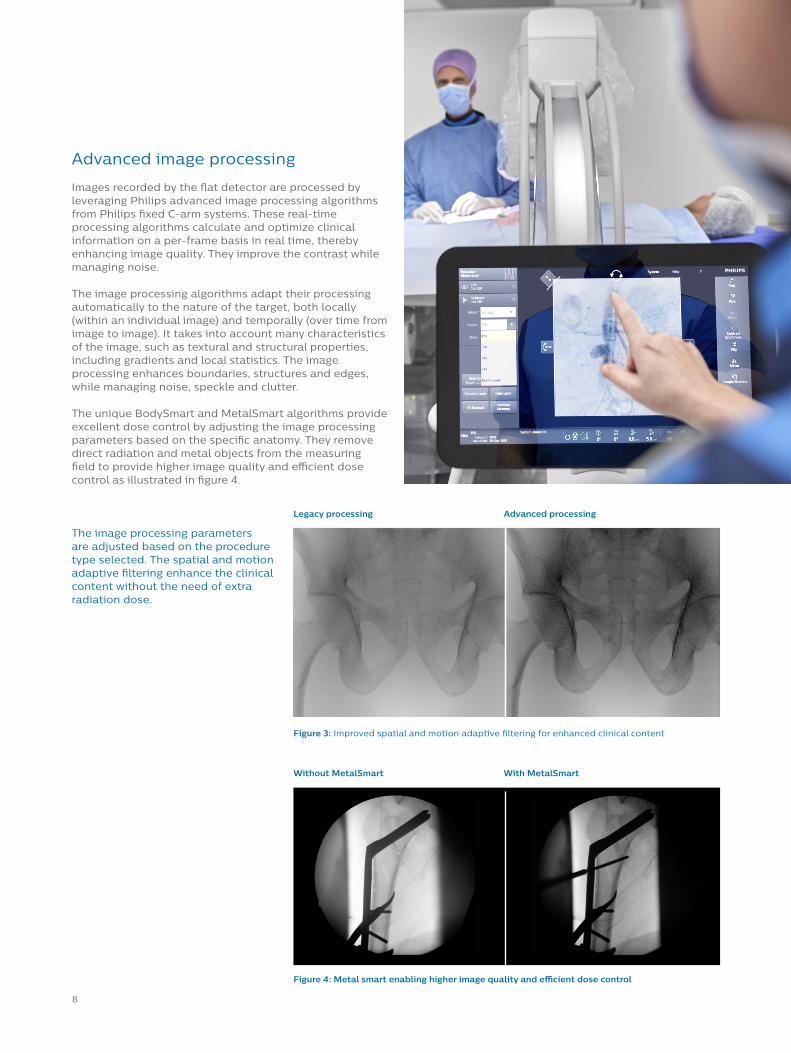
The image processing parameters are adjusted based on the procedure type selected. The spatial and motion adaptive filtering enhance the clinical content without the need of extra radiation dose.
Figure 4: Metal smart enabling higher image quality and efficient dose control
Figure 3: Improved spatial and motion adaptive filtering for enhanced clinical content
Legacy processing
Without MetalSmart
Advanced processing
With MetalSmart
Advanced image processing
Images recorded by the flat detector are processed by leveraging Philips advanced image processing algorithms from Philips fixed C-arm systems. These real-time processing algorithms calculate and optimize clinical information on a per-frame basis in real time, thereby enhancing image quality. They improve the contrast while managing noise.
The image processing algorithms adapt their processing automatically to the nature of the target, both locally (within an individual image) and temporally (over time from image to image). It takes into account many characteristics of the image, such as textural and structural properties, including gradients and local statistics. The image processing enhances boundaries, structures and edges, while managing noise, speckle and clutter.
The unique BodySmart and MetalSmart algorithms provide excellent dose control by adjusting the image processing parameters based on the specific anatomy. They remove direct radiation and metal objects from the measuring field to provide higher image quality and efficient dose control as illustrated in figure 4.

9
More control
Clinically optimized acquisition settings
The settings of the imaging parameters depend greatly on the clinical task, the procedure type, the anatomy of interest and preferred way of working. Therefore, these parameter settings are optimized and made available in preset acquisition settings. We have 30 diff erent acquisition presets developed for cardio, vascular, orthopedics, skeleton, urology, endoscopy, pain and other procedures. Each setting applies dedicated fl uoroscopy setting to help you obtain superb image quality for the anatomy of interest without applying more X-ray dose than necessary.
With Zenition, we have introduced a new X-ray mode called FluoroTap. It combines the benefi ts of the fast refresh rate of simple fl uoroscopy with image quality that comes close to a single shot. This mode will eventually also improve dose effi ciency by eliminating the need to repeat several fl uoroscopy exposures and in some cases, avoiding the use of a high dose single shot exposure.Acquisition setting presets are fl exible and programmable. If a factory preset does not suit a particular imaging need, a Philips specialist can assist in customizing a setting to make it fi t.
The Zenition system is controlled through an intuitive and easy-to-use Mobile Viewing Station (MVS) and touch screen on the C-arm stand. The relevant acquisition setting for a procedure can be selected and modifi ed on the MVS at the start of the procedure or adapted at the touch screen on the C-arm stand during the procedure. Each acquisition setting brings up an application or procedure-specifi c preset that confi gures the entire system for the appropriate image quality and dose management.
To increase control for the surgeon at table side, an optional touch screen module is available. Surgeons can easily control image acquisition settings, view and zoom images, providing greater imaging control at table side during procedures.
Highlights
• Up to 90% reduction in X-ray dose by selecting clinically optimized acquisition protocols from the wide range of dose-frame rate combinations10
• Up to 49% reduction in Dose Area Product (DAP) with Automatic Shutter Positioning protects patient anatomy and in-room personnel from unnecessary exposure10
Smart Technology
MoreControl
Better Awareness
MoreControl
Better AwAwA areness
Smart TeTeT chnology

10
Collimation, shutter & iris on LIH
The X-ray beam is automatically collimated to only irradiate the area of the detector used for imaging. The exposed patient area can be further reduced by using the manually controlled shutters and iris. Restricting the exposed patient area to the region of interest manages the patient dose required for the examination. As the amount of scattered radiation is reduced, the image quality is improved.
Iris collimation
Independently movablelead shutters
Near focus collimation
Anode
Additional beam �lter
Up to 49% reduction in Dose Area Product (DAP) with ASP10
The Zenition 50 and 70 systems incorporate a function to automatically adapt the position of the collimator shutters to the anatomy of interest. This Automatic Shutter Positioning or ASP will detect direct radiation and adjust the shutters to cover this radiation. In below figure 6, the effect is shown for imaging of a knee.
Two consecutive fluoroscopy runs of 10 seconds each have been taken; the first one without ASP and the second one with ASP activated. Both technique settings and dose values have been recorded. The tables below show that the DAP was reduced by up to 49% (from 0.044 to 0.022) with ASP activated.10
The purpose of a shutter is to restrict the area of the patient exposed to radiation to the region of interest. In order to further reduce patient dose, the collimators can be set on the last image (Last Image Hold). This allows the shutters and iris to be positioned without the need for radiation.
DAP reduced by 49% when ASP is activated for imaging of a kneeKnee kV mA Dose per run DAP per run
Without ASP 55 kV 0.67 mA 0.14 mGy 0.044 Gy.cm2
With ASP 57 kV 0.78 mA 0.18 mGy 0.022 Gy.cm2
Figure 5: Schematic showing the different components of the X-ray source assembly
Figure 6: Automatic shutter positioning or ASP effect on the imaging of a knee
Without ASP With ASP

11
Fluoro dose choices
Zenition provides a wide range of dose-frame rate combinations, enabling up to 90% X-ray dose reduction for certain anatomies. For each X-ray protocol, four fluoro dose choices are available to the user: low, normal, medium, and high. These settings range from 15 mGy/min up to 160 mGy/min, so the lowest dose level is only about 10% of the highest dose level. Each fluoro dose can be set
to three frame speeds. Switching between these modes and only using high image quality for the most challenging tasks, is an effective way of reducing dose that is under the direct influence of the user. The fluoro dose settings under the four buttons depend on the selected acquisition settings for that particular procedure.
Up to 90% dose reduction with clinically optimized acquisition protocols

12
DoseWise Zenition off ers unique dose control features*
for all Philips Zenition mobile C-arm systems:
Better awareness
Conclusion
Actual zone area AK display
The Dose indicator on the touch screen of the C-arm stand shows the actual Air Kerma rate during fl uoroscopy or exposure. When a diff erent fl uoroscopy mode is selected, the system displays the average mA curve used for the radiation in an easy-to-read numerical/graphical fashion. Furthermore, the user receives an alert when exceeding a pre-defi ned procedure dose level.
The ability to visualize this type of patient dose-related information makes the user aware of the cumulative dose exposure of diff erent regions of the patient, empowering them to take immediate action to change angulation and expose a diff erent part of the patient’s skin.
Next to (regional) Air Kerma information, the system also provides information about the DAP rate (mGy•cm2/s) during imaging. At the end of an acquisition, cumulative DAP (mGy•cm2) is displayed. The units for DAP can be adapted to the needs of the hospital facility.
Structured patient dose reporting
DICOM radiation dose structured reporting (RDSR) is the accepted standard to record and store radiation dose information from imaging modalities. RDSR contains many system attributes that help to improve knowledge of dose and system usage during procedures. The Philips Zenition system is DICOM compliant.
DoseWise is an expression of Philips longstanding commitment to managing dose to patients and staff alike, while maintaining high quality images. Supported by our proprietary smart technology and awareness programs, we give the user more control and better awareness at all times during X-ray procedures.
Smart Technology
MoreControl
Better AwarenessBetter
Awareness
Smart TeTeT chnology
MoreControl
Fluoro choicesUp to 90% reduction in X-ray dose by selecting clinically optimized acquisition protocols from the wide range of dose-frame rate combinations10
Industry leading fl at detectorUp to 60% reduction in patient skin dose for extremities and pediatrics by removing anti-scatter grid on detector10
Automatic Shutter PositioningUp to 49% reduction in Dose Area Product (DAP) with Automatic Shutter Positioning protects patient anatomy and in-room personnel from unnecessary exposure10
X-ray beam pre-fi ltration 40% reduction in patient skin dose through additional copper and aluminum beam fi ltration, which increase the quality of the X-ray beam10
Figure 7: Sample dose report that can be obtained from Zenition

13
© 2020 Koninklijke Philips N.V. All rights reserved.
www.philips.com 4522 991 62701 * SEP 2020
References1 Sansare K, et al. Early victims of X-rays: a tribute and
current perception. Dentomaxillofacial Radiology. 2011;40:123-5.
2 Balter S, et al. Fluoroscopically guided interventional procedures: a review of radiation effects on patients’ skin and hair. Radiology. 2010;254:326-41.
3 NCRP Report No. 168, Radiation Dose Management for Fluoroscopically-Guided Interventional Medical Procedures.
4 Hricak H, et al. Managing radiation use in medical imaging: a multifaceted challenge. Radiology. 2011;258:889-905.
5 The Joint Commission. Radiation overdose as a reviewable sentinel event. Accessed 20 November, 2014. http://www.jointcommission.org/assets/1/18/Radiation_Overdose.pdf
6 Roguin A, et al. Brain tumours among interventional cardiologists: a cause for alarm? Report of four new cases from two cities and a review of the literature. EuroIntervention. 2012;7:1081-6.
7 Vano E, et al. Radiation cataract risk in interventional cardiology personnel. Radiat Res. 2010; 174:490-5.
8 ICRP, 2010. Radiological Protection in Fluoroscopically Guided Procedures Performed Outside the Imaging Department. ICRP Publication 117. Ann. ICRP 40(6). Retrieved from http://www.icrp.org/.
9 Klein, LW, Donald L Milller, Stephen Balter, et al. Occupational health hazards in the interventional laboratory: time for a safer environment. Radiology 2009; 250(2):538-44
10 Data on file. Rosink H. Dose management Zenition 50/70. DHF335436. Internal technical paper. Philips Healthcare. January 2019.
11 Miller DL, et al. Minimizing radiation-induced skin injury in interventional radiology procedures. Radiology. 2002;225:329-36.
12 Boland JE, Wang LW, Love BJ, Wynne DG, Muller DW. Radiation dose during percutaneous treatment of structural heart disease. Heart Lung Circ. 2014;23(11):1075-1083. doi:10.1016/j.hlc.2014.04.258
13 Sprawls P. 1987. Physical Principles of Medical Imaging. Aspen: Aspen Publications.
14 Compared to Philips mobile C-arms preceding the Zenition 50/70 Series.
Not all products are available in all geographies. Please reach out to your Philips representative for products and services in your area.