Embed Size (px)
Citation preview
COMPARISON OF DEMINERALIZED DENTIN ANDDEMINERALIZED FREEZE DRIED BONE AS CARRIERS FOR ENAMEL
MATRIX PROTEINS IN A RAT CRITICAL SIZE DEFECT
Publication No.
Kerri Font, D.D.S.
The University of Texas Graduate School of Biomedical Sciences at San Antonio
Supervising Professor: David L. Cochran
This study evaluated the ability of Emdogain (EMD) in combination with
demineralized freeze dried bone (DFDBA) or demineralized dentin matrix (DDM) to
enhance bone regeneration as well as the ability of these carriers to maintain EMD at the
surgical site. Critical size defects were created in rat calvaria. In Part I of the experiments,
v 20050712 139
varying amounts of EMD (1 mg, 3 mg, 9 mg) with DFDBA or DDM were placed in the
defects. DFDBA, DDM, EMD alone, and propylene glycol served as controls (8
rats/group). Rats were euthanized at two and eight weeks. Histological analysis was used
to score for the presence of carrier material and new bone formation. The data were
analyzed using RIDIT analysis (p<0.05 ). In Part II of the experiments, EMD was labeled
with 1251 and placed in the defect alone or combined with DFDBA or DDM particles.
Images were acquired using gamma scintigraphy at 0, 4 hour, 1, 2, 3, 6, 13, 20 days. The
images were quantified using computer software to determine the retention of EMD in
the critical size defect.
At two weeks there was no statistically significant difference between the groups
for the amount of new bone formation. At eight weeks there was no statistically
significant difference in the new bone formation between the experimental groups with
the exception of 3 mg EMD in combination with DDM particles, which showed
statistically significant new bone formation (p=0.006 9 ). Unlike DFDBA, new bone was
not present on the surface of the DDM particles. The new bone formed around the
DFDBA particles was in intimate contact with the particle whereas; soft tissue
encapsulated the DDM particles. Though this encapsulation was unexpected, it did not
appear to interfere with the new bone formation. The 125, labeled EMD disappeared
rapidly from the defect with approximately 50% of the radioactivity remaining at 4 hours,
20% at 24 hours, and 4% on day 20. Neither carrier increased the longevity of EMD in
the defect. These results indicate that while placement of DFDBA and DDM does not
enhance new bone formation with EMD compared to EMD alone, they do not interfere
with it either.
vi
COMPARISON OF DEMINERALIZED DENTIN ANDDEMINERALIZED FREEZE DRIED BONE AS CARRIERS FOR ENAMEL
MATRIX PROTEINS IN A RAT CRITICAL SIZE DEFECT
ATHESIS
Presented to the Faculty ofThe University of Texas Graduate School of Biomedical Sciences
at San Antonioin Partial Fulfillmentof the Requirements
for the Degree ofMASTER OF SCIENCE
ByKerri Font, D.D.S.
San Antonio, Texas
May 2005
THE VIEWS EXPRESSED IN THIS ARTICLE ARETHOSE OF THE AUTHOR AND DO NOT REFLECTTHE OFFICIAL POLICY OR POSITION OF THEUNITED STATES AIR FORCE, DEPARTMENT OFDEFENSE, OR THE U.S. GOVERNMENT.

DEDICATION
This thesis dedicated to my husband , who is everything I am not and
everything I would like to be. I also would like to thank my mom and sister for all the
support and encouragement. Without you three I would not be where I am today.
iii°

ACKNOWLEGEMENTS
I would like to thank everyone who has taken time out of their schedules to help
me in obtaining this goal of a thesis. I would not have been able to complete this
document without the help of Dr. David Carmes and Dr. Howard McDonnell. Additional
thanks go to Dr. David L. Cochran, Dr. Bjorn Steffensen, and Dr. Vic Sylvia for their
direction and dedication to this project. I also like to thank Dr. Beth Goines and Dr. Luis
Medina for their help with the gamma scintigraphy data and Dr. Prihoda for statistical
analysis.
iv
TABLE OF CONTENTS
PageT itle .................................................................................................. i
A pproval ........................................................................................... ii
D edication ......................................................................................... iii
Acknowledgements ............................................................................... iv
A bstract ........................................................................................ v
Table of C ontents ................................................................................ vii
List of Figures .................................................................................. ix
List of T ables ....................................................................................... x
I. INTRODUCTION AND LITERATURE REVIEW .......................................... 1
A. Introduction ....................................................................... 1
B . O dontogenesis ........................................................................ 2
C. Periodontal regeneration ............................................................ 5
1. Demineralized Freeze Dried Bone ...................................... 6
2. Demineralized Dentin Matrix ......................... : .................... 7
3. Enamel Matrix Derivative ................................................... 8
4. Rat Calvaria Model ....................................................................... 11
D. Statement of Problem and Objectives ............................................. 11
II. MATERIALS AND METHODS .................................................. 13
A. Xenografts ......................................................................... 13
B. Enamel Matrix Derivative ......................................................... 13
C. Analysis of New Bone Formation .................................................. 13
1. Preparation for Implants ................................................. 13
2. Animal Protocol ........................................................................... 14
3. Experimental Groups .................................................................... 14
4. Surgical Protocol ........................................................................... 16
5. Histological Evaluation ................................................................ 19
D. Statistical Analysis .................................................................................. 20
E. Retention Time of Enamel Matrix Derivative ......................................... 23
1. Radiolabeled Enamel Matrix Derivative ....................................... 23
vii
2. Implant M aterial Preparation ....................................................... 24
3. Experimental Groups ..................................................................... 24
4. Surgical Protocol ............................................................................... 24
5. Gamma Scintigraphy ..................................................................... 24
F. Statistical Analysis .................................................................................. 26
III. RESULT .............................................................................. 27
IV. DISCUSSION ............................................................................ 45
V. BIBLIOGRAPHY ......................................................................................... 52
VI. VITA .................................................................................................................. 59
viii
LIST OF FIGURES
Page
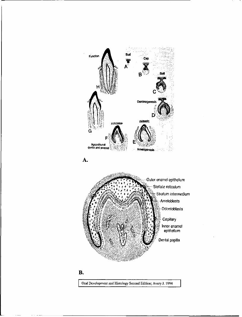
Figure 1 Stages of Tooth Development ........................................................... 3
Figure 2 Formation of 8 mm Critical Size Defect ........................................... 17
Figure 3 Schematic of Critical Size Defect ....................................................... 18
Figure 4 RIDIT Analysis of New Bone Formation at 2 and 8 weeks .............. 32
Figure 5 Histological Slide of Diluent at 2 and 8 weeks .................................. 33
Figure 6 Histological Slide of 1 mg EMD at 2 weeks ...................................... 34
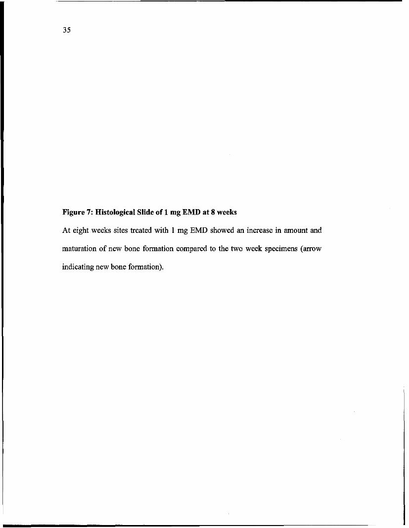
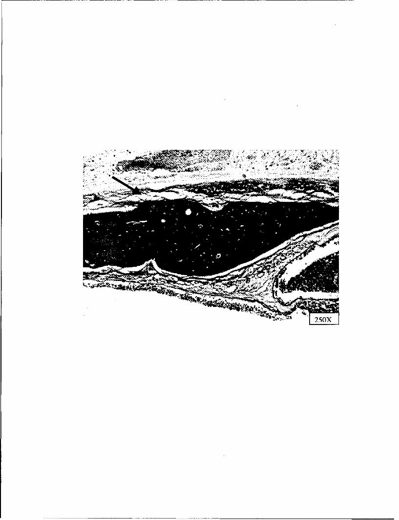
Figure 7 Histological Slide of 1 mg EMD at 8 weeks ...................................... 35
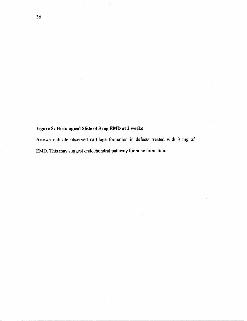
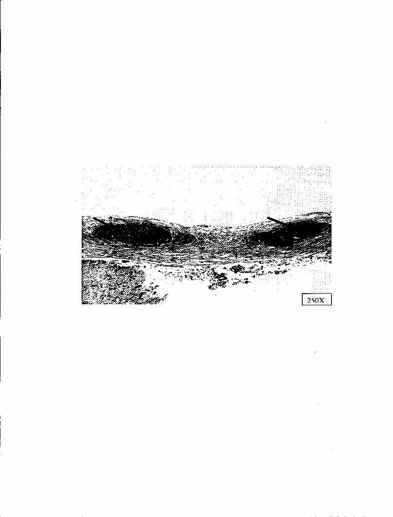
Figure 8 Histological Slide of 3 mg EMD at 2 weeks ...................................... 36
Figure 9 Histological Slide of 3 mg EMD at 8 weeks ...................................... 37
Figure 10 Histological Slide of 9 mg EMD at 2 weeks ...................................... 38
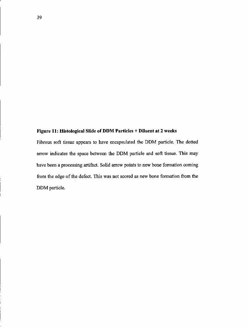
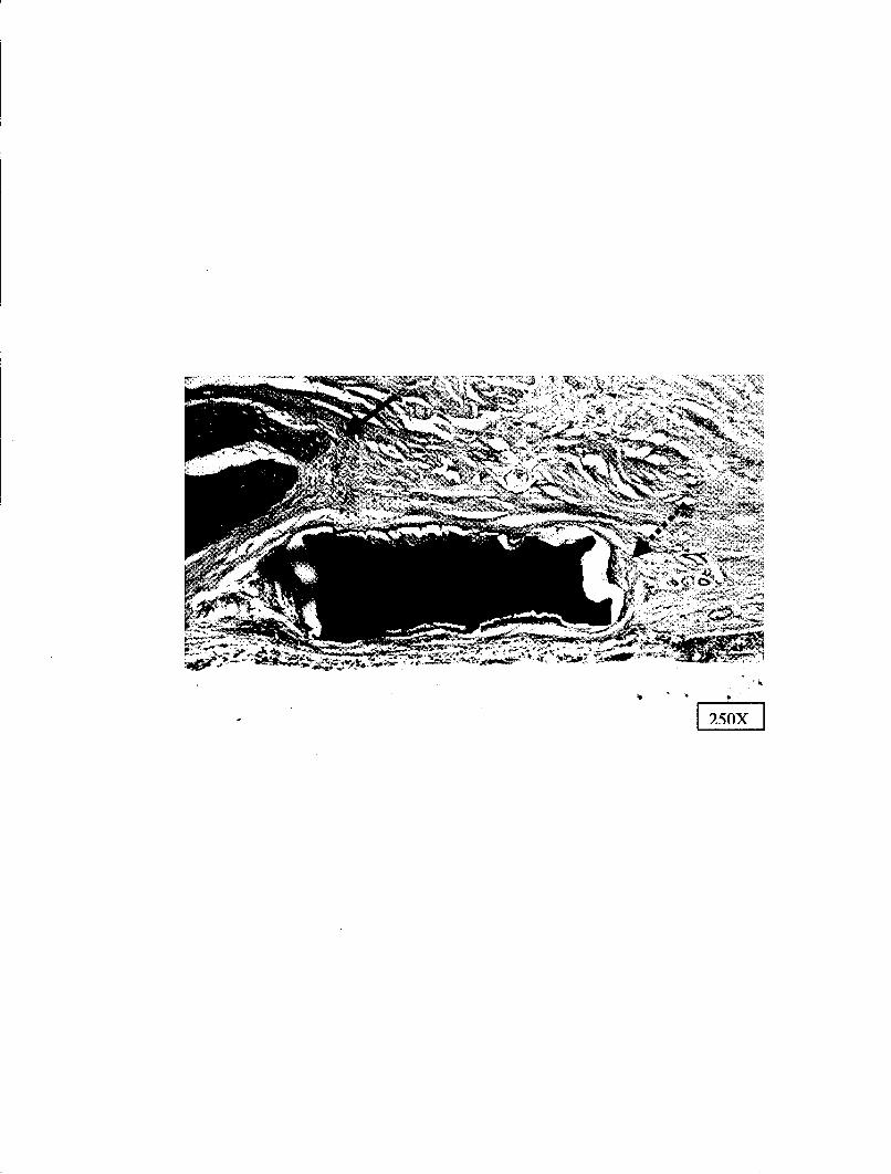
Figure 11 Histological Slide of DDM Particles + Diluent at 2 weeks .............. 39
Figure 12 Histological Slide of DDM Particles + 3 mg EMD at 8 weeks ...... 40
Figure 13 Histological Slide of DFDBA Particles + Diluent at 2 weeks ........... 41
Figure 14 Histological Slide of DFDBA Particles + 3 mg EMD at 8 weeks .......... 42
Figure 15 1251 EMD Count from Critical Size Defect ......................................... 44
ix
LIST OF TABLES
Page
Table 1 Experimental Groups ......................................................................... 15
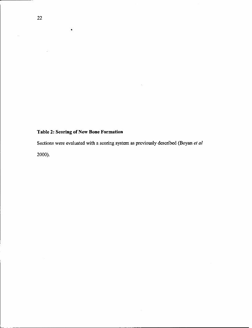
Table 2 Scoring of New Bone Formation ....................................................... 22
Table 3 New Bone Formation at 2 weeks ....................................................... 28
Table 4 New Bone Formation at 8 weeks ....................................................... 31
x
INTRODUCTION AND LITERATURE REVIEW
A. Introduction
Periodontal disease may result in destruction of periodontal attachment and
eventual loss of bone and/or teeth. About 13% of the population suffers from the
moderate to severe forms of this disease (Albander et al 1999). The initial step in treating
periodontal disease includes closed mechanical cleaning of the pockets. The goal is to
decrease the risk of disease progression and establish a healthy periodontium. Once the
disease progression has stabilized, the ideal treatment would include regeneration of
periodontal attachment and bone. Until recently, regenerative procedures were not
considered predictable and pocket reduction through a resective approach was commonly
used to maintain long term periodontal health. Treatment modalities that may promote
regeneration now include alveolar bone grafting, guided tissue regeneration, and the use
of growth factors such as those present in platelets. Stimuli are needed for the
regeneration of cementum, bone, and periodontal ligament which make up the
periodontal apparatus. This regeneration requires the coordination of a cascade of events
that may ultimately result in integrated tissue formation and include different cell types
interacting at specific times with a multitude of growth factors, hormones, and the
extracellular matrix. Alveolar bone grafting, guided tissue regeneration and growth
factors have been shown to regenerate parts of the periodontal attachment apparatus in
humans (Bowers et al 1989b, Sculean et al 1999). To increase the amount and
predictability of regeneration many different biological mediators have been
2
evaluated for their ability to stimulate reattachment and bone growth. Some of these
include differentiating polypeptides, growth factors, and extracellular matrix proteins
(Cochran, Wozney 1999). These biological mediators are naturally occurring hormones
or proteins that are purified and used in vivo in higher concentrations than would occur
naturally. Enamel matrix proteins (EMD), derived from porcine tooth germs as an
extracellular matrix, may stimulate periodontal regeneration by mechanisms that differ
from previous grafting techniques. Theoretically, EMD is able to regenerate the
periodontal apparatus by stimulating the same cells that are active in the bell stage during
tooth development when the tooth supporting apparatus is formed (Hammarstrom 1997).
B. Odontoeenesis
Human odontogenesis begins in the sixth week of gestation. The epithelial
component begins releasing transcription factors to turn on the ectomesenchymal cells.
These ectomesenchymal cells have the capacity to induce epithelial cells to differentiate
into ameloblasts. These enamel-producing cells subsequently stimulate the mesenchymal
cells to differentiate into odontoblasts. As teeth develop they pass through three stages of
growth: bud, cap, and bell. These stages are followed by apposition, root formation, and
eruption (Figure 1).
In the bud stage epithelial structures and the dental lamina are formed. The cap
stage results in the epithelial derived enamel organ and the ectomesenchymal derived
dental papilla. The dental pulp and dentin are formed from the dental papilla. The dental
follicle, which gives rise to the future periodontal ligament, alveolus, and cementum,
3
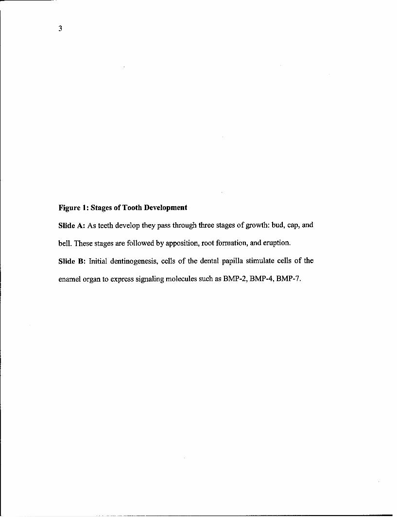
Figure 1: Stages of Tooth Development
Slide A: As teeth develop they pass through three stages of growth: bud, cap, and
bell. These stages are followed by apposition, root formation, and eruption.
Slide B: Initial dentinogenesis, cells of the dental papilla stimulate cells of the
enamel organ to express signaling molecules such as BMP-2, BMP-4, BMP-7.
SD Dud:••.
denction a e amea x
A.
G
dentin and gn"
4it• '...... Outer enamel epithelium.-''•-Stellate reticulum
Stratumi intermedium
'> ~ Odoritoblasts
Inner enamel
" Dental papilla
B.
OrlDevelopment adHistology SeodEdition; Avery 119
4
surrounds the entire cap. The bell stage is characterized by further histodifferentiation of
the cells. Cells of the dental papilla stimulate cells of the enamel organ to express
signaling molecules such as BMP-2, BMP-4, BMP-7, sonic hedgehog, and fibroblast
growth factor-4 (Gartner 1999). Immediately following the formation of the dentin
matrix, the ameloblasts produce enamel matrix. Root formation starts as dentinogenesis
and amelogenesis approach the cervical loop. The inner and outer enamel epitheliums
join to make the Hertwig's epithelial root sheath. Hertwig's epithelial root sheath
elongates until the rim folds in medially. As the Hertwig's epithelial root sheath breaks
down, cells of the dental sac migrate to become cementoblasts (Gartner 1999). Cells from
the enamel organ differentiate into the enamel matrix. Some of the signaling molecules
coming from the enamel matrix are BMP-2, insulin-like growth factor, and transforming
growth factor-P. These signaling molecules initiate the differentiation of the dental
papilla that will produce the dentin matrix (Gartner 1999).
Knowledge of the embryologic development of human teeth stimulated interest in
mimicking normal development in order to regenerate the periodontal apparatus. Due to
the cellular differentiation that occurs during the bell stage, the course of this process
became the focus of research. A new paradigm in periodontal regeneration began to form
that lead researchers to examine modulating natural growth factors as a way to selectively
increase cellular differentiation. As with many of the graft materials in periodontics,
animals served as a source for a developmental protein.
EMD is freeze dried porcine enamel matrix protein that is harvested from the
developing teeth germs of six month old piglets (Hammarstrom 1997). EMD contains
some of the same proteins that are found in immature enamel. Ninety percent of the
5
composition of EMD is amelogenins and its breakdown products. Other proteins found in
EMD are proline-rich non-amelogenins, enamelin, tuft proteins, tuftelin, and serum
proteins. Since EMD is derived from enamel matrix it is thought to primarily stimulate
cementoblast development, but it has also been shown to increase proliferation, protein
synthesis, and mineralization of periodontal ligament cells (Hammarstrom 1997). Enamel
matrix proteins are highly conserved and are similar among mammalian species and have
been shown to be safe when used with human (Brookes et al 1995).
C. Periodontal Regeneration
Periodontal regeneration is the ultimate goal of treatment for restoring and
maintaining health in periodontally diseased patients. Initially, this was an unobtainable
goal. Histological analysis of treated sites revealed that the majority of healing occurred
by long junctional epithelium (Caton et al 1980). Melcher, in 1976, introduced the theory
of guided tissue regeneration. Melcher hypothesized that the periodontal ligament was the
only source of progenitor cells for regeneration. Aukhil et al, in 1986, showed that
progenitor cells of the PDL could differentiate into cementoblasts. Melcher later revised
his theory to include bone as a source for progenitor cells (Melcher et al 1987). In 1988,
Iglhaut et al demonstrated that by excluding the epithelium and gingival connective
tissue, both bone and PDL can serve as sources of progenitor cells for regeneration.
Surgical techniques started to evolve with the knowledge that excluding the epithelial
migration was important for more predictable regeneration.
6
The pioneer of the epithelial exclusion technique for regeneration was Prichard in
1977. This technique involved removal of overlying gingival tissue leaving interdental
bone denuded (Prichard 1977). In 1983, he successfully used this technique to regenerate
intrabony defects. Though Prichard was successful with this technique, it was not
universally predictable. In the early 80's it became common practice to use non-
resorbable membranes to achieve guided tissue regeneration (GTR). Nyman et al (1982)
was the first to demonstrate that new attachment was possible with GTR in a human
model. These membranes were used to exclude the epithelium, thereby ensuring
preferential regeneration by osteoblasts and fibroblasts (Minabe 1991). Even with the
exclusion of the epithelium, complete regeneration was not always achieved. Therefore,
the need to enhance the patient's own regenerative capabilities was hypothesized. In the
1990's bone grafts were combined with the GTR technique.
1. Demineralized freeze dried bone
The gold standard of bone grafts is the patient's own bone. Autografts have both
osteogenic and osteoinductive properties. A bone graft is considered osteoinductive if it
contains proteins which will induce cells to differentiate into osteoblasts. The drawbacks
of autografts include, not having enough bone graft material and in some cases requiring
a second site surgery which increases the chance for infection and post operative
morbidity. Therefore, clinicians began to look at other sources for bone graft materials.
One source is human demineralized freeze dried bone allograft (DFDBA). It has been
shown to be a viable osteoinductive graft alternative to an autograft. Studies by Bowers et
al (1989 b, c) indicate that DFDBA stimulates regeneration of cementum, PDL, as well
7
as alveolar bone in humans. Once placed into the defect, DFDBA stays in the area and is
surrounded by host bone (Becker et al 1994). The residual DFDBA particles provide
wound stability and maintain space (Reynolds, Bowers 1996). Bone morphogenic
proteins are highly conserved proteins in the bone. These proteins have been shown to
stimulate osteoinduction (Urist et al 1983, Wang, Glimcher 1990, Wozney 1995).
Shigeyama et al (1995) isolated BMP-2, BMP-4, and BMP-7 from DFDBA. However,
studies performed by Schwartz et al (1996) have shown that the osteoinductivity varies
between DFDBA batches. Age is a major determining factor for the osteoinductive
properties (Schwartz et al 1998a). Therefore, some batches of DFDBA may be more
osteoconductive than osteoinductive. The addition of EMD to active DFDBA has been
shown to promote an increase in osteoinductivity compared to the active DFDBA alone
(Boyan et a 2000). Due to the demonstrated ability of DFDBA to stimulate regeneration,
it has been considered to be a good choice as a carrier for EMD.
2. Demineralized dentin matrix
Porcine demineralized dentin matrix derivative (DDM) is the residual product
from the production of enamel matrix protein. Demineralized dentin has been shown to
induce ectopic bone formation in rats (Bang, Urist 1967, Somerman et al 1987).
Structurally demineralized dentin is more cross-linked than bone; it has a highly porous
structure with many channels that permit rapid diffusion of reagents (Veis, Perry 1967).
Chemically demineralized bone and dentin have similar compositions (Bang, Urist 1967).
However, demineralized dentin has dentinal proteins, such as phosphophoryns, which are
rich in serine, aspartic acid and phosphorous (Dimuzio, Veis 1978). Demineralized bone
8
proteins contain more glutamic acid than aspartic acid and are lower in serine. Bone is
not as highly phosphorylated as dentin and the long acidic sequences in bone do not
contain phophorylation sites which could bind calcium ions (George et al 1993).
Phosphophoryn molecules move directly to the mineralization front and are directly
associated with mineralized collagen fibrils (Veis 1993). Similar to demineralized bone,
demineralized dentin contains bone morphogenic proteins. Unlike demineralized bone,
the bone morphogenic proteins in dentin have not been isolated (Rutherford, Fizgerald
1995). However, both demineralized bone matrix and demineralized dentin matrix
contain transforming growth factor-f3 1 (TGF-13 1) and insulin-like growth factors (IGF-I
and -II) (Finkelman et al 1990).
Carvalho et al (2004) examined bone formation in critical size bone defects in the
mandibles of rabbits. He demonstrated that the addition of demineralized dentin matrix
slices with a polytetrafluorethylene membrane accelerated new bone formation in the
rabbit mandible. Due to the similarities of demineralized dentin to demineralized bone
and the ready availability of demineralized dentin as a byproduct of EMD production,
demineralized dentin matrix may be a potential carrier for EMD.
3. Enamel matrix derivative
EMD contains some of the same proteins that are found in immature enamel. 90%
of the composition of EMD is amelogenins. Other proteins found in EMD are proline-
rich non-amelogenins, enamelin, tuft proteins, tuftelin, and serum proteins. EMD can
stimulate acellular cementum formation (Hammarstrom 1997) and enhanced periodontal
ligament cell proliferation and attachment (Gestrelius, Andersson 1997, Lyngstradaas et
9
al 2001, Hoang et al 2002). In addition research on effects of EMD on osteoblast
progenitor cells and osteoblasts suggested that EMD may interact with known regulators,
such as bone morphogenic proteins to impact bone regeneration (Iwata et al 2002). EMD
has been shown to enhance periodontal ligament regeneration and promote healing in
defects compared to surgical debridement alone (Heijl 1997, Okdua et a! 2000, Tonetti et
al 2002). EMD has been shown to selectively enhance proliferation of periodontal
ligament cells, but not epithelial cells (Gestrelius, Andersson 1997). The extracellular
matrix proteins are thought to suppress the apical growth of junctional epithelium on root
surfaces (Kawase et al 2000). EMD produces similar clinical results compared to the
clinical results of traditional GTR procedures (Pontoriero et al 1999, Silvestri et al 2000,
Sculean et al 2001, Zucchelli et al 2002, Sanz et al 2004). However, a recent study
suggested that EMD may produce a greater reduction of horizontal furcation depth
compared to similar sites treated with GTR (Jepsen et a! 2004). Histological evaluation of
intrabony defects indicates that EMD regenerates osseous defects around teeth (Sculean
et a! 2000, Cochran et a! 2003, Sculean et al 2004) and has the greatest affects in smaller
sized defects (Cochran et a! 2003, Silvestri et a! 2003). A five year follow up study from
Sculean et al (2004) indicated that the clinical results obtained from EMD were stable up
to five years. To date no longer term studies have yet to be published.
Reports show that EMD accelerates soft tissue healing (Francetti et al 2004,
Hagewald et al 2004) by a proposed mechanism including accelerated angiogenesis
(Yuan 2003). Finally EMD does not appear to enhance root coverage over traditional
connective tissue graft in mucogingival procedures (Modica et al 2000, Hagewald et al
2002, McGuire, Nunn 2003).
10
Propylene glycol alginate is used as carrier for freeze-dried EMD in clinical
procedures. Newer formulations supply the EMD in a gel solution containing 30 mg/ml
of EMD in a syringe containing 0.3 ml for a single defect or 0.7 ml for three periodontal
defects. Possibly because EMD carrier has antimicrobial properties (Sculean et al 2001,
Newman et al 2003) and unlike other grafting agents, no benefit of postoperative
antibiotics has been shown in treatments using EMD (Sculean et al 2001). So far, EMD
has been shown to be safe in multiple surgical procedures (Heard et al 2000). The body
views EMD as a "self protein" and does not produce allergic or other adverse effects after
clinical treatments (Froum et al 2004). This is due to the similarity in amino acid
sequence in pigs and humans indicating these proteins are highly conserved over time.
However, propylene glycol alginate has some inherent characteristics that are less
than ideal for a carrier of EMD. First propylene glycol has a low viscosity after rising to
body temperature and therefore makes the clinical handling of EMD a challenge. The low
viscosity of the material allows for movement outside the defect area presumably after
the proteins have precipitated in the lesion. Second, the carrier does not maintain physical
space with the potential for less defect fill. Multiple studies have evaluated using a
xenograft carrier in combination with EMD. The combination of graft material and EMD
consistently produced better clinical parameters (Lekovic et al 2000, Zucchelli et al
2003). DFDBA in combination with EMD was shown to have similar soft tissue healing
as EMD alone; but the addition of DFDBA increased the osseous fill (Gurinsky et al
2004). This investigation speculated that combining particulate graft material with EMD
would maintain EMD in the periodontal defect.
11
4. Rat Calvaria Model
A critical size defect is a defect that will not heal during the lifetime of the
animal. Testing bone regenerative materials in a critical size defects allows the
osteogenic potential of the material to be considered unequivocal. In the rat calvaria
model the critical size defect is made in the parietal bone between the frontal and
occipital bones. The calvarium is a pure membranous bone that normally heals by
intramembranous ossification. The main reasons why calvarial defects do not heal
spontaneously in adults are because there is very little bone marrow and the blood supply
is poor (Schimtz, Hollinger 1985). The first model for a rat critical size defect was 2 mm
in which the defect reportedly failed to heal in 12 weeks (Turnbull, Freeman 1974).
However, in 1982, Tagaki and Urist demonstrated that an 8 mm defect created in the
calvarium resulted in 3 mm of spontaneous healing with the remaining 5 mm healing
with fibrous connective tissue. Currently, 8 mm diameter defects are considered critical
size in the rat calvarium and are used for the testing of bone regenerative materials.
D. Statement of the Problem and Objectives
EMD has been in clinical use since 1997 and several studies have found it to have
significant but inconsistent regenerative capabilities particularly in larger defects (Yukna,
Mellonig 2000, Parodi et a? 2004). Since the low viscosity of the EMD carrier allows it to
flow away from the defect site, suturing techniques have been employed to retain the
material at the site. Although there is some evidence that EMD can regenerate one wall
defects, the absence of space maintenance properties of the EMD with the present carrier
12
may be one reason for the inconsistent regeneration observed. One way to maintain the
space in a defect and keep EMD localized to the defect would be to use a different carrier
substance with EMD.
Therefore, the objective of this study was to evaluate two carrier substances,
DFDBA and DDM, to potentially enhance the response of EMD and to test whether
EMD placed with DFDBA or DDM was retained at the surgical site. Our hypothesis was
that the addition of a carrier to EMD would increase the duration of EMD in the defect
site and thereby increase bone formation. The following specific aims were established to
fulfill this objective:
1) To evaluate the ability of EMD in combination with DFDBA or DDM to
enhance bone regeneration.
2) To evaluate the ability of DFDBA or DDM to retain EMD at the surgical
site.
MATERIALS AND METHODS
A. Xenoarafts
Human DFDBA (Musculoskeletal Transplant Foundation, Dentsply Ceramed
Dental Lot MTF 006213930502) ranging in size from 250-860 gim in diameter was
obtained in the clinical use form. Porcine DDM (XP40 Lot 0102) was obtained from
Biora AB Malmo, Sweden (Biora was acquired by Straumann (Basel, Switzerland) in
2004). These particles were of a much larger size, ranging from 1000-4100 gtm. The
DFDBA and DDM were tested in an 8 mm critical size defect to assess bone induction
ability.
B. Enamel Matrix Derivative
Porcine embryonic enamel matrix derivative was obtained from the manufacturer
(Biora AB Malmo, Sweden) in the lyophilized form available for clinical use. It was
reconstituted with propylene glycol alginate (diluent) (Biora AB Malmo, Sweden, Lot
PGA2113).
C. Analysis of New Bone Formation
1. Preparation for Implants
13
14
DFDBA (10 mg) and DDM (10 mg) particles were placed into sterile tubes. When
the EMD was indicated 1, 3, or 9 mg of EMD with propylene glycol carrier was added to
the DFDBA or DDM particles in the tubes, or placed in a separate sterile tube. On the
day of implantation the contents of the tube were placed in a sterile dampen dish and the
diluent (24 jil propylene glycol alginate) was added and mixed with a sterile spatula.
2. Animal Protocol
Sprague-Dawley (Charles River) rats 70-85 days / 300-349 grams were used for
this study. The Institutional Animal Care and Use Committee at UTHSCSA approved
this protocol prior to commencement of the study.
3. Experimental groups
The rats were divided into 14 groups per time point (2 or 8 weeks) with 8 rats in
each group (Tables 1): 1) diluent, 2) 1 mg EMD with diluent, 3) 3 mg EMD with diluent,
4) 9 mg EMD with diluent, 5) DFDBA and diluent, 6) DFDBA without diluent, 7) 1 mg
EMD with diluent and DFDBA, 8) 3 mg EMD with diluent and DFDBA, 9) 9 mg EMD
with diluent and DFDBA, 10) DDM and diluent, 11) DDM without diluent, 12) 1 mg
EMD with diluent and DDM, 13) 3 mg EMD with diluent and DDM, 14) 9 mg EMD
with diluent and DDM.

15
Table 1: Experimental Groups
The following table illustrates the division of rat groups. There were 14 groups at
each time point (two and eight weeks) with eight rats per group.
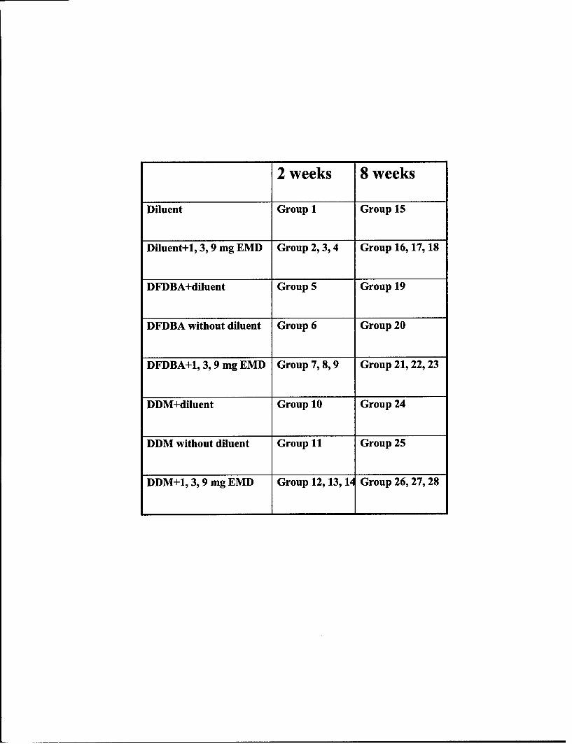
2 weeks 8 weeks
Diluent Group 1 Group 15
Diluent+l, 3,9 mg EMD Group 2, 3, 4 Group 16, 17, 18
DFDBA+diluent Group 5 Group 19
DFDBA without diluent Group 6 Group 20
DFDBA+1, 3,9 mg EMD Group 7, 8, 9 Group 21, 22, 23
DDM+diluent Group 10 Group 24
DDM without diluent Group 11 Group 25
DDM+1, 3,9 mg EMD Group 12, 13, 14 Group 26, 27, 28
16
4. Surgical Protocol
The rats were anesthetized by intramuscular injection. The anesthesia cocktail
contained 6 ml Ketamine/Acepromazine (1 ml Acepromazine (100 mg/ml) and 10 ml
Ketamine (100 mg/ml)) and 4 ml small animal Rompur (20 mg/ml). Subsequently rats
were maintained on isoflurane gas (4 L/min) with oxygen (2 L/min). The head of each rat
was shaved and disinfected using povidone iodine. A skin incision of 1.0-2.0 cm was
made on the dorsal surface of the calvarium and expanded by blunt dissection. An 8 mm
critical size defect was made in the parietal bone between the occipital and frontal bones,
taking care to leave the dura undisturbed (Figure 2, 3). The defect was created using an 8
mm Arruga trephine (Miltex, Bethpage, NY) and copious saline irrigation. After
hemostasis was achieved the rats received implantation at random from one of the
experimental groups. Wound clips were placed for primary closure of the surgical site.
The rats were placed two to a cage for healing and were allowed water and food ad
libitum. During healing no clinically observable negative effects were noted for the rats.
17
Figure 2 A and B: Formation of 8 mm Critical Size Defect
A skin incision of 1.0-2.0 cm was made on the dorsal surface of the calvarium and
expanded by blunt dissection. An 8 mm critical size defect was made in the
parietal bone between the frontal and occipital bone, taking care to leave the dura
undisturbed.
Panel A: Shows the 8 mm trephine used to create the defect.
Panel B: Shows the critical size defect with the underlying dura intact.
A.
B.
18
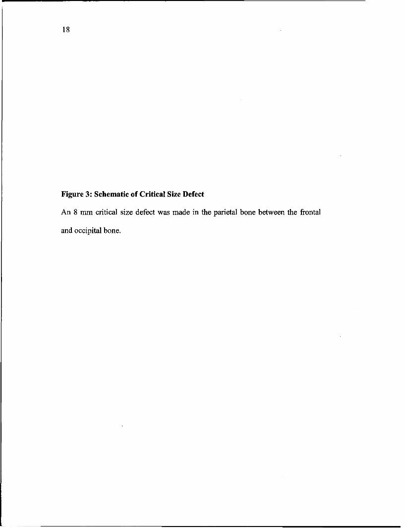
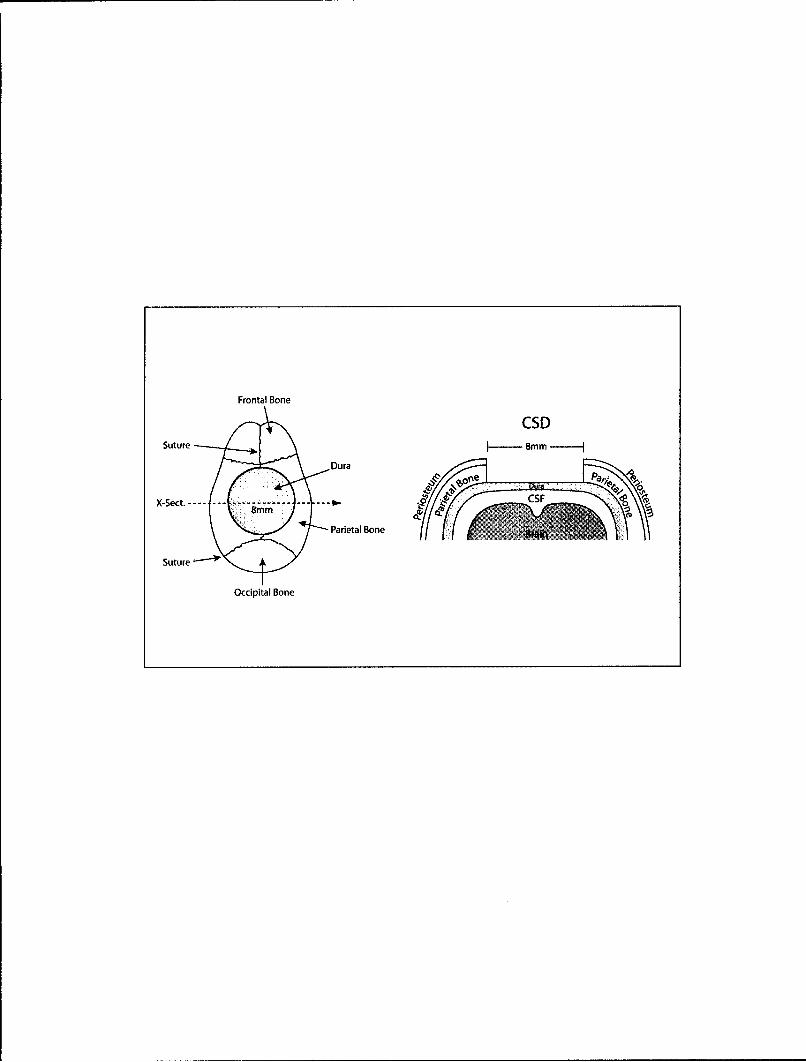
Figure 3: Schematic of Critical Size Defect
An 8 mm critical size defect was made in the parietal bone between the frontal
and occipital bone.
Frontal Bone
CSD
Suture [ 8mm - ]
Dura 0~
X-Sect.----- --- ----------- ---- -- S8mm0
Parietal Bone B
Suture
Occipital Bone
19
5. Histological Evaluation
The rats were euthanized by asphyxiation with carbon dioxide at two and eight
weeks. Tissue was recovered from the implant site and fixed in 10% formalin. The
samples were then decalcified in Surgipath (Richmond, IL) decalcifier II solution. The
samples were sectioned through the middle of the defect and placed face down on the
slide and embedded in paraffin. The samples were then sectioned at 3-4 pm each, and
stained with hemotoxylin and eosin. One section from each level was evaluated and the
data are pooled for each defect site.
The entire section was evaluated for presence of DFDBA particles, dentin matrix
particles, and new bone using a scoring system (Table 2) as previously described (Boyan
et al 2000). Briefly sections with no new bone formation or carrier were scored 0;
sections with no new bone formation but the DFDBA or DDM was present, were scored
1; sections with new bone in one site <40% of surface area were scored 2; sections with
new bone in more than one site covering >40% but <70% were scored 3; a section with
new bone formation in more than one site covering >70% of the surface area were scored
4. There was one examiner for all the slides who was blinded to the test groups. When no
DFDBA or DDM was present in the section the new bone formation was scored and the
number received a star. This test group number was then checked against the master key.
If the group contained EMD alone the score for the new bone formation was recorded. If
the test group was to contain DFDBA or DDM the score resulted in a zero and was not
included in the statistical analysis.
20
D. Statistical Analysis
RIDIT (relative to an identified distribution) analysis was used to statistically
analyze the bone score data (Fleiss 1973). RIDIT analysis is appropriate for sample data
that is qualitatively ordered because it takes advantage of ordering that exists but does not
require a gradient in proportions. In RIDIT analysis, the discrete categories represent
intervals on some unobservable distribution. The RIDIT analysis uses a reference group.
In the current study the reference group was propylene glycol alone. The reference group
has a mean RIDIT value of 0.5. All other groups are compared to the reference group.
The resulting mean RIDIT value of a group that is 0.5 indicates that there is no greater
probability of selecting a sample out of that group with more new bone formation than
out of the reference group. A RIDIT value greater than 0.5, indicates that group has a
greater probability of the outcome, which is more new bone formation. A RIDIT value
less than 0.5 indicates a lesser probability of finding greater new bone formation in that
group than in the reference group. A change of 0.1 RIDIT unit would indicate a 10%
variable probability of either an increase or decrease in bone formation compared to the
reference group. P values < 0.05 were considered significant.
Although RIDIT analysis is appropriate for qualitatively ordered categories, it is
less powerful at small sample sizes. Therefore, descriptive statistics were also determined
for the bone score data. The data were analyzed using a 3-way ANOVA followed by pair-
wise comparison of the means. The three factors were healing time, diluent, and carrier.
Analysis included all interactions of the factors, and for pair-wise comparison of the
means, the effect of each factor was adjusted for the effects of the other factors (type III).

21
In addition to analysis of the raw data, a standard square root transformation of the data
was performed. This was done because of the small number of ordered categories, as well
as to assure that the standard assumptions for valid analysis were satisfied, that the data
were normally distributed and the variances equal.
22
Table 2: Scoring of New Bone Formation
Sections were evaluated with a scoring system as previously described (Boyan et al
2000).
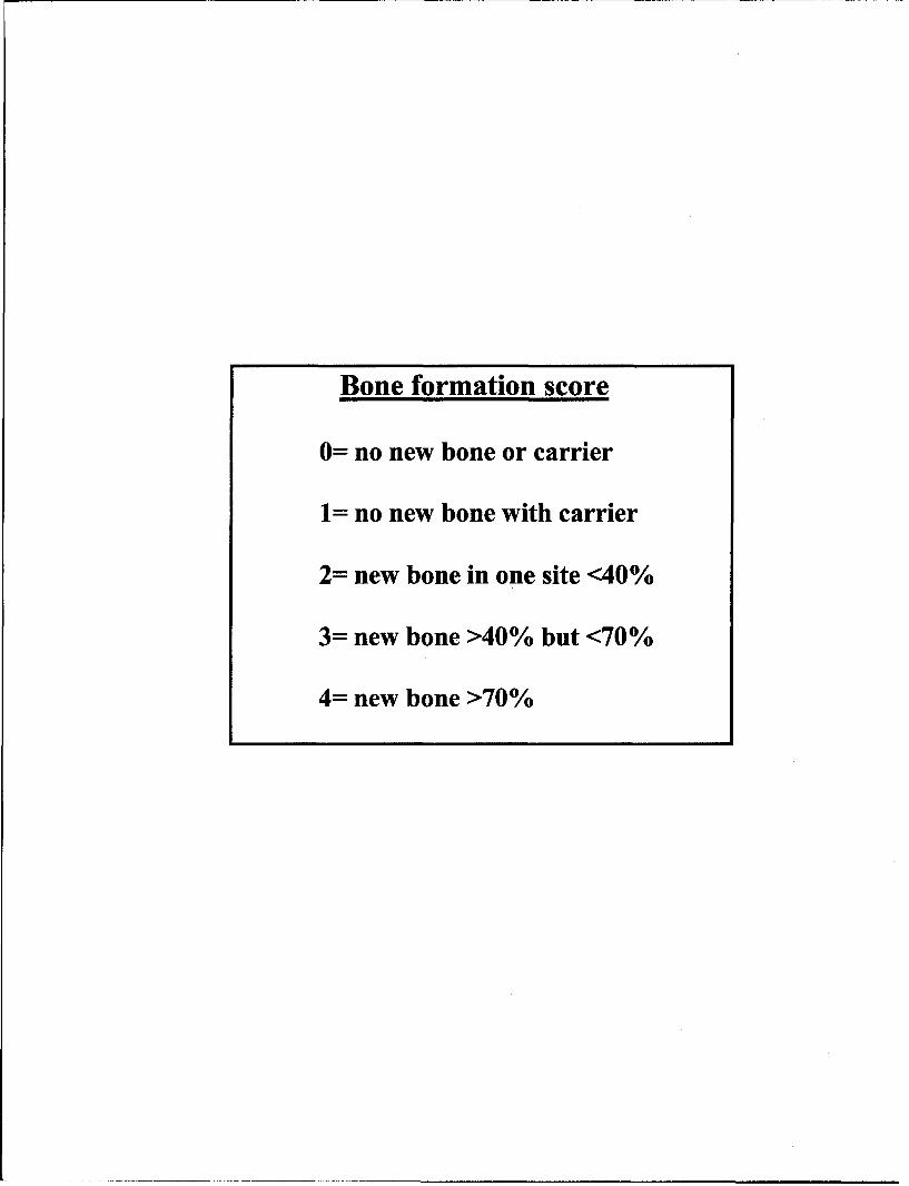
Bone formation score
0= no new bone or carrier
1= no new bone with carrier
2= new bone in one site <40%
3= new bone >40% but <70%
4= new bone >70%
23
E. Retention time of Enamel Matrix Derivative
1. Radiolabeled Enamel Matrix Derivative
EMD was radioactively labeled with 125, (Rosenberg, Teare 1977). Thirty
milligrams of EMD were dissolved in 3 ml of 0.01% acetic acid and allowed to
equilibrate at room temperature. This stock solution of EMD was further diluted to a
final concentration of lmg/ml in cold phosphate buffered saline, pH 7.2. Carrier free
Na125I obtained from PerkinElmer Life Sciences (Billerica, MA) was diluted to 5 mCi
(185 MBq) / 25 jil with PBS, pH 7.2. The iodination was performed at room temperature
in a 12 x 75 mm glass tube by the sequential addition of 5 pl Na 125I, 25 gtl EMD, and 5 gil
chloramine T (5 mg/ml in PBS, pH 7.2). After 30 seconds the reaction was stopped by
the addition of 5 jil sodium metabisulfite (10 mg/ml) and 460 gtl of 30% acetonitrile-0.15
M sodium chloride. For separation of the iodinated protein from unreacted iodide, the
entire reaction mixture was applied to a PD-10 column previously equilibrated with 30%
acetonitrile-0. 15 M sodium chloride. Elution was carried out using the same solution and
fractions of 1 ml / tube were collected. The fractions were assayed for radioactivity in a
Beckman Gamma 5500 gamma scintillation counter, and the iodination product eluted in
fractions 6 through 9 were used for the experiments. These fractions were pooled and
assayed for activity /volume in a Radix Dose Calibrator (Houston, TX). Instant TLC,
performed using SG-silica gel strips (Gelman Instrument Company, Ann Arbor, MI;
currently available from VWR International) determined the per cent of free iodine to be
less than 10% (Rosenberg, Teare 1977). The entire iodination procedure was repeated as
just described with more EMD, the iodination products from each procedure pooled, and
24
the volume reduced using a stream of nitrogen. The radioactive concentration of the final
solution was 30 ptCi / 25 jtl.
2. Implant Material Preparation
On the day of the surgery DFDBA (10 mg) or DDM (10 mg) was mixed with 25
ll of the reconstituted 1251 radiolabeled EMD (30 gCi / 25 itl).
3. Experimental 2rouDs
The rats were divided into three groups of 6 rats each: 1) radiolabeled EMD 2)
DFDBA and radiolabeled EMD 3) DDM and radiolabeled EMD.
4. Surgical Protocol
The rats were anesthetized with isoflurane gas (4 L/min) with oxygen (2 L/min).
Gas alone was used to achieve a short recovery period. The surgical procedure was the
same as Part I of the experiment. After hemostasis, 25 jil (30 gCi) of the 1251 radiolabeled
EMD was added to the defect alone or combination with either 10 mg of DFDBA or 10
mg of DDM. Primary closure of the wound was achieved with 4-0 non-resorbable
monofilament nylon sutures for primary closure. Two rats were placed to a cage for
healing and were allowed water and food ad libitum. During healing no clinically
observable negative effects were noted for the rats.
5. Gamma Scinti2raphv
25
Scintigraphic images were acquired immediately after implantation (0 hour), at 4
hours, and at 1, 2, 3, 6, 13, and 20 days. The rats were imaged under isoflurane gas
anesthesia as described above. Imaging was performed with a Picker SX-300 Spect
Gamma Camera (Picker Instruments, Cleveland, OH). Before imaging, the camera was
calibrated using 57Cobalt. Also, an extrinsic flood quality control image was obtained
with a sheet of uniformly distributed 57Cobalt. Because the energy of 125, is low for this
camera, the collimator was removed and the camera shielded with a 5 mm thick, solid
lead sheet except for a 1.0 cm hole that was placed directly over the area of the defect.
The camera head was centered over the hole and lowered to within 0.5 cm of the lead
shield. Image acquisition was performed with a MEDASYS Pinnacle computer work
station (MEDASYS, Miami, FL) interfaced to the SX-300 camera. Background
acquisitions along with 125I standards were taken daily prior to the animal acquisitions.
Control images were obtained by spotting 25 gtl containing 30 PiCi of 1251 labeled EMD
on 8 mm disks of filter paper. The dried disks were placed on the lid of a small Petri dish
and positioned directly under the hole in the lead shield for image acquisition. The
control disks were imaged for each time point in the study. A 10 minute acquisition
period was used to obtain counts that were at least 10 times greater than background.
Image analysis of uniform regions of interest containing identical pixel numbers from the
acquired images was performed using OSIRIS software (University Hospital of Geneva,
Switzerland). Retention of 1251 at the defect site was determined and corrected for
radioactive decay.
26
F. Statistical Analysis
The 125I retention data were analyzed with multiple comparison analysis, a type of
ANOVA, using a Tukey's procedure as the post-hoc test. Significance was determined at
the p < 0.05 level.
RESULTS
A. Analysis of New Bone Formation
The surgical procedures and healing were as expected with a couple of
exceptions. First, there were animal deaths, thus decreasing the groups' size, traced to
defective anesthetic cocktail. Second, there were differences in the surgical skill on the
part of the surgeon which may have contributed to the variability of the results. The
observed mean RIDIT values for the treatment groups are displayed in Tables 3, 4 and
Figure 4.
Two week observations:
At two weeks, RIDIT analysis indicated that there were no statistically significant
differences between the reference group (propylene glycol alone) and the treatment
groups (Table 3).
EMD alone: One mg of EMD resulted in the same mean RIDIT value as the
reference group. Placement of 3 mg and 9 mg of EMD resulted in 0.2 and 0.3 mean
RIDIT value respectively, which indicated there was a lower probability of finding new
bone formation in these groups than the reference group.
DFDBA group: DFDBA alone groups, with and without diluent, resulted in the
same mean RIDIT value as the reference group (0.5). When EMD was added to DFDBA
(1 mg, 3 mg, 9 mg) the resulting mean RIDIT values were less than the reference group
27
(0.2, 0.3, and 0.3 respectively). This indicated that there was a reduced probability of
finding more new bone formation in these groups than the reference group.
28
28
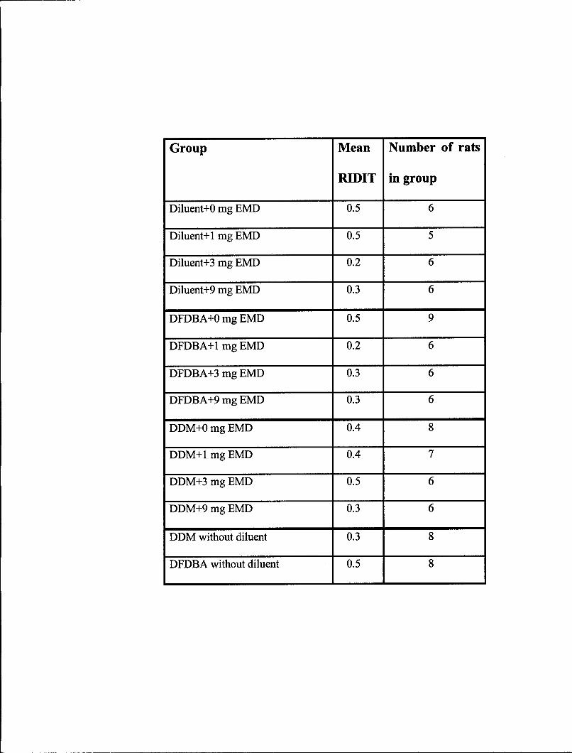
Table 3: New Bone Formation at 2 weeks
The mean RIDIT values are displayed for each group as well as the number of
animals per group. The reference group has a mean RIDIT value of 0.5. None of
the groups were statistically different at two weeks (pO0.05 statistically
significant).
Group Mean Number of rats
RIDIT in group
Diluent+0 mg EMD 0.5 6
Diluent+1 mg EMD 0.5 5
Diluent+3 mg EMD 0.2 6
Diluent+9 mg EMD 0.3 6
DFDBA+0 mg EMD 0.5 9
DFDBA+I mg EMD 0.2 6
DFDBA+3 mg EMD 0.3 6
DFDBA+9 mg EMD 0.3 6
DDM+O mg EMD 0.4 8
DDM+I mg EMD 0.4 7
DDM+3 mg EMD 0.5 6
DDM+9 mg EMD 0.3 6
DDM without diluent 0.3 8
DFDBA without diluent 0.5 8
29
DDM group: The dentin matrix alone groups, with and without diluent, resulted
in a reduced RIDIT value (0.4 and 0.3 respectively) compared to the reference group. The
dentin matrix group with the addition of 1 and 9 mg of EMD also had a reduced RIDIT
value (0.4 and 0.3 respectively) compared to the reference group. Dentin matrix particles
mixed with 3 mg of EMD resulted in the same RIDIT value (0.5) as the reference group,
indicating that there was an equal probability of finding new bone formation in either
group.
Eight week observations:
At eight weeks (Table 4), the RIDIT analysis indicated that there was more bone
formation in all the treatment groups compared to the reference group. However, with the
exception of 3 mg EMD in combination with the DDM particles (p_<0.05), these values
did not reach statistical significance.
EMD alone group: At eight weeks the mean RIDIT value of the 0 mg EMD group
was 0.3. As the concentration of EMD increased from 1 mg EMD to 9 mg EMD there
was a trend for increased bone formation (1 mg EMD=0.5; 3 mg EMD=0.6; 9 mg
EMD=0.7). These were not statistically significant differences.
DFDBA group: The mean RIDIT values for DFDBA with diluent (DFDBA+0 mg
EMD) and 1 mg EMD + DFDBA was 0.5, indicating that the amount of new bone
formation was similar to the reference group. Both 3 mg and 9 mg EMD experienced a
trend toward increased new bone formation with the same mean RIDIT value of 0.6.
30
DFDBA alone also received a mean RIDIT value of 0.6. There were no statistically
significant changes in new bone formation in any of these treatment groups.
DDM group: All treatment groups showed a mean RIDIT value greater than the
reference group. DDM with and without diluent, 1 mg and 9 mg EMD had a mean RIDIT
value of 0.6. Three mg EMD in combination with DDM particles resulted in a mean
RIDIT value of 0.8. This was the only value to achieve statistical significance (p50.05 ).
Hisotological analysis demonstrated little to no new bone formation noted away
from the margin of the defect in the diluent (propylene glycol) alone groups. In the
diluent groups soft tissue filled the majority of the defect. At eight weeks small islands of
new bone were noted in select specimens (Figure 2). Histologically the bone formation
response in the two carmer groups appeared differently. Interestingly, unlike the DFDBA
particles, the DDM particles were encapsulated with fibrous soft tissue and were not in
intimate contact with the new bone formation. Encapsulation of the DDM particles was
not expected, but it did not seem to affect the bone formation response. The separation of
the DDM particles from the surrounding fibrous soft tissue may be a processing artifact
(Figure 11, 12). The appearance of the new bone formed in contact with DFDBA
particles, appeared similar to when DFDBA is used in the ectopic model. This new bone
was in intimate contact with the DFDBA particles (Figures 13, 14). EMD alone at all
concentrations was slightly more osteoinductive than either DFDBA or DDM particles
alone. At eight weeks sites treated with EMD showed increased amount and maturation
of new bone formation (Figures 7, 9). Of note, cartilage like formation was observed at
all concentrations of EMD alone at two weeks (Figures 6, 8, 10).
31
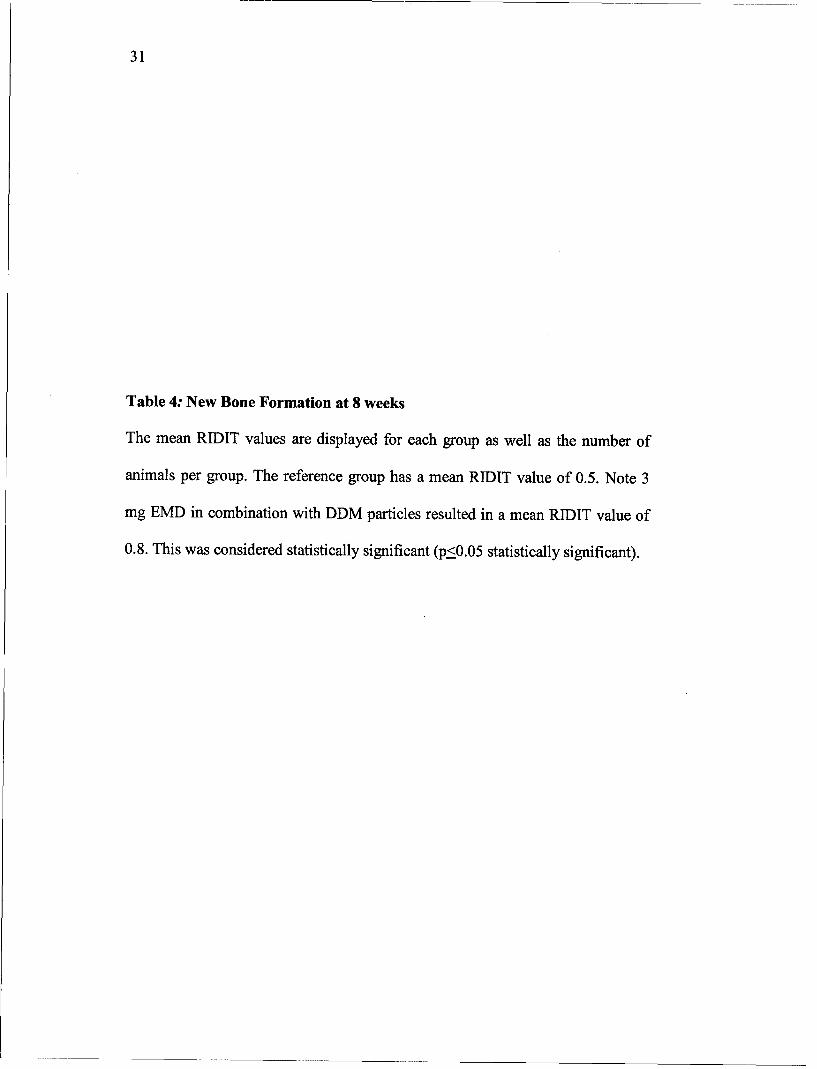
Table 4: New Bone Formation at 8 weeks
The mean RIDIT values are displayed for each group as well as the number of
animals per group. The reference group has a mean RIDIT value of 0.5. Note 3
mg EMD in combination with DDM particles resulted in a mean RIDIT value of
0.8. This was considered statistically significant (p_<0.05 statistically significant).
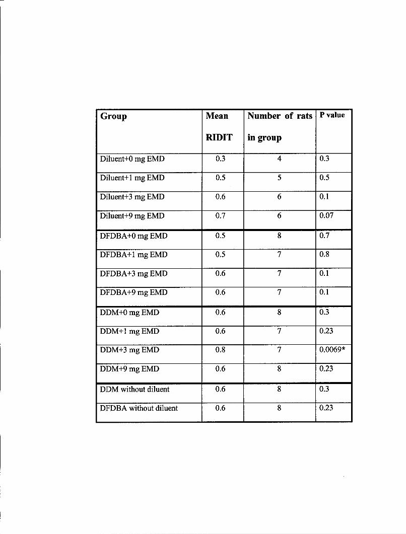
Group Mean Number of rats P value
RIDIT in group
Diluent+0 mg EMD 0.3 4 0.3
Diluent+1 mg EMD 0.5 5 0.5
Diluent+3 mg EMD 0.6 6 0.1
Diluent+9 mg EMD 0.7 6 0.07
DFDBA+0 mg EMD 0.5 8 0.7
DFDBA+1 mg EMD 0.5 7 0.8
DFDBA+3 mg EMD 0.6 7 0.1
DFDBA+9 mg EMD 0.6 7 0.1
DDM+0 mg EMD 0.6 8 0.3
DDM+I mg EMD 0.6 7 0.23
DDM+3 mg EMD 0.8 7 0.0069*
DDM+9 mg EMD 0.6 8 0.23
DDM without diluent 0.6 8 0.3
DFDBA without diluent 0.6 8 0.23
32
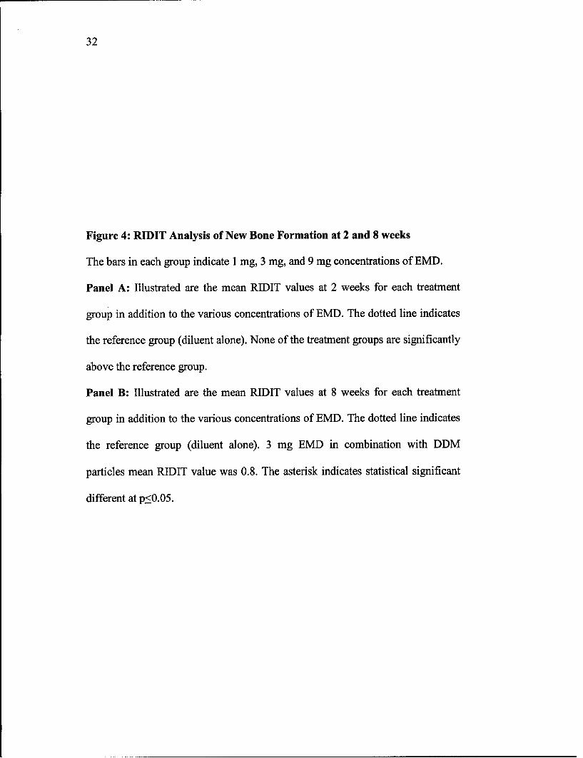
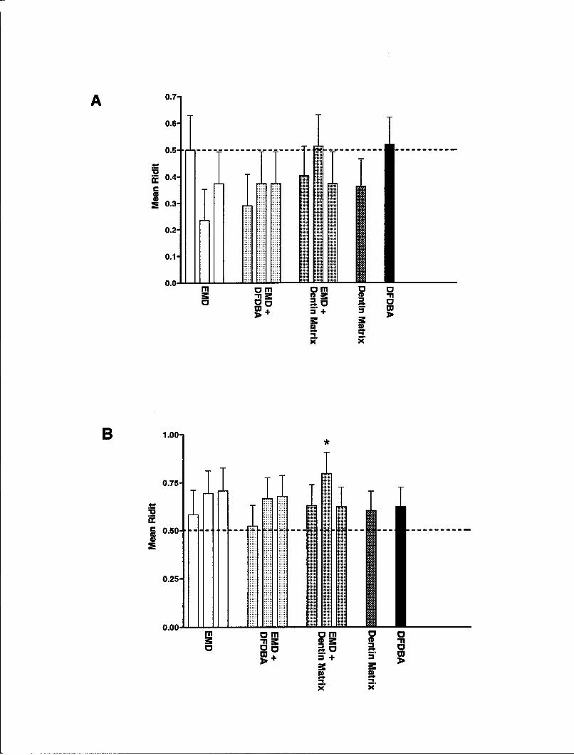
Figure 4: RIDIT Analysis of New Bone Formation at 2 and 8 weeks
The bars in each group indicate 1 mg, 3 mg, and 9 mg concentrations of EMD.
Panel A: Illustrated are the mean RIDIT values at 2 weeks for each treatment
group in addition to the various concentrations of EMD. The dotted line indicates
the reference group (diluent alone). None of the treatment groups are significantly
above the reference group.
Panel B: Illustrated are the mean RIDIT values at 8 weeks for each treatment
group in addition to the various concentrations of EMD. The dotted line indicates
the reference group (diluent alone). 3 mg EMD in combination with DDM
particles mean RIDIT value was 0.8. The asterisk indicates statistical significant
different at p<0.05.
A 0.7-
0.6-
0.5- R -
"O• .4'
0 . .. .. .. .. . .... .... .. .....=• 0.3.
0 .2 -. .... ....
0.1-
.. -. . . ......
O.OE
m 10m 9)m S) a
E -" C" EI
> >
B 1.00-
0.75-
"4-U. M."~~. 0.50.......--
0.25-
0.00 0 . . ..m aom j)-" c
33
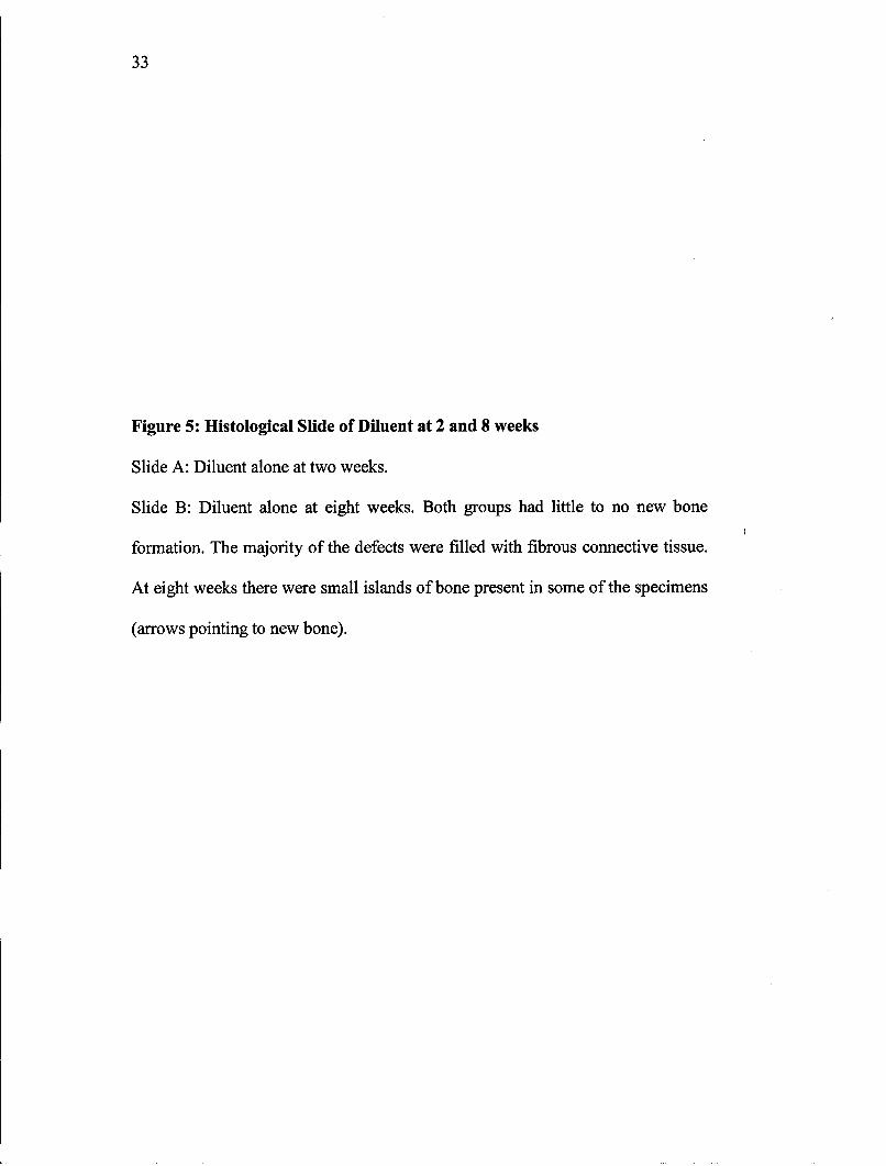
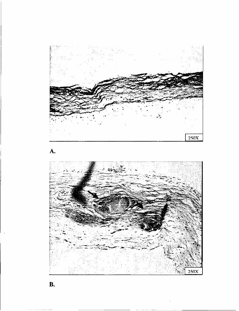
Figure 5: Histological Slide of Diluent at 2 and 8 weeks
Slide A: Diluent alone at two weeks.
Slide B: Diluent alone at eight weeks. Both groups had little to no new bone
formation. The majority of the defects were filled with fibrous connective tissue.
At eight weeks there were small islands of bone present in some of the specimens
(arrows pointing to new bone).
250X
A.
-� -
V-�-�
-�
4 -
250X
B.
34
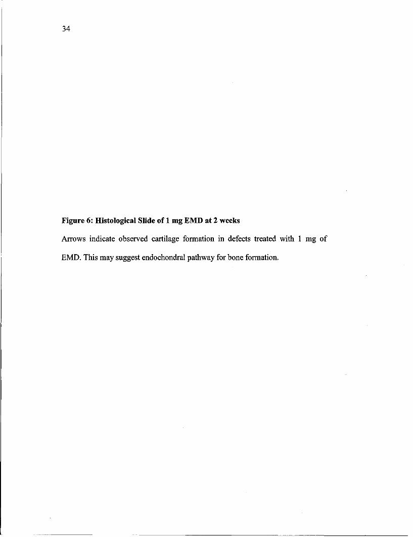
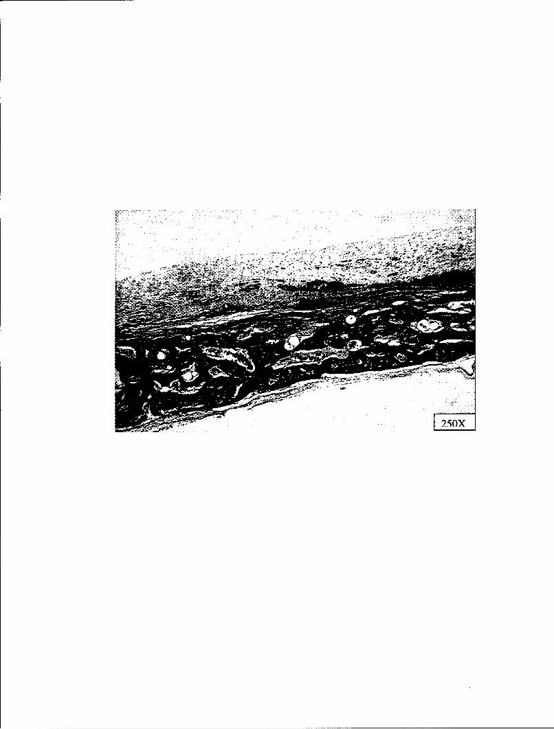
Figure 6: Histological Slide of 1 mg EMD at 2 weeks
Arrows indicate observed cartilage formation in defects treated with 1 mg of
EMD. This may suggest endochondral pathway for bone formation.
---------
35
Figure 7: Histological Slide of 1 mg EMD at 8 weeks
At eight weeks sites treated with 1 mg EMD showed an increase in amount and
maturation of new bone formation compared to the two week specimens (arrow
indicating new bone formation).
1-At-
36
Figure 8: Histological Slide of 3 mg EMD at 2 weeks
Arrows indicate observed cartilage formation in defects treated with 3 mg of
EMD. This may suggest endochondral pathway for bone formation.
-Air
P110- 4V:l 4DII
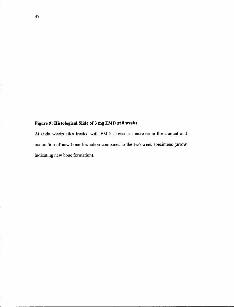
37
Figure 9: Histological Slide of 3 mg EMD at 8 weeks
At eight weeks sites treated with EMD showed an increase in the amount and
maturation of new bone formation compared to the two week specimens (arrow
indicating new bone formation).
- Opp
-25O
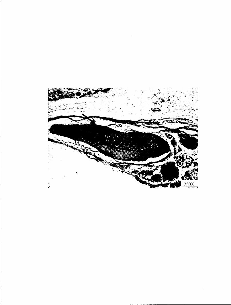
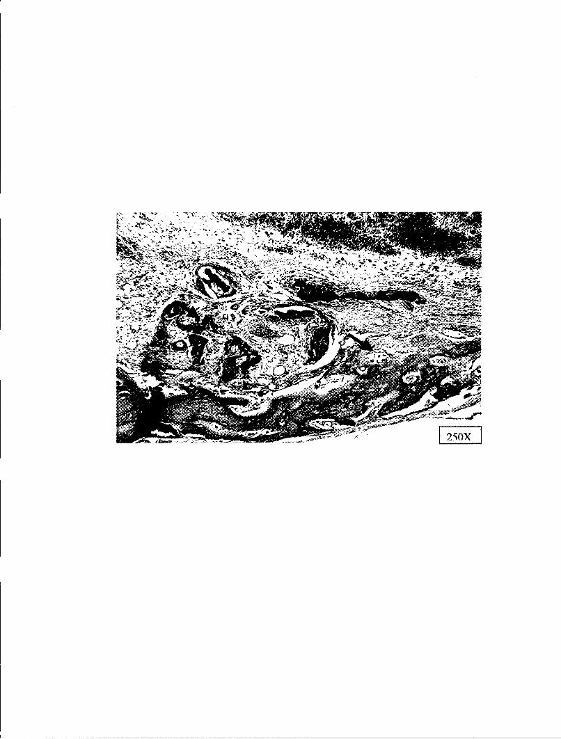
38
Figure 10: Histological Slide of 9 mg EMD at 2 weeks
Arrows indicate observed cartilage and bone formation in defects treated with 9
mg of EMD. This may suggest endochondral pathway for bone formation.
NIN
39
Figure 11: Histological Slide of DDM Particles + Diluent at 2 weeks
Fibrous soft tissue appears to have encapsulated the DDM particle. The dotted
arrow indicates the space between the DDM particle and soft tissue. This may
have been a processing artifact. Solid arrow points to new bone formation coming
from the edge of the defect. This was not scored as new bone formation from the
DDM particle.
EFII
40
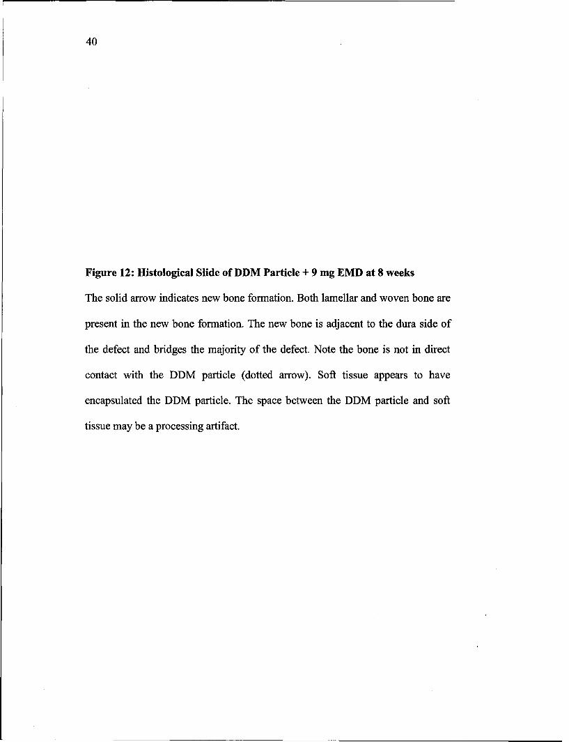
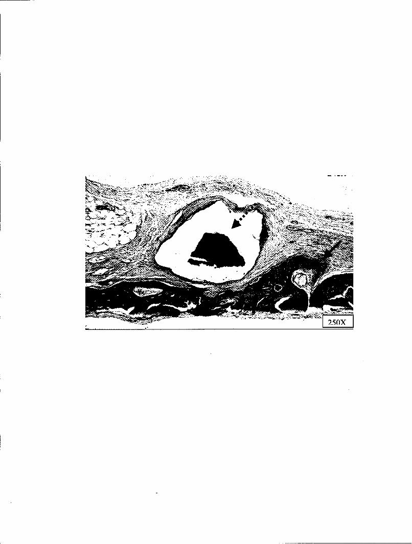
Figure 12: Histological Slide of DDM Particle + 9 mg EMD at 8 weeks
The solid arrow indicates new bone formation. Both lamellar and woven bone are
present in the new bone formation. The new bone is adjacent to the dura side of
the defect and bridges the majority of the defect. Note the bone is not in direct
contact with the DDM particle (dotted arrow). Soft tissue appears to have
encapsulated the DDM particle. The space between the DDM particle and soft
tissue may be a processing artifact.
-t7
41
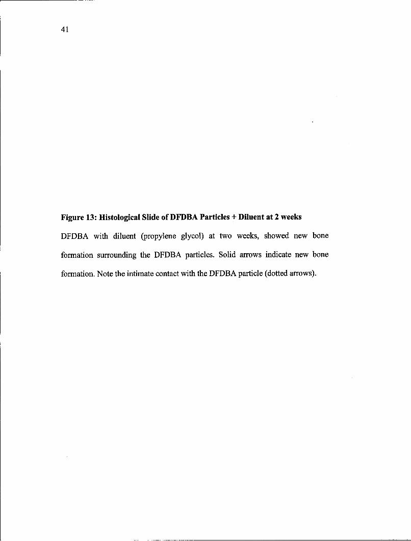
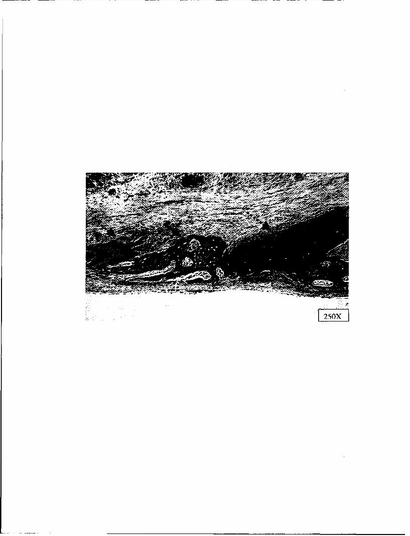
Figure 13: Histological Slide of DFDBA Particles + Diluent at 2 weeks
DFDBA with diluent (propylene glycol) at two weeks, showed new bone
formation surrounding the DFDBA particles. Solid arrows indicate new bone
formation. Note the intimate contact with the DFDBA particle (dotted arrows).
42
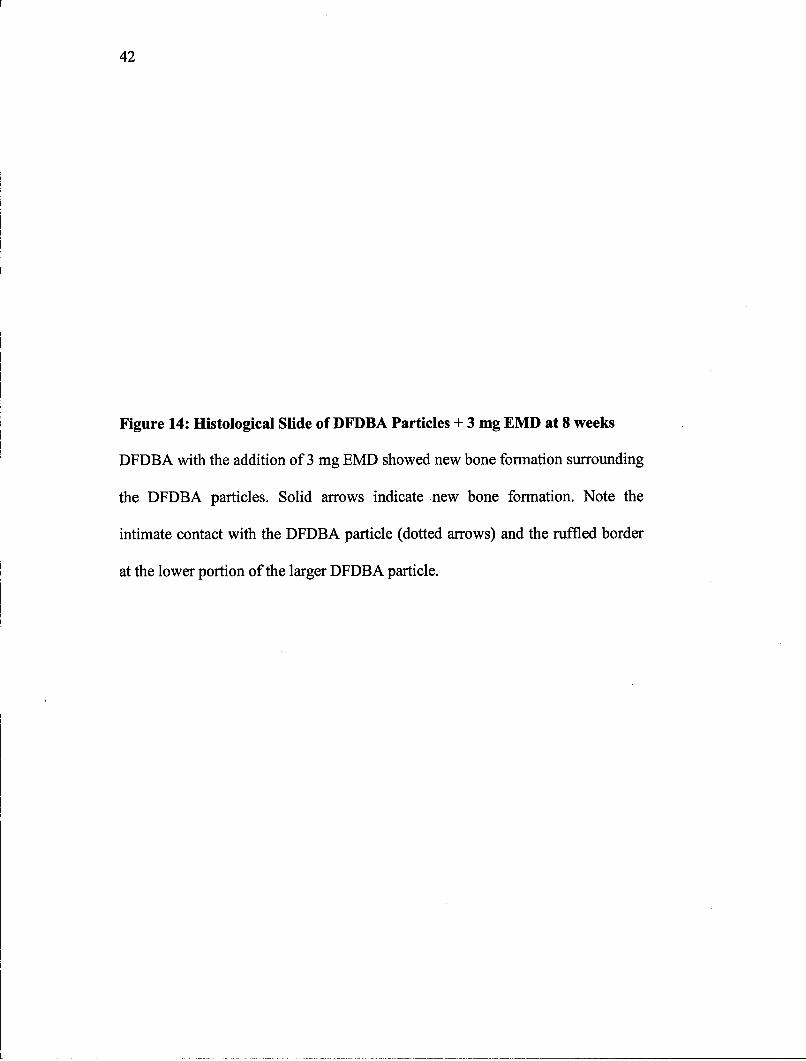
Figure 14: Histological Slide of DFDBA Particles + 3 mg EMD at 8 weeks
DFDBA with the addition of 3 mg EMD showed new bone formation surrounding
the DFDBA particles. Solid arrows indicate new bone formation. Note the
intimate contact with the DFDBA particle (dotted arrows) and the ruffled border
at the lower portion of the larger DFDBA particle.
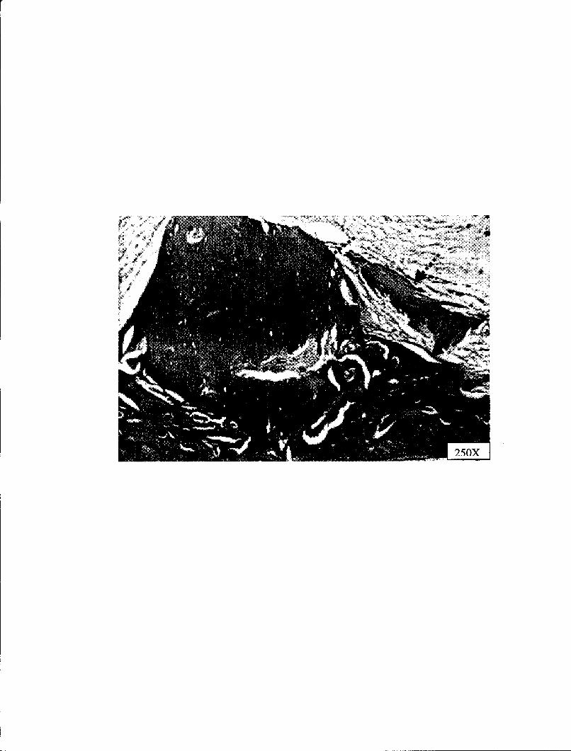
25OX
43
B. Retention time of EMD
"125I labeled EMD disappeared rapidly from the defect (Figure 15). After four
hours 50% of 30gCi/ 25 gl EMD was left in the defect. At 24 hours 20% of the total
amount of EMD placed in the defect remained. Most of the disappearance of EMD
occurred in the first 24 hours. At day 20 there was 4% of the total amount of EMD placed
in the defect. Neither DFDBA nor DDM increased the retention time of EMD in the
defect compared to EMD in the propylene glycol alone. There was a trend for EMD with
DFDBA to be retained slightly longer than EMD with propylene glycol alone in the
defect, though this was not statistically significant (p<0.05).
44
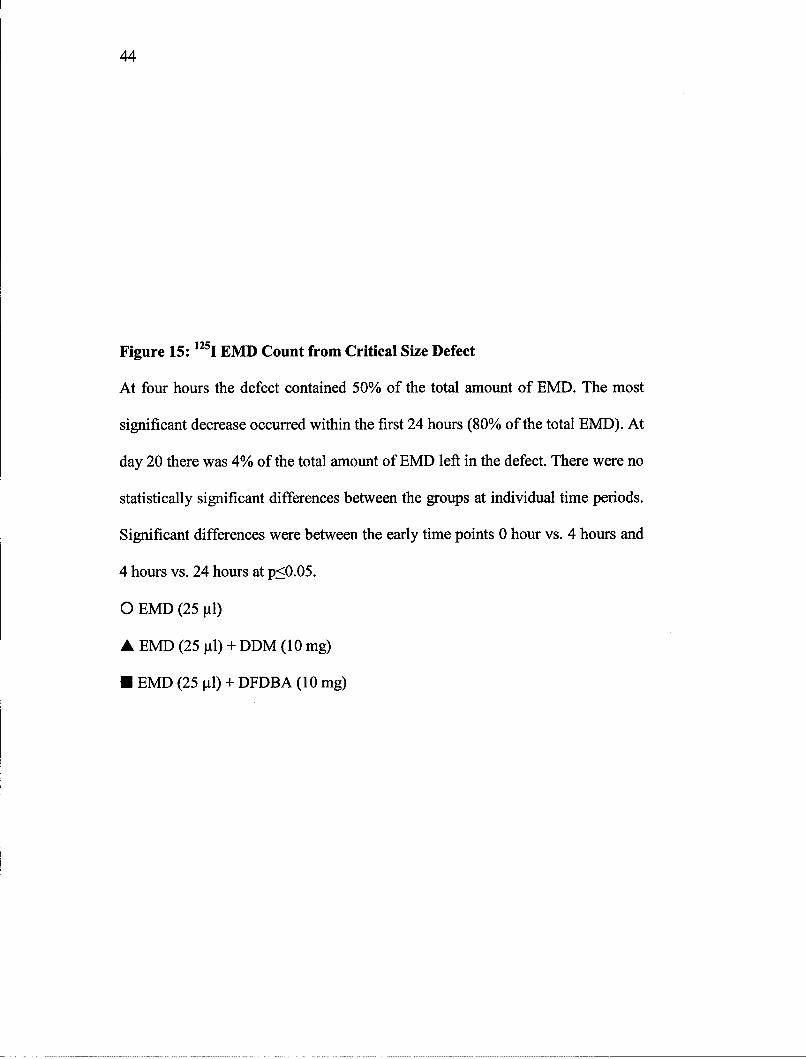
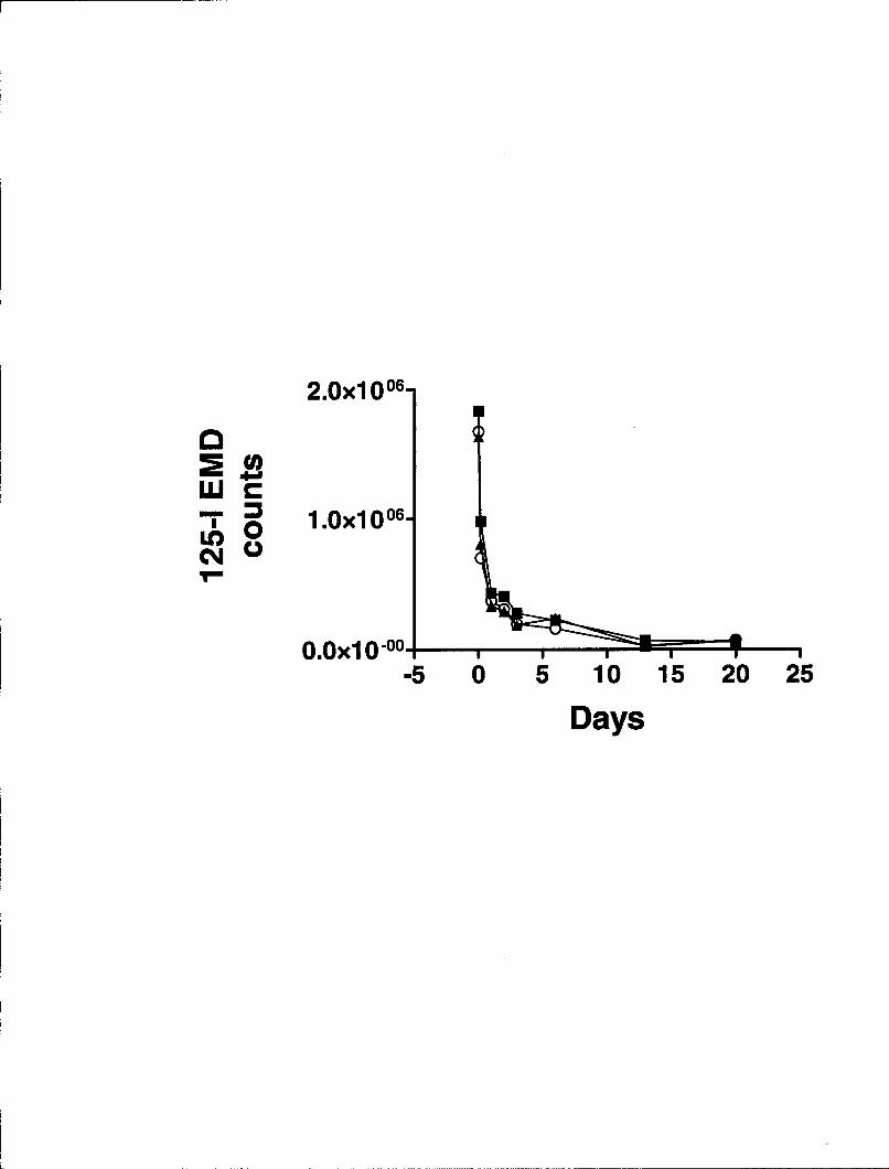
Figure 15: 1251 EMD Count from Critical Size Defect
At four hours the defect contained 50% of the total amount of EMD. The most
significant decrease occurred within the first 24 hours (80% of the total EMD). At
day 20 there was 4% of the total amount of EMD left in the defect. There were no
statistically significant differences between the groups at individual time periods.
Significant differences were between the early time points 0 hour vs. 4 hours and
4 hours vs. 24 hours at p_<0.05.
O EMD (25 pil)
A EMD (25 jtl) + DDM (10 mg)
U EMD (25 jtl) + DFDBA (10 mg)
2.Oxl006
"- 1.0x10 06-CI 0
O.Oxl 0°00
-5 0 5 10 15 20 25
Days
DISCUSSION
The critical size defect model allows for study of the bone inducing properties of
EMD, while evaluating the effects of two space maintaining cariers. The data
demonstrated that while DFDBA and DDM particles do not enhance new bone formation
with EMD over EMD alone, they do not interfere with it either. The data refutes the
hypothesis that the use of space maintaining carriers with EMD increases new bone
formation in the rat calvaria.
A. Analysis of new bone formation
The critical size defect in the rat calvarium is a proven model for the
demonstration of bone regeneration (Schmitz, Hollinger 1986). EMD has been shown to
enhance new attachment in periodontal defects (Heiji 1997). Boyan et al (2000) prepared
suggested that EMD is an osteopromotive factor because of its ability to enhance new
bone formation when used in combination with active DFDBA in an ectopic bone
formation model. DFDBA has been shown to contain bone morphogenic proteins 2, 4,
and 7 (Shigeyama et al 1995). Similarly demineralized dentin matrix particles have been
shown to contain bone morphogenic proteins, as well as TGF-13, and IGF-I and -II
(Finkelman et al 1990). Neither the DFDBA nor the DDM particles used in this study had
been assayed for osteoinductivity by their respective manufacturer. In the present study
similar amounts of new bone formation were observed at eight weeks for the DFDBA
45

46
and DDM particles when implanted alone in the critical size defect. Although the
amounts of new bone were greater in the DFDBA and DDM group compared to the
amount of new bone in the reference group (propylene glycol), the results were not
statistically different due to the variability. With a larger sample size statistical
significance would have been achieved. Group sizes for this experiment were chosen
based on results of the ectopic induction protocol used in Boyan et al (2000). Because
some animals did not survive to 8 weeks, some group sizes were reduced, thereby
affecting the statistical procedures. Also, the degree of variability observed in the critical
size defect model compared to the ectopic model, in which a more robust response has
been noted, indicates that larger sample sizes will be required to achieve statistical
significance. With a standard deviation of 0.25, which was used in our study, we would
need 18 animals per group to show a statistical difference between the data means. The
results of this study do not agree with Boyan et al (2000) findings that EMD increased
osteopromotion with DFDBA. The ectopic model is a closed system that contains
inherent wound stability from the surrounding muscles. The critical size defect in rat
calvarium is a more stringent model for regeneration due to the ability of the rats to
disturb the surgical site. In addition, in the previously mentioned study the results
demonstrated that EMD did not enhance inactive DFDBA. The DFDBA in this study
may not have been biologically active.
Histologically, the bone formation response in the two groups appeared different.
While the DFDBA particles were in intimate contact with the new bone formation, the
DDM particles were encapsulated with fibrous connective tissue from the new bone
formation. The appearance of new bone formed in intimate contact with DFDBA
47
particles is similar to that observed in the ectopic model as well. Encapsulation of the
DDM particles was not expected, but it did not seem to affect the bone formation
response. This lack of contact with new bone suggests that the DDM particles were
osteoinductive in nature or was an artifact.
EMD alone was slightly more osteoinductive at eight weeks than were DFDBA or
DDM particles alone or in combination with EMD, particularly at the higher
concentrations used. Again statistical significance was not achieved due to variability and
sample size. Of note was the cartilage-like formation was observed at all concentrations
of EMD alone groups at two weeks. This suggests that EMD may be promoting osseous
regeneration through an endochondral pathway.
This may be beguiling significant, because cartilage formation typically is not
often observed during bone formation in the critical size defect model (Wang, Glimcher
1999). Interestingly, cartilage formation was rarely observed in the experimental groups
where EMD was combined with the DFDBA or DDM particles. Further studies are
needed to assess the significance of increased cartilage-like formation with EMD alone
and exact pathway of bone formation with EMD.
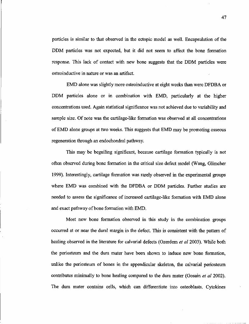
Most new bone formation observed in this study in the combination groups
occurred at or near the dural margin in the defect. This is consistent with the pattern of
healing observed in the literature for calvarial defects (Ozerdem et al 2003). While both
the periosteum and the dura mater have been shown to induce new bone formation,
unlike the periosteum of bones in the appendicular skeleton, the calvarial periosteum
contributes minimally to bone healing compared to the dura mater (Gosain et al 2002).
The dura mater contains cells, which can differentiate into osteoblasts. Cytokines
48
secreted at the wound site or liberated from implanted graft material can induce cellular
differentiation, which results in repair of the osseous defect (Wang, Glimcher 1999).
Most evidence points to a mechanism of cranial defect healing which utilizes primarily
by autocrine and paracrine signaling from the underlying dura mater (Ozerdem et al
2003). Therefore, the variability in new bone formation observed in the present study
may be explained by the quality and/or quantity of the dura mater underlying the critical
size defect. While care was taken to avoid damage to the dura during surgery, one
surgeon was more experienced than the other with the critical size defect model system.
Thus, the dura may have sustained more damage in animals operated on by the less
experienced surgeon.
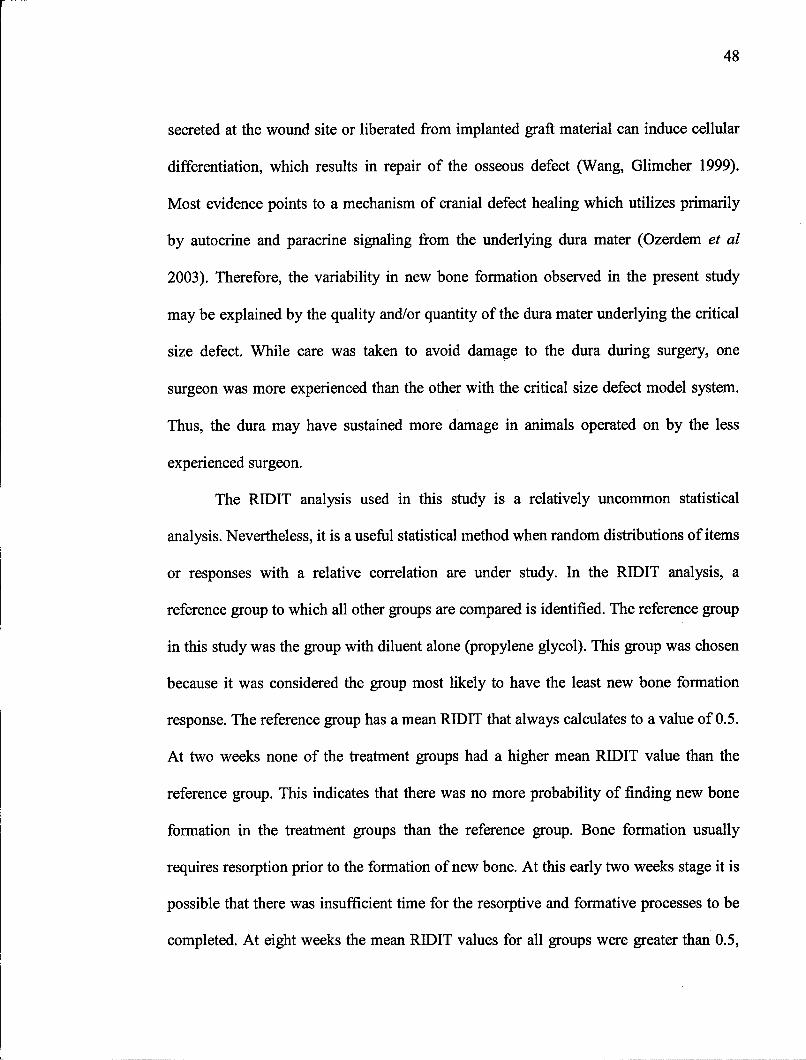
The RIDIT analysis used in this study is a relatively uncommon statistical
analysis. Nevertheless, it is a useful statistical method when random distributions of items
or responses with a relative correlation are under study. In the RIDIT analysis, a
reference group to which all other groups are compared is identified. The reference group
in this study was the group with diluent alone (propylene glycol). This group was chosen
because it was considered the group most likely to have the least new bone formation
response. The reference group has a mean RIDIT that always calculates to a value of 0.5.
At two weeks none of the treatment groups had a higher mean RIDIT value than the
reference group. This indicates that there was no more probability of finding new bone
formation in the treatment groups than the reference group. Bone formation usually
requires resorption prior to the formation of new bone. At this early two weeks stage it is
possible that there was insufficient time for the resorptive and formative processes to be
completed. At eight weeks the mean RIDIT values for all groups were greater than 0.5,
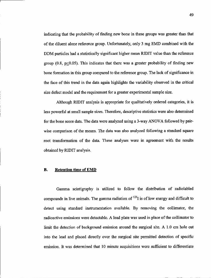
49
indicating that the probability of finding new bone in these groups was greater than that
of the diluent alone reference group. Unfortunately, only 3 mg EMD combined with the
DDM particles had a statistically significant higher mean RIDIT value than the reference
group (0.8, p<0.0 5 ). This indicates that there was a greater probability of finding new
bone formation in this group compared to the reference group. The lack of significance in
the face of this trend in the data again highlights the variability observed in the critical
size defect model and the requirement for a greater experimental sample size.
Although RIDIT analysis is appropriate for qualitatively ordered categories, it is
less powerful at small sample sizes. Therefore, descriptive statistics were also determined
for the bone score data. The data were analyzed using a 3-way ANOVA followed by pair-
wise comparison of the means. The data was also analyzed following a standard square
root transformation of the data. These analyses were in agreement with the results
obtained by RIDIT analysis.
B. Retention time of EMD
Gamma scintigraphy is utilized to follow the distribution of radiolabled
compounds in live animals. The gamma radiation of 125, is of low energy and difficult to
detect using standard instrumentation available. By removing the collimator, the
radioactive emissions were detectable. A lead plate was used in place of the collimator to
limit the detection of background emission around the surgical site. A 1.0 cm hole cut
into the lead and placed directly over the surgical site permitted detection of specific
emission. It was determined that 10 minute acquisitions were sufficient to differentiate
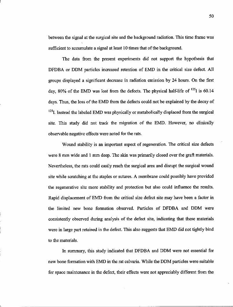
50
between the signal at the surgical site and the background radiation. This time frame was
sufficient to accumulate a signal at least 10 times that of the background.
The data from the present experiments did not support the hypothesis that
DFDBA or DDM particles increased retention of EMD in the critical size defect. All
groups displayed a significant decrease in radiation emission by 24 hours. On the first
day, 80% of the EMD was lost from the defects. The physical half-life of 1251 is 60.14
days. Thus, the loss of the EMD from the defects could not be explained by the decay of
1251. Instead the labeled EMD was physically or metabolically displaced from the surgical
site. This study did not track the migration of the EMD. However, no clinically
observable negative effects were noted for the rats.
Wound stability is an important aspect of regeneration. The critical size defects
were 8 mm wide and 1 mm deep. The skin was primarily closed over the graft materials.
Nevertheless, the rats could easily reach the surgical area and disrupt the surgical wound
site while scratching at the staples or sutures. A membrane could possibly have provided
the regenerative site more stability and protection but also could influence the results.
Rapid displacement of EMD from the critical size defect site may have been a factor in
the limited new bone formation observed. Particles of DFDBA and DDM were
consistently observed during analysis of the defect site, indicating that these materials
were in large part retained in the defect. This also suggests that EMD did not tightly bind
to the materials.
In summary, this study indicated that DFDBA and DDM were not essential for
new bone formation with EMD in the rat calvaria. While the DDM particles were suitable
for space maintenance in the defect, their effects were not appreciably different from the
51
DFDBA and neither added significantly to bone formation over EMD alone. Further
investigations using a membrane may improve the ability to detect any differences
present between the treatment groups. Likewise, increasing the sample size in subsequent
investigations will improve the ability of the model to detect significant differences in the
combination materials as well.
BIBLIOGRAPHY
Albander J., Brunelle J., Kingman A.: Destructive periodontal disease in adults 30 yearsof age and older in United States, 1988-1994. J Periodontol 1999;70:13-29.
Aukhil I., Pettersson E., Suggs C.: Guided tissue regeneration. An experimentalprocedure in beagle dogs. J Periodontol 1986;57:72-734.
Avery J.: Oral Development and Histology Second Edition. Thieme MedicalPublishers, Inc., New York; 1994.
Bang G., Urist M.: Bone induction in excavation chambers in matrix of decalcifieddentin. Arch Surg 1967;94:781-789.
Becker W., Becker B., Cafesse R.: A comparison of mineralized freeze-dried bone andautologous bone to induce bone formation in human extraction sockets. J Periodontol1994;65:1128-1133.
Bowers G., Chadroff B., Carnevale R., Mellonig J., Corio R., Emerson J., Stevens M.,Romberg E.: Histologic evaluation of new attachment apparatus formation in humans:Part II. J Periodontol 1989b;60:675-682.
Bowers G., Chadroff B., Carnevale R., Mellonig J., Corio R., Emerson J., Stevens M.,Romberg E.: Histologic evaluation of new attachment apparatus formation in humans:Part III. J Periodontol 1989c;60:683-693.
Boyan B., Weesner T., Lohmann C., Andreacchio D., Carnes D., Dean D., Cochran D.,Schwartz Z.: Porcine fetal enamel matrix derivative enhances bone formation induced bydemineralized freeze dried bone allograft in vivo. J Periodontol 2000;71:1278-1286.
Brookes S., Robinson C., Kirkham J., Bonass W.: Biochemistry and molecular biology ofamelogenin proteins of developing dental enamel. Arch Oral Biol 1995;40:1-14.
Carvalho V., Tosello D., Salgado M., Gomes M.: Histomorphometric analysis ofhomogenous demineralized dentin matrix as osteopromotive material in rabbit mandibles.Int J Oral Maxillofac Implants 2004;19:679-686.
Caton J., Nyman S., Zander H.: Histometric evaluation of periodontal surgery. II.Connective tissue attachment levels after four regenerative procedures. J ClinPeriodontol 1980;7:224-231.
Cochran D., Wozney J.: Biological mediators for periodontal regeneration. Periodontol2000 1999;19:40-58.
52
53
Cochran D., Jones A., Heiji L., Mellonig J., Schoolfield J., King G.: Periodontalregeneration with a combination of enamel matrix proteins and autogenous bone grafting.J Periodontol 2003;74:1269-1281.
Cochran D., King G., Schoolfield J., Velasquez-Plata D., Mellonig J., Jones A.: Theeffect of enamel matrix proteins on periodontal regeneration as determined byhistological analyses. J Periodontol 2003;74:1043-1055.
Dimuzio M., Veis A.: Phosphophoryns-major non-collageous proteins of rat incisorsdentin. Calcified Tissue Research 1978;25:169-178.
Finkelman R., Mohan S., Jennings J., Taylor A., Jepsen S., Baylin D.: Quantitation ofgrowth factors IGF-I, SCF/IGF-II, and TGF-3 in human dentin. J Bon Min Res1990;5:717-723.
Fleiss J.: Samples qualitatively ordered: RIDIT analysis. Statistical methods for rates andproportions John Wiley & Sons, Inc. 1973:102-108.
Francetti L., Del Fabbro M., Basso M., Testori T., Weinstein R.: Enamel matrix proteinsin the treatment of intra-bony defects: a prospective 24-month clinical trial. J ClinPeriodontol 2004;31:52-59.
Froum S., Weinberg M., Rosenberg E., Tarnow D.: A comparative study utilizing openflap debridement with and without enamel matrix derivative in the treatment ofperiodontal intrabony defects. A 12-month re-entry study. J Periodontol 2001;72:25-34.
Froum S., Weinberg M., Novak J., Mailhot J., Mellonig J., Van Dyke T., McClain P.,Papapanou P., Childers G., Cianciao S., Blieden T., Polson A., Greenstein G., Yukna R.,Wallace M., Patters M., Wagener G.: A multicenter study evaluating the sensitizationpotential of enamel matrix derivative after treatment of two infrabony defects. JPeriodontol 2004;75:1001-1008.
Gartner L.: Tooth development. Essesntials of Oral Histology and Embryology ThirdEdition. Jen House Publishing Company 1999:17-36.
George A., Sabsay B., Simonian P., Veis A.: Characterization of a novel dentin matrixacidic phophoprotein. Implications of induction of biomineralization. J BiologicalChemistry 1993;268:12624-12630.
Gestrelius S., Andersson C.: Formulation of enamel matrix derivative for surface coating.Kinetics and cell colonization. J Clin Peridodontol 1997;24:685-692.
Giannobile W., Somerman M.: Growth and amelogenin-like factors in periodontal woundhealing. A systematic review. Ann Periodontol 2003;8:193-204.
54
Gosain A., Santoro T., Song L., Capel C., Sudhakar P., Matloub H.: Osteogenesis incalvarial defects: contribution of the dura, the pericranium, and the surrounding bone inadult versus infant animals. Plast Reconstr Surg 2003; 112:515-527.
Gurinsky B., Mills M., Mellonig J.: Clinical evaluation of demineralized freeze-driedbone allograft and enamel matrix derivative versus enamel matrix derivative alone for thetreatment of periodontal osseous defects in humans. J Periodontol 2004;75:1309-1318.
Hagewald S. Spahr A. Rompola E., Haller B., Heijl L., Bernimoulin J.: Comparativestudy of Emdogain® and coronally advanced flap technique in the treatment of humangingival recessions. J Clin Periodontol 2002;29:35-41.
Hagewald S., Pischon N., Przemyslaw J., Bemimoulin J., Zimmermann B.: Effects ofenamel matrix derivative on proliferation and differentiation of primary osteoblasts. OralSurg, Oral Med, Oral Pathol, Oral Radiol, Endod 2004;98:243-249.
Hammarstrom L.: Enamel matrix, cementum development and regeneration. J ClinPeriodontol 1997;24:658-668.
Hammarstrom L., Heijl L., Gestrelius S.: Periodontal regeneration in a buccal dehiscencemodel in monkeys after application of enamel matrix proteins. J Clin Periodontol1997;24:669-677.
Heard R. Mellonig J., Brunsvold M., Lasho D., Meffert R., Cochran D.: Clinicalevaluation of wound healing following multiple exposures to enamel matrixproteinderivative in the treatment of intrabony periodontal defects. J Periodontol2000;71:1715-1721.
Heijl L.: Periodontal regeneration with enamel matrix derivative in one humanexperimental defect. A case report. J Clin Periodontol 1997;24:693-696.
Heijl L., Heden G., Svardstrom G., Ostgren A.: Enamel matrix derivative (Emdogain®)in the treatment of intrabony periodontal defects. J Clin Periodontol 1997;24:705-714.
Hoang A., Klebe R., Steffensen B., Ryu 0., Simmer J., Cochran D.: Amelogenin is a celladhesion protein. J Dent Res 2002;81:497-500.
Hogan B.: Bone morphogenetic proteins: multifunctional regulators of vertebratedevelopment. Genes Dev 1996;10:1580-1594.
Iglhaut J., Aukhil I., Simpson D., Johnston M., Koch G.: Progenitor cell kinetics duringguided tissue regeneration in experimental periodontal wounds. J Periodontol Res 1988;23:107-117.
Iwata T., Morotome Y., Tanabe T., Fukae M., Ishikawa I., Oida S.: Noggin blocksosteoinduction activity of porcine enamel extreacts. J Dent Res 2002;82:387-391.
55
Jepsen S., Heinz B., Jepsen K., Arjomand M., Hoffmiann T., Richter S., Reich E., SculeanA., Gonzales J., Bodeker R., Meyle J. A randomized clinical trial comparing enamelmatrix derivative and membrane treatment of buccal Class II furcation involvement inmandibular molars. Part I: study design and results for primary outcomes. J Periodontol2004;75:1150-1160.
Kawase T., Okuda K., Yoshie H., Bums D.: Cytostatic action of enamel matrix derivative(Emodogain®) rapidly stimulates phosphorylation of the MAP kinase family and nuclearaccumulation of smad2 in both oral epithelial and fibroblastic human cells. J PeriodontolRes 2001;36:367-376.
Lekovic V., Comargo P., Weinlaender M., Nedic M., Aleksic Z., Kenney E.: Acomparison between enamel matrix proteins used alone or in combination with bovineporous bone mineral in the treatment of intrabony periodontal defects in humans. JPeriodontol 2000;71:1110-1116.
Lyngstadaas S., Lundber E., Ekdahl H., Andersson C., Gestrelius S.: Autocrine growthfactors in human periodontal ligament cells cultured on enamel matrix derivative. J ClinPeriodontol 2001;28:181-188.
Melcher A.: On the repair potential of periodontal tissue. J Periodontol 1976;47:256-260.
Melcher A., McCulloch C., Cheong T., Nemeth E., Shiga A.: Cells from bone synthesizecementum-like and bone-like tissue in vitro and may migrate into periodontal ligament invivo. J Periodontol Res 1987;22:246-247.
McGuire M., Nunn M.: Evaluation of human recession defects treated with coronallyadvanced flaps and either enamel matrix derivative or connective tissue. Part 1:comparison of clinical parameters. J Periodontol 2003;74:1110-1125.
Minabe M.: A critical review of the biologic rationale for guided tissue regeneration. JPeriodontol 1991;62:171-179.
Modica F., Del Pizzo M., Roccuzzo M., Romagnoli R.: Coronally advanced flap for thetreatment of buccal gingival recessions with and without enamel matrix derivative. Asplit-mouth study. J Periodontol 2000;71:1693-1698.
Newman S., Coscia S., Jutwani R., Iacono V., Cutler C.: Effects of enamel matrixderivative on Porphyromonas gingivalis. J Periodontol 2003;74:1191-1195.
Nyman S., Lindhe J., Karring T., Rylander H.: New attachment following surgicaltreatment of human periodontal disease. J Clin Periodontol 1982;9:290-296.
56
Okdua K., Momose M., Miyazaki A., Murata M., Yokoyama S., Wolff L., Yoshie H.:Enamel matrix derivative in the treatment of human intrabony osseous defects. JPeriodontol 2000;71:1821-1828.
Ozerdem 0., Anlatici R., Bahar T. Roles of periosteum, dura, and adjacent bone onhealing of cranial osteonecrosis. J Craniofac Surg 2003;14:371-379.
Parodi R., Santarelli G., Gasparetto B.: Treatment of intrabony pockets with Emdogain:results at 36 months. lnt J Periodontics Restorative Dent 2004;24:57-63.
Pontoriero R., Wennstrom J., Lindhe J.: The use of barrier membranes and enamel matrixproteins in the treatment of angular bone defects: a prospective controlled clinical study.J Clin Periodontol 1999;26:833-840.
Prichard J.: Present state of the interdental denudation procedure. J Periodontol1977;48:566-569.
Reynolds M., Bowers G.: Fate of demineralized freeze-dried bone allograft in humanintrabony defects. J Periodontol 1996;67:150-157.
Rosenberg A., Teare F.: A novel rapid thin-layer chromatographic monitoring system forthe radioiodinatin of protein and polypeptides. Analytical Biochemistry 1977;77:289-292.
Rutherford B., Fitzgerald M.: A new biological approach to vital pulp therapy. Crit RevOral Biol Med 1995;6:218-229.
Sanz M., Tonetti M., Zabalegui I., Sicilia A., Blanco J., Rebelo H., Rasperini G., MeriliM., Cortellini P., Suvan J.: Treatment of intrabony defects with enamel matrix proteins orbarrier membranes: results from a multicenter practice-based clinical trial. J Periodontol2004;75:726-733.
Schwartz Z., Mellonig J., Carnes D., De La Fonaine D., Cochran D., Boyan B.: Ability ofcommercial demineralized freeze-dried bone allograft to induce new bone formation. JPeriodontol 1996;67:918-926.
Schwartz A., Sommers A., Mellonig J., Carnes D., Dean D., Cochran D., Boyan B:Ability of commercial demineralized freeze-dried bone allograft to induce new boneformation is dependent on donor age but not gender. J Periodontol 1998a;69:470-478.
Schmitz J., Hollinger J.: The critical size defect as an experimental model forcraniomandibulofacial nonunions. Clin Orthop 1986;205:299-308.
Sculean A., Donos N., Chiatella G., Windisch P., Reich E., Brecx M.: GTR withbioresorbable membranes in the treatment of intrabony defects: a clinical and histologicstudy. lnt J Periodontol Restorative Dent 1999; 19:510-509.
57
Sculean A., Donos N., Brecx M., Reich E., Karring T.: Treatment of intrabony defectswith guided tissue regeneration and enamel-matrix-proteins. J Clin Periodontol2000;27:466-472.
Sculean A., Blaes A., Arweiler N., Reich E., Donos N., Brecx M.: The effect ofpostsurgical antibiotics on the healing of intrabony defects following treatment withenamel matrix proteins. J Periodontol 2001;72:190-195.
Sculean A., Windisch P., Chiantella G., Donos N., Brecx M., Reich E.: Treatment ofintrabony defects with enamel matrix proteins and guided tissue regeneration. Aprospective controlled clinical study. J Clin Periodontol 2001;28:397-403.
Sculean A., Donos N., Schwarz F., Becker J., Brecx M., Arweiler N.: Five-year resultsfollowing treatment of intrabony defects with enamel matrix proteins and guided tissueregeneration. J Clin Periodontol 2004;7:545-549.
Sculean A., Windisch P., Chiantella G.: Human histologic evaluation of an intrabonydefect treated with enamel matrix derivative, xenograft and GTR. lnt J PeriodonticsRestorative Dent 2004;24:326-333.
Shigeyama Y., D'Errico J., Stone R., Somerman M.: Commercially-prepared allograftmaterial has biologic activity in vitro. J Periodontol 1995;66:478-487.
Silvestri M., Ricci G., Rasperini G., Sartori S., Cattaneo V.: Comparisons of treatmentsof infrabony defects with enamel matrix derivative, guided tissue regeneration with anonresorbable membrane and Widman modified flap. A pilot study. J Clin Periodontol2000;27:603-610.
Silvestri M., Sartori S., Rasperini G., Ricci G., Rota C., Cattaneo V.: Comparison ofinfrabony defects treated with enamel matrix derivative versus guided tissue regenerationwith a nonresorbable membrane. A multicenter controlled clinical trial. J ClinPeriodontol 2003;30:386-393.
Somerman M., Nathanson M., Sauk J., Manson B.: Human dentin matrix inducescartilage formation in vitro by mesenchymal cells derived from embryonic muscle. JDent Res 1987;66:1551-1558.
Takagi K., Urist M: The reaction of the dura to bone morphogenetic protein (BMP) inrepair of skull defects. Ann Surg 1982;196:100-109.
Tonetti M., Lnag N., Cortellini P., Suvan J., Adriaens P., Dubravec D., Fonzar A.,Fourmousis I., Mayfield L., Rossi R., Silvestri M., Tiedemann C., Topoll H., VangstedT., Wallkamm B.: Enamel matrix proteins in the regenerative therapy of deep intrabonydefects: A multicenter randomized controlled clinical trial. J Clin Periodontol2002;29:317-325.
58
Tumbull R., Freeman E.: Use of wounds in the parietal bone of the rat for evaluatingbone marrow for grafting into periodontal defects. J Periodontol Res 1974;9:39-43.
Urist M., DeLange R., Finerman G.: Bone cell differentiation and growth factors. Science1983;220:680-686.
Veis A., Perry A.: The phophoprotein of the dentin matrix. Biochemistry 1967;6:2409-2416.
Veis A.: Mineral-matrix interactions in bone and dentin. Journal of Bone and MineralResearch 1993;8 Suppl 2:493-497.
Wang E., Rosen V., D'Alessandro J., Bauduy M., Cordes P., Harada T., Israel D.,Hewiok R., Kerns K., LaPan P., Luxenberg D., McQuaid D., Moutsatsos I., Nove J.,Wozney J.: Recombinant human bone morphogenetic protein induces bone formation.Proc Natl Acad Sci 1990;87:2220-2224.
Wang J., Glimcher M.: Characterization of matrix-induced osteogenesis in rat calvarialbone defects: II. Origins of bone-forming cells. Calcif Tissue lnt 1999;65:486-493.
Wozney J.: The potential role of bone morphogenetic proteins in periodontalreconstruction. J Periodontol 1995;66:506-510.
Yuan K., Chen C. Lin M.: Enamel matrix derivative exhibits angiogenic effect in vitroand in a murine model. J Clin Periodontol 2003;30:732-738.
Yukna R., Mellonig J.: Histologic evaluation of periodontal healing in humans followingregenerative therapy with enamel matrix derivative. A 10-case series. J Periodontol2000;71:752-759.
Zucchelli G., Bernardi F., Montebugnoli L., DeSanctis M.: Enamel matrix proteins andguided tissue regeneration with titanium-reinforced expanded polytetrafluoroethylenemembranes in the treatment of infrabony defects: a comparative controlled clinical trial. JPeriodontol 2002;73 :3-12.
Zucchelli G., Amore C., Montebugnoli L., DeSanctis M.: Enamel matrix proteins andbovine porous bone mineral in the treatment of intrabony defects: a comparativecontrolled clinical trial. J Periodontol 2003;74:1725-1735.
VITA
Kerri Font was born mL-*.:'-.-f'•:>;21:-ý -:.'in Des Moines, Iowa, daughter of
S,}LA..'.." " and ,.. - . .. . She attended San Diego State University and
graduated with a Batchelor of Science degree in Biology with magnum cum laude
distinction. She worked one year at Scripps Clinic microbiology laboratory in San Diego,
California after which she began dental training at the University of the Pacific in San
Francisco, California.
While in dental school, she pursued periodontal research. She received Tau Kappa
Omega award in 2001, Omicron Kappa Upsilon and The American Academy of
Periodontology Student awards in 2002. In 2002 she was placed on the National Dean's
List. She then received her Doctor of Dental Science in May 2002.
In the summer of 2002, she began a Postdoctoral Program in Periodontics at
Lackland Air Force Base in conjunction with the University of Texas Health Science
Center at San Antonio. In 2003 she was accepted into the Graduate School of Biomedical
Sciences for the Masters of Science degree in Periodontics. After graduation in May
2005, she plans to practice periodontics in the Air Force at Ramstein AFB, Germany.
59
REPORT DOCUMENTATION PAGE Form Approved
R DOMB No. 0704-0188
Public reporting burden for this collection of information is estimated to average 1 hour per response, including the time for reviewing instructions, searching existing data sources,gathering and maintaining the data needed, and completing and reviewing the collection of information. Send comments regarding this burden estimate or any other aspect of thiscollection of information, including suggestions for reducing this burden, to Washington Headquarters Services, Directorate for Information Operations and Reports, 1215 JeffersonDavis Highway, Suite 1204, Arlington, VA 22202-4302, and to the Office of Management and Budget, Paperwork Reduction Project (0704-0188), Washington, DC 20503.
1. AGENCY USE ONLY (Leave blank) 2. REPORT DATE 3. REPORT TYPE AND DATES COVERED
I 5.Jul.05 THESIS4. TITLE AND SUBTITLE 5. FUNDING NUMBERS
COMPARISON OF DEMINERALIZED DENTIN AND DEMINERALIZEDFREEZE DRIED BONE AS CARRIERS FOR ENAMEL MATRIX PROTEINS IN ARAT CRITICAL SIZE DEFECT.6. AUTHOR(S)
CAPT FONT KERRI
7. PERFORMING ORGANIZATION NAME(S) AND ADDRESS(ES) 8. PERFORMING ORGANIZATION
UNIVERSITY OF TEXAS HSC AT SAN ANTONIO REPORT NUMBER
C104-1122
9. SPONSORING/MONITORING AGENCY NAME(S) AND ADDRESS(ES) 10. SPONSORING/MONITORINGTHE DEPARTMENT OF THE AIR FORCE AGENCY REPORT NUMBER
AFIT/CIA, BLDG 1252950 P STREETWPAFB OH 45433
11. SUPPLEMENTARY NOTES
12a. DISTRIBUTION AVAILABILITY STATEMENT 12b. DISTRIBUTION CODE
Unlimited distributionIn Accordance With AFI 35-205/AFIT Sup 1
13. ABSTRACT (Maximum 200 words)
DISTRIBUTION STATEMENTAApproved for Public Release
Distribution Unlimited
14. SUBJECT TERMS 15. NUMBER OF PAGES
5416. PRICE CODE
17. SECURITY CLASSIFICATION 18. SECURITY CLASSIFICATION 19. SECURITY CLASSIFICATION 20. LIMITATION OF ABSTRAC.OF REPORT OF THIS PAGE OF ABSTRACT
Standard Form 298 (Rev. 2-89) (EG)Prescribed by ANSI Std. 239.18Designed using Perform Pro, WHS/DIOR, Oct 94


![089 ' # '6& *#0 & 7Chapter 5 Tooth Tissue-engineered odontogenesis One important goal of dental research is the efficient regeneration of lost teeth [1, 2]. Tooth formation, or odontogenesis,](https://img.pdfslide.us/doc/110x75/60327f1bc15cec2a855c31cf/089-6-0-7-chapter-5-tooth-tissue-engineered-odontogenesis-one.jpg)


![Cobourne [1999] the Genetic Control of Early Odontogenesis](https://img.pdfslide.us/doc/110x75/577cd66a1a28ab9e789c508c/cobourne-1999-the-genetic-control-of-early-odontogenesis.jpg)