Embed Size (px)
Citation preview
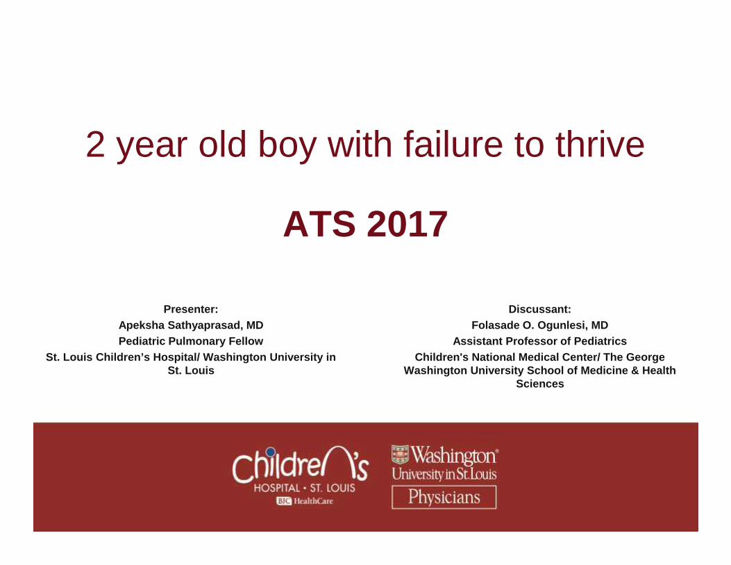
2 year old boy with failure to thrive
ATS 2017
Presenter:Apeksha Sathyaprasad, MDPediatric Pulmonary Fellow
St. Louis Children’s Hospital/ Washington University inSt. Louis
Discussant:Folasade O. Ogunlesi, MD
Assistant Professor of PediatricsChildren's National Medical Center/ The George
Washington University School of Medicine & HealthSciences
PediatricPulmonology
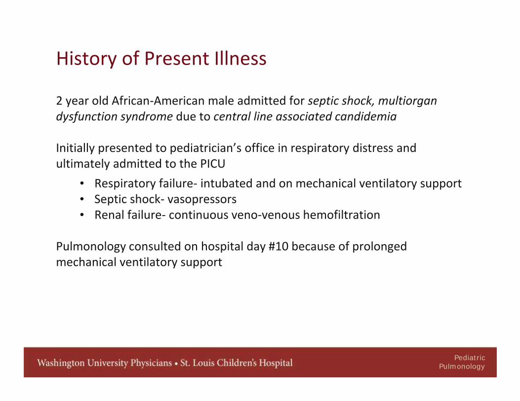
2 year old African-American male admitted for septic shock, multiorgandysfunction syndrome due to central line associated candidemia
Initially presented to pediatrician’s office in respiratory distress andultimately admitted to the PICU• Respiratory failure- intubated and on mechanical ventilatory support• Septic shock- vasopressors• Renal failure- continuous veno-venous hemofiltration
Pulmonology consulted on hospital day #10 because of prolongedmechanical ventilatory support
History of Present Illness
PediatricPulmonology
Born full-term, birth weight 3.318 Kg. Pregnancy, delivery, newborn periodwas unremarkable. Did not require oxygen support, no history of delayedpassage of meconium. No history of chronic persistent rhinitis or cough
1 year of age- chronic diarrhea and poor weight gain• Endoscopy, contrast imaging, hepatic enzymes, anti-tTG:
unremarkable• Dietary modifications (higher calorie elemental formula)• G-tube with Nissen fundoplication• Chronic intravenous hyperalimentation
Central line-associated blood stream infection• S. viridans, Klebsiella, E.coli, Enterococcus, S. aureus
History of eczema, intermittent cough and wheezing with viral illnesseswhich reportedly responded to treatment with inhaled albuterol
Past Medical History
PediatricPulmonology
Family History: Grandmother has recurrent sinusitis. No history of asthma,cystic fibrosis, recurrent infections, infertility, gastrointestinal diseases
Social history: Lives with mother and grandmother. Does not attenddaycare. No second-hand tobacco exposure. No avian or agriculturalexposures. No pets. No history of travel outside the United States
Family and social history
PediatricPulmonology
Vital signs:T: 98.8 degrees F, HR: 113 beats/min, BP: 108/68 mm Hg, RR: 40 breaths/min
Ventilator support:SIMV PRVC, FiO2 0.40, rate 34/min, Vt 95 mL, PEEP 8 cm H2O, PS 20 cm H2O
Oxyhemoglobin saturation: 100%
Growth percentiles:• Height: 80cm (1%ile)• Weight: 8.9 Kg (1%ile)
• BMI: <1%ile; BMI-Z score: -2.59• HC: 47.3 cm (18%ile)
Physical Examination
PediatricPulmonology
• HEENT: Normal• Neurologic: Sedated; Pupils equal and reactive• Pulmonary: bilateral good air entry, diffuse crackles most prominent over
the RUL, no wheezing• Cardiovascular: Regular rate and rhythm, no murmurs• Abdomen: Soft, non-distended, non-tender, no masses palpated, normal
bowel sounds• Extremities: No clubbing, cyanosis or edema• Skin: Eczematous lesions over extensor surface of the elbows and knees
Physical Exam
PediatricPulmonology
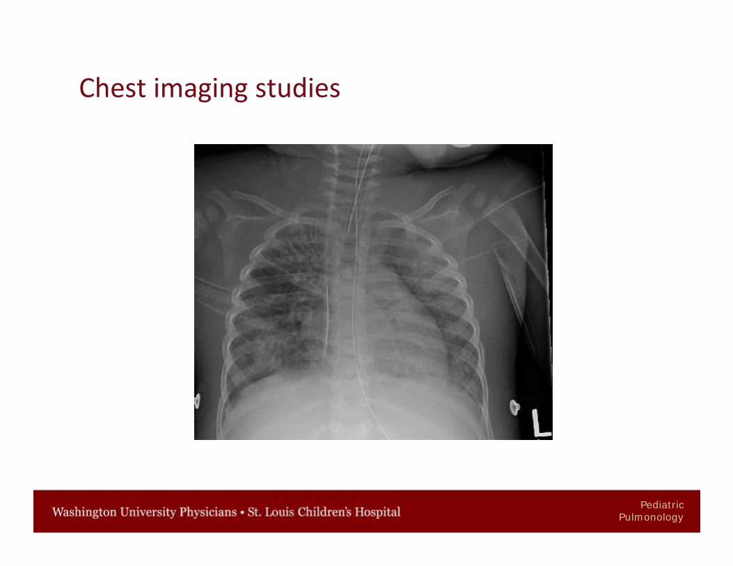
Chest imaging studies
PediatricPulmonology
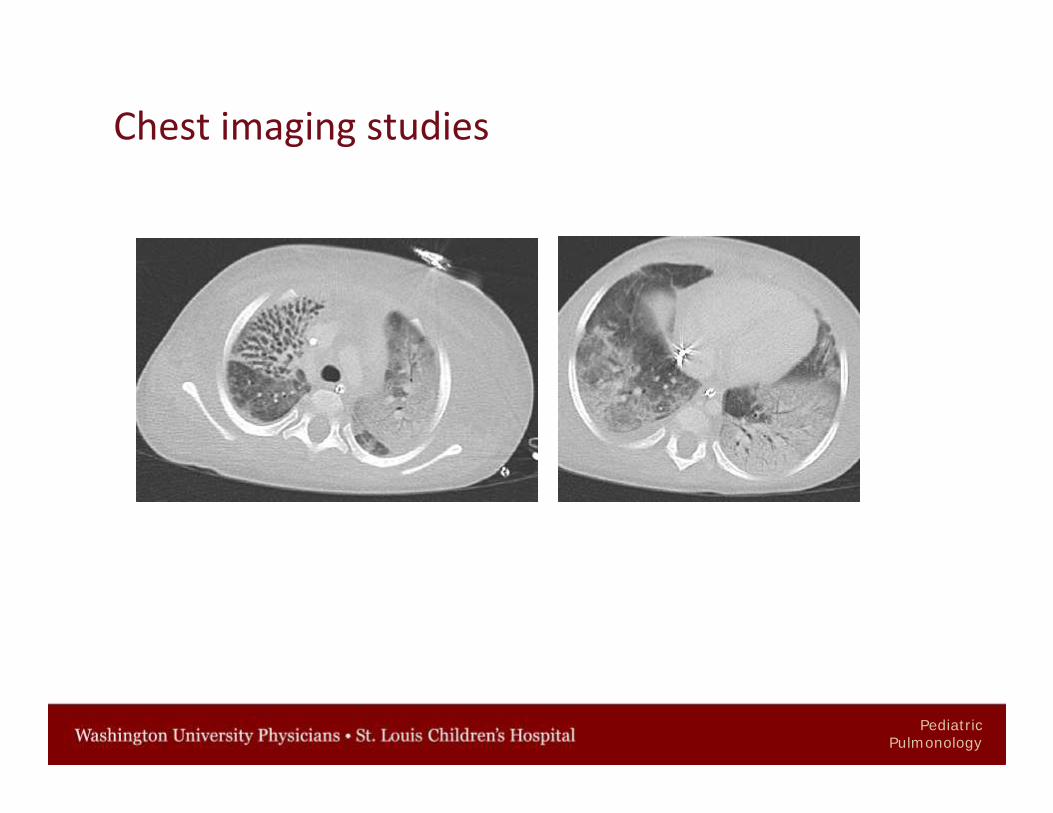
Chest imaging studies
PediatricPulmonology
• CBC: WBC 23,000/mm3 (neutrophils 71%, band neutrophils 5%,metamyelocytes 1%, lymphocytes 18%, Hgb 9.6 g/dL, Hct 36%, platelet30,000/mm3;
• ABG at time of consultation: 7.4/60/134• Blood culture (fungal): Candida tropicalis• Bronchoscopy with bronchoalveolar lavage - thick purulent secretions
throughout airways, primarily from the right upper lobe• ENT evaluation of upper airway: normal• Ciliary ultrastructure: normal• Sweat chloride concentration: 9 mmol/L• Immunological evaluation: nondiagnostic
Laboratory evaluation
PediatricPulmonology
Summary
• 2 year old African-American boy• Growth failure, malabsorption of unknown etiology, TPN dependent• Airway hyperreactivity, eczema• Recurrent central venous catheter infections• Candidemia and septic shock with multi-organ dysfunction syndrome• Right upper lobe bronchiectasis, diffuse interstitial and alveolar infiltrates
PediatricPulmonology
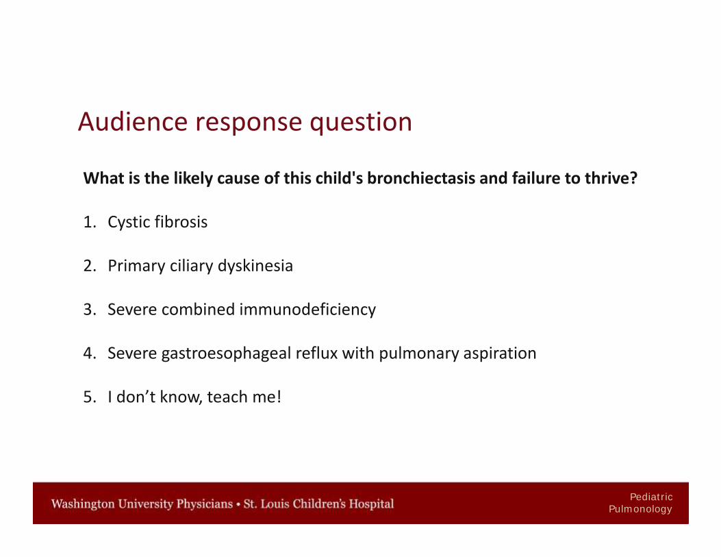
Audience response question
What is the likely cause of this child's bronchiectasis and failure to thrive?
1. Cystic fibrosis
2. Primary ciliary dyskinesia
3. Severe combined immunodeficiency
4. Severe gastroesophageal reflux with pulmonary aspiration
5. I don’t know, teach me!
PediatricPulmonology
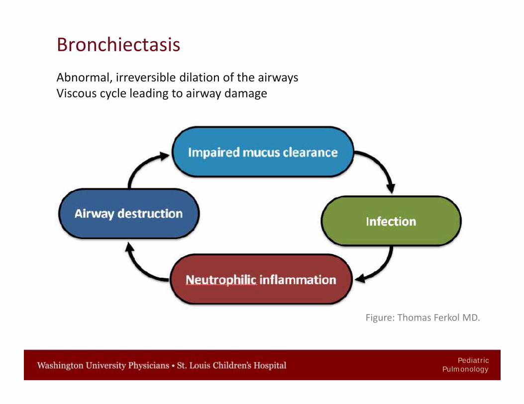
BronchiectasisAbnormal, irreversible dilation of the airwaysViscous cycle leading to airway damage
Figure: Thomas Ferkol MD.
PediatricPulmonology
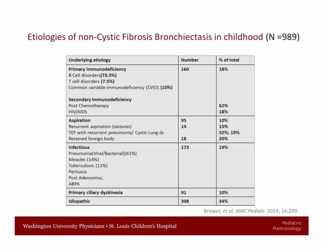
Etiologies of non-Cystic Fibrosis Bronchiectasis in childhood (N =989)
Brower, et al. BMC Pediatr. 2014; 14:299
PediatricPulmonology
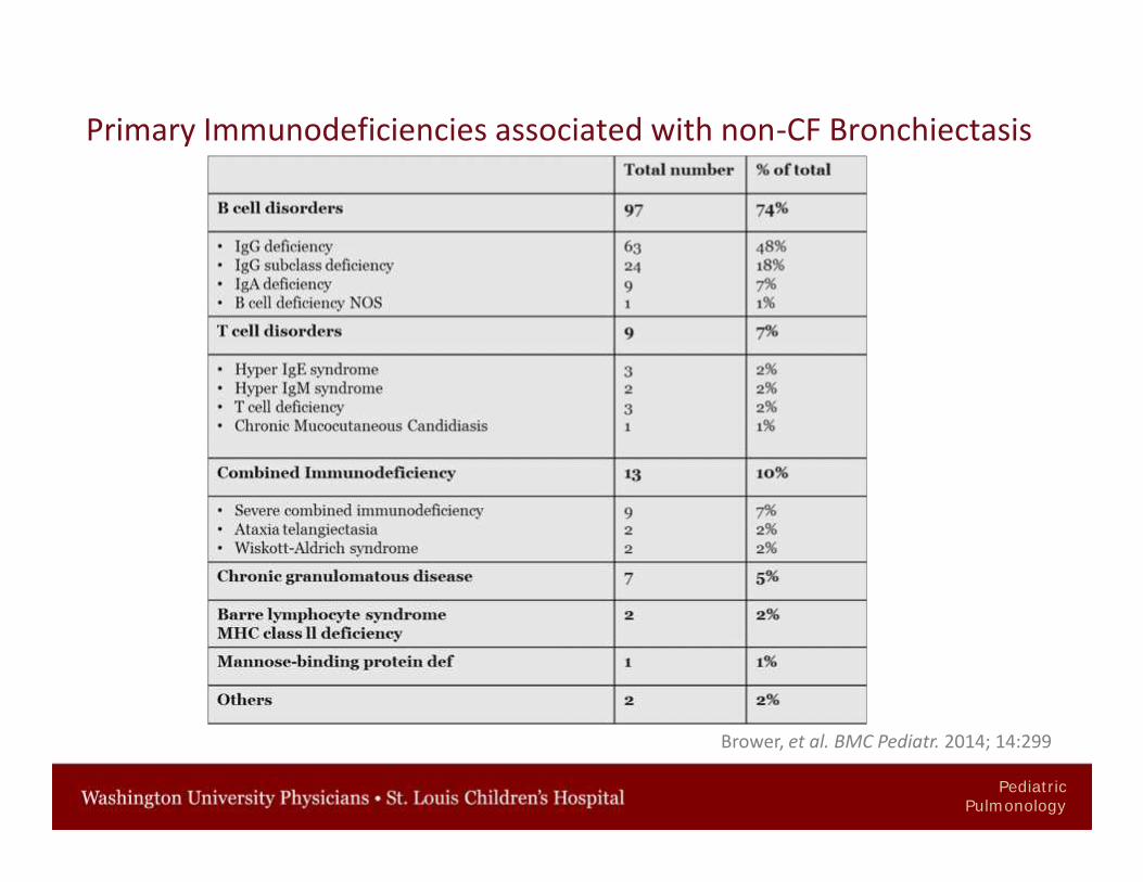
Primary Immunodeficiencies associated with non-CF Bronchiectasis
Brower, et al. BMC Pediatr. 2014; 14:299
PediatricPulmonology
• Cystic fibrosis
• Cellular immunodeficiency with recurrent pneumonia
• Primary ciliary dyskinesia
• Gastroesophageal reflux with aspiration pneumonia
• Other: Inflammatory bowel disease
Differential for bronchiectasis and poor weight gain
PediatricPulmonology
Clinical course
• Continued treatment with intravenous amphotericin B
• Non-CF bronchiectasis: airway clearance with high-frequency chest walloscillation, hypertonic saline
• Airway hyperreactivity: Daily inhaled corticosteroids and albuterol asneeded
• Patient was extubated and eventually placed on oxygen flow via nasalcannula at 1 L/min
PediatricPulmonology
Audience response question
Which of the following is not recommended for the routinemanagement of non-CF bronchiectasis?
1. Bronchodilators
2. Chest physiotherapy
3. Macrolide antibiotics
4. Recombinant human Deoxyribonuclease
PediatricPulmonology
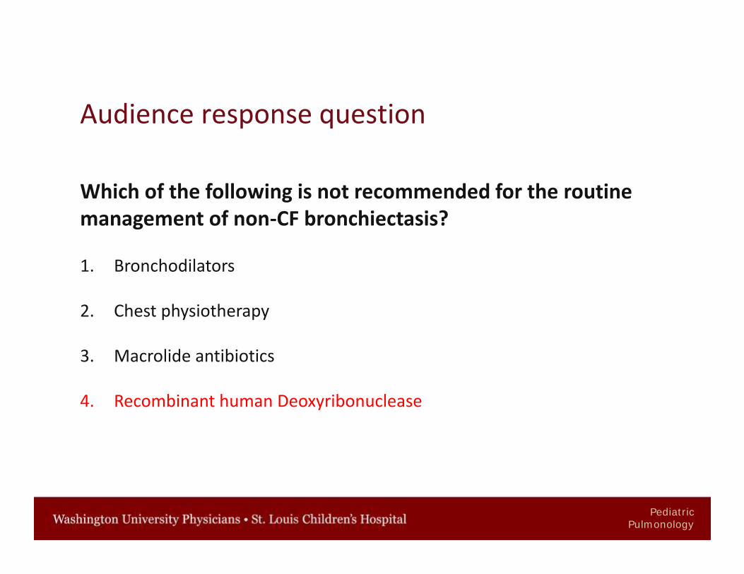
Audience response question
Which of the following is not recommended for the routinemanagement of non-CF bronchiectasis?
1. Bronchodilators
2. Chest physiotherapy
3. Macrolide antibiotics
4. Recombinant human Deoxyribonuclease
PediatricPulmonology
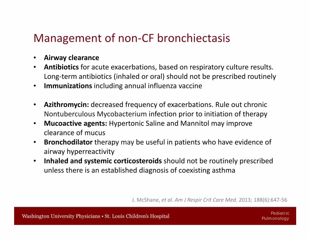
Management of non-CF bronchiectasis• Airway clearance• Antibiotics for acute exacerbations, based on respiratory culture results.
Long-term antibiotics (inhaled or oral) should not be prescribed routinely• Immunizations including annual influenza vaccine
• Azithromycin: decreased frequency of exacerbations. Rule out chronicNontuberculous Mycobacterium infection prior to initiation of therapy
• Mucoactive agents: Hypertonic Saline and Mannitol may improveclearance of mucus
• Bronchodilator therapy may be useful in patients who have evidence ofairway hyperreactivity
• Inhaled and systemic corticosteroids should not be routinely prescribedunless there is an established diagnosis of coexisting asthma
J. McShane, et al. Am J Respir Crit Care Med. 2013; 188(6):647-56
PediatricPulmonology
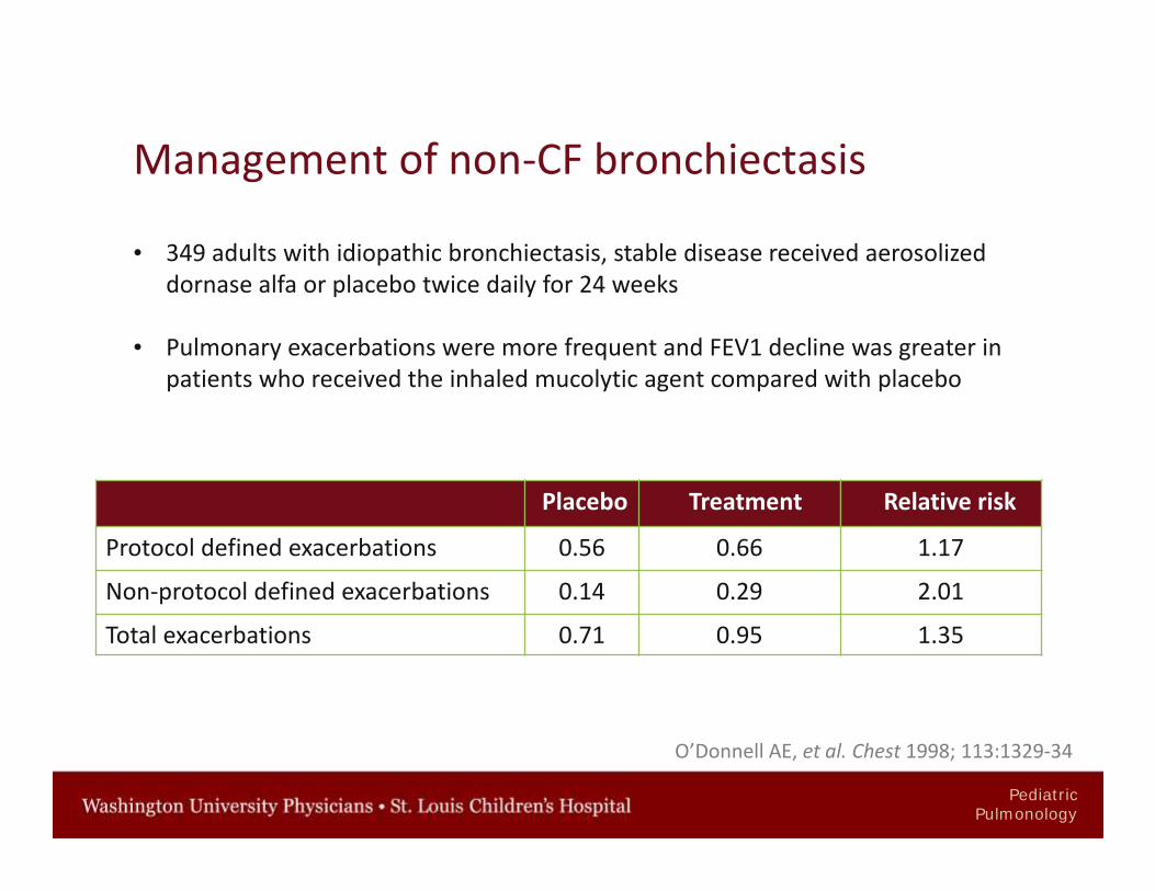
Management of non-CF bronchiectasis
• 349 adults with idiopathic bronchiectasis, stable disease received aerosolizeddornase alfa or placebo twice daily for 24 weeks
• Pulmonary exacerbations were more frequent and FEV1 decline was greater inpatients who received the inhaled mucolytic agent compared with placebo
Placebo Treatment Relative risk
Protocol defined exacerbations 0.56 0.66 1.17
Non-protocol defined exacerbations 0.14 0.29 2.01
Total exacerbations 0.71 0.95 1.35
O’Donnell AE, et al. Chest 1998; 113:1329-34
PediatricPulmonology
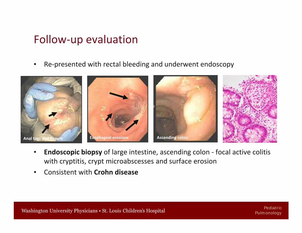
Follow-up evaluation
• Re-presented with rectal bleeding and underwent endoscopy
• Endoscopic biopsy of large intestine, ascending colon - focal active colitiswith cryptitis, crypt microabscesses and surface erosion
• Consistent with Crohn disease
Anal tags and fissure Esophageal erosions Ascending colon
PediatricPulmonology
Management• Treated with prednisone, 2mg/kg/day and gradually decreased over a 3
month period
• Azathioprine, 2mg/kg once daily
• Within 3 months, the child no longer required supplemental oxygen
PediatricPulmonology
Inflammatory Bowel Disease (IBD)Epidemiology
• Systemic disorder; unknown etiology; primarily involving the GI tract
• 25% have extra-intestinal manifestations: pulmonary involvement,arthritis, uveitis, erythema nodosum, pyoderma gangrenosum, primarysclerosing cholangitis
• Incidence: peaks in 2nd decade between ages 15 and 30 years
• All IBD patients: diagnosed in the pediatric age group:CD: 25 - 30% UC:20%
• Crohn disease (CD): patchy transmural inflammation of GI tract, frommouth to the perianal area
PediatricPulmonology
Inflammatory bowel disease and the lungs
• No precise data on the prevalence of IBD-associated lung involvement inchildren
• 7 patients less than 2 years with Crohn disease;• 1 patient - 1 month old had pulmonary infiltrates from age of 7
months.• Bronchoalveolar lavage – no infectious etiology• Open lung biopsy @ 4.5 years old- multiple granulomas with
chronic inflammation and minimal fibrosis
George Marx, The Journal of Pediatrics 2002; Vol. 140, (4)470–473;
PediatricPulmonology
Inflammatory bowel disease and the lungs -pathogenesis
Mechanism:
• Common embryological origin of the GI tract & lung• Similar immune systems in mucosa; presence of circulating immune
complexes and auto-antibodies• Adverse pulmonary effects of some drugs• Respiratory involvement may precede bowel disease presentation by
months or years• Colonic surgery may promote onset of respiratory symptoms
• Lu, De-Gan et al. World J Gastroenterol 2014; 20.1: 133–141.• Camus P et al. Eur Respir J 2000; 15:5–10,• Eaton TE et al. Thorax 1998;53:529-531.
PediatricPulmonology
• AIRWAY INFLAMMATION – most prevalent pattern ofrespiratory involvement
• AIRWAY DISEASE – Large and small airways• LUNG PARENCHYMA• PLEURAL DISEASE
Majewski et al. Arch Med Sci. 2015 10; 11(6): 1179–1188Schleiermacher, et al. J Crohns Colitis 2007; 2007, 61–69
Inflammatory bowel disease and the lungs -pathogenesis
PediatricPulmonology
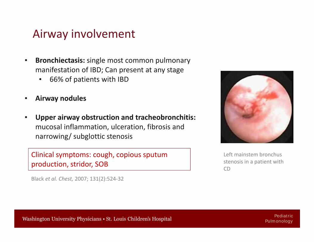
Airway involvement
• Bronchiectasis: single most common pulmonarymanifestation of IBD; Can present at any stage• 66% of patients with IBD
• Airway nodules
• Upper airway obstruction and tracheobronchitis:mucosal inflammation, ulceration, fibrosis andnarrowing/ subglottic stenosis
Black et al. Chest, 2007; 131(2):524-32
Left mainstem bronchusstenosis in a patient withCD
Clinical symptoms: cough, copious sputumproduction, stridor, SOB
PediatricPulmonology
Parenchymal disease
• Cryptogenic organizing pneumonia (COP)- may accompany pulmonary infection ormanifestation of drug toxicity
• Interstitial lung disease: Unspecified Interstitial lung disease, Eosinophilicpneumonia, Nonspecific interstitial pneumonia, Desquamative interstitialpneumonia, Usual interstitial pneumonia
• Pulmonary Interstitial Emphysema syndrome
• Fibrosing alveolitis
Majewski et al. Arch Med Sci. 2015 10; 11(6): 1179–1188Schleiermacher, et al. J Crohns Colitis 2007; 2007, 61–69
Clinical symptoms: fever, cough, dyspnea, pleuritic chest pain
PediatricPulmonology
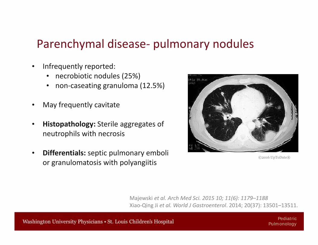
Parenchymal disease- pulmonary nodules
• Infrequently reported:• necrobiotic nodules (25%)• non-caseating granuloma (12.5%)
• May frequently cavitate
• Histopathology: Sterile aggregates ofneutrophils with necrosis
• Differentials: septic pulmonary embolior granulomatosis with polyangiitis
©2016 UpToDate®
Majewski et al. Arch Med Sci. 2015 10; 11(6): 1179–1188Xiao-Qing Ji et al. World J Gastroenterol. 2014; 20(37): 13501–13511.
PediatricPulmonology
Enteric-pulmonary fistulae
• Occurs infrequently
• Recurrent pneumonia with feculent sputum in patients with CD shouldraise suspicion of colobronchial fistula
• Colopleural fistula and fecopneumothorax are rare, but life-threateningcomplications of CD
• Abdominal and thoracic CT scans or magnetic resonance imaging (MRI)
• Treatment: mandatory surgery
Storch I et al. Inflamm Bowel Dis. 2003; 9(2):104-15Xiao-Qing Ji et al. World J Gastroenterol. 2014; 20(37): 13501–13511
PediatricPulmonology
Pleural disease:• Serositis causing pleural and pericardial effusion• Fluid is exudative containing neutrophils; may be hemorrhagic• Drug induced – Biologic agents, 5-ASA, Methotrexate
• Pneumothorax• Pleural thickening• Pleuritis
Rosenbaum AJ et al. J Clin Gastroenterol. 1983 J Clin Gastroenterol. 1983
PediatricPulmonology
Airway hyperreactivity
• Methacholine challenge:• Bronchoprovocation challenges are usually negative• Increased bronchial hyperresponsiveness in 14 children with CD
compared to control subjects, although the provocative dose causinga 20% fall in FEV1 in CD patients was greater than in the 10 asthmaticsalso tested in the study
• Inflammation of the airways by inflammatory cells and their products
Bartholo RM et al. Braz J Med Biol Res. 2005;38(2):197-203Black, et al. Chest, 2007; 131(2):524-32
PediatricPulmonology
Pulmonary function abnormalities
• Spirometry - reduced or normal FVC, FEV1, low FEV1/FVC; no significantreversibility with bronchodilator
• Fractional eNO: significantly higher in CD patients and correlatedpositively with CD activity
• Diffusion capacity for carbon monoxide :• Decreased in asymptomatic IBD patients• Significantly lower in patients with active GI disease than those in
remission
Welsh L et al. Am J Respir Crit Care Med 2012; 15;186(10):1060-1
PediatricPulmonology
Bronchoscopy
Bronchoscopy - chronic inflammation in the lungs of IBD patients
Bronchoalveolar lavage fluid (BAL)• To identify infectious etiology• Asymptomatic patients with CD• Persistently elevated alveolar lymphocytosis• An elevated CD4:CD8 ratio in the BAL, a characteristic finding in
sarcoidosis, has also been documented in patients with CD• There is no correlation with drug treatment or CD activity
Black, et al. Chest, 2007; 131(2):524-32Bewig B, et al. Respiration. 1999;66(5):467-9
PediatricPulmonology
TREATMENT
• Corticosteroids have demonstrated a high efficiency in inducing clinicalremission
• Immunomodulatory agentsAzathiorpine, Methotrexate, 6 mercaptopurine
• Biologics (anti-TNF drugs) – significant advance in CD• Infliximab, Adalimumab, Certolizumab
• Can maintain remission and improve some of the pulmonarymanifestations of IBD
PediatricPulmonology
Drugs used for IBDSeveral of these agents can cause direct pulmonary toxicity:• Organizing pneumonia (Azathioprine, 6-Mecaptopurine)• Nonspecific interstitial pneumonitis (Azathioprine, 6-Mecaptopurine,
Methotrexate, Sulfasalazine, Mesalamine)• Eosinophilic pneumonitis (Sulfasalazine, Mesalamine, Methotrexate)• Pulmonary fibrosis (Methotrexate)• Lupus-like syndrome with serositis (Biologic agents, 5-ASA, Mesalamine)• Drug-induced hypersensitivity (Methotrexate)• Opportunistic infections (Biologic agents)
Discontinuation the offending agent usually results in improvement
De-Gan Lu, et al. World J Gastroenterol 2014;7;20(1): 133-141
PediatricPulmonology
Take home points
• Maintain a high index of suspicion for lung disease in patients withinflammatory bowel disease
• Lung disease can involve airways or parenchyma and could be due to:• Underlying IBD• Opportunistic infections• Pulmonary toxicity of medications used for treatment of IBD
• Diagnostics: Chest imaging, Spirometry, DLCO and FeNO
• Bronchoscopy, Bronchoalveolar lavage, +/- Biopsy
PediatricPulmonology
Thank you!Questions?
Children's National Medical Center St. Louis Children’s Hospital