Embed Size (px)
Citation preview
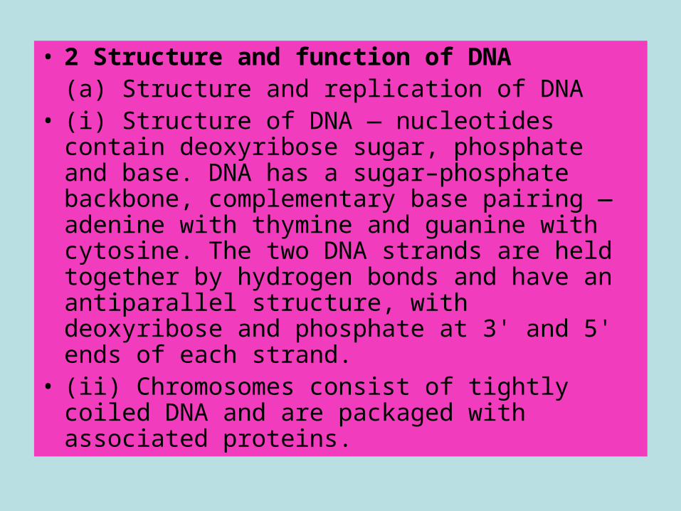
• 2 Structure and function of DNA (a) Structure and replication of DNA
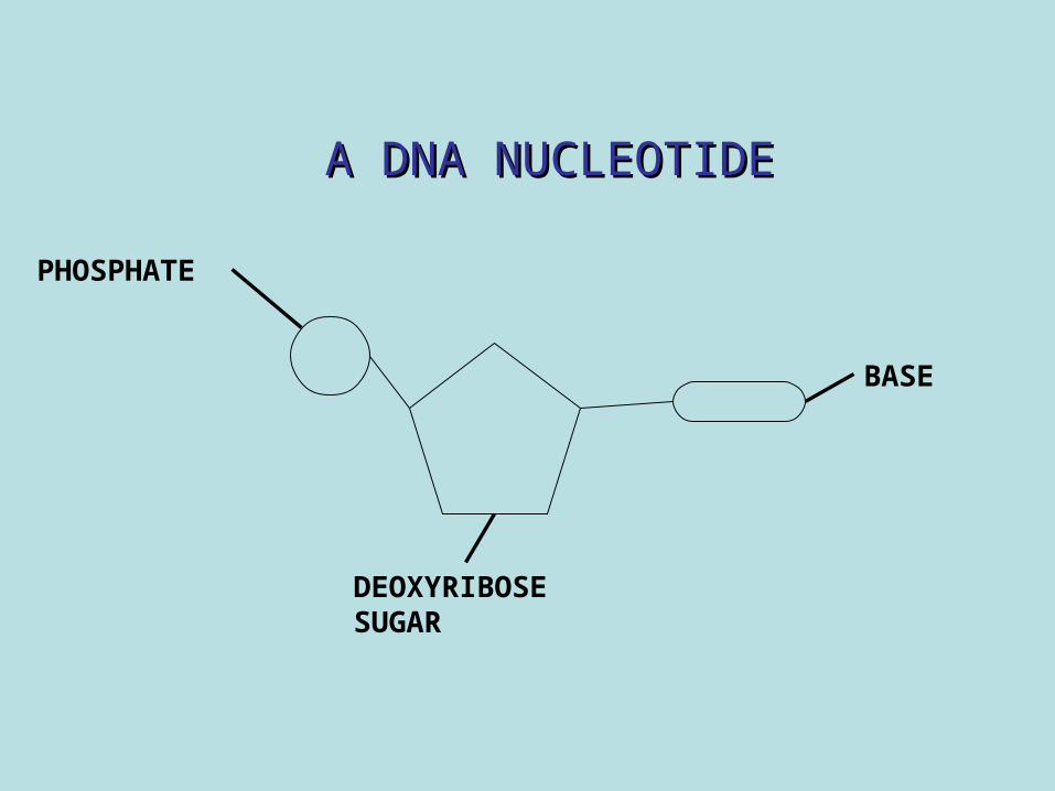
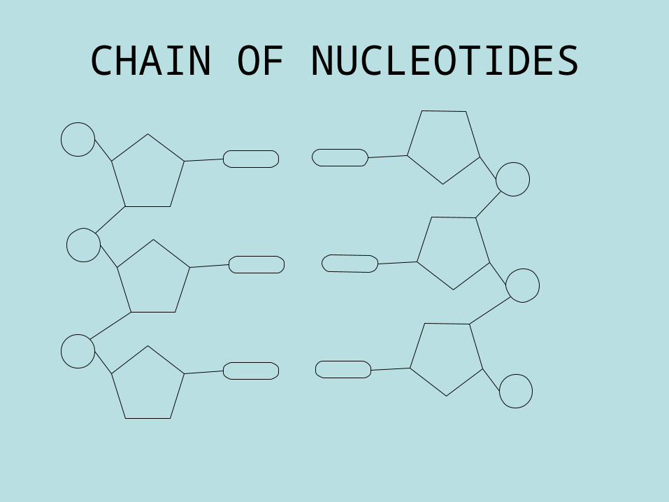
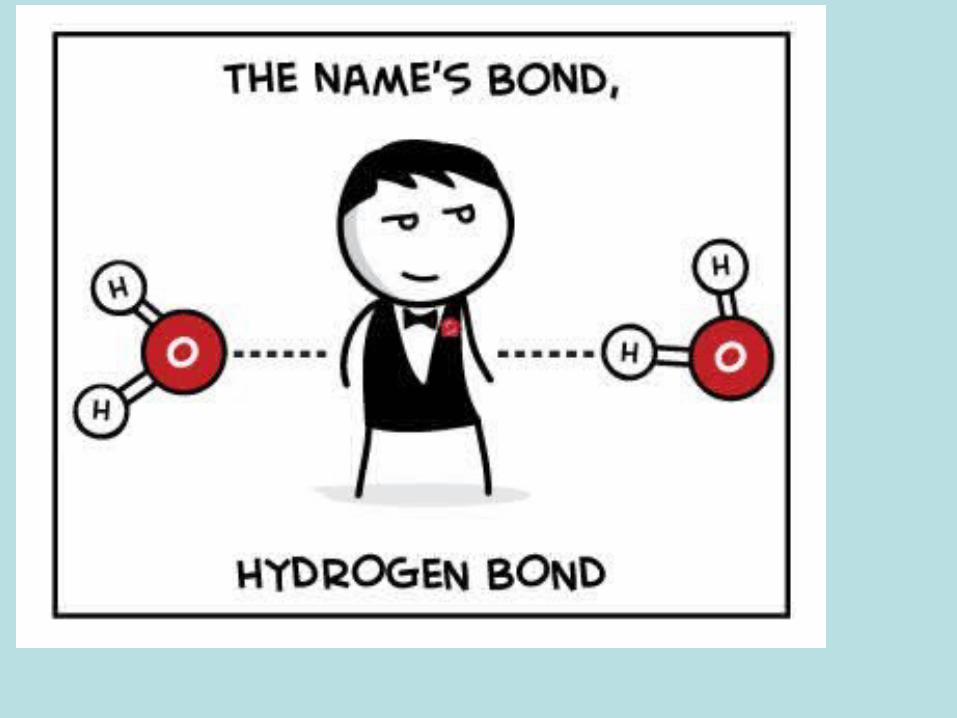
• (i) Structure of DNA — nucleotides contain deoxyribose sugar, phosphate and base. DNA has a sugar–phosphate backbone, complementary base pairing — adenine with thymine and guanine with cytosine. The two DNA strands are held together by hydrogen bonds and have an antiparallel structure, with deoxyribose and phosphate at 3' and 5' ends of each strand.
• (ii) Chromosomes consist of tightly coiled DNA and are packaged with associated proteins.
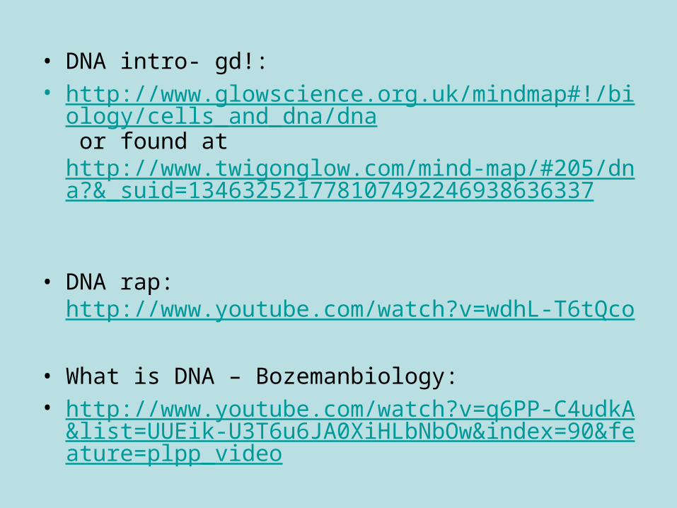
• DNA intro- gd!: • http://www.glowscience.org.uk/mindmap#!/biology/cells_
and_dna/dna or found at http://www.twigonglow.com/mind-map/#205/dna?&_suid=134632521778107492246938636337
• DNA rap: http://www.youtube.com/watch?v=wdhL-T6tQco
• What is DNA – Bozemanbiology:• http://www.youtube.com/watch?v=q6PP-C4udkA&list=U
UEik-U3T6u6JA0XiHLbNbOw&index=90&feature=plpp_video
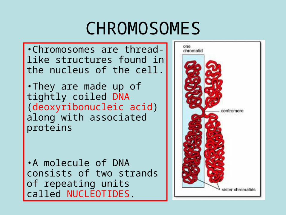
•Chromosomes are thread-like structures found in the nucleus of the cell.
•They are made up of tightly coiled DNA (deoxyribonucleic acid) along with associated proteins
•A molecule of DNA consists of two strands of repeating units called NUCLEOTIDES.
CHROMOSOMES
Videos of discoverers of DNA structure
• http://www.dnai.org/a/index.html
PHOSPHATE
BASE
DEOXYRIBOSE SUGAR
A DNA NUCLEOTIDEA DNA NUCLEOTIDE
CHAIN OF NUCLEOTIDES
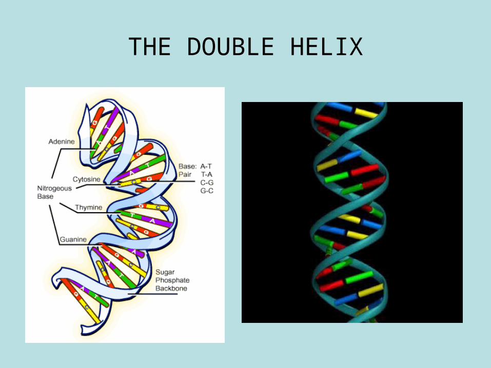
THE DOUBLE HELIX
• http://courses.scholar.hw.ac.uk/vle/scholar/session.controller?action=viewContent&back=topic&contentGUID=f830259e-992e-5044-e201-75cf99b5a3e1
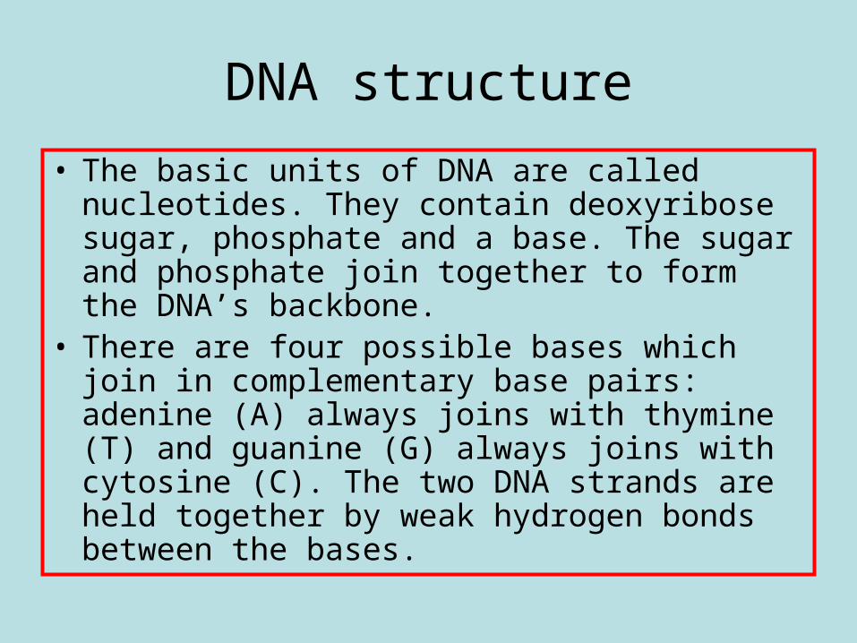
DNA structure
• The basic units of DNA are called nucleotides. They contain deoxyribose sugar, phosphate and a base. The sugar and phosphate join together to form the DNA’s backbone.
• There are four possible bases which join in complementary base pairs: adenine (A) always joins with thymine (T) and guanine (G) always joins with cytosine (C). The two DNA strands are held together by weak hydrogen bonds between the bases.
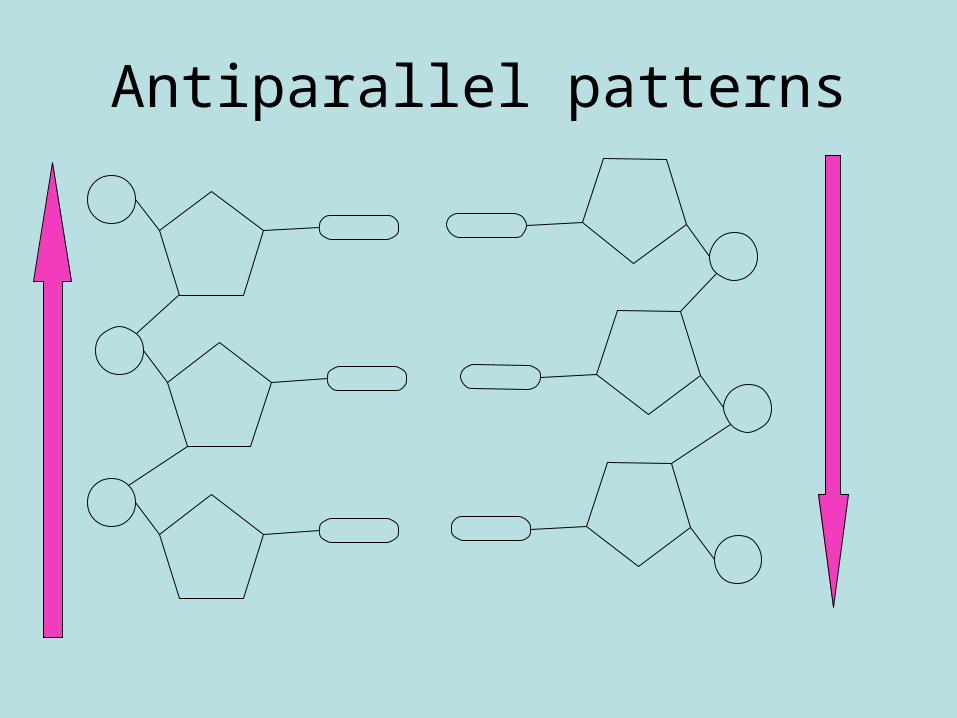
Antiparallel patterns
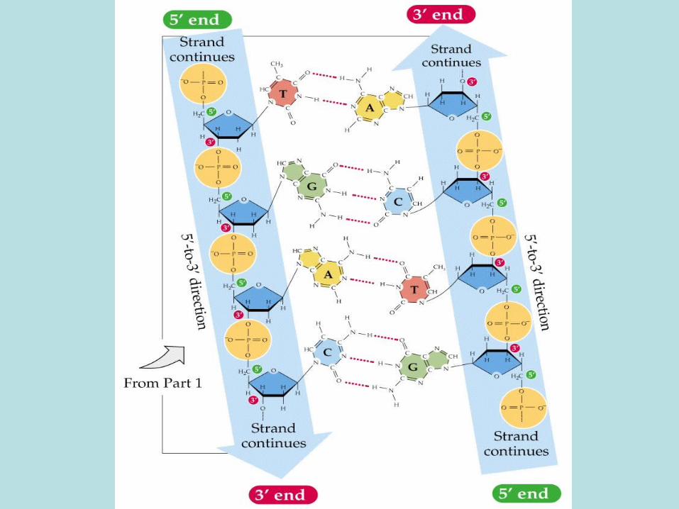
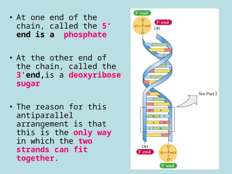
• At one end of the chain, called the 5‘ end is a phosphate
• At the other end of the chain, called the 3'end,is a deoxyribose sugar
• The reason for this antiparallel arrangement is that this is the only way in which the two strands can fit together.
• The two strands stand alongside each other but run in antiparallel directions, i.e. they run in opposite directions.
• At the end of one strand, the end finishes with a phosphate molecule and is named the 5-prime (5’) end.
• At the end of the other strand, the end finishes with a deoxyribose sugar molecule and is named 3-prime (3’) end.
• (d rhymes with 3!)
Describe the location and structure of DNA. (8 marks)
Describe the location and structure of DNA. (maximum of 8 marks):
– DNA is located on chromosomes in the nucleus.– A DNA molecule consists of two strands wound in a double helix.– Each strand consists of subunits called nucleotides.– A nucleotide consists of a deoxyribose sugar molecule, a phosphate
group and an organic / nitrogenous base.– The deoxyribose and phosphate are linked to their neighbours to form
a sugar-phosphate backbone.– There are four organic bases: adenine (A), thymine (T), guanine (G)
and cytosine (C).– Bases are linked in complementary pairs A-T and G-C.– Bases link the two DNA strands by hydrogen bonds.– The two DNA strands have an antiparallel structure / explanation.– Deoxyribose is found at the 3 end of each strand and phosphate is
found at the 5 end.
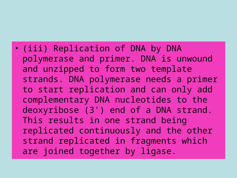
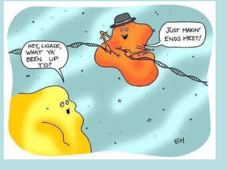
• (iii) Replication of DNA by DNA polymerase and primer. DNA is unwound and unzipped to form two template strands. DNA polymerase needs a primer to start replication and can only add complementary DNA nucleotides to the deoxyribose (3') end of a DNA strand. This results in one strand being replicated continuously and the other strand replicated in fragments which are joined together by ligase.
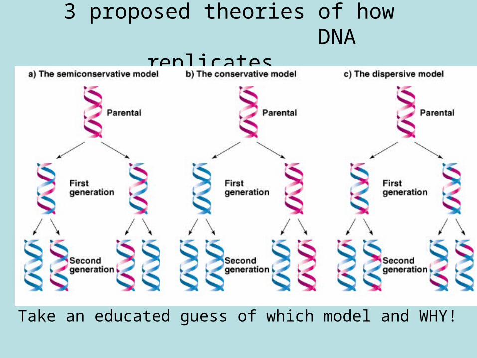
3 proposed theories of how DNA replicates....
Take an educated guess of which model and WHY!
• DNA replication – Bozemanbiology• http://www.youtube.com/watch?v=FBmO_
rmXxIw&list=UUEik-U3T6u6JA0XiHLbNbOw&index=28&feature=plpp_video
DNA replication animation!
• http://highered.mheducation.com/olcweb/cgi/pluginpop.cgi?it=swf::535::535::/sites/dl/free/0072437316/120076/micro04.swf::DNA%20Replication%20Fork.
• http://www.lpscience.fatcow.com/jwanamaker/animations/DNA%20Lecture.html
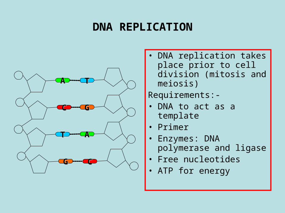
DNA REPLICATION
• DNA replication takes place prior to cell division (mitosis and meiosis)
Requirements:-• DNA to act as a
template• Primer • Enzymes: DNA
polymerase and ligase• Free nucleotides• ATP for energy
TT
GG
AA
CC
AA
CC
TT
GG
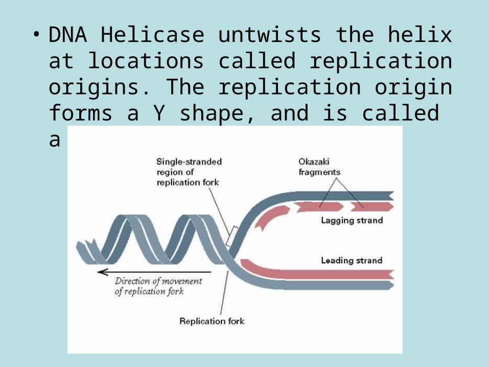
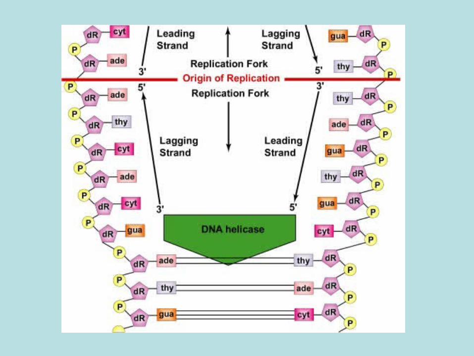
• DNA Helicase untwists the helix at locations called replication origins. The replication origin forms a Y shape, and is called a replication fork.
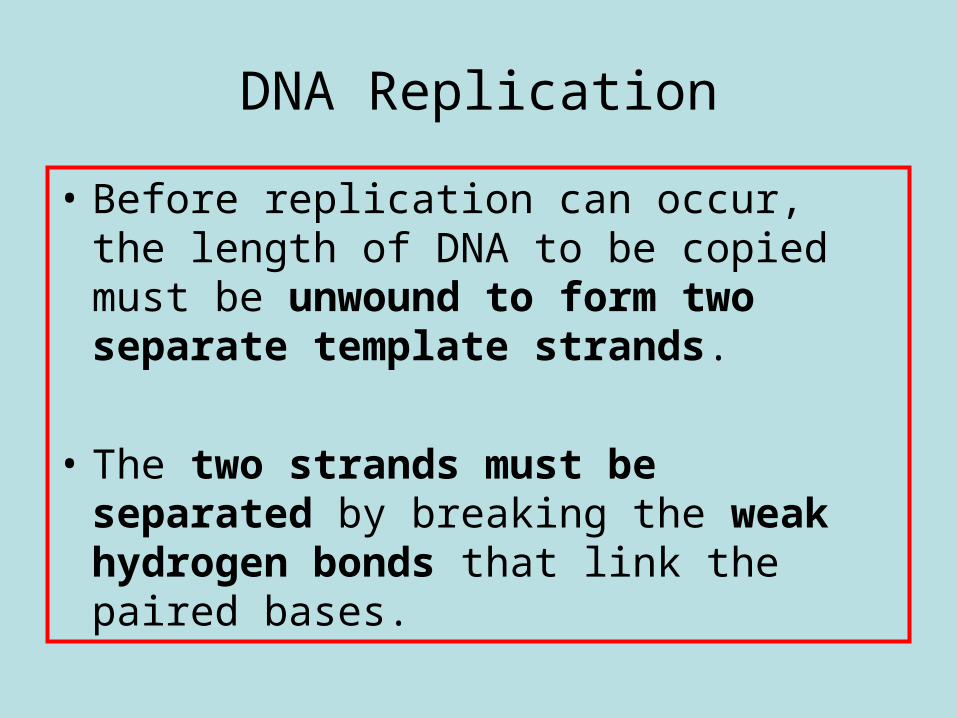
DNA Replication
• Before replication can occur, the length of DNA to be copied must be unwound to form two separate template strands.
• The two strands must be
separated by breaking the weak hydrogen bonds that link the paired bases.
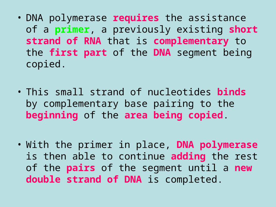
• DNA polymerase requires the assistance of a primer, a previously existing short strand of RNA that is complementary to the first part of the DNA segment being copied.
• This small strand of nucleotides binds by complementary base pairing to the beginning of the area being copied.
• With the primer in place, DNA polymerase is then able to continue adding the rest of the pairs of the segment until a new double strand of DNA is completed.
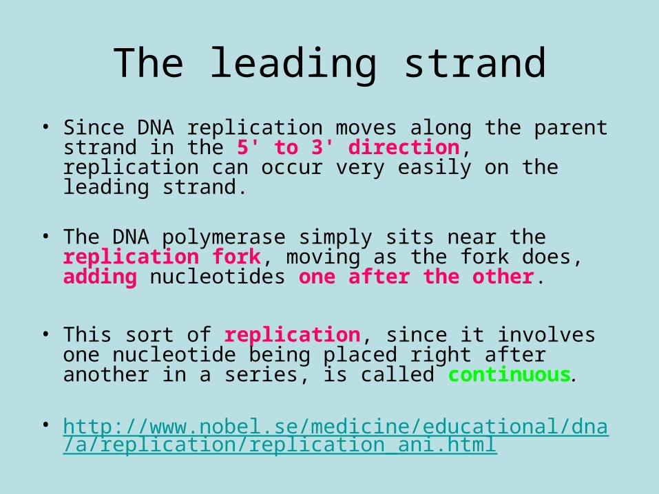
The leading strand
• Since DNA replication moves along the parent strand in the 5' to 3' direction, replication can occur very easily on the leading strand.
• The DNA polymerase simply sits near the replication fork, moving as the fork does, adding nucleotides one after the other.
• This sort of replication, since it involves one nucleotide being placed right after another in a series, is called continuous.
• http://www.nobel.se/medicine/educational/dna/a/replication/replication_ani.html
The lagging strand
• Synthesis on the lagging strand is slightly delayed in relation to synthesis on the leading strand.
• http://www.nobel.se/medicine/educational/dna/a/replication/lagging_ani.html
What you should knowDNA Replication
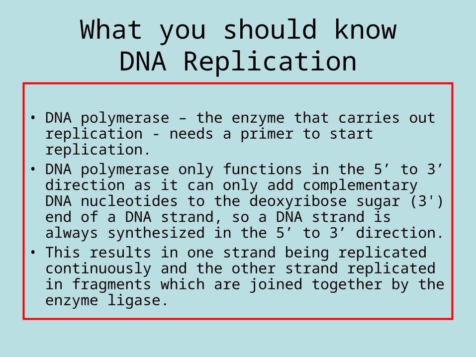
• DNA polymerase – the enzyme that carries out replication - needs a primer to start replication.
• DNA polymerase only functions in the 5’ to 3’ direction as it can only add complementary DNA nucleotides to the deoxyribose sugar (3') end of a DNA strand, so a DNA strand is always synthesized in the 5’ to 3’ direction.
• This results in one strand being replicated continuously and the other strand replicated in fragments which are joined together by the enzyme ligase.
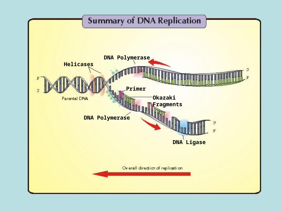
DNA Polymerase
DNA Ligase
Primer
Okazaki Fragments
DNA PolymeraseHelicases
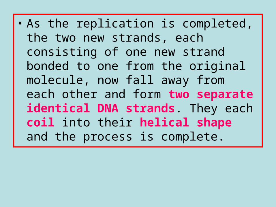
• As the replication is completed, the two new strands, each consisting of one new strand bonded to one from the original molecule, now fall away from each other and form two separate identical DNA strands. They each coil into their helical shape and the process is complete.
Give an account of the replication of DNA. (maximum of 7 marks):
– DNA is unwound and unzipped…– …by the enzyme helicase…– …to form two template strands.– DNA polymerase is the enzyme which adds nucleotides to the
new DNA strand.– DNA polymerase needs a primer to start replication.– DNA polymerase can only add complementary nucleotides to
the deoxyribose / 3 end of the DNA strand.– This results in one strand / the leading strand being
continuously replicated,– and the other strand / the lagging strand being replicated in
fragments,– which are joined together (by the enzyme ligase).
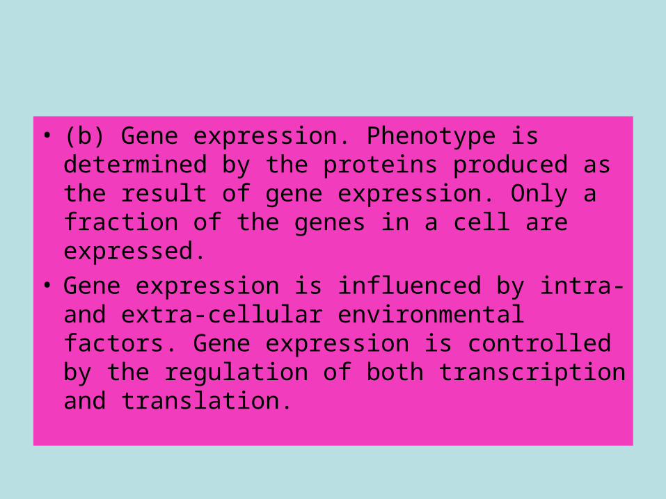
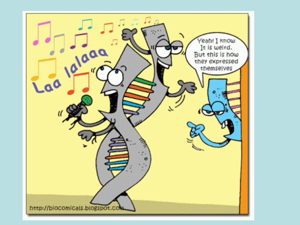
• (b) Gene expression. Phenotype is determined by the proteins produced as the result of gene expression. Only a fraction of the genes in a cell are expressed.
• Gene expression is influenced by intra- and extra-cellular environmental factors. Gene expression is controlled by the regulation of both transcription and translation.
Big Question
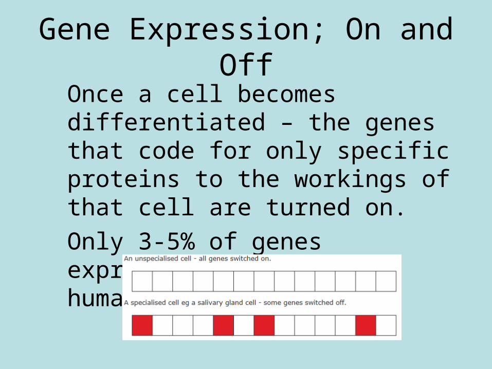
Gene Expression; On and Off
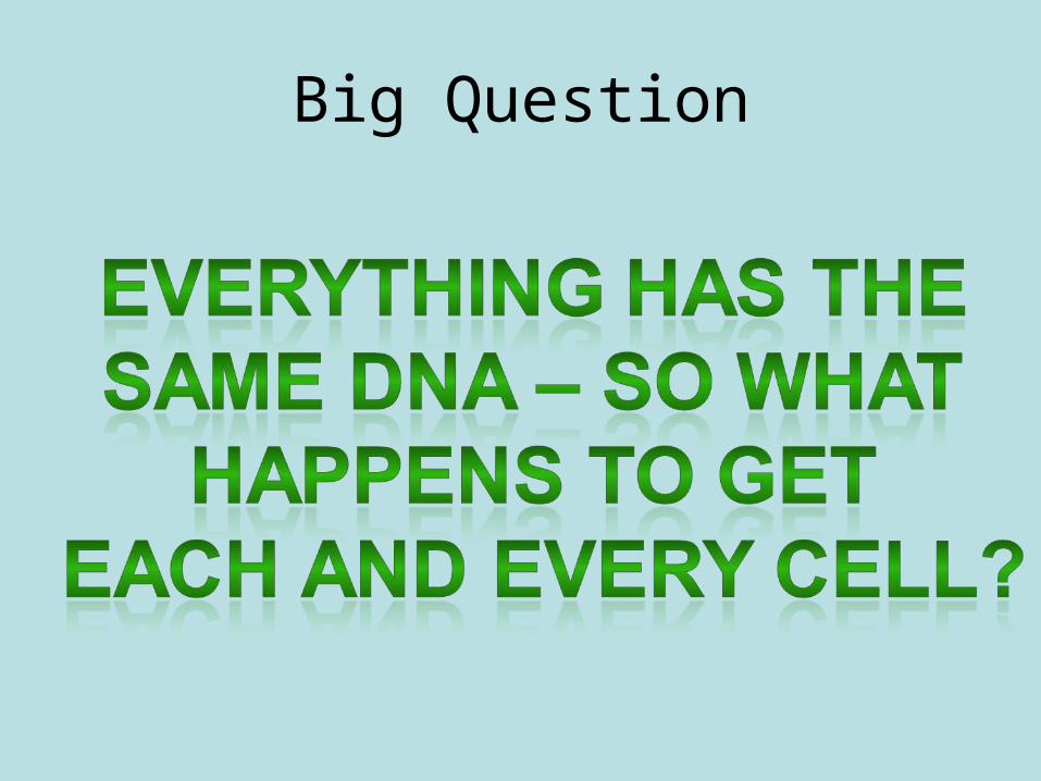
Once a cell becomes differentiated – the genes that code for only specific proteins to the workings of that cell are turned on. Only 3-5% of genes expressed in a typical human cell
Gene expression
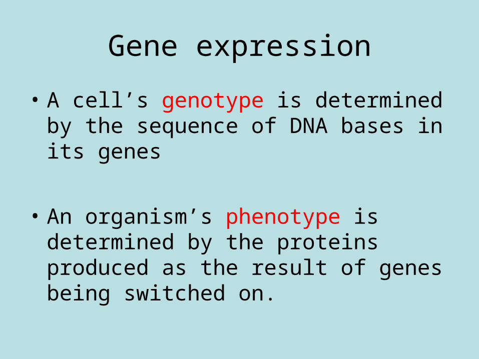
• A cell’s genotype is determined by the sequence of DNA bases in its genes
• An organism’s phenotype is determined by the proteins produced as the result of genes being switched on.
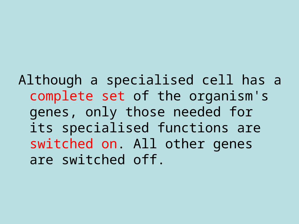
Although a specialised cell has a complete set of the organism's genes, only those needed for its specialised functions are switched on. All other genes are switched off.
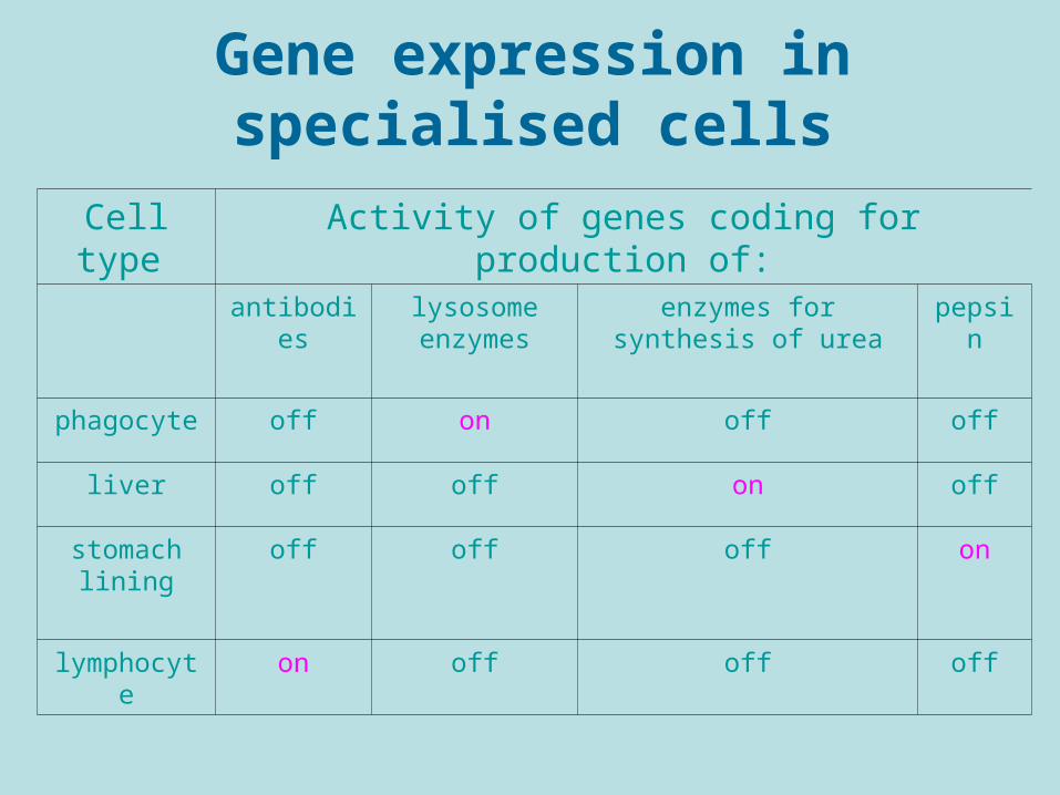
Gene expression in specialised cells
Cell type Activity of genes coding for production of:
antibodies lysosome enzymes
enzymes for synthesis of urea
pepsin
phagocyte off on off off
liver off off on off
stomach lining
off off off on
lymphocyte on off off off
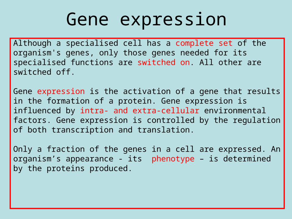
Gene expressionAlthough a specialised cell has a complete set of theorganism's genes, only those genes needed for itsspecialised functions are switched on. All other areswitched off.
Gene expression is the activation of a gene that resultsin the formation of a protein. Gene expression isinfluenced by intra- and extra-cellular environmentalfactors. Gene expression is controlled by the regulationof both transcription and translation.
Only a fraction of the genes in a cell are expressed. Anorganism’s appearance - its phenotype – is determinedby the proteins produced.
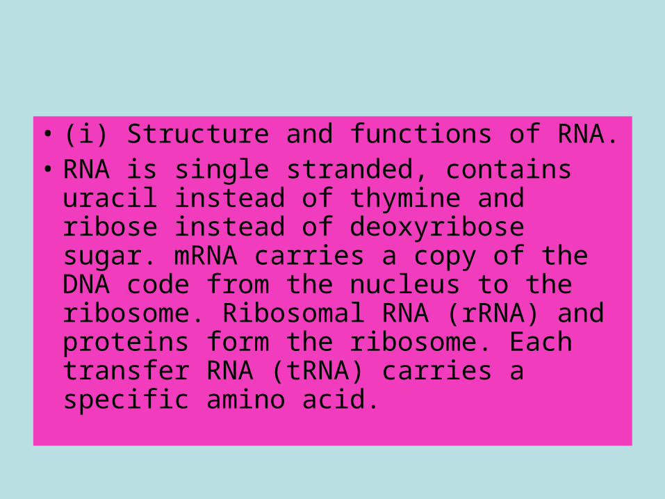
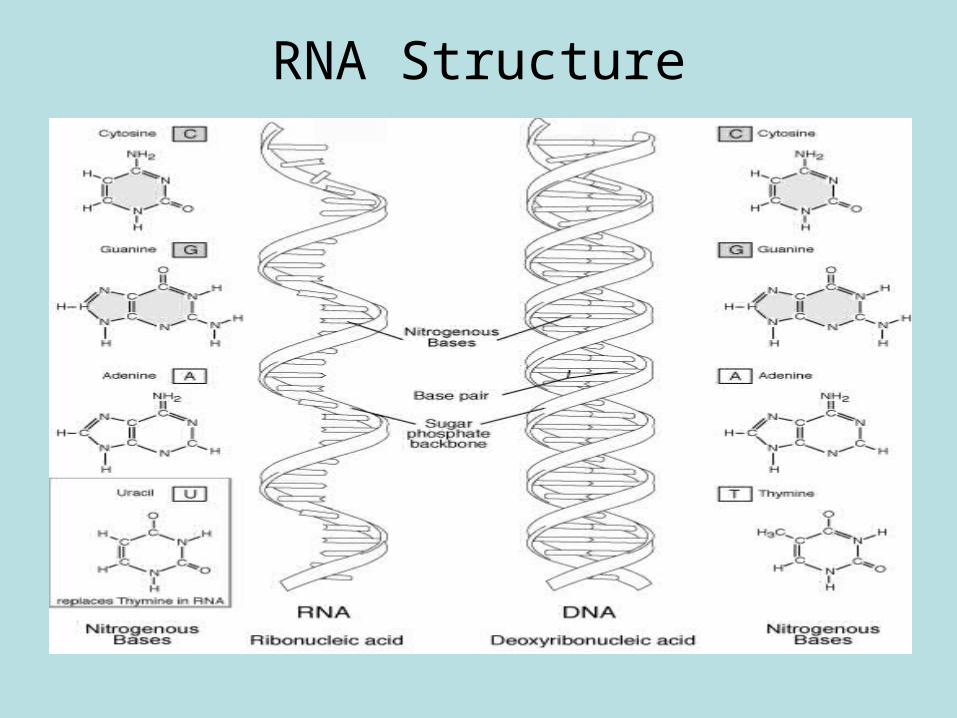
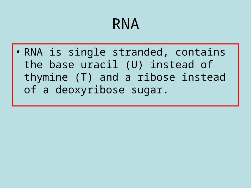
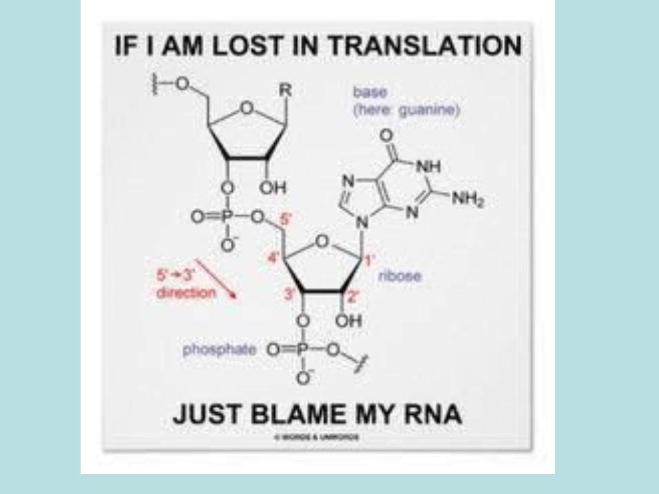
• (i) Structure and functions of RNA. • RNA is single stranded, contains uracil
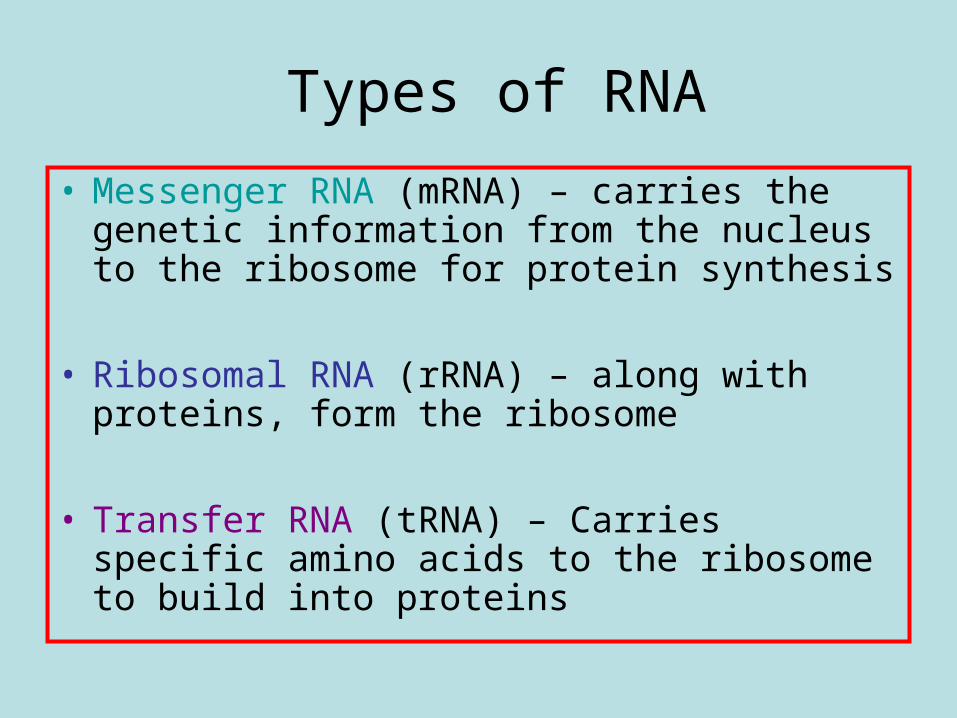
instead of thymine and ribose instead of deoxyribose sugar. mRNA carries a copy of the DNA code from the nucleus to the ribosome. Ribosomal RNA (rRNA) and proteins form the ribosome. Each transfer RNA (tRNA) carries a specific amino acid.
DNA to Protein
RNA Structure
RNA
• RNA is single stranded, contains the base uracil (U) instead of thymine (T) and a ribose instead of a deoxyribose sugar.
Types of RNA
• Messenger RNA (mRNA) – carries the genetic information from the nucleus to the ribosome for protein synthesis
• Ribosomal RNA (rRNA) – along with proteins, form the ribosome
• Transfer RNA (tRNA) – Carries specific amino acids to the ribosome to build into proteins
Venn diagram
DNA both RNA
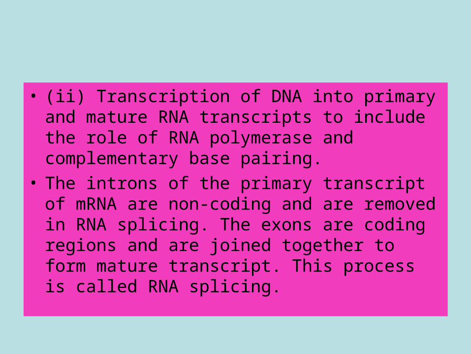
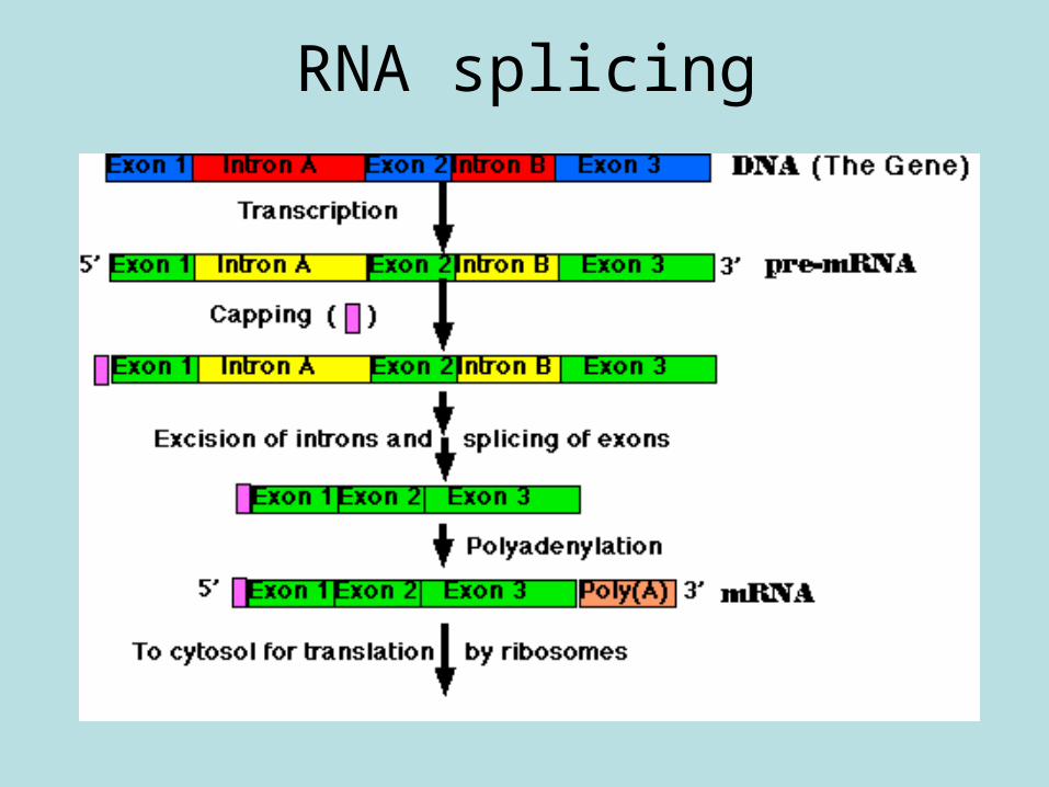
• (ii) Transcription of DNA into primary and mature RNA transcripts to include the role of RNA polymerase and complementary base pairing.
• The introns of the primary transcript of mRNA are non-coding and are removed in RNA splicing. The exons are coding regions and are joined together to form mature transcript. This process is called RNA splicing.
• How does DNA make protein?: http://www.twigonglow.com/mind-map/#205/dna?&_suid=134632521778107492246938636337
• DNA transcription and translation – Bozemanbiology• http://www.youtube.com/watch?
v=h3b9ArupXZg&list=UUEik-U3T6u6JA0XiHLbNbOw&index=27&feature=plpp_video
• • DNA RNA• http://www.youtube.com/watch?
v=xZaMi6OhsSU&feature=plcp
DNA to protein
www.yourgenome.org/downloads/animations.shtml
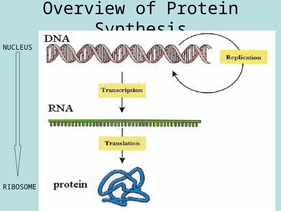
Overview of Protein Synthesis
NUCLEUS
RIBOSOME
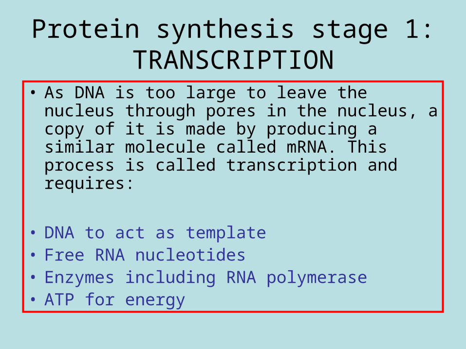
Protein synthesis stage 1: TRANSCRIPTION
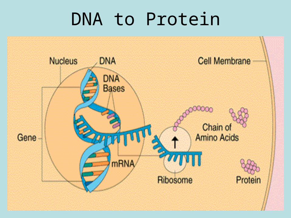
• As DNA is too large to leave the nucleus through pores in the nucleus, a copy of it is made by producing a similar molecule called mRNA. This process is called transcription and requires:
• DNA to act as template• Free RNA nucleotides• Enzymes including RNA polymerase• ATP for energy
Transcription
• http://courses.scholar.hw.ac.uk/vle/scholar/session.controller?action=viewContent&back=topic&contentGUID=16a5d6c5-d575-1e21-9bb0-f2454c2547c3
• http://www.carolina.com/teacher-resources/Video/dna-transcription-video/tr28274.tr
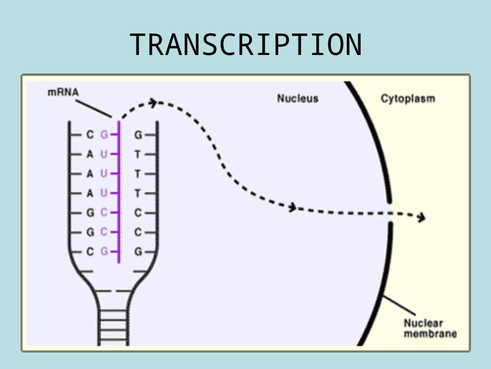
TRANSCRIPTION
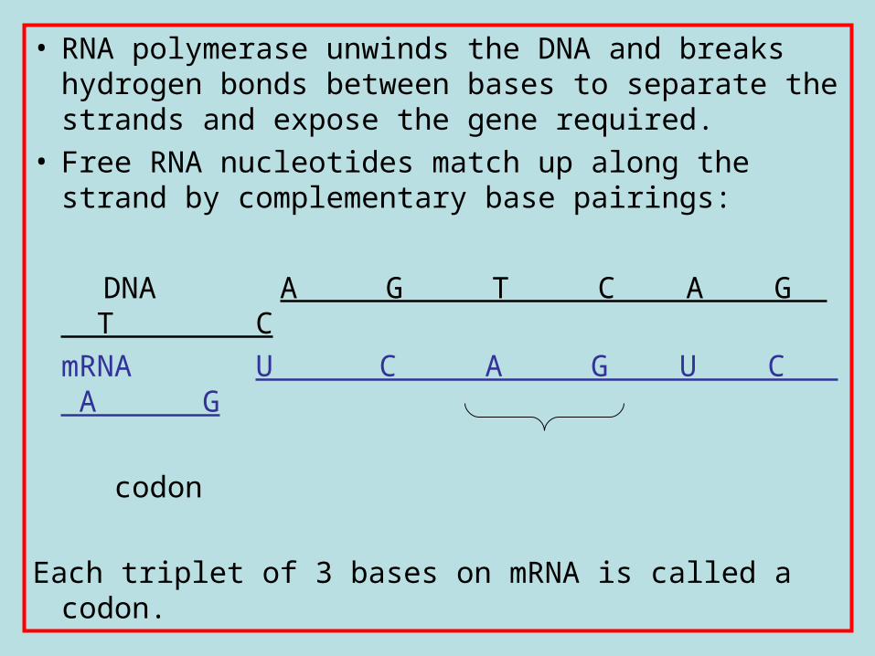
• RNA polymerase unwinds the DNA and breaks hydrogen bonds between bases to separate the strands and expose the gene required.
• Free RNA nucleotides match up along the strand by complementary base pairings:
DNA A G T C A G T CmRNA U C A G U C A G
codon
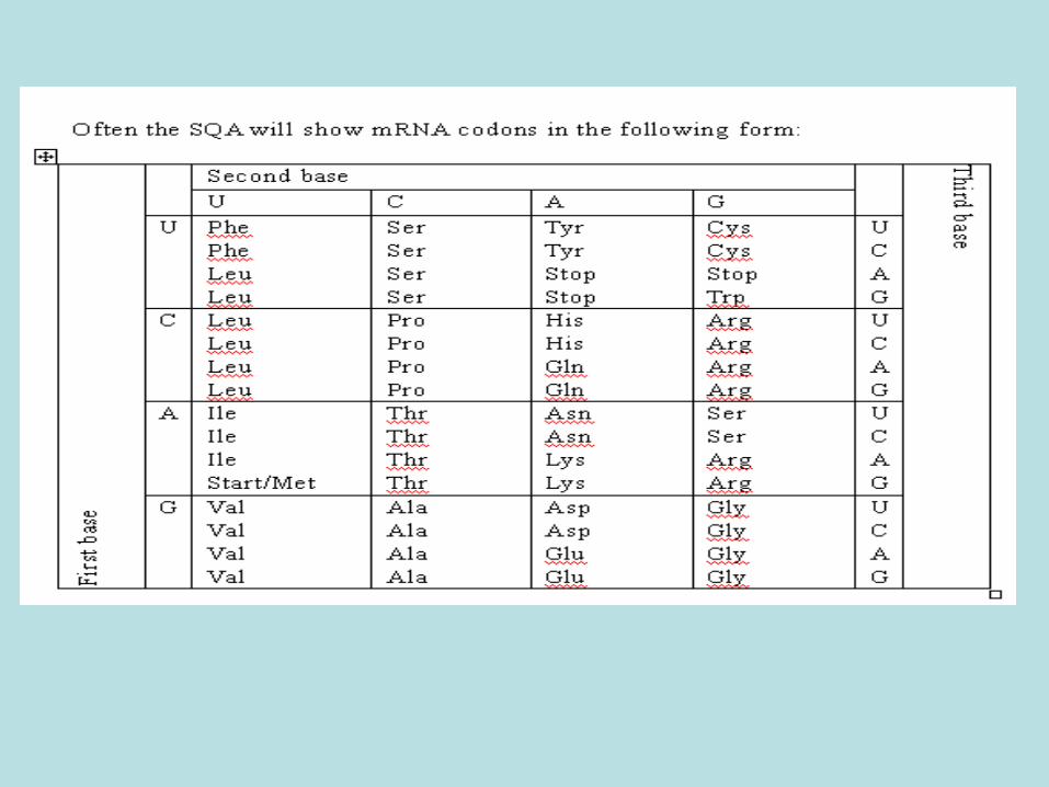
Each triplet of 3 bases on mRNA is called a codon.
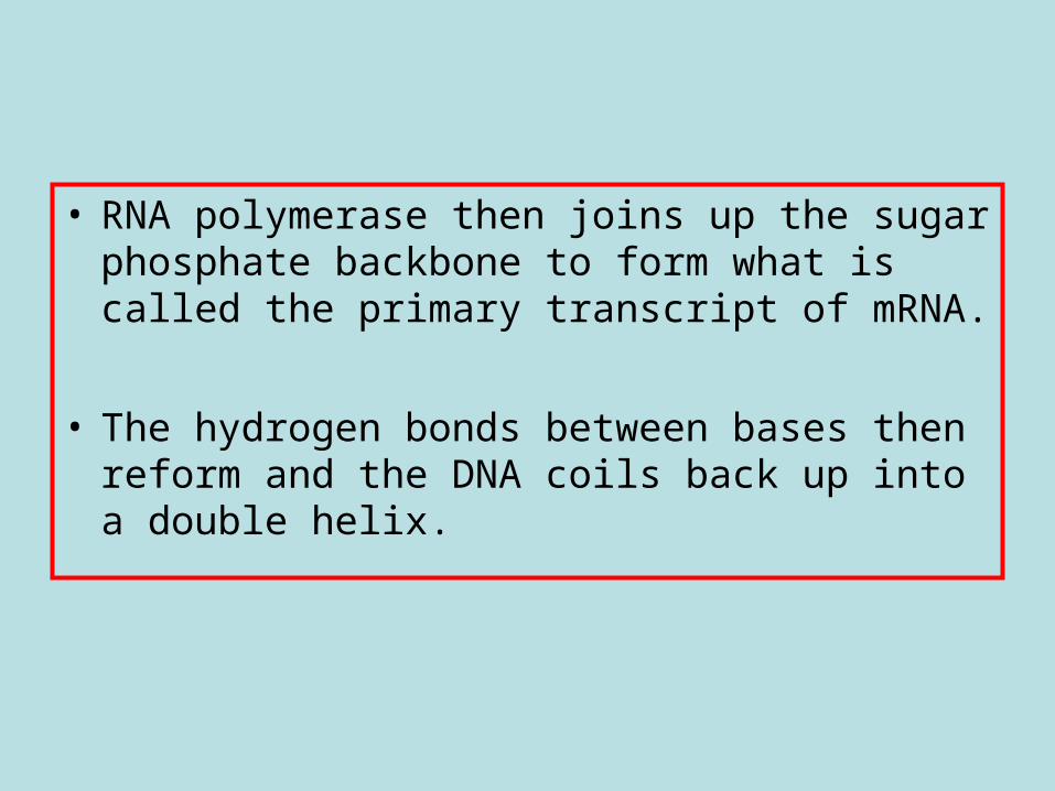
• RNA polymerase then joins up the sugar phosphate backbone to form what is called the primary transcript of mRNA.
• The hydrogen bonds between bases then reform and the DNA coils back up into a double helix.
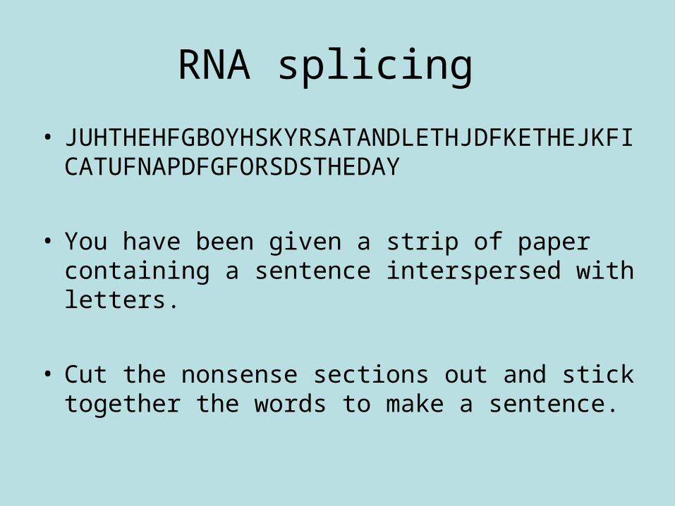
RNA splicing
• JUHTHEHFGBOYHSKYRSATANDLETHJDFKETHEJKFICATUFNAPDFGFORSDSTHEDAY
• You have been given a strip of paper containing a sentence interspersed with letters.
• Cut the nonsense sections out and stick together the words to make a sentence.
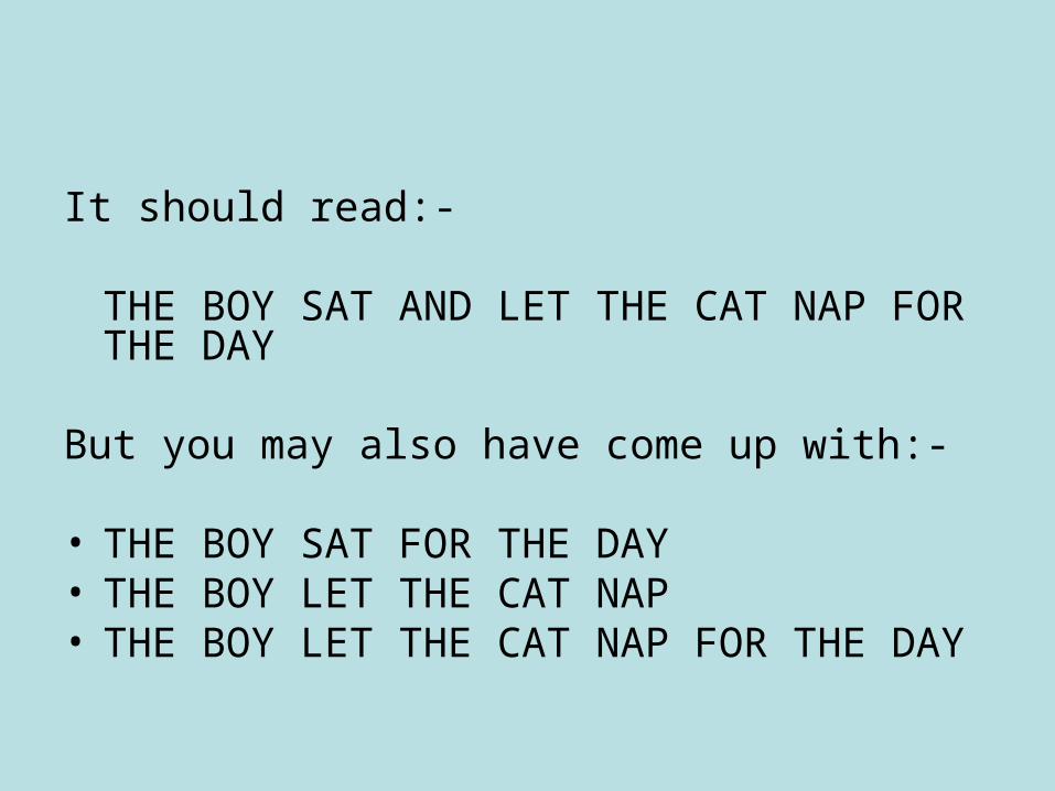
It should read:-
THE BOY SAT AND LET THE CAT NAP FOR THE DAY
But you may also have come up with:-
• THE BOY SAT FOR THE DAY• THE BOY LET THE CAT NAP• THE BOY LET THE CAT NAP FOR THE DAY
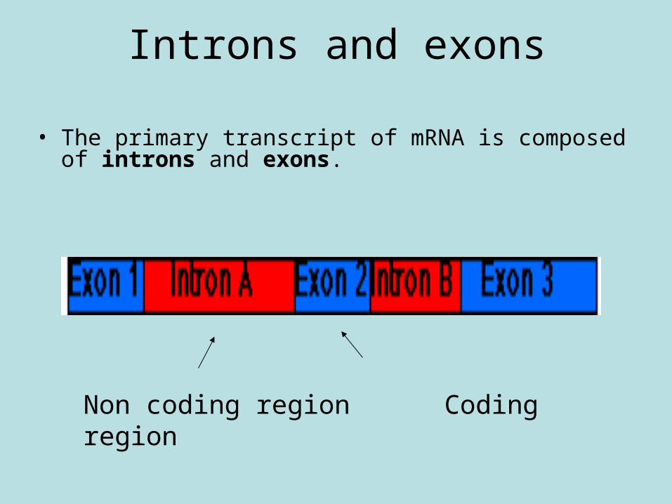
Introns and exons
• The primary transcript of mRNA is composed of introns and exons.
Non coding region Coding region
RNA splicing
• The introns of the primary transcript of mRNA are non-coding regions of the gene and are removed in RNA splicing. The exons are coding regions of the gene and are joined together to form the mature transcript of mRNA. This process is called RNA splicing.
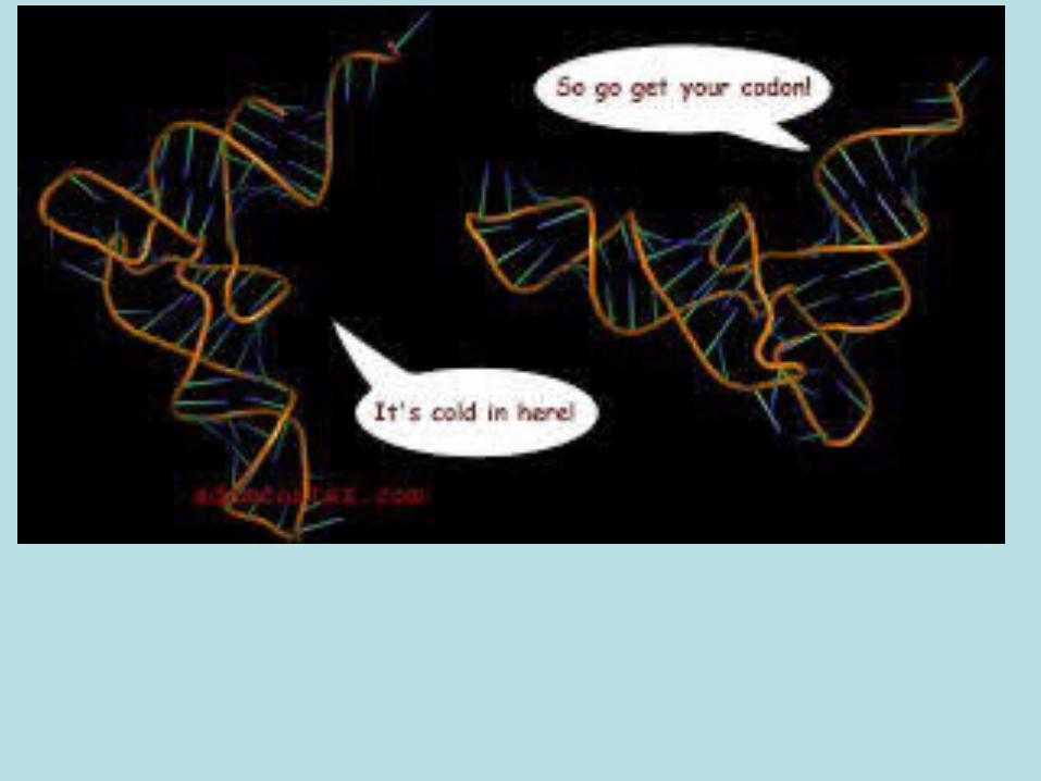
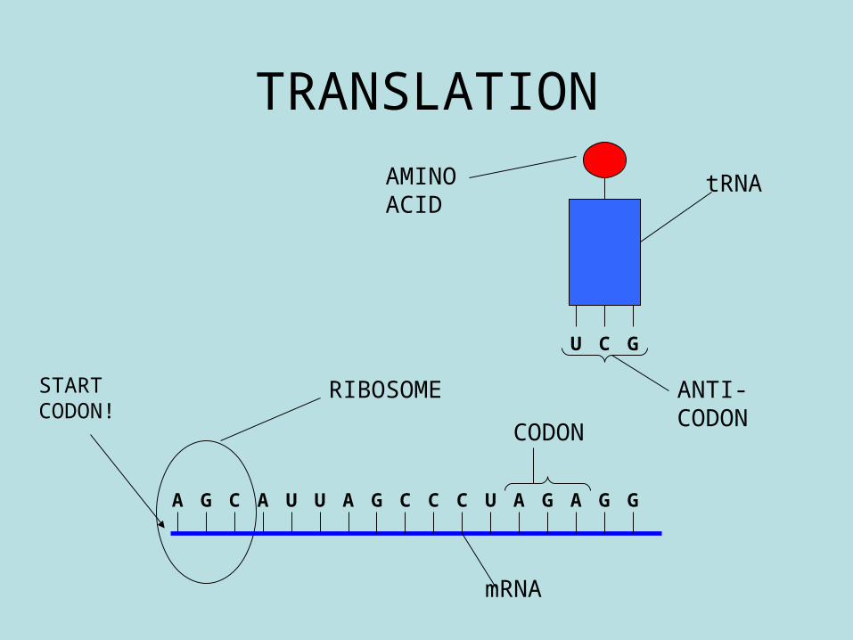
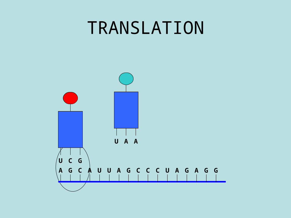
• (iii) Translation of mRNA into a polypeptide by tRNA at the ribosome.
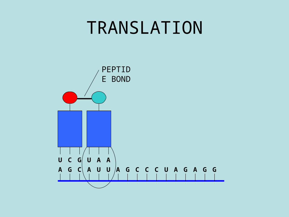
• tRNA folds due to base pairing to form a triplet anticodon site and an attachment site for a specific amino acid. Triplet codons on mRNA and anticodons translate the genetic code into a sequence of amino acids. Start and stop codons exist. Codon recognition of incoming tRNA, peptide bond formation and exit of tRNA from the ribosome as polypeptide is formed.
Protein synthesis stage 2: TRANSLATION
Changing the code on mRNA into a sequence of amino acids to form a
specific protein.
Tranlasation film
• http://www.carolina.com/teacher-resources/Video/mrna-translation-video/tr28275.tr
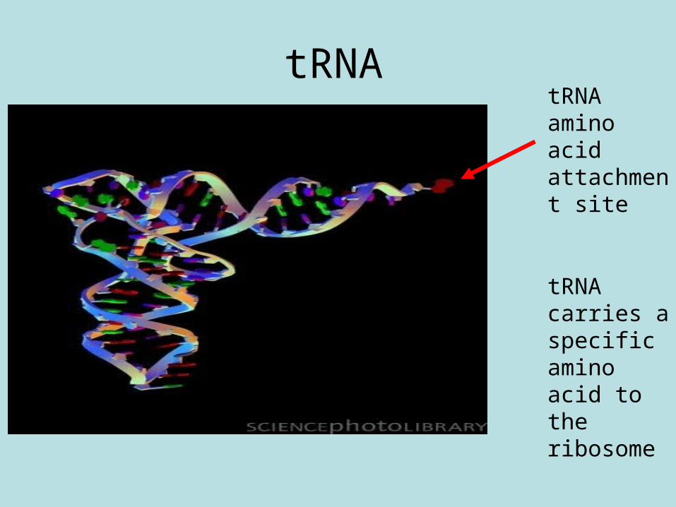
tRNAtRNA amino acid attachment site
tRNA carries a specific amino acid to the ribosome
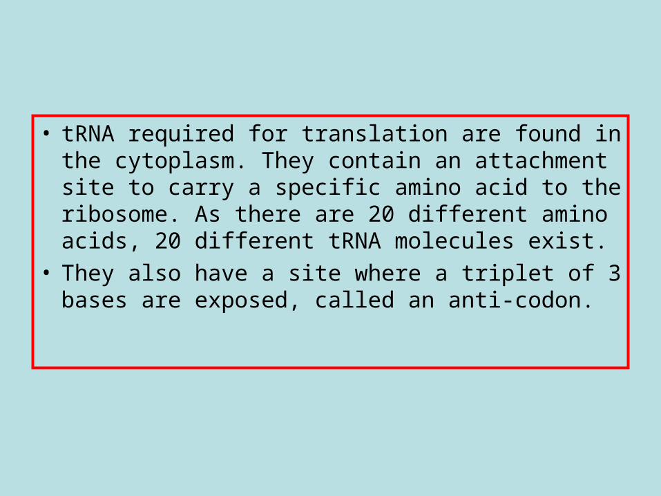
• tRNA required for translation are found in the cytoplasm. They contain an attachment site to carry a specific amino acid to the ribosome. As there are 20 different amino acids, 20 different tRNA molecules exist.
• They also have a site where a triplet of 3 bases are exposed, called an anti-codon.

• http://courses.scholar.hw.ac.uk/vle/scholar/session.controller?action=viewContent&back=topic&contentGUID=ec041196-3cb0-c55a-f4fe-e839978b92dc
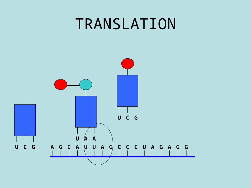
TRANSLATION
A G C UA AU CG CC U A AG GG
U C G
mRNA
tRNA
ANTI-CODON
RIBOSOME
AMINO ACID
CODON
START CODON!
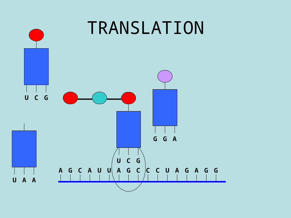
TRANSLATION
A G C UA AU CG CC U A AG GG
U C G
U A A
TRANSLATION
U A A
A G C UA AU CG CC U A AG GG
U C G
PEPTIDE BOND
TRANSLATION
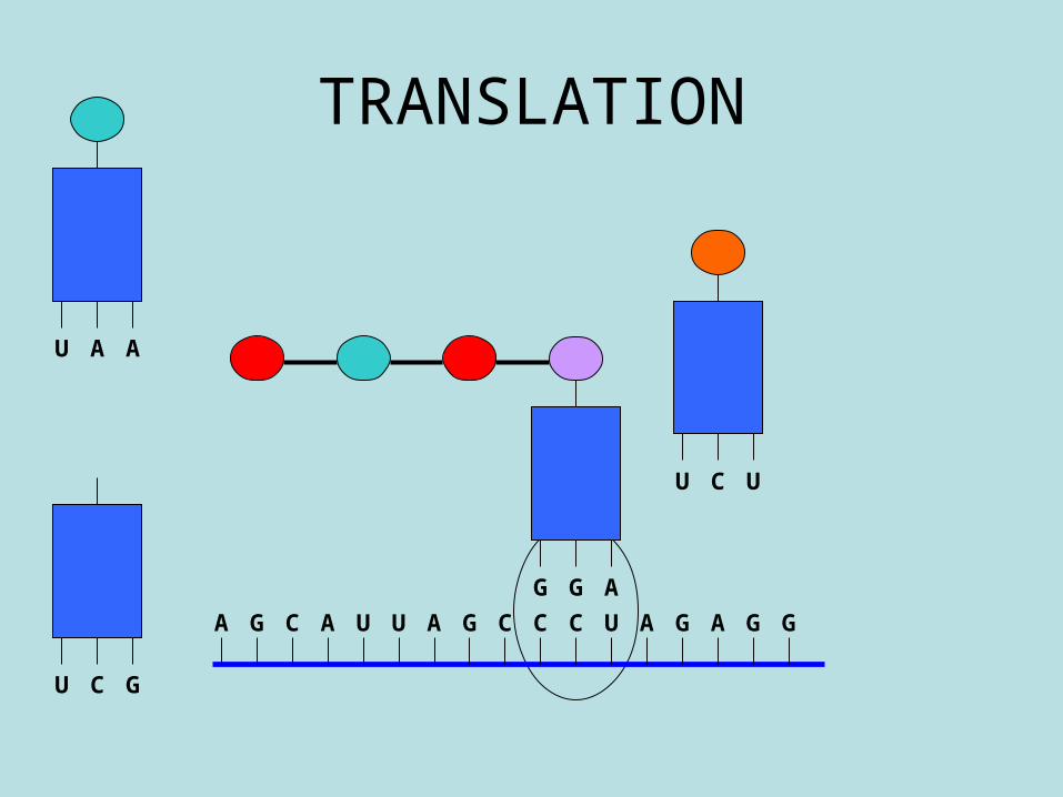
A G C UA AU CG CC U A AG GGU C G
U A A
U C G
TRANSLATION
A G C UA AU CG CC U A AG GG
U C G
U A A
U C G
G G A
TRANSLATION
A G C UA AU CG CC U A AG GG
U C G
G G A
U A A
U C U
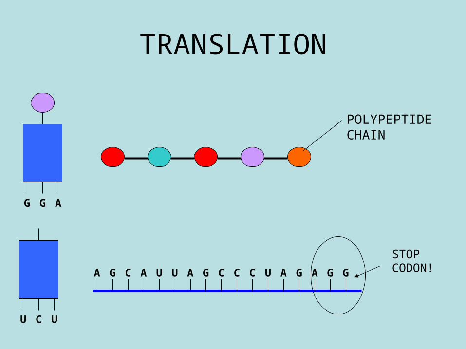
TRANSLATION
A G C UA AU CG CC U A AG GG
G G A
U C U
POLYPEPTIDE CHAIN
STOP CODON!

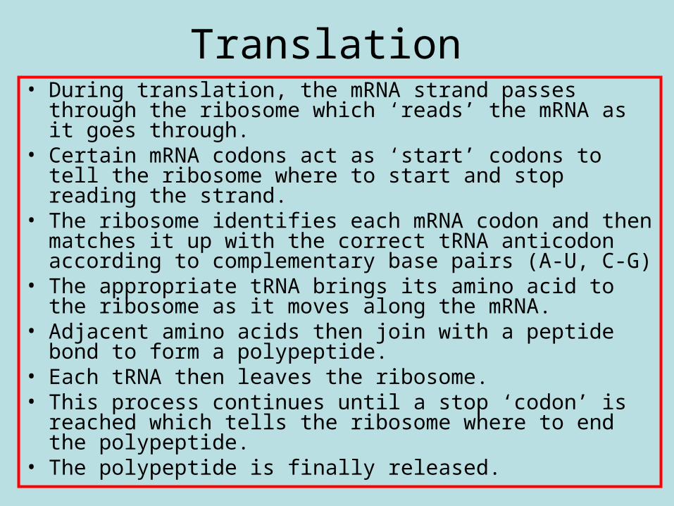
Translation • During translation, the mRNA strand passes through
the ribosome which ‘reads’ the mRNA as it goes through.
• Certain mRNA codons act as ‘start’ codons to tell the ribosome where to start and stop reading the strand.
• The ribosome identifies each mRNA codon and then matches it up with the correct tRNA anticodon according to complementary base pairs (A-U, C-G)
• The appropriate tRNA brings its amino acid to the ribosome as it moves along the mRNA.
• Adjacent amino acids then join with a peptide bond to form a polypeptide.
• Each tRNA then leaves the ribosome. • This process continues until a stop ‘codon’ is
reached which tells the ribosome where to end the polypeptide.
• The polypeptide is finally released.
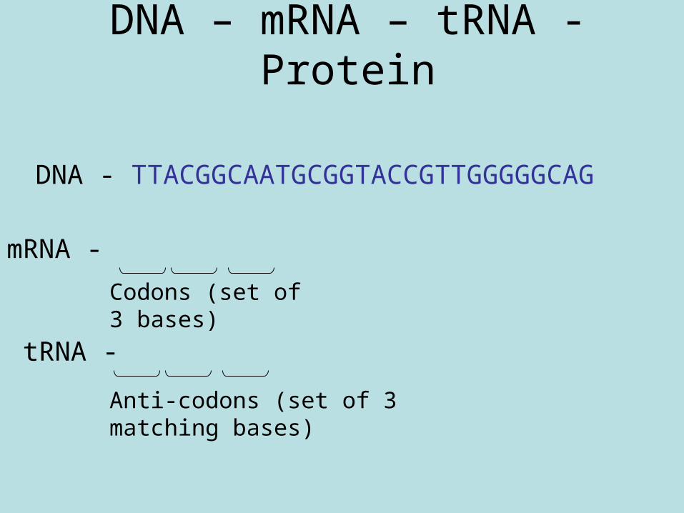
DNA – mRNA – tRNA - Protein
DNA - TTACGGCAATGCGGTACCGTTGGGGGCAG
mRNA -
Codons (set of 3 bases)
tRNA -
Anti-codons (set of 3 matching bases)
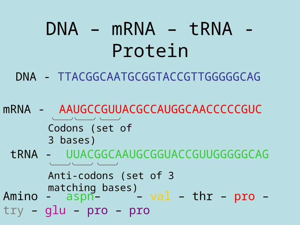
DNA – mRNA – tRNA - Protein
DNA - TTACGGCAATGCGGTACCGTTGGGGGCAG
mRNA - AAUGCCGUUACGCCAUGGCAACCCCCGUC
Codons (set of 3 bases)
tRNA - UUACGGCAAUGCGGUACCGUUGGGGGCAG
Anti-codons (set of 3 matching bases)
Amino - aspn– ala – val – thr – pro – try – glu – pro – pro
acid
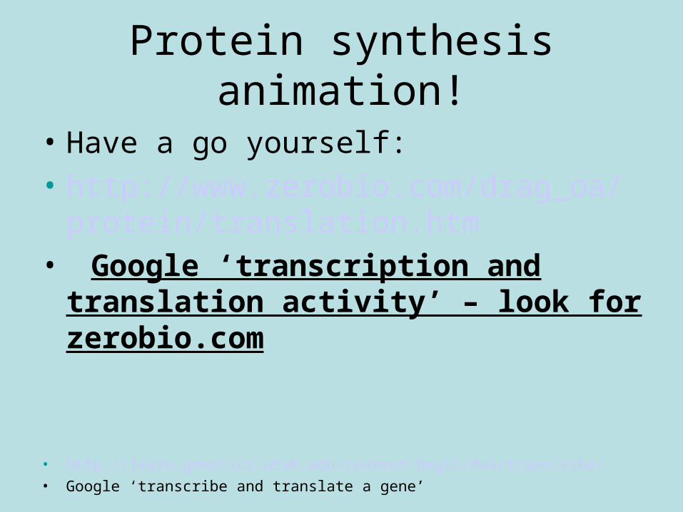
Protein synthesis animation!
• Have a go yourself:
• http://www.zerobio.com/drag_oa/protein/translation.htm
• Google ‘transcription and translation activity’ – look for zerobio.com
• http://learn.genetics.utah.edu/content/begin/dna/transcribe/• Google ‘transcribe and translate a gene’

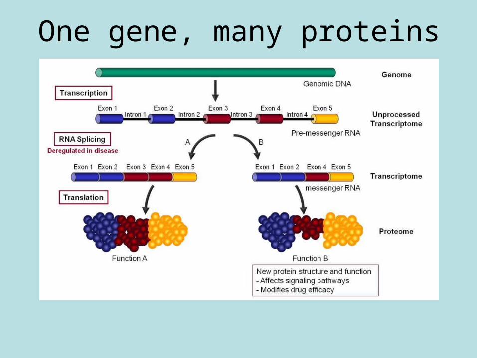
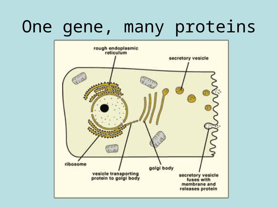
• (iv) One gene, many proteins as a result of RNA splicing and post-translational modification. Different mRNA molecules are produced from the same primary transcript depending on which RNA segments are treated as exons and introns. Post-translation protein structure modification by cutting and combining polypeptide chains or by adding phosphate or carbohydrate groups to the protein.
One gene, many proteins
One gene, many proteins
Golgi body animation
• http://www.kscience.co.uk/animations/golgi.htm
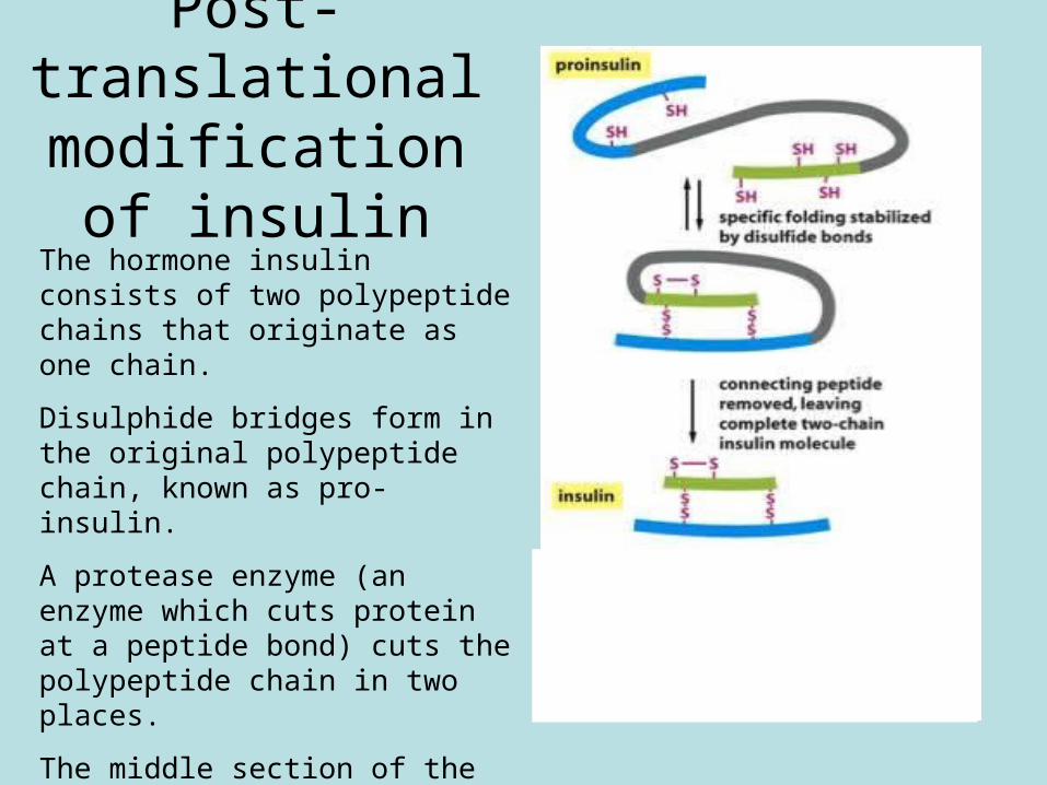
Post-translational modification of
insulinThe hormone insulin consists of two polypeptide chains that originate as one chain.
Disulphide bridges form in the original polypeptide chain, known as pro-insulin.
A protease enzyme (an enzyme which cuts protein at a peptide bond) cuts the polypeptide chain in two places.
The middle section of the protein is then removed.
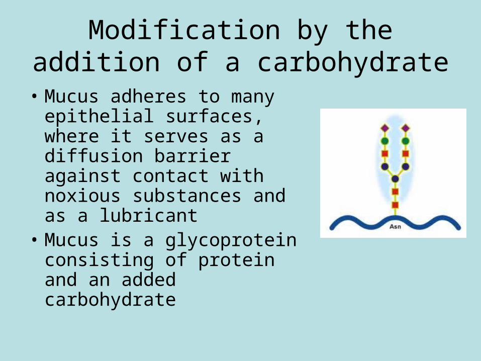
• Mucus adheres to many epithelial surfaces, where it serves as a diffusion barrier against contact with noxious substances and as a lubricant
• Mucus is a glycoprotein consisting of protein and an added carbohydrate
Modification by the addition of a carbohydrate
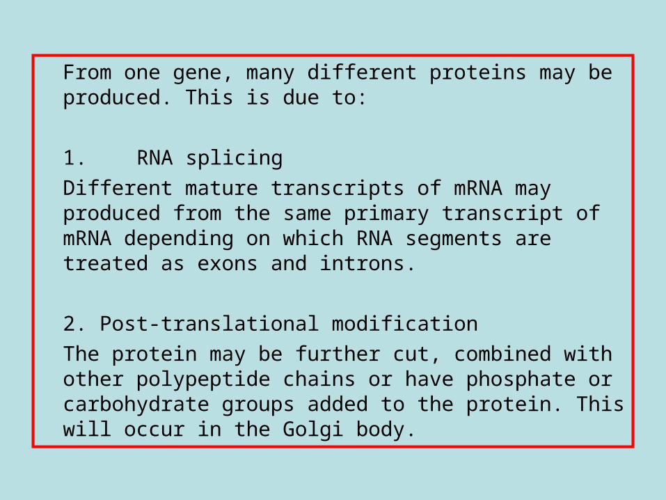
From one gene, many different proteins may be produced. This is due to:
1. RNA splicing Different mature transcripts of mRNA may produced from the same primary transcript of mRNA depending on which RNA segments are treated as exons and introns.
2. Post-translational modification The protein may be further cut, combined with other polypeptide chains or have phosphate or carbohydrate groups added to the protein. This will occur in the Golgi body.
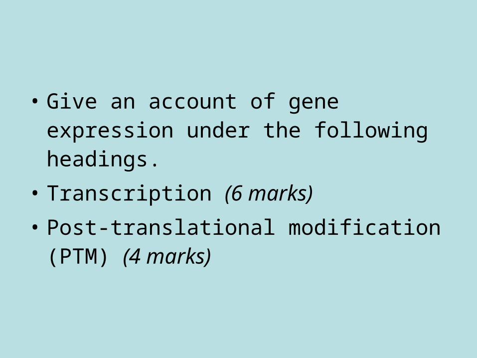
• Give an account of gene expression under the following headings.
• Transcription (6 marks)
• Post-translational modification (PTM) (4 marks)
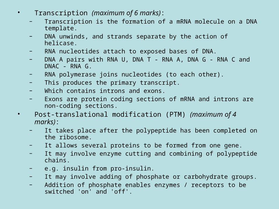
• Transcription (maximum of 6 marks):– Transcription is the formation of a mRNA molecule on a DNA
template.– DNA unwinds, and strands separate by the action of helicase.– RNA nucleotides attach to exposed bases of DNA.– DNA A pairs with RNA U, DNA T - RNA A, DNA G - RNA C and
DNAC - RNA G.– RNA polymerase joins nucleotides (to each other).– This produces the primary transcript.– Which contains introns and exons.– Exons are protein coding sections of mRNA and introns are non-
coding sections.• Post-translational modification (PTM) (maximum of 4 marks):
– It takes place after the polypeptide has been completed on the ribosome.
– It allows several proteins to be formed from one gene.– It may involve enzyme cutting and combining of polypeptide chains.– e.g. insulin from pro-insulin.– It may involve adding of phosphate or carbohydrate groups.– Addition of phosphate enables enzymes / receptors to be switched
'on' and 'off'.
Protein synthesis storyboard
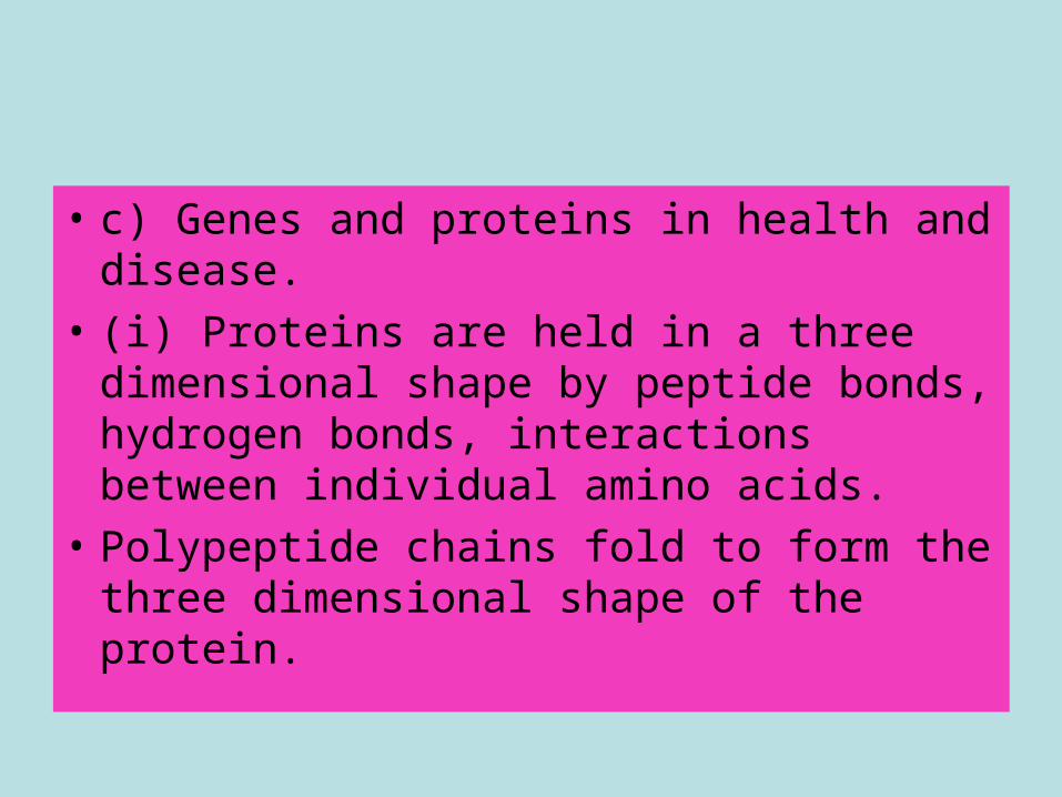
• c) Genes and proteins in health and disease.
• (i) Proteins are held in a three dimensional shape by peptide bonds, hydrogen bonds, interactions between individual amino acids.
• Polypeptide chains fold to form the three dimensional shape of the protein.

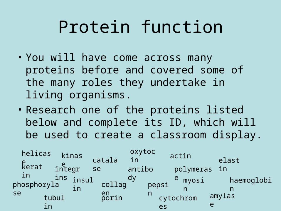
Protein function
• You will have come across many proteins before and covered some of the many roles they undertake in living organisms.
• Research one of the proteins listed below and complete its ID, which will be used to create a classroom display.
keratin
tubulin
insulin
porin
pepsin
catalase
amylase
haemoglobin
antibody
myosin
actinelastin
phosphorylase
oxytocin
cytochromes
collagen
helicase
integrins polymerase
kinase
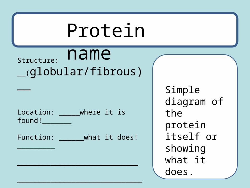
Structure:
__(globular/fibrous)__
Location: _____where it is found!_______
Function: ______what it does!_________
_____________________________
______________________________
Protein name
Simple diagram of the protein itself or showing what it does.
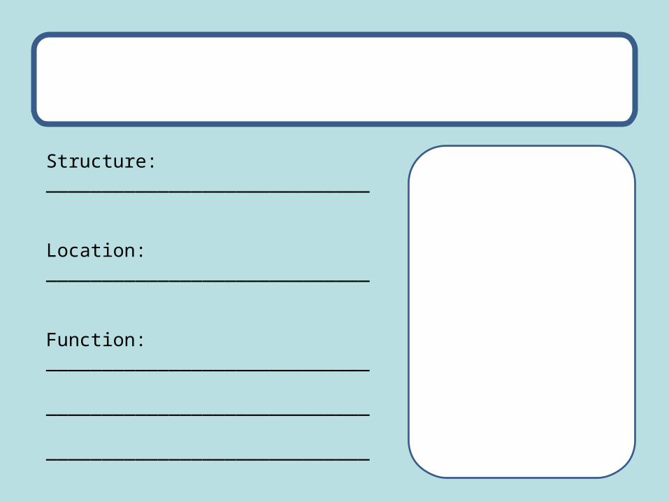
Structure: _____________________________
Location: _____________________________
Function: _____________________________
_____________________________
_____________________________
• http://courses.scholar.hw.ac.uk/vle/scholar/session.controller?action=viewContent&back=topic&contentGUID=9df68c88-6cf3-866f-f253-96ce94e448ac
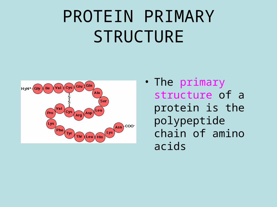
PROTEIN PRIMARY STRUCTURE
• The primary structure of a protein is the polypeptide chain of amino acids
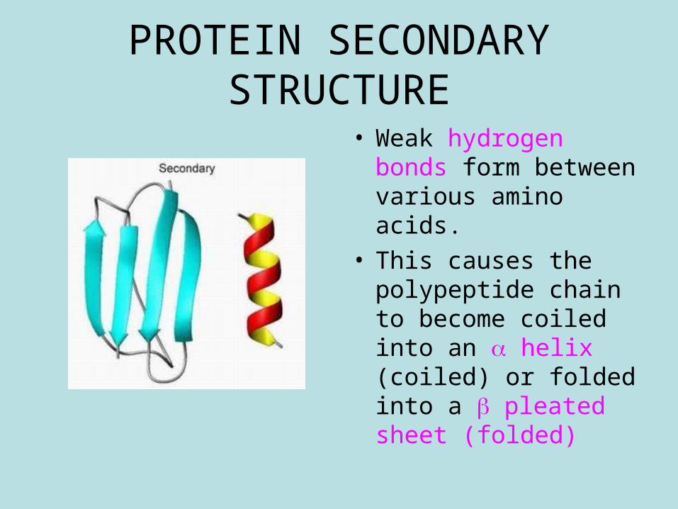
PROTEIN SECONDARY STRUCTURE
• Weak hydrogen bonds form between various amino acids.
• This causes the polypeptide chain to become coiled into an helix (coiled) or folded into a pleated sheet (folded)
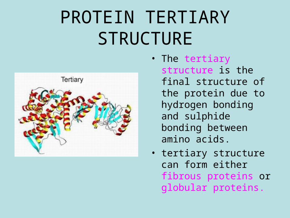
PROTEIN TERTIARY STRUCTURE
• The tertiary structure is the final structure of the protein due to hydrogen bonding and sulphide bonding between amino acids.
• tertiary structure can form either fibrous proteins or globular proteins.
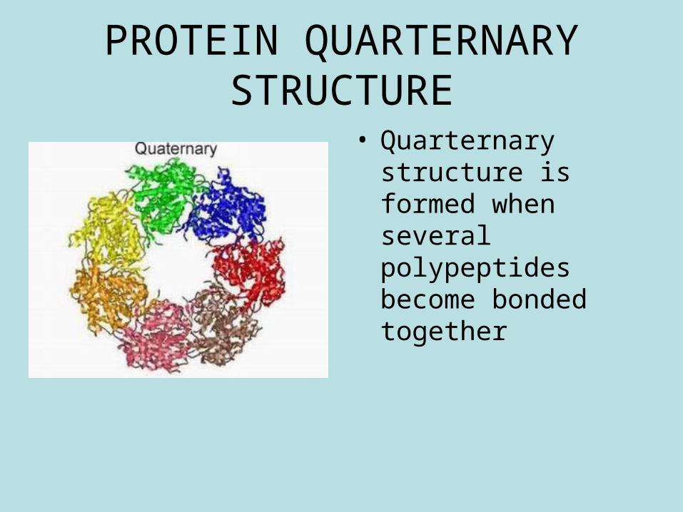
PROTEIN QUARTERNARY STRUCTURE
• Quarternary structure is formed when several polypeptides become bonded together
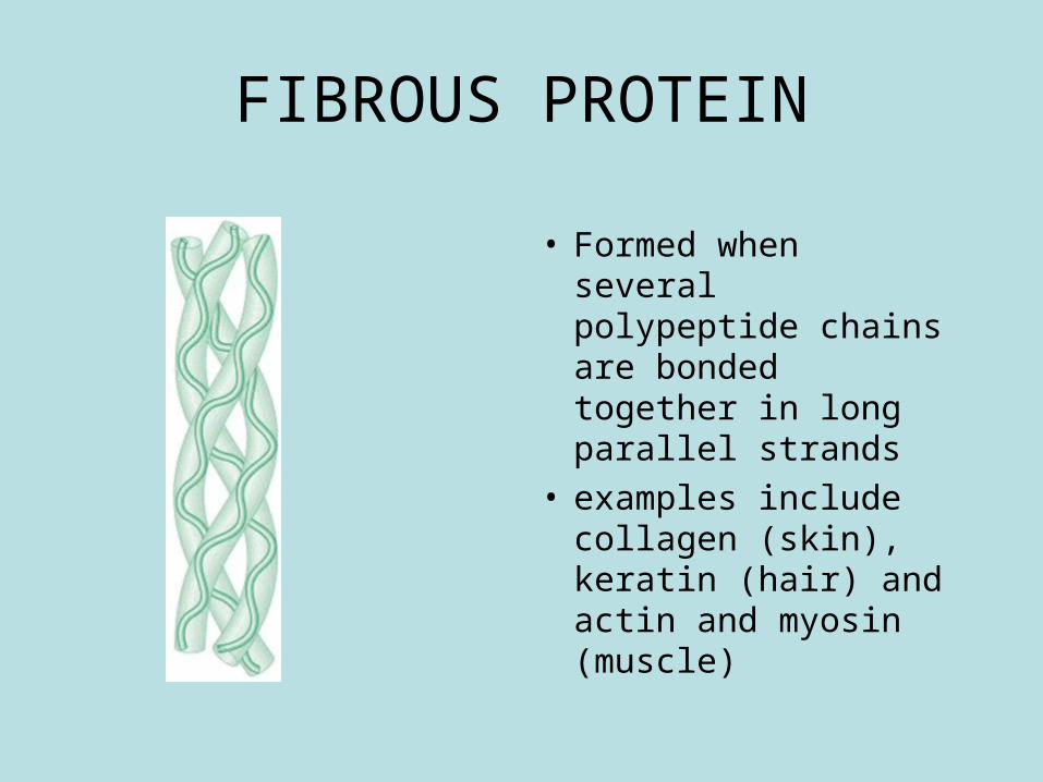
FIBROUS PROTEIN
• Formed when several polypeptide chains are bonded together in long parallel strands
• examples include collagen (skin), keratin (hair) and actin and myosin (muscle)
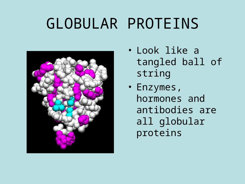
GLOBULAR PROTEINS
• Look like a tangled ball of string
• Enzymes, hormones and antibodies are all globular proteins

• http://courses.scholar.hw.ac.uk/vle/scholar/session.controller?action=viewContent&back=topic&contentGUID=2ce36382-0b0b-5ccc-ac7c-dcdd0e2453d4
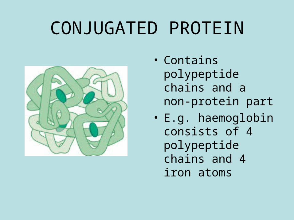
CONJUGATED PROTEIN
• Contains polypeptide chains and a non-protein part
• E.g. haemoglobin consists of 4 polypeptide chains and 4 iron atoms
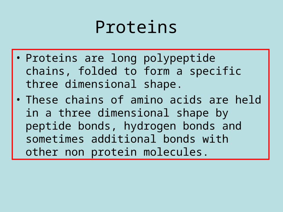
Proteins
• Proteins are long polypeptide chains, folded to form a specific three dimensional shape.
• These chains of amino acids are held in a three dimensional shape by peptide bonds, hydrogen bonds and sometimes additional bonds with other non protein molecules.
• Do protein power electrophoresis protocol
• (ii) Mutations result in no protein or a faulty protein being expressed.
• Single gene mutations involve the alteration of a DNA nucleotide sequence as a result of the substitution, insertion or deletion of nucleotides. Single-nucleotide substitutions include: missense, nonsense and splice-site mutations. Nucleotide insertions or deletions result in frame-shift mutations or an expansion of a nucleotide sequence repeat.
• The effect of these mutations on the structure and function of the protein synthesised and the resulting effects on health.
•
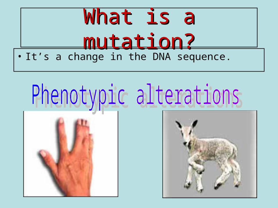
What is a mutation?What is a mutation?• It’s a change in the DNA sequence.
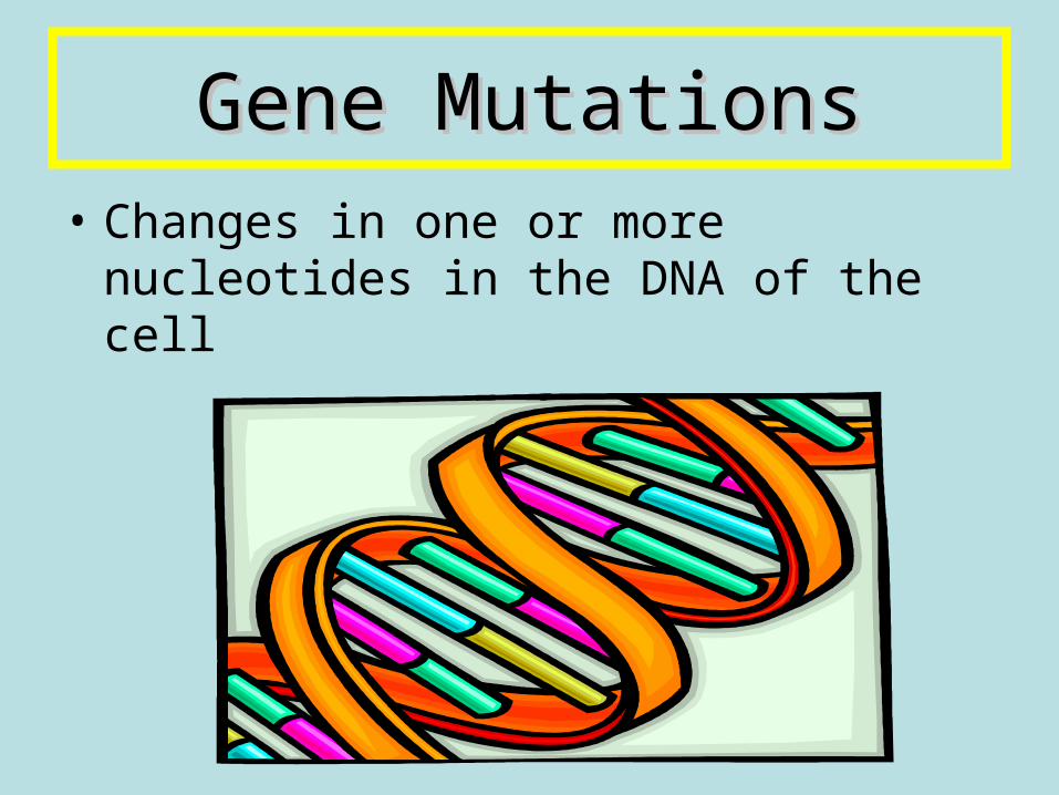
Gene MutationsGene Mutations• Changes in one or more nucleotides
in the DNA of the cell
MUTATIONS
• Occur naturally in every population• They can occur spontaneously (at
random)• An individual with a mutation is
termed a mutant
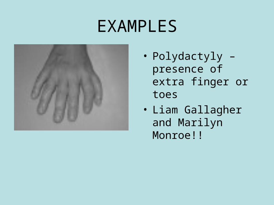
EXAMPLES
• Polydactyly – presence of extra finger or toes
• Liam Gallagher and Marilyn Monroe!!
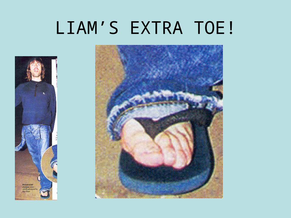
LIAM’S EXTRA TOE!
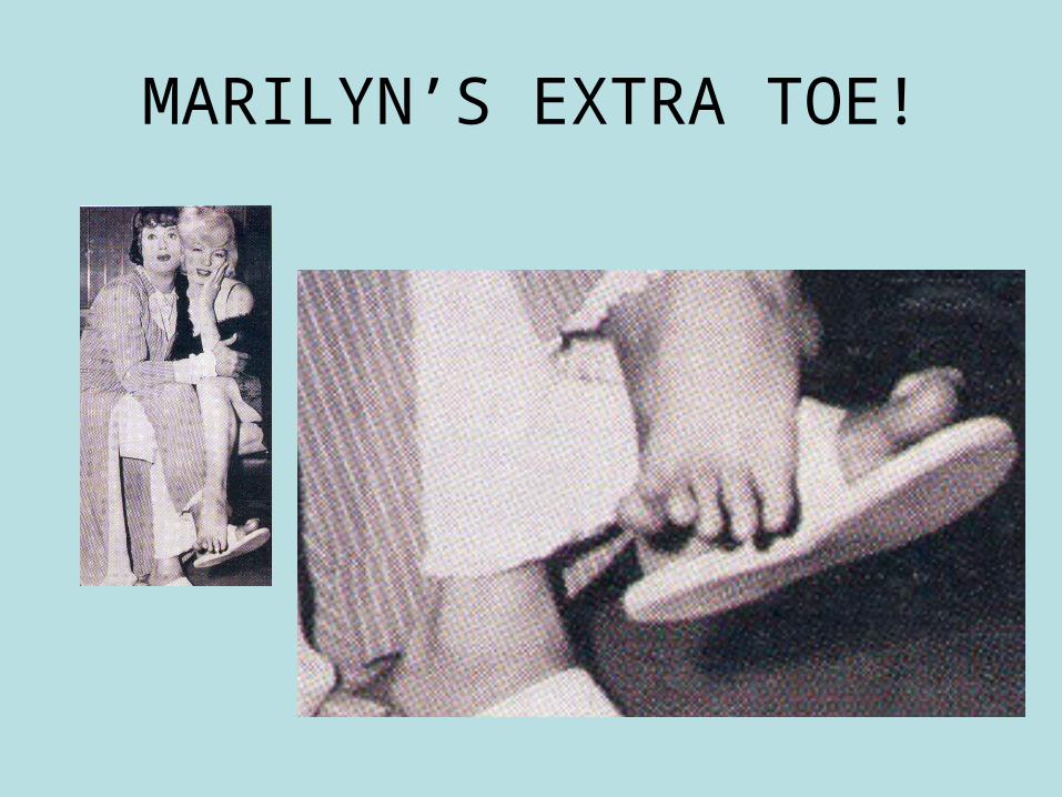
MARILYN’S EXTRA TOE!
Mutations
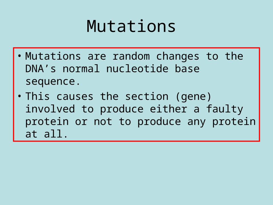
• Mutations are random changes to the DNA’s normal nucleotide base sequence.
• This causes the section (gene) involved to produce either a faulty protein or not to produce any protein at all.
• http://courses.scholar.hw.ac.uk/vle/scholar/session.controller?action=viewContent&back=topic&contentGUID=f0af9519-17b3-a64f-e476-23cff5db50e6
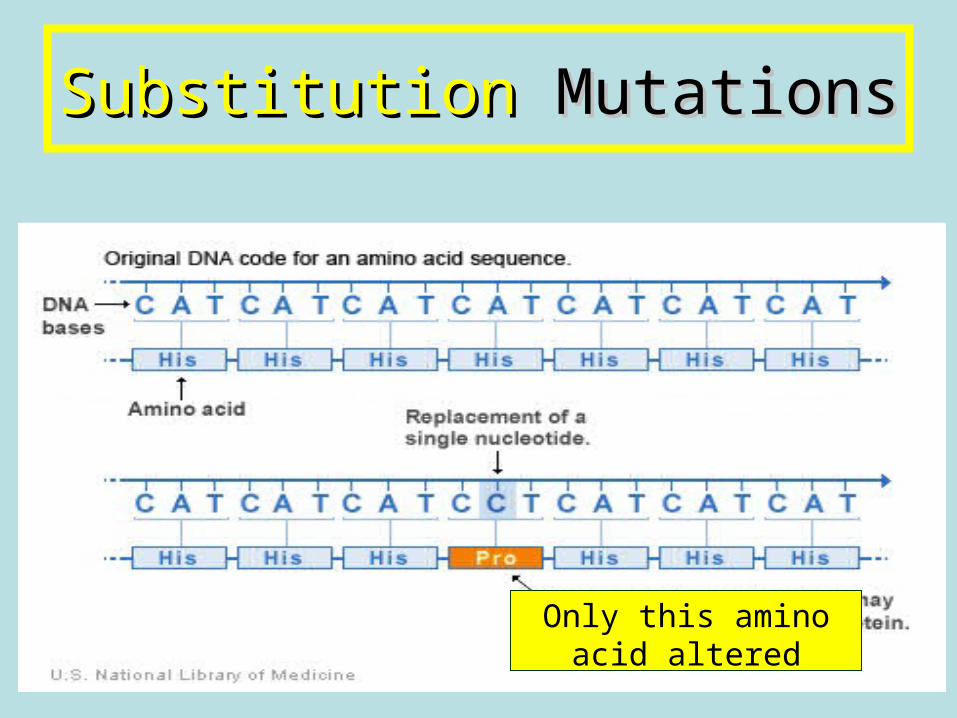
SubstitutionSubstitution Mutations Mutations
Only this amino acid altered
Mutations
• Mutations involving only one gene are called single gene mutations. There are three different types of single gene mutations:
• 1. Substitution mutations An individual nucleotide is replaced by a different nucleotide. This changes the bases in an mRNA codon.
Single nucleotide substitution mutations
• There are different types of substitution mutations, depending on where the substitution occurs:– Nonsense– Missense– Splice site
Non sense mutationsNon sense mutations
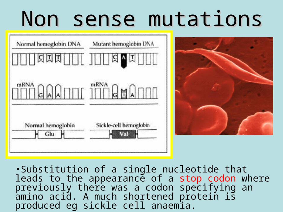
•Substitution of a single nucleotide that leads to the appearance of a stop codon where previously there was a codon specifying an amino acid. A much shortened protein is produced eg sickle cell anaemia.
Nonsense mutations
• Duchenne muscular dystrophy:• https://www.youtube.com/watch?
v=CQPLxNsN1J0&list=UUhUIp7xnLfBcNKTTjHdypfQ
• Sickle cell anaemia: • https://www.youtube.com/watch?
v=LlF_8oRs6Bw&list=UUhUIp7xnLfBcNKTTjHdypfQ
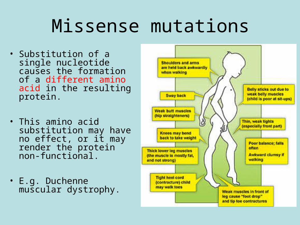
Missense mutations• Substitution of a single
nucleotide causes the formation of a different amino acid in the resulting protein.
• This amino acid substitution may have no effect, or it may render the protein non-functional.
• E.g. Duchenne muscular dystrophy.
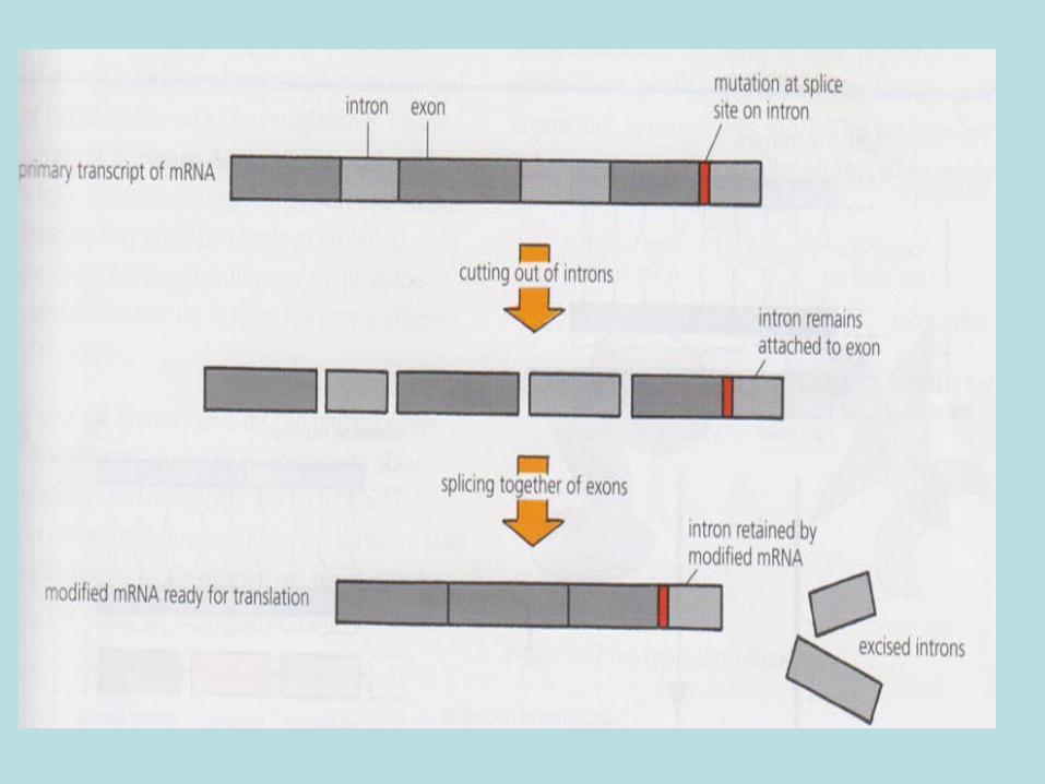
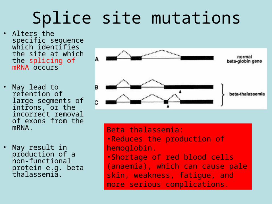
• Alters the specific sequence which identifies the site at which the splicing of mRNA occurs
• May lead to retention of large segments of introns, or the incorrect removal of exons from the mRNA.
• May result in production of a non-functional protein e.g. beta thalassemia.
Beta thalassemia: •Reduces the production of hemoglobin. •Shortage of red blood cells (anaemia), which can cause pale skin, weakness, fatigue, and more serious complications.
Splice site mutations
Substitutions mutations
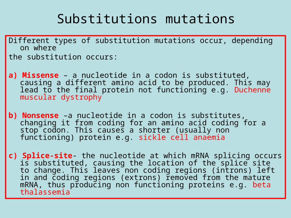
Different types of substitution mutations occur, depending on where
the substitution occurs:
a) Missense – a nucleotide in a codon is substituted, causing a different amino acid to be produced. This may lead to the final protein not functioning e.g. Duchenne muscular dystrophy
b) Nonsense –a nucleotide in a codon is substitutes, changing it from coding for an amino acid coding for a stop codon. This causes a shorter (usually non functioning) protein e.g. sickle cell anaemia
c) Splice-site- the nucleotide at which mRNA splicing occurs is substituted, causing the location of the splice site to change. This leaves non coding regions (introns) left in and coding regions (extrons) removed from the mature mRNA, thus producing non functioning proteins e.g. beta thalassemia
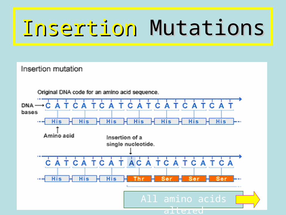
InsertionInsertion Mutations Mutations
All amino acids altered
Mutations
2. Insertion mutations
A nucleotide is added to the DNA
Nucleotide insertions cause a frame-shift mutation. This is where adding a nucleotide causes that mRNA codon and all of the codons that follow on the mRNA to change and thus produce the wrong amino acids. This leads to a very different and generally non-functional protein product. E.g. Tay-Sachs syndrome
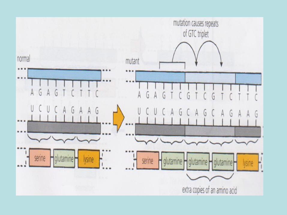
• Expansion of a nucleotide repeat:
• http://www.hhmi.org/biointeractive/tri-nucleotide-repeat
• E.g. Huntingdon’s disease:
• https://www.youtube.com/watch?v=xguyxdmeUK8&list=UUhUIp7xnLfBcNKTTjHdypfQ
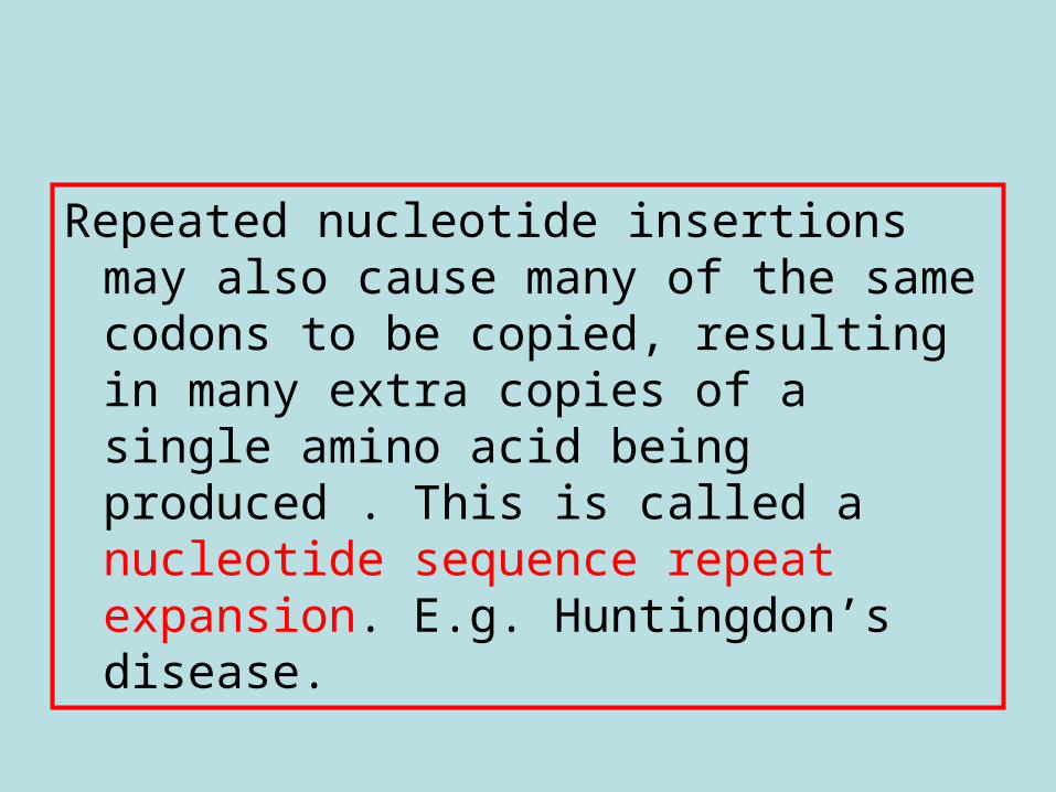
Repeated nucleotide insertions may also cause many of the same codons to be copied, resulting in many extra copies of a single amino acid being produced . This is called a nucleotide sequence repeat expansion. E.g. Huntingdon’s disease.
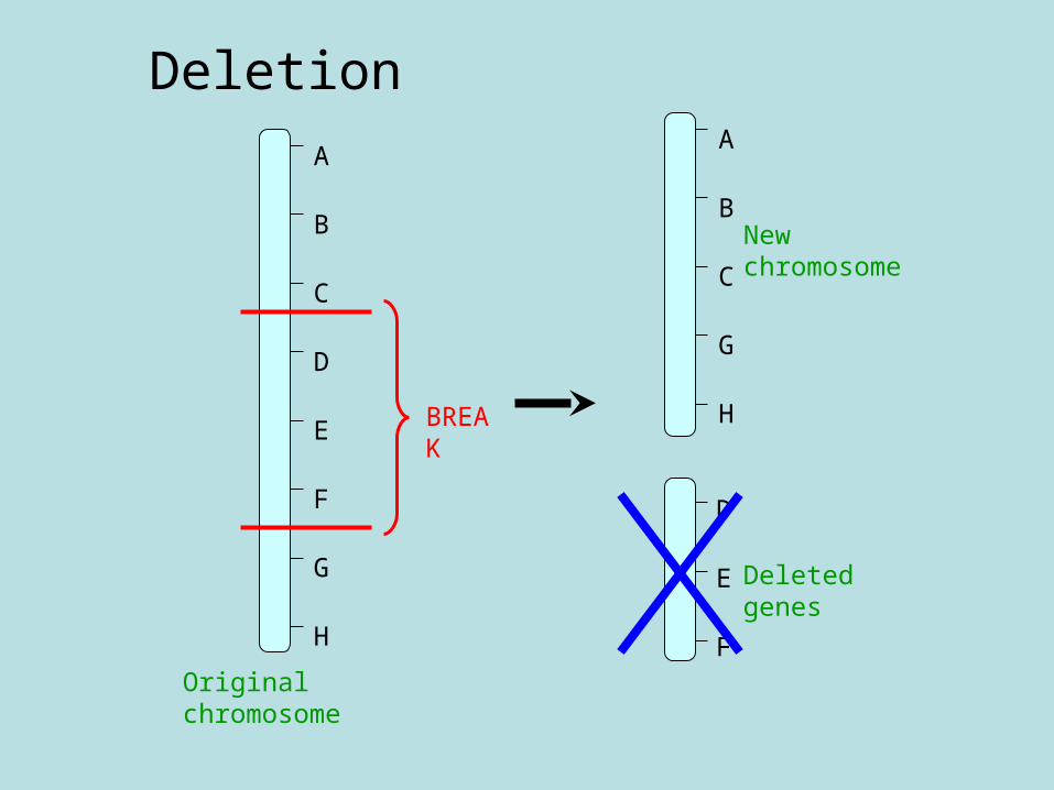
DeletionA
B
C
G
H
New chromosome
A
B
C
D
E
F
G
H
BREAK
Original chromosome
D
E
F
Deleted genes
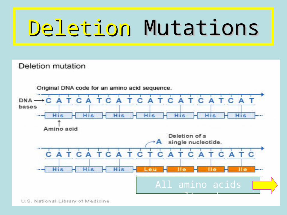
DeletionDeletion Mutations Mutations
All amino acids altered
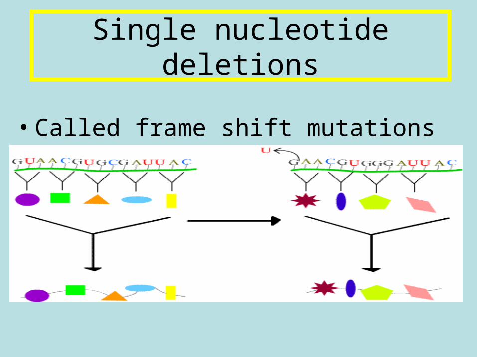
• Called frame shift mutations
Single nucleotide deletions
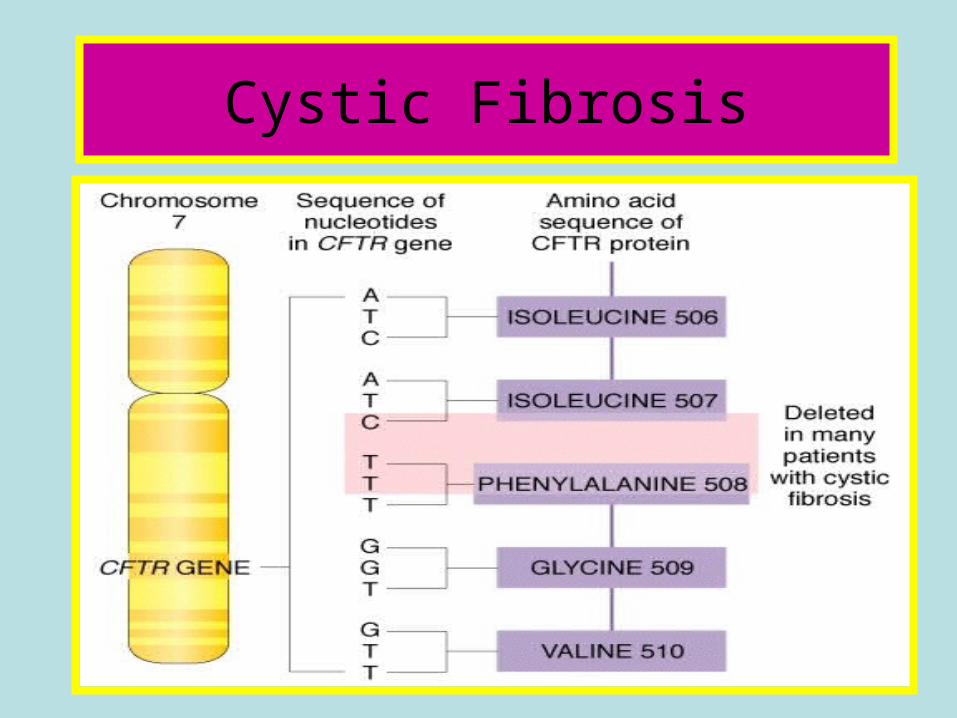
Cystic Fibrosis
Mutations
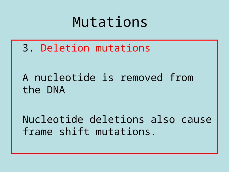
3. Deletion mutations
A nucleotide is removed from the DNA
Nucleotide deletions also cause frame shift mutations.
• Experiments investigating the effects of UV radiation on UV sensitive yeast.
Definition;
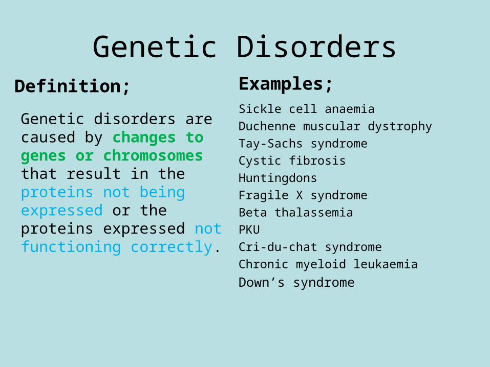
Genetic disorders are caused by changes to genes or chromosomes that result in the proteins not being expressed or the proteins expressed not functioning correctly.
Examples;Sickle cell anaemia
Duchenne muscular dystrophy
Tay-Sachs syndrome
Cystic fibrosis
Huntingdons
Fragile X syndrome
Beta thalassemia
PKU
Cri-du-chat syndrome
Chronic myeloid leukaemia
Down’s syndrome
Genetic Disorders
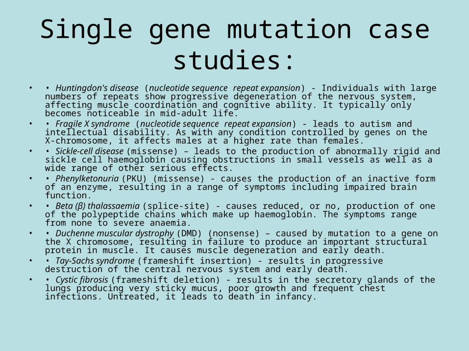
Single gene mutation case studies:
• • Huntingdon's disease (nucleotide sequence repeat expansion) - Individuals with large numbers of repeats show progressive degeneration of the nervous system, affecting muscle coordination and cognitive ability. It typically only becomes noticeable in mid-adult life.
• • Fragile X syndrome (nucleotide sequence repeat expansion) - leads to autism and intellectual disability. As with any condition controlled by genes on the X-chromosome, it affects males at a higher rate than females.
• • Sickle-cell disease (missense) – leads to the production of abnormally rigid and sickle cell haemoglobin causing obstructions in small vessels as well as a wide range of other serious effects.
• • Phenylketonuria (PKU) (missense) - causes the production of an inactive form of an enzyme, resulting in a range of symptoms including impaired brain function.
• • Beta (β) thalassaemia (splice-site) - causes reduced, or no, production of one of the polypeptide chains which make up haemoglobin. The symptoms range from none to severe anaemia.
• • Duchenne muscular dystrophy (DMD) (nonsense) – caused by mutation to a gene on the X chromosome, resulting in failure to produce an important structural protein in muscle. It causes muscle degeneration and early death.
• • Tay-Sachs syndrome (frameshift insertion) - results in progressive destruction of the central nervous system and early death.
• • Cystic fibrosis (frameshift deletion) - results in the secretory glands of the lungs producing very sticky mucus, poor growth and frequent chest infections. Untreated, it leads to death in infancy.
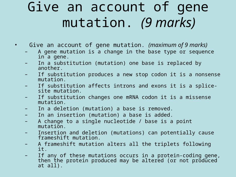
Give an account of gene mutation. (9 marks)
• Give an account of gene mutation. (maximum of 9 marks)– A gene mutation is a change in the base type or sequence in a gene.– In a substitution (mutation) one base is replaced by another.– If substitution produces a new stop codon it is a nonsense mutation.– If substitution affects introns and exons it is a splice-site mutation.– If substitution changes one mRNA codon it is a missense mutation.– In a deletion (mutation) a base is removed.– In an insertion (mutation) a base is added.– A change to a single nucleotide / base is a point mutation.– Insertion and deletion (mutations) can potentially cause frameshift
mutation.– A frameshift mutation alters all the triplets following it.– If any of these mutations occurs in a protein-coding gene, then the
protein produced may be altered (or not produced at all).
• Chromosome structure mutations — deletion; duplication; translocation.
• The substantial changes in chromosome mutations often make them lethal.
Chromosome structure mutations
• There are different types of chromosome mutations, depending on how the chromosome is altered, including:
– Deletion– Duplication– Translocation
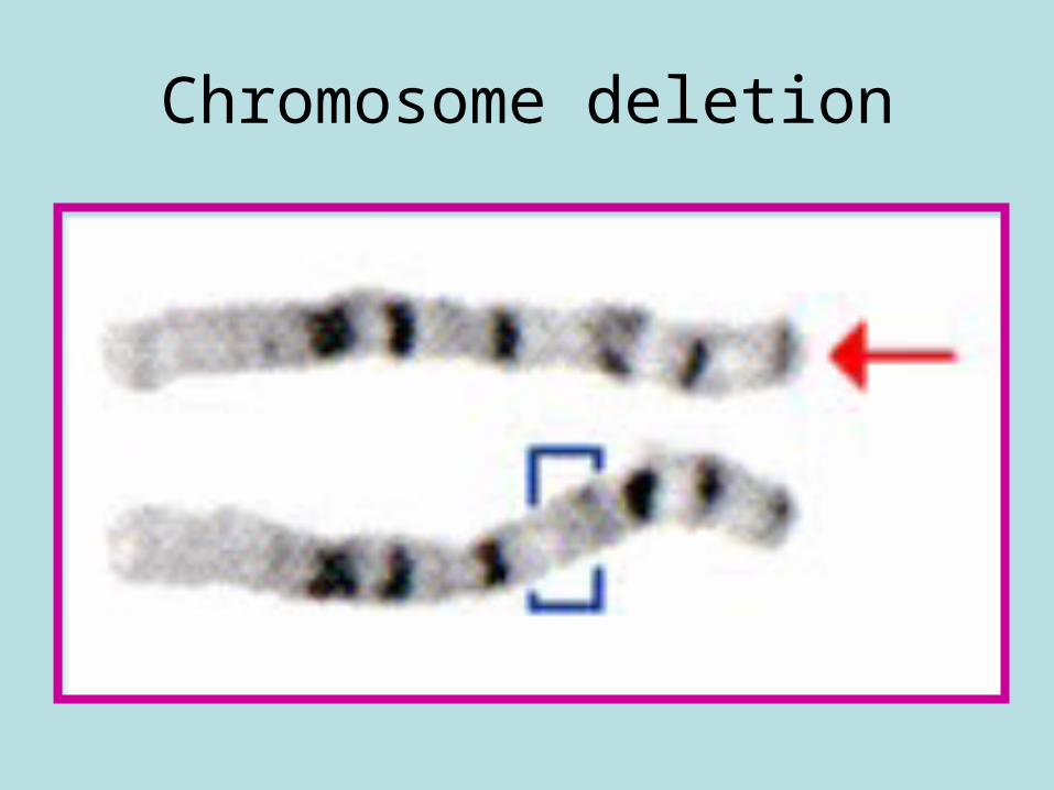
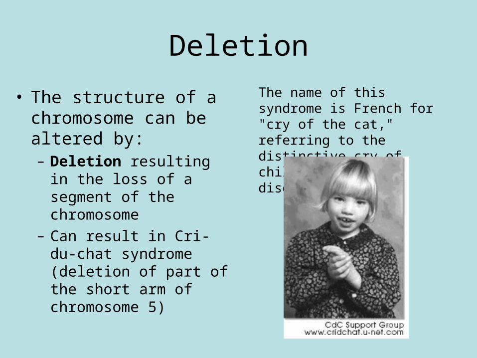
Chromosome deletion
Deletion
• The structure of a chromosome can be altered by:– Deletion resulting in the
loss of a segment of the chromosome
– Can result in Cri-du-chat syndrome (deletion of part of the short arm of chromosome 5)
The name of this syndrome is French for "cry of the cat," referring to the distinctive cry of children with this disorder.
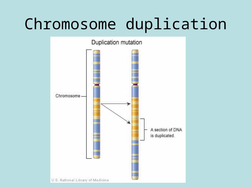
Chromosome duplication
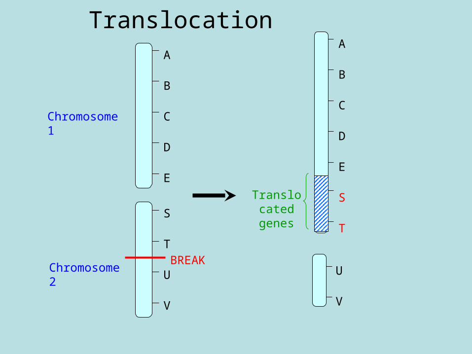
Translocation
U
V
A
B
C
D
E
S
T
Translocated
genes
A
B
C
D
E
S
T
U
V
Chromosome 1
Chromosome 2
BREAK
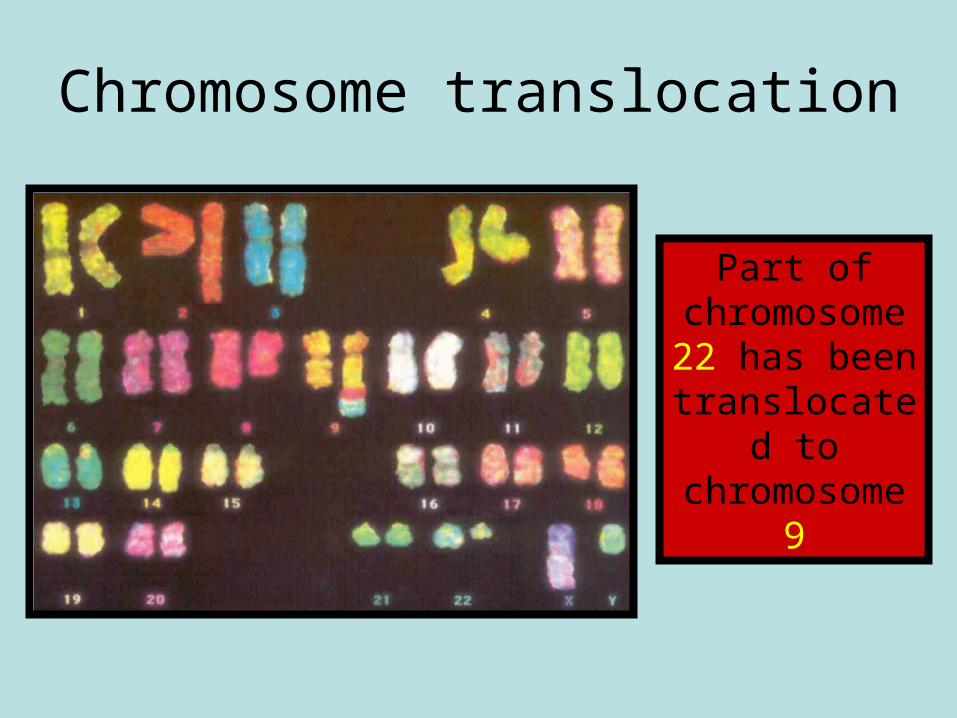
Chromosome translocation
Part of chromosome 22 has been translocated
to chromosome
9
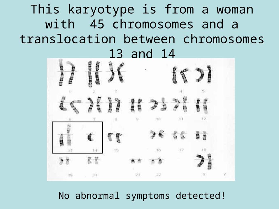
This karyotype is from a woman with 45 chromosomes and a translocation
between chromosomes 13 and 14
No abnormal symptoms detected!
Translocation sufferer film
• http://www.youtube.com/watch?v=8FGYzZOZxMw
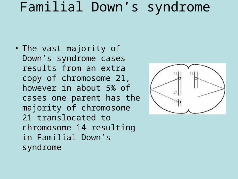
Familial Down’s syndrome
• The vast majority of Down’s syndrome cases results from an extra copy of chromosome 21, however in about 5% of cases one parent has the majority of chromosome 21 translocated to chromosome 14 resulting in Familial Down’s syndrome
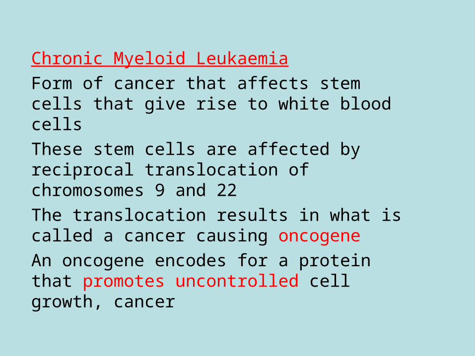
Chronic Myeloid LeukaemiaForm of cancer that affects stem cells that give rise to white blood cellsThese stem cells are affected by reciprocal translocation of chromosomes 9 and 22The translocation results in what is called a cancer causing oncogeneAn oncogene encodes for a protein that promotes uncontrolled cell growth, cancer
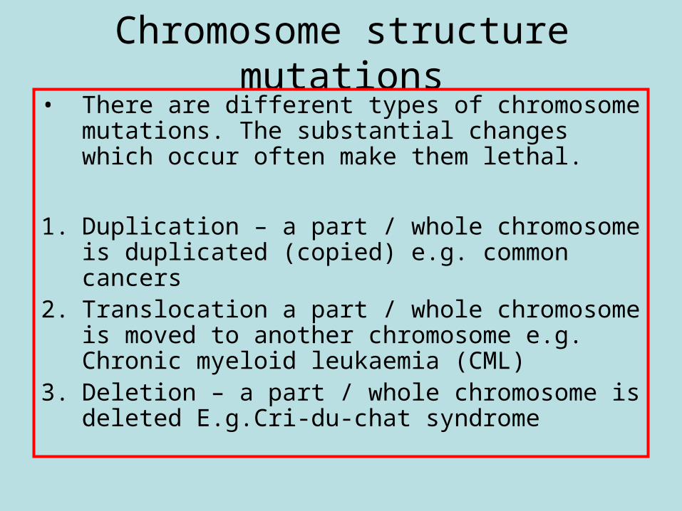
Chromosome structure mutations• There are different types of chromosome
mutations. The substantial changes which occur often make them lethal.
1. Duplication – a part / whole chromosome is duplicated (copied) e.g. common cancers
2. Translocation a part / whole chromosome is moved to another chromosome e.g. Chronic myeloid leukaemia (CML)
3. Deletion – a part / whole chromosome is deleted E.g.Cri-du-chat syndrome
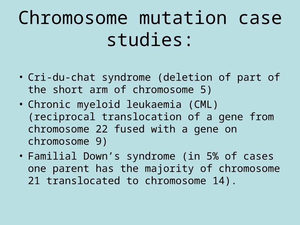
Chromosome mutation case studies:
• Cri-du-chat syndrome (deletion of part of the short arm of chromosome 5)
• Chronic myeloid leukaemia (CML) (reciprocal translocation of a gene from chromosome 22 fused with a gene on chromosome 9)
• Familial Down’s syndrome (in 5% of cases one parent has the majority of chromosome 21 translocated to chromosome 14).