Embed Size (px)
Citation preview
Chronic Kidney Disease 20
2. Guidelines for Investigation of
Chronic Kidney Disease Compilation and editing and of this volume: Dr. Chandrika Meegama(Consultant Chemical Pathologist) List of contributors Consultant Chemical Pathologists
Dr. Saroja C.Siriwardene Dr. Meliyanthi Gunatillaka Dr. Deepani Siriwardhana Dr. Eresha Jasinge
Consultant Physicians Dr. Noel Somasundaram Dr. Bandula Wijesiriwardena Dr. Rushika Lanerolle
Consultant Paediatrician Dr. Shyama De Silva
Coordinators Consultant Histopathologists
Dr. Siromi Perera Dr. Kamani Samarasinghe Dr. Modini Jayawickrama
CPSL National Guidelines / Chronic Kidney Disease 21
INDEX
CONTENT PAGE NUMBER
2.1 Introduction 22 2.2 Identification of patients with chronic kidney
disease 23 2.3 Measurement of excretory kidney function 25 2.4 Recognition of acute renal failure 33 2.5 Recognition of acute on chronic
kidney disease 34 2.6 Detection of proteinuria 34 2.7 Detection of microalbuminuria 36 2.8 Detection of haematuria 38 2.9 Renal osteodystrophy: assessment / management
in chronic kidney disease 39 2.10 Laboratory Investigations 40 2.11 Investigations in categories graded in different
levels 47 2.12 Abbreviations 48 2.13 References 49 2.14 Appendix 50
CPSL National Guidelines / Chronic Kidney Disease 22
2.1 Introduction
The Need for Sri Lankan Guidelines on Chronic Kidney Disease
Chronic kidney disease [CKD] is a major public health problem throughout the world. In Sri Lanka kidney failure is becoming increasingly common, especially in the north-central and central provinces. Applicability
These guidelines are intended to apply to adults [aged>18y] of all ages.

CPSL National Guidelines / Chronic Kidney Disease 23
2.2 Classification and Identification of Patients with Chronic Kidney Disease
This classification is based on estimated GFR, and recognizes five stages of kidney disease, as follows:
Stage 1: Normal GFR;GFR >90 mL/min/1.73m 2 with *other evidence of chronic kidney damage.
Stage 2: Mild impairment; GFR 60-89
mL/min/1.73 m 2 with *other evidence of chronic kidney damage
Stage 3: Moderate impairment; GFR 30-59 mL/min/1.73 m 2
Stage 4: Severe impairment: GFR 15-29
mL/min/1.73 m 2
Stage 5: Established renal failure (ERF): GFR <15 mL/min/1.73 m 2 or on dialysis
CPSL National Guidelines / Chronic Kidney Disease 24 *The “other evidence of chronic kidney damage”
may be one of the following:
• Persistent microalbuminuria • Persistent proteinuria
• Persistent haematuria (after exclusion of other
causes, e.g. urological disease) • Structural abnormalities of the kidneys
demonstrated on ultrasound scanning or other radiological tests, eg. polycystic kidney disease, reflux nephropathy
• Biopsy-proven chronic glomerulonephritis (most of
these patients will have microalbumuria or proteinuria, and/or haematuria)
Patients found to have a GFR of 60-89 mL/min/1.73 m 2 without one of these markers should not be considered to have CKD and should not be subjected to further investigations unless there are additional reasons to do so.
CPSL National Guidelines / Chronic Kidney Disease 25
2.3 Measurement of excretory kidney
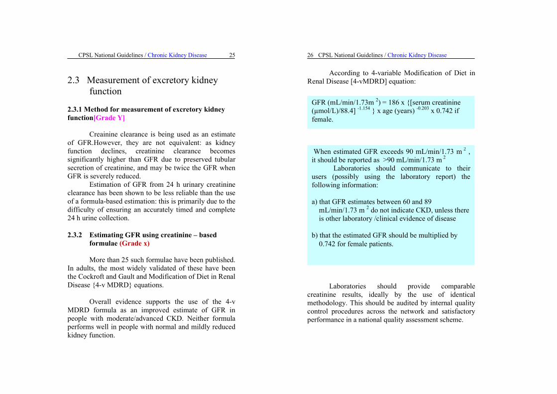
function 2.3.1 Method for measurement of excretory kidney function[Grade Y]
Creainine clearance is being used as an estimate of GFR.However, they are not equivalent: as kidney function declines, creatinine clearance becomes significantly higher than GFR due to preserved tubular secretion of creatinine, and may be twice the GFR when GFR is severely reduced.
Estimation of GFR from 24 h urinary creatinine clearance has been shown to be less reliable than the use of a formula-based estimation: this is primarily due to the difficulty of ensuring an accurately timed and complete 24 h urine collection. 2.3.2 Estimating GFR using creatinine – based
formulae (Grade x)
More than 25 such formulae have been published. In adults, the most widely validated of these have been the Cockroft and Gault and Modification of Diet in Renal Disease {4-v MDRD} equations.
Overall evidence supports the use of the 4-v
MDRD formula as an improved estimate of GFR in people with moderate/advanced CKD. Neither formula performs well in people with normal and mildly reduced kidney function.
CPSL National Guidelines / Chronic Kidney Disease 26 According to 4-variable Modification of Diet in
Renal Disease [4-vMDRD] equation:
Laboratories should provide comparable
creatinine results, ideally by the use of identical methodology. This should be audited by internal quality control procedures across the network and satisfactory performance in a national quality assessment scheme.
GFR (mL/min/1.73m 2) = 186 x {[serum creatinine (µmol/L)/88.4] -1.154 } x age (years) -0.203 x 0.742 if female.
When estimated GFR exceeds 90 mL/min/1.73 m 2 , it should be reported as >90 mL/min/1.73 m 2
Laboratories should communicate to their users (possibly using the laboratory report) the following information: a) that GFR estimates between 60 and 89
mL/min/1.73 m 2 do not indicate CKD, unless there is other laboratory /clinical evidence of disease
b) that the estimated GFR should be multiplied by
0.742 for female patients.
CPSL National Guidelines / Chronic Kidney Disease 27 Renal/pathology networks should agree a
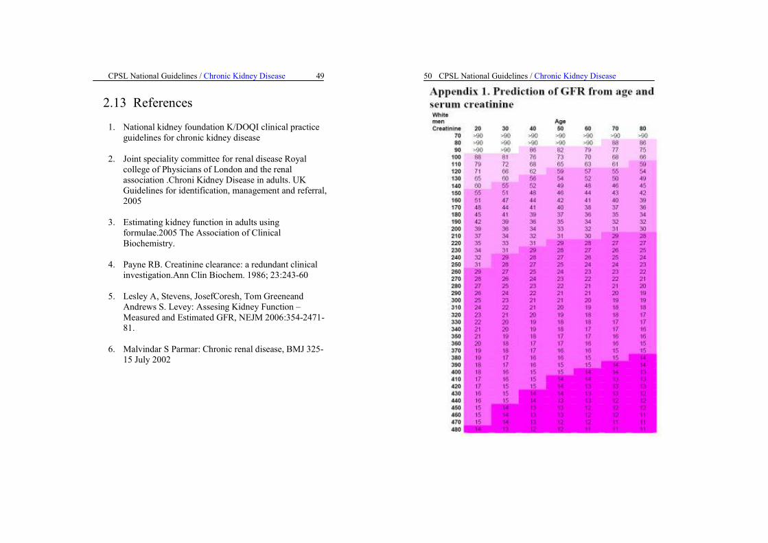
common approach to the estimation of GFR. Until these recommendations are implemented,
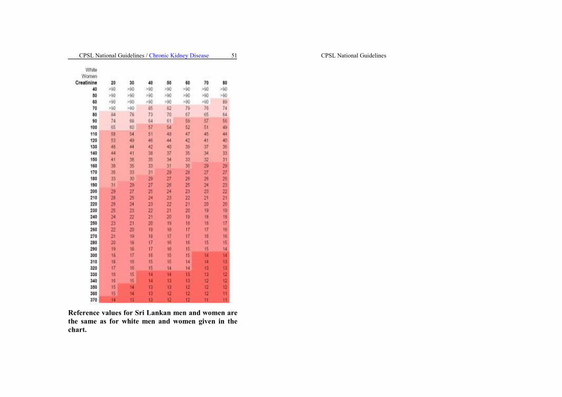
use of the prediction tables (Appendix 1) will allow estimation of GFR from age, gender, serum creatinine.
These tables give a “best case estimate” of GFR, using the lowest age and creatinine value in each cell for the calculation.
As an alternative, software systems used could
be amended to include one of these formulae and generate an estimate of GFR upon receipt of a creatinine result. However, unless this formula was used automatically every time a creatinine result was entered, this strategy would be less likely to ensure widespread use of estimated GFR. 2.3.3 When to consider creatinine clearance
measurement instead of estimated GFR [Grade x]
• Patients with unusual body habitus or diet; for
example, a person with substantial muscle wasting may have a lower GFR than suggested by the GFR estimate, even at GFR levels of less than 60ml per minute per 1.73 m2
• Patients with rapidly changing kidney function; in these patients changes in GFR estimates lag behind changes in measured creatinine clearance.
CPSL National Guidelines / Chronic Kidney Disease 28 • Patients with GFR estimates of 60ml per minute per
1.73 m2 or greater. • More accurate estimates may be necessary to
evaluate people for kidney donation, administer drugs with marked toxic effects and that are excreted by the kidneys[eg: high-dose methotrexate] or determine a person‘s eligibility for research protocols.
2.3.4 Cystatin C [Grade Z]
Cystatin C, a nonglycosylated basic protein with a low molecular mass that is freely filtered by the glomerulus, is currently under investigation as a replacement for serum creatinine in estimating the GFR. Indications for measurement of serum creatinine concentration
Serum creatinine concentration should be measured, allowing calculation of estimated GFR, at initial assessment and then at least annually in all adult patients with: previously diagnosed CKD, including
• polycystic kidney disease • reflux nephropathy • biopsy-proven chronic glomerulonephritis • persistent proteinuria • urologically unexplained persistent haematuria
CPSL National Guidelines / Chronic Kidney Disease 29
Conditions associated with a high risk of obstructive nephropathy, including
known or suspected bladder outflow obstruction
neurogenic bladder caused by spina bifida or spinal cord injury (N.B. calculated GFR may overestimate true GFR in these patients because of decreased muscle mass)
urinary diversion surgery
urinary stone disease due to primary
hyperoxaluria, cystinuria, Dent’s disease, infections (with struvite stones), anatomical abnormalities, or a stone episode rate of > 1/y
Conditions known to be associated with a high risk of silent development of CKD, including
hypertension
diabetes mellitus
heart failure
atherosclerotic coronary, cerebral, or peripheral vascular disease
CPSL National Guidelines / Chronic Kidney Disease 30
Conditions requiring long-term treatment with potentially nephrotoxic drugs, including
ACEIs and ARBs
NSAIDs
Lithium carbonate
Mesalazine and other 5-aminosalicylic
acid drugs Calcineurin inhibitors (Cyclosporin,
Tacrolimus)
Multisystem diseases that may involve the kidney, including
systemic lupuserythematosus (SLE) vasculitis myeloma rheumatoid arthritis
A first degree relative with stage 5 CKD
Frequency of measurement of serum creatinine concentration Kidney function should be measured at least annually in the risk groups outlined above. (ARF must be excluded in all patients with newly detected abnormal kidney function.)
CPSL National Guidelines / Chronic Kidney Disease 31
Minimum frequency of measurement of kidney function according to estimated GFR:
Stage 1 GFR >90 annual
Stage 2 GFR 60-89 annual
Stage 3 (known to be stable ) GFR 30-59 annual
Stage 3 (newly diagnosed or progressive) GFR 30-59 6-monthly
Stage 4 (known to be stable ) GFR 15-29 6-monthly
Stage 4 (newly diagnosed or progressive) GFR 15-29 3-monthly
Stage 5 GFR < 15 3-monthly Stable kidney function defined as change of GFR of < 2 ml/min/1.73 m 2 over 6 months or more progressive kidney damage defined as change of GFR of > 2ml/min/1.73 m 2 over 6 months or more
Kidney function should also be checked during intercurrent illness and peri-operatively in all patients with stage 2-5 CKD.
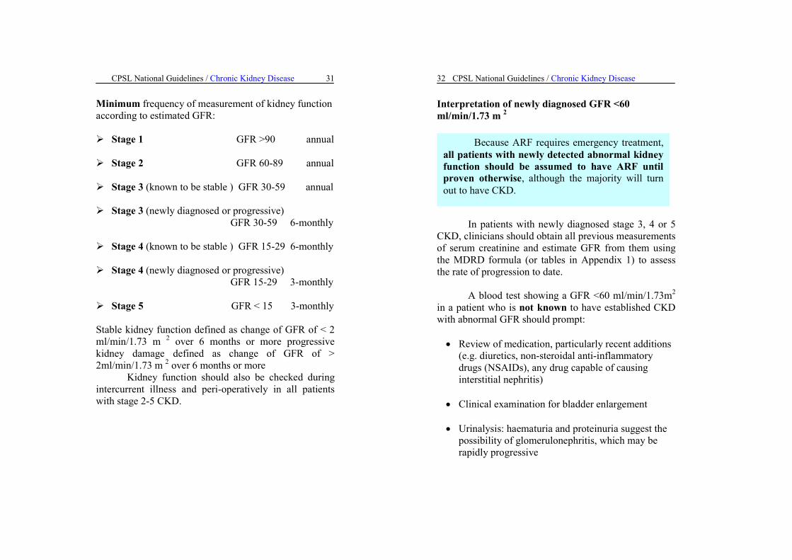
CPSL National Guidelines / Chronic Kidney Disease 32 Interpretation of newly diagnosed GFR <60 ml/min/1.73 m 2
In patients with newly diagnosed stage 3, 4 or 5
CKD, clinicians should obtain all previous measurements of serum creatinine and estimate GFR from them using the MDRD formula (or tables in Appendix 1) to assess the rate of progression to date.
A blood test showing a GFR <60 ml/min/1.73m2
in a patient who is not known to have established CKD with abnormal GFR should prompt: • Review of medication, particularly recent additions
(e.g. diuretics, non-steroidal anti-inflammatory drugs (NSAIDs), any drug capable of causing interstitial nephritis)
• Clinical examination for bladder enlargement • Urinalysis: haematuria and proteinuria suggest the
possibility of glomerulonephritis, which may be rapidly progressive
Because ARF requires emergency treatment, all patients with newly detected abnormal kidney function should be assumed to have ARF until proven otherwise, although the majority will turn out to have CKD.
CPSL National Guidelines / Chronic Kidney Disease 33 • Clinical assessment, looking for underlying
conditions such as sepsis, heart failure, hypovolaemia
• Repeat measurement of serum creatinine
concentration within a maximum of 5 days.
2.4 Recognition of Acute Renal Failure (ARF)
Formula-based estimated GFR should be
interpreted with caution in ARF, because the formulae rely on a stable serum creatinine concentration. ARF is a clinical syndrome characterised by a rapid decline in excretory function occurring over a period of hours or days. ARF should be suspected if there is a >1.5-fold rise in serum creatinine concentration, or a fall in estimated GFR of >25%, or oliguria.(defined as urine output <0.5 ml/kg/h), in the context of an acute illness. If baseline serum creatinine concentration or GFR is not known, it should be assumed that baseline GFR was 75 ml/min/1.73 m 2.
CPSL National Guidelines / Chronic Kidney Disease 34
2.5 Recognition of acute on chronic
kidney disease
A fall in estimated GFR of >25% since the last measurement of kidney function in a patient with CKD should prompt a repeat measurement of kidney function, assessment as for ARF (see preceding section) and referral if the deterioration is confirmed.
2.6 Detection of proteinuria [Grade Y]
2.6.1 Methods for detection and quantitation of proteinuria
A positive dipstick test (1+ or greater) should result in a urine sample (preferably early morning) being sent to the laboratory for confirmation by measurement of the total protein:creatinine ratio or albumin:creatinine ratio (depending on local practice).
Simultaneously, a midstream sample should be sent for culture to exclude urinary tract infection (UTI).
Urine protein:creatinine ratios >45 mg/mmol [>397mg/g creatinine] or albumin:creatinine ratios of >30mg/mmol [ >265mg/g creatinine]should be considered as positive tests for proteinuria. Positive tests for proteinuria should be followed by tests to exclude postural proteinuria, by analysis of an early morning urine sample, unless this has already been done.
CPSL National Guidelines / Chronic Kidney Disease 35
Patients with two or more positive tests for proteinuria, preferably spaced by 1 to 2 weeks, should be diagnosed as having persistent proteinuria. Indications for testing for proteinuria As part of the initial assessment of patients with;
Newly discovered GFR < 60 ml/min/1.73 m 2
Newly discovered haematuria
Newly diagnosed hypertension
Unexplained oedema
Suspected heart failure
Suspected multisystem disease, e.g. SLE, systemic vasculitis
Diabetes mellitus
As part of the annual monitoring of patients with
Biopsy-proven glomerulonephritis
Reflux nephropathy
Asymptomatic microscopic haematuria
Asymptomatic proteinuria
CPSL National Guidelines / Chronic Kidney Disease 36
Diabetes mellitus (patients with diabetes mellitus should also have annual testing for albumin:creatinine ratio if the dipstick urinalysis for protein is negative)
Monitoring for proteinuria is also required for
patients receiving treatment with gold and penicillamine.
Recommendations for frequency of monitoring are given in the British National Formulary: for penicillamine, before starting treatment and then every 1-2 weeks for the first 2 months, monthly thereafter, and in the week after any dose increase.
For intramuscular gold, before each intramuscular
injection. For oral gold, monthly. We do not recommend screening of any other
groups for proteinuria.
2.7 Detection of “microalbuminuria”[Grade Y]
2.7.1 Method for detection of
microalbuminuria
Urine albumin should be measured using an immunoassay in an early morning (preferred) or random mid-stream urine sample and expressed as an albumin:creatinine ratio.An albumin: creatinine ratio >2.5 mg/mmol [>22mg/g creatinine] in a male or >3.5 mg/mmol [>30mg/g creatinine] in a female is consistent with microalbuminuria.

CPSL National Guidelines / Chronic Kidney Disease 37 Patients demonstrating albumin:creatinine ratios
above, or equal to, this cut-off should have urine samples sent to the laboratory on two further occasions (ideally within one to three months) for albumin estimation. Patients demonstrating persistently elevated albumin: creatinine ratios in one or both of these further samples have microalbuminuria.
The diagnosis of microalbuminuria cannot be
made in the presence of an acute metabolic crisis. As far as is practicable, the best possible metabolic control of diabetes should be achieved before investigating patients for microalbuminuria. Patients should not be screened during intercurrent illness.
There is no need to exclude urinary tract infection
before diagnosing microalbuminuria unless the patient has symptoms of urinary tract infection at the time the urine sample is taken. It is important to consider other causes of increased albumin excretion, especially in the case of type 1 diabetes present for <5 years. In addition to the above caveats, these can include non-diabetic renal disease, menstrual contamination, vaginal discharge, uncontrolled hypertension, heart failure, intercurrent illness and strenuous exercise .
2.7.2 Indications for testing for microalbuminuria
Patients with diabetes mellitus who have
persistent proteinuria (as defined above) do not require testing for microalbuminuria.
CPSL National Guidelines / Chronic Kidney Disease 38 All other patients with diabetes mellitus should
undergo, as a minimum, annual testing for microalbuminuria. There is currently no proven role for screening for microalbuminuria in patients who do not have diabetes.
2.8 Detection of haematuria [Grade X]
2.8.1 Method for detection of haematuria
2.8.2 Indications for testing for haematuria
Dipstick urinalysis for blood is indicated as part
of the initial assessment of patients with • Newly detected GFR < 60 ml/min/1.73 m 2
• Newly discovered proteinuria
• Suspected multisystem disease with possible renal
involvement
Dipstick urinalysis is the test of choice for confirmation of macroscopic haematuria and for detection of microscopic haematuria. Infection, trauma, and menstruation should be excluded before confirmation of haematuria. There is no need in routine clinical practice for confirmation of haematuria by microscopy of a midstream urine sample.
CPSL National Guidelines / Chronic Kidney Disease 39 • A rise in serum creatinine concentration of >= 20%
or fall of GFR of >15% during the first 2 months after initiation of ACEI or ARB treatment.
• Unexplained hypokalaemia with hypertension.
“Screening” of unselected populations for
haematuria is not recommended.
2.9 Renal osteodystrophy: assessment/management in CKD
Antiresorptive treatment (e.g. with
bisphosphonates) for suspected or proven reduced bone mineral density should not be commenced in patients with CKD until treatable disorders of calcium, phosphate, PTH and serum 25-hydroxyvitamin D metabolism have been sought and treated.
No measurements of calcium, phosphate, or PTH
are required in stage 1 or 2 CKD unless the patient has suspected or proven reduced bone mineral density.
In stage 3 CKD, serum corrected calcium and
phosphate should be measured every 12 months. Abnormal values should be confirmed on a repeat fasting sample taken without a tourniquet. Patients with confirmed abnormalities of serum corrected calcium or phosphate should be referred to a nephrologist.
In stage 3 CKD, plasma or serum PTH should be
checked when the diagnosis of CKD stage 3 is first made.
CPSL National Guidelines / Chronic Kidney Disease 40 If the PTH is < 70 ng/L, no further checking is
required unless the patient progresses to stage 4 CKD. If the PTH is > 70 ng/L, serum 25-
hydroxyvitamin D should be checked. If the serum 25- hydroxyvitamin D is low (<80 nmol/L, 30 µg/L), therapy should be commenced with ergocalciferol or colecalciferol 800 units/day in a preparation that contains calcium carbonate or calcium lactate but not calcium phosphate; or colecalciferol 10,000 units monthly by intramuscular injection. PTH should then be rechecked after 3 months of replacement therapy. There is no need to repeat the measurement of serum 25- hydroxyvitamin D unless non-adherence or malabsorption is suspected. Vitamin D therapy should be continued long-term unless the clinical situation changes.
If the PTH is > 70 ng/L despite a normal serum
25-hydroxyvitamin D or treatment with ergocalciferol or colecalciferol, the patient should be referred to a nephrologist for specialist advice on management of hyperparathyroidism To convert PTH (ng/L) to SI units (pmol/L) multiply by 0.11
2.10 Laboratory investigations
2.10.1 Urea and Creatinine
They are waste products excreted exclusively by kidney. However the GFR has to decrease by about 40 – 60% [GFR of 30 – 60/min] for urea and creatinine to rise.
CPSL National Guidelines / Chronic Kidney Disease 41
Sample collection • Serum creatinine :
4 – 5ml of blood in a clean dry plain bottle. Separate serum from cells as early as possible Avoid haemolysed samples Avoid Cephalosporin antibiotics
• Blood urea:
3 – 4ml of blood in a clean dry plain bottle.
2.10.2 Serum electrolytes Serum Sodium
Sodium balance is maintained until the GFR falls to very low levels [5ml/min ] and is reflected by an increase in renal fractional excretion of sodium [FE Na] FE Na% = UNa x SCr x 100
SNa x Ucr UNa = urinary Na SNa = Serum Na Scr = Serum creatinine Ucr = urinary creatinine
In normal subjects the FENa% is<1%, during progreesive CKD it will increase up to a maximum of 20%. Artefactual and clinically non significant hyponatraemia; Blood taken from a drip arm [5% dextrose drip]
CPSL National Guidelines / Chronic Kidney Disease 42 Hyperglycaemia patient, diabetes mellitus or i/v infusion of glucose causing dilutional hyponatraemia in the ECF.
Hyperlipidaemia or increase in globulins
[myeloma, macroglobulinaemia] lower Na due to decrease in plasma water. Serum Potassium
K balance is maintained until the GFR falls below about 20ml/min.[serum creatinine 300-350µmol/L] Specimen Collection for serum electrolytes:
• 3-4ml of blood is collected to clean, dry plain bottle.
• Blood should not be collected from a drip arm. • Avoid muscle activity [clenching the fist ] when
collecting blood samples for electrolytes. Blood specimens should not be chilled before separation of the serum.
Factitious hyperkalaemia is common due to- Haemolysis Specimen more than 6hrs old Specimen contaminated with K-
EDTA
CPSL National Guidelines / Chronic Kidney Disease 43
2.10.3 Serum Ca and Phosphate
In chronic renal failure there is a tendency
towards a negative calcium balance and hypocalcaemia. Specimen collection and storage Serum Calcium
Serum is the preferred specimen although heparinized plasma is acceptable. 3-4 ml of blood without applying the tourniquet into acid washed bottles. Fasting specimen is preferred.
Serum should be promptly separated from the cells.Haemolyzed samples are unacceptable.
Centrifuged samples are stored at room temperature for up to 8hrs, at 40c for one day and frozen for up to 1 year. Serum Phosphate
Phosphate balance is maintained until the GFR falls below 10-20ml/min[s. creatinine of 300-400µmol/L].
Blood; 3-4 ml blood, fresh blood sample. Preferably fasting Haemolyzed samples unacceptable. Serum should be promptly separated from the
cells.
A true serum K of 6.5 mmol/L is a cause for concern and cardiac arrest can occur in patients at a K value of 8.5mmol/l.True serum K seldom rises above 9mmol/L.
CPSL National Guidelines / Chronic Kidney Disease 44
2.10.4 Acid base Hydrogen ions and plasma bicarbonate
Normal excretion of H ions and bicarbonate are maintained until the GFR falls to below 30ml/min[creat. 300µmol/ L] with a further decrease in GFR H ions retained and bicarbonate progressively falls to a level of 12 – 15mmol/L. Specimen collection of blood gas analysis
Whole blood is the preferred specimen. Specimen is collected anaerobically with lyophilized heparin anticoagulant in sterile syringe with capacities of 4-5ml.
Adequate anticoagulant[0.05mg heparin/ml blood] Anion Gap
As the GFR decreases anions such as sulphate.phosphate etc. will be retained in the blood and along with the positive hydrogen balance will result in a high anion gap metabolic acidosis A gap [mEq/L] = [Na mEq/L + K mEq/L] – [ Cl m Eq/L + Hco3. mEq/L]
2.10.5 Urinary Sediment
An important evaluation is urine analysis particularly the search for red blood cells,cellular debris and casts [ haem,granular] if any one of these is present
CPSL National Guidelines / Chronic Kidney Disease 45
the diagnosis is most likely to be acute tubular necrosis. In the normal subject and in uncomplicated pre-renal uraemia these substances are usually absent from the urine.
2.10.6 Urinary microalbumin
Urine albumin excretion is increased by physiological factors [Eg; exercise, posture]
The method of urine collection should be standardized.
Samples should not be collected after exertion, in the presence of urinary tract infections,during acute illness, immediately after surgery or after an acute fluid load.
The following urine samples are accepted for the microalbumin measurement ⇒ 24-hr urine collection. ⇒ Overnight [8-12hrs]collection ⇒ 1-2hr collection [in labor clinic] ⇒ First morning sample for simultaneous measurement
of albumin and creatinine
CPSL National Guidelines / Chronic Kidney Disease 46
The timed specimens are the most sensitive, but the albumin :creatinine ratio is practical and convenient for the patient.
2.10.7 PTH assays
Clotted blood samples sent for PTH assay need to
be separated rapidly and kept on ice. PTH can be done in plasma, blood samples anticoagulated with EDTA.
2.10.8 Creatinine clearance
24hr urine specimen and a blood sample for
creatinine [4ml of blood taken during the 24hr period of urine collection.] Avoid cephalosporin antibiotics.
24hr collection of urine: Empty bladder completely and discard the urine, note the time, from that time onwards collect the sample into a beaker and transfer into a bottle [2,5L] with preservative until 24hrs completed.
Preservative – 10ml of 6mol/L per 24hr excretion.
2.10.9 Specimen collection for ionized calcium
Specimens for ionized calcium must be transported in ice-water and analyzed within 30 minutes of collection to minimize pH change due to Co2 loss and glycolysis.
CPSL National Guidelines / Chronic Kidney Disease 47
2.11 Investigations in Categories Graded in Different Levels
Grade X – very basic Grade Y – desirable Grade Z - optional Grade X Blood urea Creatinine Serum electrolytes Urinary sediment
Serum calcium Serum phosphate Acid base Anion gap Creatinine clearance Estimated GFR by equations or By using tables Grade Y Urinary microalbumin Urinary protein: creatinine ratio Urinary alb:creatinine ratio Grade Z PTH assays 25 – hydroxyvitamin D Cystatin C
CPSL National Guidelines / Chronic Kidney Disease 48
2.12 Abbreviations ACEI Angiotensin Converting Enzyme Inhibitor AER Albumin Excretion Rate ARB Angiotensin Receptor Blocker ARF Acute Renal Failure BMD Bone Mineral Density BSA Body Surface Area CHD Coronary Heart Disease CKD Chronic Kidney Disease CT Computed Tomography CVD Cardiovascular Disease EDTA Ethylenediaminetetraacetic acid ERF Established Renal Failure ESA Erythropoiesis Stimulating Agent GFR Glomerular Filtration Rate HOT Hypertension Optimal Treatment K/DOQI Kidney Disease Outcomes Quality Initiative LVH Left Ventricular Hypertrophy MDRD Modification of Diet in Renal Disease MR Magnetic Resonance NHS National Health Service NICE National Institute for Clinical Excellence NSAID Non-Steroidal Anti-Inflammatory Drug PTH Parathyroid Hormone RAS Renin Angiotensin System RRT Renal Replacement Therapy SLE Systemic Lupus Erythematosus UTI Urinary Tract Infection
CPSL National Guidelines / Chronic Kidney Disease 49
2.13 References
1. National kidney foundation K/DOQI clinical practice guidelines for chronic kidney disease
2. Joint speciality committee for renal disease Royal
college of Physicians of London and the renal association .Chroni Kidney Disease in adults. UK Guidelines for identification, management and referral, 2005
3. Estimating kidney function in adults using
formulae.2005 The Association of Clinical Biochemistry.
4. Payne RB. Creatinine clearance: a redundant clinical
investigation.Ann Clin Biochem. 1986; 23:243-60
5. Lesley A, Stevens, JosefCoresh, Tom Greeneand Andrews S. Levey: Assesing Kidney Function – Measured and Estimated GFR, NEJM 2006:354-2471-81.
6. Malvindar S Parmar: Chronic renal disease, BMJ 325-
15 July 2002
CPSL National Guidelines / Chronic Kidney Disease 50
CPSL National Guidelines / Chronic Kidney Disease 51
Reference values for Sri Lankan men and women are the same as for white men and women given in the chart.
CPSL National Guidelines