Embed Size (px)
Citation preview
2
Chapter 5
The Human Body

3
Objectives
• There are no 1985 objectives for this chapter.
4
Introduction
• Anatomy– The study of structures and components of an
organism• Physiology
– The study of the body functions of a living organism• Pathophysiology
– The study of the body functions of a living organism in an abnormal state
5
The Structure of the Human Body (1 of 3)
• Cells – Most basic component of an organism
• Tissues– A group of similar cells working together to perform a
common function• Organs
– Different types of tissues working together to perform a particular function
6
The Structure of the Human Body (2 of 3)
• Organ systems– Groups of organs that work together– May be located together or apart – Combined, they form an organism– Carry out vital functions

7
The Structure of the Human Body (3 of 3)
• Organ systems include:– Musculoskeletal, circulatory, respiratory, nervous,
gastrointestinal, urinary, reproductive, immune, endocrine, lymphatic, integumentary, and special sensory
• Homeostasis– Balanced internal environment– System of checks and balances
8
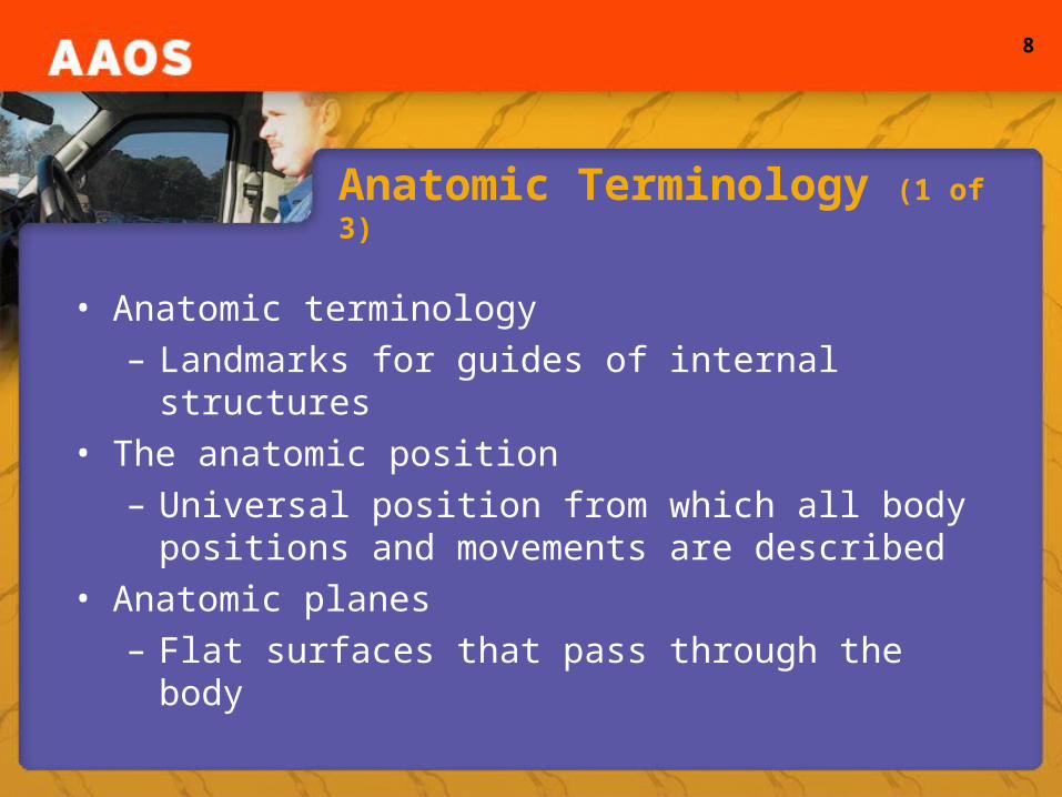
Anatomic Terminology (1 of 3)
• Anatomic terminology– Landmarks for guides of internal structures
• The anatomic position– Universal position from which all body positions and
movements are described • Anatomic planes
– Flat surfaces that pass through the body
9
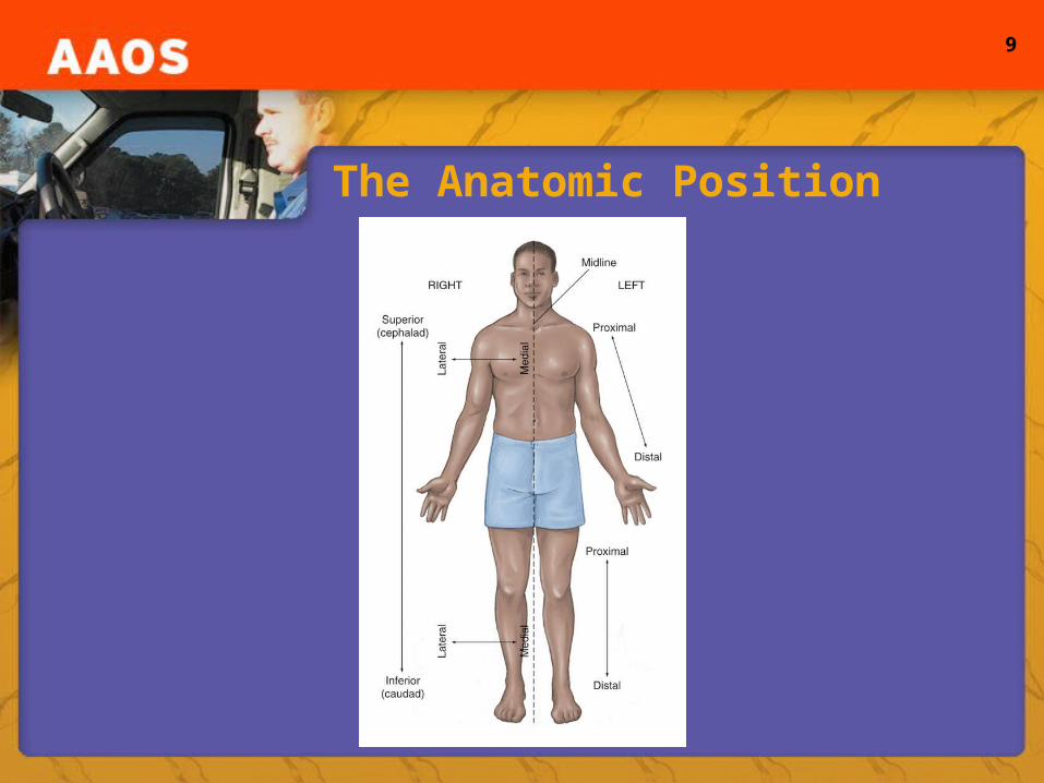
The Anatomic Position
10
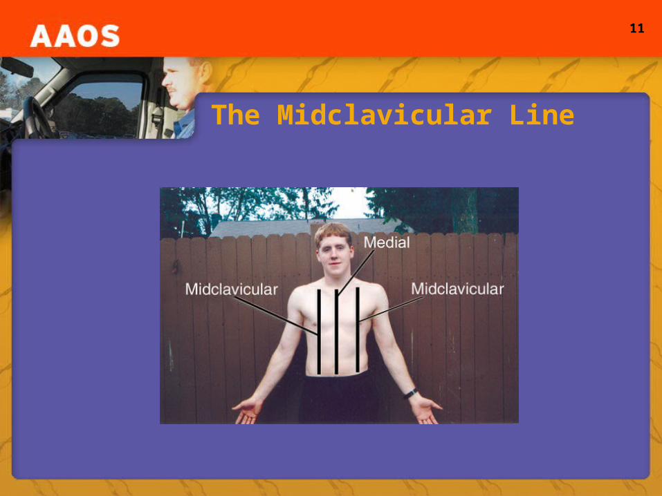
• Proximal and distal• Midline• Midaxillary line• Midclavicular line
Anatomic Terminology (2 of 3)
• Frontal plane– Anterior and posterior
• Transverse plane– Cranial and cephalad
• Median plane– Medial and lateral
• Sagittal plane
11
The Midclavicular Line

12
• Other Directional Terms– Bilateral– Contralateral– Ipsilateral
Anatomic Terminology (3 of 3)
• Directional Terms– Right and left– Superior and inferior– Superficial and deep– Ventral and dorsal– Palmar and plantar– Apex
13
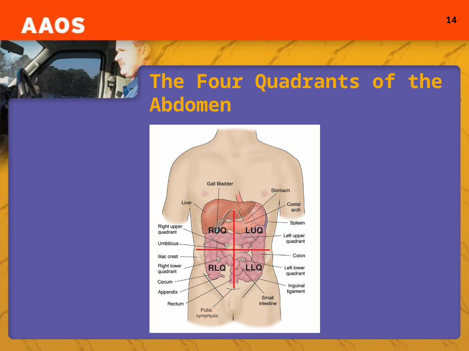
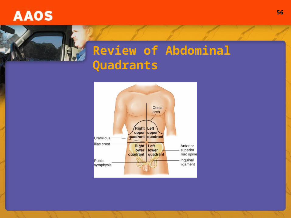
Abdominal Quadrants
• Abdomen– Two imaginary lines divide this area into four parts– Inferior tip of sternum to the genital area; iliac crest
across the umbilicus– Right upper quadrant, left upper quadrant, right lower
quadrant, left lower quadrant– Each quadrant contains specific organs
14
The Four Quadrants of the Abdomen
15
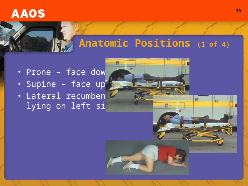
Anatomic Positions (1 of 4)
• Prone – face down • Supine – face up• Lateral recumbent –
lying on left side
16
Anatomic Positions (2 of 4)
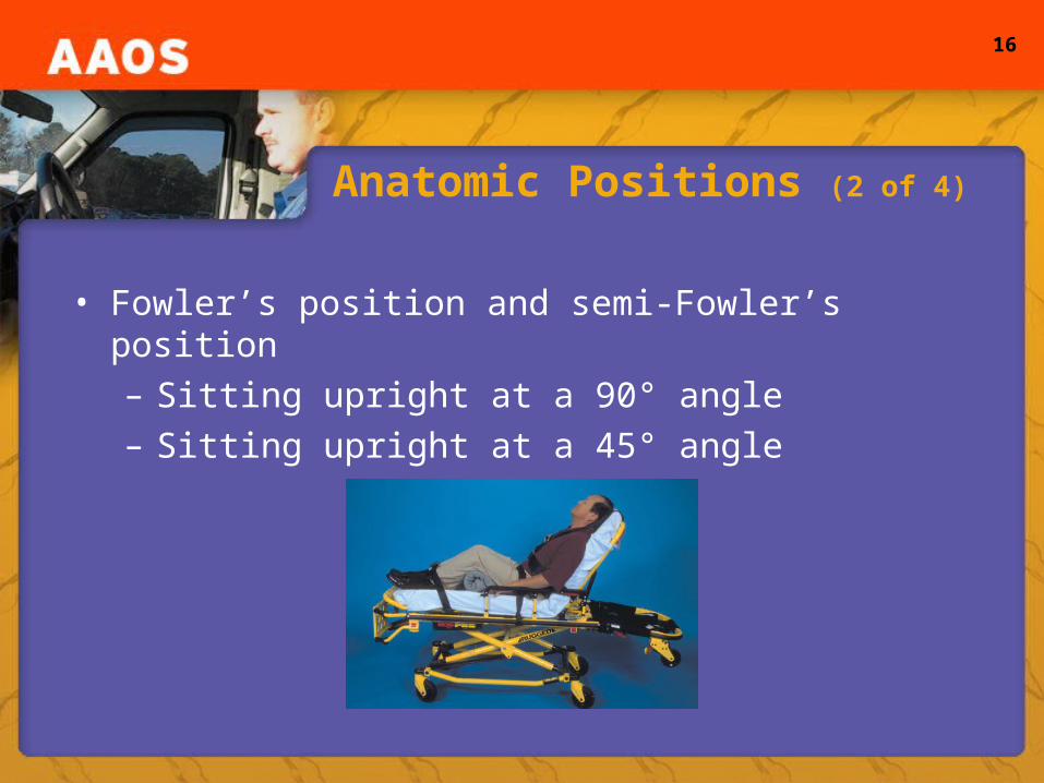
• Fowler’s position and semi-Fowler’s position– Sitting upright at a 90° angle– Sitting upright at a 45° angle
17
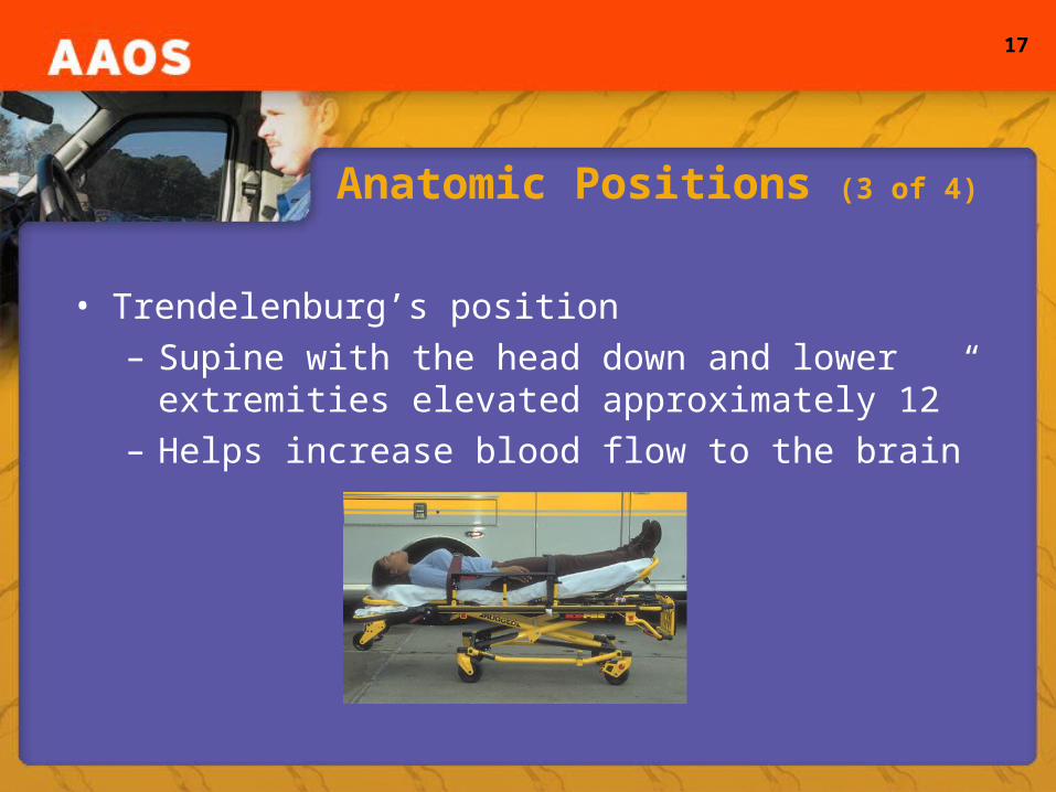
Anatomic Positions (3 of 4)
• Trendelenburg’s position– Supine with the head down and lower extremities
elevated approximately 12”– Helps increase blood flow to the brain
18
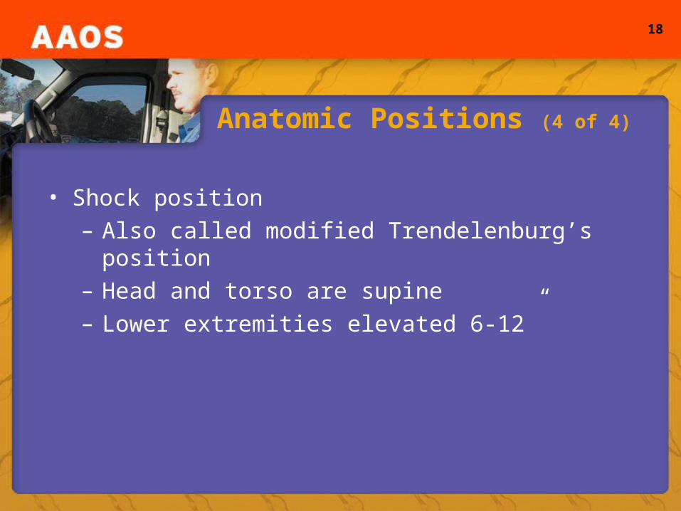
Anatomic Positions (4 of 4)
• Shock position– Also called modified Trendelenburg’s position– Head and torso are supine– Lower extremities elevated 6-12”
19
Movements and Positions (1 of 2)
• Movements– From simple to complicated, movements can be
broken down into a series of components and described with specific terms
• Range of Motion (ROM)– Full distance that a joint can be moved– Flexion
• Moving a distal part of an extremity toward the trunk
20
Movements and Positions (2 of 2)
• ROM– Extension– “Hyper”
• Supination and pronation• Internal and external rotation• Abduction and adduction
21
Cellular Transport Mechanisms
• Permeability of the cell membrane– Selective permeability
• Allows some substances to pass, but not others• Maintains environments
– Electrolytes• Chemicals dissolved in blood, made of salt or acid
substances that become iconic conductors when dissolved in a solvent
22
Diffusion
• Diffusion– The movement of solutes, which are particles such
as salts, dissolved in a solvent• Concentration gradient
– Difference in concentrations of the substance on either side of the membrane
– Small molecules diffuse more easily than large ones; watery solutions diffuse faster than viscous solutions
23
Osmosis
• Osmosis– The movement of a solvent, such as water, from an
area of low solute concentration to high concentration• Osmotic pressure
– Measure of the tendency of water to move by osmosis across a membrane
• Crenation• Lysis
24
Permeability of the Cell Membrane
• Facilitated diffusion– The process in which a carrier molecule moves
substances in or out of cells from high to low concentration
• Active transport– The movement of a substance against a
concentration or gradient
25
Endocytosis and Exocytosis
• Endocytosis– Uptake of material through the cell membrane by a
membrane-bound droplet that forms within the protoplasm of the cell
– Phagocytosis or “cell eating”– Pinocytosis or “cell drinking”
• Exocytosis– The release of secretions from the cells
26
Cellular Metabolism
• Metabolism– The sum of all the physical and chemical processes
that produce and maintain the body– Two phases:
• Anabolism or “building phase”• Catabolism or “break down phase”
– Body’s fuel• Carbohydrates (mostly glucose), fats, and proteins
27
Cellular Respiration
• Respiration– Process of using glucose, fat, and proteins to
generate energy– Creates energy in the form of adenosine triphosphate
(ATP)– Takes place within the mitochondria– Krebs cycle– Glycolysis

28
Tissues (1 of 4)
• Epithelial tissue and glands– Type of tissue that covers all external body surfaces
and forms secreting portions of glands– Lines hollow organs in the body– Provides a protective barrier – Functions in absorption of nutrients– Functions in secretion of body substances
29
Tissues (2 of 4)
• Connective tissue– Connects other types of tissue together– Extracellular matrix – Adipose tissue
• Contains large amounts of lipids
– Other types of connective tissues• Aid in formation of blood vessels• Part of the body’s self-defenses
30
Tissues (3 of 4)
• Muscle Tissue– Located within the substance of the body and
invariably enclosed by connective tissue– Classified by structure and function– Structural
• Striated or nonstriated
– Function• Voluntary and involuntary
– Cardiac muscle
31
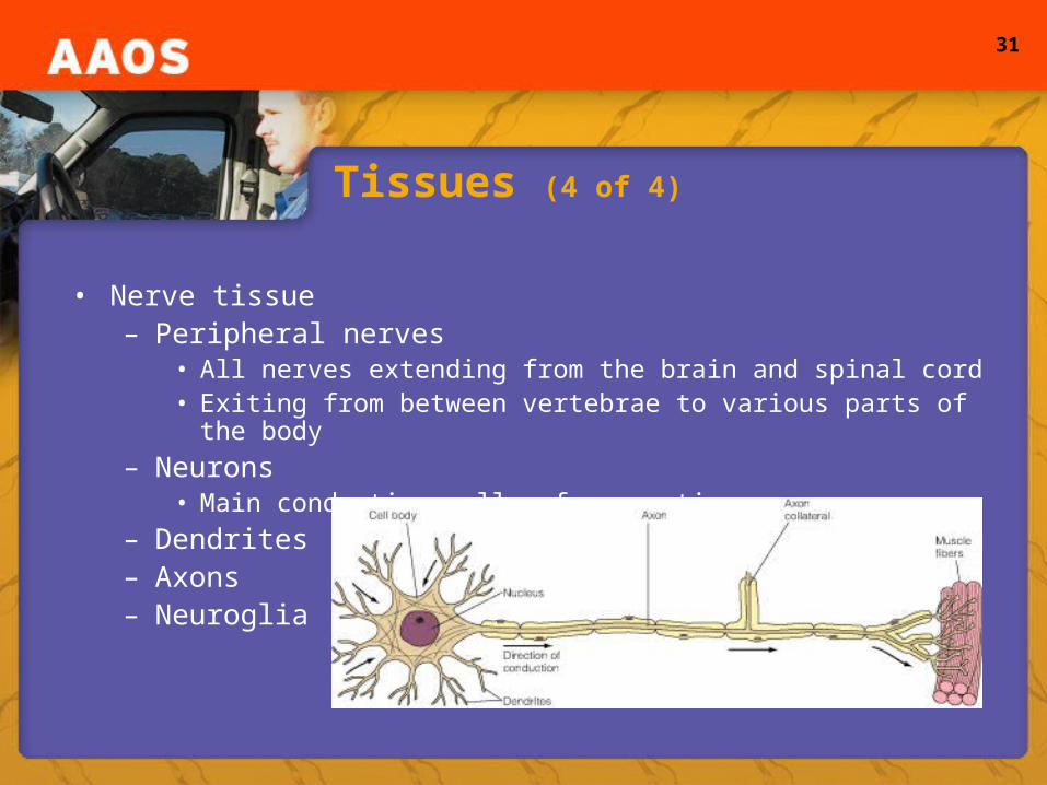
Tissues (4 of 4)
• Nerve tissue– Peripheral nerves
• All nerves extending from the brain and spinal cord• Exiting from between vertebrae to various parts of the body
– Neurons• Main conducting cells of nerve tissue
– Dendrites– Axons– Neuroglia
32
Integumentary System (1 of 2)
• Integumentary system– Outer surface of the body– Skin, nails, hair, and sweat and oil glands– Largest organ in the body; accounts for 15% of body
weight• Function of the skin
– Protection from the environment– Regulation of body temperature– Transmission of information to the brain
33
Integumentary System (2 of 2)
• Anatomy of the skin– Epidermis– Subcutaneous tissue– Sebaceous glands– Dermis– Sweat glands– Hair follicles

34
Anatomy of the Skin
35
Skeletal System
• Axial skeleton– Forms the upright part of the body– Consists of:
• Hyoid, skull, vertebral column, ribs, and sternum
• Appendicular skeleton– Attached to the axis as appendages– Consists of:
• Shoulder and pelvic girdles, upper and lower extremities

36
The Skull (1 of 3)
• Skull– Consists of 28 bones in three anatomic groups:
auditory ossicles, cranium, and face– Cranial vault
• Encases, protects the brain• Parietal, temporal, frontal, occipital, sphenoid, and ethmoid
bones• Foramen magnum
37
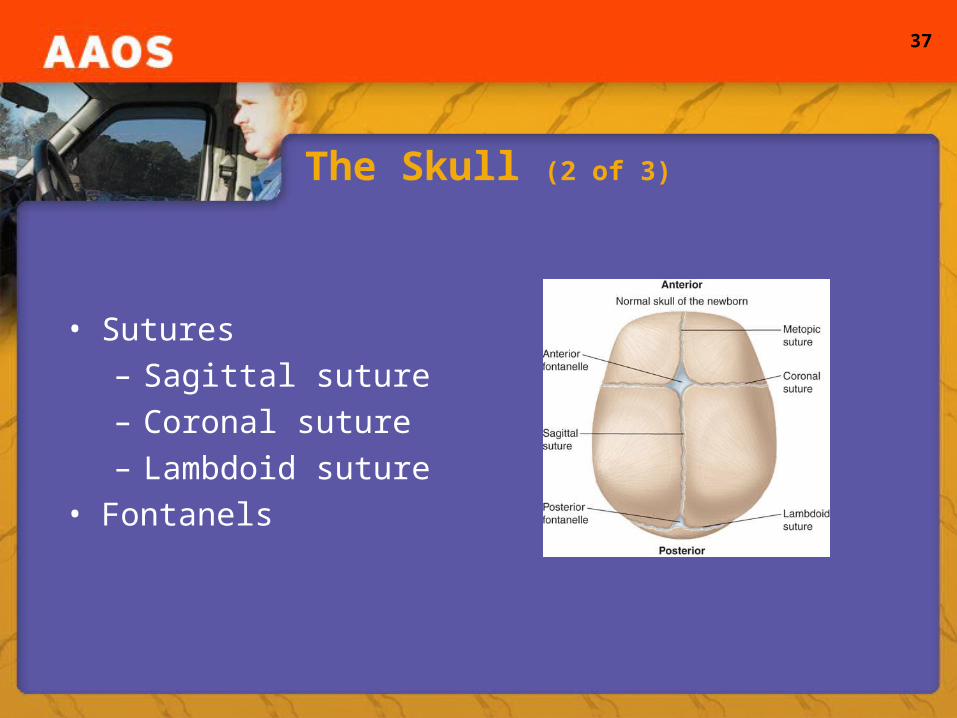
The Skull (2 of 3)
• Sutures– Sagittal suture– Coronal suture– Lambdoid suture
• Fontanels
38
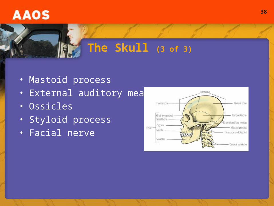
The Skull (3 of 3)
• Mastoid process• External auditory meatus• Ossicles• Styloid process• Facial nerve
39
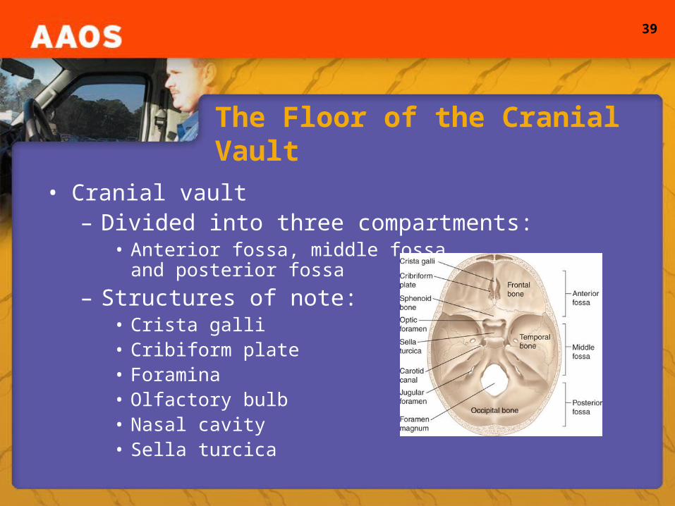
The Floor of the Cranial Vault
• Cranial vault– Divided into three compartments:
• Anterior fossa, middle fossa, and posterior fossa
– Structures of note:• Crista galli• Cribiform plate• Foramina• Olfactory bulb• Nasal cavity• Sella turcica

40
The Base of the Skull
• Base of the skull– Complex and full of foramina
• Structures of note:– Occipital condyles– Palatine bone– Zygomatic arch
41
The Facial Bones
• Facial bones– Frontal and ethmoid bones part of the cranial vault
and the face– Composed of 14 bones– Include:
• Maxillae, mandible, zygoma, palatine, nasal, lacrimal, vomer, and inferior nasal concha bones
– Protect the eyes, nose, and tongue and provide attachment points for muscles involved in mastication

42
Bones of the Orbit
• Orbits– Cone-shaped fossae– Enclose and protect the eyes– Contain blood vessels, nerves, and fat– Created by the frontal, sphenoid, zygomatic, maxilla,
lacrimal, ethmoid, and palatine bones– Blow to the eye can result in fracture of the orbit floor
(blowout fracture)

43
Bones of the Nose
• Nasal bones– Composed of several
portions of the facial bones• Structures of note:
– Nasal septum– Paranasal sinuses
44
The Mandible and Temporomandibular Joint
• Mandible– Large movable bone– Composed of the lower jaw and teeth
• Structures of note:– Rami– Mandibular notch– Temporomandibular joint
45
The Hyoid Bone
• Hyoid– “Floats”– Not actually part of the skull– Supports the tongue and serves as a point of
attachment for neck and tongue muscles
46
The Neck (1 of 2)
• Neck– Contains several important structures
• C1-C7• Upper portion of the trachea and esophagus
– Useful landmarks• Adam’s apple (upper part of the thyroid cartilage)• Cricoid cartilage• Cricothyroid membrane• Cartilaginous rings
47
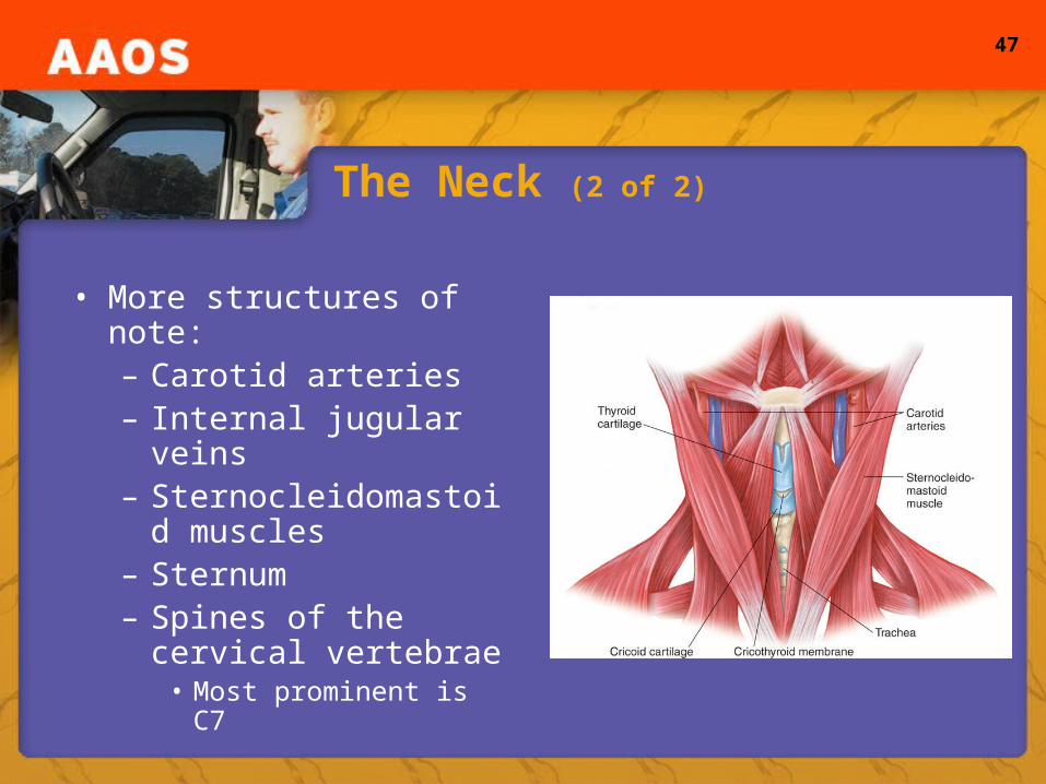
The Neck (2 of 2)
• More structures of note:– Carotid arteries– Internal jugular veins– Sternocleidomastoid
muscles– Sternum– Spines of the cervical
vertebrae• Most prominent is C7
48
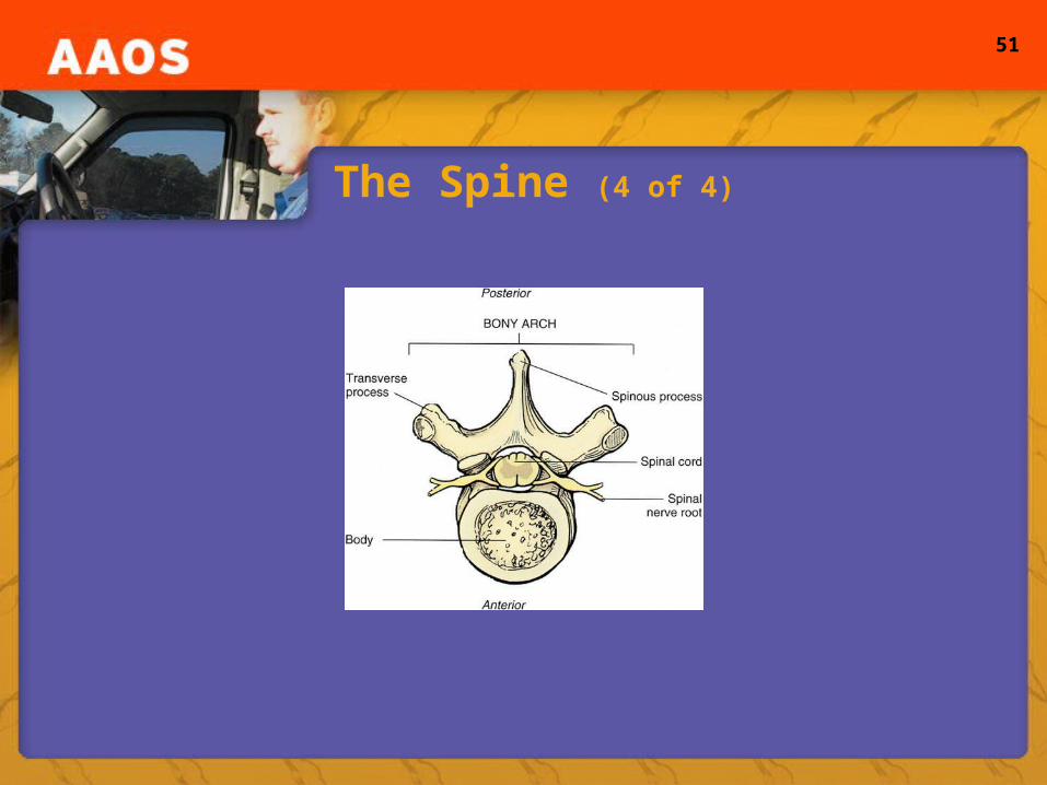
The Spine (1 of 4)
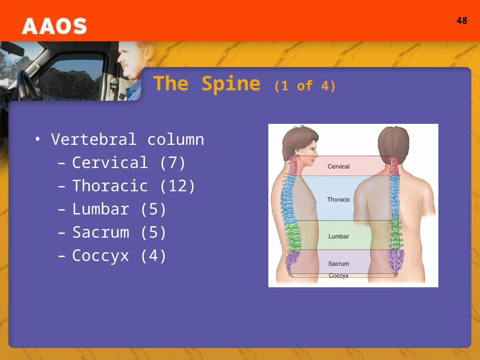
• Vertebral column– Cervical (7)– Thoracic (12)– Lumbar (5)– Sacrum (5)– Coccyx (4)
49
The Spine (2 of 4)
• Atlas (C1)– Point at which the head rotates
• Axis (C2)– Dens or odontoid process
• Spinal cord– Extension of the brain– Carries messages between the body and brain– Exits skull through foramen magnum– Protected by the vertebrae
50
The Spine (3 of 4)
• The vertebrae– Anterior portion consists of a solid block called “the
body”– Posterior part called the “bony arch”– Series of arches form a tunnel that runs the length of
the spine called the “spinal canal” which encases and protects the spinal cord
– Vertebrae are connected by ligaments– Intervertebral discs
51
The Spine (4 of 4)
52
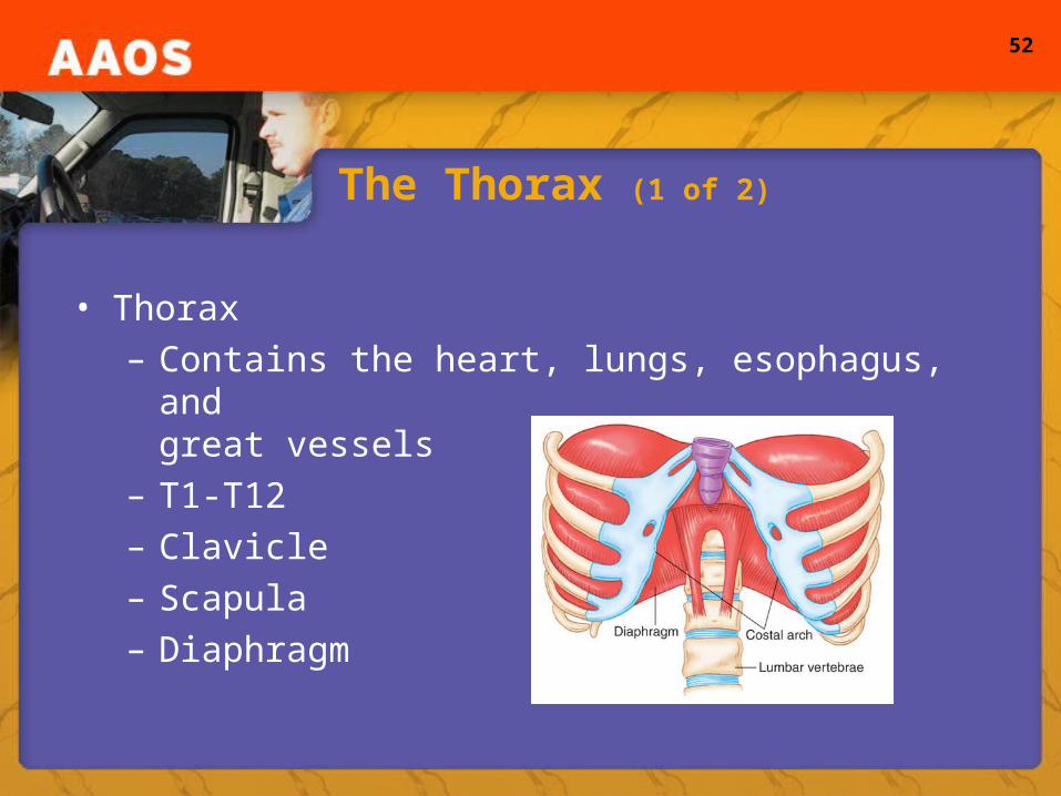
The Thorax (1 of 2)
• Thorax– Contains the heart, lungs, esophagus, and
great vessels– T1-T12– Clavicle– Scapula– Diaphragm
53
The Thorax (2 of 2)
• Anterior aspects– Sternum:
• Manubrium, xiphoid process, angle of Louis
– 12 pairs of ribs• Costal arch• Floating ribs
• Posterior aspects– Costovertebral angle (junction of the spine and the
tenth ribs)
54
Diaphragm/Organs and Vascular Structures
• Diaphragm– Muscular dome – Separates thorax and abdomen– Involved in respiration– Anteriorly attaches to costal arch; posteriorly to
lumbar vertebrae• Organs and vascular structures
– Pulmonary artery– Anatomic landmarks
55
The Abdomen
• The Abdomen– Second major body cavity– Contains organs of digestion and excretion– Separated by:
• Diaphragm, muscular walls, imaginary plane extending from the pubic symphysis through the sacrum
– Shares organs with the pelvis (depending on posture)– Quadrants
56
Review of Abdominal Quadrants
57
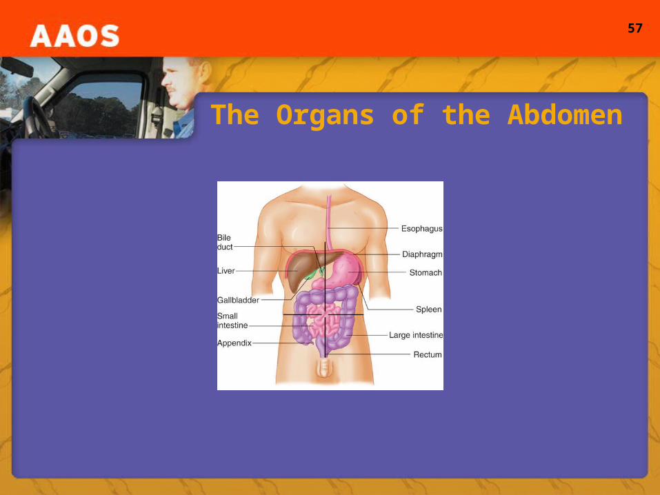
The Organs of the Abdomen

58
Organs and Vascular Structures (1 of 2)
• RUQ– Liver, gallbladder, portion of the colon/small intestine
• LUQ– Stomach, spleen, portion of the colon/small intestine
• RLQ– Cecum, appendix
• LLQ– Descending and sigmoid portions of the colon
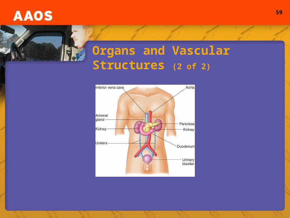
• Retroperitoneal– Kidneys
59
Organs and Vascular Structures (2 of 2)
60
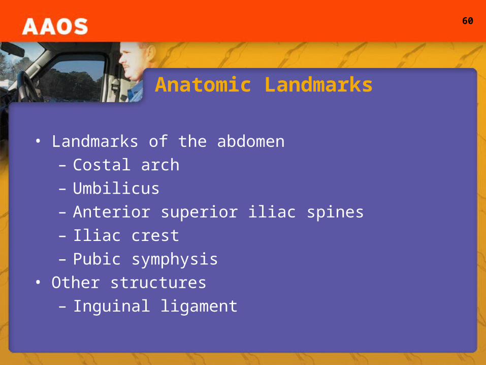
Anatomic Landmarks
• Landmarks of the abdomen– Costal arch– Umbilicus– Anterior superior iliac spines– Iliac crest– Pubic symphysis
• Other structures– Inguinal ligament

61
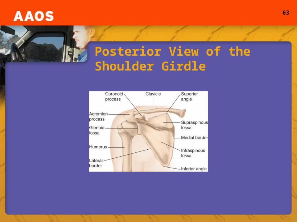
The Appendicular Skeleton (1 of 2)
• Shoulder girdle– Attaches upper extremity to the body– Composed of scapula and clavicle
• Shoulder joint– Acromion process – Ball and socket joint– Glenoid fossa– Motions include: flexion, extension, abduction,
adduction, rotation, and circumduction
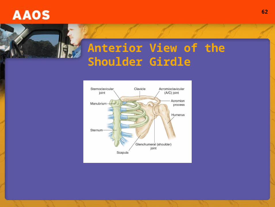
62
Anterior View of the Shoulder Girdle
63
Posterior View of the Shoulder Girdle

64
Anterior View of the Shoulder Joint
65
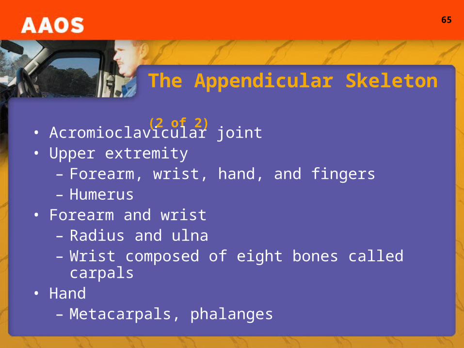
The Appendicular Skeleton (2 of 2)
• Acromioclavicular joint• Upper extremity
– Forearm, wrist, hand, and fingers– Humerus
• Forearm and wrist– Radius and ulna– Wrist composed of eight bones called carpals
• Hand– Metacarpals, phalanges
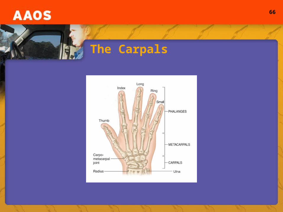
66
The Carpals
67
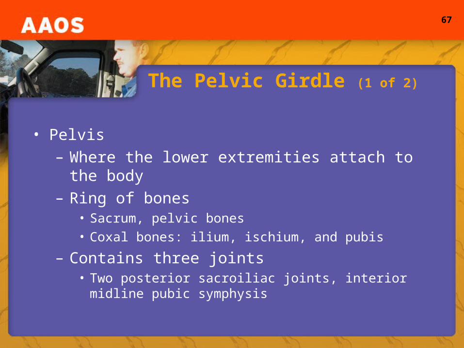
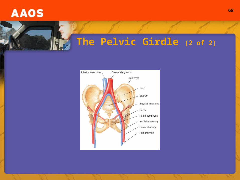
The Pelvic Girdle (1 of 2)
• Pelvis– Where the lower extremities attach to the body– Ring of bones
• Sacrum, pelvic bones• Coxal bones: ilium, ischium, and pubis
– Contains three joints• Two posterior sacroiliac joints, interior midline pubic
symphysis
68
The Pelvic Girdle (2 of 2)

69
The Lower Extremity
• Lower extremity– Composed of: hip, thigh, knee, leg, ankle, foot, and
toes• Structures of note:
– Femur (longest, strongest bone in the body)– Femoral head (articulates with pelvic girdle)– Acetabulum– Greater trochanter

70
The Leg, Knee, and Ankle
• Leg– Patella– Medial malleolus– Lateral malleolus
• Knee– Hinge joint– Contains ligaments within the join
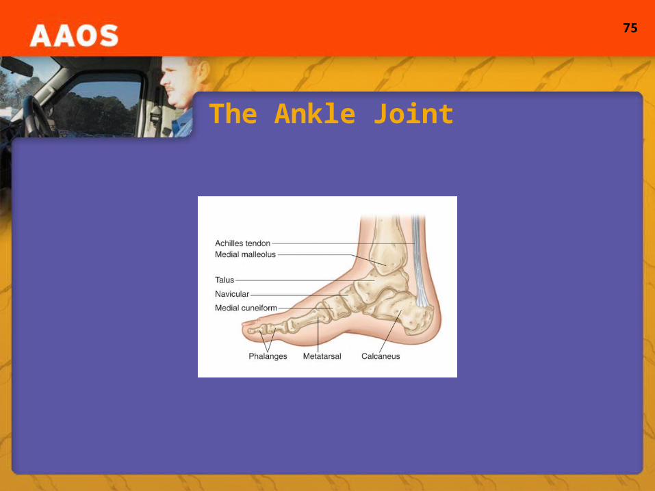
• Ankle– Phalanges, metatarsals

71
Cartilage, Tendons, and Ligaments
• Cartilage– All are connective tissues– Synovial fluid
• Tendons– Periosteum– Connects muscle to bone
• Ligaments– Tough, white bands of tissue– Connect bone to bone
72
Bones: Their Growth and Organization (1 of 2)
• Bones– Specialized form of connective tissue– Protect internal organs– Storage site for minerals– Consist of collagen and hydroxyapatite– Living substances that require blood supply– Terms:
• Osteoblasts, osteocyte, osteoclasts, lamellae, lacuna, canaliculi
73
Bones: Their Growth and Organization (2 of 2)
• Bones– Classified according to shape
• Long, short, and flat
– Long bones• Consist of diaphysis, epiphyses, and physis
– Two main types:• Compact and cancellous
– Growth• Appositional and endochondral
74
Joints
• Joints– When two bones contact– Consist of ends of bones and connective and
supporting tissue– Named by combining names of the two bones
• Joint capsule• ROM
– Determined by extent ligaments hold together
75
The Ankle Joint
76
The Musculoskeletal System
• Skeletal muscle– Voluntary, striated– Under control of the nervous system
• Smooth muscle– Involuntary– Responds to stretching, heat, and waste removal
• Cardiac muscle– Involuntary, rich blood supply, own electrical system
– “automaticity”
77
The Nervous System (1 of 2)
• Nervous system– Complex array of structures– Controls voluntary and involuntary body functions– Major divisions include:
• Central, peripheral, and autonomic
– Composed of specialized tissue that conducts electrical impulses between the brain and body
78
The Nervous System (2 of 2)
• Nervous system terminology:– Synapse– Presynaptic terminal– Synaptic cleft– Postsynaptic cleft– Neurotransmitters– Synaptic vesicles– Nerve fibers– “Fight or flight” and “Feed or breed”
79
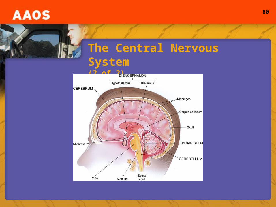
• The central nervous system– Brain and spinal cord
• Cerebrum– Left and right– Longitudinal fissure– Lobes
• Frontal
• Parietal
• Occipital
• Temporal
The Central Nervous System(1 of 2)
80
The Central Nervous System(2 of 2)
81
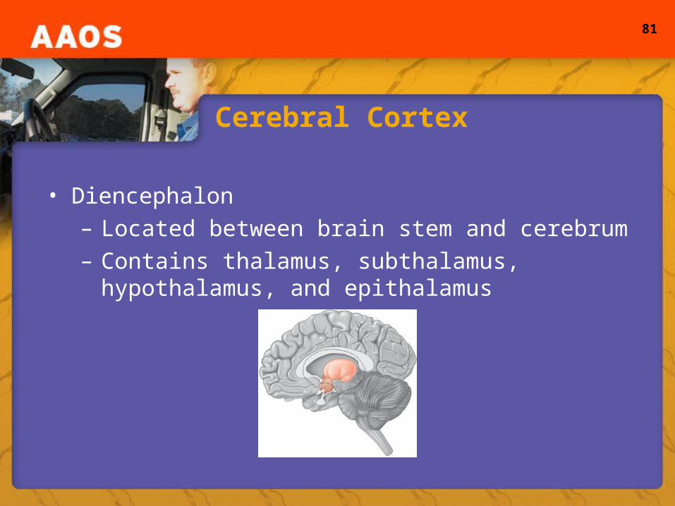
Cerebral Cortex
• Diencephalon– Located between brain stem and cerebrum– Contains thalamus, subthalamus, hypothalamus, and
epithalamus
82
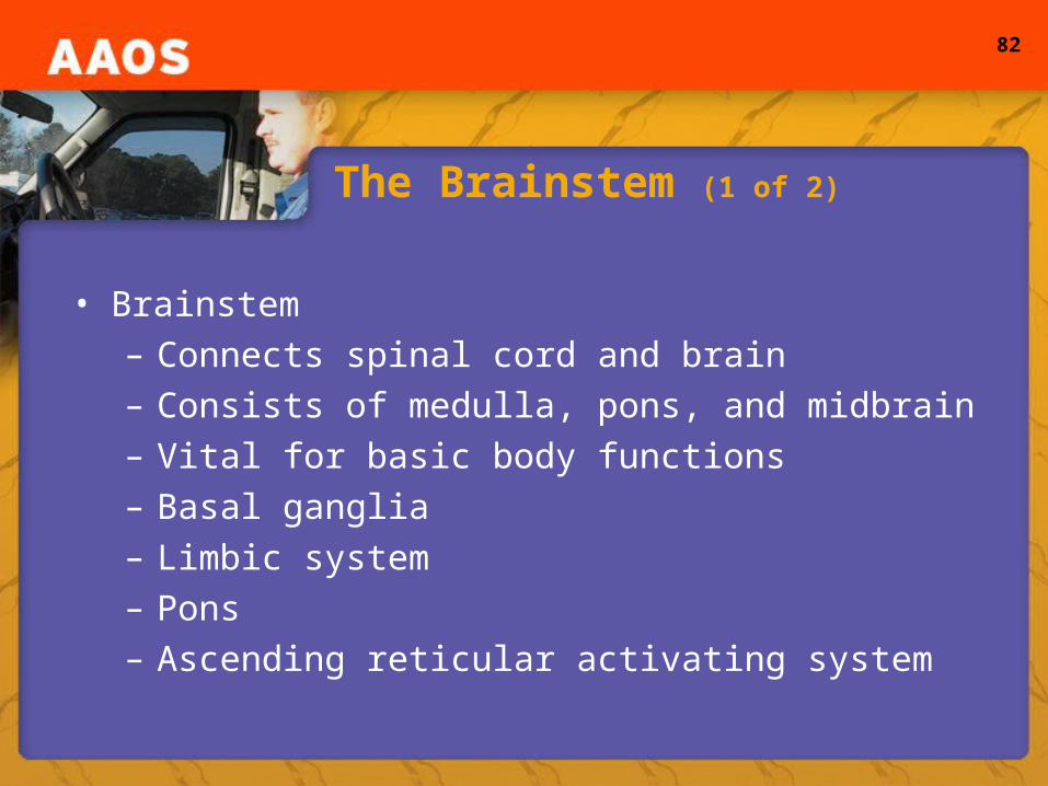
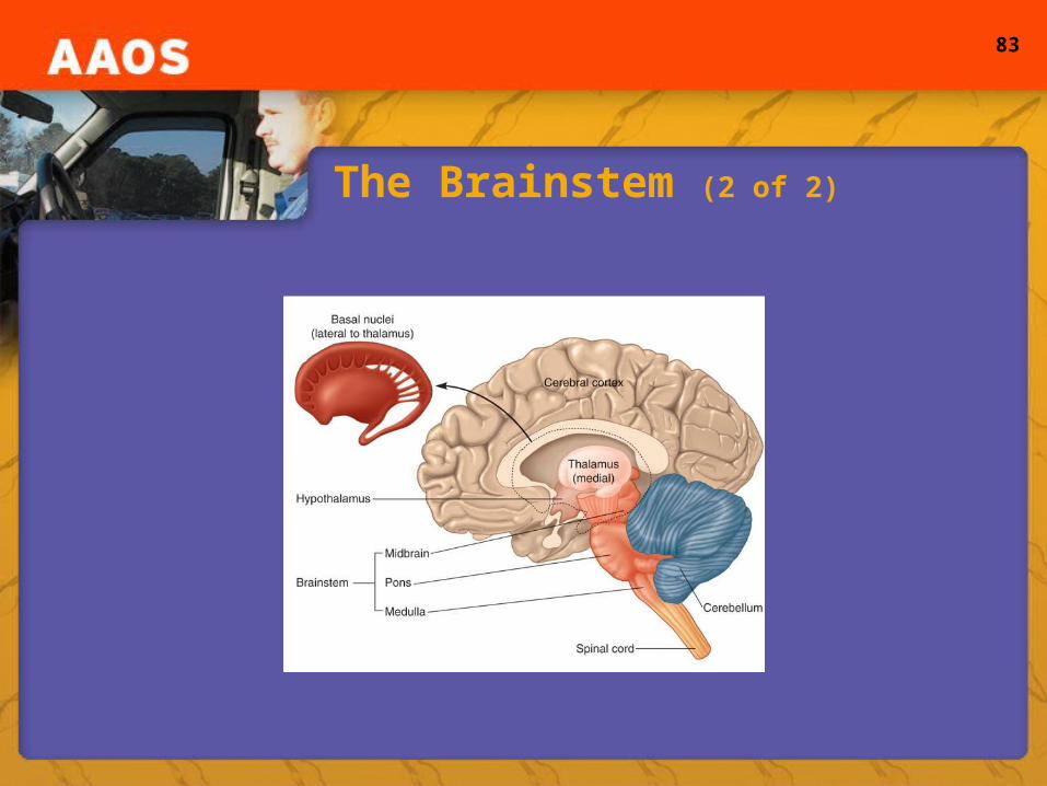
The Brainstem (1 of 2)
• Brainstem– Connects spinal cord and brain– Consists of medulla, pons, and midbrain– Vital for basic body functions– Basal ganglia– Limbic system– Pons– Ascending reticular activating system
83
The Brainstem (2 of 2)

84
The Cerebellum and Meninges
• Cerebellum– Communicates with other regions of the CNS– Cerebellar peduncles– Essential for coordinating muscle movements
• Meninges– Encloses the CNS– Dura mater– Arachnoid– Pia mater
85
The Spinal Cord
• Spinal cord– Continuation of the CNS– Composed of nerve fibers– Extends to the level of L2– Cauda equina– Vertebral canal– Ascending fibers (afferent tracts)– Action potentials– Descending fibers (efferent tracts)

86
The Peripheral Nervous System
• Peripheral nervous system– Consists of nerves from CNS to peripheral structures– Ganglia– Spinal nerves– Ventral root, dorsal root, dorsal root ganglion– Intervertebral foramen– Dermatomes

87
The Plexuses
• Cervical plexus– Most important nerve is the phrenic nerve
• Brachial plexus– Divided into rami, trunks, divisions, cords, and
branches– Axillary, radial, musculocutaneous, median, and ulnar
• Lumbosacral Plexus– Six major nerves: obturator, femoral, tibial, common
peroneal, sciatic, deep peroneal, and superficial peroneal

88
The Phrenic Nerve
89
The Cranial Nerves (1 of 2)
• Cranial nerves– 12 pairs– Functions:
• Olfactory (I) – smell• Optic – (II) sight• Oculomotor (III) – motion of the eyeballs/upper lid• Trochlear (IV) – downward gaze• Trigeminal (V) – sensation to scalp, face, and lower jaw
90
The Cranial Nerves (2 of 2)
• Functions continued:– Abducens (VI) – lateral eye movement– Facial (VII) – facial expression, sense of taste– Vestibulocochlear (VIII) – hearing, balance– Glossopharyngeal (IX) – pharyngeal muscles– Vagus (X) – parasympathetic fibers of
thorax/abdominal organs– Spinal accessory (XI) – soft palate– Hypoglossal (XII) – tongue, C1-C3
91
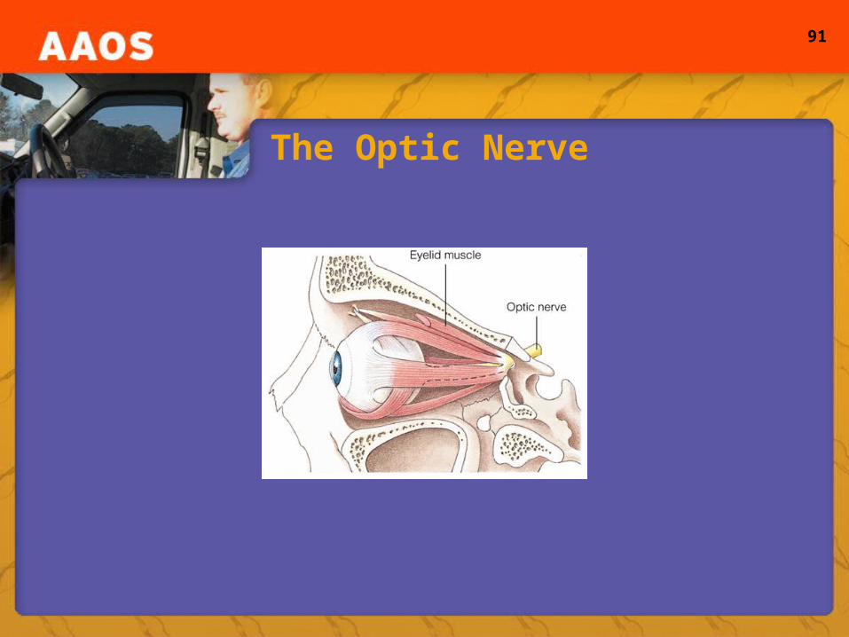
The Optic Nerve
92
The Autonomic Nervous System
• ANS– Operates without conscious control– Sympathetic and parasympathetic– Terms: sympathetic pathway and adrenal glands
• Preganglionic and postganglionic neurons– Separated by ganglionic synapse– Neuroeffector cells
93
Neurotransmitters and Receptors
• Sympathetic and parasympathetic – Secrete one of two neurotransmitters– Both secrete acetylcholine from preganglionic fibers;
diffuse across nicotinic receptors– Acetylcholine destroyed by acetylcholinesterase– Muscarinic receptors– Alpha and beta receptors

94
The Endocrine System
• Endocrine system– Made of various glands– Hormones regulate body functions– Targets tissues– Endocrine versus exocrine glands– Prostaglandins– Steroids and thyroid hormones– Negative feedback or positive feedback
95
The Pituitary Gland and Hypothalamus (1 of 2)
• Pituitary gland– Known as the the “master gland”– Located at the base of the brain
• Hypothalamus– Basal portion of diencephalon– Regulates function of pituitary gland
96
The Pituitary Gland and Hypothalamus (2 of 2)
• Posterior pituitary lobe– Antidiuretic hormone or vasopressin
• Constricts blood vessels and raises BP• Target tissue is the kidney
– Oxytocin• Causes smooth muscles (uterus) to contract• Lactation
• Anterior pituitary lobe– Not considered part of the CNS
97
Hormones
• Growth hormone– Stimulates growth (long bones)– Produced by hypothalamus
• Thyroid-stimulating hormone• Adrenocorticotropic hormone
– Corticosteroids stimulated by stress• Reproduction-regulating hormones
– Regulate production of eggs and sperm
98
The Thyroid Gland
• Thyroid– Large gland at the base of the neck– Two lobes connected by the isthmus– Contains follicles filled with thyroglobulin– Between the follicles are parafollicular cells
• Produce calcitonin
– Thyroid gland produces triiodothyronine and tetraiodothyronine

99
The Parathyroid Glands
• Usually four in number• Produce and secrete parathyroid hormone
– Maintains normal levels of calcium in the blood and normal neuromuscular function

100
The Pancreas
• Pancreas– Belongs to both endocrine and digestive systems– Retroperitoneum– Produces insulin and glucagon– Islet of Langerhans
• Alpha and beta cells
– Glycogen

101
The Adrenal Glands (1 of 2)
• Adrenal glands– Supra-renal glands– Manufacture and secrete certain
sex hormones– Medulla
• Epinephrine and norepinephrine
– Adrenal cortex• Divided into three zones• Aldosterone
102
The Adrenal Glands (2 of 2)
• Zona fasiculata– Glucocorticoids
• Cortisol: regulates blood glucose, metabolizes fat tissue, inhibits inflammation
– Hypothalamic-pituitary-adrenal axis• Regulates secretion of corticosteroids
• Zona reticularis– Androgens
• Androstendedione
103
The Reproductive Glands and Hormones
• Gonads– Testes
• Testosterone
– Ovaries• Estrogen• Progesterone• Human chorionic gonadatropin (hCG)
104
Blood and Its Components
• Blood– Substance consists of plasma and formed elements
• Red blood cells, white blood cells, and platelets
– Adult male contains about 5 liters of blood– Plasma
• Watery, straw-colored fluid• Accounts for >50% of total blood volume• Made of 92% water and 8% other substances
105
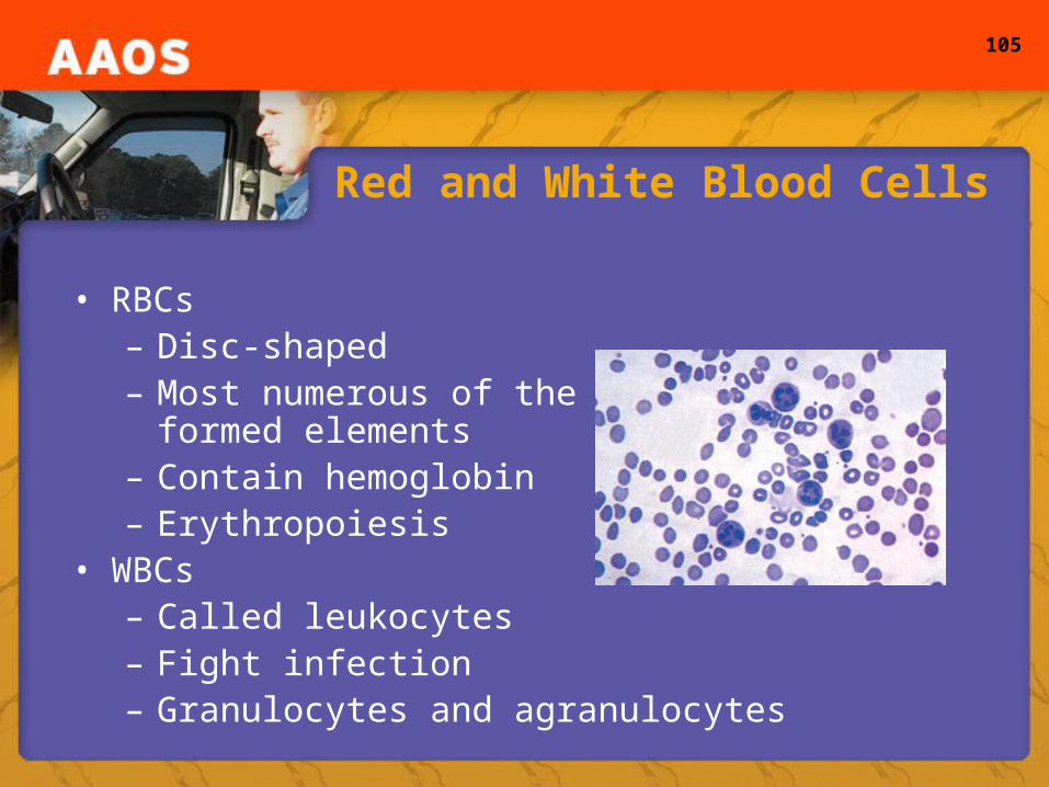
Red and White Blood Cells
• RBCs– Disc-shaped– Most numerous of the
formed elements– Contain hemoglobin– Erythropoiesis
• WBCs– Called leukocytes– Fight infection– Granulocytes and agranulocytes
106
Platelets and Blood Clotting
• Platelets– Necessary in clot formation– Hemostasis– Chemicals are released, activation of platelets– Formation of thrombin– Conversion of fibrinogen to fibrin– Binds to platelet plug forming a clot– Activation of t-PA
107
The Heart (1 of 2)
• Location and major structures of the heart– Located behind the sternum – About the size of the closed fist of the person it
belongs to• Mediastinum• Myocardium
108
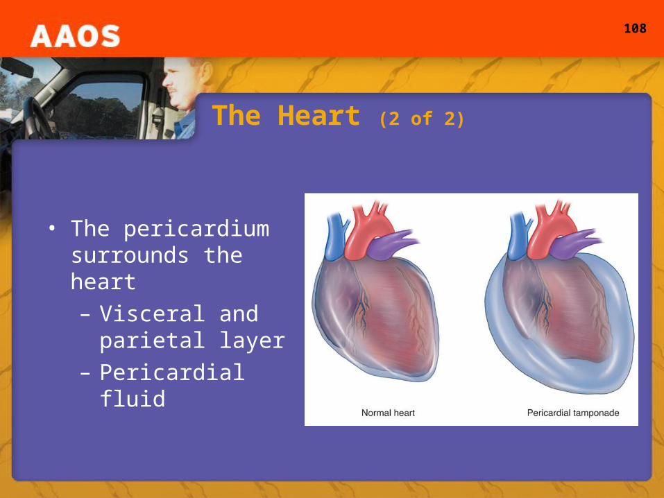
The Heart (2 of 2)
• The pericardium surrounds the heart– Visceral and
parietal layer– Pericardial fluid
109
Blood Flow Within the Heart (1 of 2)
• Superior and inferior vena cava– Return deoxygenated blood from the body to the right
atrium• Blood passes into the right ventricle• Blood is pumped through the pulmonic valve into the
pulmonary artery and to the lungs
110
Blood Flow Within the Heart (2 of 2)
• Freshly oxygenated blood is returned to the left atrium through the pulmonary veins
• Blood flows through the mitral valve into the left ventricle, which pumps the oxygenated blood through the aortic valve into the aorta and then the entire body
111
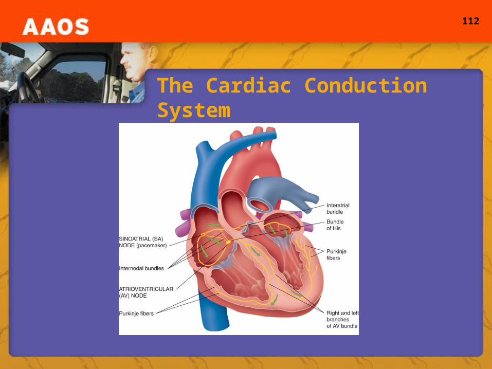
Electrical Properties of the Heart and Conduction System
• Electrical stimulus– Caused by a set of chemical changes within the
myocardial cells– “Automaticity”
• Conduction system– Six parts: SA node, AV node, bundle of His, right and
left bundle branches, and Purkinje fibers
112
The Cardiac Conduction System
113
Valves of the Heart
• Atrioventricular valves– Tricuspid valve– Mitral valve
• Cusps• Papillary muscles• Chordae tendineae cordis
– Semilunar valves• Pulmonic valve• Aortic valve
114
Regulation of Heart Function
• Chronotropic state– Contraction
• Dromotropic state– Rate of conduction
• Inotropic state– Strength of contraction
• Baroreceptors and chemoreceptors• Alpha and beta effects

115
The Cardiac Cycle
• Cardiac cycle– Contraction results in pressure changes in the
chambers– Systole– Cardiac output– Circulatory system– Ejection fraction: CO = SV x HR– Starling’s Law of the Heart– Preload and afterload

116
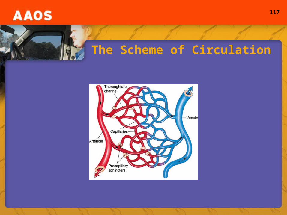
The Vascular System
• The general scheme of blood circulation– Transported through arteries and veins– Arterioles, capillaries, and venules
• Circulation to the heart– Coronary arteries– LAD– Circumflex coronary arteries
117
The Scheme of Circulation
118
Pulmonary and Systemic Arterial Circulation
• Pulmonary circulation– Carries blood from the right side of the heart to the
lungs and back• Systemic arterial circulation
– head and neck– upper extremity– thoracic aorta– abdominal aorta– pelvis and lower extremity
119
Systemic Venous Circulation
• Head and neck• Upper extremity• Thorax• Abdomen and pelvis
– Hepatic portal system• Lower extremity
– Femoral, popliteal, dorsalis pedis, and posterior tibial arteries
120
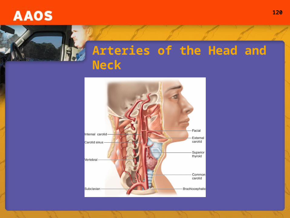
Arteries of the Head and Neck
121
Physiology of the Circulatory System
• Terms: – Pulse, radial artery, blood pressure, diastole
• Normal circulation in adults– Vessels adjust to hold 100% of the blood volume– Perfusion: meets the cell needs
• Inadequate circulation in adults– Automatic adjustment during blood loss to maintain
adequate pressure
122
The Lymphatic System
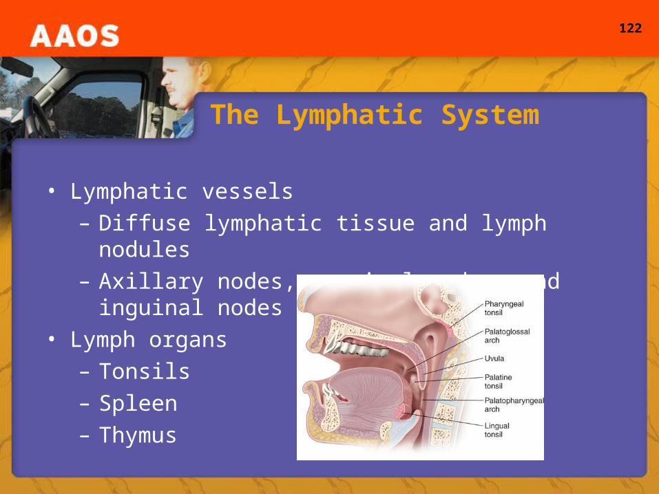
• Lymphatic vessels– Diffuse lymphatic tissue and lymph nodules– Axillary nodes, cervical nodes, and inguinal nodes
• Lymph organs– Tonsils – Spleen – Thymus
123
The Respiratory System
• Respiratory system– Breathing, gas exchange, and entrance of air
• Upper airway– Mouth, nasal cavity, and oral cavity– Uvula, epiglottis, nares, conchae, and turbinates
• Lower airway– Larynx, vocal cords, glottis, bronchi, alveoli, lungs,
pleura, and bronchial arteries and veins

124
Respiratory Physiology
• Physiology– Primary function to exchange gases at the
alveolocapillary membrane– 21% oxygen in the air– Pulmonary function tests and spirometry– Tidal volume, residual volume, vital capacity, and
FEVI• Respiratory center
– Located in the medulla oblongata
125
The Digestive System (1 of 2)
• How digestion works– Complex chemical process
• Anatomy of the digestive system– Mouth– Salivary glands– Oropharynx– Esophagus– Stomach– Pancreas
126
The Digestive System (2 of 2)
• More anatomy of the digestive system– Liver– Bile ducts– Small intestine– Large intestine– Appendix– Rectum
127
The Urinary System
• Urinary system – Controls the discharge of waste materials filtered
from the blood by the kidneys– Kidneys– Ureter– Urinary bladder– Urethra
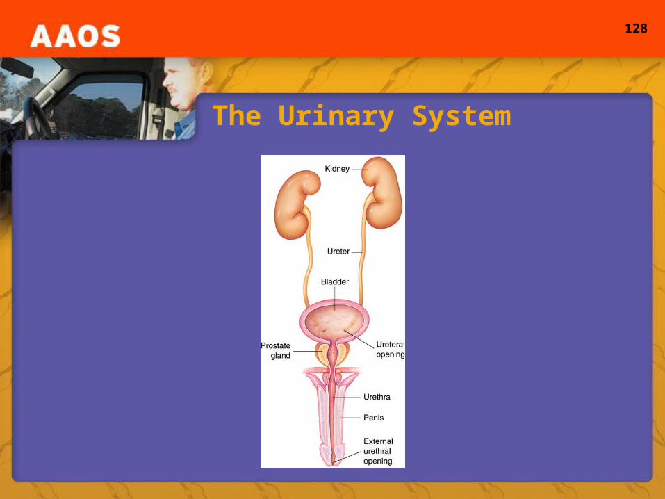
128
The Urinary System
129
The Genital System
• Genital system– Controls reproductive processes
• Male reproductive system and organs– Testicles, vas deferens, seminal vesicles, prostate
gland, urethra, and penis• Female reproductive system and organs
– Ovaries, fallopian tubes, uterus, cervix, and vagina
130
Fluids and Electrolytes (1 of 2)
• Body fluid balance– Total body water averages 50-70% of total body
weight– Intracellular and extracellular fluid– Intravascular fluid and interstitial fluid– Fluid balance is the process of maintaining
homeostasis through equal intake and output– ADH, thirst, kidneys, and water shifts
131
Fluids and Electrolytes (2 of 2)
• Acid-base balance– Acids and bases– Alkalotic versus acidotic– pH range 0-14 with 7.0 being neutral
• Buffer systems– Act as fast defenses for acid-base challenges– Absorb hydrogen ions when they are in excess