Embed Size (px)
Citation preview
1
1H Chemical Shifts in NMR. Part 191. Carbonyl Anisotropies and Steric Effects in
aromatic aldehydes and ketones.
Raymond J. Abraham* and Mehdi Mobli
Chemistry Department, The University of Liverpool, P.O.Box 147, Liverpool L69 3BX
and
Richard J.Smith, GlaxoSmithKline, New Frontiers Science Park, Harlow, Essex CM19 5AW
The 1H Chemical Shifts of benzaldehyde, 2-chloro, 2-hydroxy and 2-methoxy
benzaldehyde, acetophenone, 2-methoxy and 2-hydroxy acetophenone, indanone, anthraquinone,
fluorenone, anthrone, α-tetralone, 2,4,6-trimethyl acetophenone, 9-acetylanthracene, 9-
anthranaldehyde and benzosuberone were obtained and completely assigned in CDCl3 and
DMSO solution. In anthrone a keto-enol tautomerism (anthrone/9-hydroxyanthracene) was
observed by NMR in hydrogen bonding solvents but not chloroform. The % of enol is linearly
dependant on the Kamlett β hydrogen bonding parameter of the solvent, not the solvent relative
permitivity.
The chemical shift data allowed the determination of the carbonyl substituent chemical
shifts (SCS) in these molecules. These were analysed in terms of the carbonyl electric field,
magnetic anisotropy and steric effects for long-range protons together with a model (CHARGE7)
for the calculation of the two-bond and three bond effects. The SCS of the carbonyl bond was
reproduced with an asymmetric magnetic anisotropy acting at the midpoint of the carbonyl bond
with values of ∆χ parll and ∆χ perp of 6.36 and –11.88 (10-30 cm3/molecule) plus a steric term
from the oxygen atom and the C=O electric field effect. The short range effects of the carbonyl
group on the aldehyde proton were modelled using the appropriate β functions in the CHARGE
routine. For the 9-substituted anthracenes the Hückel π calculation was modified to account for
the 1H chemical shifts of the H-10 protons. This model gave a comprehensive calculation of the 1H chemical shifts of these aromatic aldehydes and ketones. For the data set of 129 chemical
shifts ranging from 2.5 to 11.5 δ the rms error of the observed vs calculated shifts was 0.094
ppm.
The CO anisotropy and oxygen shielding differ appreciably from the corresponding
values for the aliphatic aldehydes and ketones but are similar to the values for the CO group of
amides, illustrating the effect of conjugation on these parameters. The model was used in the
2
conformational analysis of some related compounds. In 2-chlorobenzaldehyde the chemical shift
calculations support a non-planar molecule with the aldehyde/ ring dihedral angle in the trans
conformer of ca 250. In the strained 7-membered ring of benzosuberone, the model was used to
test calculated geometries. The ab initio geometry at the B3LYP(6-31++G(d,p) ) level gave the
best agreement with the observed shifts.
Introduction
The influence of the carbonyl group on the chemical shifts of neighbouring protons has
been of interest since the early days of NMR, the low field chemical shift of the aldehyde proton
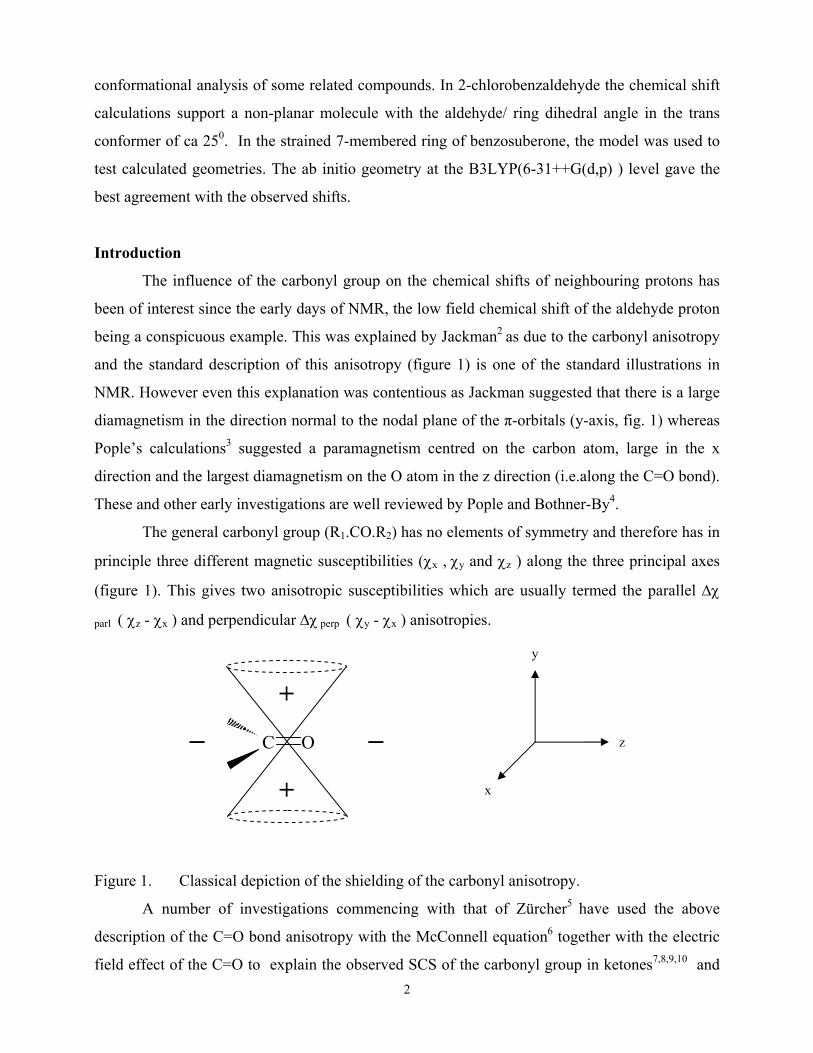
being a conspicuous example. This was explained by Jackman2 as due to the carbonyl anisotropy
and the standard description of this anisotropy (figure 1) is one of the standard illustrations in
NMR. However even this explanation was contentious as Jackman suggested that there is a large
diamagnetism in the direction normal to the nodal plane of the π-orbitals (y-axis, fig. 1) whereas
Pople’s calculations3 suggested a paramagnetism centred on the carbon atom, large in the x
direction and the largest diamagnetism on the O atom in the z direction (i.e.along the C=O bond).
These and other early investigations are well reviewed by Pople and Bothner-By4.
The general carbonyl group (R1.CO.R2) has no elements of symmetry and therefore has in
principle three different magnetic susceptibilities (χx , χy and χz ) along the three principal axes
(figure 1). This gives two anisotropic susceptibilities which are usually termed the parallel ∆χ
parl ( χz - χx ) and perpendicular ∆χ perp ( χy - χx ) anisotropies.
Figure 1. Classical depiction of the shielding of the carbonyl anisotropy.
A number of investigations commencing with that of Zürcher5 have used the above
description of the C=O bond anisotropy with the McConnell equation6 together with the electric
field effect of the C=O to explain the observed SCS of the carbonyl group in ketones7,8,9,10 and
C O _ _
+
+
y
x
z
3
peptides11. None of these calculated the chemical shifts of the protons vicinal to the carbonyl
group. An investigation which did include all the protons in the ketones studied was given in a
previous part of this series12. This data was subsequently refined12a using the CHARGE7 routine,
including the carbonyl anisotropy and electric field plus an oxygen but not a carbon steric term.
Apart from the investigations mentioned above no calculation of the 1H shifts in these
compounds has been given. In particular a general calculation of the 1H chemical shifts for
carbonyl compounds using the ab initio GIAO method has not been reported to date, the basis set
dependance of such calculations being a severe problem. A recent investigation by Lampert et
al13 compared the observed vs calculated NMR chemical shifts for phenol and benzaldehyde and
for 13 substituted derivatives, using a variety of basis sets and computational procedures within
the Gaussian94 program. The calculated shielding of the aromatic protons with respect to
methane varied by ca 0.5 – 1.0 ppm. depending on the procedure and basis set used and this may
well represent the limit of accuracy of such calculations.
The classical investigations above considered only aliphatic ketones apart from that of
Williamson on peptides11 and therefore the anisotropy for a saturated carbonyl group was
obtained. However when the carbonyl is attached to an aromatic group it will be conjugated and
this is likely to affect the carbonyl anisotropy. This was noted by Jackman2 who suggested that
the inconsistency of Pople’s model when applied to amides may be due to the assumption that the
anisotropy of the carbonyl group in amides is similar to that in aldehydes and ketones.
The effect of conjugation on the carbonyl group anisotropy may also be determined from
the chemical shielding tensor. Wasylishen et al.14 used MASNMR to determine the chemical shift
tensors for the carbonyl carbon of acetaldehyde, 3,4-dibenzyloxy-benzaldehyde and 3,4-
dimethoxy benzaldehyde. There was a significant difference (>25%) between the shift tensors for
the carbonyl carbon of acetaldehyde and those for the carbonyl carbons of the benzaldehydes,
which were identical (within the error margin of the observations). These results suggest strongly
that the anisotropy of the carbonyl group should be treated separately for aromatic and aliphatic
systems and this is the basis for the present investigation.
We present here the complete assignment of a the 1H NMR spectra of benzaldehyde (1),
2-chloro, 2-hydroxy and 2-methoxy benzaldehyde (2, 3, 4), acetophenone (5), 2-methoxy and 2-
hydroxy acetophenone (6, 8), indanone (7), anthraquinone (9), fluorenone (10), anthrone (11), α-
tetralone (12), 2,4,6-trimethyl acetophenone (13), 9-acetylanthracene (14), 9-anthranaldehyde
(15) and benzosuberone (16) (scheme 1) in CDCl3 and DMSO. The solvent of choice in
4
pharmaceutical investigations is often DMSO rather than CDCl3 thus we present here the data for
both solvents as part of a comprehensive comparison of 1H chemical shifts in the two solvents.
The compounds were selected on the basis that they had the carbonyl group in as many different
orientations w.r.t the ring system as possible. These provide sufficient data for a complete
analysis of the aromatic carbonyl substituent effects and we shall show that the C=O anisotropy
for these molecules is very different from that for aliphatics and indeed more similar to that for
amides.
Theory
As the theory has been given previously1,12,15 only a brief summary of the latest version
(CHARGE7) will be given here. The theory distinguishes between short-range substituent effects
over one, two and three bonds, which are attributed to the electronic effects of the substituents
and long-range effects due to the electric fields, steric effects and anisotropy of the substituents.
Short range effects.
The CHARGE scheme calculates the effects of neighbouring atoms on the partial atomic
charge of the atom under consideration based upon classical concepts of inductive and resonance
contributions. If we consider an atom I in a four atom fragment I-J-K-L the partial atomic charge
on I is due to three effects. There is a α effect from atom J given by the difference in the
electronegativity of atoms I and J. A β effect from atom K proportional to both the
electronegativity of atom K and the polarisability of atom I. There is also a γ effect from atom L
given by the product of the atomic polarisabilities of atoms I and L for I = H and L = F, Cl, Br, I.
However for chain atoms (C,N,O,S etc.) the γ effect (i.e. C.C.C.H) is parameterised separately and is
given by A+Bcosθ where θ is the C.C.C.H dihedral angle and A and B empirical parameters.
The total charge is given by summing these effects and the partial atomic charges (q)
converted to shift values using eqn.1
δ = 160.84q - 6.68 (1)
Long range effects.
The effects of distant atoms on the proton chemical shifts are due to steric, anisotropic and
electric field contributions. H..H steric interactions are shielding in alkanes and deshielding in
aromatics and X..H (X = C, O, Cl, Br, I) interactions deshielding, according to a simple r-6
dependence (eqn. 2) where aS is the steric coefficient for any given atom.
δ steric = aS / r 6 (2)
The effects of the electric field of the C-X bonds (X= H, F, Cl, Br, I, O) on the C-H protons
5
are obtained from the component of the electric field along the C-H bond. The electric field for a
single bonded atom (e.g. =O ) is calculated as due to the charge on the oxygen atom and an equal and
opposite charge on the attached carbon atom. The vector sum gives the total electric field at the
proton and the component of this field along the CH bond is proportional to the proton chemical
shift.
The magnetic anisotropy of a bond with cylindrical symmetry (e.g. C≡C ) is obtained from
the appropriate McConnell eqn (eqn 3) .
δanis = ∆χ (3cos2ϕ−1)/ 3R3 (3)
In eqn. 3 R is the distance from the perturbing group to the nucleus of interest in Å, ϕ is
the angle between the vector R and the symmetry axis and ∆χ the anisotropy of the C≡C bond.
( ∆χ = χ parl
- χ perp ) where χ parl and χ perp are the susceptibilities parallel and perpendicular to the
symmetry axis respectively.
For a non-symmetric group such as the carbonyl group eqn 3 is replaced by the full
McConnell eqn (eqn 4) where θ1 and θ2 are the angles between the radius vector R and the x and
z axes respectively (fig. 1) and ∆χparl (χz−χx ) and ∆χperp (χy −χx ) are the parallel and
perpendicular anisotropy for the C=O bond respectively.
δanis = [∆χparl (3cos2θ1-1) + ∆χperp (3cos2θ2-1)] / 3R3 (4)
For aromatic compounds it is necessary to include the shifts due to the aromatic ring
current and the π electron densities in the aromatic ring1. The equivalent dipole approximation
was used to calculate the ring current shifts to give eqn. 5. In eqn. 5, R is the distance of the
proton from the benzene ring centre, θ the angle of the R vector with the ring symmetry axis, µ
the equivalent dipole of the aromatic ring and ƒc the π-electron current density for the ring,
being 1.0 for substituted benzenes.
δrc = ƒc µ (3cos2 θ-1) / R3 (5)
The π electron densities are calculated from Huckel theory16. The standard coulomb and
resonance integrals for the Huckel routine are given by eqn.6, where α0
αr = α0 + hrβ0 (6)
βrs= krsβ0
and β0 are the coulomb and resonance integrals for a carbon 2pZ atomic orbital and hr and krs the
factors modifying these integrals for orbitals other than sp2 carbon. For substituted aromatics the
values of the coefficients hr and krs in eqn.6 for the orbitals involving hetero atoms have to be
6
found. These were obtained so that the π densities calculated from the Huckel routine reproduce
the π densities from ab initio calculations.
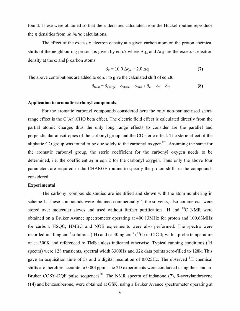
The effect of the excess π electron density at a given carbon atom on the proton chemical
shifts of the neighbouring protons is given by eqn.7 where ∆qα and ∆qβ are the excess π electron
density at the α and β carbon atoms.
δπ = 10.0 ∆qα + 2.0 ∆qβ (7)
The above contributions are added to eqn.1 to give the calculated shift of eqn.8.
δtotal = δcharge + δsteric + δanis + δel + δπ + δrc (8)
Application to aromatic carbonyl compounds.
For the aromatic carbonyl compounds considered here the only non-parametrised short-
range effect is the C(Ar).CHO beta effect. The electric field effect is calculated directly from the
partial atomic charges thus the only long range effects to consider are the parallel and
perpendicular anisotropies of the carbonyl group and the CO steric effect. The steric effect of the
aliphatic CO group was found to be due solely to the carbonyl oxygen12a. Assuming the same for
the aromatic carbonyl group, the steric coefficient for the carbonyl oxygen needs to be
determined, i.e. the coefficient aS in eqn 2 for the carbonyl oxygen. Thus only the above four
parameters are required in the CHARGE routine to specify the proton shifts in the compounds
considered.
Experimental
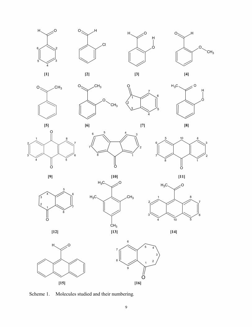
The carbonyl compounds studied are identified and shown with the atom numbering in
scheme 1. These compounds were obtained commercially17, the solvents, also commercial were
stored over molecular sieves and used without further purification. 1H and 13C NMR were
obtained on a Bruker Avance spectrometer operating at 400.13MHz for proton and 100.63MHz
for carbon. HSQC, HMBC and NOE experiments were also performed. The spectra were
recorded in 10mg cm-3 solutions (1H) and ca.30mg cm-3 (13C) in CDCl3 with a probe temperature
of ca 300K and referenced to TMS unless indicated otherwise. Typical running conditions (1H
spectra) were 128 transients, spectral width 3300Hz and 32k data points zero-filled to 128k. This
gave an acquisition time of 5s and a digital resolution of 0.025Hz. The observed 1H chemical
shifts are therefore accurate to 0.001ppm. The 2D experiments were conducted using the standard
Bruker COSY-DQF pulse sequences18. The NMR spectra of indanone (7), 9-acetylanthracene
(14) and benzosuberone, were obtained at GSK, using a Bruker Avance spectrometer operating at
7
700.13 MHz for the proton experiments, a Bruker Avance operating at 500.13 MHz for the
carbon, HSQC and HMBC experiments and a Bruker Avance operating at 399.87 MHz for the
NOE experiments. 9-methoxyanthracene was synthesised by methylating the enol tautomer of
anthrone according to Meek et al19.
Conformational
The geometries of the polycyclic molecules were obtained using the molecular mechanics
program PCMODEL Version 7.020 with the MMFF94 forcefield. For the smaller molecules the
geometries were further optimised using the Gaussian98 programme at the B3LYP/6-31G**
level21. It has been shown22 that the DFT level of theory generally obtained better geometries
compared to other theoretical levels, especially for compounds with intra molecular hydrogen
bonding. Anthraldehyde was also optimised at the B3LYP/6-31G** level as this was the
geometry used previously for anthracene parameterisation23. 9-acetylanthracene was too large to
run at these higher levels of theory but the 9-methoxy - and 9-hydroxy-anthracene were run at
B3LYP/3-21G* level. All the calculations were carried out on a PC.
OH
X
X
O H
cis trans
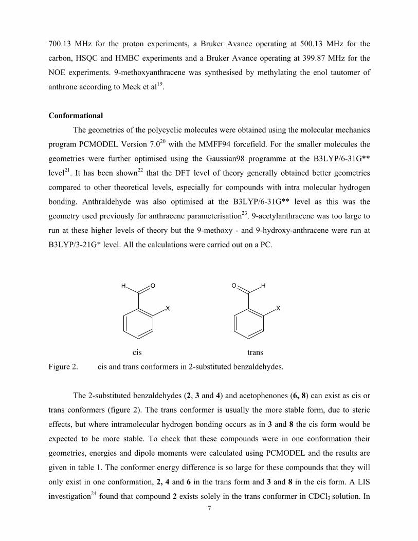
Figure 2. cis and trans conformers in 2-substituted benzaldehydes.
The 2-substituted benzaldehydes (2, 3 and 4) and acetophenones (6, 8) can exist as cis or
trans conformers (figure 2). The trans conformer is usually the more stable form, due to steric
effects, but where intramolecular hydrogen bonding occurs as in 3 and 8 the cis form would be
expected to be more stable. To check that these compounds were in one conformation their
geometries, energies and dipole moments were calculated using PCMODEL and the results are
given in table 1. The conformer energy difference is so large for these compounds that they will
only exist in one conformation, 2, 4 and 6 in the trans form and 3 and 8 in the cis form. A LIS
investigation24 found that compound 2 exists solely in the trans conformer in CDCl3 solution. In
8
all these compounds except 2 (see below) the carbonyl is coplanar with the aromatic ring. The
remaining polycyclic compounds can only exist in one conformation. Compounds 7, 9, 10, 11
and 15 are planar, 12 has an envelope cyclohexenone ring and 13 and 14 have the acetyl group
orthogonal to the aromatic ring. LIS studies of the conformations of 725, 1226, 1327, and 14 and
1528 in chloroform solution agreed with these results.
All these compounds were used for the parametrisation except for 2 and 16. In 2 the
PCMODEL geometry has a CO/ring dihedral angle of ca 400 but the ab initio geometry is planar.
In 16 again the ring geometries for the molecular mechanics and various ab initio basis sets differ
considerably. These molecules were therefore omitted from the calculations and will be
considered subsequently.
Spectral Assignments
The spectra were obtained in CDCl3 and DMSO. The assignments of the spectra of benzaldehyde
1 and acetophenone 5 are straightforward. Those of 2-chlorobenzaldehyde 223, 2-
hydroxybenzaldehyde 329, indan-1-one 725, 2-hydroxyacetophenone 829, fluoren-9-one 1030,
2,4,6-trimethylacetophenone 1327, 9-anthraldehyde and 9-acetylanthracene, 14, 1528 have been
given previously. The chemical shifts given here of dilute samples (approx. 5-10mg/cm3) in
CDCl3 agree with this data, although in some cases the chemical shifts varied slightly due to
concentration effects.
Table 1. Energies (kcal/mol), Dipole moments (D) and CO/Ring Dihedral Angles of the trans
and cis conformers of 2-substituted benzaldehydes and acetophenones.
Energy Energy ∆Ε Dipole Dipole CO/Ring Dihedral
Angle
trans cis Εcis−Εtrans (t) (c) (t) (c)
[1] Benzaldehyde 31.26 4.37 0
[2] 2-Chloro-benzaldehyde 27.18 33.06 5.88 4.14 6.59 0 0
[3] 2-Hydroxy benzaldehyde 33.67 25.78 -7.89 2.90 4.87 11.2 0 [4] 2-Methoxybenzaldehyde 39.63 47.61 7.98 5.09 5.56 0 0
[5] Acetophenone 36.10 4.37 0 [6] 2-Methoxy-acetophenone 47.19 5.75 4.17 29.1 [8] 2 Hydroxy acetophenone 36.36 30.57 -5.79 6.02 5.01 17.6 0
9
O C H 3
O
O 1
2
3 4 5
6
78
O C H3
O
H O C H 3
OCH3
O
1
2
3456
7 8
[1] [2] [3] [4] [5] [6] [7] [8] [9] [10] [11]
[12] [13] [14]
[15] [16] Scheme 1. Molecules studied and their numbering.
OH
2
45 3
6
O H
Cl
O H
OCH3
OH
O
H
O
1
2
3
45
67
O
12
345
6
7
8
10
CH3CH3
CH3
CH3 OCH3 O
12
3
4 56
7
8
10
O
1 2 3
4 5
6
7 8
O
1 2
3
4 5
6
7
8
9
OH
10
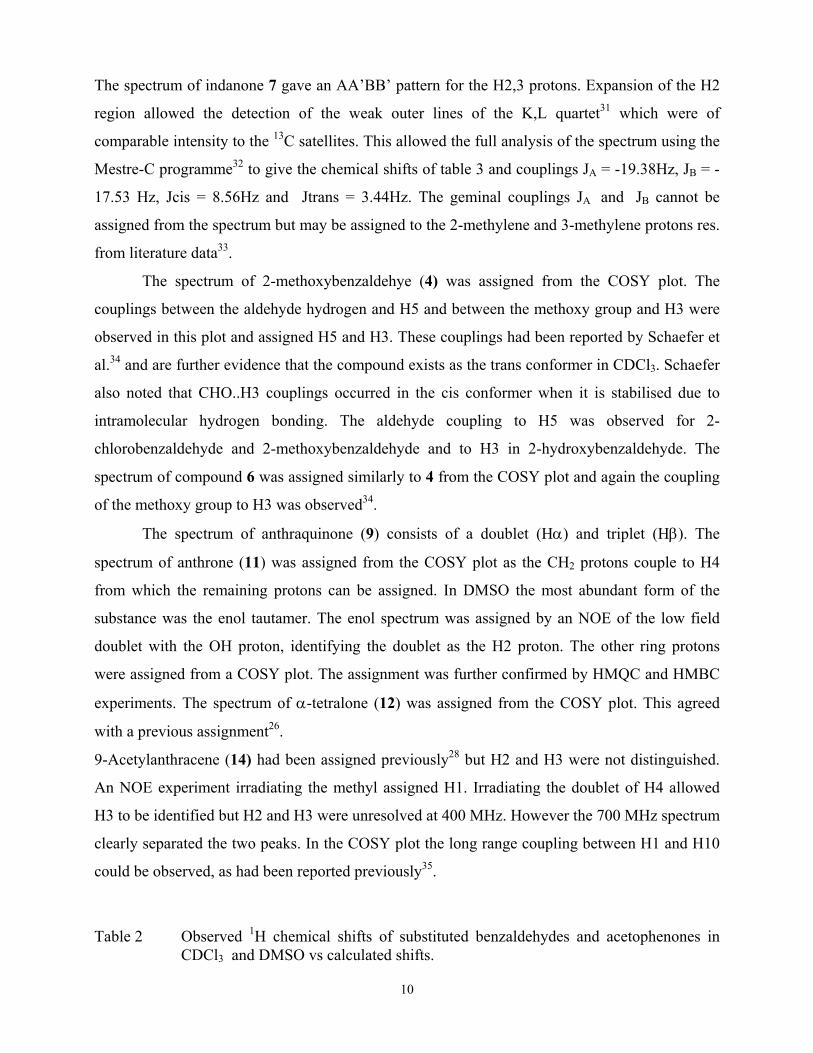
The spectrum of indanone 7 gave an AA’BB’ pattern for the H2,3 protons. Expansion of the H2
region allowed the detection of the weak outer lines of the K,L quartet31 which were of
comparable intensity to the 13C satellites. This allowed the full analysis of the spectrum using the
Mestre-C programme32 to give the chemical shifts of table 3 and couplings JA = -19.38Hz, JB = -
17.53 Hz, Jcis = 8.56Hz and Jtrans = 3.44Hz. The geminal couplings JA and JB cannot be
assigned from the spectrum but may be assigned to the 2-methylene and 3-methylene protons res.
from literature data33.
The spectrum of 2-methoxybenzaldehye (4) was assigned from the COSY plot. The
couplings between the aldehyde hydrogen and H5 and between the methoxy group and H3 were
observed in this plot and assigned H5 and H3. These couplings had been reported by Schaefer et
al.34 and are further evidence that the compound exists as the trans conformer in CDCl3. Schaefer
also noted that CHO..H3 couplings occurred in the cis conformer when it is stabilised due to
intramolecular hydrogen bonding. The aldehyde coupling to H5 was observed for 2-
chlorobenzaldehyde and 2-methoxybenzaldehyde and to H3 in 2-hydroxybenzaldehyde. The
spectrum of compound 6 was assigned similarly to 4 from the COSY plot and again the coupling
of the methoxy group to H3 was observed34.
The spectrum of anthraquinone (9) consists of a doublet (Hα) and triplet (Hβ). The
spectrum of anthrone (11) was assigned from the COSY plot as the CH2 protons couple to H4
from which the remaining protons can be assigned. In DMSO the most abundant form of the
substance was the enol tautamer. The enol spectrum was assigned by an NOE of the low field
doublet with the OH proton, identifying the doublet as the H2 proton. The other ring protons
were assigned from a COSY plot. The assignment was further confirmed by HMQC and HMBC
experiments. The spectrum of α-tetralone (12) was assigned from the COSY plot. This agreed
with a previous assignment26.
9-Acetylanthracene (14) had been assigned previously28 but H2 and H3 were not distinguished.
An NOE experiment irradiating the methyl assigned H1. Irradiating the doublet of H4 allowed
H3 to be identified but H2 and H3 were unresolved at 400 MHz. However the 700 MHz spectrum
clearly separated the two peaks. In the COSY plot the long range coupling between H1 and H10
could be observed, as had been reported previously35.
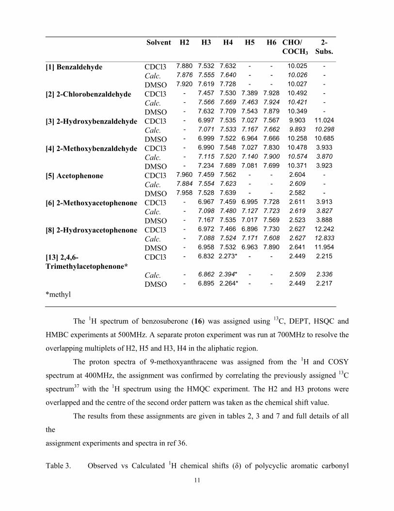
Table 2 Observed 1H chemical shifts of substituted benzaldehydes and acetophenones in
CDCl3 and DMSO vs calculated shifts.
11
Solvent H2 H3 H4 H5 H6 CHO/ COCH3
2-Subs.
[1] Benzaldehyde CDCl3 7.880 7.532 7.632 - - 10.025 - Calc. 7.876 7.555 7.640 - - 10.026 - DMSO 7.920 7.619 7.728 - - 10.027 - [2] 2-Chlorobenzaldehyde CDCl3 - 7.457 7.530 7.389 7.928 10.492 - Calc. - 7.566 7.669 7.463 7.924 10.421 - DMSO - 7.632 7.709 7.543 7.879 10.349 - [3] 2-Hydroxybenzaldehyde CDCl3 - 6.997 7.535 7.027 7.567 9.903 11.024 Calc. - 7.071 7.533 7.167 7.662 9.893 10.298 DMSO - 6.999 7.522 6.964 7.666 10.258 10.685[4] 2-Methoxybenzaldehyde CDCl3 - 6.990 7.548 7.027 7.830 10.478 3.933 Calc. - 7.115 7.520 7.140 7.900 10.574 3.870 DMSO - 7.234 7.689 7.081 7.699 10.371 3.923 [5] Acetophenone CDCl3 7.960 7.459 7.562 - - 2.604 - Calc. 7.884 7.554 7.623 - - 2.609 - DMSO 7.958 7.528 7.639 - - 2.582 - [6] 2-Methoxyacetophenone CDCl3 - 6.967 7.459 6.995 7.728 2.611 3.913 Calc. - 7.098 7.480 7.127 7.723 2.619 3.827 DMSO - 7.167 7.535 7.017 7.569 2.523 3.888 [8] 2-Hydroxyacetophenone CDCl3 - 6.972 7.466 6.896 7.730 2.627 12.242 Calc. - 7.088 7.524 7.171 7.608 2.627 12.833 DMSO - 6.958 7.532 6.963 7.890 2.641 11.954[13] 2,4,6-Trimethylacetophenone*
CDCl3 - 6.832 2.273* - - 2.449 2.215
Calc. - 6.862 2.394* - - 2.509 2.336 DMSO - 6.895 2.264* - - 2.449 2.217
*methyl
The 1H spectrum of benzosuberone (16) was assigned using 13C, DEPT, HSQC and
HMBC experiments at 500MHz. A separate proton experiment was run at 700MHz to resolve the
overlapping multiplets of H2, H5 and H3, H4 in the aliphatic region.
The proton spectra of 9-methoxyanthracene was assigned from the 1H and COSY
spectrum at 400MHz, the assignment was confirmed by correlating the previously assigned 13C
spectrum37 with the 1H spectrum using the HMQC experiment. The H2 and H3 protons were
overlapped and the centre of the second order pattern was taken as the chemical shift value.
The results from these assignments are given in tables 2, 3 and 7 and full details of all
the
assignment experiments and spectra in ref 36.
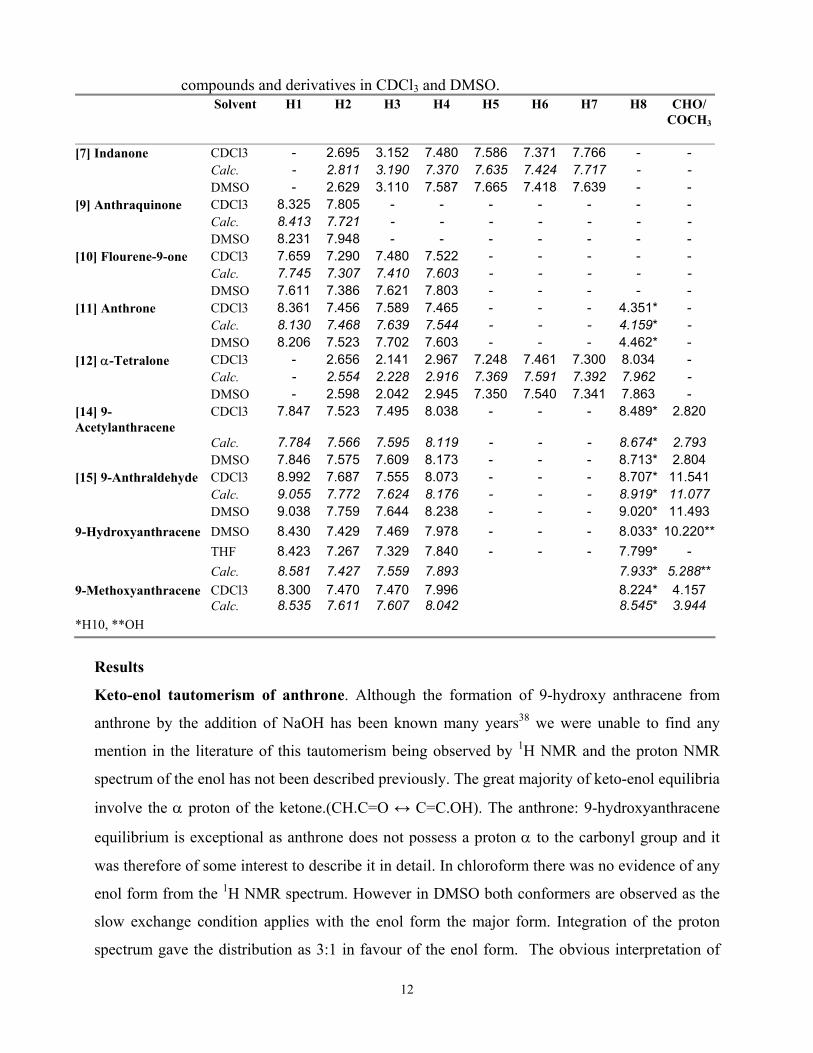
Table 3. Observed vs Calculated 1H chemical shifts (δ) of polycyclic aromatic carbonyl
12
compounds and derivatives in CDCl3 and DMSO. Solvent H1 H2 H3 H4 H5 H6 H7 H8 CHO/
COCH3
[7] Indanone CDCl3 - 2.695 3.152 7.480 7.586 7.371 7.766 - - Calc. - 2.811 3.190 7.370 7.635 7.424 7.717 - - DMSO - 2.629 3.110 7.587 7.665 7.418 7.639 - - [9] Anthraquinone CDCl3 8.325 7.805 - - - - - - - Calc. 8.413 7.721 - - - - - - - DMSO 8.231 7.948 - - - - - - - [10] Flourene-9-one CDCl3 7.659 7.290 7.480 7.522 - - - - - Calc. 7.745 7.307 7.410 7.603 - - - - - DMSO 7.611 7.386 7.621 7.803 - - - - - [11] Anthrone CDCl3 8.361 7.456 7.589 7.465 - - - 4.351* - Calc. 8.130 7.468 7.639 7.544 - - - 4.159* - DMSO 8.206 7.523 7.702 7.603 - - - 4.462* - [12] α-Tetralone CDCl3 - 2.656 2.141 2.967 7.248 7.461 7.300 8.034 - Calc. - 2.554 2.228 2.916 7.369 7.591 7.392 7.962 - DMSO - 2.598 2.042 2.945 7.350 7.540 7.341 7.863 - [14] 9-Acetylanthracene
CDCl3 7.847 7.523 7.495 8.038 - - - 8.489* 2.820
Calc. 7.784 7.566 7.595 8.119 - - - 8.674* 2.793 DMSO 7.846 7.575 7.609 8.173 - - - 8.713* 2.804 [15] 9-Anthraldehyde CDCl3 8.992 7.687 7.555 8.073 - - - 8.707* 11.541 Calc. 9.055 7.772 7.624 8.176 - - - 8.919* 11.077 DMSO 9.038 7.759 7.644 8.238 - - - 9.020* 11.4939-Hydroxyanthracene DMSO 8.430 7.429 7.469 7.978 - - - 8.033* 10.220**
THF 8.423 7.267 7.329 7.840 - - - 7.799* -
Calc. 8.581 7.427 7.559 7.893 7.933* 5.288**9-Methoxyanthracene CDCl3 8.300 7.470 7.470 7.996 8.224* 4.157 Calc. 8.535 7.611 7.607 8.042 8.545* 3.944 *H10, **OH
Results
Keto-enol tautomerism of anthrone. Although the formation of 9-hydroxy anthracene from
anthrone by the addition of NaOH has been known many years38 we were unable to find any
mention in the literature of this tautomerism being observed by 1H NMR and the proton NMR
spectrum of the enol has not been described previously. The great majority of keto-enol equilibria
involve the α proton of the ketone.(CH.C=O ↔ C=C.OH). The anthrone: 9-hydroxyanthracene
equilibrium is exceptional as anthrone does not possess a proton α to the carbonyl group and it
was therefore of some interest to describe it in detail. In chloroform there was no evidence of any
enol form from the 1H NMR spectrum. However in DMSO both conformers are observed as the
slow exchange condition applies with the enol form the major form. Integration of the proton
spectrum gave the distribution as 3:1 in favour of the enol form. The obvious interpretation of
13
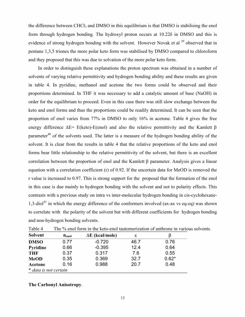
the difference between CHCl3 and DMSO in this equilibrium is that DMSO is stabilising the enol
form through hydrogen bonding. The hydroxyl proton occurs at 10.22δ in DMSO and this is
evidence of strong hydrogen bonding with the solvent. However Novak et al 39 observed that in
pentane 1,3,5 triones the more polar keto form was stabilised by DMSO compared to chloroform
and they proposed that this was due to solvation of the more polar keto form.
In order to distinguish these explanations the proton spectrum was obtained in a number of
solvents of varying relative permittivity and hydrogen bonding ability and these results are given
in table 4. In pyridine, methanol and acetone the two forms could be observed and their
proportions determined. In THF it was necessary to add a catalytic amount of base (NaOH) in
order for the equilibrium to proceed. Even in this case there was still slow exchange between the
keto and enol forms and thus the proportions could be readily determined. It can be seen that the
proportion of enol varies from 77% in DMSO to only 16% in acetone. Table 4 gives the free
energy difference ∆E= E(keto)-E(enol) and also the relative permittivity and the Kamlett β
parameter40 of the solvents used. The latter is a measure of the hydrogen bonding ability of the
solvent. It is clear from the results in table 4 that the relative proportions of the keto and enol
forms bear little relationship to the relative permittivity of the solvent, but there is an excellent
correlation between the proportion of enol and the Kamlett β parameter. Analysis gives a linear
equation with a correlation coefficient (r) of 0.92. If the uncertain data for MeOD is removed the
r value is increased to 0.97. This is strong support for the proposal that the formation of the enol
in this case is due mainly to hydrogen bonding with the solvent and not to polarity effects. This
contrasts with a previous study on intra vs inter-molecular hydrogen bonding in cis-cyclohexane-
1,3-diol41 in which the energy difference of the conformers involved (ax-ax vs eq-eq) was shown
to correlate with the polarity of the solvent but with different coefficients for hydrogen bonding
and non-hydrogen bonding solvents.
Table 4 The % enol form in the keto-enol tautomerization of anthrone in various solvents. Solvent nenol ∆E (kcal/mole) ε β DMSO 0.77 -0.720 46.7 0.76 Pyridine 0.66 -0.395 12.4 0.64 THF 0.37 0.317 7.6 0.55 MeOD 0.35 0.369 32.7 0.62* Acetone 0.16 0.988 20.7 0.48 * data is not certain
The Carbonyl Anisotropy.
14
The 1H chemical shifts in CDCl3 in tables 2 and 3 are sufficient to allow the full
parameterisation in the CHARGE routine for aromatic carbonyl groups. All the data in the tables
were used except the chemical shifts for 2-chlorobenzaldehyde (2) and benzosuberone (16) (see
later) and also the hydroxyl hydrogens of compounds 3 and 8. This gave a total of 129 shifts
ranging from 2.8 to 11.5δ. As stated previously there are only four variables to be determined.
These are the C(Ar).CHO beta effect, the carbonyl anisotropy ∆χparl and ∆χperp and the oxygen
steric effect. The values of the parameters were obtained by use of a non-linear least mean square
program CHAP842 which compares the observed vs. calculated chemical shifts. This gave ∆χparl =
6.36, ∆χperp = –11.88 (10-30 cm3/molecule) and the oxygen steric coefficient as =38.4ppm Å6. The
rms error was 0.094 ppm for the whole dataset. The calculated and observed shifts are given in
tables 2 and 3 and it can be seen that the agreement is excellent with the largest error for the ring
protons ca 0.15ppm. This demonstrates the applicability of the CHARGE scheme to this
important class of compounds. The values of the CO anisotropy and oxygen steric coefficient
found here will be considered later.
A small change was made to the π calculation in CHARGE (eqn. 6) for the 9-substituted
anthracenes. In the unmodified routine the H10 proton of anthraldehyde was calculated at much
too large δ value (calc. 9.10 vs obs. 8.71). Conversely the H10 proton of 9-hydroxy anthracene
was calculated at too low δ value (calc. 7.57 vs obs. 7.80). The calculated SCS have the correct
signs (note H10 in anthracene is 8.43δ)23 but are much too large. This difference is not due to the
carbonyl anisotropy or to steric or electric field effects as these effects decrease very rapidly with
distance (see table 5). Hückel theory tends to exagerate the π charges in compounds with very
polarisable π systems such as the middle ring of anthracene and this was the reason for these
anomalies. This effect did not happen with the 9-acetyl anthracene as the acetyl group is
orthogonal to the ring, thus there is no conjugation with the π system.
In CHARGE the resonance integral coefficient (krs eqn 6) is –1.0 for benzenoid
aromatics. To account for the polarisability of the middle ring of anthracene this coefficient for
the C9,10 bonds with the α-carbons was modified. The two results above were used to optimise
this value. Decreasing the value of this resonance integral to –1.25 gave reasonable agreement for
both molecules and these calculated values are given in table 3. Most interestingly the chemical
shift of H10 in 9-methoxyanthracene is also upfield of anthracene. The SCS of the methoxy
group at H10 is –0.21ppm which is comparable to that of the hydroxy group (-0.36) even though
both the MM and ab initio calculations gave the methoxy group orthogonal to the anthracene ring
15
and in consequence show no π effect. The observed SCs could be due to hyperconjugation or
possibly to large vibrational motion of the methoxy group.
The use of CHARGE for Conformational Analysis
The CHARGE routine gives proton chemical shifts for the conformationally rigid
molecules considered in good agreement with the observed shifts. A related question is whether
the CHARGE routine can be used in conformationally mobile compounds to obtain
conformational information. The compounds we wish to consider here are 2-chlorobenzaldehyde
and benzosuberone. However before these are attempted it is necessary to consider a well-
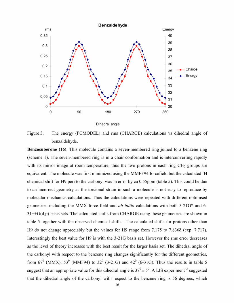
defined case such as benzaldehyde. The energy profile obtained from PCMODEL is shown in
figure 3. The CHARGE routine gives an accurate calculation of the proton chemical shifts for the
planar molecule but would it also reproduce the effects in the non-planar conformations? To test
this the shifts were calculated for the various orientations of the aldehyde group and the rms
deviation of the observed vs calculated shifts obtained. This curve is also shown in figure 3 with
the energy profile. The ordinates differ in the two plots but the overall good agreement of the two
curves is strong support for the use of CHARGE in the conformational analysis of these
compounds.
2-chlorobenzaldehyde (2). The theoretical calculations gave conflicting geometries for the
stable trans conformer. Gaussian98 using the B3LYP density function theory with the 6-31G**
basis set gave a planar molecule which was also the case with the MMF94 forcefield of
PCMODEL. In contrast the MMX force field in PCMODEL gave a minimum energy for a 400
ring/aldehyde torsional angle. Thus the proton chemical shifts were obtained from CHARGE for
100 rotations of the aldehyde from the plane and compared with the observed data. The best
agreement was for a torsional angle of 250 with an rms error of 0.085ppm. There is no
experimental data to support this result but it would appear a reasonable value.
16
Figure 3. The energy (PCMODEL) and rms (CHARGE) calculations vs dihedral angle of
benzaldehyde.
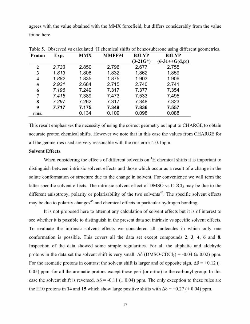
Benzosuberone (16). This molecule contains a seven-membered ring joined to a benzene ring
(scheme 1). The seven-membered ring is in a chair conformation and is interconverting rapidly
with its mirror image at room temperature, thus the two protons in each ring CH2 groups are
equivalent. The molecule was first minimized using the MMFF94 forcefield but the calculated 1H
chemical shift for H9 peri to the carbonyl was in error by ca 0.55ppm (table 5). This could be due
to an incorrect geometry as the torsional strain in such a molecule is not easy to reproduce by
molecular mechanics calculations. Thus the calculations were repeated with different optimised
geometries including the MMX force field and ab initio calculations with both 3-21G* and 6-
31++G(d,p) basis sets. The calculated shifts from CHARGE using these geometries are shown in
table 5 together with the observed chemical shifts. The calculated shifts for protons other than
H9 do not change appreciably but the values for H9 range from 7.175 to 7.836δ (exp. 7.717).
Interestingly the best value for H9 is with the 3-21G basis set. However the rms error decreases
as the level of theory increases with the best result for the larger basis set. The dihedral angle of
the carbonyl with respect to the benzene ring changes significantly for the different geometries,
from 670 (MMX), 530 (MMF94) to 320 (3-21G) and 420 (6-31G). Thus the results in table 5
suggest that an appropriate value for this dihedral angle is 370 ± 50. A LIS experiment43 suggested
that the dihedral angle of the carbonyl with respect to the benzene ring is 56 degrees, which
0
0.05
0.1
0.15
0.2
0.25
0.3
0.35
0 90 180 270 36030
31
32
33
34
35
36
37
38
39
40
Charge
Energy
Benzaldehyderms Energy
Dihedral angle
17
agrees with the value obtained with the MMX forcefield, but differs considerably from the value
found here.
Table 5. Observed vs calculated 1H chemical shifts of benzosuberone using different geometries. Proton Exp. MMX MMFF94 B3LYP
(3-21G*) B3LYP
(6-31++G(d,p)) 2 2.733 2.850 2.796 2.677 2.755 3 1.813 1.808 1.832 1.862 1.859 4 1.882 1.835 1.875 1.903 1.906 5 2.931 2.684 2.715 2.740 2.741 6 7.196 7.249 7.317 7.377 7.354 7 7.415 7.389 7.473 7.533 7.495 8 7.297 7.262 7.317 7.348 7.323 9 7.717 7.175 7.349 7.836 7.557
rms. 0.134 0.109 0.098 0.088 This result emphasises the necessity of using the correct geometry as input to CHARGE to obtain
accurate proton chemical shifts. However we note that in this case the values from CHARGE for
all the geometries used are very reasonable with the rms error ≈ 0.1ppm.
Solvent Effects.
When considering the effects of different solvents on 1H chemical shifts it is important to
distinguish between intrinsic solvent effects and those which occur as a result of a change in the
solute conformation or structure due to the change in solvent. For convenience we will term the
latter specific solvent effects. The intrinsic solvent effect of DMSO vs CDCl3 may be due to the
different anisotropy, polarity or polarisability of the two solvents44. The specific solvent effects
may be due to polarity changes45 and chemical effects in particular hydrogen bonding.
It is not proposed here to attempt any calculation of solvent effects but it is of interest to
see whether it is possible to distinguish in the present data set intrinsic vs specific solvent effects.
To evaluate the intrinsic solvent effects we considered all molecules in which only one
conformation is possible. This covers all the data set except compounds 2, 3, 4, 6 and 8.
Inspection of the data showed some simple regularities. For all the aliphatic and aldehyde
protons in the data set the solvent shift is very small. ∆δ (DMSO-CDCl3) = -0.04 (± 0.02) ppm.
For the aromatic protons in contrast the solvent shift is larger and of opposite sign, ∆δ = +0.12 (±
0.05) ppm. for all the aromatic protons except those peri (or ortho) to the carbonyl group. In this
case the solvent shift is reversed, ∆δ = -0.11 (± 0.04) ppm. The only exception to these rules are
the H10 protons in 14 and 15 which show large positive shifts with ∆δ = +0.27 (± 0.04) ppm.
18
A possible interpretation of these effects is as follows. The anisotropic and polar
contributions to ∆δ are very small except for protons very close to the carbonyl group. The major
effect for aromatic protons is due to the different polarisabilities of the two solvents and this
effect is enhanced for very polarisable entities such as the middle ring of the anthracenes 14 and
15.
The largest specific solvent shifts are as expected with the hydroxy compounds 3 and 8.
However most intriguingly the hydroxy protons in these compounds are shielded in DMSO
compared to CDCl3 , by 0.34 and 0.29 ppm. res. even though the DMSO solvent is undoubtedly
hydrogen bonding to these protons. A possible interpretation is that when the intra-molecular
hydrogen bond in CDCl3 is replaced by the inter-molecular H-bond in DMSO, the electric field
and steric contributions to the OH chemical shift are ca the same, but the large anisotropic effect
of the carbonyl on the intramolecular OH proton has now been replaced by the much less
anisotropic SO group, giving rise to the observed shielding effect. In addition in 3 there is a large
deshielding of the aldehyde proton in DMSO and this could be due to an increased % of the trans
conformer in this solvent.
In the other confomationally mobile compounds 2, 4, and 6 the solvent effects generally
follow the non-specific trends outlined above, thus there is no evidence from these shifts that
there is a substantial % of the more polar cis conformer in DMSO.
Discussion
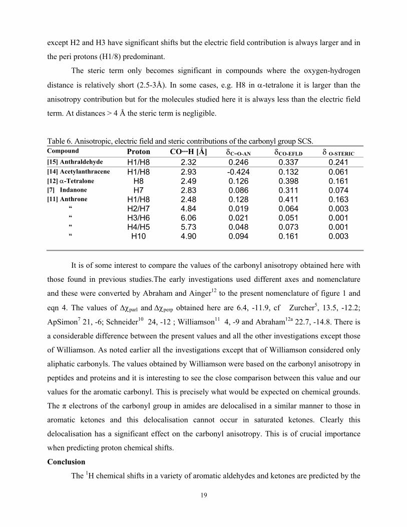
In CHARGE the components of the carbonyl substituent effect are given explicitly thus it
is of interest to determine the proportions of the carbonyl anisotropy, electric field and steric
effect at the various protons in the molecules. As the anisotropy and electric field are
propoprtional to r-3 and the steric effect to r-6 obviously the steric effect will be significant only
for near protons. Some illustrative examples are given in table 6 together with the C=O···H
distance.
Comparison of the results for the near H1/H8 protons in 9-anthraldehyde (15) and 9-
acetylanthracene (14) is of interest as in 15 the molecule is planar but in 14 the acetyl group is
perpendicular to the anthracene ring plane. The effect of the carbonyl anisotropy is strongly
deshielding in 15 but strongly shielding in 14. In contrast the electric field and steric effects are
the same sign in both molecules but much larger in 15 due to the closer proximity of the carbonyl
group and H1/8. Comparison of the anisotropy and electric field contributions is well illustrated
by the results for anthrone (11). They are both long range and all the protons of the compound
19
except H2 and H3 have significant shifts but the electric field contribution is always larger and in
the peri protons (H1/8) predominant.
The steric term only becomes significant in compounds where the oxygen-hydrogen
distance is relatively short (2.5-3Å). In some cases, e.g. H8 in α-tetralone it is larger than the
anisotropy contribution but for the molecules studied here it is always less than the electric field
term. At distances > 4 Å the steric term is negligible.
Table 6. Anisotropic, electric field and steric contributions of the carbonyl group SCS. Compound Proton CO···H [Å] δC=O-AN δCO-EFLD δ O-STERIC [15] Anthraldehyde H1/H8 2.32 0.246 0.337 0.241 [14] Acetylanthracene H1/H8 2.93 -0.424 0.132 0.061 [12] α-Tetralone H8 2.49 0.126 0.398 0.161 [7] Indanone H7 2.83 0.086 0.311 0.074 [11] Anthrone H1/H8 2.48 0.128 0.411 0.163 “ H2/H7 4.84 0.019 0.064 0.003 “ H3/H6 6.06 0.021 0.051 0.001 “ H4/H5 5.73 0.048 0.073 0.001 “ H10 4.90 0.094 0.161 0.003
It is of some interest to compare the values of the carbonyl anisotropy obtained here with
those found in previous studies.The early investigations used different axes and nomenclature
and these were converted by Abraham and Ainger12 to the present nomenclature of figure 1 and
eqn 4. The values of ∆χparl and ∆χperp obtained here are 6.4, -11.9, cf Zurcher5, 13.5, -12.2;
ApSimon7 21, -6; Schneider10 24, -12 ; Williamson11 4, -9 and Abraham12a 22.7, -14.8. There is
a considerable difference between the present values and all the other investigations except those
of Williamson. As noted earlier all the investigations except that of Williamson considered only
aliphatic carbonyls. The values obtained by Williamson were based on the carbonyl anisotropy in
peptides and proteins and it is interesting to see the close comparison between this value and our
values for the aromatic carbonyl. This is precisely what would be expected on chemical grounds.
The π electrons of the carbonyl group in amides are delocalised in a similar manner to those in
aromatic ketones and this delocalisation cannot occur in saturated ketones. Clearly this
delocalisation has a significant effect on the carbonyl anisotropy. This is of crucial importance
when predicting proton chemical shifts.
Conclusion
The 1H chemical shifts in a variety of aromatic aldehydes and ketones are predicted by the
20
CHARGE routine to within 0.1ppm. This together with previous results for aliphatic carbonyl
compounds allows the CHARGE programme to predict the 1H chemical shift of any aldehyde
and ketone to essentially experimental accuracy. The carbonyl anisotropy in the aromatic
ketones was shown to be similar to that in amides but much less than the value in aliphatic
ketones..
The keto/enol tautomerism in anthrone/9-hydroxy anthracene was observed by NMR and
the percentage of enol was shown to be proportional to the Kamlett β hydrogen bonding effect of
the solvent and not to the solvent polarity.
Acknowledgements
We thank the EPSRC and GSK for a CASE research studentship (M.M.) and GSK for the
750 MHz spectrum of 7 and 14. We thank Dr. M.H. Abraham for helpful discussions on the solvent
parameter scales.
References
1.Part 18, R.J.Abraham and M.Reid, J. Chem. Soc. Perkin Trans.2, 2002, 1081.
2. L.M.Jackman, Nucl.Mag.Res.Spec, p 112-30, Pergamon Press,1959
3. J.A.Pople, J..Chem.Phys, 1962, 37, 53,60.
4. A.A.Bothner-By and J.A.Pople, Ann.Rev.Phys.Chem., 1965, 16, 43.
5. R.F. Zurcher, Prog.Nuclear.Mag.Res.Spec, 1967, 2, 205.
6. H.M.McConnell, J..Chem.Phys, 1957, 27, 226.
7.a) J.W.ApSimon, P.V.DeMarco and D.W. Mathieson , Tetrahedron,1970, 26, 119.
b) J.W.ApSimon and H.Beierbeck, Canad J.Chem.,1971, 49, 1328.
8. J. Homer and D.Callagham, J.Chem.Soc.A, 1968, 439.
9. K.J.Toyne, Tetrahedron,1973, 29, 3889.
10. H.J.Schneider,U.Buchheit,N.Becker,G.Shmidt and U.Siehl, J.Am.Chem.Soc.,1985,107,7027.
11. a) M.P.Williamson and T.Asakura, J.Mag.Res., 1991,94,557.
b) ibid,1993, B101,63.
c) M.P.Williamson, T.Asakura, E.Nakamura and M.Demura, J.Biomol.NMR,1992,2,83.
12. R.J.Abraham and N.J.Ainger, J.Chem.Soc.Perk.Trans.2,1999, 441.
12a. R.J.Abraham, unpublished results.
13. H.Lampert, W.Mikenda, A.Karpfen and H.Kahlig,J.Phys.Chem.,1997,101,9610.
14. G. Wu, M.D. Lumsden, G.C. Ossenkamp, K. Eichele and R.E. Wasylishen, J. Phys. Chem., 1995. 99 ,15806.
15. R.J.Abraham, Progress in NMR Spectroscopy, 1999,35,85.
21
16. a) R.J.Abraham and P.E.Smith, J.Comp. Chem.,1987, 9, 288.
b) R.J.Abraham and P.E.Smith, J.Comp. Aid. Molec. Design,1989, 3, 175.
17. Aldrich Chem. Co. Eastmank Kodak Co., Rochester, USA.
18. Bruker XWINNMR version 3.0, Bruker AM, Silbersteifen, D-7512 Germany.
19. J.S.Meek, P.A.Monroe, C.J.Bouboulis, J.Org.Chem.,1963, 28,2572.
20. PC Model 7.0. 1998, Serena Software: Box 3076, Bloomington, USA.
21. M.J.Frisch, G.W.Trucks, H.B.Schlegel, G.E.Scuseria, M.A.Robb, J.R.Cheeseman,
V.G.Zakrzewski, J.A.Montgomery, Jr., R.E.Stratmann, J.C.Burant, S.Dapprich, J.M.Millam,
A.D.Daniels, K.N.Kudin, M.C.Strain, O.Farkas, J.Tomasi, V.Barone, M.Cossi, R.Cammi,
B.Mennucci, C.Pomelli, C.Adamo, S.Clifford, J.Ochterski, G.A.Petersson, P.Y.Ayala,
Q.Cui, K.Morokuma, D.K.Malick, A.D.Rabuck, K.Raghavachari, J.B.Foresman,
J.Cioslowski, J.V.Ortiz, A.G.Baboul, B.B.Stefanov, G.Liu, A.Liashenko, P.Piskorz,
I.Komaromi, R.Gomperts, R.L.Martin, D.J.Fox, T.Keith, M.A.Al-Laham, C.Y.Peng,
A.Nanayakkara, M.Challacombe, P.M.W.Gill, B.Johnson, W.Chen, M.W.Wong, J.L.Andres,
C.Gonzalez, M.Head-Gordon, E.S.Replogle and J. A. Pople., GAUSSIAN 98, Revision A9.
1998, Gaussian inc.: Pittsburg PA.
22. J.B.Foresman and A.Frisch, Exploring Chemistry with Electronic Structure Methods,
Gaussian
inc.,1993,Pittsburgh,USA.
23. R.J.Abraham,M.Canton,M.Reid and L.Griffiths, J.Chem.Soc.Perk.Trans.2,2000, 803.
24. R.J.Abraham,D.J.Chadwick and F.Sancassan, J.Chem.Soc.Perk.Trans.2,1984,1037.
25. R.J.Abraham,D.J.Chadwick and F.Sancassan, J.Chem.Soc.Perk.Trans.2,1989,1377.
26. R.J.Abraham and M.S.Lucas, J.Chem.Soc.Perk.Trans.2,1988,1269.
27. R.J.Abraham,H.A.Bergen and D.J.Chadwick, J.Chem.Soc.Perk.Trans.2,1983,1161.
28. R.J.Abraham,D.J.Chadwick and F.Sancassan, J.Chem.Soc.Perk.Trans.2,1988,169.
29. H. Lampert, W. Mikenda, A. Karpfen and H. Kählig, J. Phys. Chem. A, 1997. 101, 9610.
30. F. Sancassan, G. Petrillo and R.J. Abraham, J. Chem. Soc., Perkin Trans 2, 1995, 1965.
31. R.J.Abraham, Analysis of High Resolution NMR Spectra, ch 4, Elsevier, London, 1971.
32. C. Cobas, J. Cruces and F. J. Sardina, MestRe-C. 2000, Universidad de Santiago de Compostela,
33. R.J.Abraham, J.Fisher and P.Loftus, Introduction to NMR Spectroscopy, p.23, J. Wiley 1988
34.a) T. Schaefer, R. Sebastian, D.M. McKinnon, P.W. Spevack, K.J. Cox and C.S. Takeuchi, Can. J. Chem, 1993. 71, 960.
22
b) T. Schaefer, J. Peeling and A. Wildman, Org. Mag. Res, 1984. 22, 477.
35. H.Günther, NMR Spectroscopy. 2nd ed. 1995: John Wiley &Sons.
36. M.Mobli, Annual report, Liverpool University, 2002.
37. J.L.Marshall, A.M.Ihrig and D.E.Miller J.Mag.Res.,1974,16, 439,
38. K.H.Meyer, Justus Liebigs Ann. Chem, 1911. 379, 70.
39. P.Novak, D. Skare, S. Sekusak and D. Vikic-Topic, Croatia Chemica Acta, 2000. 73,1153.
40. M.J. Kamlet, J.M. Abboud, M.H. Abraham, R.W. Taft, J. Org. Chem., 1983. 48, 2877
41. R.J. Abraham, E.J. Chambers and W.A. Thomas, J. Chem. Soc. Perkin Trans. 2, 1993,1061.
42. Kuo SS, Computer Applications of Numerical Methods, ch 8,Addison-Wesley, London, 1972.
43. J.Epsztajn, A. Bieniek, J.Z. Brzezinski and H. Kalinowski, Tetrahedron, 1986. 42, 3559.
44. P.Laszlo, Progress in NMR Spectroscopy, 1968, 3, 203.
45. R.J.Abraham and E.Bretschneider, ch 13, Internal Rotation in Molecules, Ed. W.J.Orville-
Thomas,Academic Press, NY, 1974.