Embed Size (px)
DESCRIPTION
Script
Citation preview

Comprehensive Functional Maps of the Antigen-binding Site of an Anti-ErbB2 Antibody Obtained withShotgun Scanning Mutagenesis
Felix F. Vajdos1, Camellia W. Adams1, Timothy N. Breece2
Leonard G. Presta3, Abraham M. de Vos1 and Sachdev S. Sidhu1*
1Department of ProteinEngineering, Genentech Inc.1 DNA WaySouth San FranciscoCA 94080, USA
2Department of ProcessSciences, Genentech Inc.1 DNA WaySouth San FranciscoCA 94080, USA
3Department of ImmunologyGenentech Inc., 1 DNA WaySouth San Francisco, CA 94080USA
Shotgun scanning combinatorial mutagenesis was used to study theantigen-binding site of Fab2C4, a humanized monoclonal antibody frag-ment that binds to the extracellular domain of the human oncogeneproduct ErbB2. Essentially all the residues in the Fab2C4 complementaritydetermining regions (CDRs) were alanine-scanned using phage-displayedlibraries that preferentially allowed side-chains to vary as the wild-type oralanine. A separate homolog-scan was performed using libraries thatallowed side-chains to vary only as the wild-type or a similar amino acidresidue. Following binding selections to isolate functional clones, DNAsequencing was used to determine the wild-type/mutant ratios at eachvaried position, and these ratios were used to assess the contributions ofeach side-chain to antigen binding. The alanine-scan revealed that mostof the side-chains that contribute to antigen binding are located in theheavy chain, and the Fab2C4 three-dimensional structure revealed thatthese residues fall into two groups. The first group consists of solvent-exposed residues which likely make energetically favorable contacts withthe antigen and thus comprise the functional-binding epitope. The secondgroup consists of buried residues with side-chains that pack against otherCDR residues and apparently act as scaffolding to maintain the func-tional epitope in a binding-competent conformation. The homolog-scaninvolved subtle mutations, and as a result, only a subset of the side-chainsthat were intolerant to alanine substitutions were also intolerant to homo-logous substitutions. In particular, the 610 A2 functional epitope surfacerevealed by alanine-scanning shrunk to only 369 A2 when mapped withhomologous substitutions, suggesting that this smaller subset of side-chains may be involved in more precise contacts with the antigen. Theresults validate shotgun scanning as a rapid and accurate method fordetermining the functional contributions of individual side-chainsinvolved in protein–protein interactions.
q 2002 Elsevier Science Ltd. All rights reserved
Keywords: phage display; protein engineering; combinatorial mutagenesis;antibody; shotgun scanning*Corresponding author
Introduction
Monoclonal antibodies have proven invaluable as reagents in biological chemistry, and more recently, astherapeutic agents.1 The field of antibody engineering is concerned with technologies that can be used to
0022-2836/02/$ - see front matter q 2002 Elsevier Science Ltd. All rights reserved
Present address: L. G. Presta, DNAX Research Institute of Molecular and Cellular Biology, Inc., 901 CaliforniaAvenue, Palo Alto, CA 94304, USA.
E-mail address of the corresponding author: [email protected]
Abbreviations used: BSA, bovine serum albumin; CC, correlation coefficient; CDR, complementarity determiningregion; CDR-Hn, (where n ¼ 1, 2, or 3), heavy chain CDR 1, 2, or 3; CDR-Ln, (where n ¼ 1, 2, or 3), light chain CDR 1, 2,or 3; cP3, C-terminal domain of the M13 bacteriophage gene-3 minor coat protein; ECD, extracellular domain; ELISA,enzyme-linked immunosorbant assay; Fab, antigen-binding fragment; Fv, variable fragment; PBS, phosphate-bufferedsaline; rmsd, root mean square deviation; wt, wild-type.
doi:10.1016/S0022-2836(02)00264-4 available online at http://www.idealibrary.com onBw
J. Mol. Biol. (2002) 320, 415–428
dissect and rationalize the requirements for anti-body structure and function.2 This knowledge canthen be used to improve or alter particular anti-body–antigen interactions, or even to engineercompletely novel-binding specificities.
The specificity and affinity of an antibody for itscognate antigen is determined by the sequenceand structure of the variable fragment (Fv): aheterodimer consisting of the N-terminal domainsof the heavy and light chains. Even within the Fv,antigen binding is primarily mediated by thecomplementarity determining regions (CDRs), sixhypervariable loops (three each in the heavy andlight chains) which together present a large con-tiguous surface for potential antigen binding.Aside from the CDRs, the Fv also contains morehighly conserved framework segments whichconnect the CDRs and are mainly involved insupporting the CDR loop conformations,3,4
although in some cases, framework residues alsocontact antigen.5,6 As an important step to under-standing how a particular antibody functions, itwould be very useful to assess the contributions
of each CDR side-chain to antigen binding, and inso doing, to produce a functional map of the anti-gen-binding site.
Site-directed mutagenesis is a powerful tool formapping binding energetics at protein–proteininterfaces.7,8 In this process, individual DNAcodons are systematically altered and the corre-sponding mutant proteins are expressed, purified,and assayed for activity relative to the wild-type.The effects of individual side-chain substitutionscan then be assessed in terms of DDGmut – wt, thedifference in binding free energy between themutant and wild-type protein. By analyzing panelsof point mutants, a detailed map of the bindingenergetics can be obtained, but the process can bevery laborious because individual mutant proteinsmust be made and analyzed separately. In par-ticular, a comprehensive analysis of an antigen-binding site would ideally encompass all CDRresidues, and this would require the analysis ofdozens or even hundreds of point mutants.9,10
Recently, a general and rapid combinatorialmutagenesis strategy has been developed forexploring protein structure and function.11
“Shotgun scanning” mutagenesis uses phage-displayed libraries of protein mutants constructedusing degenerate codons with restricted diversity.For example, codons may be chosen to preferen-tially allow the wild-type (wt) or alanine in thecase of a shotgun alanine-scan. The library pool isthen subjected to binding selections to enrich forclones that retain affinity for a binding partner,and following selection, DNA sequencing is usedto determine the ratio of wild-type/mutant (wt/mut) at each varied position. This ratio can beused to assess binding contributions of each side-chain with good correlation to those obtained withtraditional site-directed mutagenesis. The methodis very rapid because many side-chains are simul-taneously scanned with a single library, and theanalysis is based on DNA sequencing whichcircumvents the need for protein purification andbiophysical analysis.
We used the shotgun scanning approach tostudy the antigen-binding site of a humanizedmonoclonal antibody (humAb2C4) that binds tothe extracellular domain of the human receptortyrosine kinase ErbB2 (ErbB2-ECD, Kd ¼ 8:5 nM),and in so doing, inhibits tumor growth (C.W.A.,unpublished results). The antigen-binding portionof humAb2C4 was displayed on M13 bacterio-phage in an Fab format (Fab2C4), i.e. a heterodimerconsisting of the light chain and the variable andfirst constant domains of the heavy chain. Weconducted two different shotgun scans, with eachscan covering essentially the complete sequencesof all six CDRs. With a shotgun alanine-scan, weassessed the effects of removing all side-chainatoms past the b-carbon, fairly drastic mutationsthat can be used to infer the roles of individualside-chains in protein structure and function.7
We also conducted a more subtle scan, termed ashotgun homolog-scan, in which we substituted
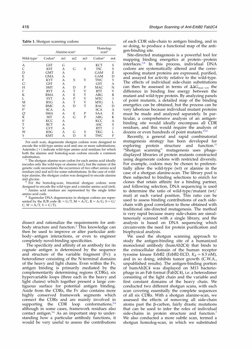
Table 1. Shotgun scanning codons
Alanine-scana
Homolog-scanb
Wild-typec Codond m1 m2 m3 Codond m4
A GST G KCT SC KST A G S TSC SD GMT A GAM EE GMA A GAM DF KYT A S V TWC YGp GST A GST AH SMT A D P MAC NIp RYT A T V RTT VK RMA A E T ARG RL SYT A P V MTC IM RYG A T V MTG LNp RMC A D T RAC DPp SCA A SCA AQp SMA A E P SAA ER SST A G P ARG KSp KCC A KCC AT RCT A ASC SV GYT A RTT IW KSG A G S TKG LY KMT A D S TWC F
For each scan, degenerate shotgun codons were designed toencode the wild-type amino acid and one or more substitutions.Asterisks ( p ) indicate wild-type amino acid residues for whichboth the alanine and homolog-scan codons encode a commonsubstitution.
a The shotgun alanine-scan codon for each amino acid ideallyencodes only the wild-type or alanine (m1), but the nature of thegenetic code necessitates the occurrence of two other amino acidresidues (m2 and m3) for some substitutions. In the case of wild-type alanine, the shotgun codon was designed to encode alanineand glycine.
b For the homolog-scan, binomial shotgun codons weredesigned to encode the wild-type and a similar amino acid (m4).
c Amino acid residues are represented by the single letteramino acid code.
d Equimolar DNA degeneracies in shotgun codons are repre-sented by the IUB code (K ¼ G/T, M ¼ A/C, R ¼ A/G, S ¼ G/C, W ¼A/T, Y ¼ C/T).
416 Shotgun Scanning of Anti-ErbB2 Fab2C4

each wild-type residue with a similar amino acid,to gain insight into which positions require preciseside-chain geometries and chemistry. When themutagenesis results were mapped onto the three-dimensional crystal structure of Fab2C4, each scanprovided a comprehensive view of how the CDRside-chains contribute to the formation of a func-
tional antigen-binding site. The two views aredistinct yet complementary: together, they providea clearer understanding of antibody structure andfunction than would be possible with either scanalone.
Results
Shotgun alanine-scan of Fab2C4
For the shotgun alanine-scan, we replaced wtcodons with degenerate codons that ideallyencoded the wt amino acid or alanine (m1 inTable 1), although the nature of the genetic code
Figure 1. Sequences of the Fab2C4 CDRs. The sequence of each CDR is shown along with the position of each resi-due in the numbering scheme† of Kabat et al.37 Residues shown to be important for ErbB2-ECD binding in either theshotgun alanine or homolog-scan are shown in bold or underlined, respectively (Fwt/mut .10, see Tables 3 and 4).Asterisks ( p ) indicate residues that were not analyzed in the shotgun scans.
Table 2. Fab2C4 shotgun scanning libraries
Mutated regions Diversity
Library CDRs ResiduesShotguncodons
Mutagenicoligonucleotides Theoretical Actual
HAa H1,H2,H3
T28, T30, D31, Y32, T33, D50, V51, N52, N53, S54, I58,N60, Q61, N95, L96, P98, S99
Alanine H1-A1, H2-A1,H3-A1
3.3 £ 107 1.5 £ 1010
HAb H1,H2,H3
D35, P52a, G55, G56, S57, Y59, R62, F63, K64, G65, G97,F99a, Y99b, F100, D101, Y102
Alanine H1-A2, H2-A2,H3-A2
1.7 £ 107 2.4 £ 1010
LAa L1,L2,L3
Q27, D28, S30, I31, G32, S50, S52, Y53, Y55, Y91, Y92, I93,Y94, Y96
Alanine L1-A1, L2-A1,L3-A1
8.3 £ 107 1.4 £ 1010
LAb L1,L2,L3
K24, A25, S26, V29, V33, A34, A51, R54, T56, Q89, Q90,P95, T97
Alanine L1-A2, L2-A2,L3-A2
1.6 £ 104 2.5 £ 1010
HHa H1,H3
T28, T30, D31, Y32, T33, M34, D35, N95, L96, G97, P98,S99, F99a, Y99b, F100, D101, Y102
Homolog H1-H, H3-H 1.3 £ 105 2.4 £ 1010
HHb H2 D50, V51, N52, P52a, N53, S54, G55, G56, S57, I58, Y59,N60, Q61, R62, F63, K64, G65
Homolog H2-H 1.3 £ 105 2.2 £ 1010
LH L1,L2,L3
K24, A25, S26, Q27, D28, V29, S30, I31, G32, V33, A34,S50, A51, S52, Y53, R54, Y55, T56, Q89, Q90, Y91, Y92, I93,
Y94, P95, Y96, T97
Homolog L1-H, L2-H,L3-H
1.3 £ 108 2.4 £ 1010
Libraries were designed to replace the codons for the indicated residues with either alanine-scan or homolog-scan shotgun codons(Table 1). Libraries were constructed using the indicated mutagenic oligonucleotides (see Materials and Methods), and in each case,the theoretical diversity (the number of amino acid combinations encoded by the mutagenic oligonucleotides) was exceeded at least100-fold by the actual diversity of the constructed library.
† Antibody residues are designated by a letter in lowercase italics denoting the heavy or light chain (h or l,respectively), followed by the amino acid in the one-letter code, followed by the position in the chain. Forexample, h D101 denotes an aspartic acid residue atposition 101 in the heavy chain.
Shotgun Scanning of Anti-ErbB2 Fab2C4 417

necessitated two other amino acid substitutions forsome residues (m2 and m3 in Table 1). In positionswhere alanine was the wt, we used a degeneratecodon that encoded alanine or glycine. The sixCDRs of Fab2C4 encompass a total of 64 residues(Figure 1). We constructed two libraries (HAa andHAb) that together covered 33 of the 37 heavychain CDR residues and two libraries (LAa andLAb) that together covered all 27 light chain CDRresidues (Figure 1 and Table 2). Each library con-tained .1010 unique members, and thus in eachcase, the theoretical diversity for combinatorialmutagenesis at the scanned positions wasexceeded by at least 100-fold (Table 2).
Phage pools from each library were subjected totwo different selections. The first selection (displayselection) isolated variants capable of binding to amonoclonal antibody specific for the epitope tagfused to the N terminus of the Fab2C4 light chain.The second selection (antigen selection) isolatedvariants capable of binding to ErbB2-ECD. Closeto 100 binding clones were sequenced from each
selection; the sequences were aligned, and at eachmutated position, the occurrences of wt oreach designed substitution were tabulated (seeMaterials and Methods for details). For each selec-tion, these data were used to calculate the wt/mutratio for each mutation at each position (Tables 3and 4).
Because the wt/mut ratio is the statistical prefer-ence for the wt relative to the mutant, it correlateswith the effect of each mutation on the selectedtrait (i.e. binding to the anti-tag antibody orErbB2-ECD). Ratios greater than or less than 1indicate deleterious or beneficial mutations,respectively.
The anti-tag antibody selected for phage variantsthat displayed assembled Fab2C4 fragments con-taining both the heavy and light chains. This isbecause the heavy chain was fused directly to abacteriophage coat protein while the epitope tagwas fused to the light chain N terminus. Thus, theanti-tag antibody only binds to phage particlesthat contain a light chain associated with the
Table 3. Fab2C4 light chain shotgun scan
Wt/mut ratios
Antigen selection Display selection Fwt/mut
Residuea Wt/m1 Wt/m2 Wt/m3 Wt/m4 Wt/m1 Wt/m2 Wt/m3 Wt/m4 m1 m2 m3 m4
K24 0.89 4.2 0.96 0.88 0.42 0.79 0.52 1.0 2.1 5.3 1.8 0.86A25 3.7 2.8 2.0 1.6 1.8 1.8S26p 3.5 2.8 2.9 1.5 1.2p 1.9p
Q27p 0.67 1.5 2.5 0.51 0.88 1.2 0.94 0.73 0.76 1.3p 2.7 0.70p
D28 1.1 1.8 0.99 1.9 1.1 1.0V29 6.1 3.5 2.5 2.0 2.4 1.8S30p 1.8 1.1 1.5 0.87 1.1p 1.3p
I31p 0.91 2.8 0.57 0.64 1.7 2.7 0.56 0.55 0.53 3.7 1.0p 1.2p
G32p 3.3 4.8 2.9 3.9 1.1p 1.2p
V33 16 3.1 3.3 2.8 4.8 1.1A34 16 5.5 3.6 2.5 4.6 2.2
S50p 1.0 0.78 1.3 0.87 0.77p 0.89p
A51 1.7 1.6 0.90 0.85 1.9 1.8S52p 1.3 1.2 1.5 1.7 0.85p 0.70p
Y53 1.9 97 4.4 1.4 1.6 3.5 1.2 1.3 1.2 28 3.7 1.1R54 3.2 4.1 1.9 3.0 1.7 3.7 1.0 2.4 1.8 1.1 1.9 1.3Y55 32 80 53 4.8 1.4 2.3 0.89 0.95 23 35 60 5.1T56 0.49 0.88 0.89 0.76 0.55 1.2
Q89p 8.8 10 70 3.6 0.77 2.4 3.4 1.9 11 4.2p 21 1.8p
Q90p 2.4 1.1 .36 0.67 0.88 1.9 2.3 0.71 2.7 0.58p .16 0.94p
Y91 .166 .166 166 0.94 1.8 3.5 0.97 1.2 .92 .47 138 0.76p
Y92 1.2 3.7 1.1 0.88 1.3 2.1 0.84 0.6 0.96 1.8 0.76 1.5I93p 1.7 1.6 0.81 0.69 1.7 1.5 0.64 0.53 1.0 1.1 1.3p 1.3p
Y94 6.7 30 5.5 1.3 1.9 3.0 1.7 0.63 3.6 10 3.2 2.0P95p 13 9.7 1.1 1.74 12p 5.6p
Y96 0.99 .66 2.1 0.36 2.1 18 2.2 0.91 0.48 .3.7 0.95 0.40T97 0.56 0.28 0.89 0.35 0.62 0.80
For each of the listed light chain residues, the effect of each mutation (Table 1) was assessed using data from either the alanine-scanlibraries (m1, m2, and m3) or the homolog-scan libraries (m4) described in Table 2. The wt/mut ratios were determined from thesequences of binding clones isolated after selection for binding to either the ErbB2-ECD (antigen selection) or an anti-tag antibody(display selection). The function ratio (Fwt/mut) for each mutation was derived by dividing the antigen selection wt/mut ratio by thedisplay selection wt/mut ratio. Fwt/mut provides a quantitative estimate of the effect of each mutation on the binding affinity ofFab2C4 for ErbB2-ECD. Deleterious effects are indicated by Fwt/mut values greater than 1.0, and mutations that have large deleteriouseffects (Fwt/mut .10) are shown in bold text. In cases where a particular mutation was not observed amongst the antigen selectionsequences, only a lower limit could be defined for the wt/mut ratio and the Fwt/mut (indicated by a greater than sign). Asterisks ( p )indicate residues for which the alanine and homolog-scan codons encoded a common substitution.
a Residues are denoted by the single letter amino acid code and are numbered according to the scheme of Kabat et al.37
418 Shotgun Scanning of Anti-ErbB2 Fab2C4

phage-displayed heavy chain. Most of the wt/mutratios for the display selection were close to 1.0,indicating that the mutations did not significantlyaffect Fab2C4 display levels (Tables 3 and 4). How-ever, several mutations exhibited wt/mut ratiossignificantly greater than 1.0 (e.g. h P52aA, h Y59A,h F63A), suggesting that these mutations reduceddisplay. Conversely, for a few mutations, wt/mutratios significantly less than 1.0 suggest that thesemutations may actually increase display (e.g.h D35A, h L96A).
In the selection for binding to ErbB2-ECD,mutations could effect the selection either byaltering the level of Fab2C4 display (as in thedisplay selection), or alternatively, by directly orindirectly altering the side-chains that make
binding contacts with the antigen. In this selection,alanine substitutions at three light chain positions(Table 3) and 21 heavy chain positions (Table 4)exhibited wt/mut ratios greater than 10.
To obtain a quantitative estimate of each muta-tion’s effect on ErbB2-ECD binding affinity, wedivided the wt/mut ratio from the antigen selec-tion by the wt/mut ratio from the display selec-tion. This operation corrected for effects onFab2C4 display and provided a number which wetermed the function ratio (Fwt/mut). As we haveshown previously, the Fwt/mut value for eachmutation is approximately equal to the corre-sponding ratio of equilibrium binding constants(Ka,wt/Ka,mut),
11 and thus, it provides a goodestimate of the effect of each mutation on the
Table 4. Fab2C4 heavy chain shotgun scan
Wt/mut ratios
Antigen selection Display selection Fwt/mut
Residuea Wt/m1 Wt/m2 Wt/m3 Wt/m4 Wt/m1 Wt/m2 Wt/m3 Wt/m4 m1 m2 m3 m4
T28 4.5 0.94 0.7 0.47 6.4 2.0T30 0.33 0.27 0.7 0.39 0.47 0.69D31 170 29 1.4 1.1 120 26Y32 .161 .161 .161 17 2.0 3.1 1.1 0.85 .81 .52 .150 20T33 20 8.9 0.94 0.38 21 23M34 NDb ND ND 2.2 ND ND ND 0.88 ND ND ND 2.5D35 2.8 14 0.14 0.90 20 15
D50 170 .91 0.24 0.41 710 .220V51 10 1.3 1.1 1.8 9.4 0.73N52p .168 168 84 .91 0.41 0.34 0.80 0.83 .410 490p 110 .110p
P52ap 72 14 6.1 0.62 12p 23p
N53p .166 166 .166 .91 1.4 0.97 2.6 0.57 .120 170p .64 .160 p
S54p 84 .91 0.33 1.1 260p .83 p
G55p 14 90 0.40 2.9 34p 31p
G56p 0.60 0.36 5.0 2.6 0.12p 0.14p
S57p 7.0 0.47 4.4 0.86 1.6p 0.55p
I58p 45 45 4.5 2.1 0.86 0.95 0.51 0.61 53 47 8.8p 3.4p
Y59 33 .59 9.8 0.78 8.7 10.4 1.8 0.58 3.8 .5.7 5.4 1.3N60p 4.8 4.4 120 3.0 1.2 0.91 15 1.8 4.0 4.8p 8.0 1.7p
Q61p 2.6 0.98 1.1 0.69 0.53 0.42 2.0 0.71 4.8 2.3p 0.55 0.97p
R62 4.3 .44 4.0 1.3 1.2 15 0.24 1.2 3.6 2.9 17 1.0F63 26 26 4.6 3.2 6.6 2.2 8.8 4.0 4.4 12 0.52 0.81K64 54 54 6.0 0.57 4.9 7.7 2.7 0.67 12 7.0 2.2 0.85G65p 5.8 9.1 2.50 3.9 2.3p 2.4p
N95p .170 21 .170 21 1.8 2.0 2.1 3.1 .98 11p 84 6.9p
L96 23 .45 0.35 1.5 0.11 0.33 0.19 1.2 210 .140 1.8 1.3G97p .78 89 3.3 2.1 .24 p 42p
P98p .178 29 1.9 0.44 .94 p 65p
S99p 2.8 7.0 0.55 1.6 5.0p 4.4p
F99a .75 .75 .75 10 2.4 5.4 1.3 1.1 .31 14 58 9.1Y99b .74 74 74 1.7 0.8 4.1 1.7 0.49 .93 18 44 3.5F100 77 .77 77 17 2.6 5.9 1.5 5.1 30 13 51 3.3D101 9.1 .87 1.1 2.5 8.3 .35Y102 8.3 7.5 3.2 2.8 2.3 1.9 2.1 0.92 3.6 3.9 1.5 3.0
For each of the listed heavy chain residues, the effect of each mutation (Table 1) was assessed using data from either the alanine-scan libraries (m1, m2, and m3) or the homolog-scan libraries (m4) described in Table 2. The wt/mut ratios were determined fromthe sequences of binding clones isolated after selection for binding to either the ErbB2-ECD (antigen selection) or an anti-tag antibody(display selection). The function ratio (Fwt/mut) for each mutation was derived by dividing the antigen selection wt/mut ratio by thedisplay selection wt/mut ratio. Fwt/mut provides a quantitative estimate of the effect of each mutation on the binding affinity ofFab2C4 for ErbB2-ECD. Deleterious effects are indicated by Fwt/mut values greater than 1.0, and mutations that have large deleteriouseffects (Fwt/mut .10) are shown in bold text. In cases where a particular mutation was not observed amongst the antigen selectionsequences, only a lower limit could be defined for the wt/mut ratio and the Fwt/mut (indicated by a greater than sign). Asterisks ( p )indicate residues for which the alanine and homolog-scan codons encoded a common substitution.
a Residues are denoted by the single letter amino acid code and are numbered according to the scheme of Kabat et al.37
b ND indicates that these values were not determined, because we forgot to include this residue in the alanine-scan libraries.
Shotgun Scanning of Anti-ErbB2 Fab2C4 419

equilibrium binding constant between Fab2C4 andErbB2-ECD. Alanine substitutions at three lightchain positions and 19 heavy chain positionsexhibited Fwt/Ala values greater than 10, indicatingthat side-chains at these positions contributesignificantly to the binding affinity of Fab2C4 forErbB2-ECD (Tables 3 and 4, Figure 2).
Shotgun homolog-scan of Fab2C4
In the shotgun homolog-scan libraries, eachscanned position was represented by a binomialcodon that encoded only the wild-type and asimilar amino acid (Table 1). We constructed two
libraries (HHa and HHb) that together covered 34heavy chain CDR residues and a single library(LH) that covered all 27 light chain CDR residues(Figure 1 and Table 2). As with the alanine-scans,the library diversities were sufficient to exceed thetheoretical diversities by at least 100-fold (Table 2).
Each library was subjected to separate selectionsfor binding to anti-tag antibody or ErbB2-ECDand Fwt/mut values were determined for eachmutation, as described above for shotgun alanine-scanning. The Fwt/mut values for many homologsubstitutions were significantly lower than thosefor the corresponding alanine substitutions; nolight chain residues and only 13 heavy chain
Figure 2. Fwt/mut values measuring the effects of Fab2C4 CDR mutations on the binding affinity for ErbB2-ECD.Values are shown for either alanine (black bars) or homolog (white bars) substitutions. Data for (a) the light chainwere from Table 3, and data for (b) the heavy chain were from Table 4 (except the mutation hM34A for which theEC50,mut/EC50,wt-value from Table 5 was plotted).
420 Shotgun Scanning of Anti-ErbB2 Fab2C4
residues exhibited Fwt/mut values greater than 10(Tables 3 and 4, Figure 2).
Comparison of Fwt/mut values for identicalmutations in different libraries
While most substitutions in the homolog-scanwere designed to be different from those in thealanine-scan, there was some overlap (residueswith asterisks in Table 1). Glycine, proline andserine were substituted with alanine in both scans.Furthermore, for asparagine, glutamine, and iso-
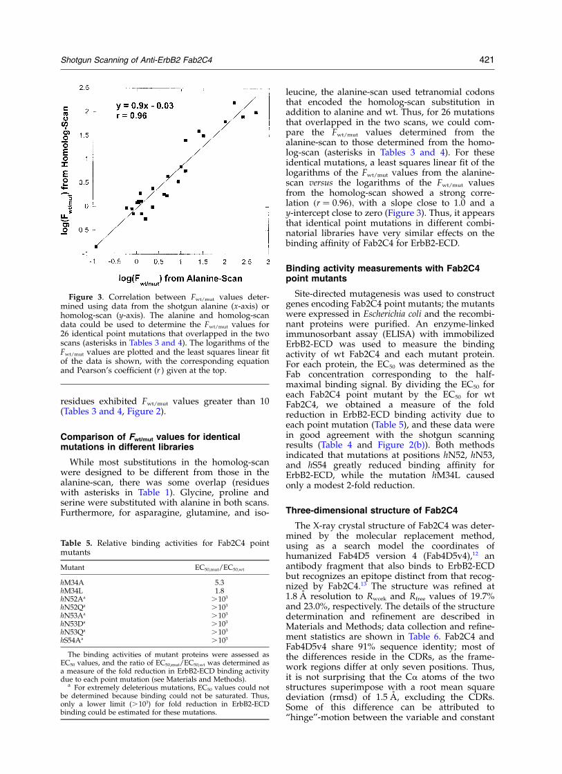
leucine, the alanine-scan used tetranomial codonsthat encoded the homolog-scan substitution inaddition to alanine and wt. Thus, for 26 mutationsthat overlapped in the two scans, we could com-pare the Fwt/mut values determined from thealanine-scan to those determined from the homo-log-scan (asterisks in Tables 3 and 4). For theseidentical mutations, a least squares linear fit of thelogarithms of the Fwt/mut values from the alanine-scan versus the logarithms of the Fwt/mut valuesfrom the homolog-scan showed a strong corre-lation ðr ¼ 0:96Þ; with a slope close to 1.0 and ay-intercept close to zero (Figure 3). Thus, it appearsthat identical point mutations in different combi-natorial libraries have very similar effects on thebinding affinity of Fab2C4 for ErbB2-ECD.
Binding activity measurements with Fab2C4point mutants
Site-directed mutagenesis was used to constructgenes encoding Fab2C4 point mutants; the mutantswere expressed in Escherichia coli and the recombi-nant proteins were purified. An enzyme-linkedimmunosorbant assay (ELISA) with immobilizedErbB2-ECD was used to measure the bindingactivity of wt Fab2C4 and each mutant protein.For each protein, the EC50 was determined as theFab concentration corresponding to the half-maximal binding signal. By dividing the EC50 foreach Fab2C4 point mutant by the EC50 for wtFab2C4, we obtained a measure of the foldreduction in ErbB2-ECD binding activity due toeach point mutation (Table 5), and these data werein good agreement with the shotgun scanningresults (Table 4 and Figure 2(b)). Both methodsindicated that mutations at positions hN52, hN53,and hS54 greatly reduced binding affinity forErbB2-ECD, while the mutation hM34L causedonly a modest 2-fold reduction.
Three-dimensional structure of Fab2C4
The X-ray crystal structure of Fab2C4 was deter-mined by the molecular replacement method,using as a search model the coordinates ofhumanized Fab4D5 version 4 (Fab4D5v4),12 anantibody fragment that also binds to ErbB2-ECDbut recognizes an epitope distinct from that recog-nized by Fab2C4.13 The structure was refined at1.8 A resolution to Rwork and Rfree values of 19.7%and 23.0%, respectively. The details of the structuredetermination and refinement are described inMaterials and Methods; data collection and refine-ment statistics are shown in Table 6. Fab2C4 andFab4D5v4 share 91% sequence identity; most ofthe differences reside in the CDRs, as the frame-work regions differ at only seven positions. Thus,it is not surprising that the Ca atoms of the twostructures superimpose with a root mean squaredeviation (rmsd) of 1.5 A, excluding the CDRs.Some of this difference can be attributed to“hinge”-motion between the variable and constant
Figure 3. Correlation between Fwt/mut values deter-mined using data from the shotgun alanine (x-axis) orhomolog-scan (y-axis). The alanine and homolog-scandata could be used to determine the Fwt/mut values for26 identical point mutations that overlapped in the twoscans (asterisks in Tables 3 and 4). The logarithms of theFwt/mut values are plotted and the least squares linear fitof the data is shown, with the corresponding equationand Pearson’s coefficient (r ) given at the top.
Table 5. Relative binding activities for Fab2C4 pointmutants
Mutant EC50,mut/EC50,wt
hM34A 5.3hM34L 1.8hN52Aa .103
hN52Qa .103
hN53Aa .103
hN53Da .103
hN53Qa .103
hS54Aa .103
The binding activities of mutant proteins were assessed asEC50 values, and the ratio of EC50,mut/EC50,wt was determined asa measure of the fold reduction in ErbB2-ECD binding activitydue to each point mutation (see Materials and Methods).
a For extremely deleterious mutations, EC50 values could notbe determined because binding could not be saturated. Thus,only a lower limit (.103) for fold reduction in ErbB2-ECDbinding could be estimated for these mutations.
Shotgun Scanning of Anti-ErbB2 Fab2C4 421
domains, as the rmsd between the two structuresdecreases to 0.7 A or 0.9 A when the superpositionis performed using only the constant domains orthe variable domain frameworks, respectively. Allresidues in the CDRs of Fab2C4 are well ordered,with the exception of h L100. The disorder in thisregion appears to be correlated with disorderaround the immediately adjacent CDR-H1 residueh Y32. In comparison with Fab4D5v4, there is asingle amino acid deletion in the sequence ofFab2C4 that occurs in CDR-H3 and is accommo-dated by a completely altered backbone trajectory.Conservation of a hydrophobic patch in this regionis maintained by the aromatic ring of h F105 inFab2C4 lying in the same position as h W99 inFab4D5v4, despite the fact that these residues areat opposite ends of CDR-H3. The only differencein the light chain frameworks occurs at position66, where an arginine in Fab4D5v4 is substitutedby a glycine in Fab2C4, causing the polypeptidebackbone to undergo a significant rearrangement.
Discussion
Antibody affinity and specificity is predomi-nantly dictated by the six CDR loops that together
form the antigen-binding site. With the exceptionof CDR-H3, the conformations of the CDR main-chains do not vary greatly within different anti-bodies, and they can be classified into a limitednumber of “canonical structures”.14 Thus, themajor determinants of antibody specificity and affi-nity are the CDR side-chains. Side-chains can beclassified as buried or solvent exposed on thebasis of their solvent accessible surface area, andthese classifications have implications for the
Table 6. Data collection and refinement statistics forFab2C4
A. Unit cellSpace group P21
a (A) 41.97b (A) 64.25c (A) 79.44b (deg.) 105.44
B. Diffraction dataResolution (A) 15–1.8 (1.9–1.8)a
No. of reflections 85,734No. of unique reflections 36,884Rmerge
b 0.065 (0.328)a
Completeness (%) 97.6 (97.6)a
I/s(I ) 5.2 (1.4)a
Redundancy 2.3 (2.3)a
C. RefinementRwork
c 0.197Rfree
c 0.230No. of protein atoms 3323No. of water molecules 382No. of sulfate ions 2Average Bprotein (A2) 22.6Average Bwater molecules (A2) 33.8Average Bsulfate (A2) 61.1Rmsd bond length (A) 0.005Rmsd angles (deg.) 1.4Rmsd bonded Bs (A2) 1.8
a Values for the outer resolution shell are given inparantheses.
b Rmerge ¼ ShklðlIhkl 2 kIhklllÞ=ShklkIhkll; where Ihkl is the intensityof reflection hkl, and kIhkll is the average intensity of multipleobservations.
c Rwork ¼ SlFo 2 Fcl=SFo; where Fo and Fc are the observedand calculated structure factor amplitudes, respectively. Rfree isthe R-factor for a randomly selected 5% of reflections whichwere not used in the refinement.
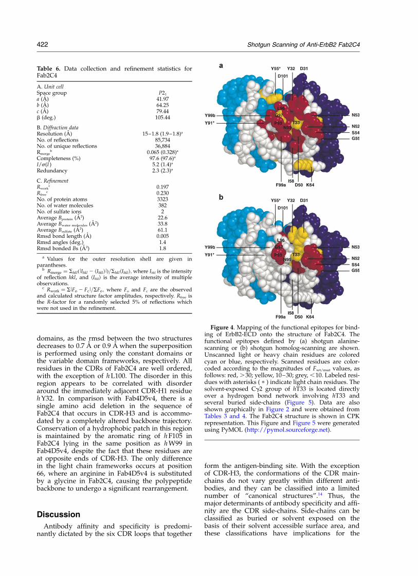
Figure 4. Mapping of the functional epitopes for bind-ing of ErbB2-ECD onto the structure of Fab2C4. Thefunctional epitopes defined by (a) shotgun alanine-scanning or (b) shotgun homolog-scanning are shown.Unscanned light or heavy chain residues are coloredcyan or blue, respectively. Scanned residues are color-coded according to the magnitudes of Fwt/mut values, asfollows: red, .30; yellow, 10–30; grey, ,10. Labeled resi-dues with asterisks ( p ) indicate light chain residues. Thesolvent-exposed Cg2 group of hT33 is located directlyover a hydrogen bond network involving hT33 andseveral buried side-chains (Figure 5). Data are alsoshown graphically in Figure 2 and were obtained fromTables 3 and 4. The Fab2C4 structure is shown in CPKrepresentation. This Figure and Figure 5 were generatedusing PyMOL (http://pymol.sourceforge.net).
422 Shotgun Scanning of Anti-ErbB2 Fab2C4
functional role of each CDR side-chain in antigenbinding.
Solvent-exposed side-chains cover the surface ofthe antigen-binding site where they can potentiallymake direct binding contacts with the antigen.Solvent-exposed side-chains that become buriedupon antigen binding are components of the“structural-binding epitope”, and within thisgroup, those that make energetic contributions tothe binding interaction constitute the “functional-binding epitope”.8,15 In contrast, buried side-chainsare unable to make significant direct contact withantigen, but they can still contribute to antigenbinding by acting as “scaffolding” residues thatpack against residues in the functional epitope,and in so doing, maintain the structural integrityof the antigen-binding site.16
Accurate definition of the structural-bindingepitope can only be achieved with an antibody–antigen co-crystal structure which provides athree-dimensional view of the binding interface.However, as we show here, considerable insightsinto antibody function can be gained with compre-hensive CDR mutagenesis data in conjunction withthe structure of an unbound antigen-binding site.Such a database can reveal both the functionalepitope that makes energetically favorable contactswith antigen, and also, the buried scaffolding side-chains that serve to maintain the functional epitopein a binding-competent conformation.
The functional-binding epitope of Fab2C4
When the shotgun alanine-scan results weremapped onto the X-ray crystal structure of Fab2C4(Figure 4(a)), they revealed a functional epitopecomprising two solvent-exposed “ridges”. Oneridge is composed of residues h I58, h P98, h F99a,
and h Y99b while the other contains residuesh D31, h Y32, hN52, hN53, and hS54. These residuesare all in the heavy chain, suggesting that much ofthe binding energy is derived from heavy chaininteractions. Intriguingly, the most important func-tional epitope residues revealed by the shotgunhomolog-scan (Figure 4(b)) constitute a subset ofthe residues found to be important in the shotgunalanine-scan. In particular, three residues inCDR-H2 (hN52, hN53, and hS54) form a smallpatch that was highly conserved in both scans,suggesting that this surface makes precise contactswith the antigen ErbB2-ECD. This prediction isfurther supported by affinity measurements withpoint-mutated proteins which also showed thatthese residues are extremely intolerant to substi-tutions (Table 5).
The functional epitope defined by the shotgunalanine-scan covers a surface area of 610 A2 in theFab2C4 structure, with almost all of this area(608 A2) being confined to the heavy chain. In con-trast, the functional epitope defined by the shotgunhomolog-scan covers only 369 A2. Thus, the func-tional epitope can be divided into two distinctregions. One subset of the surface appears tomake relatively non-specific interactions withErbB2-ECD, while a much smaller surface appearsto make more specific contacts with the antigen.Interestingly, this smaller surface, revealed asfunctionally important by both scans, is restrictedentirely to the heavy chain; the side-chainsfound to be important only in the alanine-scan arelocated at the dimer interface between the lightand heavy chains. The size of the functionalepitope identified by the alanine-scan is similar tothat of structural-binding epitopes revealed byX-ray crystal structures of antibody–antigencomplexes.17,18
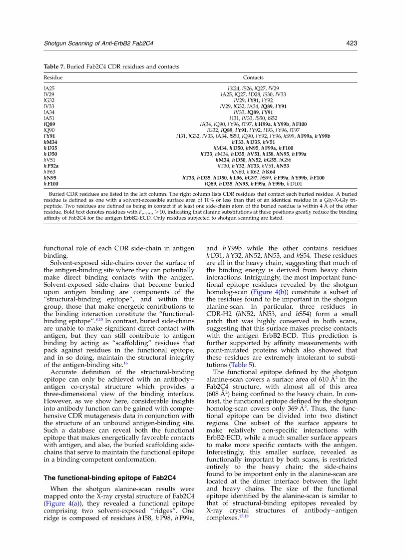
Table 7. Buried Fab2C4 CDR residues and contacts
Residue Contacts
lA25 l K24, lS26, lQ27, lV29lV29 lA25, lQ27, l D28, lS30, lV33lG32 lV29, l Y91, l Y92lV33 lV29, lG32, lA34, lQ89, l Y91lA34 lV33, lQ89, l Y91lA51 l I31, lV33, lS50, lS52lQ89 lA34, lQ90, l Y96, lT97, h H99a, h Y99b, h F100lQ90 lG32, lQ89, l Y91, l Y92, l I93, l Y96, lT97l Y91 l I31, lG32, lV33, lA34, lS50, lQ90, l Y92, l Y96, hS99, h F99a, h Y99bhM34 hT33, h D35, hV51h D35 hM34, h D50, hN95, h F99a, h F100h D50 hT33, hM34, h D35, hV51, h I58, hN95, h F99ahV51 hM34, h D50, hN52, hG55, hG56h P52a hT30, h Y32, hT33, hV51, hN53h F63 hN60, h R62, h K64hN95 hT33, h D35, h D50, h L96, hG97, hS99, h F99a, h Y99b, h F100h F100 lQ89, h D35, hN95, h F99a, h Y99b, h D101
Buried CDR residues are listed in the left column. The right column lists CDR residues that contact each buried residue. A buriedresidue is defined as one with a solvent-accessible surface area of 10% or less than that of an identical residue in a Gly-X-Gly tri-peptide. Two residues are defined as being in contact if at least one side-chain atom of the buried residue is within 4 A of the otherresidue. Bold text denotes residues with Fwt/Ala .10, indicating that alanine substitutions at these positions greatly reduce the bindingaffinity of Fab2C4 for the antigen ErbB2-ECD. Only residues subjected to shotgun scanning are listed.
Shotgun Scanning of Anti-ErbB2 Fab2C4 423

The role of scaffold residues in antigen binding
Of the 61 CDR residues that were subjected toshotgun scanning, 17 can be classified as buried(Table 7), if a buried residue is defined as onewith a solvent accessible surface area of 10% orless than that of an identical residue in a Gly-X-Gly tripeptide. Substitutions for eight of the nineburied light chain residues have little effect onErbB2-ECD binding. l Y91 is the only buried lightchain residue which was found to be intolerantto substitution in the alanine-scan (Table 3 andFigure 2(a)). However, the homolog-scan showedthat substitution of l Y91 with phenyalanine iswell tolerated, indicating that the contribution ofthe tyrosine side-chain to antigen-binding resultsfrom the volume occupied by the large, hydro-phobic phenyl ring rather than the side-chainhydroxyl group. l Y91 makes contact with anumber of CDR residues, including heavy chainresidues that contribute significantly to antigenbinding (Table 7), and thus, it appears to buttressthe functional-binding epitope. Similar buttressinginteractions have been shown to effect the
binding affinities of other antigen–antibodyinteractions.16,19
In contrast to the light chain residues, mostburied heavy chain residues appear to be involvedin scaffolding interactions that are critical for anti-gen binding. All eight buried heavy chain residuesthat we scanned preferred wt over alanine, withsix residues exhibiting Fwt/ala values greater than10 (Table 4 and Figure 2(b)). The buried heavychain residues make numerous contacts with func-tionally important CDR residues (Table 7), and inso doing, apparently fix the Fab2C4 functionalepitope in a productive binding conformation.
Antibody CDRs contain an unusually largeproportion of buried hydrophilic residues, in com-parison with other protein folds or even otherregions of antibody structure.20 Indeed, many ofthe buried CDR residues in Fab2C4 are hydrophilic(Table 7), and several of these side-chains partici-pate in important scaffolding interactions that areessential for high affinity antigen binding (Figure2(b)). In particular, it appears that the confor-mations of the heavy chain CDRs are maintainednot only by hydrophobic contacts, but also by ahydrogen bond network involving buried CDRside-chains (Figure 5). Although the Fab2C4crystals were grown at near neutral pH, the buriedside-chains of h D35 and h D50 are oriented nearlydirectly towards each other and their Od1 atomsare in close proximity (2.4 A distance), suggestingthat one or both of these carboxylates exist in aprotonated state. The hydrogen bond network alsoinvolves the side-chains of hN95, hT33 and h W47,and thus, it connects residues from each of thethree heavy chain CDRs. The need for precisegeometry and chemistry within the network isreflected in the intolerance of the interacting side-chains to substitution not only with alanine, butalso with homologous side-chains (Table 4 andFigure 2(b)).
Reproducibility and reliability ofshotgun scanning
Shotgun scanning uses statistical analysis ofcombinatorial libraries to predict the functionalcontributions of individual protein side-chains.For these predictions to be valid, two importantconditions must be met. First, the prediction forany individual mutation should be independent ofthe combinatorial library used for the scan, or inother words, the predicted effect of an identicalmutation in two different libraries should be thesame. Second, the shotgun scanning predictionsshould be in good agreement with results obtainedwith affinity measurements for point-mutatedproteins. The Fab2C4 shotgun scan results meetboth criteria.
The alanine and homolog-scan data setscontained 26 overlapping Fwt/mut values (asterisksin Tables 3 and 4), allowing us to compare thepredicted functional effects for identical mutationsobtained from different libraries. As shown in
Figure 5. The buried hydrogen bond network in theheavy chain of Fab2C4. Side-chain atoms are shown assticks colored as follows: carbon, gray; oxygen, red;nitrogen, blue. Main chains are shown as gray tubesand hydrogen bonds are shown as dashed magentalines. The hydrogen bond network ties together residuesfrom each of the three heavy chain CDRs, and with theexception of hT33, all the interacting side-chains areburied. The g-OH group of hT33 is anchored by thehydrogen bonds, resulting in the Cg2 group beingsolvent exposed at the base of a central depression inthe Fab2C4 functional epitope (Figure 4).
424 Shotgun Scanning of Anti-ErbB2 Fab2C4

Figure 3, the two scans gave remarkably similarestimates of functional importance for identicalmutations. Thus, data from different libraries canbe compared with each other with high confidence,as the results for individual mutations are highlyreproducible and independent of the library.Furthermore, these results suggest that most side-chains in the Fab2C4 CDRs act additively andindependently in the binding interaction withErbB2-ECD, since the effects of individualmutations do not appear to be significantly influ-enced by mutations at other CDR positions. Theshotgun scanning predictions are also in goodagreement with affinity data for purified pointmutants. Shotgun homolog-scanning predictedthat the mutation hM34L should result in a modest2.5-fold reduction in affinity (Table 4), and indeed,the introduction of this mutation into Fab2C4reduced binding to ErbB2-ECD by about 2-fold(Table 5). In contrast, shotgun scanning identifiedresidues hN52, hN53, and hS54 as critical com-ponents of a small solvent-exposed patch that washighly intolerant to substitutions. Again, affinitymeasurements with purified proteins confirmedthis prediction, as all substitutions at these sitesresulted in Fab2C4 mutants with greatly compro-mised antigen-binding activity (Table 5).
Thus, the results of the two shotgun scans werein good agreement with each other and with theresults obtained with purified point-mutatedproteins. In conjunction with the Fab2C4 crystalstructure, the scanning data provided compre-hensive functional maps of the Fab2C4 antigen-binding site. Together, the alanine and homolog-scanning maps allowed for the accurate definitionof both the functional binding epitope, and also,the buried scaffolding side-chains that hold thisepitope in a binding-competent conformation. Thedata should be useful in guiding the design ofnew phage-displayed libraries for further optimi-zation of the binding interaction between Fab2C4and ErbB2-ECD. Furthermore, the general shotgunscanning technology should facilitate the rapidanalysis of many other protein–proteininteractions.
Materials and Methods
Oligonucleotides
DNA degeneracies are represented in the IUB code(K ¼ G/T, M ¼ A/C, R ¼ A/G, S ¼ G/C, W ¼A/T,Y ¼ C/T). Degenerate codons are shown in bold text.The following mutagenic oligonucleotides were used forlibrary constructions:
H1-A1: GCA GCT TCT GGC TTC RCT TTC RCTGMT KMT RCT ATG GAC TGG GTC CGT
H1-A2: GCA GCT TCT GGC TTC ACC TTC ACCGAC TAT ACC ATG GMT TGG GTC CGT CAG GCC
H2-A1: CTG GAA TGG GTT GCA GMT GYT RMCCCT RMC KCC GGC GGC TCT RYT TAT RMC SMACGC TTC AAG GGC CGT
H2-A2: CTG GAA TGG GTT GCA GAT GTT AATSCA AAC AGT GST GST KCC ATC KMT AACCAG SST KYT RMA GST CGT TTC ACT CTG AGT
H3-A1: TAT TAT TGT GCT CGT RMC SYT GGASCA KCC TTC TAC TTT GAC TAC
H3-A2: TAT TAT TGT GCT CGT AAC CTG GSTCCC TCT KYT KMT KYT GMT KMT TGG GGTCAA GGA ACC
H1-H: GCA GCT TCT GGC TTC ASC TTC ASCGAM TWC ASC MTG GAM TGG GTC CGT CAGGCC
H2-H: GGC CTG GAA TGG GTT GCA GAM RTTRAC SCA RAC KCC GST GST KCC RTT TWCRAC SAA CGC TTC AAG GCC CGT
H3-H: TAT TAT TGT GCT CGT RAC MTC GSTSCA KCC TWC TWC TWC GAM TWC TGG GGTCAA GGA ACC
L1-A1: ACC TGC AAG GCC AGT SMA GMT GTGKCC RYT GST GTC GCC TGG TAT CAA
L1-A2: GTC ACC ATC ACC TGC RMA GST KCCCAG GAT GYT TCT ATT GGT GYT GST TGG TATCAA CAG AAA CCA
L2-A1: AAA CTA CTG ATT TAC KCC GCT KCCKMT CGA KMT ACT GGA GTC CCT TCT
L2-A2: AAA CTA CTG ATT TAC TCG GST TCCTAC SST TAC RCT GGA GTC CCT TCT CGC
L3-A1: TAT TAC TGT CAA CAA KMT KMT RYTKMT CCT KMT ACG TTT GGA CAG GGT
L3-A2: GCA ACT TAT TAC TGT SMA SMA TATTAT ATT TAT SCA TAC RCT TTT GGA CAG GGTACC
L1-H: GTC ACC ATC ACC TGC ARG KCC KCCSAA GAM RTT KCC RTT GST RTT KCC TGG TATCAA CAG AAA CCA
L2-H: AAA CTA CTG ATT TAC KCC KCC KCCTWC ARG TWC ASC GGA GTC CCT TCT CGC
L3-H: GCA ACT TAT TAC TGT SAA SAA TWCTWC RTT TWC SCA TWC ASC TTT GGA CAGGGT ACC
Construction of shotgun scanning libraries
To display Fab2C4 on the surface of M13 bacterio-phage, we modified a previously described phagemid(pS1602) designed for the display of human growthfused to the C-terminal domain of the M13 gene-3minor coat protein (cP3).21 Standard molecular biologytechniques were used to replace the fragment of pS1602encoding human growth hormone with a DNA fragmentencoding the light and heavy chains of Fab2C4. Theresulting phagemid (pC2C4) contained a bicistronicgene under the control of the IPTG-inducible Ptac
promoter.22 The first open reading frame encoded a poly-peptide consisting of the maltose binding proteinsecretion signal, followed by an epitope tag (amino acidsequence: MADPNRFRGKDLGG),23 followed by theFab2C4 light chain. The second open reading frameencoded a polypeptide consisting of the stII secretionsignal,24 followed by the Fab2C4 heavy chain, followedby cP3. Expression in E. coli resulted in the periplasmicsecretion of free Fab2C4 light chain and Fab2C4 heavychain fused to cP3, and the two chains spontaneouslyassociated to assemble functional Fab2C4. E. coli culturesharboring pC2C4 were coinfected with M13-VCS helperphage (Stratagene) and grown at 37 8C without IPTGinduction, resulting in the production of M13 bacterio-phage displaying Fab2C4 in a monovalent format.
Shotgun Scanning of Anti-ErbB2 Fab2C4 425

Libraries were constructed using previously describedmethods25 with appropriately designed “stop template”versions of pC2C4. For each library, we used a stop tem-plate which contained TAA stop codons within each ofthe CDRs to be mutated. The stop template was used asthe template for the Kunkel mutagenesis method26 withmutagenic oligonucleotides (see above) designed tosimultaneously repair the stop codons and introducemutations at the desired sites. The libraries are describedin Table 2.
Library sorting and binding assays
NUNC 96-well Maxisorp immunoplates were coatedovernight at 4 8C with capture target (either ErbB2-ECDor an anti-tag antibody at 5 mg/ml) and blocked for twohours with bovine serum albumin (BSA) (Sigma). Phagefrom the libraries described above were propagated inE. coli XL1-blue with the addition of M13-VCS helperphage. After overnight growth at 37 8C, phage wereconcentrated by precipitation with PEG/NaCl and resus-pended in phosphate-buffered saline (PBS), 0.5% (w/v)BSA, 0.1% (v/v) Tween 20 (Sigma), as describedpreviously.25 Phage solutions (1013 phage/ml) wereadded to the coated immunoplates. Following a two-hour incubation to allow for phage binding, the plateswere washed 12 times with PBS, 0.05% Tween 20.Bound phage were eluted with 0.1 M HCl for tenminutes and the eluant was neutralized with 1.0 M Trisbase. Eluted phage were amplified in E. coli XL1-blueand used for further rounds of selection.
Individual clones from each round of selection weregrown in a 96-well format in 500 ml of 2YT brothsupplemented with carbenicillin and M13-VCS, and theculture supernatants were used directly in phageELISAs25 to detect phage-displayed Fab2C4 variants thatbound to either ErbB2-ECD or anti-tag antibody. Afterone round of selection for anti-tag binding or two roundsof selection for ErbB2-ECD binding, greater than 50% ofthe clones exhibited positive phage ELISA signals atleast 2-fold greater than signals on control plates coatedwith BSA. These positive clones were subjected to DNAsequence analysis.
DNA sequencing and analysis
Culture supernatants containing phage particles wereused as templates for PCRs that amplified DNA frag-ments containing the light and heavy chain genes. ThePCR primers were designed to add M13(-21) and M13Runiversal sequencing primers at either end of the ampli-fied fragment, thus facilitating the use of these primersin sequencing reactions. Amplified DNA fragmentswere sequenced using Big-Dye terminator sequencingreactions which were analyzed on an ABI Prism 370096-capillary DNA analyzer (PE Biosystems, Foster City,CA). All reactions were performed in a 96-well format.
The sequences were analyzed with the programSGCOUNT as described previously.11 SGCOUNT alignedeach DNA sequence against the wild-type DNAsequence by using a Needleman–Wunch pairwise align-ment algorithm, translated each aligned sequence ofacceptable quality, and tabulated the occurrence of eachnatural amino acid at each position. For the ErbB2-ECDbinding selection, the number of analyzed clones areindicated in parenthesis following the name of eachlibrary: HAa (178), HAb (67), LAa (167), LAb (85), HHa(89), HHb (64), LH (65). For the anti-tag antibody-
binding selection, the following number of clones wereanalyzed for each library: HAa (94), HAb (91), LAa(183), Lab (72), HHa (73), HHb (73), LH (84).
ELISA for measuring the binding of Fab2C4 mutantsto ErbB2-ECD
Fab2C4 and mutant proteins were produced using apreviously described expression vector with an alkalinephosphatase promoter.27 Fab2C4 mutants were con-structed using the Kunkel site-directed mutagenesismethod.26 For protein production, expression plasmidswere transformed into E. coli 16C9, cultures were grownin modified AP5 medium, and Fab proteins werepurified using protein G-sepharose CL-4B (AmershamPharmacia), as described previously.27 Fab proteins werebuffer exchanged into 10 mM sodium succinate,140 mM NaCl (pH 6.0), and concentrated using a Centri-con-10 (Amicon). Protein concentrations were deter-mined by quantitative amino acid analysis.
The binding activities of Fab2C4 and mutants weremeasured with an ELISA. NUNC 96-well maxisorpimmunoplates were coated overnight at 4 8C withErbB2-ECD (1 mg/ml in 50 mM carbonate buffer (pH9.6)). The plates were blocked for one hour at roomtemperature with ELISA diluent buffer (PBS, 0.5% BSA,0.05% Tween 20). Serial dilutions of Fab protein wereincubated on the ErbB2-ECD-coated plates for twohours at room temperature, and the plates were washedwith PBS, 0.05% Tween 20. Bound Fab proteins weredetected with biotinylated murine anti-human kappachain antibody followed by streptavidin/horseradishperoxidase conjugate (Sigma), using 3,30,5,50-tetramethylbenzidine (TMB) as substrate (Kirkegaard and PerryLaboratories, Gaithersburg, MD). Titration curves werefit with a four-parameter non-linear regression curve-fitting program (KaleidaGraph, Synergy Software) todetermine the EC50 values, the Fab concentrations corre-sponding to half-maximal binding signals. The foldreduction in ErbB2-ECD binding activity due to eachpoint mutation was determined by dividing the EC50 forthe Fab2C4 point mutant by the EC50 for wild-typeFab2C4 (Table 5).
Purification of Fab2C4 protein forX-ray crystallography
Fab 2C4 was produced in E. coli 16C9 using a pre-viously described expression vector with an alkalinephosphatase promoter.27 Cells were pelleted by centrifu-gation, resuspended in PBS (5 ml/g of cell paste), anddisrupted by three passes through a MicrofluidizerHC8000 (7000 PSI). The suspension was adjusted to50 mM magnesium sulfate and 0.2% (v/v) polyethyleneimine, stirred for 30 minutes, and centrifuged for onehour in a Sorval RC3B centrifuge (4500 rpm) to removesolids. The supernatant was decanted and sterile filtered(0.22 mm). A Gammabind þ protein G Sepharose column(Amersham Pharmacia) was equilibrated in PBS, thesupernatant was loaded at 40 ml/ml of resin, and elutedin 0.1 M acetic acid, 25 mM NaCl (pH 2.8). The eluantwas adjusted to pH 6.0 with 2.0 M Tris base, mixed withan equal volume of 1.0 M sodium sulfate, sterile filtered(0.22 mm), and loaded on a J. T. Baker Hi-Propyl column(Mallinckrodt-Baker, Phillipsburg, NJ) at 5 mg/ml ofresin. Fab2C4 protein was eluted with a 20-columnvolume gradient from 0.5 M sodium sulfate, 10 mMsuccinate (pH 5.0) to 10 mM succinate (pH 5.0). The
426 Shotgun Scanning of Anti-ErbB2 Fab2C4

Hi-propyl pool was diluted to a conductivity of 2.5 mSwith purified water and loaded on a J. T. Baker ABXcolumn (Mallinckrodt-Baker, Phillipsburg, NJ) at1.0 mg/ml of resin. The ABX column was eluted with a20 column volume gradient from 50 mM Hepes (pH 7.1)to 100 mM NaCl, 50 mM Hepes (pH 7.1). The resultingpool was concentrated in an Amicon stirred cell with aYM10 membrane (Millipore, Bedford, MA) and formu-lated in 10 mM succinate (pH 5.0) on a sephadex G-25column (Amersham Pharmacia) (0.1 ml/ml of resin).
Crystallization and data collection
Prior to crystallization experiments, Fab2C4 proteinwas dialyzed against deionized water, then concentratedto 22.5 mg/ml using a Centricon 10 microconcentrator(Millipore, Bedford, MA). Crystallization was performedwith the sitting drop vapor diffusion method and used2 ml protein solution and 2 ml precipitant (35–45%saturated (NH4)2SO4, 0.1 M Tris–HCl (pH 8.0)). After 24hours, the drops were microseeded with 1.0 ml of serialdilutions of a fresh seed stock solution. Thin, plate-likecrystals appeared within hours, and grew to a maximumsize of 0.1 mm £ 0.05 mm £ 0.01 mm within one week.Crystals were harvested into a cryo-protectant solutionconsisting of 45% saturated (NH4)2SO4, 0.1 M Tris–HCl,20% (v/v) glycerol (pH 8.0), and immediately flashcooled in liquid nitrogen. Initially, a data set complete to2.7 A was collected using a Mar image plate detectormounted on a rotating anode X-ray source operating ata wavelength of 1.5418 A. This data set was used for themolecular replacement calculations and for the initialrefinement steps. Later, a separate 1.8 A data set wascollected from the same specimen at the AdvancedLight Source, Beamline 5.0.2 at a wavelength of 1.1 A.The data were integrated using the programMOSFLM28,29 and scaled using the program SCALA.30,31
Intensities were converted to amplitudes by the methodof French & Wilson32 using the program TRUNCATE,and Free R flags were assigned using the programUNIQUE.31 Data statistics are reported in Table 6.
Structure determination and refinement
The structure was determined by molecular replace-ment using the coordinates of Fab4D5 version 4 (PDBcode 1fvd)12 and the program AMORE.31,33 All databetween 10 A and 3 A were used. The highest peak inthe rotation function search had a correlation coefficient(CC) of 0.28, while the next highest peak had a CC of0.18. The correct solution was readily identified in thetranslation function, with a CC of 0.38 (next highest0.20) and an R-factor of 49% (next best 54.2%). Rigidbody refinement further improved the model, producinga CC of 47.3%, and an R-factor of 47.2%. The singlemolecule in the asymmetric unit of these crystals resultsin a solvent content of 42% and a correspondingMatthews coefficient of 2.1 A3/Da. At this point, non-identical residues between Fab4D5 and Fab2C4 wereremoved from the model and the model was subjectedto rigid body refinement using the program CNX34
(MSI, San Diego, CA), and all data between 6.0 A and2.7 A, treating the variable and constant domains of theheavy and light chains as independent groups. Thisreduced the R-factor to 46.9% ðRfree ¼ 46:6%Þ: Torsionangle molecular dynamics, followed by Powell minimi-zation and grouped (main-chain and side-chain) B-factorrefinement reduced the R-factor against 8–2.7 A data to
32.3% ðRfree ¼ 42%Þ: Automatic refinement continuedusing the high resolution data, this time using theCartesian molecular dynamics simulated annealingprotocol in CNX. The R-factor at this point was 27.1%ðRfree ¼ 30:5%Þ: Inspection of SigmaA weighted maps32,35
clearly indicated the conformations of the omitted resi-dues, and 12 cycles of automatic refinement, followedby manual rebuilding, resulted in the final model,which consists of residues 1–214 of the light chain, resi-dues 1–223 of the heavy chain, 382 water molecules,and two sulfate ions. The final R-factor was 19.7%ðRfree ¼ 23:0%Þ: The refinement statistics are shown inTable 6.
Protein Data Bank accession number
The coordinates and structure factors of Fab2C4 havebeen deposited with the Research Collaboratory forStructural Bioinformatics PDB36 under accession number1L7I.
Acknowledgments
We thank Alan Zhong for DNA sequencing and theGenentech DNA synthesis group for oligonucleotides.We thank the staff of the Macromolecular Crystal-lography Facility at the Advanced Light Source(Berkeley, CA) for assistance with the high-resolutionX-ray data set. The Advanced Light Source is supportedby the Director, Office of Science, Office of Basic EnergySciences, Materials Sciences Division, of the USDepartment of Energy under Contract No. DE-AC03-76SF00098 at Lawrence Berkeley National Laboratory.
References
1. Hudson, P. J. (1999). Recombinant antibody con-structs in cancer therapy. Curr. Opin. Immunol. 11,547–557.
2. Dall’Acqua, W. & Carter, P. (1998). Antibody engi-neering. Curr. Opin. Struct. Biol. 8, 443–450.
3. Chothia, C., Lesk, A. M., Tramontano, A., Levitt, M.,Smith-Gill, S. J., Air, G. et al. (1989). Conformationsof immunoglobulin hypervariable regions. Nature,342, 877–883.
4. Tramontano, A., Chothia, C. & Lesk, A. M. (1990).Framework residue 71 is a major determinant of theposition and conformation of the second hyper-variable region in the VH domains of immunoglobu-lins. J. Mol. Biol. 215, 175–182.
5. Padlan, E. A., Silverton, E. W., Sheriff, S., Cohen,G. H., Smith-Gill, S. J. & Davies, D. R. (1989). Struc-ture of an antibody–antigen complex: crystal struc-ture of the HyHEL-10 Fab-lysozyme complex. Proc.Natl Acad. Sci. USA, 86, 5938–5942.
6. Bentley, G. A., Boulot, G., Riottot, M. M. & Poljak,R. J. (1990). Three-dimensional structure of an idio-tope–anti-idiotope complex. Nature, 348, 254–257.
7. Wells, J. A. (1991). Systematic mutational analyses ofprotein–protein interfaces. Methods Enzymol. 202,390–411.
8. Clackson, T. & Wells, J. A. (1995). A hot spot of bind-ing energy in a hormone–receptor interface. Science,267, 383–386.
Shotgun Scanning of Anti-ErbB2 Fab2C4 427

9. Kelley, R. F. & O’Connell, M. P. (1993). Thermo-dynamic analysis of an antibody functional epitope.Biochemistry, 32, 6828–6835.
10. Chen, G., Dubrawsky, I., Mendez, P., Georgiou, G. &Iverson, B. L. (1999). In vitro scanning mutagenesisof all the specificity determining residues in anantibody binding site. Protein Eng. 12, 349–356.
11. Weiss, G. A., Watanabe, C. K., Zhong, A., Goddard,A. & Sidhu, S. S. (2000). Rapid mapping of proteinfunctional epitopes by combinatorial alaninescanning. Proc. Natl Acad. Sci. USA, 97, 8950–8954.
12. Eigenbrot, C., Randal, M., Presta, L., Carter, P. &Kossiakoff, A. A. (1993). X-ray structures of the anti-gen-binding domains from three variants ofhumanized anti-p185HER2 antibody 4D5 and com-parison with molecular modeling. J. Mol. Biol. 229,969–995.
13. Fendly, B. M., Winget, M., Hudziak, R. M., Lipari,M. T., Napier, M. A. & Ullrich, A. (1990). Characteri-zation of murine monoclonal antibodies reactive toeither the human epidermal growth factor orHER2/neu gene product. Cancer Res. 50, 1550–1558.
14. Chothia, C., Lesk, A. M., Tramontano, A., Levitt, M.,Smith-Gill, S. J., Air, G. et al. (1989). Conformationsof immunoglobulin hypervariable regions. Nature,342, 877–883.
15. Clackson, T., Ultsch, M. H., Wells, J. A. & de Vos,A. M. (1998). Structural and functional analysis ofthe 1:1 growth hormone:receptor complex revealsthe molecular basis for receptor affinity. J. Mol. Biol.277, 1111–1128.
16. Schildbach, J. F., Near, R. I., Bruccoleri, R. E., Haber,E., Jeffrey, P. D., Novotny, J. et al. (1993). Modulationof antibody affinity by a non-contact residue. ProteinSci. 2, 206–214.
17. Janin, J. & Chothia, C. (1990). The structure ofprotein–protein recognition sites. J. Biol. Chem. 265,16027–16030.
18. Davies, D. R., Sheriff, S. & Padlan, E. A. (1988). Anti-body–antigen complexes. J. Biol. Chem. 263,10541–10544.
19. Hawkins, R. E., Russell, S. J., Baier, M. & Winter, G.(1993). The contribution of contact and non-contactresidues of antibody in the affinity of binding toantigen. J. Mol. Biol. 234, 958–964.
20. Padlan, E. A. (1990). On the nature of antibody com-bining sites: unusual structural features that mayconfer on these sites an enhanced capacity for bind-ing ligands. Proteins: Struct. Funct. Genet. 7, 112–124.
21. Sidhu, S. S. (2000). Phage display in pharmaceuticalbiotechnology. Curr. Opin. Biotechnol. 11, 610–616.
22. Amman, E. & Brosius, J. (1985). “ATG vectors” forregulated high-level expression of cloned genes inEscherichia coli. Gene, 40, 183–190.
23. Lasky, L. A. & Dowbenko, D. J. (1984). DNAsequence analysis of the type-common glycoprotein-D genes of herpes simplex virus types 1 and 2.DNA, 3, 23–29.
24. Lowman, H. B., Bass, S. H., Simpson, N. & Wells, J. A.(1991). Selecting high-affinity binding proteins bymonovalent phage display. Biochemistry, 30,10832–10838.
25. Sidhu, S. S., Lowman, H. B., Cunningham, B. C. &Wells, J. A. (2000). Phage display for selection ofnovel binding peptides. Methods Enzymol. 328,333–363.
26. Kunkel, T. A., Roberts, J. D. & Zakour, R. A. (1987).Rapid and efficient site-specific mutagenesis withoutphenotypic selection. Methods Enzymol. 154, 367–382.
27. Presta, L. G., Chen, H., O’Connor, S. J., Chisholm, V.,Meng, Y. G., Krummen, L. et al. (1997). Humani-zation of an anti-vascular endothelial growth factormonoclonal antibody for the therapy of solid tumorsand other disorders. Cancer Res. 57, 4593–4599.
28. Powell, H. R. (1999). The Rossman Fourier auto-indexing algorithm in MOSFLM. Acta Crystallog.sect. D, 55, 1690–1695.
29. Leslie, A. G. W. (1992). Recent changes to theMOSFLM package for processing film and imageplate data. Joint CCPR ESF-EAMCB Newsletter ProteinCrystallog. 26.
30. Evans, P. R. (1993). Data reduction. Proceedings ofCCP4 Study Weekend
31. Collaborative Computational Project No. 4 (1994).The CCP4 suite: programs for protein crystal-lography. Acta Crystallog. sect. D, 50, 760–763.
32. French, S. & Wilson, K. (1978). On the treatment ofnegative intensity observations. Acta Crystallog. sect.A, 34, 517–525.
33. Navaza, J. (1994). AmoRe: an automated package formolecular replacement. Acta Crystallog. sect. D, 50,157–163.
34. Brunger, A. T., Adams, P. D., Clore, G. M., DeLano,W. L., Gros, P., Grosse-Kunstleve, R. W. et al. (1998).Crystallography and NMR system: a new softwaresuite for macromolecular structure determination.Acta Crystallog. sect. D, 54, 905–921.
35. Read, R. J. (1986). Improved Fourier coefficients formaps using phases from partial structures witherrors. Acta. Crystallog. sect. D, 42, 140–149.
36. Berman, H. M., Westbrook, J., Feng, Z., Gilliland, G.,Bhat, T. N., Weissig, H. et al. (2000). The ProteinData Bank. Nucl. Acids Res. 28, 235–242.
37. Kabat, E. A., Wu, T. T., Redi-Miller, M., Perry, H. M.& Gottesman, K. S. (1987). Sequences of Proteins ofImmunological Interest, 4th edit., National Institutesof Health, Bethesda, MD.
Edited by I. Wilson
(Received 14 January 2002; received in revised form 19 March 2002; accepted 22 March 2002)
428 Shotgun Scanning of Anti-ErbB2 Fab2C4