Embed Size (px)
Citation preview
(19) United States (12) Patent Application Publication (10) Pub. No.: US 2014/0328754 A1
US 20140328754A1
HINO et al. (43) Pub. Date: Nov. 6, 2014
(54) RADIOACTIVE METAL-LABELED Related U.S. Application Data ANT-CADHERN ANTIBODY (63) Continuation of application No. 13/578.462, filed on
(71) Applicants: Akihiro HINO, Sammu-shi (JP); Akio Aug. 10, 2012, now Pat. No. 8,815,211, filed as appli Nagano, S u-shi (JP); Masahiko cation No. PCT/JP2011/052759 on Feb. 9, 2011. Watanabe, Sammu-shi (JP); Tadasi O O Matsuura, Tokyo (JP); Hirokazu Satoh, (30) Foreign Application Priority Data Tokyo (JP); Fumiko Nomura, Tokyo Feb. 10, 2010 (JP 2010-028O28 (JP); Katsuyuki Mitomo, Tokyo (JP) eb. 1U, (JP) .................................
(72) Inventors: Akihiro HINO, Sammu-shi (JP); Akio Publication Classification Nagano, Sammu-shi (JP); Masahiko Watanabe, Sammu-shi (JP); Tadasi (51) Int. Cl. Matsuura, Tokyo (JP); Hirokazu Satoh, A615 L/It (2006.01) Tokyo (JP); Fumiko Nomura, Tokyo (52) U.S. Cl. (JP); Katsuyuki Mitomo, Tokyo (JP) CPC .................................. A61K 51/1027 (2013.01)
USPC ..................... 424/1.53: 530/387.9; 530/387.3 (73) Assignees: Fujifilm RI Pharma Co., LTD, Tokyo
(JP); Perseus Proteomics Inc., Tokyo (JP) (57) ABSTRACT
(21) Appl. No.: 14/307,589 A radioactive metal-labeled anti-cadherin antibody which is
(22) Filed: Jun. 18, 2014 obtained by binding a radioactive metallic element to an anti-cadherin antibody via a metal-chelating reagent.

US 2014/0328754 A1 Nov. 6, 2014 Sheet 1 of 14 Patent Application Publication
;-------------------+------------~--~~~*~~~~ ~~~~~~~~••••?•••••••••••••••••••?~~~~~~~~~~~~~ ž?$*******
************************
š?. 888; it {x :::::::::::::: ••••••*********************««««««««««^-~g

Patent Application Publication Nov. 6, 2014 Sheet 2 of 14 US 2014/0328754 A1

US 2014/0328754 A1 Nov. 6, 2014 Sheet 3 of 14
i: .
Patent Application Publication
US 2014/0328754 A1 Nov. 6, 2014 Sheet 4 of 14 Patent Application Publication
: 8. 88::::: a

US 2014/0328754 A1 Nov. 6, 2014 Sheet 5 of 14 Patent Application Publication
r:rs:-------------
$3:3: a x : 3::::::: 8:33:883:
****************
US 2014/0328754 A1
8:88 8:8; 38
Nov. 6, 2014 Sheet 6 of 14
*::: :
Patent Application Publication

US 2014/0328754 A1
8:33:838; 38:::::::8: ; :
Nov. 6, 2014 Sheet 7 of 14 Patent Application Publication

Patent Application Publication Nov. 6, 2014 Sheet 8 of 14 US 2014/0328754 A1
US 2014/0328754 A1 Nov. 6, 2014 Sheet 9 of 14 Patent Application Publication
| |
§§§§§.
* 3::::::::::::::::::::
2.3.3
s: 8
::::
3: 88:8 ? :
...tx....:

US 2014/0328754 A1 Nov. 6, 2014 Sheet 10 of 14 Patent Application Publication
W.
. :-

US 2014/0328754 A1 Nov. 6, 2014 Sheet 11 of 14 Patent Application Publication
3:
&

US 2014/0328754 A1 Nov. 6, 2014 Sheet 12 of 14 Patent Application Publication

3×3×3×3×2×3.33 33.338 3×3×3
US 2014/0328754 A1
:::::::::
xx is W-88 . ----
::::::::
Nov. 6, 2014 Sheet 13 of 14 Patent Application Publication
US 2014/0328754 A1 Nov. 6, 2014 Sheet 14 of 14 Patent Application Publication

US 2014/0328754 A1
RADIOACTIVE METAL-LABELED ANT-CADHERN ANTIBODY
CROSS-REFERENCE TO RELATED APPLICATIONS
0001. This application is a continuation of application Ser. No. 13/578,462 filed Aug. 10, 2012, allowed and incorpo rated herein by reference, which is a National Stage of PCT/ JP11/052,759 filed Feb. 9, 2011 and claims the benefit of JP 2010-028028 filed Feb. 10, 2010.
TECHNICAL FIELD
0002 The present invention relates to a radioactive metal labeled anti-cadherin antibody which highly specifically accumulates in cancer cells, and to a cancer therapeutic agent and a cancer diagnostic agent each containing the antibody.
BACKGROUND ART
0003. There is keen demand for new cancer therapy for the treatment of cancer, which is now the leading cause of death. Currently, cancer therapies such as Surgical therapy, radio therapy, and chemotherapy (by use of an anti-cancer agent) are employed. Even after Surgery, an anti-cancer agent is employed in postoperative therapy. 0004 Currently employed anti-cancer agents include an alkylating agent, an antimetabolite, an alkaloide anti-cancer agent, an antibiotic anti-cancer agent, and a platinum agent. The treatment effects of these agents are not completely sat isfactory. Some agents are not cancer cell-specific and fre quently cause adverse side effects, which is problematic. Under Such circumstances, there is demand for development of more effective anti-cancer agents. I0005. Meanwhile, cadherin is a Ca"-dependent adhesion molecule which is expressed on the cell surface. Examples of known cadherin species include classic cadherins such as E cadherin, N cadherin, and P cadherin (CDH3); as well as protocadherin, and desmosomal cadherin. These cadherins are known to bind homophylicly, to form an adherence junc tion, and to link to the cytoskeletal system (actin filaments) via intracellular catenin and are considered to control cell adhesion by Such a mechanism. 0006. In addition to cell adhesion, cadherin is thought to relate to embryogenesis, morphogenesis, synaptogenesis, synaptic plasticity, and infiltration and metastasis of cancer. Thus, an anti-cadherin antibody is reported to be useful for cancer therapy (Patent Documents 1 to 3).
PRIOR ART DOCUMENTS
Patent Documents
Patent Document 1: Japanese Kohyo (PCT) Patent Publication No. 2005-522982
Patent Document 2: Japanese Kohyo (PCT) Patent Publication No. 2008-538909
Patent Document 3: Japanese Kohyo (PCT) Patent Publication No. 2009-528257
SUMMARY OF THE INVENTION
Problems to be Solved by the Invention 0007. However, the anti-cancer effect of the anti-cadherin antibody is not satisfactory, and there has been demand for development of a more potent cancer therapeutic agent.
Nov. 6, 2014
0008 Thus, an object of the present invention is to provide a radioactive metal-labeled anti-cadherin antibody which can be highly accumulated in cancer tissue. Another object is to provide a cancer therapeutic agent which contains the anti body as an active ingredient and which exhibits high anti cancer effect. Still another object is to provide a cancer diag nostic agent which can predict the efficacy of a cancer therapeutic agent and confirm the therapeutic effect thereof.
Means for Solving the Problems 0009. The present inventors have conducted extensive studies to attain the aforementioned objects, and have found that a radioactive metal-labeled anti-cadherin antibody in which a radioactive metallic element is bound to a anti-cad herin antibody via a metal-chelating reagent is accumulated specifically in the cancer tissue of a cancer-bearing animal, and that the anti-cancer effect thereof is particularly remark ably enhanced as compared to the unlabeled anti-cadherin antibody-administration group. The present invention has been accomplished on the basis of these findings. 0010. Accordingly, the present invention provides a radio active metal-labeled anti-cadherin antibody which is obtained by binding a radioactive metallic element to an anti-cadherin antibody via a metal-chelating reagent, and a cancer therapeutic agent and a cancer diagnostic agent each containing, as an active ingredient, the radioactive metal labeled anti-cadherin antibody. 0011. The present invention also provides the radioactive metal-labeled anti-cadherin antibody for use in the treatment or diagnosis of cancer. 0012. The present invention also provides use of the radio active metal-labeled anti-cadherin antibody for producing a cancer therapeutic agent or a cancer diagnostic agent. 0013 The present invention also provides a method for the treatment or diagnosis of cancer, containing administering an effective amount of the radioactive metal-labeled anti-cad herin antibody to a subject in need thereof.
Effects of the Invention
0014. The cancer therapeutic agent containing, as an active ingredient, the radioactive metal-labeled anti-cadherin antibody of the present invention is highly accumulated in cancertissue and exhibits high cancertissue-shrinking effect. Therefore, by use of the cancer therapeutic agent, cancer therapy can be effectively performed without causing adverse side effects. Also, by use of the cancer diagnostic agent of the present invention, the efficacy of the cancer therapeutic agent of the present invention can be predicted, and the therapeutic effect thereof can be confirmed.
BRIEF DESCRIPTION OF THE DRAWINGS
(0015 FIG. 1 Graphs showing the result of the affinity of antibodies evaluated by flow cytometry. 0016 FIG.2 Bio-distribution of Ga-DOTA-PPMX2016 antibody (adding ratio of 1:10) 96 hours after administration thereof.
0017 FIG.3 Bio-distribution of Ga-DOTA-PPMX2016 antibody (adding ratio of 1:3) 96 hours after administration thereof.
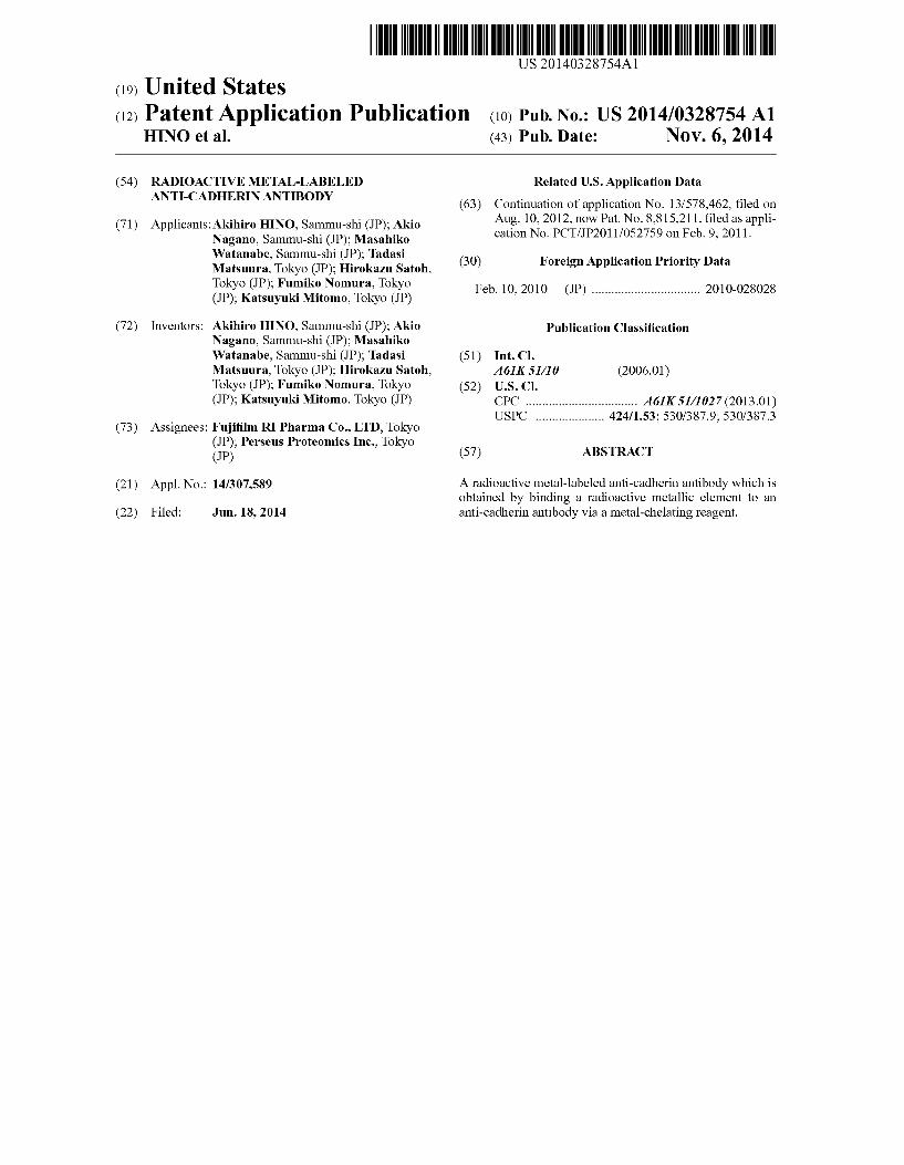
0018 FIG. 4 Bio-distribution of Ga-DOTA-PPMX2029 antibody (adding ratio of 1:10) 96 hours after administration thereof.
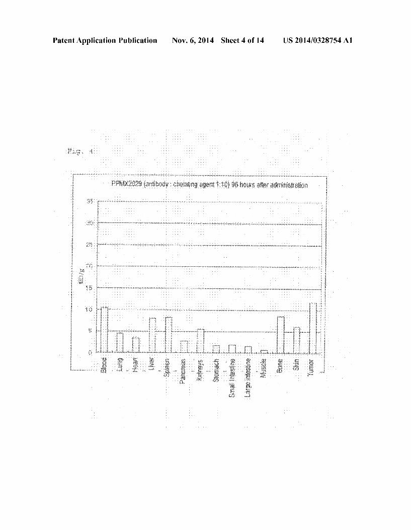
US 2014/0328754 A1
0019 FIG.5 Bio-distribution of Ga-DOTA-PPMX2029 antibody (adding ratio of 1:3) 96 hours after administration thereof. 0020 FIG. 6 Bio-distribution of Ga-DOTA-PPMX2025 antibody (adding ratio of 1:10) 96 hours after administration thereof. 0021 FIG. 7 Bio-distribution of Ga-DOTA-PPMX2025 antibody (adding ratio of 1:3) 96 hours after administration thereof. 0022 FIG.8 Bio-distribution of Ga-DOTA-PPMX2025 antibody in case of the added antibody-DOTA ratios of 1:1, 1:3, and 1:1096 hours after administration thereof. 0023 FIG.9 Bio-distribution of '''In-DOTA-PPAT-052 27c antibody (adding ratio of 1:3) 96 hours after administra tion thereof. 0024 FIG. 10 Bio-distribution of '''In-DOTA-PPAT-052 27c antibody (adding ratio of 1:3) 48 hours after administra tion thereof. 0025 FIG. 11 Bio-distribution of '''In-DOTA-PPAT-052 28c antibody (adding ratio of 1:3) 48 hours and 96 hours after administration thereof. 0026 FIG. 12 Anti-tumor effect of 'Y-DOTA PPMX2029 antibody (adding ratio of 1:3) in xenograft model. 0027 FIG. 13 Anti-tumor effect of'Y-DOTA-PPAT-052 27c antibody (adding ratio of 1:3) in xenograft model. 0028 FIG. 14 Photoimages showing the results of immu nohistochemical staining for confirming CDH3 protein expression.
MODES FOR CARRYING OUT THE INVENTION
0029. The radioactive metal-labeled anti-cadherin anti body of the present invention is a labeled anti-cadherin anti body to which a radioactive metallic element is bound to via a metal-chelating reagent. The cancertherapeutic agent or the cancer diagnostic agent contains the radioactive metal-la beled anti-cadherin antibody. 0030 The anti-cadherin antibody is not particularly lim
ited, so long as the antibody specifically binds to cadherin. Examples of cadherin include E cadherin, N cadherin, and P cadherin. Of these, P cadherin is more preferred. 0031. The anti-cadherin antibody encompasses a mono clonal antibody, a polyclonal antibody, an antibody maintain ing ability of binding specifically to an antigenic determinant group and variants and derivatives of an antibody such as T-cell receptor fragment. 0032. The type of the anti-cadherin antibody is not par
ticularly limited, and there may be appropriately employed antibodies such as a mouse antibody, a human antibody, a rat antibody, a rabbit antibody, a sheep antibody, a camel anti body, and a chicken antibody; and gene recombinant antibod ies which are intentionally modified so as to reduce the het ero-antigenicity to human Such as a chimeric antibody and a humanized antibody. The recombinant antibody may be pro duced through a known method. The chimeric antibody is an antibody formed of variable regions of a heavy chain and a light chain of a mammalian antibody other than human anti body, for example mouse antibody and constant regions of a heavy chain and a light chain of a human antibody and may be produced by linking a DNA fragment encoding the variable region of the mouse antibody to a DNA fragment encoding the constant region of the human antibody, incorporating the resultant fragment into an expression vector, and incorporat ing the vector into host cells (see, for example, Cabilly S. et
Nov. 6, 2014
al., Proc. Natl. Acad. Sci. USA, 1984, 81 (11) 3273-7: Morri son et al., Proc. Natl. Acad. Sci. USA, 1984, 81 (21), 6851-5: and European Patent Application Laid-Open No. 171496). The humanized antibody, which is also called a reshaped antibody, is an antibody produced through transplantation of a complementarity determining region (CDR) of a mamma lian antibody other than human antibody, e.g., a mouse anti body into a CDR of a human antibody, and gene recombina tion techniques therefor are generally known. Specifically, a DNA sequence including a CDR of a mouse antibody linked to a framework region (FR) of a human antibody is synthe sized through PCR using several oligonucleotides which are produced to have overlapped part at an end thereof. The thus-obtained DNA fragment is linked to a DNA fragment encoding the constant region of the human antibody, Subse quently the resultant fragment is incorporated into an expres sion vector and the vector is incorporated into host cells to thereby produce the humanized antibody (see EP2394.00 A and WO 96/02576A). The FR of the human antibody linked via the CDR is selected from FRs having a CDR which forms a Suitable antigen-binding site. If needed, an amino acid in the FR of the variable region of the antibody may be substituted such that a CDR of the reshaped antibody forms an appropri ate antigen-binding site (Sato, K. et al., Cancer Res., 1993, 53. 851-856). 0033. The amino acid sequence of the chimeric antibody or humanized antibody preferably has an identity of 1005 to that of the Vh or V1 region of cDNA expressing a deposited hybridoma. Due to genetic modification, an antibody having an identity in amino acid sequence of 90% or higher is also preferred. In the process of humanization or chimerization, there has been conventionally carried out such controlled residue Substitution for improving binding to an antigen. Such an antibody having a partially modified sequence is essentially considered to be an antibody originating from the original hybridoma. 0034 Methods for producing a chimeric antibody and a humanized antibody based on a genetic engineering tech nique have been already known. Specifically, the Vh and VL sequences of a monoclonal antibody serving as a confirmed group is genetically modified, and then chimerization or humanization is performed through a routine technique. 0035. The method for recovering a human antibody is also known. In one procedure, human lymphocytes are sensitized in vitro with an antigen of interest or with cells expressing the antigen, and the thus-sensitized lymphocytes are fused with human myeloma cells, e.g., U266, to thereby produce a human antibody of interest having a binding activity to the antigen (see JP-B-1989-59878). Alternatively, a human anti body of interest may be recovered through immunization, with an antigen of interest, of a transgenic animal having a complete repertory of the human antibody gene (see WO 93/12227, WO 92/03918, WO 94/02602, WO 94/25585, WO96/34096, and WO96/33735). Also known is a technique for recovering a human antibody through panning by use of a human antibody library. In one procedure, a variable region of a human antibody is expressed as a single-chain antibody (ScPV) on the phage surface through the phage display method, and a phage which binds the antigen can be selected. Through gene analysis of the thus-selected phage, a DNA sequence encoding the variable region of the human antibody which binds to the antigen can be determined. When the DNA sequence of the Sclv which binds the antigenis elucidated, an appropriate expression vector can be produced from the

US 2014/0328754 A1
sequence, whereby a human antibody of interest can be recovered. These methods are widely known (see WO 92/01047, WO92/20791, WO 93/06213, WO 93/11236, WO 93/19172, WO95/01438, and WO95/15388). 0036. These anti-cadherin antibodies may be a low mol ecule antibody such as an antibody fragment, a modified antibody or the like, so long as the ability of recognizing the entire or a part of the protein encoded by the cadherin gene is maintained. Examples of the antibody fragment include Fab, Fab'. F(ab')2, Fv, and Diabody. Such an antibody fragment may be produced by constructing a gene encoding the anti body fragment, incorporating the gene into an expression vector, and expressing the vector in appropriate host cells (see, for example, Co., M. S. et al., J. Immunol. (1994) 152, 2968-2976: Better, M. and Horwitz, A. H., Methods Enzy mol. (1989) 178, 476-496: Pluckthun, A. and Skerra, A., Methods Enzymol. (1989) 178, 497-515; Lamoyi, E., Meth ods Enzymol. (1986) 121, 652-663; Rousseaux, J. et al., Methods Enzymol. (1986) 121, 663-669; and Bird, R. E. and Walker, B. W., Trends Biotechnol. (1991) 9, 132-137). 0037. As a modified antibody, an antibody which is bound to any of various molecules such as polyethylene glycol (PEG) may be used. Such a modified antibody may be pro duced through chemical modification of the obtained anti body. The antibody modification technique has already been established in the art.
0038. In the present invention, there may be also employed a Sugar chain modified antibody for potentiating cytotoxic activity. Techniques of modifying the sugar chain in an anti body have already been known (e.g., WO 00/61739 and WO 02/31140). 0039. The anti-cadherin antibody of the present invention also encompasses a multi-specificantibody having specificity to two or more different antigens. A typical example of Such a molecule may be one which can bind two antigens (i.e., a bi-specific antibody). The “multi-specific antibody of the present invention includes an antibody having a specificity to two or more (e.g., three) antigens. The multi-specific anti body may be a full-length antibody or a fragment of such an antibody (e.g., F(ab') bi-specific antibody). 0040. The anti-cadherin antibody of the present invention and the antibody fragment thereof may be produced through any Suitable method such as in Vivo, cultured cells, in vitro translation reaction, and recombinant DNA expression sys tem
0041 Techniques of producing monoclonal antibodies and antibody-producing cells (hybridomas) are generally known in the art (Campbell, “Monoclonal Antibody Technol ogy: Laboratory Techniques in Biochemistry and Molecular Biology. Elsevier Science Publishers, Amsterdam, The Netherlands, 1984; and St. Grothet al., J. Immunol. Methods 35: 1-21, 1980). In one specific procedure, a protein or a fragment thereof encoded by a cadherin gene serving as an immunogen is Subcutaneously or intraperitoneally injected for immunization to any animal (e.g., mouse or rabbit) which is known to produce an antibody. In immunization, an adju vant may be employed, and Such an adjuvant is well known in the art.
0042. The polyclonal antibody may be produced by iso lating an anti-serum containing antibodies from an immu nized animal and screening for the presence of an antibody having a target specificity through a technique well known in the art (e.g., ELISA, Western blotting, or radioimmunoassay).
Nov. 6, 2014
0043. The monoclonal antibody may be produced by removing spleen cells from an immunized animal and fusing the cells with myeloma cells, to thereby produce hybridomas which can produce monoclonal antibodies. Hybridoma cells producing an antibody which can recognize a protein of inter estora fragment thereofmay be selected based on a technique well known in the art (e.g., ELISA, Western blotting, or radioimmunoassay). Then, the hybridoma Secreting an anti body of interest is cloned, and the obtained cells are cultured under appropriate conditions. The thus-secreted antibody is recovered and purified through a method well known in the art (e.g., ion-exchange column chromatography or affinity chro matography). In an alternative procedure, a human mono clonal antibody may be produced by use of a Xenomouse strain (see Green, J. Immunol. Methods 231: 11-23, 1999; and Wells, Eek, Chem. Biol. 2000 August: 7(8): R185-6). Currently, monoclonal antibody production based on phage display involving no immunization is carried out. The mono clonal antibody of the present invention is a single-molecular species antibody produced by single-species of antibody producing cells or a DNA fragment obtained therefrom and encoding the antibody. The monoclonal antibody may be produced through any of the aforementioned methods. 0044) The DNA fragment encoding a monoclonal anti body can be readily isolated and sequenced through a routine method (e.g., by use of an oligonucleotide probe which can binds specifically to genes encoding the heavy chain and light chain of the monoclonal antibody). A hybridoma cell is a preferred starting material for producing such a DNA frag ment. After isolation, Such a DNA fragment is inserted into an expression vector, and the vector is recombined to host cells such as E. coli cells, monkey COS cells, Chinese hamster ovary (CHO) cells or myeloma cells in which no immuno globulin is produced unless the cells are transformed. The monoclonal antibody of interest is produced by the recombi nant host cells. In an alternative mode, an antibody or an antibody fragment can be isolated from an antibody phage library produced through a technique of McCafferty et al. (Nature 348: 552-554 (1990)). 0045. The host cell employed for monoclonal antibody expression is preferably a mammal-origin host cell. A host cell most Suited to a monoclonal antibody to be expressed may be selected. The host cell is not limited and typical examples thereof include CHO-originating cell line (Chinese hamster ovary cell), CV1 (monkey kidney), COS (CV1 derivative expressing SV40T antigen), SP2/0 (mouse myeloma), P3x63-Ag3.653 (mouse myeloma), 293 (human kidney), and 293T (293 derivative expressing SV40T anti gen). The host cell system may be obtained from a commer cial facility, the American Tissue Culture Collection (ATCC), or an organization which published a relevant document. 0046. The host cell is preferably a dhfr gene expression defective CHO-originating cell line (deletion in dhfr gene expression) or SP2/0 (see Urland, G. et al., Effect of gamma rays at the dihydrofolate reductase locus: deletions and inver sions: Somat. Cell. Mol. Genet. Vol. 12, 1986, p. 5555-566: and Schulman, M. et al. A better cell line for making hybri domas secreting specific antibodies, Nature Vol. 276, 1978, p. 269-270). The host cell is more preferably a DHFR-deleted CHO. Transfection of a plasmid into host cells may be per formed through any technique. Transfection technique is not limited and specific examples thereof include transfection (including calcium phosphate method, DEAE method, lipo fection, and electroporation), DNA incorporation by use of an

US 2014/0328754 A1
envelope (e.g., Sendai virus), micro-injection, and infection by use of a viral (e.g., retrovirus or adenovirus) vector (see Current Protocols in Molecular Biology, Chapter 9 Introduc tion of DNA into Mammalian Cells, John Wiley and Sons, Inc.). Among them, incorporation of a plasmid into host cells through electroporation is particularly preferred. 0047. The recognition site in cadherin of the anti-cadherin antibody of the present invention is preferably a region from 1 to 655 of SEQID NO: 2. 0048. The anti-cadherin antibody of the present invention
is preferably produced from a hybridoma PPMX2016, PPMX2025, PPMX2029, PPAT-052-02, PPAT-052-03, PPAT-052-09, PPAT-052-24, PPAT-052-25, PPAT-052-26, or PPAT-052-28, oran transgenic CHO cell line PPAT-052-27c, PPAT-052-02c, PPAT-052-03c, PPAT-052-09c, PPAT-052 21c, PPAT-052-24c, PPAT-052-25c, PPAT-052-26c, PPAT 052-28c, or PPAT-052-29c. In the present specification, the numbers attached to PPMX or PPAT are given to either cor responding antibody-producing cells or antibodies produced by the antibody-producing cells. 0049. A radioactive metal which is bound to the anti cadherin antibody is preferably a cytotoxic radioactive metal when the radioactive metal-labeled anti-cadherin antibody is used as a cancer therapeutic agent, and a non-cytotoxic radio active metal when the radioactive metal-labeled anti-cadherin antibody is used as a cancer diagnostic agent. 0050 Examples of the cytotoxic radioactive metal include yttrium-90 ('Y), rhenium-186 (Re), rhenium-188 (Re), copper-67 (Cu), iron-59 (Fe), strontium-89 (Sr), gold-198 (Au).)mercury-203 (Hg), lead-212 (212Pb).)dysprosium-165 (Dy), ruthenium-103 (Ru), bismuth-212 (212Bi), ('Bi) bismuth-213 (Bi).)hol mium-166 ("Ho), samarium-153 (153Sm), and lutetium 177 (77Lu). 0051. Among these radioactive metals, 'Y. Sm, and '77Luare preferred, from the viewpoints of half-life, radiation energy, ease of labeling reaction, percent of labeling, com plex stability, etc. 0052 A non-cytotoxic radioactive metal suitably employed in a cancer diagnostic agent is not limited and examples thereof include technetium-99m ("Tc), indium 111)indium-113m (""In), gallium-67 ('Ga), gallium-68 (Ga), thallium-201 ('Tl), chromium-51 (Cr), cobalt-57 (Co), cobalt-58 (Co), cobalt-60 ('Co), strontium-85 (Sr), mercury-197 ('Hg), and copper-64 (Cu). 0053 For bonding a radioactive metallic element to the anti-cadherin antibody, in a preferred mode, a metal-chelat ing reagent is reacted with the anti-cadherin antibody, and the product is further reacted with a radioactive metallic element, to thereby form a complex. In the thus-produced modified antibody, the radioactive metallic element is bound to the anti-cadherin antibody via the metal-chelating reagent. 0054 Examples of the metal-chelating reagent for form ing Such a complex include (1) quinoline derivatives such as 8-hydroxyquinoline, 8-acetoxyquinoline, 8-hydroxyquinal dine, oxyquinoline Sulfate, O-acetyloxine, O-benzoyloxine, O-p-nitrobenzoyloxine, and quinolone compounds having a quinoline skeleton (e.g., norfloxacin, ofloxacin, enoxacin, ciprofloxacin, lomefloxacin, tosfloxacin, fleroxacin, and sparfloxacin); (2) compounds such as chloranilic acid, alumi non, thiourea, pyrogallol, cupferron, Bismuthiol (II), galloyl gallic acid, thiolide, 2-mercaptobenzothiazole, and tetraphe nylarsonium chloride; (3) ethylenediaminetetraacetic acid (EDTA), diethylenetriaminepentaacetic acid (DTPA), and
Nov. 6, 2014
compounds having a similar skeleton (dihydroxyethylgly cine, diaminopropanolte traacetic acid, ethylenediamine diacetic acid, ethylenediaminedipropionic acid hydrochlo ride, hydroxyethylethylenediaminetriacetic acid, ethylenedi aminetetrakis(methylenesulfonic acid), glycol ether diamine tetraacetic acid, hexamethylenediaminetetraacetic acid, hydroxyethyliminodiacetic acid, iminodiacetic acid, diami nopropanetetraacetic acid, nitrilotriacetic acid, nitrilotripro pionic acid, nitrilotris(methylenesulfonic acid) trisodium salt, triethylenetetraminehexaacetic acid, methyl DTPA, cyclohexyl DTPA, aminobenzyl EDTA, isothiocyanobenzyl EDTA, isothiocyanobenzyl DTPA methylisothiocyanoben Zyl DTPA, cyclohexylisothiocyanobenzyl DTPA, maleimi dopropylamidobenzyl EDTA, maleimidopentylamidobenzyl EDTA, maleimidodecylamidobenzyl EDTA, maleimidopen tylamidobenzyl DTPA, and maleimidodecylamidobenzyl DTPA); and (4)1,4,7,10-tetraazacyclododecane-1,4,7,10-tet raacetic acid (DOTA), 14.7-triazacyclononane-1,4,7-triace tic acid (NOTA), 14.8, 11-tetraazacyclotetradecane-1,4,8,11 tetraacetic acid (TETA), 1,4,7,10-tetraazacyclododecane (Cyclen), 14.8, 11-tetraazacyclotetradecan (Cyclam), isothiocyanobenzyl DOTA, and isothiocyanobenzyl NOTA. 0055 Among these metal-chelating reagents, isothiocy anobenzyl DOTA, methylisothiocyanobenzyl DTPA, cyclo hexylisothiocyanobenzyl DTPA are preferred, from the view points of ease of incorporation reaction of metal-chelate to antibody, percent of labeling, complex stability, etc. 0056. The radioactive metallic element may be bound to the anti-cadherin antibody through a routine method. In one procedure, a metal-chelating reagent is reacted with an anti cadherin antibody, to thereby prepare a label precursor, and the precursor is reacted with a radioactive metallic element. 0057. In the cancer therapeutic agent and cancer diagnos
tic agent of the present invention, the ratio by mole of anti cadherin antibody to metal-chelating reagent is important for accumulation in cancer cells and anti-cancer effect. The mole ratio (anti-cadherin antibody:chelating reagent) is preferably 1:0.1 to 1:4.5, more preferably 1:0.5 to 1:3. In order to attain Such mole ratios, the anti-cadherin antibody and the chelating reagent are preferably added to react at a ratio of 1:0.1 to 1:less than 5, particularly preferably 1:1 to 1:3. The number of chelate molecule(s) per anti-cadherin antibody may be calcu lated by measuring molecular weight through MALDI-TOF mass analysis or a similar technique, and comparing the molecular weight of an un-modified antibody to that of a modified antibody (U.S. Pat. No. 7,514,078, Lu et al., J. Pharm. Sci. 94(4), 2005, p. 788-797, and Tedesco et al., J. Clin. Onco. 23 (16S), 2005, 4765). Alternatively, the number of chelate molecule(s) per anti-cadherin antibody may be determined through chelatometric titration. One known method employs an alkaline earth metal colorimetric reagent (arsenazo III) (Bradyr et al., Nucl. Med. Biol. 31, 795-802, 2004, and Dadachova et al., Nucl. Med. Biol. 26, 977-982, 1999). 0058. The cancer therapeutic agent or cancer diagnostic agent of the present invention may be provided as a labeled formulation or a kit formulation containing a label precursor. Eitherformulation may be employed in the present invention. In the case of labeled formulation, a cancer therapeutic agent or a cancer diagnostic agent containing a labeled anti-cad herin antibody may be administered as is. In the case of a kit formulation, the agent may be administered after labeling with a radioactive metallic element of interest.

US 2014/0328754 A1
0059. The anti-cadherin antibody containing a radioactive metallic element bound thereto highly accumulates in cancer tissue and exhibits high cancer cell-toxic activity. Thus, the antibody is a useful cancer therapeutic agent which less dam ages the tissue other than cancer tissue and which has high safety. Also, the anti-cadherin antibody containing a radioac tive metallic element bound thereto has an anti-cancer activ ity remarkably higher than that of a corresponding anti-cad herin antibody. The anti-cancer activity is remarkably high particularly when the mole ratio of antibody to chelating agent is 1:0.1 to 1:4.5. 0060. The cancer therapeutic agent of the present inven tion may be used in combination with another anti-cancer agent. Examples of Such an anti-cancer agent include an alkylating agent, an antimetabolite, a microtubule inhibitor, an antibiotic anti-cancer agent, a topoisomerase inhibitor, a platinum agent, a molecular target drug, a hormone agent, and a biologics. Examples of the alkylating agent include nitrogen mustard anti-cancer agents (e.g., cyclophosphamide), nitrosourea anti-cancer agents (e.g., ranimustine), and dacar bazine. Examples of the antimetabolite include 5-FU, UFT, carmofur, capecitabine, tegafur, TS-1, gemcitabine, and cyt arabine. Examples of the microtubule inhibitor include alka loid anti-cancer agents (e.g., Vincristine) and taxane anti cancer agents (e.g., docetaxel and paclitaxel). Examples of the antibiotic anti-cancer agent include mitomycin C, doxo rubicin, epirubicin, daunorubicin, and bleomycine. Examples of the topoisomerase inhibitor include irinotecan and nogite can having topoisomerase I inhibiting activity and etoposide having topoisomerase II inhibiting activity. Examples of the platinum agent include cisplatin, paraplatin, medaplatin, and oxaliplatin. Examples of the molecular target drug include trastuzumab, rituximab, imatinib, gefitinib, erlotinib, bevaci Zumab, bortezomib, Sunitinib, and Sorafenib. Examples of the hormone agent include dexamethasone, finasteride, and tamoxifen. Examples of the biologics include interferons C. B, and Y and interleukin 2. 0061 The cancer therapeutic agent of the present inven tion may be used in combination with a cancer therapy. Examples of the cancer therapy include Surgery, radiation therapies (including gamma knife therapy, Cyber knife therapy, boron neutron capture therapy, and proton beam/ heavy ion beam therapy), MR-guided focused ultrasound Surgery, cryotherapy, radiofrequency ablation, percutaneous ethanol injection therapy, and embolotherapy. 0062. The cancer therapeutic agent of the present inven tion is effective on various cancers of a mammal (including a human). Examples of the target cancer include carcinomas Such as pharyngeal cancer, laryngeal cancer, tongue cancer, lung cancer, breast cancer, esophageal cancer, stomach can cer, colorectal cancer, uterine cancer, ovarian cancer, liver cancer, pancreatic cancer, gallbladder cancer, kidney cancer, prostatic cancer, malignant melanoma, and thyroid cancer, and sarcomas such as osteosarcoma, chondrosarcoma, rhab domyosarcoma, leiomyosarcome, liposarcoma, angiosar coma, fibrosarcoma, leukemia, malignant lymphoma, and myeloma. 0063. The cancer therapeutic agent of the present inven tion may be dissolved in an aqueous solution, preferably a physiologically adaptable buffer Such as Hanks solution, Ringer's Solution, or buffered physiological saline. Also, the therapeutic agent may have the form of Suspension, Solution, emulsion, or the like in an oily or aqueous vehicle.
Nov. 6, 2014
0064. The dosage of the cancer therapeutic agent of the present invention, which varies in accordance with the Symp tom, administration route, body weight, age, etc. of a patient in need thereof, is preferably, for example, 37 to 3,700 MBq. for one treatment of adult. 0065. The cancer therapeutic agent of the present inven tion is generally administered parenterally. For example, the cancer therapeutic agent is injected (e.g., Subcutaneously, intravenous, intramuscle, intraperitoneally) or administered transdermally, transmucosally, transnasally, transplu monarily, etc. 0066. The cancer diagnostic agent of the present invention may be used in tumor imaging. In the case where a patient has a tumor in which CDH3 protein is expressed, the cancer diagnostic agent of the present invention accumulates in the tumor. Thus, the tumor can be imaged by detecting radiation by means of an apparatus such as a single photon emission computed tomograph (SPECT), a positron emission tomo graph (PET), or a scintillation camera. For example, by use of the cancer diagnostic agent of the present invention, the thera peutic effect of the cancer therapeutic agent of the present invention can be predicted before administration of the thera peutic agent. The diagnostic agent is administered to a patient before the treatment, and the tumor is imaged. When high accumulation is observed, the superior effect of the therapeu tic agent can be predicted as potent. The diagnostic agent may be used for determining the therapeutic effect. The diagnostic agent of the present invention is administered to a patient who has received the treatment with the therapeutic agent of the present invention or any other treatment so as to image the tumor. Through monitoring the time-dependent variation in accumulation of the diagnostic agent, the expansion or shrinkage of the tumor over time can be observed. 0067. The antibody for use as a diagnostic agent prefer ably recognizes an epitope competitive to a therapeutic agent. More preferably, the antibody recognizes the same epitope as that recognized by the therapeutic agent. Most preferably, the therapeutic agent and the diagnostic agent are the same anti body. 0068. The cancer diagnostic agent of the present invention
is generally administered to a subject intravenously. How ever, the cancer diagnostic agent may also be administered arterially. The dosage thereof, which varies in accordance with the symptom, administration route, body weight, age, etc. of a patient in need thereof, is preferably, for example, 37 to 1,120 MBq for one treatment of adult.
EXAMPLES
0069. The present invention will next be described in detail by way of examples, which should not be construed as limiting the invention thereto.
Example 1
Production of Soluble CDH3 Antigen (0070 Soluble CDH3 (sCDH3) protein in which the C-ter minal transmembrane region had been deleted was produced to serve as an immunogen for producing an anti-CDH3 anti body.
(1) Production of Soluble CDH3 Antigen Expression Vector (0071 PCR was performed by use of a CDH3 full-length cDNA as a template and a forward primer (SEQ ID NO:3:

US 2014/0328754 A1
CGCGGTACCATGGGGCTCCCTCGT, (hCDH3FullFW)) and a reverse primer (SEQID NO: 4: CCGTCTAGATAAC CTCCCTTCCAGGGTCC, (hCDH3SolbRV)), which had been designed so as to amplify a segment corresponding to the CDH3 extracellular region (1-654 in SEQ ID NO: 2, hereinafter referred to as SCDH3cDNA). The reaction was performed by use of KOD-Plus (product of Toyobo) and under the following conditions: 94°C.-15 sec, 55° C.-30 sec, and 68° C.-90 sec (30 cycles)). 0072 After completion of the PCR reaction, the reaction mixture was subjected to agarose gel electrophoresis, and a gel piece containing a band of a target size (about 2.0 kbp) was cut out. The targetsCDH3cDNA was recovered from the gel piece by use of a QIA quick gel extraction kit (product of Quiagen). 0073. In order to insert SCDH3cDNA into an expression vector pEF4/myc-HisB, SCDH3cDNA was treated with two restriction enzymes Kipni and Xbal. The thus-obtained frag ment was inserted into pEF4/myc-HIsB which had been treated with the same restriction enzymes Kipni and Xbal, by use of T4 DNA ligase through a routine technique, whereby an expression vector pEF4-sCDH3-myc-His was yielded.
(2) Expression of Soluble CDH3 Protein 0074 According to a protocol of an FuGENE6 transfec tion reagent, 8x10 CHO cells were inoculated to a 10-cm diameter dish on the day before transfection, and the cells were cultured overnight. Thereafter, an expression vector pEF4-sCDH3-myc-His (8 ug) and an FuGENE6 regent (16 were mixed with serum-free RPMI 1640 medium (400 uL), and the mixture was allowed to stand at room temperature for 15 minutes. The resultant mixture was added to the cell cul ture liquid for transfection. Two days after transfection, clon ing was performed through limiting dilution by use of a selection reagent (Zeocin). 0075 Soluble CDH3-expressing CHO cells were selected through Western blotting by use of an anti-c-Myc monoclonal antibody (product of SANTA CRUZ BIOTECHNOLOGY). Cell lines which exhibited high level of secretion into the culture Supernatant and high proliferation were selected to obtaina soluble CDH3-expressing CHO cell line (EXZ1702). The thus-selected soluble CDH3-expressing CHO cells (EXZ1702) were cultured for 72 hours in three roller bottles (each culture area: 1,500 cm) with serum-free medium CHO-S-SFM-II (333 mL/bottle) (product of Invitrogen), and the culture supernatants were recovered. The thus-obtained culture Supernatant was subjected to affinity chromatography by means of HisTrap (registered trademark) HP column (product of GE Healthcare Bio-science) and gel filtration chromatography by means of Superdex (registered trade mark) 200 pg column (product of GE Healthcare Bio-sci ence), to thereby acquire soluble CDH3 protein.
Example 2
Establishment of CDH3-Expressing CHO Cell Line 0076 For obtaining a cell line for anti-CDH3 antibody screening, a CHO cell line expressing full length CDH3 was established.
(1) Production of CDH3 Gene Expression Vector 0077. In order to insert full-length human CDH3 DNA represented by SEQ ID NO: 1 into a mammal expression
Nov. 6, 2014
vector pEF4/myc-HisB (product of Invitrogen), the full length human CDH3 DNA was treated with two restriction enzymes KipnI (product of Takara BioInc.) and Xbal (product of Takara Bio Inc.) at 37° C. for one hour. The thus-obtained fragment was inserted into pEF4/myc-HisB which had been treated with the same restriction enzymes KipnI and Xbal, by use of T4DNA ligase (product of Promega) through a routine technique, whereby an expression vector pEF4-CDH3-myc His was produced.
(2) Acquisition of Stable CDH3-Expressing Line 0078. According to a protocol of an FuGENE (registered trademark) 6 transfection reagent (product of Roche Diag nostics K.K.), 8x10 CHO cells were inoculated to a 10-cm diameter dish on the day before transfection, and the cells were cultured overnight. Thereafter, an expression vector pEF4-CDH3-myc-His (8 g) and an FuGENE6 regent (16 uL) were mixed with serum-free RPMI 1640 medium (prod uct of SIGMA-ALDRICH) (400 uL), and the mixture was allowed to stand at room temperature for 15 minutes. The resultant mixture was added to the cell culture liquid for transfection. Two days after transfection, cloning was per formed through limiting dilution by use of a selection reagent (Zeocin). (0079 Clones of CDH3 full-length expressing CHO were selected through Western blotting by use of an anti-c-Myc monoclonal antibody (product of SANTA CRUZBIOTECH NOLOGY). As a result, a CDH3 full-length expressing CHO cell line (EXZ1501) was selected as a cell line which exhib ited high level of expression and high proliferation. The reac tion between EXZ1501 and a commercial anti-CDH3 anti body (product of R&D SYSTEMS) was confirmed through flow cytometry. That is, CDH3 protein expression on the cell membrane of EXZ1501 was confirmed.
Example 3
Production of Anti-CDH3 Monoclonal Antibody
(1) Production of Monoclonal Antibody by Use of Soluble CDH3 Protein as an Immunogen 0080 Soluble CDH3 protein (50 g) dissolved in physi ological saline was mixed with an equal amount of Titer MAX Gold (registered trademark) (product of Titer Max), and the mixture was intraperitoneally and Subcutaneously injected to MRL/lpr mice (Japan SLC inc.) for initial immu nization. Subsequent immunization procedures were per formed by intraperitoneally and Subcutaneously injecting, to the mice, a mixture of soluble CDH3 protein (25 ug) and Titer-MAX Gold prepared in the same manner. Three days after final immunization, spleen cells were prepared from the mice under aseptic conditions, and the cells were fused with mouse myeloma cells SP2/O—Ag14 or P3-X63-Ag8.653 through a generally employed polyethylene glycol method.
(2) Selection of Anti-CDH3 Hybridomas
I0081. Selection of anti-CDH3 antibodies were performed through flow cytometry by use of a full-length CDH3-ex pressing CHO cell line (EXZ1501). I0082 Specifically, full-length CDH3-expressing CHO cells (EXZ1501) were removed from a culture plate by treat ing with 2 mM EDTA-PBS and suspended in FACS solution
Antibody-Producing
US 2014/0328754 A1
to a cell concentration of 1x10° cells/mL. The cell suspension was inoculated to a 96-well plate to a concentration of 50 LL/well, and a hybridoma culture Supernatant was added thereto, followed by reaction at 4° C. for 60 minutes. The plate was washed twice with FACS solution (200 uL/well), and Alexa Fluor 488-labeled anti-mouse IgG goat F(ab')2 (product of Invitrogen) was added thereto, followed by reac tion at 4° C. for 30 minutes. Subsequently, the plate was washed twice with FACS solution, and flow cytometry was performed, to thereby select hybridomas producing an anti body which binds to CDH3-expressing CHO cells. As a result, 40 clones PPMX2016 to PPAT-052-28 were obtained. Through flow cytometry, it was confirmed that all the hybri domas reacted with CDH3-expressing CHO cells (EXZ1501) and NCI-H358 but do not react with CHO cells. Antibodies were purified from the hybridoma culture supernatant by means of Protein G column and employed in the Subsequent experiments. Among the selected hybridomas, PPMX2016 (NITE BP-897), PPMX2025 (NITE BP-898), PPMX2029 (NITE BP-899), PPAT-052-02 (NITE BP-1034), PPAT-052 03 (NITE BP-1035), PPAT-052-09 (NITE BP-1036), PPAT 052-24 (NITE BP-1037), PPAT-052-25 (NITE BP-1038), PPAT-052-26 (NITE BP-1039), and PPAT-052-28(NITE BP-1040) were deposited with Incorporated Administrative Agency, the National Institute of Technology and Evaluation, Patent Microorganisms Depositary (2-5-8, KaZusakamatari, Kisarazu-shi, Chiba, Japan) on Feb. 10, 2010 and Jan. 18, 2011.
Example 4
Cloning of Antibody Genes 0083 (1) A DNA fragment encoding the V-region of a mouse monoclonal antibody to human CDH3 was cloned through the following procedure. Cytoplasmic RNA was iso lated from the mouse hybridoma cells through a method disclosed in a document (Gough, “Rapid and quantitative preparation of cytoplasmic RNA from small numbers of cells.” Analytical Biochemistry, 173, p. 93-95 (1988)), with the proviso that instead of the dissolution buffer disclosed in the document, a TNE buffer (i.e., 25 mM Tris-HCl, pH: 7.5; 1% NP-40: 150 mM NaCl; 1 mM EDTA, pH: 8.0)was employed. More specifically, 5x10 hybridoma cells were suspended in the TNE buffer (200LL), to thereby dissolve cell membranes, and cell nuclei were removed through centrifu gation. To the thus-obtained cytoplasma Supernatant (about 200 uL), an extraction buffer (10 mM Tris-HCl, pH: 7.5; 0.35M NaCl; 1% (w/v) SDS: 10 mM EDTA, pH: 8.0:7M urea) (200 uL) was added. The mixture was subjected to extraction with phenol and chloroform. To the thus-obtained RNA solution, glycogen (product of Roche, Cat No. 901393) serving as a carrier was added. Then, ethanol was added to precipitate the product. The RNA precipitate was dissolved in sterilized distilled water (10 to 50 LL) to a cytoplasmic RNA concentration of 0.5 to 2 ug/uL. (2) Production of cDNA Library from RNA Prepared from Hybridomas 0084. For synthesizing a single-strand cDNA, there was prepared a reaction mixture (20 LL) containing the above prepared cytoplasmic RNA (0.5 to 3 ug), 50 mM Tris-HCl (pH: 8.3, room temperature), 75 mMKC1, 3 mM MgCl, and 10 mM DTT), a random primer (100 ng), 0.5 mM dNTP and Superscript II (reverse transcriptase, product of Invitrogen) (200 units). The mixture was incubated at 42° C. for 50
Nov. 6, 2014
minutes. The thus-synthesized cDNA library was employed as a template of polymerase chain reaction (PCR) without performing further treatment.
(3) Amplification of a Gene Encoding a Variable Region of Anti-CDH3 Antibody Through PCR I0085 All the primers employed in the experiments were synthesized by Hokkaido System Science Co., Ltd. a. Primers for Use in PCR Amplification of a Gene Encoding Mouse L-Chain V-Region I0086. The following two primer sets were employed: (i) a DNA primer having, at the 5' end, a homology to the FR1 part and 4-set primers having, at the 3' end, a homology to a J-chain gene in the mouse L-chain, and (ii) 7-set primers having, at the 5' end, a homology to the L-chain signal part and an antisense primer having, at the 3' end, a homology to the KC part (KVL antisense primer). Polymerase chain reac tion was performed by use of the two primer sets, whereby a mouse immunoglobulin L-chain variable region DNA frag ment was obtained from the cDNA. The primer sequences are as follows.
(i) 4-Set Sense Primers for Mouse L-Chain Variable Region Cloning I0087. According to “Phage Display—A Laboratory Manual-, Barbas Burton Scott Silverman, PROTOCOL 9.5, 17 sense primers and 3 reverse primers were synthesized by Hokkaido System Science Co., Ltd.
VK Sense (FR1 Part) I0088 A mixture of the following 17 primers was employed as a VK sense primer.
(degeneracy 2) : SEO ID NO : 5
5 - GAY ATC CAG CTG ACT CAG CC-3
(degeneracy 4) : SEQ ID NO: 6
s' - GAY ATT GTT CTC WCC CAG TC-3'
(degeneracy 8) : SEO ID NO : 7
s' - GAY ATT GTG. MTM ACT CAG TC-3'
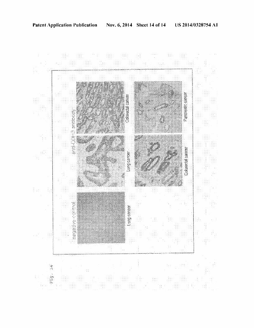
(degeneracy 8) : SEQ ID NO: 8
s' GAY ATT GTG YTR ACA CAG TC-3'
(degeneracy 8) : SEO ID NO : 9
5 GAY ATT GTR ATG ACM CAG TC-3
(degeneracy 16) : SEO ID NO: 10
5 GAY ATT MAG ATR AMC CAG TC-3
(degeneracy 12): SEO ID NO: 11
5 GAY ATT CAG ATG AYD CAG TC-3
(degeneracy 4) : SEO ID NO: 12
5 GAY ATY CAG ATG ACA CAG AC-3
(degeneracy 4) : SEO ID NO: 13
s' GAY ATT GTT CTC AWC. CAG TC-3'
US 2014/0328754 A1
- Continued
(degeneracy 8) : SEO ID NO: 4.
5 GAY ATT GWG CTS ACC CAA TC-3
(degeneracy 16) : SEO ID NO: 5
s' GAY ATT STR ATG ACC CAR TC-3'
(degeneracy 16) : SEO ID NO: 6
5 GAY RTT KTG ATG ACC CAR AC-3
(degeneracy 12): SEO ID NO: 7
5 GAY ATT GTG ATG ACB CAG KC-3
(degeneracy 4) : SEO ID NO: 8
5 GAY ATT GTG ATA ACY CAG GA-3
(degeneracy 4) : SEO ID NO: 19
5 GAY ATT GTG ATG ACC CAG WT-3
(degeneracy 2) : SEO ID NO: 2O
5 GAY ATT GTG ATG ACA CAA CC-3
(degeneracy 2) : SEO ID NO: 21
s' GAY ATT TTG. CTG ACT CAG TC-3'
J Antisense (4-Set Primers)
J1/J2 Antisense Primer (1) 0089
(degeneracy 8) : SEO ID NO: 22
5 - GGS ACC AAR CTG GAA. ATM AAA-3'
J4 Antisense Primer (2)
0090
SEO ID NO: 23 5 - GGG ACA. AAG TTG GAA. ATA AAA-3" :
J5 Antisense Primer (3)
0091
SEO ID NO: 24 5 - GGG ACC AAG CTG GAG CTG AAA-3" :
J1/J2, J4, J5 Antisense Primer Mixture (4)
(ii) 7-Set Primers for Mouse L-Chain Variable Region Cloning VK Sense (Signal Peptide Part)
0092. The primers were obtained through nucleotide sequence modification of a mouse Ig-primer set (Novagen; Merck, Cat. No. 69831-3) such that restriction enzyme sites were removed. A-set sense primer
Nov. 6, 2014
SEO ID NO: 25 5-ATGRAGWCACAKWCYCAGGTCTTT-3 :
B-Set Sense Primer
0093
SEO ID NO: 26 5 - ATGGAGACAGACACACTCCTGCTAT-3 :
C-Set Sense Primer
0094)
SEO ID NO: 27 5 - ATGGAGWCAGACACACTSCTGYTATGGGT-3 :
D-Set Sense Primer (Mixture of the Following 2 Primers) 0.095
SEO ID NO: 28 s' - ATGAGGRCCCCTGCTCAGWTTYTTGGIWTCTT-3 :
SEO ID NO: 29 5 - ATGGGCWTCAAGATGRAGTCACAKWYYCWGG-3 :
E-Set Sense Primer (Mixture of the Following 3 Primers) 0.096
SEO ID NO: 3 O s' - ATGAGTGTGCYCACTCAGGTCCTGGSGTT-3 :
SEO ID NO: 31 s' - ATGTGGGGAYCGKTTTYAMMCTTTTCAATTG-3 :
SEO ID NO: 32 s' - ATGGAAGCCCCAGCTCAGCTTCTCTTCC-3' :
F-Set Sense Primer (Mixture of the Following 4 Primers) O097
SEO ID NO: 33 s' - ATGAGIMMKTCIMTTCAITTCYTGGG-3 :
SEO ID NO: 34 s' - ATGAKGTHCYCIGCTCAGYTYCTIRG-3 " :
SEO ID NO: 35 s' - ATGGTRTCCWCASCTCAGTTCCTTG-3 :
SEO ID NO: 36 5'-ATGTATATATGTTTGTTGTCTATTTCT-3 '' .
G-Set Sense Primer (Mixture of the Following 4 Primers) 0098
SEO ID NO : 37 s' - ATGAAGTTGCCTGTTAGGCTGTTGGTGCT-3 :
SEO ID NO: 38 s' - ATGGATTTWCARGTGCAGATTWTCAGCTT-3 :

US 2014/0328754 A1
- Continued SEO ID NO: 39
5'-ATGGTYCTYATWTCCTTGCTGTTCTGG-3 :
SEO ID NO: 40 s' - ATGGTYCTYATWTTRCTGCTGCTATGG-3 :
KVL Antisense Primer
0099
SEO ID NO: 41 ACTGGATGGTGGGAAGATGGA:
B. Primers for Use in PCR Amplification of a Gene Encoding Mouse H-Chain V-Region 0100. The following two primer sets were employed 4-set primers having, at the 5' end, a homology to the mouse H-chain signal part and a primer having, at the 3' end, a homology to the KC part; and 1-set primers having, at the 5' end, a homology to the FR1 part and 2-set primers having, at the 3' end, a homology to the mouse H-chain constant region (IGHC). Polymerase chain reaction was performed by use of the two primer sets, whereby a mouse immunoglobulin H-chain variable region DNA fragment was isolated from the cDNA. The primer sequences are as follows.
(i) Primers for Mouse H-Chain Variable Region Cloning VH Sense (Signal Part: 4-Set Primers) 0101 These primers were synthesized according to Cur rent Protocols in Immunology (John Wiley and Sons, Inc.), Unit 2.12 Cloning, Expression, and Modification of Antibody V Regions (Table 2.12.2).
(degeneracy: 32) : SEO ID NO: 42
s' - ATG GRA. TGS AGC TGK GTM ATS CTC TT-3'
(degeneracy: 8) : SEO ID NO: 43
s' - ATG RAC TTC GGG YTG AGC TKG GTT TT-3'
SEO ID NO: 44 s" - ATG GCT GTC TTG. GGG CTG CTC TTC T-3 '' .
(degeneracy: 32) : SEO ID NO: 45
s' - ATG GRC AGR CTT ACW TYY-3'
(ii) Primers for Mouse H-Chain Variable Region Cloning VH Sense (FR1 Part) 0102 These primers were designed by nucleotide sequence modification of sense primers disclosed in a docu ment (Tanetal, “Superhumanized Antibodies: Reduction of Immunoogenic Potential by Complementarity-Determining Region Grafting with Human Germline Sequences: Applica tion to an Anti-CD281, Journal of Immunology 169 (2002), p. 1119-1125).
(degeneracy: 256): SEO ID NO: 46
S-SAG GTS MAR CTK SAG SAG TCW GG-3
Nov. 6, 2014
VHAntisense (Antisense Primer Common to 3 and 4) 0103) The primer was designed through degeneration of the nucleotide sequence so that the primer can be annealed with all the isoforms of mouse IgG.
(degeneracy: 6): SEO ID NO: 47
s' - CAS CCC CAT CDG TCT ATC C-3'
Example 5
Production of Chimera Anti-CDH3 Immunoglobulin Expression Vector
Production of Expression Plasmid 0104. Through PCR employing DNA Engine (Peltier Thermal Cycler, MJ Research, Bio-Rad), each variable region of the L-chain and the H-chain of an anti-CDH3 mouse monoclonal antibody was amplified by use of the primers described in Example 4. Each of the thus-amplified DNA fragments was incorporated into a Sub-cloning vector pGEM (product of Promega). The nucleotide sequence of the DNA fragment was determined by use of a universal primer which binds to T7 an 6. SP6 promoter of the vector. The thus obtained nucleotide sequences of the L-chain and H-chain variable regions of the anti-CDH3 antibody were searched by IMGT/V-QUEST Search page (http://imgt.cines.fr/IMGT vduest/valuest?livret=0&Option mouseIg), whereby completion of cloning of the antibody genes was confirmed. 0105 Next, a gene encoding the human CK region was linked to the cloned gene encoding the V region of the L-chain of the anti-CDH3 antibody, and a gene encoding the human CK1 region was linked to the gene encoding the V region of the H-chain. The thus-designed L-chain and H-chain chi mericantibody genes were synthesized in full length by Gen Script. At the time, frequency of codon usage was optimized So as to obtain efficient gene expression in producing cells (according to a method disclosed in Kim et al., Codon opti mization for high-level expression of human erythropoietin (EPO) in mammalian cells, Gene, 199, 1997, p. 293-301). Specifically, in the case of L-chain, for the purpose of effec tive translation, an essential DNA sequence (Kozak, M., J. At least six nucleotides preceding the AUG initiator codon enhance translation in mammalian cells. J. Mol. Biol. 196, p. 947-950, 1987), signal peptide of mouse IGKV, the V region of the L-chain of the anti-CDH3 antibody, and the human CK region were juxtaposed in this order, and restriction enzyme sites were added to both ends (Nhe on the 5' side and EcoRI on the 3' side). The chimera H-chain was prepared in the same manner. Each of the synthesized genes was cut with NheI and EcoRI, and the cut fragment was incorporated into an expres sion vector pCAGGS between the Nhel site and the EcoRI site, to thereby produce an anti-CDH3 chimeric antibody L-chain expression vector pCAGGS-IGK and H-chain expression vector pCAGGS-IGH.
Example 6
Production of Chimera Anti-CDH3 Immunoglobulin Stable Expression Vector
0106 For realizing high-level expression of a genetically modified antibody gene in CHO cells, there was produced an

US 2014/0328754 A1
expression vector into which a dihydrofolate reductase (dhfr) gene linked to a CMV promoter sequence and having poly A signal had been incorporated. 0107 For producing a chimeric antibody-stably express ing/producing cell line, there was produced a pCAGGS expression vector into which a dhfr gene had been incorpo rated. Specifically, into pCAGGS-IGH and pCAGGS-IGK, which are transient expression vectors, a dhfr gene having a CMV promoter and polyA signal was incorporated. Each of a mouse dhfr gene having a CMV promoter and a Kozak sequence and SV40 poly A signal was amplified through PCR. These genes in mixture form were linked together through PCR, and an HindIII site was added to both ends of the linked product, to thereby acquire a gene fragment of HindIII-CMV promoter-Kozak-dhfr-poly A-HindIII. The fragment was inserted into pCAGGS-IGH or pCAGGS-IGK at the HindIII sites, to thereby obtain pCAGGS-IGH-CMVp dhfr-A and pCAGGS-IGK-CMVp-dhfr-A. These expression vectors enable chimeric antibody expression with a CAG promoter, and expression of a dhfr gene with a CMV pro moter, whereby a chimeric antibody can be effectively pro duced through gene amplification.
Example 7
Establishing a Chimera Anti-CDH3-Producing CHO Cell Line
0108 CHO dhfr cells (G. Urlaub et al., Isolation of Chi nese hamster cell mutants deficient in dihydrofolate reductase activity, Proc. Natl. Acad. Sci. USA 77, p. 4216-4220, 1980) were simultaneously transformed by use of two plasmids (linear plasmids obtained by cutting circular plasmids with Pvul in an amplicillin-resistant gene); i.e., a pCAGGS-IGK CMV-dhfr-A vector for chimera anti-CDH3 L chain expres sion and a pCAGGS-IGH-CMV-dhfr-A vector for chimera anti-CDH3 H chain expression. Electroporation was per formed by means of Amaxa (product of Lonza). DNA frag ment (2 ug/sample, in the case of L-chain plasmid or H-chain plasmid) was added to a 0.1 mL Amaxa electroporation CHO buffer containing 3x10 cells and a pulse was applied. 0109 The cells which had undergone electroporation were added to an Iscove’s Modified Dulbecco medium (IMDM: free of HT) containing 10% dialyzed FBS that is free of HT (H: hypoxanthine, T: thymidine). Three days after transfection, the medium was changed to an IMDM medium free of 10% dialyzed FBS, 2 mM L-glutamine, and HT, and neo-- transformed cells were selected by use of 1 mg/mL G418, to thereby acquire clones of a chimeric antibody-pro ducing positive cell line. Subsequently, gene amplification was performed by use of the clones selected by using G418. 2-Round amplification was performed in 250 nM and 1,000 nM methotrexate (MTX), and cell lines which can produce a chimera CDH3 antibody (about 50 to 100 mg/L-culture supernatant) were established. The thus established chimera anti-CDH3 antibody-stably expressing CHO cell lines were deposited with Incorporated Administrative Agency, the National Institute of Technology and Evaluation, Patent Microorganisms Depositary.
TABLE 1.
Cell line Accession No.
PPAT-052-02c PPAT-052-03c
NITEBP-1041 NITEBP-1042
Nov. 6, 2014
TABLE 1-continued
Cell line Accession No.
PPATOS2-09c PPATOS2-21c PPATOS2-24c PPATOS2-2Sc PPATOS2-26c PPATOS2-27C PPAT-052-28c PPATOS2-29c
NITEBP-1043 NITEBP-1044 NITEBP-1045 NITEBP-1046 NITEBP-1047 NITEBP-1048 NITEBP-1049 NITEBP-1050
Example 8
Acquisition of Purified Antibodies 0110. The antibodies were purified from the culture super natant by use of protein A.
Example 9
Confirmation of Affinity 0111. Through a competitive method, the affinity of the mouse anti-CDH3 antibody was compared with that of the chimera anti-CDH3 antibody. In the competitive method, the affinity of the anti-CDH3 antibody was determined through flow cytometry (BD, FACS Calibur) by use of cancer cells NCI-H358, which are known to be CDH3 high expression cells. 0112 Specifically, an antibody serially diluted sample (400 ug/mL to 24 ng/mL) (50 uL) and an Alexa488-labeled antibody (4 ug/mL) (50 uL) were added to and mixed on a 96-well plate. NCI-H358 cells were removed from a culture plate through treatment with 2 mM EDTA-PBS, and the cells were suspended in an FACS solution (1% BSA PBS) to a concentration of 1.5x10"/mL. An aliquot (100 uL) of the Suspension was added to the wells containing the antibody mixture. After addition, reaction was performed at room tem perature for 60 minutes, and the plate was washed twice with an FACS solution (200 uL/well). Subsequently, the fluores cence intensity (GEO mean) of each well was determined through flow cytometry. 0113. The percent of binding inhibition of the Alexa488 labeled antibody was calculated from a GEO mean value, as compared with that obtained by the reaction only with the Alexa488-labeled antibody (1 pg/mL). The antibody concen tration showing 50% inhibition was calculated, and the data were compared. 0114 FIG. 1 shows the affinity evaluation of PPMX2016 (mouse antibody) and PPAT-052-27c (chimeric antibody thereof). Virtually no difference in affinity was observed between the two antibodies.
Example 10
Production of Labeled Antibodies
(1) Bonding DOTA to Antibody
(0.115. An antibody was dissolved in a buffer (50 mM Bicine-NaOH, 150 mM. NaCl, pH: 8.5) to an antibody con centration of 10 mg/mL. Separately, isothiocyanobenzyl DOTA (B-205, product of Macrocyclics) was dissolved in DMSO to a concentration of 10 mg/mL. The two solutions

US 2014/0328754 A1
were mixed together so as to adjust the ratio by mole of antibody to DOTA 1:1 (adding ratio 1:1), 1:3 (adding ratio 1:3), or 1:10 (adding ratio 1:10), and the mixture was stirred and allowed to stand at 25°C. for 17 hours. After termination of reaction, the reaction mixture was purified by means of a desalting column (PD-10, product of GE Healthcare, 17-0435-01) with PBS. The following antibodies were used: PPMX2016, PPMX2025, PPMX2029, PPAT-052-27c, and PPAT-052-28c.
(2) Determination of Percent of Chelate Incorporation 0116. The percent of chelate incorporated into antibody was determined through chelatometric titration. The modifi cation antibody protein concentration was determined through a customary method in advance, and the number of moles of modification antibody was calculated from the molecular weight of IgG. To 1-mg/mL standard copper Solu tion (100 whose concentration had been determined through atomic absorption spectrometry, an arsenazo III reagent (0.776 mg) and metal-free 5 Mammonium acetate (product of Sigma Aldrich) solution (3 mL) were added, and ultrapure water was added to the solution to adjust the final volume to 10 mL. The resultant solution was stored at room temperature in the dark to prepare the arsenazo III solution. DOTA was dissolved in ultrapure water, to thereby prepare a DOTA standard Solution. A modification antibody was dissolved in ultrapure water, to thereby prepare a modification antibody solution. The DOTA standard solution or the modification antibody solution (10 uL) was admixed with the arsenazo III solution (190 uL), and the mixture was incubated at 37°C. for 30 minutes. Subsequently, the absorbance of the mixture was measured at a wavelength of 630 nm. A standard curve was drawn from the absorbance measurements of DOTA standard solutions. By the standard curve, the number of DOTA mol ecule(s) bound to the modification antibody was calculated (average number of DOTA modification). 0117 Table 2 shows the results (DOTA-adding ratio and actual average number of modifying DOTA). As is clear from Table 2, the number of DOTA molecules bound to the anti body was found to be determined by the adding ratio of DOTA.
TABLE 2
AV. no. of Antibody to DOTA-adding ratio modifying DOTA
PPMX2025 (adding ratio 1:1) O.9 PPMX2025 (adding ratio 1:3) 2.0 PPMX2025 (adding ratio 1:10) 5.3 PPMX2016 (adding ratio 1:1) 0.7 PPMX2016 (adding ratio 1:3) 2.1 PPMX2016 (adding ratio 1:10) 5.2 PPAT-052-27c (addding ratio 1:3) 1.8
(Note) PPAT-052-27c was tested only at an adding ratio of 1:3.
(3) Preparation of Ga-, or 'Y-Labeled Antibodies 0118 (i) Labeling with 7Ga or '''In 0119 Each of the purified PPMX2016, PPMX2025, PPMX2029, and PPAT-052-27c antibodies and PPAT-052 28c antibody was dissolved in a buffer (0.25M ammonium acetate-HCl, pH: 5.5) to a concentration of 6 mg/mL. A 'GaCl, solution (product of Fuji Film RI Pharma) or a
Nov. 6, 2014
'''InCls solution (product of MDS Nordion Inc.) was added to the antibody solution, and the mixture was incubated at 45° C. for one hour. (ii) Labeling with 'Y 0120 Each of the purified PPMX2029 and PPAT-052-27c antibodies was dissolved in a buffer (0.25M ammonium acetate-HCl, pH: 5.5) to a concentration of 6 mg/mL. A 'YCls solution (product of Nuclitec) was added to the anti body solution, and the mixture was incubated at 45° C. for one hour. (iii) Determination of Percent of Labeling I0121. An aliquot of the labeling reaction mixture was sampled and subjected to thin-layer chromatography (61885, product of PALL), to thereby determine the percent of label ing. Physiological saline was employed as an eluent, and the radioactivity was measured at the top and bottom ends of a strip by means of a y-counter. The percent of labeling was calculated by the following equation.
Percent of labeling (bottom end count (top end count-bottom end count))x100(%)
I0122) When the percent of labeling reached 90% or higher, the labeled antibody was used in the subsequent experiment. The labeled antibodies were purified a desalting column(PD-10, product of GE Healthcare, 17-0435-01) with PBS.
Example 11
Investigation of Relationship Between Percent of Chelate-Incorporation and Bio-Distribution (i.e.,
Distribution in the Body) 0123 PPMX2016, PPMX2025, and PPMX2029 were investigated in terms of bio-distribution by virtue of differ ence of percent of chelate incorporation values (DOTA-add ingratio 1:1, 1:3, and 1:10). (0.124 Firstly, NCI-H358 was cultured in a 10% FBS-con taining RPMI1640 medium, and the cultured cells were sub cutaneously transplanted to the right ventral region of each of the nude mice (female, 7-week old, CLEAJapan Inc.) at a cell concentration of 1x10⁷ cells/mouse. The mice were bred until the average tumor volume reached 100 to 150 mm. (0.125. Then, to the NCI-H358-transplanted mice, 'Ga DOTA-PPMX2016 antibody (adding ratio 1:3 and 1:10), 'Ga-DOTA-PPMX2025 antibody (adding ratio 1:3 and 1:10), or (Ga-DOTA-PPMX2029 antibody(adding ratio 1:3 and 1:10) was administered at a dose of 370 kBq/mouse. 0.126 Ninety-six hours after administration, the mice were sacrificed to anatomy, and tissues and the tumor were removed. The weight of each tissue and the tumor weight were measured, the radioactivity was measured by means of a y-counter and % ID/g was calculated by the following equation.
% ID g (accumulated RI amount? total administered RIamountx100(%))/weight (g) E2
0127 FIGS. 2 to 7 show the results. All the tested antibod ies exhibited enhanced accumulation in the tumor, at a DOTA-adding ratio of 1:3, as compared with the case at a ratio of 1:10. Such enhanced accumulation results in enhancement of therapeutic effect. In addition, adverse side effects, which would otherwise be caused by retention of a radioactive Substance in non-targeted organs, can be avoided. I0128 FIG. 8 shows the result of administration of 'Ga DOTA-PPMX2025 antibody (use ratio 1:1, 1:3, and 1:10) to
US 2014/0328754 A1
non-cancer-bearing nude mice (female, 7-week old, CLEA Japan Inc.) at 370 kBq/mouse. The antibody was accumulated highly in the liver when the adding ratio was 1:10, whereas accumulation in the liver was low when the ratio was 1:3 or 1:1, which indicates that adverse side effects such as radio active damage are prevented on the liver.
Example 12
Investigation of Behavior of Anti-Caldina Chimeric Antibody in the Body
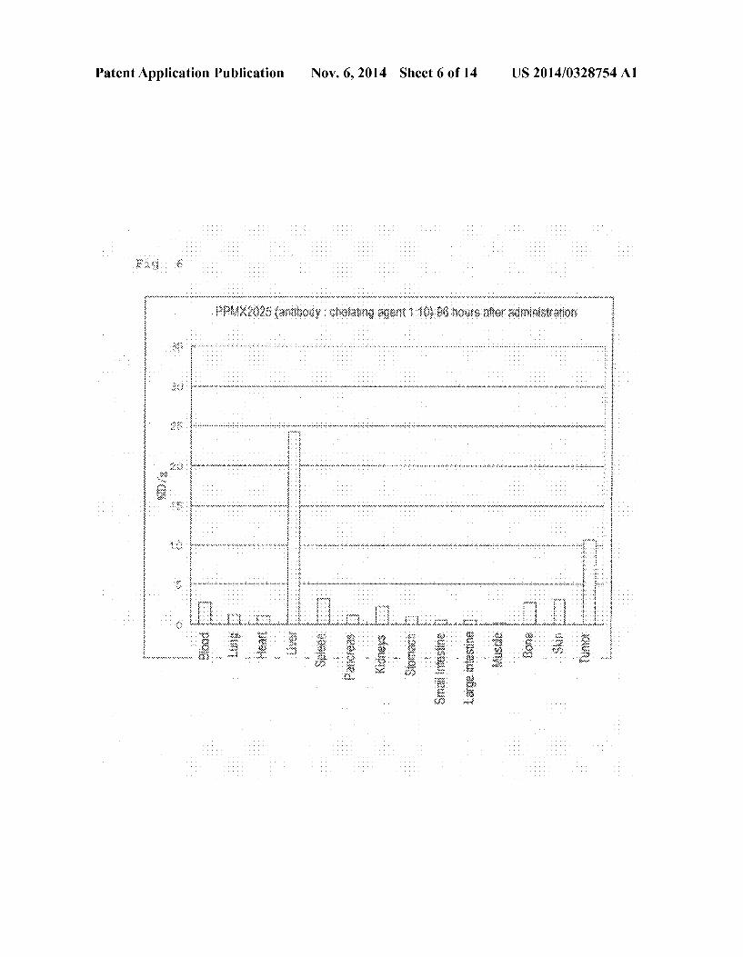
0129. The behaviors of chimera antibodies PPAT-052-27c and PPAT-052-28c in the body were investigated. 0130 Firstly, NCI-H1373 was cultured in a 10% FBS containing RPMI1640 medium, and the cultured cells were Subcutaneously transplanted into to the right ventral region of each of the nude mice (female, 9-week old, CLEAJapan Inc.) at a cell concentration of 4x10 cells/mouse. The mice were bred until the average tumor volume reached 100 to 150 mm. 0131 Then, to the NCI-H1373-transplaned mice, '''In DOTA-PPAT-052-27c (adding ratio 1:3) or '''In-DOTA PPAT-052-28c (adding ratio 1:3) at a dose of 370 kBq/mouse. 0132) Forty-eight or ninety-six hours after administration, the mice were sacrificed to anatomy, and tissues and the tumor were removed. The weight of each tissue and the tumor weight were measured, the radioactivity was measured by means of a y-counter and % ID/g was calculated. 0.133 FIGS. 9 to 11 show the results. PPAT-052-27c exhibited a percent accumulation in the tumor as high as 46% ID/g 48 hours after administration. PPAT-052-28c exhibited a percent accumulation in the tumor as high as 41% ID/g 48 hours after administration and 52% ID/g 96 hours after administration.
Example 13
Xenograft Test
0134 NCI-H358 was cultured in a 10% FBS-containing RPMI1640 medium, and the cultured cells were subcutane ously transplanted into to the right ventral region of each of the nude mice (female, 7-week old, CLEAJapan Inc.) at a cell concentration of 1x10" cells/mouse. 0135. The NCI-H358-transplaned mice were divided into six groups (n=8). 'Y-DOTA-PPMX2029 antibody (adding ratio 1:3) was administered at a dose of 7.4MBq/mouse, 5.6 MBq/mouse, 3.7 MBq/mouse, and 1.9 MBq/mouse. As con trol groups, unlabeled PPMX2029 was administered at a dose of 80 ug/mouse, and physiological saline was administered at a dose of 100LL/mouse. In all the groups, administration was performed when the average tumor volume reached 100 to 150 mm.
SEQUENCE LISTING
<16 Os NUMBER OF SEO ID NOS: 47
<21 Os SEQ ID NO 1 &211s LENGTH: 24.90 &212s. TYPE: DNA
<213> ORGANISM: Homo sapiens 22 Os. FEATURE:
<221s NAME/KEY: CDS
Nov. 6, 2014
0.136. After administration, the body weight and the tumor volume were measured twice a week (every 3 or 4 days). This observation wad continued to day 51 after administration. 0.137 FIG. 12 shows the test results. 'Y-DOTA PPMX2029 antibody (adding ratio 1:3) exhibited anti-tumor effect proportional to the radioactivity. I0138 Separately, NCI-H1373 was cultured in a 10% FBS containing RPMI1640 medium, and the cultured cells were Subcutaneously transplanted into to the right ventral region of each of the nude mice (female, 7-week old, CLEAJapan Inc.) at a cell concentration of 5x10° cells/mouse. (0.139. The NCI-H1373-transplaned mice were divided into four groups (n=8). 'Y-DOTA-PPAT-052-27.c antibody (adding ratio 1:3) was administered at a dose of 5.6 MBq/ mouse and 3.7 MBq/mouse. As control groups, unlabeled PPMX2029 was administered at a dose of 60 ug/mouse, and physiological saline was administered at a dose of 100 LL/mouse. In all the groups, administration was performed when the average tumor volume reached 100 to 150 mm. 0140. After administration, the body weight and the tumor volume were measured twice a week (every 3 or 4 days). This observation wad continued to day 26 after administration. 0141 FIG. 13 shows the test results. 'Y-DOTA-PPAT 052-27c (adding ratio 1:3) exhibited anti-tumor effect pro portional to the radioactivity.
Example 14 Immunohistochemical Staining
0142. CDH3 protein expression in a clinical cancer speci men was confirmed by immunohistochemical staining of a cancer specimen tissue array. 0.143 AS cancer specimen tissue arrays, employed were tissues of pancreatic cancer (adenocarcinoma), lung cancer (adenocarcinoma), lung cancer (squamous cell carcinoma), and colorectal cancer (adenocarcinoma), which are the prod ucts of Shanghai Outdo Biotech Co., Ltd. 0144. Each tissue array slide was dewaxed and activated with 10 mM Tris 1 mM EDTA (pH: 9.0) at 95° C. for 40 minutes. Endogenous peroxidase in the array slide was inac tivated with a blocking agent, which is included in the ENVI SION--Kit (product of Dako). Subsequently, the tissue array slide was reacted with 5 g/mL anti-CDH3 antibody 610227 (product of BD BIOSCIENCE) or with 5 g/mL anti-HBs antibody Hyb-3423 (negative control) at 4°C. overnight. The antibody solution was washed out, and the tissue array slide was further reacted with a polymer secondary antibody reagent which is included in the ENVISION--Kit at room temperature for 30 minutes. The slide was then color-devel oped by a coloring reagent which is included in the ENVI SION--Kit, and nuclear staining was performed by use of a hematoxylin/eosin Solution. (0145 FIG. 14 shows the results. Cancer cells were stained by anti-CDH3 antibody, but normal cells were not stained.


US 2014/0328754 A1 Nov. 6, 2014 16
- Continued
35 4 O 45
Glin Ala Leu Gly Llys Val Phe Met Gly Cys Pro Gly Glin Glu Pro Ala SO 55 6 O
Lieu. Phe Ser Thir Asp Asn Asp Asp Phe Thr Val Arg Asn Gly Glu Thir 65 70 7s 8O
Val Glin Glu Arg Arg Ser Lieu Lys Glu Arg ASn Pro Lieu Lys Ile Phe 85 90 95
Pro Ser Lys Arg Ile Lieu. Arg Arg His Lys Arg Asp Trp Val Val Ala 1OO 105 11 O
Pro Ile Ser Val Pro Glu Asn Gly Lys Gly Pro Phe Pro Glin Arg Lieu. 115 12 O 125
Asn Glin Lieu Lys Ser Asn Lys Asp Arg Asp Thir Lys Ile Phe Tyr Ser 13 O 135 14 O
Ile Thr Gly Pro Gly Ala Asp Ser Pro Pro Glu Gly Val Phe Ala Val 145 150 155 160
Glu Lys Glu Thr Gly Trp Lieu. Lieu. Lieu. Asn Llys Pro Lieu. Asp Arg Glu 1.65 17O 17s
Glu Ile Ala Lys Tyr Glu Lieu. Phe Gly. His Ala Val Ser Glu Asn Gly 18O 185 19 O
Ala Ser Val Glu Asp Pro Met Asn Ile Ser Ile Ile Val Thr Asp Glin 195 2OO 2O5
Asn Asp His Llys Pro Llys Phe Thr Glin Asp Thr Phe Arg Gly Ser Val 210 215 220
Lieu. Glu Gly Val Lieu Pro Gly Thr Ser Val Met Glin Val Thr Ala Thr 225 23 O 235 24 O
Asp Glu Asp Asp Ala Ile Tyr Thr Tyr Asn Gly Val Val Ala Tyr Ser 245 250 255
Ile His Ser Glin Glu Pro Lys Asp Pro His Asp Leu Met Phe Thir Ile 26 O 265 27 O
His Arg Ser Thr Gly Thr Ile Ser Val Ile Ser Ser Gly Lieu. Asp Arg 27s 28O 285
Glu Lys Val Pro Glu Tyr Thr Lieu. Thir Ile Glin Ala Thr Asp Met Asp 29 O 295 3 OO
Gly Asp Gly Ser Thir Thir Thr Ala Val Ala Val Val Glu Ile Lieu. Asp 3. OS 310 315 32O
Ala Asn Asp Asn Ala Pro Met Phe Asp Pro Glin Llys Tyr Glu Ala His 3.25 330 335
Val Pro Glu Asn Ala Val Gly His Glu Val Glin Arg Lieu. Thr Val Thr 34 O 345 35. O
Asp Lieu. Asp Ala Pro Asn. Ser Pro Ala Trp Arg Ala Thr Tyr Lieu. Ile 355 360 365
Met Gly Gly Asp Asp Gly Asp His Phe Thr Ile Thr Thr His Pro Glu 37 O 375 38O
Ser Asn Glin Gly Ile Lieu. Thir Thr Arg Lys Gly Lieu. Asp Phe Glu Ala 385 390 395 4 OO
Lys Asn Gln His Thr Lieu. Tyr Val Glu Val Thr Asn Glu Ala Pro Phe 4 OS 41O 415
Val Lieu Lys Lieu Pro Thr Ser Thr Ala Thr Ile Val Val His Val Glu 42O 425 43 O
Asp Val Asn Glu Ala Pro Val Phe Val Pro Pro Ser Lys Val Val Glu 435 44 O 445

US 2014/0328754 A1 Nov. 6, 2014 17
- Continued
Val Glin Glu Gly Ile Pro Thr Gly Glu Pro Val Cys Val Tyr Thr Ala 450 45.5 460
Glu Asp Pro Asp Llys Glu Asn. Glin Lys Ile Ser Tyr Arg Ile Lieu. Arg 465 470 47s 48O
Asp Pro Ala Gly Trp Lieu Ala Met Asp Pro Asp Ser Gly Glin Val Thr 485 490 495
Ala Val Gly. Thir Lieu. Asp Arg Glu Asp Glu Glin Phe Val Arg Asn. Asn SOO 505 51O
Ile Tyr Glu Val Met Val Lieu Ala Met Asp Asn Gly Ser Pro Pro Thr 515 52O 525
Thr Gly Thr Gly Thr Lieu. Lieu. Lieu. Thir Lieu. Ile Asp Val Asn Asp His 53 O 535 54 O
Gly Pro Val Pro Glu Pro Arg Glin Ile Thr Ile Cys Asn Glin Ser Pro 5.45 550 555 560
Val Arg Glin Val Lieu. Asn. Ile Thr Asp Lys Asp Lieu. Ser Pro His Thr 565 st O sts
Ser Pro Phe Glin Ala Gln Lieu. Thir Asp Asp Ser Asp Ile Tyr Trp Thr 58O 585 59 O
Ala Glu Val Asn. Glu Glu Gly Asp Thr Val Val Lieu. Ser Lieu Lys Llys 595 6OO 605
Phe Lieu Lys Glin Asp Thr Tyr Asp Val His Lieu. Ser Lieu. Ser Asp His 610 615 62O
Gly Asn Lys Glu Glin Lieu. Thr Val Ile Arg Ala Thr Val Cys Asp Cys 625 630 635 64 O
His Gly His Val Glu Thr Cys Pro Gly Pro Trp Lys Gly Gly Phe Ile 645 650 655
Lieu Pro Val Lieu. Gly Ala Val Lieu Ala Lieu. Lieu. Phe Lieu. Lieu. Lieu Val 660 665 67 O
Lieu. Lieu. Lieu. Lieu Val Arg Llys Lys Arg Lys Ile Lys Glu Pro Lieu. Lieu. 675 68O 685
Lieu Pro Glu Asp Asp Thr Arg Asp Asn Val Phe Tyr Tyr Gly Glu Glu 69 O. 695 7 OO
Gly Gly Gly Glu Glu Asp Glin Asp Tyr Asp Ile Thr Glin Lieu. His Arg 7 Os 71O 71s 72O
Gly Lieu. Glu Ala Arg Pro Glu Val Val Lieu. Arg Asn Asp Wall Ala Pro 72 73 O 73
Thir Ile Ile Pro Thr Pro Met Tyr Arg Pro Arg Pro Ala Asn Pro Asp 740 74. 7 O
Glu Ile Gly Asn. Phe Ile Ile Glu Asn Lieu Lys Ala Ala Asn. Thir Asp 7ss 760 765
Pro Thr Ala Pro Pro Tyr Asp Thr Lieu. Leu Val Phe Asp Tyr Glu Gly 770 775 78O
Ser Gly Ser Asp Ala Ala Ser Lieu. Ser Ser Lieu. Thir Ser Ser Ala Ser 78s 79 O 79. 8OO
Asp Glin Asp Glin Asp Tyr Asp Tyr Lieu. Asn. Glu Trp Gly Ser Arg Phe 805 810 815
Llys Llys Lieu Ala Asp Met Tyr Gly Gly Gly Glu Asp Asp 82O 825
<210s, SEQ ID NO 3 &211s LENGTH: 24

US 2014/0328754 A1 18
- Continued
&212s. TYPE: DNA
<213> ORGANISM: Artificial sequence 22 Os. FEATURE:
<223> OTHER INFORMATION: Designed DNA based on CDH3 gene
<4 OOs, SEQUENCE: 3
cgcggtacca tdggctic cc ticgt. 24
<210s, SEQ ID NO 4 &211s LENGTH: 29 &212s. TYPE: DNA
<213> ORGANISM: Artificial sequence 22 Os. FEATURE:
<223> OTHER INFORMATION: Designed DNA based on CDH3 gene
<4 OOs, SEQUENCE: 4
ccgt.ctagat aacctic cct t c cagggtc.c 29
<210s, SEQ ID NO 5 &211s LENGTH: 2O &212s. TYPE: DNA
<213> ORGANISM: Artificial sequence 22 Os. FEATURE:
<223> OTHER INFORMATION: Designed primer based on anti-CDH3 antibody gene
<4 OOs, SEQUENCE: 5
gayatc.ca.gc tigacticagoc 2O
<210s, SEQ ID NO 6 &211s LENGTH: 2O &212s. TYPE: DNA
<213> ORGANISM: Artificial sequence 22 Os. FEATURE:
<223> OTHER INFORMATION: Designed primer based on anti-CDH3 antibody gene
<4 OOs, SEQUENCE: 6
gayattgttct cwcccagtic 2O
<210s, SEQ ID NO 7 &211s LENGTH: 2O &212s. TYPE: DNA
<213> ORGANISM: Artificial sequence 22 Os. FEATURE:
<223> OTHER INFORMATION: Designed primer based on anti-CDH3 antibody gene
<4 OO > SEQUENCE: 7
gayattgttgm timacticagtic 2O
<210s, SEQ ID NO 8 &211s LENGTH: 2O &212s. TYPE: DNA
<213> ORGANISM: Artificial sequence 22 Os. FEATURE:
<223> OTHER INFORMATION: Designed primer based on anti-CDH3 antibody gene
<4 OOs, SEQUENCE: 8
gayattgttgy tracacagtic 2O
<210s, SEQ ID NO 9 &211s LENGTH: 2O
Nov. 6, 2014

US 2014/0328754 A1 19
- Continued
&212s. TYPE: DNA
<213> ORGANISM: Artificial sequence 22 Os. FEATURE:
<223> OTHER INFORMATION: Designed primer based on anti-CDH3 antibody gene
<4 OOs, SEQUENCE: 9
gayattgtra tigacmcagtic 2O
<210s, SEQ ID NO 10 &211s LENGTH: 2O &212s. TYPE: DNA
<213> ORGANISM: Artificial sequence 22 Os. FEATURE:
<223> OTHER INFORMATION: Designed primer based on anti-CDH3 antibody gene
<4 OOs, SEQUENCE: 10
gayatt maga tramccagtic 2O
<210s, SEQ ID NO 11 &211s LENGTH: 2O &212s. TYPE: DNA
<213> ORGANISM: Artificial sequence 22 Os. FEATURE:
<223> OTHER INFORMATION: Designed primer based on anti-CDH3 antibody gene
<4 OOs, SEQUENCE: 11
gayatt caga tigaydcagtic 2O
<210s, SEQ ID NO 12 &211s LENGTH: 2O &212s. TYPE: DNA
<213> ORGANISM: Artificial sequence 22 Os. FEATURE:
<223> OTHER INFORMATION: Designed primer based on anti-CDH3 antibody gene
<4 OOs, SEQUENCE: 12
gayatycaga tigacacagac 2O
<210s, SEQ ID NO 13 &211s LENGTH: 2O &212s. TYPE: DNA
<213> ORGANISM: Artificial sequence 22 Os. FEATURE:
<223> OTHER INFORMATION: Designed primer based on anti-CDH3 antibody gene
<4 OOs, SEQUENCE: 13
gayattgttct cawccagtic 2O
<210s, SEQ ID NO 14 &211s LENGTH: 2O
&212s. TYPE: DNA
<213> ORGANISM: Artificial sequence 22 Os. FEATURE:
<223> OTHER INFORMATION: Designed primer based on anti-CDH3 antibody gene
<4 OOs, SEQUENCE: 14
gayattgwgc tsa.cccaatc 2O
Nov. 6, 2014
US 2014/0328754 A1 20
- Continued
<210s, SEQ ID NO 15 &211s LENGTH: 2O &212s. TYPE: DNA
<213> ORGANISM: Artificial sequence 22 Os. FEATURE:
<223> OTHER INFORMATION: Designed primer based on anti-CDH3 antibody gene
<4 OOs, SEQUENCE: 15
gayatt stra tigacccartc 2O
<210s, SEQ ID NO 16 &211s LENGTH: 2O &212s. TYPE: DNA
<213> ORGANISM: Artificial sequence 22 Os. FEATURE:
<223> OTHER INFORMATION: Designed primer based on anti-CDH3 antibody gene
<4 OOs, SEQUENCE: 16
gayrttktga tigacccarac 2O
<210s, SEQ ID NO 17 &211s LENGTH: 2O &212s. TYPE: DNA
<213> ORGANISM: Artificial sequence 22 Os. FEATURE:
<223> OTHER INFORMATION: Designed primer based on anti-CDH3 antibody gene
<4 OOs, SEQUENCE: 17
gayattgttga tigacbcagkc 2O
<210s, SEQ ID NO 18 &211s LENGTH: 2O &212s. TYPE: DNA
<213> ORGANISM: Artificial sequence 22 Os. FEATURE:
<223> OTHER INFORMATION: Designed primer based on anti-CDH3 antibody gene
<4 OOs, SEQUENCE: 18
gayattgttga taacyCagga 2O
<210s, SEQ ID NO 19 &211s LENGTH: 2O &212s. TYPE: DNA
<213> ORGANISM: Artificial sequence 22 Os. FEATURE:
<223> OTHER INFORMATION: Designed primer based on anti-CDH3 antibody gene
<4 OOs, SEQUENCE: 19
gayattgttga tigacccagWt 2O
<210s, SEQ ID NO 2 O &211s LENGTH: 2O &212s. TYPE: DNA
<213> ORGANISM: Artificial sequence 22 Os. FEATURE:
<223> OTHER INFORMATION: Designed primer based on anti-CDH3 antibody gene
<4 OOs, SEQUENCE: 2O
gayattgttga tigacacalacc 2O
Nov. 6, 2014

US 2014/0328754 A1 21
- Continued
<210s, SEQ ID NO 21 &211s LENGTH: 2O &212s. TYPE: DNA
<213> ORGANISM: Artificial sequence 22 Os. FEATURE:
<223> OTHER INFORMATION: Designed primer based on anti-CDH3 antibody gene
<4 OOs, SEQUENCE: 21
gayattittgc tigacticagtic 2O
<210s, SEQ ID NO 22 &211s LENGTH: 21 &212s. TYPE: DNA
<213> ORGANISM: Artificial sequence 22 Os. FEATURE:
<223> OTHER INFORMATION: Designed primer based on anti-CDH3 antibody gene
<4 OOs, SEQUENCE: 22
ggsaccaarc tdgaaatmaa a 21
<210s, SEQ ID NO 23 &211s LENGTH: 21 &212s. TYPE: DNA
<213> ORGANISM: Artificial sequence 22 Os. FEATURE:
<223> OTHER INFORMATION: Designed primer based on anti-CDH3 antibody gene
<4 OOs, SEQUENCE: 23
ggga caaagt taaataaa a 21
<210s, SEQ ID NO 24 &211s LENGTH: 21 &212s. TYPE: DNA
<213> ORGANISM: Artificial sequence 22 Os. FEATURE:
<223> OTHER INFORMATION: Designed primer based on anti-CDH3 antibody gene
<4 OOs, SEQUENCE: 24
gggaccalagc tiggagctgaa a 21
<210s, SEQ ID NO 25 &211s LENGTH: 24 &212s. TYPE: DNA
<213> ORGANISM: Artificial sequence 22 Os. FEATURE:
<223> OTHER INFORMATION: Designed primer based on anti-CDH3 antibody gene
<4 OOs, SEQUENCE: 25
atgragwcac alkwcycaggit ctitt 24
<210s, SEQ ID NO 26 &211s LENGTH: 25 &212s. TYPE: DNA
<213> ORGANISM: Artificial sequence 22 Os. FEATURE:
<223> OTHER INFORMATION: Designed primer based on anti-CDH3 antibody gene
<4 OOs, SEQUENCE: 26
Nov. 6, 2014

US 2014/0328754 A1 22
- Continued
atggagacag acacactic ct gctat 25
<210s, SEQ ID NO 27 &211s LENGTH: 29 &212s. TYPE: DNA
<213> ORGANISM: Artificial sequence 22 Os. FEATURE:
<223> OTHER INFORMATION: Designed primer based on anti-CDH3 antibody gene
<4 OOs, SEQUENCE: 27
atggagWCag acacact Sct gy tatgggit 29
<210s, SEQ ID NO 28 &211s LENGTH: 32 &212s. TYPE: DNA
<213> ORGANISM: Artificial sequence 22 Os. FEATURE:
<223> OTHER INFORMATION: Designed primer based on anti-CDH3 antibody gene
22 Os. FEATURE:
<221 > NAMEAKEY: misc feature <222s. LOCATION: (27) . . (27) <223> OTHER INFORMATION: n at position 27 is i
<4 OOs, SEQUENCE: 28
atgaggrocc ctdct cagwt tyttggnwitc tt 32
<210s, SEQ ID NO 29 &211s LENGTH: 31 &212s. TYPE: DNA
<213> ORGANISM: Artificial sequence 22 Os. FEATURE:
<223> OTHER INFORMATION: Designed primer based on anti-CDH3 antibody gene
<4 OOs, SEQUENCE: 29
atgggcWitca agatgragtic acakWyycWg g 31
<210s, SEQ ID NO 3 O &211s LENGTH: 29 &212s. TYPE: DNA
<213> ORGANISM: Artificial sequence 22 Os. FEATURE:
<223> OTHER INFORMATION: Designed primer based on anti-CDH3 antibody gene
<4 OOs, SEQUENCE: 30
atgagtgtgc ydacticaggit cctggsgtt 29
<210s, SEQ ID NO 31 &211s LENGTH: 31
&212s. TYPE: DNA
<213> ORGANISM: Artificial sequence 22 Os. FEATURE:
<223> OTHER INFORMATION: Designed primer based on anti-CDH3 antibody gene
<4 OOs, SEQUENCE: 31
atgtggggay cqktttyamm ctitttcaatt g 31
<210s, SEQ ID NO 32 &211s LENGTH: 28 &212s. TYPE: DNA
Nov. 6, 2014

US 2014/0328754 A1 Nov. 6, 2014 23
- Continued
<213> ORGANISM: Artificial sequence 22 Os. FEATURE:
<223> OTHER INFORMATION: Designed primer based on anti-CDH3 antibody gene
<4 OOs, SEQUENCE: 32
atggaag.ccc cagcticagot totct tcc 28
<210s, SEQ ID NO 33 &211s LENGTH: 26 &212s. TYPE: DNA
<213> ORGANISM: Artificial sequence 22 Os. FEATURE:
<223> OTHER INFORMATION: Designed primer based on anti-CDH3 antibody gene
22 Os. FEATURE:
<221 > NAMEAKEY: misc feature <222s. LOCATION: (6) . . (6) <223> OTHER INFORMATION: n at position 6 is i 22 Os. FEATURE:
<221 > NAMEAKEY: misc feature <222s. LOCATION: (12) ... (12) <223> OTHER INFORMATION: n at position 12 is i 22 Os. FEATURE:
<221 > NAMEAKEY: misc feature <222s. LOCATION: (18) ... (18) <223> OTHER INFORMATION: n at position 18 is i
<4 OOs, SEQUENCE: 33
atgagnmmkt cnmtt cantt cytggg 26
<210s, SEQ ID NO 34 &211s LENGTH: 26 &212s. TYPE: DNA
<213> ORGANISM: Artificial sequence 22 Os. FEATURE:
<223> OTHER INFORMATION: Designed primer based on anti-CDH3 antibody gene
22 Os. FEATURE:
<221 > NAMEAKEY: misc feature <222s. LOCATION: (12) ... (12) <223> OTHER INFORMATION: n at position 12 is i 22 Os. FEATURE:
<221 > NAMEAKEY: misc feature <222s. LOCATION: (24) . . (24) <223> OTHER INFORMATION: n at position 24 is i
<4 OOs, SEQUENCE: 34
atgakgthcy cngcticagyt yotnrg 26
<210s, SEQ ID NO 35 &211s LENGTH: 25 &212s. TYPE: DNA
<213> ORGANISM: Artificial sequence 22 Os. FEATURE:
<223> OTHER INFORMATION: Designed primer based on anti-CDH3 antibody gene
<4 OOs, SEQUENCE: 35
atgg trtc cw cascticagtt cottg 25
<210s, SEQ ID NO 36 &211s LENGTH: 27
&212s. TYPE: DNA
<213> ORGANISM: Artificial sequence 22 Os. FEATURE:
<223> OTHER INFORMATION: Designed primer based on anti-CDH3 antibody gene
US 2014/0328754 A1 24
- Continued
<4 OOs, SEQUENCE: 36
atgtatatat gtttgttgtc. tatttct 27
<210s, SEQ ID NO 37 &211s LENGTH: 29 &212s. TYPE: DNA
<213> ORGANISM: Artificial sequence 22 Os. FEATURE:
<223> OTHER INFORMATION: Designed primer based on anti-CDH3 antibody gene
<4 OO > SEQUENCE: 37
atgaagttgc Ctgttaggct gttggtgct 29
<210s, SEQ ID NO 38 &211s LENGTH: 29 &212s. TYPE: DNA
<213> ORGANISM: Artificial sequence 22 Os. FEATURE:
<223> OTHER INFORMATION: Designed primer based on anti-CDH3 antibody gene
<4 OOs, SEQUENCE: 38
atggatt two argtgcagat twit cagott 29
<210s, SEQ ID NO 39 & 211 LENGTH: 27 &212s. TYPE: DNA
<213> ORGANISM: Artificial sequence 22 Os. FEATURE:
<223> OTHER INFORMATION: Designed primer based on anti-CDH3 antibody gene
<4 OOs, SEQUENCE: 39
atgg tyctya tvtccttgct gttctgg 27
<210s, SEQ ID NO 4 O &211s LENGTH: 27 &212s. TYPE: DNA
<213> ORGANISM: Artificial sequence 22 Os. FEATURE:
<223> OTHER INFORMATION: Designed primer based on anti-CDH3 antibody gene
<4 OOs, SEQUENCE: 4 O
atgg tyctya tvttrc togct gctatgg 27
<210s, SEQ ID NO 41 &211s LENGTH: 21 &212s. TYPE: DNA
<213> ORGANISM: Artificial sequence 22 Os. FEATURE:
<223> OTHER INFORMATION: Designed primer based on anti-CDH3 antibody gene
<4 OOs, SEQUENCE: 41
actggatggt giggaagatgg a 21
<210s, SEQ ID NO 42 &211s LENGTH: 26 &212s. TYPE: DNA
<213> ORGANISM: Artificial sequence 22 Os. FEATURE:
Nov. 6, 2014
US 2014/0328754 A1 25
- Continued
<223> OTHER INFORMATION: Designed primer based on anti-CDH3 antibody gene
<4 OOs, SEQUENCE: 42
atggratgsa gctgkgtmat Sct citt 26
<210s, SEQ ID NO 43 &211s LENGTH: 26 &212s. TYPE: DNA
<213> ORGANISM: Artificial sequence 22 Os. FEATURE:
<223> OTHER INFORMATION: Designed primer based on anti-CDH3 antibody gene
<4 OOs, SEQUENCE: 43
atgractt.cg ggytgagctkggttitt 26
<210s, SEQ ID NO 44 &211s LENGTH: 25 &212s. TYPE: DNA
<213> ORGANISM: Artificial sequence 22 Os. FEATURE:
<223> OTHER INFORMATION: Designed primer based on anti-CDH3 antibody gene
<4 OOs, SEQUENCE: 44
atggctgtct togggctgct Cttct 25
<210s, SEQ ID NO 45 &211s LENGTH: 18
&212s. TYPE: DNA
<213> ORGANISM: Artificial sequence 22 Os. FEATURE:
<223> OTHER INFORMATION: Designed primer based on anti-CDH3 antibody gene
<4 OOs, SEQUENCE: 45
atggrc-agric ttacwtyy 18
<210s, SEQ ID NO 46 &211s LENGTH: 23 &212s. TYPE: DNA
<213> ORGANISM: Artificial sequence 22 Os. FEATURE:
<223> OTHER INFORMATION: Designed primer based on anti-CDH3 antibody gene
<4 OOs, SEQUENCE: 46
saggtsmarc tilisagsagtic Wogg 23
<210s, SEQ ID NO 47 &211s LENGTH: 19
&212s. TYPE: DNA
<213> ORGANISM: Artificial sequence 22 Os. FEATURE:
<223> OTHER INFORMATION: Designed primer based on anti-CDH3 antibody gene
<4 OOs, SEQUENCE: 47
cascc.c catc digt citaticc 19
Nov. 6, 2014
US 2014/0328754 A1
1: A radioactive metal-labeled anti-cadherin antibody, which is obtained by binding a radioactive metallic element to an anti-cadherin antibody via a metal-chelating reagent,
wherein a mole ratio of the anti-p-cadherin antibody to the metal-chelating reagent is from 1:0.1 to 1:4.5.
2: The radioactive metal-labeled anti-p-cadherin antibody of claim 1, wherein the mole ratio of the anti-p-cadherin antibody to the metal-chelating reagent is from 1:0.5 to 1:3.
3: The radioactive metal-labeled anti-p-cadherin antibody of claim 1, wherein the anti-p-cadherin antibody is bonded to a region 1 to 655 of SEQID NO:2.
4: The radioactive metal-labeled anti-p-cadherin antibody of claim 1, wherein the metal-chelating reagent is selected from the group consisting of isothiocyanobenzyl DOTA, methylisothiocyanobenzyl DTPA and cyclohexylisothiocy anobenzyl DTPA.
5: The radioactive metal-labeled anti-p-cadherin antibody of claim 1, wherein the anti-p-cadherin antibody is a mono clonal antibody, a recombinant antibody, a chimericantibody thereof, a humanized antibody, or a fragment thereof.
6: The radioactive metal-labeled anti-p-cadherin antibody of claim 1, wherein the radioactive metallic element is cyto toxic radioactive metal suitable for treating a p-cadherin expressing cancer.
7: The radioactive metal-labeled anti-p-cadherin antibody of claim 6, wherein the cytotoxic radioactive metal is selected from the group consisting of yttrium-90 ('Y), rhenium-186 (Re), rhenium-188 (Re), copper-67 ('Cu), iron-59 (Fe), strontium-89 (Sr), gold-198 (Au), dysprosium 165 ("Dy), ruthenium-103 ('Ru), holmium-166 ("Ho), samarium-153 ('Sm), and lutetium-177 ("77Lu).
8: The radioactive metal-labeled anti-p-cadherin antibody of claim 6, wherein the cytotoxic radioactive metal is yttrium 90 (Y).
9: The radioactive metal-labeled anti-p-cadherin antibody of claim 1, wherein the radioactive metallic element is non cytotoxic radioactive metal suitable for diagnosing a p-cad herin expressing cancer.
10: The radioactive metal-labeled anti-p-cadherin anti body of claim 9, wherein the non-cytotoxic radioactive metal is selected from the group consisting of technetium-99m ("-Tc), indium-111 ('''In), indium-113m (''"In), gal lium-67 ('Ga), gallium-68 (Ga), thallium-201 ('Tl), cobalt-57 (7Co), strontium-85 (Sr), and copper-64 (Cu).
11: The radioactive metal-labeled anti-p-cadherin anti body of claim 9, the non-cytotoxic radioactive metal is indium-111 ('''In) or copper-64 (Cu).
12: A p-cadherin expressing cancer therapeutic agent com prising as an active ingredient the radioactive metal-labeled anti-p-cadherin antibody of claim 6.
13: A p-cadherin expressing cancer diagnosing agent com prising as an active ingredient the radioactive metal-labeled anti-p-cadherin antibody of claim 9.
26 Nov. 6, 2014
14: A metal-chelating reagent-binding anti-p-cadherin antibody, obtained by a process comprising binding a metal chelating reagent to an anti-p-cadherin antibody,
wherein a mole ratio of the anti-p-cadherin antibody to the metal-chelating reagent is from 1:0.1 to 1:4.5.
15: The metal chelating reagent-binding anti-p-cadherin antibody of claim 14, wherein the mole ratio of the anti-p- cadherinantibody to the metal-chelating reagent is from 1:0.5 to 1:3.
16: The metal chelating reagent-binding anti-p-cadherin antibody of claim 14, wherein the anti-p-cadherin antibody is bonded to a region 1 to 655 of SEQID NO:2
17: The metal chelating reagent-binding anti-p-cadherin antibody of claim 14, wherein the metal-chelating reagent is selected from the group consisting of isothiocyanobenzyl DOTA, methylisothiocyanobenzyl DTPA and cyclohexyl isothiocyanobenzyl DTPA.
18: The metal chelating reagent-binding anti-p-cadherin antibody of claim 14, wherein the anti-p-cadherin antibody is a monoclonal antibody, a recombinant antibody, a chimeric antibody, a humanized antibody, or a fragment thereof.
19: Akit suitable for preparing a radioactive metal-labeled anti-p-cadherin antibody, comprising a metal chelating reagent-binding anti-p-cadherin antibody of claim 14.
20: The kit of claim 19, wherein the anti-p-cadherin anti body is a monoclonal antibody produced by an antibody producing cell of an accession number of NITE BP-897, NITE BP-898, NITE BP-899, NITE BP-1040, NITE BP-1044, NITE BP-1048, NITEBP-1049 or NITEBP-1050, or a recombinant antibody thereof or a chimeric antibody thereof or a humanized antibody thereof, or a fragment of any of these antibodies.
21: A method for producing a metal-chelating reagent binding anti-p-cadherin antibody; comprising
adding an anti-p-cadherin antibody and a metal-chelating reagent at a ratio of 1:0.1 to 1:less than 5 to react.
22: The method of claim 21, wherein the anti-p-cadherin antibody and the metal chelating reagent are added at a ratio of 1:1 to 1:less than 3 to react.
23: A method for treating a p-cadherin expressing cancer, comprising:
administering an effective amount of the radioactive metal labeled anti-p-cadherin antibody of claim 6 to a subject in need thereof.
24: A method for diagnosing a p-cadherin expressing can cer, comprising:
administering an effective amount of the radioactive metal labeled anti-p-cadherin antibody of claim 9 to a subject in need thereof, and
diagnosing the p-cadherin expressing cancer. k k k k k