Embed Size (px)
Citation preview
(19) United States (12) Patent Application Publication (10) Pub. No.: US 2016/0159909 A1
US 20160159909A1
EHNINGER (43) Pub. Date: Jun. 9, 2016
(54) GENETIC MODIFIED PLURI- OR (30) Foreign Application Priority Data MULTIPOTENT STEM CELLS AND USES THEREOF May 5, 2015 (EP) .................................. 151663473
(71)
(72)
(73)
(21)
(22)
(60)
Applicant: GEMoaB Monoclonals GmbH, Dresden (DE)
Inventor: Armin EHNINGER, Dresden (DE)
Assignee: GEMoaB Monoclonals GmbH, Dresden (DE)
Appl. No.: 14/959,784
Filed: Dec. 4, 2015
Related U.S. Application Data Provisional application No. 62/088,578, filed on Dec. 6, 2014.
Publication Classification
(51) Int. Cl. C07K 6/28 (2006.01) A6135/28 (2006.01)
(52) U.S. Cl. CPC ............. C07K 16/2809 (2013.01); A61K 35/28
(2013.01); C07K 16/2803 (2013.01); C07K 2317/31 (2013.01); C07K 2317/76 (2013.01)
(57) ABSTRACT The invention concerns pluri- or multipotent stem cells (SCs), e.g. human pluri- or multipotent stem cells (hSCs) engineered to express a multispecific antibody and which furtherexpress, on their surface, a human immune cell co-stimulatory ligand or an active fragment thereof.

Patent Application Publication Jun. 9, 2016 Sheet 1 of 14 US 2016/0159909 A1
SFrt ity his
Figure 1A
^ s: x: 0. *
?tu for self-a- ...-- --/
Figure 1B
Patent Application Publication Jun. 9, 2016 Sheet 2 of 14 US 2016/0159909 A1
Figure 1C
ka kDa t3. 3.
f SS
35
s
s
Figure 1D

Patent Application Publication Jun. 9, 2016 Sheet 3 of 14 US 2016/0159909 A1
6000 Ash, n = 2 vector control A CD33-C3
Figure 1E

Patent Application Publication Jun. 9, 2016 Sheet 4 of 14 US 2016/0159909 A1
O-3 ces
anti-C33 Aix CO33-03 SA: an:i-Ci:S rais : 33-33 sai
*:38,3 8:Fis203
| Figure 2A
K93 SC.
C33-C3 SA CO33-C3 SA
Figure 2B

Patent Application Publication Jun. 9, 2016 Sheet 5 of 14 US 2016/0159909 A1
se
2 gir: Ü girl 200 gim C33-C3 SA {33-338A C33-C3 }SA
&
(, 9, 82% ; : *:::: MF-235
:3:
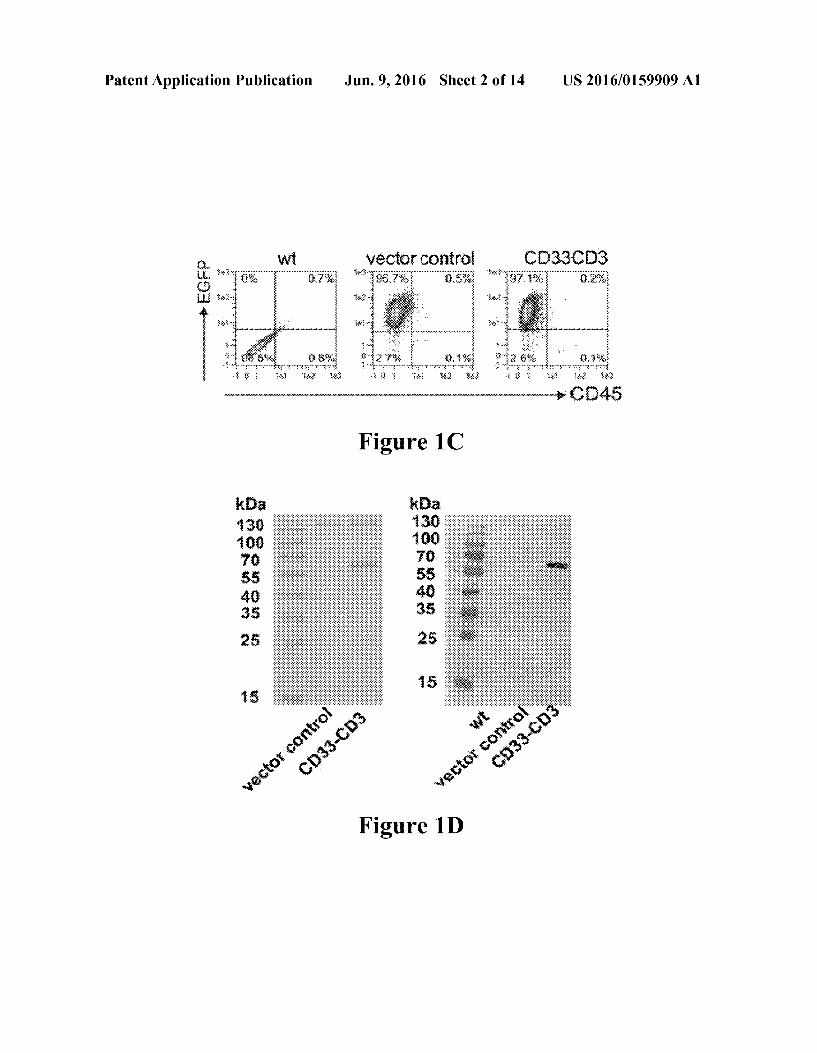
Figure 2C

Patent Application Publication Jun. 9, 2016 Sheet 6 of 14 US 2016/0159909 A1
Figure 3A
: 8.
g E: E333e3:-3 iss E. E.333,388.83.3 s: N: 333-33
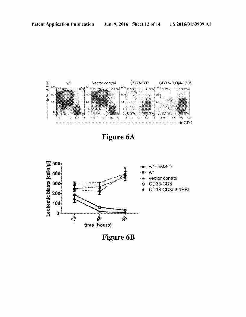
s
s
is As east extractise is
Figure 3B

Patent Application Publication Jun. 9, 2016 Sheet 7 of 14 US 2016/0159909 A1
8. ::::::::::::
: 333-3
s i. m (333 x3 first
its cessfweil
Figure 3C

Patent Application Publication Jun. 9, 2016 Sheet 8 of 14 US 2016/0159909 A1
w 8.
3: 333-33
- - - - is exisfy:
8:8 see . . . . . bab - - - - - - - - - 3-3 it is . . . . & + 3 + 4 + 8 + i.
Figure 3D
CD33-CD3 38.
sector Extro 33-C32-3. 38
Figure 4A

Patent Application Publication Jun. 9, 2016 Sheet 9 of 14 US 2016/0159909 A1
s
S.
s ol S s s S. s S.
hMSC celisweil hSC cells we 2. 48
vector Citro 33-C3
C3- 3 3-33 5:
- s Figure 4B 98

Patent Application Publication Jun. 9, 2016 Sheet 10 of 14 US 2016/0159909 A1
Or
8
60 O

Patent Application Publication Jun. 9, 2016 Sheet 11 of 14 US 2016/0159909 A1
Patent Application Publication Jun. 9, 2016 Sheet 12 of 14 US 2016/0159909 A1
''
wie S& • Nix it * - yector Corio g C33-3
33.3 -83.3
tire heirs
Figure 6B

Patent Application Publication Jun. 9, 2016 Sheet 13 of 14 US 2016/0159909 A1
. i 3.
Figure 6C
w vector contro
33-C3 EC 33-C3 - E8.
Figure 6D

Patent Application Publication Jun. 9, 2016 Sheet 14 of 14 US 2016/0159909 A1
O:
8
8
who ieaiser grasig
{ 2 3. : s
weeks after x
Figure 7

US 2016/O 1599.09 A1
GENETIC MODIFIED PLUR- OR MULTIPOTENT STEM CELLS AND USES
THEREOF
CROSS-REFERENCE TO RELATED APPLICATIONS
0001. This application claims priority benefit of European Patent Application No. EP 15 16634.7.3, filed May 5, 2015, and U.S. Provisional Patent Application Ser. No. 62/088,578, filed Dec. 6, 2014, the disclosures of which are hereby incor porated by reference herein in their entirety.
FIELD OF THE INVENTION
0002 The invention concerns pluri- or multipotent stem cells (SCs), e.g. human pluri- or multipotent stem cells (hSCs) engineered to express a multispecific antibody and which further expresses, on its surface, a human immune cell co-stimulatory ligand or an active fragment thereof.
BACKGROUND OF THE INVENTION
0003. The use of recombinant bispecific antibodies (bsabs) for retargeting effector T lymphocytes towards cancer cells is recently emerging as a promising immunotherapeutic tool for the treatment of hematologic malignancies and other cancers. The high efficacy of this class of molecules in redi recting specifically CD8+ and CD4+ T cells to any chosen tumor associated antigen (TAA) on the Surface of tumor cells by cross-linking their activating CD3 receptor has been reported in many preclinical and clinical studies. Nonethe less, due to their low molecular mass, bsabs have a short half-life in vivo and consequently have to be continuously administered to patients over prolonged time spans of several weeks to achieve clinical responses (Schlereth et al. 2005, Stork et al. 2008, Bargou et al. 2008, Handgretinger et al. 2011). External medical pump devices as currently used in clinical trials can be a potential source of infection. Thus there is a need to improve the route of application for this highly effective class of drugs for treating disorders such as cancer. An alternative to continuous infusion through external medi cal pump devices are gene-modified cells, which continu ously produce and secrete bsabs for their life-time in the body of the patient. 0004 Kasuya, et al. (Kasuya et al., Int. J. Mol Med, 25, 2010, 209-215) report the production of a bispecific anti HER2 and anti-CD16 antibody by embryonic fibroblasts that have been transplanted into immunodeficient mice. 0005. In addition Compte and colleagues, (Compte, et al., Stem Cells, 2099, 27, 753-760) disclose a tumor immuno therapy by use of gene-modified human mesenchymal stem cells (hMSC) for producing a bispecific anti-CEA and anti CD3 antibody. 0006 Furthermore Frank, et al. (Frank, et al., PloSone, 2009, 4. e831) used neural stem cells (NSC) as a novel plat form for tumor specific delivery of therapeutic antibodies. 0007 Although several advances in the use of genetically modified cells for producing therapeutic antibodies have been made, there are still limitations that have to be overcome. 0008. The present invention is directed to overcoming these and other deficiencies in the art.
SUMMARY OF THE INVENTION
0009. It is an object of the present invention to provide genetically modified cells engineered to produce multispe
Jun. 9, 2016
cificantibodies and an improved method for in situ delivery of therapeutic antibodies by these genetically modified cells. 0010. The objective is solved by using pluri- or multipo tent stem cells according to the present invention. 0011. In one aspect, provided herein are pluri- or multipo tent stem cells engineered to express a multispecific antibody, which further express on their Surface a human immune cell co-stimulatory ligand or an active fragment thereof. 0012. The term pluri- or multipotent stem cell (SC) refers to any stem cell other than an embryonic stem cellor stem cell derived from embryo or fetus. The term pluri- or multipotent stem cell (SC) excludes cells from embryo or fetus and includes cells that can be found in children, as well as in adults or that is generated by genetic manipulation of a Somatic cell. 0013 The term pluri- or multipotent stem cell (SC) refers to stem cells comprising mesenchymal stem cells (MSC), neural stem cells (NSC), endothelial progenitor cells (EPC), hematopoietic stem cells (HSC) and induced pluripotent stem cell (iPSC). Preferably the stem cells are human stem cells. 0014. In a preferred embodiment the pluri- or multipotent stem cells are human mesenchymal stem cells (hMSC). 0015. In certain embodiments, the human immune cell co-stimulatory ligand or an active fragment thereof, which can be recombinantly expressed on the cell surface of SCs, provided herein is selected from a group of CD28 ligands CD80 (B7-1) and CD86 (B7-2), CD137 ligand (CD137L, 4-1BBL), Ox40L ligand OX40L (CD252), CD27 ligand CD70 (CD27L), Inducible T-cell Costimulator (ICOS) ligand ICOSL (CD275), lymphocyte function-associated antigen (LFA) 1 ligand intercellular adhesion molecule (ICAM)-1 (CD54), -2 (CD102), and -3 (CD50), and ligands of the sig naling lymphocytic activation molecule (SLAM) family e.g. 2B4 (CD244) ligand SLAMF2 (CD48). 0016. In certain embodiments the pluri- or multipotent stem cells are engineered to express multispecific antibodies, comprising at least a target cell specific binding domain and at least an immune cell specific binding domain. 0017. The two specific domains (target cell and immune cell) are preferably expressed as a fusion protein. 0018. The target cell specific binding domain is preferably specific for a tumor associated antigen or an antigen of a pathogen, parasite or parasitoid. 0019 More preferred, the target cell specific binding domain is specific for an antigen selected from Surface, cyto plasmic or nuclear antigens like the La/SSB antigen, CD19, CD20, CD22, CD33, CD38, CD56, CD157, CD123, CCR5, STEAP1, prostate stem cell antigen PSCA, prostate specific membrane antigen PSMA, members of the epidermal growth factor receptor family, preferably EGFR (ErbB-1), HER2/ neu (ErbB-2), Her3 (ErbB-3) and Her4 (ErbB-4) and mutants thereof, members of the ephrin receptor family, preferably EphA1-10 or EphB1-6, embryonic antigens, preferably car cinoembryonic antigen CEA, fetal acethylcholine receptor, CEACAM-6, CEACAM-7. 0020 Next most preferred, the target cell specific binding domain is specific for an antigen selected from members of the Rho family of GTPases, members of the high mobility group proteins, bladder tumor antigen (BTA), CA125, CD2, CD4, CD8, CD10, CD23, CD30, CD44, CD52, CD99, CD135, CD274, Carboanhydrase IX, Endosialin, fibroblast activating protease-alpha (FAP-alpha), FBP. gpA33, gp100, c-Met (hepatocyte growth factor receptor), MCSP and TIM 3, members of the vascular endothelia growth factor family (VEGFR 1-3), epithelia cell adhesion molecule EpCAM,

US 2016/O 1599.09 A1
alphafetoprotein AFP members of the mucin protein family, preferably MUC1 or MUC16, follicle stimulating hormone receptor (FSHR), the human high molecular weight-mela noma-associated antigen (HMW-MAA), folate binding pro tein FBP a-Folate receptor, ligands of the NKG2D receptor, members of the epithelia glycoprotein family, preferably EGP-2, EGP-4 or EGP-40, members of the insulin-like growth factor receptor family, preferably IGF-1R, diasialo gangliosides, preferably GD2 or GD3, members of the car bonic anhydrase family, preferably CAIX, members of the carbohydrate antigen family, preferably Ley, including mutants of the named proteins and protein families and viral envelope proteins, preferably gp120, gp41 or EBOV GP. 0021 Alternatively preferred, the target cell binding domain is composed of the alpha and beta or the gamma and delta chains of a T cell receptor (TCR) or fragments thereof. Such TCR-derived binding moieties recognize and bind to peptides presented by human leukocyte antigen class (HLA) I and II protein complexes. Preferred examples are, but are not limited to, TCRs specific for viral peptides, peptides derived from proteins like EGFR family, survivin, Sry-like high motil ity group box (SOX) protein family, melanoma-associated antigens, preferably autoimmunogenic cancer/testis antigen NY-ESO-1, members of the melanoma antigen family A MAGEA, the preferentially expressed antigen in melanoma PRAIVIE, and leukemia-associated antigens, preferably wilms tumor gene 1 WT1. The target cell binding moiety could also comprise ligands to proteins and protein com plexes, further on referred as receptors. Such ligands may bind to, but are not limited to, cytokine receptors, preferably IL-13 receptor, ligands of the NKG2D receptor, ligands to the EGFR family members, or auto-reactive TCRs. 0022. For the generation of permanent SCs expressing multispecific antibodies, e.g. bispecific antibodies, cDNA encoding a recombinant antibody, preferably multispecific antibody, Such as bispecific antibody construct can, for example, be cloned into a viral vector (e.g., self-inactivating retroviral vector), such as a retroviral vector or lentiviral vector, preferably p6NST50 to generate a transfer vector. In this viral vector, the expression of a recombinant antibody (e.g., multispecific antibody, Such as bispecific antibody) can be driven by a Suitable promoter, Such as a viral promoter, e.g., spleen focus forming virus (SFFV)-derived internal pro moter. In certain embodiments, an enhanced green fluores cence protein (EGFP)-Zeocin fusion protein can be co-ex pressed by an internal ribosomal entry site (IRES). Lentiviral particles pseudotyped with an envelope protein, e.g., the Vesicular Stomatitis Virus envelope 120 (VSV-G), can be generated by transient transfection of a packaging cell, e.g., HEK293T cells, and virus supernatant are harvested and used to stably transduce SCs, such as SCP-1 cells. 0023 For ectopic expression of a co-stimulary ligand,
e.g., the co-stimulatory 4-1BB ligand (CD137L), on the sur face of SCs, such as SCP-1 cells, a viral vector, such as a retroviral vector or a lentiviral vector, can be used which harbors an appropriate mammalian promoter, such as an internal minimal human elongation factor 1 alpha promoter. 0024. In a particular aspect, SCs can be co-transfected with two or more expression vectors (e.g., viral vectors) encoding polypeptides that associate (e.g., covalently or non covalently) to form antigen binding domains (e.g., antigen binding domains comprising a VH and a VL) of an antibody (e.g., multispecific antibody, such as bispecific antibody) expressed by SCs.
Jun. 9, 2016
0025. In certain embodiments, at least 1 pg. 5 pg. 10 pg. 50 pg, 75 pg. 100 pg. 250 pg of antibody are produced per cell over a period of 48 hours. In other embodiments, at most 1 pg. 5 pg. 10 pg. 50 pg. 75 pg. 100 pg. 250 pg of antibody are produced per cell over a period of 48 hours. In certain embodiments, 5-10, 5-50, 10-50, 25-75, or 8-70 pg of anti body are produced per cell over a period of 48 hours. 0026. In specific aspects, provided herein are multispe cific antibodies, such as bispecific antibodies, which are recombinantly expressed by SCs, wherein the multispecific antibodies comprising at least a target cell specific binding domain and at least an immune cell specific binding domain. 0027. The two specific domains (target cell and immune cell) are preferably expressed as a fusion protein. 0028. The target cell specific binding domain is preferably specific for a tumor associated antigen or an antigen of a pathogen, parasite or parasitoid. 0029. The tumor antigen can be associated, but is not restricted to, hematopoietic and lymphoid malignancies like leukemia (e.g. acute lymphoblastic leukemia, acute myelog enous leukemia or chronic lymphocytic or myelogenous leu kemia), lymphoma (e.g. non-Hodgkin and Hodgkin lympho mas) or myeloma. It can be also associated with carcinoma (e.g. breast, prostate, lung, pancreas or colon carcinoma). sarcoma (e.g. osteosarcoma, Ewings sarcoma), or blastoma (e.g. neuroblastoma, glioblastoma multifome). 0030. In certain aspects, a bispecific antibody provided herein is a single chain bispecific antibody. In a particular embodiment, a bispecific antibody provided herein, such as a single chain bispecific antibody, is humanized (e.g., human ized bispecific antibody targeting CD33 and CD3). 0031. As used herein, the terms “variable region' or “vari able domain refer to a portion of an antibody, generally, a portion of an antibody light or heavy chain, typically about the amino-terminal 110 to 120 amino acids in a mature heavy chain and about the amino-terminal 90 to 100 amino acids in a mature light chain. Variable regions comprise complemen tarity determining regions (CDRs) flanked by framework regions (FRs). Generally, the spatial orientation of CDRs and FRs are as follows, in an N-terminal to C-terminal direction: FR1-CDR1-FR2-CDR2-FR3-CDR3-FR4. Without wishing to be bound by any particular mechanism or theory, it is believed that the CDRs of the light and heavy chains are primarily responsible for the interaction of the antibody with antigen and for the specificity of the antibody for an epitope. 0032. In a specific embodiment, numbering of amino acid positions of antibodies described herein is according to the EU Index, as in Kabat et al. (1991) Sequences of Proteins of Immunological Interest, Fifth Edition, U.S. Department of Health and Human Services, NIH Publication No. 91-3242. In certain embodiments, the variable region is a human vari able region. In certain embodiments, the variable region com prises murine (e.g., mouse or rat) CDRS and human frame work regions (FRs). In particular embodiments, the variable region is a primate (e.g., human or non-human primate) vari able region. In certain embodiments, the variable region com prises murine (e.g., mouse or rat) CDRS and primate (e.g., human or non-human primate) framework regions (FRS). As a non-limiting example, a variable region described herein is obtained from assembling two or more fragments of human sequences into a composite human sequence. 0033. In certain aspects, the CDRs of an antibody can be determined according to (i) the Kabat numbering system (Kabat et al. (1971) Ann. NY Acad. Sci. 190:382-391 and,

US 2016/O 1599.09 A1
Kabat et al. (1991) Sequences of Proteins of Immunological Interest, Fifth Edition, U.S. Department of Health and Human Services, NIH Publication No. 91-3242); or (ii) the Chothia numbering scheme, which will be referred to herein as the “Chothia CDRs (see, e.g., Chothia and Lesk, 1987, J. Mol. Biol., 196:901-917: Al-LaZikani et al., 1997, J. Mol. Biol., 273:927-948; Chothia et al., 1992, J. Mol. Biol., 227:799 817: Tramontano A et al., 1990, J. Mol. Biol. 215(1): 175-82: and U.S. Pat. No. 7,709.226); or (iii) the ImMunoGeneTics (IMGT) numbering system, for example, as described in Lefranc, M.-P., 1999, The Immunologist, 7:132-136 and Lefranc, M.-P. et al., 1999, Nucleic Acids Res., 27:209-212 (“IMGT CDRs’); or (iv) MacCallum et al., 1996, J. Mol. Biol., 262:732-745. See also, e.g., Martin, A., “Protein Sequence and Structure Analysis of Antibody Variable Domains, in Antibody Engineering, Kontermann and Dibel, eds., Chapter 31, pp. 422-439, Springer-Verlag, Berlin (2001). 0034. In a specific aspect, provided herein are SCs expressing multispecific antibodies, e.g., bispecific antibod ies, such as single chain bispecific antibodies, comprising a VL and a VH targeting a tumor associated antigen or an antigen of a pathogen, parasite or parasitoid. In specific aspects, multispecific antibodies, such as bispecific antibod ies, e.g., single chain bispecific antibodies, described herein comprise one or more linkers (e.g., one, two, three, four, or five, or more linkers). 0035. In a certain embodiment, such a linker described herein comprises repetitions, for example, at least one, two, three, four or five repetitions, of glycine-serine, e.g., GS linkers. 0036. In a specific embodiment, provided herein are SCs expressing a multispecific antibody, e.g., bispecific antibody, Such as a single chain bispecific antibody, which specifically binds to a tumor associated antigen or an antigen of a patho gen, parasite or parasitoid comprising a heavy chain variable region (VH) and a light chain variable region (VL). 0037. In specific embodiments, multispecific antibodies, Such as, bispecific antibodies, e.g., single chain bispecific antibodies, described herein comprises at the N-terminus a signal peptide, e.g., an Ig kappa leader sequence as signal peptide, for protein secretion. 0038. In certain embodiments, multispecific antibodies, Such as, bispecific antibodies, e.g., single chain bispecific antibodies, described herein comprises at the C-terminus a tag, for example, myc-tag or and histidine (his)-tag (e.g., 6xhis-tag) for protein purification and/or detection. 0039. In certain embodiments, antibodies (e.g., multispe
cific antibodies, such as bispecific antibodies) described herein can be of any type (e.g., IgG, IgE, IgM, Ig|D, IgA or IgY), any class, (e.g., IgG, IgG, IgGs. IgG4, IgA1 or IgA2), or any subclass (e.g., IgG or IgG or a mixture thereof) of immunoglobulin molecules. 0040. In specific embodiments, multispecific antibodies, Such as, bispecific antibodies, e.g., single chain bispecific antibodies, described herein do not comprise an antibody constant region. 0041 Methods for making multispecific (e.g. bispecific antibodies) have been described, see, for example, U.S. Pat. Nos. 7,951,917, 7,183,076, 8,227,577, 5,837,242, 5,989,830, 5,869,620, 6,132,992, and 8,586,713. 0042. In certain embodiments, the presently disclosed methods and uses enhance the T cell response against a cancer cell. Without being bound by theory, the improved T cell
Jun. 9, 2016
response against a cancer cell is accomplished by targeting of pluri- or multipotent stem cells to the cancer cell in a patient. In certain embodiments, at least 1x10, 2x10,3x10, 4x10, 5x10, 6x10, 7x10, 8x10, 9x10, 1x10, 2x10, 3x10, 4x10, 5x10, 6x10, 7x10, 8x10,9x10, or at least 1x10⁷ SCs as described herein are administered to a patient. In certain embodiments, at most 1x10, 2x10, 3x10, 4x10, 5x10, 6x10, 7x10, 8x10, 9x10, 1x10, 2x10, 3x10, 4x10, 5x10, 6x10, 7x10, 8x10, 9x10, or at least 1x10⁷ SCs as described herein are administered to a patient. In a more specific embodiment between 5x10° and 6x10° SCs as described herein are administered to a patient. 0043. In certain embodiments, at least 1 mg, 2 mg, 3 mg. 4 mg, 5 mg, 6 mg, 7 mg, 8 mg, 9 mg, or at least 10 mg cell mass ofan SC as described hereincan be transplanted into a patient. In certain embodiments, at most 1 mg, 2 mg, 3 mg, 4 mg, 5 mg, 6 mg, 7 mg, 8 mg.9 mg, or at least 10 mg cell mass of an SC as described herein can be transplanted into a patient. In certain embodiments, about 1 mg, 2 mg, 3 mg, 4 mg, 5 mg, 5.5 mg, 6 mg, 7 mg, 8 mg.9 mg, or at least 10 mg cell mass of an SC as described herein can be transplanted into a patient. 0044 SCs can be used as in situ producer of anti-cancer therapeuticals. In certain embodiments, autologous gene modified SCs as described herein can be injected intrave nously into patients and, without being bound by theory, migrate to the tumor site and deliver their payload locally. 0045. In certain embodiment, the use of SCs for in situ production of anti-cancer therapeutics (eg. multispecific or bispecific antibodies) results in continuous delivery of the anti-cancertherapeuticals inapatient. More specifically, Such continuous production can last for at least 1, 2, 3, 4, 5, 6, 7, 8, 9, 10, 11, 12, 13, or 14 days, at least 1, 2, 3, 4, 5, 6, 7, 8, 9, 10 weeks, or at least 1, 2, 3, 4, 5, 6, 7, 8, 9, 10, 11, or 12 months, or at least 1, 2, 3, 4, 5, 6, 7, 8, 9, 10 years. More specifically, Such continuous production can last for at most 1, 2, 3, 4, 5, 6, 7, 8, 9, 10, 11, 12, 13, or 14 days, at least 1, 2, 3, 4, 5, 6, 7, 8, 9, 10 weeks, or at least 1, 2, 3, 4, 5, 6, 7, 8, 9, 10, 11, or 12 months, or at least 1, 2, 3, 4, 5, 6, 7, 8, 9, 10 years. 0046. In specific aspects, provided herein are methods for treating cancer, such as acute myeloid leukemia (AML) in a Subject by administering a therapeutic effective amount of SCs expressing an antibody, e.g., multispecific antibody. Such as bispecific antibody and a T cell co-stimulatory ligand. Such as the co-stimulatory 4-1BB ligand (CD137L). Also provided herein are related methods for inducing/enhancing T cell proliferation and related methods for activating T cells using such SCs. 0047. In particular embodiments, provided herein are methods for managing, treating, preventing or protecting against AML in a Subject (e.g. human Subject) in need thereof, administering to the subject a therapeutically effective amount of SCs expressing an antibody, e.g., a multispecific antibody, such as a bispecific antibody (e.g. anti-CD33-anti CD3 bispecific antibody), and a T cell co-stimulatory ligand, such as the co-stimulatory 4-1BB ligand (CD137L). 0048. In a particular embodiment, provided herein is a method for inducing or enhancing T cell proliferation com prising T cells interacting with an effective amount of SCs expressing an antibody, e.g., multispecific antibody, such as bispecific antibody (for example, anti-CD33-anti-CD3 bispe cific antibody), and a T cell co-stimulatory ligand, such as the co-stimulatory 4-1BB ligand (CD137L). In a specific embodiment, provided herein is a method for inducing or enhancing T cell proliferation in a subject in need thereof,

US 2016/O 1599.09 A1
comprising a therapeutic effective amount of SCs expressing an antibody, e.g., multispecific antibody, such as bispecific antibody (for example, anti-CD33-anti-CD3 bispecific anti body), and optionally a T cell co-stimulatory ligand. Such as the co-stimulatory 4-1BB ligand (CD137L). 0049. In particular embodiments, T cell proliferation is increased by at least 10%, 20%, 30%, 40%, 50%, 60%, 70%, 80%, 90%, 100%, 150%, 200%, 250%, 300%, 400%, or 500% or more in presence of SCs compared to their absence, as determined by methods known in the art, for example, those described herein. 0050. In a particular embodiment, provided herein is a method for inducing or enhancing T cell activation, compris ing T cells interacting with an effective amount of SCs expressing an antibody, e.g., multispecific antibody, such as bispecific antibody (for example, anti-CD33-anti-CD3 bispe cific antibody), and a T cell co-stimulatory ligand, such as the co-stimulatory 4-1BB ligand (CD137L). In a specific embodiment, provided herein is a method for inducing or enhancing T cell activation in a Subject in need thereof, com prising a therapeutic effective amount of SCs expressing an antibody, e.g., multispecific antibody, such as bispecific anti body (for example, anti-CD33-anti-CD3 bispecific anti body), and a T cell co-stimulatory ligand, Such as the co stimulatory 4-1BB ligand (CD137L). 0051. In particular embodiments, T cell activation is increased by at least 10%, 20%, 30%, 40%, 50%, 60%, 70%, 80%, 90%, 100%, 150%, 200%, 250%, 300%, 400%, or 500% or more in presence of SCs compared to their absence, as determined by methods known in the art, for example, those described herein (e.g., Examples Section). 0052. These and other objects, features, and advantages of
this invention will become apparent from the following detailed description of the various aspects of the invention taken in conjunction with the accompanying drawings.
BRIEF DESCRIPTION OF THE DRAWINGS
0053 For the purpose of illustrating aspects of the present invention, there are depicted in the drawings certain embodi ments of the invention. However, the invention is not limited to the precise arrangements and instrumentalities of the embodiments depicted in the drawings. Further, as provided, like reference numerals contained in the drawings are meant to identify similar or identical elements. 0054 FIGS. 1A-1E: Generation of bsab CD33-CD3 releasing gene-modified hMSCs. FIG. 1A: Schematic repre sentation of the structure of the bsab CD33-CD3 constructed as single-chain bispecific tandem fragment variable (Sch sTaFv). The VH and VL domains of each scFv were human ized by CDR grafting and connected via a linker comprised of three repeats of four glycine and one serine residues 3*(Gly4Ser). The N-terminus of the bsab construct contains a signal peptide (SP) for the secretion of the bsab into the cell culture medium, whereas its C-terminus tag harbors a myc (myc) and his-tag (his) used for immunochemical Ab detec tion and purification. FIG. 1B: Schematic illustrating binding of the bsab to its target antigens CD3 and CD33 cross-links a T cell and a tumor cell and establishes an immune synapse. FIG. 1C: Transgene expression analysis of parental and trans duced hMSCs was performed by flow cytometry. The per centages of living CD45- and EGFP+ cells are shown. Dead cells were excluded by propidium iodide staining. FIG. 1D: Purified fractions of the bsab CD33-CD3 Secreted in the culture medium were separated on SDS-gels and thereafter
Jun. 9, 2016
stained either with Coomassie brilliant blue(R) or analyzed by Western blotting. FIG. 1E: The quantitative analysis of the released bsab was performed by ELISA. hMSCs cells were seeded at limiting cell densities and the antibody concentra tion ng/ml in culture medium was determined after 48 h of culture. Results represent the means-SD of two independent experiments. 0055 FIGS. 2A-2C. Antigen binding properties of the hMSC secreted recombinant protein. FIG. 2A: CD33+ MOLM-13 cells (left panels) and T cells (right panels) were stained with either the maternal anti-CD33 and anti-CD3 mAbs following by a PE-conjugated anti-mouse IgG second ary Ab (black graphs) or the anti-CD33-anti-CD3 bsab and a FITC-labeled anti-myc Ab (black graphs). As controls, samples were stained with secondary antibodies alone (white graphs). FIG.2B: BSAb binding to CD33- HEK293T cells or SCP-1 wt cells was analyzed in parallel by flow cytometry after staining with the bsab and the anti-myc/FITC Ab. FIG. 2C: The accessibility of the bsab to the CD33 antigen on MOLM-13 cells was tested after pretreatment with 2, 20 and 200 ug/ml of the maternal anti-CD33 mAb respectively. 0056 FIGS. 3A-3D: Efficient T cell-mediated killing of target cell lines with varying CD33 expression levels is induced in the presence of bsab-releasing hMSCs. FIG. 3A: HEK293T, OCI-AML3, U937 and MOLM-13 were analyzed for CD33 surface expression levels by staining with anti CD33/PE mAb (in black) or matched isotype control Ab (in grey) respectively. Numbers represent mean fluorescence intensity (MFI) of total cells. FIG. 3B: In a standard chro mium release assay 51Cr labeled CD33+ MOLM-13 cells and CD33- HEK293T cells were incubated with freshly iso lated T cells at effector to target (e:t) cell ratio of 5:1 for 20h with decreasing concentrations of the purified bsab CD33 CD3. MeaniSD of 2 independent donors is shown. FIG. 3C: Specific cell lysis of AML cell lines U937 (upper) and MOLM-13 (lower) measured with standard chromium release assay. Freshly isolated CD3+ T cells were co-cultured for 10 and 20h with 51Cr labeled CD33+ target cells at an eit cell ratio of 5:1 in the presence of hMSC lines seeded at different concentrations 48 h before adding effector T cells and target cells. Data are presented as meansitSD from two or three different donors, respectively. FIG. 3D: Decreasing densities of 51Cr labeled gene-modified hMSCs were co cultured with PBMCs in the presence or absence of CD33+ MOLM-13 cells at an e:t ratio of 5:1. After 20 h of co incubation the specific hMSCs lysis was examined via chro mium release assay. Data shown as meantSD from two inde pendent donors. 0057 FIGS. 4A-4B: T cell-mediated tumor cell killing elicited by hMSC-produced anti-CD33-anti-bsab is enhanced by co-stimulation from 4-1BBL. FIG. 4A: For the analysis of the 4-1BBL transgene expression hMSCs were stained with PE-conjugated anti-CD137L antibody and the Surface expression of the immunoligand as well as intracel lular EGFP signal correlating with bsab CD33-CD3 expres sion were analyzed by flow cytometry. Positive cells are shown as percentages of all analyzed cells. Dead cells were excluded by propidium iodide counter-staining. Quadrant position was placed based on isotype control staining (not shown). FIG. 4B: In a flow cytometry-based cytotoxicity assay eFluoró70 proliferation dye-labeled CD33+OCI AML3 cells were incubated with pan T cells at an et ratio of 1:1 for 24h, 48 h (upper) and 96 h (lower) in the presence or absence of gene-modified hMSCs seeded at limiting densities

US 2016/O 1599.09 A1
48 h prior to the experiment. Target cell numbers counted at the indicated time points were normalized to the control sample with only target cells. Data represent the means-SD of three different donors. Statistical significance was deter mined using one-way ANOVA with Bonferroni multiple comparison test. **p-0.01. 0058 FIGS. 5A-5B: The presence of the co-stimulatory 4-1BBL signal dramatically increases cytokine secretion and T cell expansion. FIG. 5A: Secretion of pro-inflammatory cytokines by T cells was determined after 24h culture upon their cross-linkage with the transgenic CD33-expressing HEK293T cells via hMSC-produced CD33-CD3 bsab in the presence or absence of the hMSC-presented co-stimulatory 4-1BBL. FIG. 5B: After 6 days of co-cultivation with target cells T cell counts were investigated and T cell expansion was calculated as ratio of T cell number at day 6 to T cell number seeded at day O. Data are shown as means-SD of three or four individual donors. Statistical significance was determined using one-way ANOVA with Bonferroni multiple compari son test. *p-0.05; **p<0.01: ***p<0.001. 0059 FIGS. 6A-6D: Antitumor effect of redirected AML patient-derived T cells against autologous AML blasts via hMSC-produced bsab CD33-CD3 and surface presented 4-1BBL molecule. FIG. 6A: 1x10⁵ AML patient-derived MNCs were cultured together with 48 h pre-seeded 1x10⁴ hMSCs. After 96 h of co-cultivation the percentages of Sur viving HLA-DR-- AML blasts and CD3+ T cells were deter mined respectively as proportions of all CD45+ cells by flow cytometry analysis. FIG. 6B: Total AML blasts number after 24, 48 and 96 h of co-incubation was calculated. The average of surviving cells and the SD of triplets are shown for one representative donor out of three. FIG. 6C: Total numbers of CD3-CD123+HLA-DR+CD45+AML blasts (left) and CD3+ CD123-HLA-DR-CD45+ T cells (right) after 96 h of co cultivation with or without control/bsab- and 4-1BBL-ex pressing hMSCs are reported for three independent donors. Numbers of each Subpopulation were calculated according to their relative percentages as determined by staining for spe cific cell surface markers. FIG. 6D: Absolute autologous T cell number was measured after 96 hand overall expansion of the cells in the presence of hMSCs was determined. Data are presented as means-SD from three different donors. Statisti cal significance was determined using one-way ANOVA with Bonferroni multiple comparison test. *p-0.05; **p<0.01; ***p<0.001. 0060 FIG. 7: BSAb-releasing hMSCs prevent the estab lishment of AML in NOD/SCID IL2Ry-/- (NSG) mice. (a) Kaplan-Meier survival analysis of NOD/SCID IL2Ry-/- mice after intravenous injection of T cells and MOLM-13 AML cells at an et ratio of 5:1 together with vector control containing (dashed line) or bsab-releasing (black line) hMSCs, indicated as control (n=5) and treatment group (n=4) respectively. A log-rank test was performed to determine the statistical significance in Survival between the groups from ongoing experiment. **p-0.01.
DETAILED DESCRIPTION OF THE INVENTION
0061. In a further aspect, provided herein are mesenchy mal stem cells (MSCs), such as human MSCs (hMSCs), engineered to express an antibody, e.g., multispecific anti body, such as a bispecific antibody targeting CD33 and CD3. and, optionally, a human T cell co-stimulatory ligand, Such as the human T cell co-stimulatory 4-1BB ligand (CD137L) or an active fragment thereof. In specific aspects, also provided
Jun. 9, 2016
herein are methods for treating acute myeloid leukemia (AML) using such hMSCs, as well as related methods for inducing/enhancing T cell proliferation and related methods for activating T cells. 0062. In one aspect provided herein are human mesenchy mal stem cells (hMSCs) engineered to express a multispecific antibody, Such as a bispecific antibody targeting CD33 and CD3, and, optionally, a human T cell co-stimulatory ligand, such as the human T cell co-stimulatory 4-1BB ligand (CD137L) or an active fragment thereof. In specific aspects, also provided herein are methods for treating acute myeloid leukemia (AML) using such hMSCs, as well as related meth ods for inducing/enhancing T cell proliferation and related methods for activating T cells using such hMSCs. 0063. In certain embodiments, the presently disclosed methods and uses enhance the T cell response against a cancer cell. Without being bound by theory, the improved T cell response against a cancer cell is accomplished by targeting of hMSCs to the cancer cell inapatient. In certain embodiments, at least 1x10, 2x10, 3x10, 4x10, 5x10, 6x10, 7x10, 8x10, 9x10, 1x10, 2x10, 3x10°, 4x10, 5x10, 6x10, 7x10, 8x10, 9x10, or at least 1x10⁷ MSCs as described herein are administered to a patient. In certain embodiments, at most 1x10, 2x10, 3x10, 4x10, 5x10, 6x10, 7x10, 8x10, 9x10, 1x10, 2x10, 3x10, 4x10, 5x10, 6x10, 7x10, 8x10, 9x10, or at least 1x10⁷ MSCs as described herein are administered to a patient. In a more specific embodiment between 5x10° and 6x10 MSCs as described herein are administered to a patient. 0064. In certain embodiments, at least 1 mg, 2 mg, 3 mg. 4 mg, 5 mg, 6 mg, 7 mg, 8 mg, 9 mg, or at least 10 mg cell mass of an MSC as described herein can be transplanted into a patient. In certain embodiments, at most 1 mg, 2 mg, 3 mg. 4 mg, 5 mg, 6 mg, 7 mg, 8 mg, 9 mg, or at least 10 mg cell mass of an MSC as described herein can be transplanted into a patient. In certain embodiments, about 1 mg, 2 mg, 3 mg. 4 mg, 5 mg, 5.5 mg, 6 mg, 7 mg. 8 mg.9 mg. or at least 10 mg cell mass of an MSC as described herein can be transplanted into a patient. 0065 MSCs can be used as in situ producer of anti-cancer therapeutics. In certain embodiments, autologous gene modified MSCs as described herein can be injected intrave nously into patients promoting their migration to the tumor site and the local delivery of their payload without being bound by theory. In other embodiments, MSCs could be trapped into an artificial scaffold matrix and transplanted Subcutaneously in proximity of a tumor site. Again, without being bound by theory, thus the potential supportive role in tumorangiogenesis processes could be reduced, ensuring that transplanted MSCs cannot escape from their confined artifi cial environment. An additional benefit of this strategy can be that the delivery of the anti-cancer agent can be controlled and stopped after tumor clearance by removing the scaffold Compte et al. (2007) Cancer Gene Ther: 14(4): 380-388.). 0066. In certain embodiment, the use of MSCs to in situ production of anti-cancer therapeutics (eg. bispecific anti bodies) results in continuous delivery of the anti-cancerthera peutic in a patient. More specifically, such continuous pro duction can be production for at least 1,2,3,4,5,6,7,8,9, 10. 11, 12, 13, or 14 days, at least 1,2,3,4,5,6,7,8,9, 10 weeks, or at least 1,2,3,4,5,6,7,8,9, 10, 11, or 12 months, or at least 1, 2, 3, 4, 5, 6, 7, 8, 9, 10 years. More specifically, such continuous production can be production for at most 1,2,3,4, 5, 6, 7, 8, 9, 10, 11, 12, 13, or 14 days, at least 1, 2, 3, 4, 5, 6,

US 2016/0159909 A1
7, 8, 9, 10 weeks, or at least 1, 2, 3, 4, 5, 6, 7, 8, 9, 10, 11, or 12 months, or at least 1, 2, 3, 4, 5, 6, 7, 8, 9, 10 years.
Mesenchymal StemCells 0067. Any mesenchymal stem cells known to the skilled artisan can be used with the methods and compositions pro vided herein. For example, methods for isolating primary hMSCs have been described, see, e.g., Penfornis and Pochampally, 2011, Methods Mol Biol. 698:11-21; and Qiao et al., 2008, Cell Biol. International, 32: 8-15. Mesenchymal stem cells, or MSCs, are multipotent stromal cells that can differentiate into a variety of cell types, including bone, car tilage, and fat cells. MSCs can be derived from bone marrow. 0068. In a specific aspect, provided herein are mesenchy mal stem cells (MSCs), such as human MSCs (hMSCs), recombinantly expressing an antibody (e.g., multispecific antibody, such as bispecific antibody, for example, anti CD33-anti-CD3 bispecific antibody), and optionally a T cell co-stimulatory ligand, such as the co-stimulatory 4-1BB ligand (CD137L). 0069. In a particular aspect, hMSCs provided herein are engineered to express multispecific antibodies, e.g., bispe cific antibodies (e.g., bispecific antibodies targeting CD33 and CD3) are derived from the single-cell-picked clone 1 (SCP-1) cell line (Wicker et al., 2008, J. Cell Mol Med: 12: 1347-1359), derived from human mesenchymal stem cells and immortalized by lentiviral transduction of the gene cod ing for the human telomerase reverse transcriptase (hTERT). 0070. In a certain aspect, hMSCs provided herein engi neered to express multispecific antibodies, e.g., bispecific antibodies (e.g., bispecific antibodies targeting CD33 and CD3) are derived from primary hMSCs. (0071. For the generation of permanenth MSCs expressing multispecific antibodies, e.g., bispecific antibodies (e.g., bispecific antibodies targeting CD33 and CD3), cDNA encoding a recombinant antibody (e.g., multispecific anti body, such as bispecific antibody, for example, anti-CD33 anti-CD3 bispecific antibody) construct can, for example, be cloned into a a viral vector (e.g., self-inactivating retroviral vector), such as a retroviral vector or lentiviral vector (e.g., p6NST50) to generate a transfer vector. In this viral vector, the expression of a recombinant antibody (e.g., multispecific antibody, such as bispecific antibody, for example, anti CD33-anti-CD3 bispecific antibody) can be driven by a suit able promoter, such as a viral promoter, e.g., spleen focus forming virus (SFFV)-derived internal promoter. In certain embodiments, an enhanced green fluorescence protein (EGFP)-Zeocin fusion protein can be co-expressed by an internal ribosomal entry site (IRES). Lentiviral particles pseudotyped with an envelope protein, e.g., the Vesicular Stomatitis Virus envelope 120 (VSV-G), can be generated by transient transfection of a packaging cell, e.g., HEK293T cells, and virus supernatant are harvested and used to stably transduce hMSCs, such as SCP-1 cells. 0072 For ectopic expression of a co-stimulary ligand, e.g., the co-stimulatory 4-1BB ligand (CD137L), on the sur face of hMSCs, such as SCP-1 cells, a viral vector, such as a retroviral vector or a lentiviral vector, can be used which harbors an appropriate promoter, such as an internal minimal human elongation factor 1alpha promoter. (0073. In a particular aspect, hMSCs can be co-transfected with two or more expression vectors (e.g., viral vectors) encoding polypeptides that associate (e.g., covalently or non covalently) to form antigen binding domains (e.g., antigen
Jun. 9, 2016
binding domains comprising a VH and a VL) of an antibody (e.g., multispecific antibody, such as bispecific antibody, for example, anti-CD33-anti-CD3 bispecific antibody) expressed by hMSCs. 0074. In certain embodiments, at least 1 pg. 5 pg. 10 pg. 50 pg. 75 pg. 100 pg. 250 pg of antibody are produced per cell over a period of 48 hours. In other embodiments at most 1 pg. 5 pg. 10 pg. 50 pg. 75 pg. 100 pg. 250 pg of antibody are produced per cell over a period of 48 hours. In certain embodiments, 5-10, 5-50, 10-50, 25-75, or 8-70 pg of anti body are produced per cell over a period of 48 hours.
Antibodies
0075). In specific aspects, provided herein are antibodies, such as bispecific antibodies, which are recombinantly expressed by hMSCs. In a particular embodiment, such bispecific antibodies target CD33, e.g., human CD33. In a specific embodiment, such bispecific antibodies target CD33, e.g., human CD33, and CD3, e.g., human CD3. In certain aspects, a bispecific antibody provided herein is a single chain bispecific antibody. In a particular embodiment, a bispecific antibody provided herein, such as a single chain bispecific antibody, is humanized (e.g., humanized bispecific antibody targeting CD33 and CD3). 0076. As used herein, the terms “variable region' or “vari able domain” refer to a portion of an antibody, generally, a portion of an antibody light or heavy chain, typically about the amino-terminal 110 to 120 amino acids in a mature heavy chain and about the amino-terminal 90 to 100 amino acids in a mature light chain. Variable regions comprise complemen tarity determining regions (CDRs) flanked by framework regions (FRs). Generally, the spatial orientation of CDRs and FRs are as follows, in an N-terminal to C-terminal direction: FR1-CDR1-FR2-CDR2-FR3-CDR3-FR4. Without wishing to be bound by any particular mechanism or theory, it is believed that the CDRs of the light and heavy chains are primarily responsible for the interaction of the antibody with antigen and for the specificity of the antibody for an epitope. In a specific embodiment, numbering of amino acid positions of antibodies described herein is according to the EU Index, as in Kabat et al. (1991) Sequences of Proteins of Immuno logical Interest, Fifth Edition, U.S. Department of Health and Human Services, NIH Publication No. 91-3242. In certain embodiments, the variable region is a human variable region. In certain embodiments, the variable region comprises murine (e.g., mouse or rat) CDRs and human framework regions (FRs). In particular embodiments, the variable region is a primate (e.g., human or non-human primate) variable region. In certain embodiments, the variable region com prises murine (e.g., mouse or rat) CDRs and primate (e.g., human or non-human primate) framework regions (FRS). As a non-limiting example, a variable region described herein is obtained from assembling two or more fragments of human sequences into a composite human sequence. 0077. In certain aspects, the CDRs of an antibody can be determined according to (i) the Kabat numbering system (Kabat et al. (1971) Ann. NY Acad. Sci. 190:382-391 and, Kabat et al. (1991) Sequences of Proteins of Immunological Interest, Fifth Edition, U.S. Department of Health and Human Services, NIH Publication No. 91-3242); or (ii) the Chothia numbering scheme, which will be referred to herein as the “Chothia CDRs” (see, e.g., Chothia and Lesk, 1987, J. Mol. Biol., 196:901-917: Al-Lazikani et al., 1997, J. Mol. Biol. 273:927-948; Chothia et al., 1992, J. Mol. Biol., 227:799

US 2016/O 1599.09 A1
817: Tramontano A et al., 1990, J. Mol. Biol. 215(1): 175-82: and U.S. Pat. No. 7,709.226); or (iii) the ImMunoGeneTics (IMGT) numbering system, for example, as described in Lefranc, M.-P., 1999, The Immunologist, 7:132-136 and Lefranc, M.-P. et al., 1999, Nucleic Acids Res., 27:209-212 (“IMGT CDRs’); or (iv) MacCallum et al., 1996, J. Mol. Biol., 262:732-745. See also, e.g., Martin, A., “Protein Sequence and Structure Analysis of Antibody Variable Domains, in Antibody Engineering, Kontermann and Dibel, eds., Chapter 31, pp. 422-439, Springer-Verlag, Berlin (2001). 0078. In a specific aspect, provided herein are hMSCs expressing multispecific antibodies, e.g., bispecific antibod ies, such as single chain bispecific antibodies, comprising a VL and a VH targeting CD33 (e.g., human CD33). In specific aspects, multispecific antibodies, such as, bispecific antibod ies, e.g., single chain bispecific antibodies, described herein comprise one or more linkers (e.g., one, two, three, four, or five, or more linkers). In a certain embodiment, such a linker described herein comprises repetitions, for example, at least one, two, three, four or five repetitions, of glycine-serine, e.g., GS linkers. In certain specific embodiments, a bispecific antibody for use with the methods and compositions dis closed herein consists of or comprises the following structure: (humanized heavy chain variable region of anti-CD33)-(GS) -(humanized light chain variable region of anti-CD33)- AAARG (SEQ ID NO: 1)-(humanized heavy chain variable region of anti-CD3)-(GS)-(humanized light chain variable region of anti-CD3). In certain even more specific embodi ments, such a bispecific antibody further comprises at its N-terminus a signal peptide suitable for secretion of the bispecific antibody from the hMSC and/or an AAQPA (SEQ ID NO:2) sequence between the signal peptide and the humanized heavy chain variable region of anti-CD33; and/or a myc tag at the C-terminus; and/or a histidine tag at the C-terminus. 0079 Non-limiting examples of antibodies comprising a VL and a VH targeting CD33 have been described, for example, U.S. Patent Application Publication No. US 2012/ 0251554A1 (e.g., antibodies comprising CDRs of anti-CD33 antibody DRB1 or DRB2); Stamova et al., 2011, Molecular Immunol., 49: 474-482; Arndt et al., 2013, Leukemia, 27: 964-967; and Arndt et al., 2014, Leukemia, 28: 59-69, each of which is incorporated herein by reference in its entirety. 0080. In a specific embodiment, provided herein are hMSCs expressing a multispecific antibody, e.g., bispecific antibody, Such as a single chain bispecific antibody, which specifically binds to CD33 (e.g., human CD33) comprising a heavy chain variable region (VH) and a light chain variable region (VL), wherein (i) the VH comprises complementarity determining region CDR1 comprising the amino acid sequence DYVVH (SEQ ID NO:3), CDR2 comprising the amino acid sequence YINPYNDGTKYNEKFKG (SEQ ID NO:4), and CDR3 comprising the amino acid sequence DYRYEVYGMDY (SEQ ID NO:5); and (ii) the VL com prises CDR1 comprising the amino acid sequence TASSSV NYIH (SEQ ID NO:6), CDR2 comprising the amino acid sequence TSKVAS (SEQ ID NO:7), and CDR3 comprising the amino acid sequence QQWRSYPLT (SEQID NO:8). 0081. In a specific embodiment, provided herein are hMSCs expressing a multispecific antibody, e.g., bispecific antibody, Such as a single chain bispecific antibody, which specifically binds to CD33 (e.g., human CD33) comprising a heavy chain variable region (VH) and a light chain variable
Jun. 9, 2016
region (VL), wherein (i) the VH comprises CDR1, CDR2 and CDR3 comprising amino acid sequences as set forth in Table 1; and (ii) the VL comprises CDR1, CDR2 and CDR3 com prising amino acid sequences as set forth in Table 2.
TABLE 1.
VH CDRs of representative anti-CD33 antibodies
WH CDR1 WH CDR2 WH CDR3
SYYIH
(SEO ID NO: 9) WIYPGNDDISYNOKFXG (SEQ ID NO: 13)
EWRLRYFDW
(SEO ID NO: 17)
DYNMH
(SEQ ID NO: 1.O) YIYPYNGGTGYNOKFKS (SEQ ID NO: 14)
GRPAMDY
(SEQ ID NO: 18)
DYNMY
(SEQ ID NO: 11) YIDPYKGGTIYNOKFKG (SEQ ID NO: 15)
QMITAYYFDY (SEQ ID NO: 19)
DYWLH
(SEQ ID NO: 12) LINTYNGDVRYNOKFMG (SEQ ID NO: 16)
DYRYEYYAMDY
(SEQ ID NO: 2O)
TABLE 2
VL, CDRs of representative anti-CD33 antibodies
WL CDR1 WL CDR2 WL CDR3
KSSOSVFFSSSOKNYLA (SEQ ID NO: 21)
WASTRES
(SEQ ID NO: 25) HOYLSSRT
(SEQ ID NO: 29)
RASESWDNYGISFMN
(SEQ ID NO: 22) ASNOGS
(SEQ ID NO: 26) OOSKEVPWT
(SEQ ID NO: 3 O)
KASODINKYIA (SEQ ID NO: 23)
TSTLOP (SEO ID NO: 27)
LOYDNLLT (SEQ ID NO: 31)
SANSSWSYIH
(SEQ ID NO: 24) TSKLAS
(SEQ ID NO: 28) OOWTSHPLT
(SEQ ID NO: 32)
I0082 In a specific aspect, provided herein are hMSCs expressing multispecific antibodies, e.g., bispecific antibod ies, such as single chain bispecific antibodies, comprising (i) aVL and a VH targeting CD33 (e.g., human CD33) and (ii) a VL and a VH targeting CD3 (e.g., human CD3). I0083) Non-limiting examples of antibodies comprising a VL and a VH targeting CD3 have been described, for example, U.S. Pat. Nos. 7,728,114 and 5,929.212; and PCT International Patent Application Publication Nos. WO 2014/ 047231 and WO 2004/108158; each of which is incorporated herein by reference in its entirety. I0084. In a specific embodiment, provided herein are hMSCs expressing a multispecific antibody, e.g., bispecific antibody, such as a single chain bispecific antibody, which specifically binds to CD33 (e.g., human CD33) and CD3 (e.g., human CD3), comprising a heavy chain variable region (VH) and a light chain variable region (VL) of a humanized antibody which specifically binds to human CD33, wherein (i) the VH comprises complementarity determining region CDR1 comprising the amino acid sequence DYVVH (SEQ ID NO:3), CDR2 comprising the amino acid sequence YIN PYNDGTKYNEKFKG (SEQ ID NO:4), and CDR3 com prising the amino acid sequence DYRYEVYGMDY (SEQID NO:5); and (ii) the VL comprises CDR1 comprising the amino acid sequence TASSSVNYIH (SEQID NO:6), CDR2 comprising the amino acid sequence TSKVAS (SEQ ID NO:7), and CDR3 comprising the amino acid sequence QQWRSYPLT (SEQ ID NO:8). Non-limiting examples of

US 2016/O 1599.09 A1
fully humanized anti-CD33-anti-CD3 bispecific antibodies have been described, e.g., see Arndt et al., 2013, Leukemia, 27: 964-967. 0085. In a specific embodiment, an anti-CD33-anti-CD3 bispecific antibody has the following configuration from the N-terminus to the C-terminus: heavy chain variable region targeting CD33 (VH/CD33)-linker1-light chain variable region targeting CD33 (VL/CD33)-linker2-heavy chain vari able region targeting CD3 (VH/CD3)-linker3-light chain variable region targeting CD3 (VL/CD3). 0086. In certain embodiments, the CDRs for the anti-CD3 portion of a multispecific or bispecificantibody are the CDRs of MT-301 (see, e.g., Arndt et al. 2014, Leukemia 28:59-69). 0087. In specific embodiments, multispecific antibodies, Such as, bispecific antibodies, e.g., single chain bispecific antibodies, described herein comprises at the N-terminus, a signal peptide, e.g., an Ig leader sequence as signal peptide, for protein secretion. 0088. In certain embodiments, multispecific antibodies, Such as, bispecific antibodies, e.g., single chain bispecific antibodies, described herein comprises at the C-terminus a tag, for example, myc-tag or and histidine (his)-tag (e.g., 6xhis-tag) for protein purification and/or detection. 0089. In certain embodiments, antibodies (e.g., multispe
cific antibodies, such as bispecific antibodies) described herein can be of any type (e.g., IgG, IgE, IgM, Ig|D, IgA or
Jun. 9, 2016
IgY), any class, (e.g., IgG, IgG, IgGs. IgG4, IgA1 or IgA2), or any Subclass (e.g., IgG or IgG or a mixture thereof) of immunoglobulin molecule. 0090. In specific embodiments, multispecific antibodies, Such as, bispecific antibodies, e.g., single chain bispecific antibodies, described herein do not comprise an antibody constant region. 0091 Methods for making multispecific (e.g. bispecific antibodies) have been described, see, for example, U.S. Pat. Nos. 7,951,917, 7,183,076, 8,227,577, 5,837,242, 5,989,830, 5,869,620, 6,132,992, and 8,586,713.
Ligands of Co-Stimulatory Molecules 0092. In a specific aspect, provided herein are hMSCs that recombinantly express an antibody (e.g., multispecific anti body, such as bispecific antibody, for example, anti-CD33 anti-CD3 bispecific antibody), and optionally a T cell co stimulatory ligand. Such as the co-stimulatory 4-1BB ligand (CD137L). 0093. Non-limiting examples of co-stimulatory receptors having ligands, which can be recombinantly expressed on the cell surface of hMSCs provided herein include, but are not limited to, those described in Table 3, e.g., CD28 ligand B7-1 and B7-2, Ox40L ligand OX40L, CD27 ligand CD70 (CD27L), LFA-1 ligand ICAM-1, -2, and -3, and SLAM ligand.
TABLE 3
Co-Stimulatory Receptors
Co stimulatory Molecule
4-1BB (CD137) CD28
Ox40 (CD134)
CD27
CD40 ligand (CD154)
ICOS Ligand
Description Genbank Accession No.
(Watts & DeBenedette, Current Opinion in Immunology, 1999. 11:286-293). CD28 is the primary co-stimulatory receptor for inducing high level IL-2 production and Survival of naive CD4+ T cells. Ligand: B7-1 and B7-2 (Watts & DeBenedette, Current Opinion in Immunology, 1999. 11:286-293). Promotes Th2 response; Sustains proliferation of Th1 and Th2 effectors; Enhances IL-2 and Th2 cytokine production: Up regulated on CD4+ T cells Ligand: OX40L (Watts & DeBenedette, Current Opinion in Immunology, 1999. 11:286-293). CD27 is a tumor necrosis factor receptor; Functions in T cell-B cell and T cell-T cell interations; Role in expansion of T cells after CD28 co stimulation. Ligand: CD70 (CD27L) (Watts & DeBenedette, Current Opinion in Immunology, 1999. 11:286-293). Primarily expressed on activated T cells, acts as a costimulatory molecule van Kooten C, Banchereau J (2000). “CD40-CD40 ligand'. J. Leukoc. Biol. 67 (1): 2-17. Inducible T-cell Costimulator (CD278) is a CD28-superfamily costimulatory molecule that is expressed on activated T cells. (Watts & DeBenedette, Current Opinion in Immunology, 1999. 11:286-293).
UO3397.1
JO2988.1
S76792.1
NM OO1242.4
NG OO7279.1
NM 012092.3

US 2016/O 1599.09 A1
TABLE 3-continued
Jun. 9, 2016
Co-Stimulatory Receptors
Co stimulatory Molecule Description
LFA-1 Lymphocyte function-associated antigen 1: Adhesion and cytoskeleton reorganization during T cell activation. Ligand: ICAM-1, -2, -3 Watts & DeBenedette, Current Opinion in Immunology, 1999. 11:286-293). Signaling lymphocytic activation molecule. SLAM is a CD2-related Surface receptor expressed by activated T cells and B cells. (Henning, Eur J. Immunol. 2001 Sep; 31 (9): 2741-50; Watts & DeBenedette, Current Opinion in Immunology, 1999. 11: 286-293). Ligand: SLAM cell Surface receptor expressed on natural killer cells (NK cells) and T cells mediating non-major histocompatibility complex (MHC) restricted killing. Latchman Y. McKay PF, Reiser H (1998) “Identification of the 2B4 molecule as a counter-receptor for CD48.J. Immunol. 161 (11): 5809-12.
SLAM AYO4OSS4.1
2B4 (CD244) BCO28073.1
Uses and Methods
0094. In specific aspects, provided herein are methods for treating cancer, such as acute myeloid leukemia (AML) in a Subject by administering a therapeutically effective numbers of hMSCs expressing an antibody, e.g., multispecific anti body, such as bispecific antibody (for example, anti-CD33 anti-CD3 bispecific antibody), and optionally a T cell co stimulatory ligand. Such as the co-stimulatory 4-1BB ligand (CD137L). In certain embodiments, the cancer is a type of cancer associated with expression of CD33, such as AML and one binding domain of the multispecific antibody is an anti-CD33 antibody. Also provided herein are related meth ods for inducing/enhancing T cell proliferation and related methods for activating T cells using such hMSCs. 0095. In particular embodiments, provided herein are methods for managing, treating, preventing or protecting against AML in a subject (e.g., human Subject) in need thereof, comprising administering to the Subject a therapeu tically effective amount of hMSCs expressing an antibody, e.g., multispecific antibody, such as bispecific antibody (for example, anti-CD33-anti-CD3 bispecific antibody), and optionally a T cell co-stimulatory ligand. Such as the co stimulatory 4-1BB ligand (CD137L). 0096. In a particular embodiment, provided herein is a method for inducing or enhancing T cell proliferation com prising contacting T cells with an effective amount of hMSCs expressing an antibody, e.g., multispecific antibody, such as bispecific antibody (for example, anti-CD33-anti-CD3 bispe cific antibody), and optionally a T cell co-stimulatory ligand, such as the co-stimulatory 4-1BB ligand (CD137L). In a specific embodiment, provided herein is a method for induc ing or enhancing T cell proliferation in a subject in need thereof, comprising a therapeutic effective amount of hMSCs expressing an antibody, e.g., multispecific antibody, such as bispecific antibody (for example, anti-CD33-anti-CD3 bispe
Genbank Accession No.
NM 001114380.1
cific antibody), and optionally a T cell co-stimulatory ligand, such as the co-stimulatory 4-1BB ligand (CD137L). 0097. In particular embodiments, T cell proliferation is increased by at least 10%, 20%, 30%, 40%, 50%, 60%, 70%, 80%, 90%, 100%, 150%, 200%, 250%, 300%, 400%, or 500% or more in the presence of hMSCs secreting an anti body and optionally a T cell co-stimulatory ligand, as com pared to unmodified hMSC or in the absence of hMSCs as determined by methods known in the art, for example, those described herein (e.g., Examples Section). 0098. In a particular embodiment, provided herein is a method for inducing or enhancing T cell activation, compris ing T cells stimulated by an effective amount of hMSCs expressing an antibody, e.g., multispecific antibody, such as bispecific antibody (for example, anti-CD33-anti-CD3 bispe cific antibody), and optionally a T cell co-stimulatory ligand, such as the co-stimulatory 4-1BB ligand (CD137L). In a specific embodiment, provided herein is a method for induc ing or enhancing T cell activation in a Subject in need thereof, comprising a therapeutic effective amount of hMSCs expressing an antibody, e.g., multispecific antibody, such as bispecific antibody (for example, anti-CD33-anti-CD3 bispe cific antibody), and optionally a T cell co-stimulatory ligand, such as the co-stimulatory 4-1BB ligand (CD137L). 0099. In particular embodiments, T cell activation is increased by at least 10%, 20%, 30%, 40%, 50%, 60%, 70%, 80%, 90%, 100%, 150%, 200%, 250%, 300%, 400%, or 500% or more in the presence of hMSCs secreting an anti body and optionally a T cell co-stimulatory ligand as com pared to unmodified hMSC or in the absence of hMSCs, as determined by methods known in the art, for example, those described herein (e.g., Examples Section).
EXAMPLES
0100. The examples in this section are offered by way of illustration, and not by way of limitation.

US 2016/O 1599.09 A1
Example 1 0101 Bispecific antibodies (bsabs) engaging T cells against cancer cells are an emerging promising immunothera peutic tool for the treatment of leukemia and solid tumors. Due to their low molecular mass, bsabs have a short half-life and have to be continuously infused in patients over pro longed time spans of several weeks to achieve clinical responses. As an alternative to continuous intravenous infu sion through extra-corporal medical devices the use of mes enchymal stem cells (MSCs) as autonomous cellular machines for the production and secretion of bsabs. 0102. A humanized anti-CD33-anti-CD3 bsab which is capable to redirect human T-cells against CD33 expressing leukemic cells was expressed in the immortalized human MSC line SCP-1. See, Stamova S, et al. (2011) Leukemia: 256): 1053-1056; Stamova S, et al. (2012) Anal Biochem: 4232:261-268; Arndt C, et al. (2011) Blood: 118: 663-664: Arndt C, et al. (2013) Blood: 12221:930; Arndt C, et al. (2013) Leukemia: 27: 964-967. Gene-modified SCP-1 cells expressed the bsab at sufficient amounts to redirect T cells efficiently against CD33 presenting target cells both in vitro as well as in an immunodeficient mouse model. The killing effect was independent of the antigen density on target cells. Moreover, T cells from patients suffering from acute myeloid leukemia (AML) in blast crisis eliminated autologous leuke mic cells over time in the presence of the bsab secreting MSCs. The immune response against AML cells could be further enhanced by providing T cells an additional co-stimu lus via the CD137-CD137 ligand axis through CD137L expression on MSCs. 0103) T-cell engaging bispecific antibodies (bsabs) have been described. See, e.g., Stamova S, et al. (2012), Antibod ies: 12: 172-198; Stamova S, et al. (2011), Leukemia: 256: 1053-1056; Feldmann A. et al. (2012), J Immunol: 1896: 3249-3259; Bargou R, et al. (2008), Science: 3211: 974 977; Handgretinger R, et al. (2011), Leukemia: 25: 181-184: Schlereth B, et al. (2006) Cancer Immunol Immunother 555 : 503-514; Stork R, et al. (2008) J Biol Chem: 28312): 7804-7812. Human mesenchymal stem cells (MSCs) have been described Hamada H, et al. (2005) Cancer Sci: 963: 149-156: Compte M, et al. (2013) Biomatter:31: e-pub doi: 10.4161/biom.23897. Without being bound by theory MSCs have a limited immunogenicity and are even poorly recog nized by HLA incompatible hosts Le Blanc K, et al. (2003) Exp Hematol: 31.10: 890-896; Gotherstrom C, et al. (2004) Am J Obstet Gynecol: 1901: 239-245: Uccelli A, et al. (2008) Autoimmunity: 418): 592-595 and have a tendency to accumulate at the site of tumors including metastatic lesions Ren C, et al. (2008) Stem Cells: 269): 2332-2338: Kim S M, et al. (2008) Cancer Res: 68231: 9614-9623; Uchibori R, et al. (2009) J Gene Med: 115): 373-381). Fur ther, without being bound by theory, adoptive transfer of MSCs may provide an immunosuppressive environment helpful to reduce GvHD reactions e. g. von Bonin M, et al. (2009) Bone Marrow Transplant: 433: 245-251; Wehner R, et al. (2009) Haematologica: 948: 1151-1156. 0104 CD33 is predominantly expressed on myeloid-de rived cells and found overexpressed on bone marrow cells from patients with AML as well as leukemic stem cells Eh ninger A, et al. (2014) Blood Cancer Journal: 4, e218: doi: 10.1038/bc.2014.39). Current conventional AML therapies do not achieve long-term remissions depending on age and subtype of the disease, therefore new adjuvant therapeutic strategies are urgently needed, especially for the elimination
Jun. 9, 2016
of minimal residual disease (MRD). It is demonstrated herein that gene-modified MSCs are able to (i) express the CD33 CD3 specific bsab at high levels, and (ii) mediate an efficient lysis of AML blasts by human primary T-cells from both healthy donors and AML patients.
Cell Lines
0105. The human acute myeloid leukemia (AML) cell lines U937 (ACC 5) and MOLM-13 (ACC 554) were cultured in RPMI 1640 medium containing 10% of fetal calf serum (FCS, Biochrom AG), 100 ug/ml penicillin/streptomycin (Biochrom AG), whereas OCI-AML3 (ACC 582), HEK293T (ACC 635) and HEK293T genetically modified to ectopically express CD33 Arndt C, et al. (2011) Blood: 118: 663-664 were cultured in DMEM medium (10% FCS, 100 g/ml penicillin/streptomycin). The recently described single-cell picked clone 1 (SCP-1) cell line Böcker W. et al. (2008) J Cell Mol Med: 124: 1347-1359, derived from human mes enchymal stem cells and immortalized by lentiviral transduc tion of the gene coding for the human telomerase reverse transcriptase (hTERT), were grown in RPMI 1640 medium (10% FCS, 100 ug/ml penicillin/streptomycin). All cell lines were maintained at 37° C. and 5% CO2. Generation of Recombinant bsab-Releasing hMSCs 0106 The development of the fully humanized anti CD33-anti-CD3 bsab is already described Arndt C, et al. (2013) Leukemia: 27: 964-967. For the generation of perma nent hMSCs releasing the bsab, the cDNA encoding the recombinant Ab construct was cloned into the lentiviral vec tor p6NST50 Koristka S, et al. (2013) J Autoimmun: 42: 105-116 to generate the transfer vector p6NST50. bsab. EGFP-Zeo. In this self-inactivating lentiviral vector, the expression of the CD33-CD3 bsab is driven by a spleen focus forming virus (SFFV)-derived internal promoter. An enhanced green fluorescence protein (EGFP)-Zeocin fusion protein is co-expressed by an internal ribosomal entry site (IRES). Lentiviral particles pseudotyped with the Vesicular Stomatitis Virus envelope (VSV-G) were generated by tran sient transfection of HEK293T cells and virus supernatant was harvested as recently described Cartellieri M. et al. (2014) PLoS One: 94: e93745. doi:10.1371/journal.pone. 0.093745 and used to stably transduce SCP-1 cells. For the ectopic expression of the co-stimulatory 4-1BB ligand (CD137L) on SCP-1 cell surface a lentiviral vector was used which harbors an internal minimal human elongation factor 1alpha promoter instead of the SFFV promoter.
Flow-Cytometry Analysis
0107 Transduced and parental wild type cells were stained with anti-CD45/VioBlue, anti-CD33/PE, anti-CD90/ VioBlue, anti-CD105/PE and anti-CD73/APC (all Miltenyi Biotec, Bergisch-Gladbach, Germany) monoclonal antibod ies (mAbs) to analyze the hMSC marker profile. To monitor for 4-1BBL transgene expression SCP-1 cells were stained with an anti-CD137L/PE (BD Bioscience, Heidelberg, Ger many) mAb. Cells stained only with matched isotype control Ab (in grey) served as negative control. Samples were ana lyzed using a MACSQuant Analyzer R and MACSQuantify software (both Miltenyi Biotec). Expression, Purification and Quantitative Analysis of the Recombinant bsab 0.108 Culture supernatants of the stably transduced hMSCs were analyzed by sodium dodecyl sulfate-polyacry

US 2016/O 1599.09 A1
lamide gel electrophoresis (SDS-PAGE) for the His-tagged anti-CD33-anti-CD3 bsab purified by a single-step affinity chromatography on Ni-NTA columns (Qiagen, Hilden, Ger many), and Western blotting using anti-penta-his mAb (Qiagen) and an alkaline phosphatase (AP)-coupled anti mouse IgG antibody (Dianova, Hamburg, Germany) as pre viously described e. g. Feldmann A. et al. (2011) Prostate: 7 19: 998-1011. The amount of anti-CD33-anti-CD3 bsab secreted was quantified by enzyme-linked immunosorbent assay (ELISA) as follow: SCP-1 cells were seeded as triplets in 96-well plates at decreasing cell densities. After 48 h of culture the Supernatant of each sample was collected and added at a defined dilution to the wells of F 16 MaxiSorp ELISA plates (Nunc), beforehand coated with 1.5 lug/ml mouse anti-pentaHis capture antibody (Qiagen), capable of binding the C-terminal histidine (his)-tag of the CD33-CD3 bsab. Standard samples were prepared as a 2-fold serial dilu tion from purified CD33-CD3bsab (GEMoaB, Dresden, Ger many) starting from 100 ng/ml. For binding of the captured bSab, a detection solution was prepared by diluting anti-Myc HRP detection Ab (Miltenyi) 1:1000 in blocking buffer, and added to the samples for 2 hat RT. The substrate solution containing 100 g/ml TMB, 10% DMSO, 0.05 Mphosphate/ citrate buffer and 30% H2O, was added per well for 15 minto develop a color reaction and stopped by adding 1 MHSO stop solution. The optical density was measured at 450 nm using the SUNRISE Microplate Reader (Tecan, Maennedorf, Switzerland) and used to calculate the concentration of hMSC-released bsab. Indirect immunofluorescence analysis was performed to examine the binding properties of the hMSCs-released bsab. A total of 5x10 CD33" or CD33 cell lines and PBMCs were stained with 20 ug/ml of the purified bsab. The bispecific construct was detected by flow cytometry using FITC-conjugated antibody against the myc-tag (Milte nyi) Feldmann A. et al. (2012) J Immunol: 1896: 3249 3259; Feldmann A, et al. (2011) Prostate: 719): 998-1011. CD3' T cells were discriminated using an anti-CD3/VioBlue mAb, whereas anti-CD56/APC (Miltenyi) and anti-16/PE (BD Biosciences) mAbs were used to identify CD3-CD56" CD16" NK cells. Maternal anti-CD33 and anti-CD3 mAbs were used as control and detected with a PE-conjugated Goat F(ab) anti-Mouse IgG (Fcgamma) Ab (Immunotech, Marseille, France). T Cells Isolation from Human Healthy Donors 0109 Human peripheral blood mononuclear cells (PB MCs) isolated from buffy coats of healthy volunteers after their informed consent were prepared by gradient centrifuga tion over polydextranbased separating solution Biocol (Bio chrom). Pan T cells were freshly isolated from PBMCs by negative selection using the pan T cell isolation kit (Miltenyi Biotec), human CD4" and CD8" T cells by negative selection using the CD4 and CD8" T cell isolation kit (Miltenyi Bio tec), respectively. Human T cells were cultured in RPMI 1640 medium containing 10% of fetal calf serum (FCS, Biochrom AG), 100 g/ml penicillin/streptomycin (Biochrom AG), and 50 U/ml IL-2 (ImmunoTools, Friesoythe, Germany) over night before starting the experiments the next day. T cells were washed twice with RPMI 1640 medium without any additives before added to the experimental cultures.
'Cr-Release and Flow-Cytometry Based Cytotoxicity Assay 0110. The killing of CD33" target tumor cells by T-cells redirected via hMSCs-released anti-CD33-anti-CD3 bsab was examined either by standard 'Cr release assays or by
Jun. 9, 2016
flow cytometry-based assays as recently established Cartel lieri M., et al. (2014) PLoS One: 94: e93745. doi:10.1371/ journal.pone.0093745; Feldmann A. et al. (2011) Prostate: 719:998-1011: Koristka S, et al. (2012) JImmunol: 1883: 1551-1558).
Determination of Cytokine Concentration 0111. To determine amounts of secreted IFN-Y, IL-10, TNF-C., and IL-2 from co-cultures, cell-free supernatants were collected at the indicated time and analyzed for cytokine secretion using OptEIA ELISA Sets (BD Biosciences) according to the manufacturer's protocol. The absorption of the samples was measured after 30 min, and the obtained values were used to calculate the concentration of the cytok ines in the samples, according to the values obtained for the standard series provided by the manufacturer.
T-Cell Activation and Proliferation Assays 0112 The expression of the activation markers CD69 and CD25 were analyzed to determine the activation state of T cells. As target cells 1x10" transgenic CD33-expressing HEK293T cells were co-cultured with 5x10 untouched pan T cells in the presence or absence of 1x10'SCP-1 cells seeded 48 h before starting the experiment. After indicated time points, Supernatant of each sample was collected and the cells of one triplet were pooled and stained with a mixture of anti-CD3/VioBlue, anti-CD69/FITC, anti-CD4/PerCP (all purchased from Miltenyi) and anti-CD25/PE (BD Bio Sciences). T cell proliferation assays were performed as pre viously described Koristka S, et al. (2013) J Autoimmun: 42: 105-116; Koristka S, et al. (2012) J Immunol: 1883: 1551 1558). Flow-Cytometry Killing Assay with Fresh AML Samples 0113 Mononuclear cell (MNC) samples from AML patients with hyperleukocytosis were obtained from leuka pheresis products with informed consent and approval by the local institutional review board. MNCs were prepared by gradient centrifugation over polydextran based separating solution Biocol (Biochrom). The redirection of autologous T cell towards AML blasts by the bsabs CD33-CD3-releasing hMSCs was investigated by co-cultivation of 1x10' 48 h pre-cultured genetically modified hMSCs with 1x10 AML patient-derived MNCs. The specific killing of myeloid cells was analyzed with MACSQuant(R) Analyzer (Miltenyi Bio tec) at indicated time points. Moreover, MNCs of one triplet were pooled and stained with a mixture of anti-CD3/PECy7. anti-CD4/PerCP mAbs (Miltenyi) in order to analyze the relative percentage ofT cells; and anti-CD123/PE, anti-HLA DR/FITC and anti-CD45/VioBlue mabs (Miltenyi Biotec) to discriminate the myeloid cell populations. Living cells were identified by being propidium iodide negative.
Mouse Model
0114) NOD/SCIDIL2Ry-/- (NSG) mice were kept under standardized environmental conditions and received auto claved food, water, and bedding. Prior to the injection, bsab releasing or vector control containing hMSCs (5x10) were cultured for 48 hours and subsequently incubated together with 1x10MOLM-13 cells and freshly isolated human T cells at an et ratio of 5:1. Following additional 24 hours of co-culture the mixed cell population was administrated intra venously (i.v.) to two groups of eight- to ten-week-old NSG mice via the tail vein. One group (5 animals) received

US 2016/O 1599.09 A1
MOLM-13 cells, T cells and vector control hMSCs, whereas the other group (4 animals) was treated with MOLM-13 cells, T cells and bsab-secreting hMSCs. Mice were daily moni tored for posture, activity, fur texture and skin integrity. Ani mals were sacrificed according to local ethical committee guidelines when displayed pathological score and more than 15% body weight loss. The survival rate of bsab-treated or untreated mice was determined.
Statistical Analysis 0115 Statistical analysis was performed using GraphPad Prism Software (La Jolla, Calif., USA). One-way analysis of variance (ANOVA) with Bonferroni Multiple Comparison test was used for statistical significance when multiple experi ments were compared. Survival data were analyzed by using a Kaplan-Meier Survival analysis with a log rank method of statistics (***P<0.001, **P<0.01, *P<0.05). 0116. The bsab CD33-CD3 was constructed as a single chain bispecific tandem fragment variable (scBsTaFV). A schematic representation of the protein structure is given in FIG. 1A. The VHandVL domains of each scFv were human ized by CDR grafting and connected via a linker comprised of three repeats of four glycine and one serine residues 3*(Gly4Ser). The N-terminus of the bsab construct contains a signal peptide (SP) for the secretion of the bsab into the cell culture medium, whereas its C-terminus tag harbors a myc and his-tag used for immunochemical Ab detection and puri fication. Binding of the bsab to its target antigens CD3 and CD33 cross-links a T cell and a tumor cell and establishes an immune synapse (FIG. 1B). The parental SCP-1 cell line was examined by flow cytometry at different passages to charac terize the surface protein expression. SCP-1 cells expressed the typical MSCs marker proteins CD90, CD105 and CD73 whereas they did not display expression of CD33 antigen or hematopoietic antigens CD45 and CD34. The genetically modified hMSC lines used in this work were generated from the hTERT-immortalized single-cell derived hMSC line SCP-1 Böcker W. et al. (2008) J Cell Mol Med: 124: 1347-1359 using lentiviral genetransfer to stably express the bsab CD33-CD3. For the identification and selection of suc cessfully transduced cells with bsab gene, the EGFP-Zeocin fusion gene was co-expressed under the same promoter through an internal ribosomal entry site (IRES). A lentiviral vector containing only the EGFP-Zeocin expression cassette was used for generation of a vector control cell line, whereas untransduced SCP-1 cell line served as wild type (wt) con trol. Following the SCP-1 cells transduction each hMSC line (wt, vector control and CD33-CD3) was analyzed for intrac ellular EGFP signal by flow cytometry (FIG. 1C). All cell lines were selected to nearly 100% purity. Next, it was tested if the genetically modified MSCs secrete the bsab. Therefore culture medium was run over affinity chromatography col umns to purify bsab CD33-CD3 through the C-terminal his tag. Indeed, a protein with the expected size of ~60 kDa was detected in the elution fraction by comassie staining (FIG. 1D). Western blotting confirmed the presence of a his-tagged protein at the corresponding molecular size (FIG. 1D). After successful bsab production the amount of bsab CD33-CD3 secreted by the modified MSCs was quantified by ELISA. The maximum bsab concentration obtained from the bsab releasing hMSCs under these experimental conditions reached 4400 ng/ml at a starting density of 10 hMSC cells/ well (FIG. 1E). Considering the sample volume of 200 ul it was calculated that a single MSC releases approximately 8.8
Jun. 9, 2016
pg. bsab in 48 h. However, the highest calculated Abamount released by a single MSC was obtained at the lowest seeding density (10 cells/well) reaching 73.7 pg/cell in 48 h. From the antibody release curve it was concluded that the amount of released bsab starts to remain stable above 5x10" cells/well under the chosen experimental conditions. This observation is most likely due to a rapid nutrient consumption at high seeding densities of hMCSs during the 48 h cultivation period, which in turn down regulates protein synthesis includ ing bsab production in the cells. Next the binding specificity of the purified bsab released by the gene-modified hMSCs was analyzed. In agreement with previous analysis, the bind ing of both parental monoclonal antibodies lead to a strong shift in MFI (FIG.2A). The binding capabilities of the MSC released anti-CD33-anti-CD3 bsab is dependent on surface expression of the respective antigen (FIG. 2A), while no binding can be detected on antigen-negative e.g. HEK293T and SCP-1 cells (FIG. 2B). Furthermore, MSC-released bsab shows a strong binding to the CD33 antigen which can be blocked in a concentration-dependent manner with the paren tal CD33-specific mab (FIG. 2C). In contrast to the anti CD33 domain, the binding to the CD3 complex on T cells via the anti-CD3 domain is hardly detectable (FIG. 2A). 0117. The capability of the MSC-released bsab to redirect T lymphocytes specifically in an antigen-dependent manner was evaluated with various AML cell lines expressing low (OCI-AML3), intermediate (U937) and high (MOLM-13) levels of CD33 antigen (FIG. 3A). HEK293T cells were included in these experiments as a CD33 negative control cell line. Using MOLM-13 cells as targets, it could be demon strated that human T cells were redirected and mediated target cell killing in a concentration-dependent way in the prescence of the purified anti-CD33-anti-CD3 bsab, whereas CD33 negative HEK293T cells were not attacked by human T cells in the prescence of the bsab (FIG.3B). Next, retargeting of T lymphocytes to AML cells via in situbsab-releasing modified hMSCs was evaluated. Therefore, 1x10 CD3+ T cells were incubated with 'Cr labeled CD33" AML cell lines at an eit cell ratio of 5:1 in the presence of 48 h-cultured hMSC lines. The cross-linkage of T cells and target cells via hMSC-re leased bsab resulted in effective killing of target cells already at earlier time points independently of the CD33 antigen density on the surface of both analyzed cell lines (FIG. 3C). After prolonged incubation time of 20 ha large portion of target cells were lysed even at low MSC seeding densities (FIG. 3C). No relevant difference in the context of specific target cell lysis between the samples with different hMSC densities was detected, Suggesting that T cells can be effi ciently redirected against tumor cells even at very low bsab concentrations released by the lowest cell density used in this experimental setting (5x10° cells/well). On the contrary no specific lysis was detected in the presence of the wt and vector control hMSC lines, confirming that the observed tumor cell killing was strictly dependent on the bsab release (FIG. 3C). According to the findings herein, the killing efficacy of the hMSC-released bSab CD33-CD3 is within the concentration range of previously published bsab CD33-CD3 data Sta mova S, et al. (2011) Leukemia: 256: 1053-1056; Arndt C, et al. (2013) Leukemia: 27: 964-967: Arndt C, et al. (2014) Leukemia: 281: 59-69. Moreover, to further exclude any bsab-dependent off-target effect on bsab releasing MSCs, additional killing assays were performed. Therefore, 48 h-cultured 'Cr labeled gene-modified MSCs were incubated with peripheral blood mononuclear cells to better resemble a

US 2016/O 1599.09 A1
natural in vivo situation characterized by the presence of a more complex cell population. No off-target lysis of bsab expressing hMSCs was observed after 20 h of co-culture of hMSCs and PBMCs in the presence or absence of CD33" target cells (FIG. 3D), whereas parallel experiments per formed under the same conditions showed efficient killing of CD33" target cells. The obtained results clearly indicate that MSC-secreted bsab is able to redirect human T cells and induce a specific target cell lysis by triggering an efficient T cell activation.
0118. The bsab-releasing hMSCs were further modified by co-expression of the 4-1BBL molecule on the cell surface to prove that the additional co-stimulus led to an enhanced T cell-mediated response even against AML cells expressing low levels of CD33 antigen. Therefore the bsab CD33-CD3 producing hMSC line was additionally genetically modified to express the 4-1BBL molecule. Surface expression of 4-1BBL in the newly generated sub-line could be verified by flow cytometry (FIG. 4A). Next, the immunotherapeutic effect exerted by the modified hMSC sub-lines was investi gated by flow cytometry-based cytotoxicity assay. In order to define the lowest hMSCs density leading to a relevant target cell killing effect, hMSCs were seeded at very low concen trations and cultured for 48 h. Subsequently freshly isolated human T cells and EFLUOR(R670-labeled CD33 low OCI AML3 target cells were co-incubated together with the modi fied hMSCs at an e:t ratio of 1:1, which better resembles natural in vivo conditions of AML patients in acute blast crisis having low T cell numbers. The numbers of living target cells were quantified after 24 and 48 h by flow cytometry. T cell mediated target cell lysis triggered by the bsab released from both immunotherapeutic hMSC lines (CD33-CD3 and CD33-CD3+4-1BBL) was delayed and could not be observed before the 48 h time point (FIG. 4B). Co-stimulation by 4-1BBL presented on the surface of the modified hMSC line did not significantly enhance the killing abilities of bsab redirected T cells under these experimental conditions (FIG. 4B). 0119. As it is believed that 4-1BB-mediated co-stimula tion is mainly involved in late phases of immune activation and, as Such, a longer time may be required to observe a specific T cell response Arndt C, et al. (2014) Leukemia: 281: 59-69, the co-incubation time period under the same experimental conditions was prolonged. Thus, the percent ages of living EFLUORR670-positive tumor cells were fur ther analyzed after 96 h. After a longer co-incubation time the additional T cell stimulation by hMSC presented 4-1BBL ameliorated the T cell response towards CD33low target cells compared to the hMSCs expressing only the bsab, leading to a more pronounced specific tumor cell killing even at the lowest hMSCs density of 10 MSCs/well (FIG. 4B). 0120 In order to verify the effects of the co-stimulatory signal via 4-1BBL/4-1BB interaction T cell activation, release of pro-inflammatory cytokines and T cell expansion were investigated. In these experiments transgenic CD33" HEK293T cells, negative for T cell co-stimulatory ligands like B7.1, B7.2, Ox40, 4-1BB and ICOS ligand, were used as target cells instead of CD33 native AML cell lines to exclude additional T cell stimulation exerted by co-stimulatory mol ecules. 0121 Analyzing the amount of pro-inflammatory cytok ines secreted into the Supernatants, dramatic differences were detected. Co-stimulation via 4-1BBL expressed on the modi fied hMSCs increased significantly the levels of IFN-Y and
Jun. 9, 2016
TNF-C. secretion up to 8-fold for TNF and more than 10-fold for IFN-Y (FIG. 5A). On the contrary, T cell cross-linkage with CD33" HEK293T cells without the co-stimulatory sig nal provoked lower IFN-Y and TNF-C. release, while nearly no cytokines were detectable in presence of the wt and vector control hMSCs (FIG. 5A). In accordance to the increased cytokine secretion, cross-linking with immunoligand expressing hMSCs induced stronger proliferation of bsab activated T cells leading to higher expansion rates of T cells in the presence of the 4-1BBL presenting hMSCs (FIG. 5B). T cells expanded approximately two to three fold in the pres ence of hMSCs expressing the co-stimulatory ligand, whereas no increase in absolute T cells numbers could be observed either in the presence of the CD33-CD3-releasing hMSCs or the parental wt and vector control sub-lines respec tively (FIG. 5B). I0122. In the next step. CD33-CD3 bsab-producing hMSCs were co-cultivated with AML patient-derived MNCs to investigate their capability to redirect autologous T cells towards AML blasts. Freshly isolated MNCs were pheno typed via immunostaining to measure the proportions of CD45", CD123" and HLA-DR AML blasts and CD45", CD3 T cells respectively. As in previous experimental set tings, MSC sub-lines were seeded 48 h beforehand and then co-cultivated with patient-derived MNCs. The relative per centages and the absolute cell numbers of either leukemic cells or leukocyte populations were monitored by flow cytometry upon staining for specific Surface markers after 24, 48 and 96 h of co-culture to quantify tumor cell killing and T cell expansion respectively. For one representative donor, already 48 h after starting the co-culture, absolute leukemic blast numbers dramatically decreased, leading to a nearly complete eradication of tumor cells within 96 h (FIG. 6A, FIG. 6B). On the contrary in the presence of the control hMSCs the number of leukemic blasts remained nearly stable over time. I0123. The AML blasts number strongly decreased in the presence of both bsab-releasing hMSCs in comparable man ner (FIG. 6A, FIG. 6B, FIG. 6C), handoff note, the presence of the 4-1BB co-stimulatory signal significantly enhanced the proliferation of T cell population after 96 h of co-culture, leading to a doubling of total T cell number and to a pro nounced T cell expansion, approximately 3 fold higher com pared to the expansion attained in the other samples (FIG. 6C, right panel and FIG. 6D). Altogether, these results show that the continuous delivery of bsab CD33-CD3 together with a constant stimulation of T cells by modified MSCs improve specific killing of autologous AML blasts and increase patient-derived T cells proliferation over time. Moreover, the data show that a co-stimulatory signal forbsab-redirected and -activated T cells need not necessarily be mediated via inter action with the target cells but can also be provided by inde pendent cells. 0.124. The efficacy of the bsab-secreting gene-modified cell system was investigated in vivo by co-injection of 24 h-preincubated MOLM-13 cells, T cells and bsab CD33-CD3 releasing- or vector control containing hMSC lines in NOD/ SCID IL2RY mice. Daily monitoring of the animals revealed that the mice of the control group injected with vector control-modified hMSCs developed evident signs of leukemic cells engraftment between the second and the third week after transplantation displaying weight loss, reduced locomotion, hunched posture and ruffled fur (FIG. 7). In contrast mice of the treatment group receiving the bsab-re leasing hMSCs appeared to be protected from onset of the
US 2016/O 1599.09 A1
disease during the experiment. At the time when all untreated control mice had to be killed, all treated mice were still alive and did not show signs of disease (FIG. 7, significance of survival **p<0.01). 0.125 Together the data presented herein underline that continuous in situ delivery of bsab CD33-CD3 by genetically modified hMSCs can represent a valid alternative to the exog enous administration of short-lived immunoagents for anti gen-specific immunotherapy of AML patients. Moreover, the immunosuppressive potential of MSCs does not limit their use as in vivo bsab factory. 0126 All references (e.g., publications or patents or patent applications) cited herein are incorporated herein by refer
SEQUENCE LISTING
<16 Os NUMBER OF SEO ID NOS: 32
<21 Os SEQ ID NO 1 &211s LENGTH: 5 212s. TYPE: PRT
<213> ORGANISM: Artificial Sequence 22 Os. FEATURE: 223 OTHER INFORMATION: Chain 22 Os. FEATURE:
<221s NAMEAKEY: CHAIN <222s. LOCATION: (1) . . (5)
<4 OOs SEQUENCE: 1
Ala Ala Ala Arg Gly 1. s
<21 Os SEQ ID NO 2 &211s LENGTH: 5 212s. TYPE: PRT
<213> ORGANISM: Artificial Sequence 22 Os. FEATURE: 223 OTHER INFORMATION: Chain 22 Os. FEATURE:
<221s NAMEAKEY: Chain <222s. LOCATION: (1) . . (5)
<4 OOs SEQUENCE: 2
Ala Ala Glin Pro Ala 1. 5
<21 Os SEQ ID NO 3 &211s LENGTH: 5 212s. TYPE: PRT
<213> ORGANISM: Homo sapiens
<4 OOs SEQUENCE: 3
Asp Tyr Val Val His 1. 5
<21 Os SEQ ID NO 4 &211s LENGTH: 17 212s. TYPE: PRT
<213> ORGANISM: Homo sapiens
<4 OOs SEQUENCE: 4
Jun. 9, 2016
ence in their entirety and for all purposes to the same extent as if each individual reference (e.g., publication or patent or patent application) was specifically and individually indi cated to be incorporated by reference in its entirety for all purposes.
I0127. Although preferred embodiments have been depicted and described in detail herein, it will be apparent to those skilled in the relevant art that various modifications, additions, Substitutions, and the like can be made without departing from the spirit of the invention and these are there fore considered to be within the scope of the invention as defined in the claims which follow.
Tyr Ile ASn Pro Tyr Asn Asp Gly Thr Llys Tyr Asn. Glu Lys Phe Llys 1. 5 1O
Gly
15
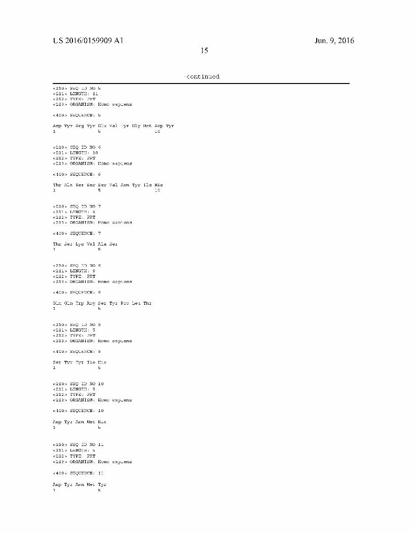
US 2016/O 1599.09 A1
<210s, SEQ ID NO &211s LENGTH: 11 212. TYPE: PRT
&213s ORGANISM:
<4 OOs, SEQUENCE:
Asp Tyr Arg Tyr 1.
<210s, SEQ ID NO &211s LENGTH: 10 212. TYPE: PRT
&213s ORGANISM:
<4 OOs, SEQUENCE:
Thir Ala Ser Ser 1.
<210s, SEQ ID NO &211s LENGTH: 6 212. TYPE: PRT
&213s ORGANISM:
<4 OOs, SEQUENCE:
Thir Ser Llys Val 1.
<210 SEQ ID NO &211s LENGTH: 9 212. TYPE: PRT
&213s ORGANISM:
<4 OOs, SEQUENCE:
Glin Gln Trp Arg 1.
<210s, SEQ ID NO &211s LENGTH: 5 212. TYPE: PRT
&213s ORGANISM:
<4 OOs, SEQUENCE:
Ser Tyr Tyr Ile 1.
<210s, SEQ ID NO &211s LENGTH: 5 212. TYPE: PRT
&213s ORGANISM:
<4 OOs, SEQUENCE:
Asp Tyr Asn Met 1.
<210s, SEQ ID NO &211s LENGTH: 5
212. TYPE: PRT &213s ORGANISM:
<4 OOs, SEQUENCE:
Asp Tyr Asn Met 1.
5
Homo sapiens
5
Glu Val Tyr Gly Met Asp Tyr 5 1O
6
Homo sapiens
6
Ser Val Asn Tyr Ile His 5 1O
Homo sapiens
7
Ala Ser
Homo sapiens
8
Ser Tyr Pro Leu. Thr 5
Homo sapiens
9
His
10
Homo sapiens
10
His
11
Homo sapiens
11
Tyr 5
15
- Continued
Jun. 9, 2016
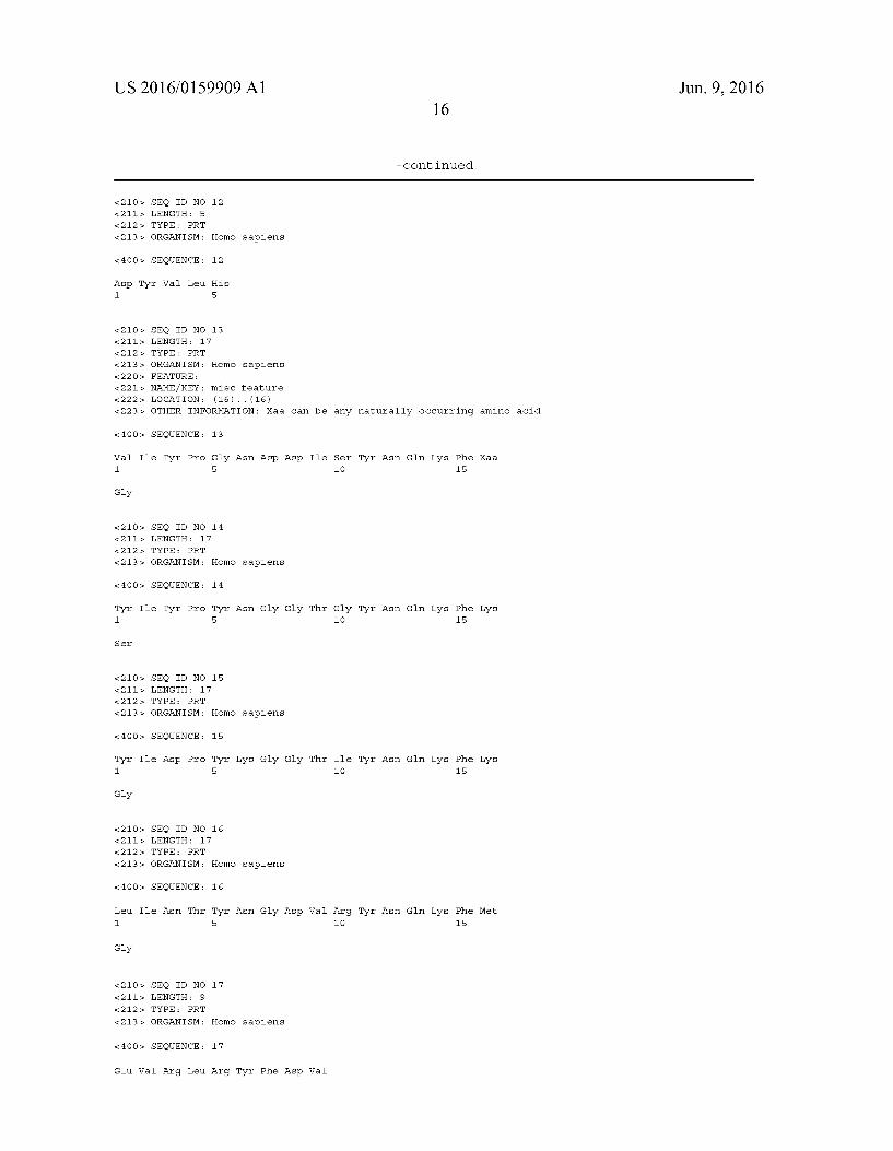
US 2016/O 1599.09 A1 16
- Continued
<210s, SEQ ID NO 12 &211s LENGTH: 5 212. TYPE: PRT
<213> ORGANISM: Homo sapiens
<4 OOs, SEQUENCE: 12
Asp Tyr Val Lieu. His 1. 5
<210s, SEQ ID NO 13 &211s LENGTH: 17 212. TYPE: PRT
<213> ORGANISM: Homo sapiens 22 Os. FEATURE:
<221 > NAMEAKEY: misc feature <222s. LOCATION: (16) ... (16) <223> OTHER INFORMATION: Xaa can be any naturally occurring amino acid
<4 OOs, SEQUENCE: 13
Val Ile Tyr Pro Gly Asn Asp Asp Ile Ser Tyr Asn Gln Llys Phe Xaa 1. 5 1O 15
Gly
<210s, SEQ ID NO 14 &211s LENGTH: 17 212. TYPE: PRT
<213> ORGANISM: Homo sapiens
<4 OOs, SEQUENCE: 14
Tyr Ile Tyr Pro Tyr Asn Gly Gly Thr Gly Tyr Asn Gln Llys Phe Lys 1. 5 1O 15
Ser
<210s, SEQ ID NO 15 &211s LENGTH: 17 212. TYPE: PRT
<213> ORGANISM: Homo sapiens
<4 OOs, SEQUENCE: 15
Tyr Ile Asp Pro Tyr Lys Gly Gly Thr Ile Tyr Asn Gln Llys Phe Lys 1. 5 1O 15
Gly
<210s, SEQ ID NO 16 &211s LENGTH: 17 212. TYPE: PRT
<213> ORGANISM: Homo sapiens
<4 OOs, SEQUENCE: 16
Lieu. Ile Asn. Thir Tyr Asn Gly Asp Val Arg Tyr Asn Gln Llys Phe Met 1. 5 1O 15
Gly
<210s, SEQ ID NO 17 &211s LENGTH: 9 212. TYPE: PRT
<213> ORGANISM: Homo sapiens
<4 OOs, SEQUENCE: 17
Glu Val Arg Lieu. Arg Tyr Phe Asp Wall
Jun. 9, 2016

US 2016/O 1599.09 A1 Jun. 9, 2016 17
- Continued
<210s, SEQ ID NO 18 &211s LENGTH: 7 212. TYPE: PRT
<213> ORGANISM: Homo sapiens
<4 OOs, SEQUENCE: 18
Gly Arg Pro Ala Met Asp Tyr 1. 5
<210s, SEQ ID NO 19 &211s LENGTH: 10 212. TYPE: PRT
<213> ORGANISM: Homo sapiens
<4 OOs, SEQUENCE: 19
Gln Met Ile Thr Ala Tyr Tyr Phe Asp Tyr 1. 5 1O
<210s, SEQ ID NO 2 O &211s LENGTH: 11 212. TYPE: PRT
<213> ORGANISM: Homo sapiens
<4 OOs, SEQUENCE: 2O
Asp Tyr Arg Tyr Glu Tyr Tyr Ala Met Asp Tyr 1. 5 1O
<210s, SEQ ID NO 21 &211s LENGTH: 17 212. TYPE: PRT
<213> ORGANISM: Homo sapiens
<4 OOs, SEQUENCE: 21
Lys Ser Ser Glin Ser Val Phe Phe Ser Ser Ser Gln Lys Asn Tyr Lieu. 1. 5 1O 15
Ala
<210s, SEQ ID NO 22 &211s LENGTH: 15 212. TYPE: PRT
<213> ORGANISM: Homo sapiens
<4 OOs, SEQUENCE: 22
Arg Ala Ser Glu Ser Val Asp Asn Tyr Gly Ile Ser Phe Met Asn 1. 5 1O 15
<210s, SEQ ID NO 23 &211s LENGTH: 11
212. TYPE: PRT
<213> ORGANISM: Homo sapiens
<4 OOs, SEQUENCE: 23
Lys Ala Ser Glin Asp Ile Asn Llys Tyr Ile Ala 1. 5 1O
<210s, SEQ ID NO 24 &211s LENGTH: 10 212. TYPE: PRT
<213> ORGANISM: Homo sapiens

US 2016/O 1599.09 A1 Jun. 9, 2016 18
- Continued
<4 OOs, SEQUENCE: 24
Ser Ala Asn Ser Ser Val Ser Tyr Ile His 1. 5 1O
<210s, SEQ ID NO &211s LENGTH: 7 212. TYPE: PRT
&213s ORGANISM:
<4 OOs, SEQUENCE:
Trp Ala Ser Thr 1.
<210s, SEQ ID NO &211s LENGTH: 6 212. TYPE: PRT
&213s ORGANISM:
<4 OOs, SEQUENCE:
Ala Ser Asn. Glin 1.
SEQ ID NO LENGTH: 6 TYPE PRT ORGANISM:
< 4 OO SEQUENCE:
Thir Ser Thir Lieu. 1.
<210s, SEQ ID NO &211s LENGTH: 6 212. TYPE: PRT
&213s ORGANISM:
<4 OOs, SEQUENCE:
Thir Ser Lys Lieu. 1.
<210s, SEQ ID NO &211s LENGTH: 8 212. TYPE: PRT
&213s ORGANISM:
<4 OOs, SEQUENCE:
His Glin Tyr Lieu. 1.
<210s, SEQ ID NO &211s LENGTH: 9 212. TYPE: PRT
&213s ORGANISM:
<4 OOs, SEQUENCE:
Glin Glin Ser Lys 1.
<210s, SEQ ID NO &211s LENGTH: 8
212. TYPE: PRT &213s ORGANISM:
25
Homo sapiens
25
Arg Glu Ser 5
26
Homo sapiens
26
Gly Ser
27
Homo sapiens
27
Glin Pro
28
Homo sapiens
28
Ala Ser
29
Homo sapiens
29
Ser Ser Arg Thr 5
3 O
Homo sapiens
3 O
Glu Val Pro Trp Thr 5
31
Homo sapiens

US 2016/O 1599.09 A1 Jun. 9, 2016
- Continued
<4 OOs, SEQUENCE: 31
Lieu. Glin Tyr Asp Asn Lieu. Lieu. Thr 1. 5
<210s, SEQ ID NO 32 &211s LENGTH: 9 212. TYPE: PRT
<213> ORGANISM: Homo sapiens
<4 OOs, SEQUENCE: 32
Gln Glin Trp Thr Ser His Pro Leu. Thr 1. 5
What is claimed is: 1. A pluri- or multipotent stem cell (SC) engineered to
express a multispecific antibody, which further expresses, on its surface, a human T cell co-stimulatory ligand or an active fragment thereof.
2. The SC of claim 1, wherein the pluri- or multipotent stem cells (SC) are human pluri- or multipotent stem cells (hSC).
3. The SC of claim 1, wherein the T cell co-stimulatory ligand or an active fragment thereof is selected from a group of CD28 ligand B7-1 and B7-2, Ox40L ligand OX40L, 4-1BB ligand 4-1BBL (CD137L), CD27 ligand CD70 (CD27L), CD40 ligand CD154, ICOS ligand ICOSL, LFA-1 ligand ICAM-1, -2, and -3, 2B4 ligand and SLAM ligand.
4. The SC of claim 1, wherein the SCs are engineered to express multispecific antibodies, preferably bispecific anti bodies, comprising at least a target cell specific binding domain and at least a immune cell specific binding domain, wherein the specific domains are preferably expressed as a fusion protein.
5. The SC of claim 4, wherein the immune cell specific binding domain is specific for an activating immune cell receptor.
6. The SC of claim 5, wherein the immune cell specific binding domain is specific to an activating immune cell recep tor like CD2, CD3 epsilon and Zeta chain, CD4, CD8, CD16, DAP12, NKG2D.
7. The SC of claim 6 wherein the target cell specific binding domain is specific for a tumor associated antigenoran antigen of a pathogen, parasite or parasitoid.
8. The SC of claim 7, the target cell specific binding domain is specific for an antigen selected from Surface, cytoplasmic or nuclear antigens like the La/SSB antigen, CD19, CD20, CD22, CD33, CD38, CD56, CD157, CD123, CCR5, STEAP1, prostate stem cell antigen PSCA, prostate specific membrane antigen PSMA, members of the epidermal growth factor receptor family, preferably EGFR (ErbB-1), HER2/ neu (ErbB-2), Her3 (ErbB-3) or Her4 (ErbB-4) or mutants thereof, members of the ephrin receptor family, preferably EphA1-10 or EphB1-6, embryonic antigens, preferably car cinoembryonic antigen CEA, fetal acethylcholine receptor, CEACAM-6, CEACAM-7.
9. The SC of claim 7, wherein the target cell specific binding domain is specific for an antigen selected from mem bers of the Rho family of GTPases, members of the high mobility group proteins, bladder tumor antigen (BTA), CA125, CD2, CD4, CD8, CD10, CD23, CD30, CD44, CD52,
CD99, CD135, CD274, Carboanhydrase IX, Endosialin, fibroblast-activating protease-alpha (FAP-alpha), FBP. gpA33, gp100, c-Met (hepatocyte growth factor receptor), MCSP and TIM-3, members of the vascular endothelia growth factor family (VEGFR 1-3), epithelia cell adhesion molecule EpCAM, alphafetoprotein AFP members of the mucin protein family, preferably MUC1 or MUC16, follicle stimulating hormone receptor (FSHR), the human high molecular weight-melanoma-associated antigen (HMW MAA), folate binding protein FBP a-Folate receptor, ligands of the NKG2D receptor, members of the epithelia glycopro tein family, preferably EGP-2, EGP-4 and EGP-40, members of the insulin-like growth factor receptor family (e.g. IGF 1R), diasialogangliosides, preferably GD2, GD3, members of the carbonic anhydrase family, preferably CAIX, members of the carbohydrate antigen family, preferably Ley, including mutants of the named proteins and protein families and viral envelope proteins, preferably gp120, gp41 or EBOV GP).
10. The SC of claim 7, wherein the target cell binding domain is composed of the alpha and beta or the gamma and delta chains of a T cell receptor (TCR) or fragments thereof, wherein such TCR-derived binding moieties recognize and bind to peptides presented by human leukocyte antigen class (HLA) I and II protein complexes, preferably viral peptides, peptides derived from proteins like EGFR family, survivin, Sry-like high motility group box (SOX) protein family, mela noma-associated antigens, preferably autoimmunogenic can cer/testis antigen NY-ESO-1, members of the melanoma anti gen family MAGE A, the preferentially expressed antigen in melanoma PRAME, and leukemia-associated antigens, pref erably wilms tumor gene 1 WT1 or wherein the target cell binding moiety comprise ligands to proteins and protein com plexes, preferably cytokine receptors, preferably IL-13 receptor, ligands of the NKG2D receptor, ligands to the EGFR family members, or auto-reactive TCRs.
11. The SC of claim 1, wherein the SCs are mesenchymal stem cells (MSCs), preferably human MSCs (hMSCs).
12. A method of making a SC of claim 1, wherein the method comprises introducing into an SC a cDNA encoding the multispecific antibody and a cDNA encoding the a human T cell co-stimulatory ligand.
13. A pharmaceutical composition comprising the SC of claim 1.
14. The pharmaceutical composition of claim 13 further comprising a pharmaceutically acceptable carrier.
k k k k k