Embed Size (px)
Citation preview
18
Detection of Salmonella spp. Presence in Food
Anna Zadernowska and Wioleta Chajęcka University of Warmia and Mazury in Olsztyn, Faculty of Food Sciences
Chair of Industrial and Food Microbiology Poland
1. Introduction
The analysis of food products for presence of pathogenic microorganisms is one of the basic steps to control safety and quality of food. Development of new, fast, and reliable identification methods for biological threats are necessary to meet the safety standards of food products and risk management. Salmonella spp., a marker of food products safety, is widely distributed foodborne pathogen. The standard culture methods to detect the presence of microorganisms in food products are well developed; although these methods require 4 to 5 days to obtain presumptive positive or negative results. These tests are time-consuming and can take up to 7 days depending on the realization of biochemical and serological confirmations. In addition, sensitivity of cultures can be affected by antibiotic treatment, inadequate sampling, and a small number of viable microorganisms in samples. Standardized classical culture methods are still in use by many labs, especially by regulatory agencies, because they are harmonized methods, looked at as the “gold standards” in food diagnostics and thus overall well accepted. These are important aspects in international trade and compliance testing. A serious drawback is that, although they demand no expensive infrastructure and are rather cheap in consumables, they are laborious to perform, demand large volumes usage of liquid and solid media and reagents, and encompass time-consuming procedures both in operation and data collection. As an alternative to time-consuming culture methods, several approaches have been developed to accelerate detection of pathogenic microorganisms in food products. In the present work, besides the standard method of Salmonella spp. detection in food products (ISO 6579:2003) some alternative detection methods have been presented.
2. Taking samples for tests
The first stage of microbiological analysis of food consists in taking and preparing a sample for analyses. Incorrect sampling can lead to obtaining false negative or false positive results. When talking about taking samples, the term “representative sample” is often used. The sample should reflect the image of the product from which it originates as precisely as possible. It is quite easy to take a representative sample from liquid products, e.g. milk, if the milk has been sufficiently mixed before taking the sample. On the other hand, when the subject of examination is a product of high viscosity, with slow flow or of a heterogeneous structure, then it is very difficult to assess the microbiological quality of the entire batch (e.g. a barrel or a
www.intechopen.com
Salmonella – A Dangerous Foodborne Pathogen 394
truckload) by examining only one 25-gram sample. The answer to the question concerning the required number of single samples is extremely difficult. In view of the high costs of microbiological tests, the number of samples is generally limited. In a microbiological laboratory, samples are taken with the use of sterile tools, e.g. spoons, scalpels, knives, spatulas and pipettes. Frozen products should be first thawed at below 5°C (for not longer than 12 hours). In the case of deeply frozen samples, sterile drills are used for sampling. Determination of Salmonella sp. in food products always consists in detecting the presence of those bacteria in a specified amount of the product (generally 25g/ml, very rarely 10g/ml), but the number of those microorganisms in food is not determined. Both in the classical method and in its modifications, the first stage of detection is non-selective enrichment. This is crucial, since food production involves its technological treatment, e.g. heating, which can cause the death of most cells or cause sub-lethal injured. Omission of the stage of pre-enrichment of the sample and inoculating the material directly on the solid medium can give false negative results. If the examined material includes a very low number of living cells, or the cells have been sub-lethally damaged during the technological processes, we may not receive macroscopically-visible colonies on the solid medium. In such a case there is a risk of releasing the product to market although it does not satisfy safety criteria. During the storage of such a product, damaged cells can be repaired and bacteria can proliferate to a level that would be hazardous for the consumers. There are many methods to determine Salmonella sp. in food and, for this reason, the present study focuses on the classical culture method – the application of a Vidas device – as the only fully automated one. Additionally, the PCR method (a commonly-applied alternative to the plate method) and the FISH method (which is still not popular, although work on its optimization is ongoing) are also described.
3. A classical culture method of detecting Salmonella
Detection of the presence of Salmonella pursuant to Commission Regulation (EC) No 2073/2005 (microbiological criteria for foodstuff) as amended, is carried out according to the ISO 6579 standard - Microbiology of food and animal feeding stuffs - Horizontal method for detection of Salmonella spp.(ISO, 2002). Pursuant to the above regulation, detection of Salmonella in food should be carried out for such products as raw meat, meat products intended for consumption in the raw state, gelatine, cheese, butter, cream, unpasteurized milk, powdered milk, eggs and products containing raw eggs, crustaceans, molluscs, fruit and vegetables, unpasteurized juice, powdered infant formulas and dietary food for special medical purposes. Standard ISO 6579 2003 (Microbiology of food and animal feeding stuffs - Horizontal method for detection of Salmonella spp.)includes four stages of the detection process and depending on the need to obtain confirmations, it lasts from 5 to 7 days:
Pre-enrichment in non-selective liquid medium
Selective enrichment in liquid media
Plating on selective media
Serological and biochemical identification of suspected colonies During the first stage, in order to proliferate and regenerate damaged cells, the culture is performed on liquid peptone water at 37°C for 18±2 hours. Buffered peptone water is applied for non-selective enrichment of Salmonella sp. For such products as cocoa or chocolate products, peptone water is applied with an addition of casein or skimmed milk
www.intechopen.com
Detection of Salmonella spp. Presence in Food 395
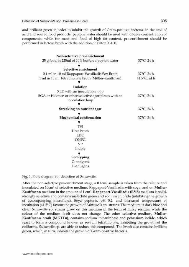
and brilliant green in order to inhibit the growth of Gram-positive bacteria. In the case of acid and soured food products, peptone water should be used with double concentration of components, while for meat and food of high fat content, pre-enrichment should be performed in lactose broth with the addition of Triton X-100.
Non-selective pre-enrichment 25 g food in 225ml of 10% buffered pepton water 37°C, 24 h Selective enrichment 0.1 ml in 10 ml Rappaport-Vassiliadis Soy Broth 37°C, 24 h 1 ml in 10 ml Tetrathionate broth (Müller-Kauffman) 41.5°C, 24 h Isolation XLD with an inoculation loop BGA or Hektoen or other selective agar plates with an
inoculation loop 37°C, 24 h
Streaking on nutrient agar 37°C, 24 h Biochemical confirmation 37°C, 24 h TSI Urea broth LDC ONPG VP Indole Serotyping O-antigens H-antigens
Fig. 1. Flow diagram for detection of Salmonella.
After the non-selective pre-enrichment stage, a 0.1cm3 sample is taken from the culture and inoculated on 10cm3 of selective medium, Rappaport-Vassiliadis with soya, and on Muller-Kauffmann medium in the amount of 1 cm3. Rappaport-Vassiliadis (RVS) medium is solid, strongly selective and contains malachite green and sodium chloride (inhibiting the growth of accompanying microflora). Soya peptone, pH 5.2, and increased temperature of incubation (41.5°C) favour the growth of Salmonella sp. strains. The medium is dark blue and clear. Salmonella sp. strains grow on this medium in the form of milky residue, while the colour of the medium itself does not change. The other selective medium, Muller-Kauffmann broth (MKTTn), contains sodium thiosulphate and potassium iodide, which react to form a compound known as sodium tetrathionate, inhibiting the growth of the coliforms. Salmonella sp. are able to reduce this compound. The broth also contains brilliant green, which, in turn, inhibits the growth of Gram-positive bacteria.
www.intechopen.com
Salmonella – A Dangerous Foodborne Pathogen 396
After incubation at 37°C for 48±3 hours, cultures are inoculated on two selective media, so as to receive individual colonies. The first of them is XLD (xylose lysine deoxycholate) agar. The other can be chosen by the laboratory, and it can be BGA (brilliant green agar), Hektoen or Wilson-Blair agar for example. XLD agar contains lactose, saccharose, L-lysine, sodium thiosulphate, sodium deoxycholate,
ferric ammonium citrate (III) and phenol red. Differential agents of the agar include:
lactose, saccharose, xylose, lysine and sodium thiosulphate, from which hydrogen sulfide
is released, forming in reaction with iron salts (III) black residue of iron sulfide in the
centre of the colony. The pH indicator is phenol red. The agar makes it possible to
determine the sugar fermentation ability. Incubation is carried out at 37ºC for 24±3 hours.
Typical colonies can be colourless, very light, slightly shiny and transparent (colour of the
medium) with a dark tinted centre, surrounded by a light red area and yellow edge, or of
pink to red colour, with a black centre or without a black centre. H2S (–) colonies are
colourless or light pink with darker centres, and lactose (+) colonies are yellow or without
the characteristic blackening.
BGA. Differential factors of this agar are sugars: saccharose and lactose. Brilliant green is a
selective agent. Typical colonies are transparent, colourless or light pink, and the colour
around colonies changes from pink to light red.
Hektoen agar. Selective agents include bile salts, inhibiting the growth of Gram (+) bacteria
Differential factors are three sugars: lactose, saccharose and salicin. Increased lactose
content ensures that bacteria fermenting this sugar with a delay are not omitted. Bacteria
colonies producing hydrogen sulfide had a dark centre as a result of the reaction between
hydrogen sulfide and iron (III). Typical colonies of Salmonella sp. are green, with or
without a black centre.
Wilson-Blair agar. This is a strongly selective and differential medium for Salmonella,
including S. Typhi isolated from food. Salmonella spp., depending on the strain, grow in the
form of black colonies surrounded with an area of black medium or dark brown and brown
without this area. A characteristic feature of Salmonella spp. colonies is a metallic, shining
surface as a result of produced hydrogen sulfide, forming a metallically-black residue in
reaction with iron ions. The growth of Gram-positive bacteria and other Enterobacteriaceae,
including Shigella spp., is strongly inhibited by brilliant green and bismuth sulfite present in
the medium.
Rambach-agar chromogenic medium – with sodium deoxycholate, proplylene glycol and
chromogenic mix. Colonies of Salmonella sp. are red as a result of glycol fermentation,
lactose positive bacteria from the coli group, due to the activity of galactosidase, destroy a
bound between the components of chromogenic mix and released chromophore gives those
colonies a blue-violet or blue-green colouring. Salmonella Typhi and Salmonella Paratyphi
form colourless or yellowish colonies on this medium.
New selective media have been developed based on biochemical characteristic of Salmonella
such as ┙-galactosidase activity in the absence of ┚-galactosidase activity, C8-esterase
activity, catabolism of glucuronate, glycerol and propylene glycol, hydrolysis of X-5-Gal,
and H2S production. e.g. SMID agar (BioNerieux, France), Rainbow Salmonella agar (Biolog,
USA), CHROMagar Salmonella (CHROM agar, France), chromogenic Salmonella esterase agar
(PPR Diagnostics Ltd, UK), Compass Salmonella agar (Biokar diagnostics, France), and
chromogenic ABC medium (Lab M. Ltd., UK) (Maciorowski et al., 2006; Manafi, 2000; Perry
et al., 2007; Schonenbrucher et al., 2008)
www.intechopen.com
Detection of Salmonella spp. Presence in Food 397
MEDIUM REACTIONS/ENZYMES RESULTS
NEGATIVE POSITIVE
TSIa
Acid production (if the butt is yellow, and the
slope is red, acid production is only from
glucose)
Butt red Butt yellow
TSIa Acid production from lactose and/or sucrose
Surface red Surface yellow
TSIa Gas production No air bubbles in
butt Air bubbles in butt
TSIa H2S production No black colour Black colour
UREA BROTH Urease Yellow Rose pink – deep
cerise
LCD TEST Lysine decarboxylase A yellow/brown
colour
A purple colour (and a
yellow/brown colour in the LDC control medium if
used)
ONPG ┚-Galactosidase Remain
colourless Yellow
VOGES PROSKAUER
Acetoin production Remain
colourless A pink/red colour
INDOLE Indole production Yellow ring Red / pink ring
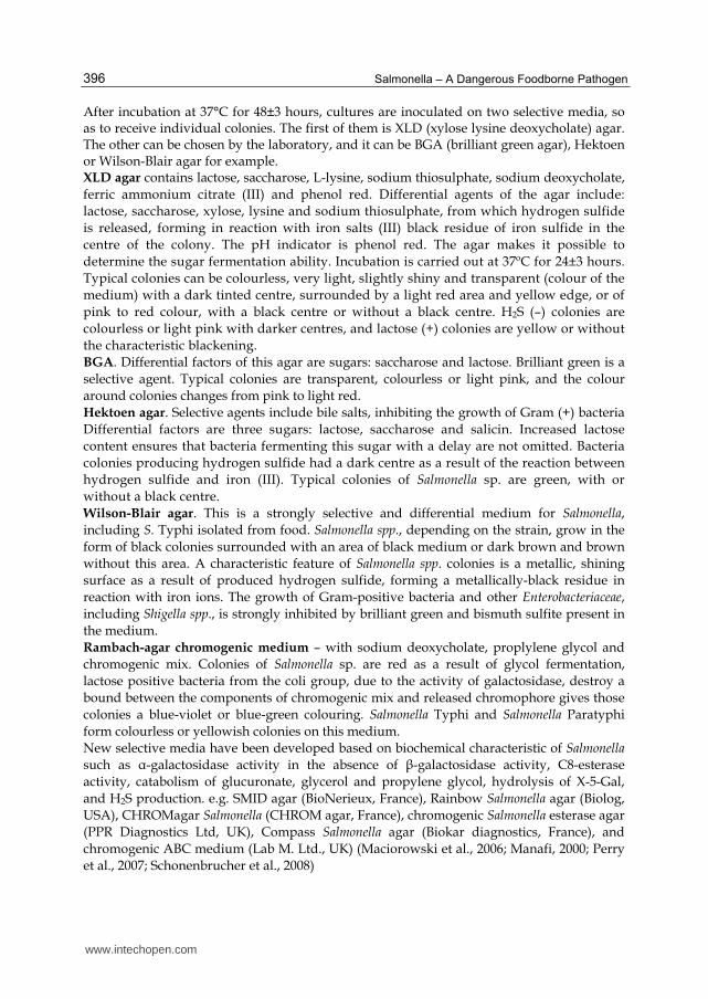
Table 1. Interpretation table. aRegarding TSI: Read the colour of the butt and of the surface of the medium; ALK: A red colour corresponding to no acid production; NC: No change in the colour of the medium ; A: A yellow colour corresponding to acid production; G: Gas production in the butt; H
2S production; +: Black colour; -: No black colour
After 48 h incubation at 37°C, a preliminary identification is made on the basis of the appearance of colonies grown on selective media. Five characteristic colonies are selected from each plate and are plating on the nutrient agar medium, followed by biochemical examinations. In order to perform these examinations, biochemical tests are carried out on the following media:
TSI medium (Triple-sugar iron agar)
Christensen medium with urea (urease production)
peptone medium with tryptophan (indole production)
medium with lysine (lysine decarboxylation)
Clark medium (V-P reaction)
ONPG medium (┚-galactosidase detection)
www.intechopen.com
Salmonella – A Dangerous Foodborne Pathogen 398
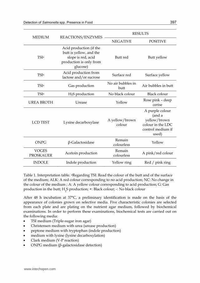
Test Positive or negative
reaction
Percentage of Salmonella inoculations showing the
reaction1)
TSI glucose (acid formation) TSI glucose (gas formation) TSI lactose TSI sucrose TSI hydrogen sulfide Urea splitting Lysine decarboxylation ┚-Galactosidase reaction Voges-Proskauer reaction Indole reaction
+ + - - + - + - - -
100
91.92)
99.23)
99.5 91.6 99
94.64) 98.43)
100 98.9
1) These percentages indicate only that not all strains of Salmonella show the reactions marked + or -. These percentages may vary from country to country and from food product to food product. 2) Salmonella Typhi is anaerogenic. 3) The Salmonella subspecies III (Arizona) gives positive or negative lactose reactions but is always ┚-galactosidase positive. The Salmonella subspecies II gives a negative lactose reaction, but gives a positive ┚-galactosidase reaction. For the study of strains, it may be useful to carry out complementary biochemical tests. 4 S. Paratyphi A is negative.
Table 2. Biochemical results for Salmonella.
Triple-sugar iron agar is used for differentiation of Enterobactericeae according to their ability to ferment lactose, sucrose and glucose. The colour of the slope and the butt and gas production are noted. Acid production from fermentation of one or more of the sugars results in a yellow colour because the phenol red indicator turns yellow at low pH. Very little glucose is present in the medium, so if a bacteria, like Salmonella, only ferments glucose then only a little acid will be formed. On the slope, the acid will be oxidised by the air and by the breakdown of protein in the medium and the colour will remain red while the butt is yellow. H
2S production from thiosulphate will be seen as black areas in the medium due to
FeS production. Gas production from fermentation of sugars will be seen as gas bubbles in the medium. The medium is only lightly inoculated. Christensen medium with urea. Urea medium tests for high urea activity. It is the most common method to detect urease production by Enterobacteriaceae (1):
2 2 3 22NH CO H O 2NH CO (1)
The phenol red turns red at alkaline pH so a positive reaction is shown as the development of a red-pink colour. Tryptone/tryptophane medium for indole reaction. The media is used for testing the liberation of indole from tryptophane. When Kovacs reagent containing amyl alcohol and p-dimethylaminobenzaldehyde is added, indole can be extracted into the amyl alcohol layer by shaking a little. Indole and p-dimethylaminobenzaldehyde produces a red or pink colour.
L-Lysine decarboxylation medium for the LDC test. The LDC broth is used for the test of production of lysine decarboxylase. This enzyme decarboxylates lysine to yield the alkaline
www.intechopen.com
Detection of Salmonella spp. Presence in Food 399
compound cadaverin and 2CO . A paraffin oil layer is added after inoculation to keep the pH alkaline. Often glucose is metabolised in the beginning of the incubation period and a yellow colour develops in the media after some hours of incubation, but later the media turns purple if the lysin decarboxylase is present because of formation of the alkaline compound cadaverin. As other compounds in the media could be broken down to alkaline compounds, the LDC control media without lysine is also inoculated, a layer of paraffin oil added and it is incubated at the same time. If both the LDC media and the LDC control media turn purple, it cannot be shown that lysine decarboxylase is present and the test is evaluated as negative. Medium VP. This is a test for acetoin production from glucose. The acetoin produced is oxidised to diacetyl, which produces a red colour with ┙-naphtol at alkaline pH. A positive reaction is seen as a very pale red colour. ONPG medium. This medium shows the presence of ┚-galactosidase producing bacteria. ┚-galactosidase liberates o-nitrophenol, which is yellow at alkaline pH, from ONPG. The reaction is positive if a yellow colour develops. API. Determination of biochemical features of the examined bacteria can also involve the application of API 20E tests (Biomerieux), aimed at identification of bacteria from the family Enterobacteriaceae. The API 20E system facilitates the 24-hour identification of Enterobacteriaceae as well as 24 or 48-hour identification of other Gram negative bacteria. The API 20E strip consists of microtubes containing dehydrated substrates for the demonstration of enzymatic activity and carbohydrate (CHO) fermentation. The substrates are reconstituted by adding a bacterial suspension. After incubation, the metabolic end products are detected by indicator systems or the addition of reagents. CHO fermentation is detected by colour change in the pH indicator. Serological tests. These tests are carried out for strains of bacteria which have been classified into the Salmonella genus on the basis of their biochemical features, in order to detect the presence of somatic O, capsular Vi and flagellar H antigens. The examinations are carried out by slide agglutination on the basis of Kauffmann-White antigenic schema. Polyvalent and monovalent serums should be used to determine somatic antigens, and anti-Vi and anti – H serums to detect the presence of Vi and H antigen. Determination of flagellar antigens makes it possible to determine the serological type of the examined bacteria. Culture methods are labor intensive and time consuming when handling many samples. In addition, detection can be prevented by the presence of other competing microorganisms during cultural enrichment, and the selective agar media have a very poor specificity creating an abundance of false positives (such as Citrobacter or Proteus) (Manafi, 2000). Therefore, there is a need for Salmonella detection methods that provide results more rapidly with sensitivity similar to or greater than, the conventional methods.
4. Polymerase chain reaction
Due to its high sensitivity, specificity, and rapid results, PCR is an efficient alternative to conventional microbiological culture methods to detect specific types of microorganisms in foods, water, and environmental samples (Moganedi et al., 2007; Glynn et al., 2006; Piknova´et al., 2002). The International Standardization Organization (ISO) recently published standards which address the PCR methodology for the detection of food-borne pathogens (Tomás et al., 2009).
www.intechopen.com
Salmonella – A Dangerous Foodborne Pathogen 400
Gene Target
Matrices Enrichment Limit of
detection Primers (1-forward; 2-reverse;
3-probe) References
ttrBCA
chicken
20 h < 3
CFU/ml
1: CTC ACC AGG AGA TTA CAA CAT GG
Malorny et al., 2004
minced meat
2: AGC TCA GAC CAA AAG TGA CCA TC
fish 3: CAC CGA CGG CGA GAC CGA CTT T
fimC
103
CFU/ml
1: ATA AAT CCG GCG GCC TGA TG
Seo et al., 2006
ice cream without 2: TGG TAT CGA CGC CTT TAT CTG AGA
3: TTA CAC CGG AGT GGA TTA AAC GGC TGG G
invA
salmon
16h
2,5-5 CFU/25g
1: GTG AAA TAA TCG CCA CGT TCG GGC AA
Hein et al., 2006
chicken meat
(salmon, chicken)
2: TCA TCG CAC CGT CAA AGG AAC CGT AA
milk 5
CFU/25ml (milk)
3: TTA TTG GCG ATA GCC TGG CGG TGG GTT TTG TTG
invA
35°C-24h
(pre-) 1: AAC GTG TTT CCG TGC GTA AT
Cheng et al., 2008
chili powder
41°C-24h 0,04
CFU/g 2: TCC ATC AAA TTA GCG GAG GC
shrimp (selective)
3: TGG AAG CGC TCG CAT TGT GG
oriC
1: TCACCTGCGACAGCCATGA
McCarthy et al., 2009
cheddar
2: TGAGCATCGCCATCGGCAT
turkey meat
48 h (selective)
6,1 x 101 CFU/ml
3: ATTCCAGCAGTCGGCCATAGCTG (Set I)
cooked turkey meat
1: CATTGATGCCATGGGTGACART
2: CGTGACGATAATCCGTGTAC
3: TACACGAGTCACTAAATCCTTCAGT (Set II)
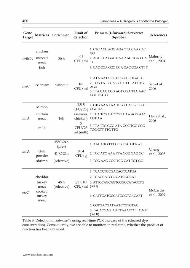
Table 3. Detection of Salmonella using real-time PCR.increase of the released dye concentration). Consequently, we are able to monitor, in real time, whether the product of reaction has been obtained.
www.intechopen.com
Detection of Salmonella spp. Presence in Food 401
4.1 Conventional PCR
PCR – polymerase chain reaction – is an enzymatic reaction replicating a DNA molecule. A critical condition of the PCR course is the presence of an enzyme, thermostable DNA polymerase. Substrates of the reaction are deoxyribonucleotides (building blocks of newly synthesized DNA molecules) and oligonucleotides (starters), which on a complementary basis attach to the replicated DNA strand. Polymerase chain reaction is a thermal process, with cyclically changing temperatures. One cycle leading to synthesis of one copy of DNA from one matrix molecule is composed of the following stages: DNA denaturation (90-95°C), attachment of starters (the so-called annealing, 50-65°C) and strand elongation. Reaction under laboratory conditions is carried out in a device known as thermocycler. Usually, one PCR reaction is made of 35-40 cycles, which result in creating billions of copies of an individual DNA molecule (with a logarithmic increase of the product in each cycle).
4.2 Real–time PCR
Real-time PCR is a polymerase chain reaction observed in real time, is an improvement of the classical PCR method (Al-Soud et al., 2005; Bansal et al., 2006). A real-time PCR thermocycler is equipped with an optical system which collects information about the course of reaction, cycle by cycle. TaqMan® probes labelled with fluorescent dyes make an additional component of PCR real time reactions. A probe is an oligonucleotide designed to bind highly specifically to a replicated fragment of DNA, therefore – in case of the analysed application in food microbiology – to “detect” a sequence which is characteristic for the required pathogen. The probe should bind specifically and strongly, which can be ensured by special protein molecules (a MGB molecule in TaqMan® Applied Biosystems probes). Polymerase, while synthesising a new strand, moves along the matrix, encountering a probe on its way. Because of its exonucleolytic activity, the enzyme starts to “unstick” the probe from the matrix and afterwards to destroy it, releasing a fluorescent dye. An optical system of the thermocycler triggers and then receives dye glow, which becomes more intense with each cycle (a logarithmic increase of the amount of product results in a logarithmic A forty-cycle real-time PCR reaction lasts about 1.5 hours. After adding the time needed to isolate DNA from the analysed sample (up to 30 minutes), the entire determination of the presence or the lack of pathogen lasts 2 hours (while using the Applied Biosystems TaqMan® Pathogen Detection System). Samples for determination are taken from pre-enrichment cultures on buffered peptone water after 18 h incubation at 37°C, so the total time from the collection of a sample to the final results does not exceed 24 hours. Chen et al. (2000) evaluated the TaqMan system for the detection of Salmonella that utilizes primers and probes developed from a novel target sequence (invA). The detection limit was below 3 CFU/25 g or 25 ml when raw milk, ground beef and ground pork inoculated with Salmonella were pre-enriched overnight. Malorny et al. (2004) used specifically designed primers and a probe target within the ttrRSBCA locus, and included internal amplification control, which is coamplified with the same primers as the Salmonella DNA in the assay. The diagnostic accuracy was shown to be 100% compared to the traditional culture method when 110 various food samples (chicken rinses, minced meat, fish, and raw milk) were investigated for Salmonella by real-time PCR including a pre-enrichment step in buffered peptone water. A very frequent target of species-specific Salmonella PCR assays is the invasion protein invA gene, and several invA-based PCR assays have been already developed and validated (Malorny et al. 2003a; b).
www.intechopen.com
Salmonella – A Dangerous Foodborne Pathogen 402
Validated PCR methods are available from Bio-Rad, Roche, Qualicon/Oxoid, Genesystems, AES Chemunex, Applied BioSystems, Idaho Technology Inc., Lantmännen, IEH Laboratries and Consulting Group, ADNucleis and BioControl systems. Validation is an important step in the process of standardizing a method because it provides evidence that the new method gives similar results and is in agreement with the currently used reference method (Patel et al., 2006). One major difficulty with PCR is the presence of compounds that inhibit the PCR reaction.
These compounds can contaminate the DNA templates extracted from food samples and
may in turn generate false-negative results (Elizaquivel et al., 2008). Therefore, evaluation
and elimination of PCR inhibitory compounds are important steps in the development of
PCR and real-time PCR assays (Abu Al-Soud et al., 2000). The PCR procedure is sufficiently
sensitive such that, in theory, only a few template molecules are required to initiate the
synthesis reactions (Uyttendaele et al., 2003). However, an enrichment step is still required
to detect small numbers of Salmonella in food samples. This step may consist of non-selective
enrichment with buffered peptone water (BPW) and selective enrichment with Rappaport-
Vassiliadis. These enrichment broth have been directly utilized for Salmonella DNA template
preparation. However, limited research has been conducted to quantitatively evaluate the
effects of the enrichment broths using conventional PCR assays and even less using a real-
time PCR protocol. Therefore, identifying and eliminating the PCR inhibitory effects of the
enrichment broths is key to enhancing the performance of PCR assays in detecting
Salmonella in foods.
4.3 Multiplex polymerase chain reaction (PCR)
Multiplex PCR is a variant of the PCR technique in which two or more loci are
simultaneously amplified in the same reaction. Multiplex PCR can be described as a
specific and sensitive in vitro amplification of DNA with distinguishable size products
from the same or different organisms in a single reaction (Jasson et al., 2010; Fitzgerald et
al., 2007). In this methodology several specific primer sets are combined into a single PCR
assay. MPCR is undoubtedly useful to rapidly establish simultaneous detection of
multiple virulence factors (Fach et al., 2009) or combined detection of multiple isolates
(Kawasaki et al., 2009; Settanni & Corsetti, 2007). A convened format for MPCR is the
GeneDisc (PALL) (Beutin et al., 2009).
Recently, multiplex real-time PCR assays have been applied to detect more than two gene
sequences in a single reaction by using spectrally distinct dye-labeled probes (TaqMan
system) (Elizaquivel et al., 2008). This technology could potentially save time and effort in
the laboratory and thus may lower testing-related costs incurred by the food industry
(Elizaquivel et al., 2008).
5. Fish (fluorescent in situ hybridization)
The literature provides a limited number of reports concerning the application of the FISH technique for food examination (Ootsubo et al., 2003), while it is broadly applied in microbiology of environment, histopathology, histoimmunology, cytogenetics. Initially, it was developed in order to identify and to determine the number of bacterial cells in water ecosystem environments (Skowrońska & Zmysłowska, 2006), deposits, rhizosphere and soil.
www.intechopen.com
Detection of Salmonella spp. Presence in Food 403
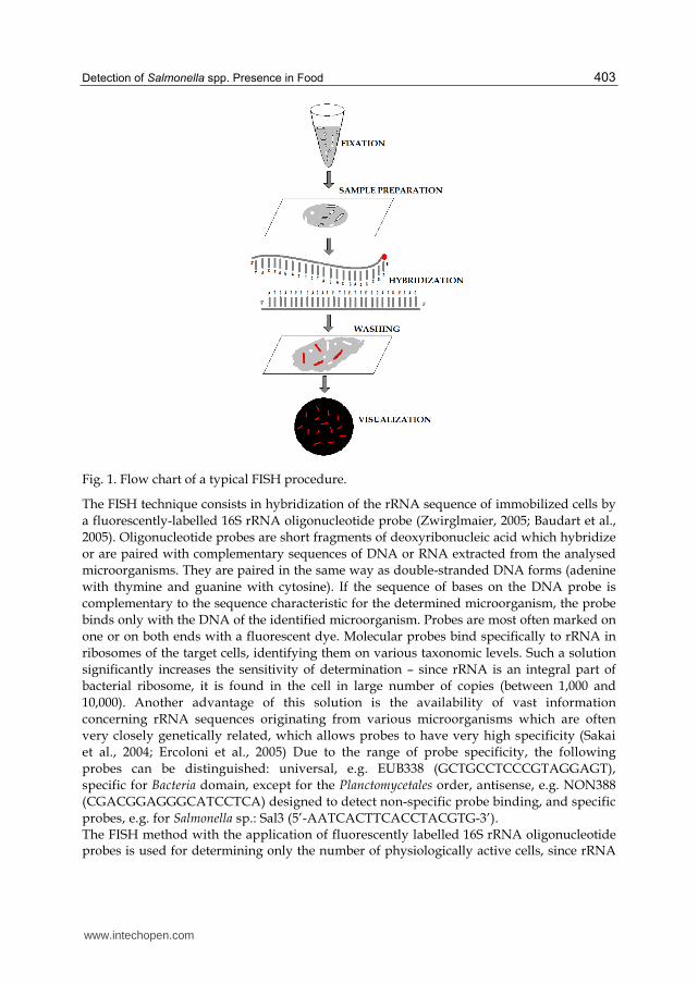
Fig. 1. Flow chart of a typical FISH procedure.
The FISH technique consists in hybridization of the rRNA sequence of immobilized cells by
a fluorescently-labelled 16S rRNA oligonucleotide probe (Zwirglmaier, 2005; Baudart et al.,
2005). Oligonucleotide probes are short fragments of deoxyribonucleic acid which hybridize
or are paired with complementary sequences of DNA or RNA extracted from the analysed
microorganisms. They are paired in the same way as double-stranded DNA forms (adenine
with thymine and guanine with cytosine). If the sequence of bases on the DNA probe is
complementary to the sequence characteristic for the determined microorganism, the probe
binds only with the DNA of the identified microorganism. Probes are most often marked on
one or on both ends with a fluorescent dye. Molecular probes bind specifically to rRNA in
ribosomes of the target cells, identifying them on various taxonomic levels. Such a solution
significantly increases the sensitivity of determination – since rRNA is an integral part of
bacterial ribosome, it is found in the cell in large number of copies (between 1,000 and
10,000). Another advantage of this solution is the availability of vast information
concerning rRNA sequences originating from various microorganisms which are often
very closely genetically related, which allows probes to have very high specificity (Sakai
et al., 2004; Ercoloni et al., 2005) Due to the range of probe specificity, the following
probes can be distinguished: universal, e.g. EUB338 (GCTGCCTCCCGTAGGAGT),
specific for Bacteria domain, except for the Planctomycetales order, antisense, e.g. NON388
(CGACGGAGGGCATCCTCA) designed to detect non-specific probe binding, and specific
probes, e.g. for Salmonella sp.: Sal3 (5’-AATCACTTCACCTACGTG-3’). The FISH method with the application of fluorescently labelled 16S rRNA oligonucleotide probes is used for determining only the number of physiologically active cells, since rRNA
www.intechopen.com
Salmonella – A Dangerous Foodborne Pathogen 404
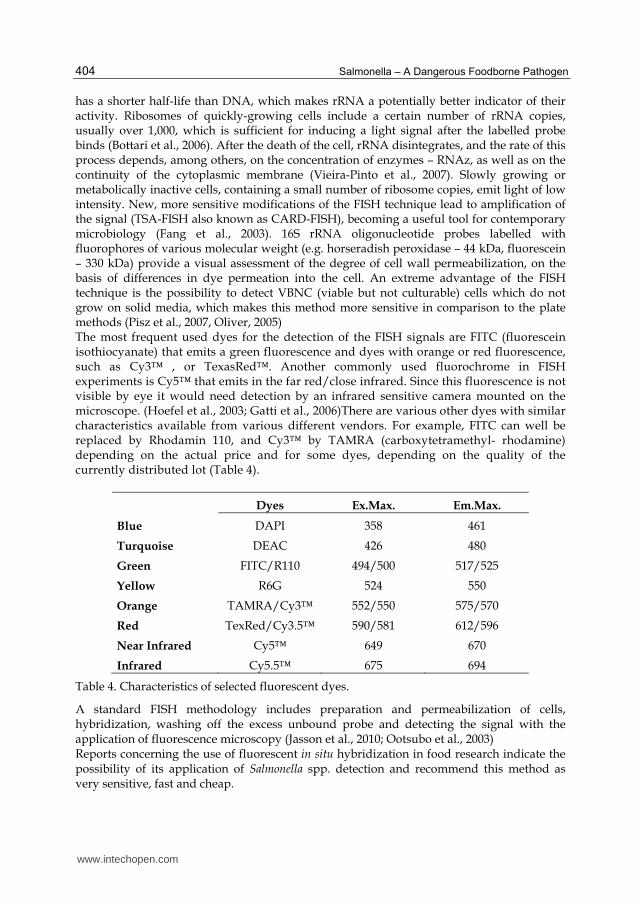
has a shorter half-life than DNA, which makes rRNA a potentially better indicator of their activity. Ribosomes of quickly-growing cells include a certain number of rRNA copies, usually over 1,000, which is sufficient for inducing a light signal after the labelled probe binds (Bottari et al., 2006). After the death of the cell, rRNA disintegrates, and the rate of this process depends, among others, on the concentration of enzymes – RNAz, as well as on the continuity of the cytoplasmic membrane (Vieira-Pinto et al., 2007). Slowly growing or metabolically inactive cells, containing a small number of ribosome copies, emit light of low intensity. New, more sensitive modifications of the FISH technique lead to amplification of the signal (TSA-FISH also known as CARD-FISH), becoming a useful tool for contemporary microbiology (Fang et al., 2003). 16S rRNA oligonucleotide probes labelled with fluorophores of various molecular weight (e.g. horseradish peroxidase – 44 kDa, fluorescein – 330 kDa) provide a visual assessment of the degree of cell wall permeabilization, on the basis of differences in dye permeation into the cell. An extreme advantage of the FISH technique is the possibility to detect VBNC (viable but not culturable) cells which do not grow on solid media, which makes this method more sensitive in comparison to the plate methods (Pisz et al., 2007, Oliver, 2005) The most frequent used dyes for the detection of the FISH signals are FITC (fluorescein isothiocyanate) that emits a green fluorescence and dyes with orange or red fluorescence, such as Cy3™ , or TexasRed™. Another commonly used fluorochrome in FISH experiments is Cy5™ that emits in the far red/close infrared. Since this fluorescence is not visible by eye it would need detection by an infrared sensitive camera mounted on the microscope. (Hoefel et al., 2003; Gatti et al., 2006)There are various other dyes with similar characteristics available from various different vendors. For example, FITC can well be replaced by Rhodamin 110, and Cy3™ by TAMRA (carboxytetramethyl- rhodamine) depending on the actual price and for some dyes, depending on the quality of the currently distributed lot (Table 4).
Dyes Ex.Max. Em.Max.
Blue DAPI 358 461
Turquoise DEAC 426 480
Green FITC/R110 494/500 517/525
Yellow R6G 524 550
Orange TAMRA/Cy3™ 552/550 575/570
Red TexRed/Cy3.5™ 590/581 612/596
Near Infrared Cy5™ 649 670
Infrared Cy5.5™ 675 694
Table 4. Characteristics of selected fluorescent dyes.
A standard FISH methodology includes preparation and permeabilization of cells, hybridization, washing off the excess unbound probe and detecting the signal with the application of fluorescence microscopy (Jasson et al., 2010; Ootsubo et al., 2003) Reports concerning the use of fluorescent in situ hybridization in food research indicate the possibility of its application of Salmonella spp. detection and recommend this method as very sensitive, fast and cheap.
www.intechopen.com
Detection of Salmonella spp. Presence in Food 405
Vieira-Pinto et al., (2005) compared FISH methods with the classical plate method for detection of Salmonella spp. Out of 47 samples of pork tonsils, 16 (34%) were positive for Salmonella spp detection by the FISH method with the application of 23S rRNA Sal3 probe. Out of 31 negative results obtained by FISH method, one sample was positive for Salmonella spp. detection by the plate method. Similar results were obtained by Fang et al., (2003), who detected Salmonella sp. species in 56 samples of food products by the FISH method (23S rRNA Sal3), while 28 samples were not positive for Salmonella spp. detection by the plate method. Huge number of positive results can derive from the presence of cells slightly damaged or occurrence of factors inhibiting their growth in food products, which can transfer cells to VBNS state. The authors suggest that FISH method seems to be less prone to diverse physical-chemical properties of preserved food products (temperature, concentration of NaCl, pH), which can work as a stress factor for Salmonella spp. cells. Presence of microflora can be another reason of high number of positive results obtained by the FISH method as compared to the plate methods. The conclusions drawn from the research show the need for continuous improvement of the methodology and selecting and/or designing more specific probes. This is related to the varied chemical and microbiological composition of food (the so-called matrix), which can lead to errors in reading. Therefore, a relatively fast assessment of the quality and safety of food requires not only the selection of probes for individual species of microorganisms, but first of all optimal preparation of food samples for examination purposes on the basis of the matrix. Preparation of samples is understood as proper filtration and centrifugation at various parameters in order to eliminate large particles, and also the choice of optimal digestion conditions or permeabilization of the cell wall of microorganisms (e.g. with lysozyme, proteinase K, achromopeptidase, paraformaldehyde, ethanol etc.) occurring in the examined food. Proper preparation of samples and cells prevents non-specific absorption of the probe on cell elements and easier penetration of cell cytoplasm.
6. VIDAS
VIDASTM (BioMérieux) is an automated enzyme-linked fluorescent assay (ELFA) method based on the detection of Salmonella by using specific antibodies coated on the inner surface of a tip-like disposable pipette which is introduced into the VIDAS system along with the VIDAS Salmonella strip containing the boiled Salmonella culture. VIDAS Immuno-concentration Salmonella (ICS) is a fully automated method for the concentration of Salmonella from foods. It replaces traditional selective enrichment procedures with an automated immunological capture and specific release process (Yeh et al., 2002). The method is based on multistage reaction. The kit contains so called reagent stripes, that is a set of wells with reagents sealed tightly inside, and pipettes, which inner sides are coated with antibodies against specific antigens. The amount of 500 µl of the sample after selective enrichment stage on RVS is introduced to the first well and a strip is placed in the immunoanalyser chamber. Reaction suspension is cyclically pulled up and down by pipettes. A pipet tip-like device, the solid-phase receptacle (SPR) serves as the solid phase as well as the pipet for the assay. The SPR is coated with anti-Salmonella antibodies absorbed on the surface. A final enzymatic step releases the captured Salmonella into a well. Detection of Salmonella antigens is based on enzyme-linked fluorescent immunoassay performed in the automated VIDAS instrument. ASPR serves as the solid phase as well as the pipet for the assay. The SPR is coated with a cocktail of highly specific monoclonal
www.intechopen.com
Salmonella – A Dangerous Foodborne Pathogen 406
antibodies. All of the assay steps are performed automatically by the VIDAS instrument. For the detection of Salmonella by VIDAS SLM, the sample is inoculated into lactose broth and incubated for 18 h at 37°C (non-selective pre-enrichment). Subsequently, 0.1 ml of this medium is inoculated into Rappaport–Vassiliadis broth and 1 ml into tetrathionate broth, and then incubated for 8 h at 42°C and 8 h at 37°C, respectively. Then, 1 ml of each broth is inoculated separately into 10 ml of M-broth and incubated at 42°C for 18 h. Finally, 1 ml of each broth is placed in a tube, which is heated for 15 min at 100°C. Following pre-enrichment, immuno-concentration, and postenrichment of test portions, an aliquot of the boiled test suspension is placed into the reagent strip and is cycled in and out of the SPR for a specific length of time. Salmonella antigens, if present, bind to the monoclonal antibodies coating the interior of the SPR. All other unbound material is washed away. Antibodies conjugated with alkaline phosphatase are cycled in and out of the SPR, binding to any Salmonella antigen bound to the SPR wall. The final wash step removes unbound conjugate. The substrate, 4-methyl umbelliferyl phosphate, is converted by the enzyme on SPR wall to the fluorescent product, 4-methyl umbelliferone. The intensity of fluorescence is measured by the optical scanner in VIDAS. The fluorescence intensity is measured twice at 450 nm. The first result is related to the background, the second it the value after incubation of the substrate with enzyme. Based on that, the apparatus calculates the result of the test and interprets it as a positive or negative one. RFV (Relative Fluorescence Value) is calculated as the difference between the sample and background fluorescences. The printed report contains the RFV value of the sample, RFV value of the standard, and test value (TV), which is a quotient of the sample value and standard value. A result was interpreted by the apparatus as positive, if TV ≥ 0.23, while as negative if TV ≤ 0.23. Results are interpreted after the test values and control are compared to thresholds stored in the computer. A positive result requires confirmation with classical culture methods, that is streak plating on two plates with selection growth medium. For confirmation, previously prepared and stored under cold conditions broth culture of the investigated sample is used. Based on the comparative studies with the standard plate method, it can be concluded that the VIDAS system can be use to get fast results; however, because these results can be false positive then they have to be confirmed by culture method (Yeh et al., 2002; Zadernowska et al., 2010; Walker et al., 2001) Problems with detection of some Salmonella spp. serotypes were observed during detection by the immunoenzymatic method. This may be caused by weak binding of antibodies, which is confirmed by results obtained by other authors. Vitek Immunodiagnostic Assay System (VIDAS, BioMérieux) are currently used in the meat and poultry processing industries (Maciorowski et al., 2006). Several validation studies have been reported that the detection rate of VIDAS systems were comparable to that of culture method (Yeh et al., 2002) and real-time PCR (Uyttendaele et al., 2003) for detecting of Salmonella in food. VIDAS Salmonella Xpress (VIDAS SLMX) is most rapid method for the detection of Salmonella than VIDAS SLM. The results are obtained as little as 17 hours. The method has been simplified with a single enrichment in buffer peptone water and just one pipeting step. A broad incubation time of 16 to 24 hours simplifies the laboratory workflow, enabling all samples to be processed as they arrive during the day. This test is validated for raw beef and veal meats (including frozen), not flavoured and pasteurized egg products. VIDAS UP Salmonella is a new generation of assay based on the latest technology available for pathogen screening: Phage recombinant protein. Bacteriophages are viruses
www.intechopen.com
Detection of Salmonella spp. Presence in Food 407
infecting bacteria. Phages are extremely host-specific. Most bacteria can be infected by particular phages and it is common that a given phage can recognize and infect only one or a few strains or species of bacteria (Hagens & Loessner, 2007). The specificity of these phages is partly mediated by tail-associated proteins that distinctively recognize surface molecules of susceptible bacteria (Kretzer et al., 2007). Bacteriophages or proteins of bacteriophages have been included in various ways in detection methods for pathogens (Favrin et al., 2003) Although there is a need to perform a collaborative study to further evaluate the methods before it can be concluded that their performances are equal, both the PCR and the ELFA-based assay could provide a rapid and user-friendly screening method for detection of Salmonella in food (Uyttendaele et al., 2003; Priego et al., 2009; Kumar et al., 2008; Szabo et al., 2008)
7. Conclusion
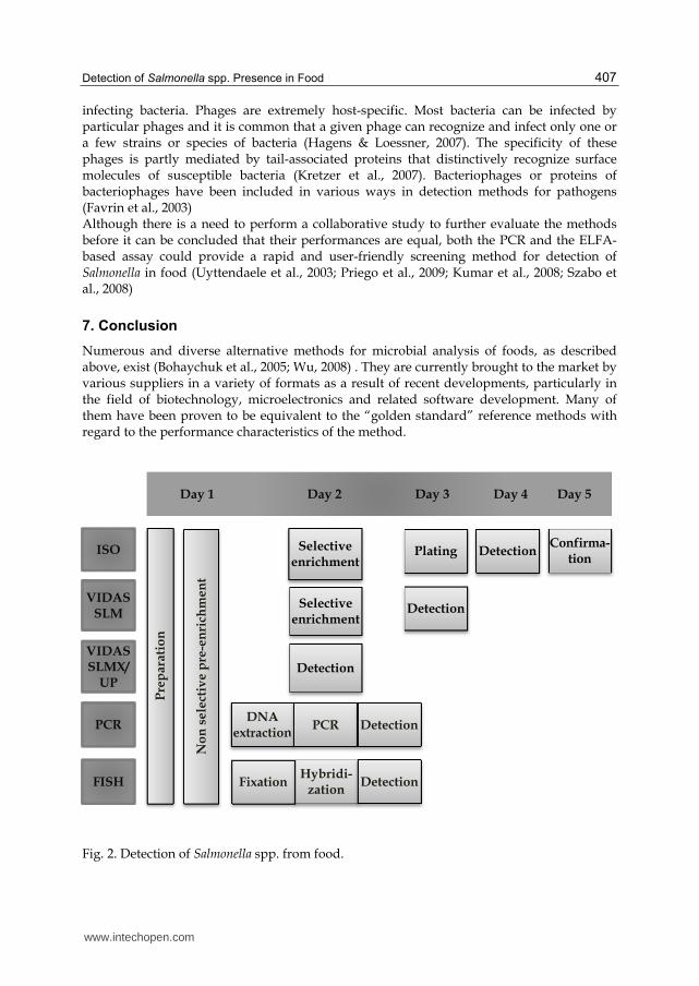
Numerous and diverse alternative methods for microbial analysis of foods, as described above, exist (Bohaychuk et al., 2005; Wu, 2008) . They are currently brought to the market by various suppliers in a variety of formats as a result of recent developments, particularly in the field of biotechnology, microelectronics and related software development. Many of them have been proven to be equivalent to the “golden standard” reference methods with regard to the performance characteristics of the method.
Fig. 2. Detection of Salmonella spp. from food.
Day 1 Day 2 Day 3 Day 4 Day 5
ISO
VIDAS SLM
VIDAS SLMX/
UP
PCR
FISH
Pre
pa
rati
on
No
n s
ele
ctiv
e p
re-e
nri
chm
en
t
Selective enrichment
Selective enrichment
Detection
DNA extraction
PCR Detection
Fixation Hybridi-
zation Detection
Plating Detection Confirma-
tion
Detection
www.intechopen.com
Salmonella – A Dangerous Foodborne Pathogen 408
Due to an overload of alternative methods and/or formats on the market, food business operators or competent authority, for which microbial analysis of food is only a supporting tool in the assurance of food safety, have difficulties in deciding which method is best fit for their purpose in their particular context. (Jasson et al., 2010) Evolution in alternative rapid methods, mainly immunological and molecular methods, focus on the combination of available techniques e.g. combination of immunocapture and PCR and/or by elaboration of new formats optimizing reading and registration software rather than introducing new principles of detection or enumeration.
8. Acknowledgment
This study was supported by the Grant N N312 491340 from the National Science Centre, Poland.
9. References
Al-Soud, W. A., Ouis I. S., Ljungh, D. Q. Li., Wadström, S. T. (2005). Characterization of the PCR inhibitory effect of bile to optimize real-time PCR detection of Helicobacter species. FEMS Immunology & Medical Microbiology 44:177-182
Al-Soud, W. A.; & Radstrom, P. (2000). Effects of amplification facilitators on diagnostic PCR in the presence of blood, feces, and meat. Journal of Clinical Microbiology, Vol. 38, pp. 4463-70
Bansal, N. S., Gray, V., & McDonell, F. (2006). Validated PCR assay for the routine detection of Salmonella in food. Journal Food Protection, Vol. 69, pp. 282-7
Baudart, J., Olaizola, A., Coallier, J., Gauthier V., Laurent P. (2005). Assessment of a new technique combining a viability test, whole-cell hybridization and laser-scanning cytometry for the direct counting of viable Enterobacteriaceae cells in drinking water. FEMS Microbiology Letters., Vol. 243,pp. 405-409
Beutin, L., Jahn, S., Fach, P. (2009). Evaluation of the ‘GeneDisc’ real-time PCR system for detection of enterohaemorrhagic Escherichia coli (EHEC) O26, O103, O111, O145 and O157 strains according to their virulence markers and their O- and Hantigen- associated genes. Journal of Applied Microbiology. Vol. 106, pp.1122-1132
Bohaychuk, V.M., Gensler, G.E., King, R.K.,Wu, J.T., McMullen, L.M. (2005). Evaluation of detection methods for screening meat and poultry products for the presence of foodborne pathogens. Journal Food Protection. Vol. 68, pp. 2637-2647
Bottari, B., Ercolini, D., Gatti, M., Neviani, E. (2006). Application of FISH technology for microbiological analysis: current state and prospects. Applied Microbiology and Biotechnology. Vol. 73, pp. 485-494
Chen, W., G. Martinez, and A. Mulchandani. (2000). Molecular beacons: a real-time polymerase chain reaction assay for detecting Salmonella. Analytical Biochemistry Vol. 280, pp. 166-72
Cheng, C. M., Lin, W., Van, K. T., Phan, L., Tran, N. N. & Farmer, D. (2008). Rapid detection of Salmonella in foods using real-time PCR. Journal Food Protection, Vol.71, No.12, pp. 2436-41
Elizaquivel, P., & Aznar, R. (2008). A multiplex RTi-PCR reaction for simultaneous detection of Escherichia coli O157:H7, Salmonella spp. and Staphylococcus aureus on fresh, minimally processed vegetables. Food Microbiology, Vol.25, No.5, pp. 705-13
www.intechopen.com
Detection of Salmonella spp. Presence in Food 409
Ercolini, D., Hill, P.J., Dodd, C.E.R. (2005). Development of a fluorescence in situ hybridization method for cheese using a 16S rRNA probe. Journal of Microbiological Methods. Vol.52, pp. 267-271
Fach, P., Micheau, P., Mazuet, C., Perelle, S., Popoff, M. (2009). Development of real-time PCR tests for detecting botulinum neurotoxins A, B, E, F producing Clostridium botulinum, Clostridium baratii and Clostridium butyricum. Journal of Applied Microbiology, Vol.107, pp. 465-473
Fang, Q.; Brockmann, S.; Botzenhart, K., Wiedenmann, A. (2003). Improved detection of Salmonella sp. in foods by fluorescent in situ hybridization with 23S rRNA probes: a comparison with conventional cultural methods. Journal of Food Protection, Vol.66, pp.723-731
Favrin, S.J., Jassim, S.A., Griffiths, M.W. (2003). Application of a novel immunomagnetic separation-bacteriophage assay for the detection of Salmonella enteritidis and Escherichia coli O157: H7 in food. International Journal of Food Microbiology. Vol.85, pp. 63-71
Fitzgerald, C.; Collins, M.; van Duyne, S.; Mikoleit, M.; Brown, T.; Fields, P. (2007). Multiplex, bead-based suspension array for molecular determination of common Salmonella serogroups. Journal of Clinical Microbiology, Vol.45, No.10, pp.3323-3334
Gatti M., Bernini V., Lazzi C., Neviani E.: Fluorescence microscopy for studying the viability of micro-organisms in natural whey starters. Letters of Applied Microbiology. 2006, 42, 338–343
Glynn, B., Lahiff, S.,Wernecke, M., Barry, T., Smith, T.J., Maher, M. (2006). Current and emerging molecular diagnostic technologies applicable to bacterial food safety. International Journal of Dairy Technology. Vol.59, pp.126-139
Hagens, S., Loessner, M.J. (2007). Application of bacteriophages for detection and control of foodborne pathogens. Applied Microbiology and Biotechnology. Vol.76, pp. 513-519
Hein, I., Flekna, G., Krassnig, M. & Wagner, M. (2006). Real-time PCR for the detection of Salmonella spp. in food: An alternative approach to a conventional PCR system suggested by the FOOD-PCR project. Journal of Microbiological Methods. Vol. 66(3), pp. 538-47
Hoefel D., Grooby W.L., Monis P.T., Andrews S., Saint C.P. (2003). A comparative study of carboxyfluorescein diacetate and carboxyfluorescein diacetate succinimidyl ester as indicators of bacterial activity. Journal of Microbiological Methods, Vol.52, pp. 379– 388
International Standards Organization. ISO 6579 Microbiology of food and animal feeding stuffs - Horizontal method for detection of Salmonella spp. 6579:2002
Jasson, V.; Jacxsens, L., Luning P., Rajkovic, A., Uyttendaele, M. (2010). Alternative microbial methods: An overview and selection criteria. Food Microbiology, Vol.27, pp. 710-730
Kawasaki, S.; Fratamico, P.M.; Horikoshi, N.; Okada, Y.; Takeshita, K., Sameshima, T.; Kawamoto, S. (2009). Evaluation of a multiplex PCR system for simultaneous detection of Salmonella spp., Listeria monocytogenes, and Escherichia coli O157: H7 in foods and in food subjected to freezing. Foodborne Pathogens and Disease. Vol.6, pp. 81-89
Kretzer, J.W., Lehmann, R., Schmelcher, M., Banz, M., Kim, K.P., Korn, C., Loessner, M.J. (2007). Use of high-affinity cell wall-binding domains of bacteriophage endolysins
www.intechopen.com
Salmonella – A Dangerous Foodborne Pathogen 410
for immobilization and separation of bacterial cells. Applied and Environmental Microbiology. Vol.73, pp. 1992-2000
Kumar, R., Surendran, P.K., Thampuran, N. (2008). Evaluation of culture, ELISA and PCR assays for the detection of Salmonella in seafood. Letters. in Applied. Microbiology. Vol.46, pp. 221-226
Maciorowski, K. G., Herrera, P., Jones, F. T.,Pillai, S. D., Ricke, S. C. (2006). Cultural and immunological detection methods for Salmonella spp. in animal feeds - A Review. Veterinary Research Communications, Vol. 30, pp.127-37
Malorny, B, Tassios, P.T., Rådström, P., Cook, N., Wagner, M., Hoorfar, J. (2003b). Standardization of diagnostic PCR for the detection of foodborne pathogens. Applied and Environmental Microbiology, Vol.83, No.1, (May 2003), pp.39-48
Malorny, B., Hoorfar, J., Bunge, C., Helmuth, R. (2003a). Multicenter Validation of the Analytical Accuracy of Salmonella PCR: towards an International Standard. Applied and Environmental Microbiology, Vol. 69, No. 1,(January 2003), pp. 290-296
Malorny, B., Paccassoni, E., Fach, P., Bunge, C., Martin, A., Helmuth, R. (2004). Diagnostic real-time PCR for detection of Salmonella in food. Applied and Environmental Microbiology, Vol.70, pp.7046-52
Manafi, M. (2000). New developments in chromogenic and fluorogenic culture media. International Journal of Food Microbiology, Vol. 60, pp. 205-18
McCarthy, N., Reen, F. J., Buckley, J. F., Frye, J. G., Boyd, E. F. & Gilroy, D. (2009). Sensitive and rapid molecular detection assays for Salmonella enterica Serovars Typhimurium and Heidelberg. Journal of Food Protection, Vol.72, No.11, pp. 2350-7
Moganedi, K. L. M., Goyvaerts, E. M. A., Venter, S. N., Sibara, M. M. (2007). Optimisation of the PCR-invA primers for the detection of Salmonella in drinking and surface waters following pre-cultivation step. Water SA 33, 196-202
Oliver J.D. (2005). The viable but nonculturable state in bacteria. Journal of Microbiology, 43, 93-100
Ootsubo M., Shimizu T., Tanaka R., Sawabe T., Tajima K., Ezura Y. (2003). Seven-hour fluorescence in situ hybridization technique for enumeration of Enterobacteriaceae in food and environmental water sample. Journal of Applied Microbiology. Vol.95, pp.1182–1190
Patel, J. R., Bhagwat, A. A., Sanglay, G. C., and Solomon, M. B. (2006). Rapid detection of Salmonella from hydrodynamic pressure-treated poultry using molecular beacon real-time PCR. Food Microbiology. Vol.23, pp.39–46
Perry, J.D., Freydiere, A.M. (2007). The application of chromogenic media in clinical microbiology. Journal of Applied. Microbiology. Vol.103, pp.2046-2055
Piknova , L., Stefanovicova´, A., Drahovska´ , H., Sa´sik, M., & Kuchta, T. (2002). Detection of Salmonella in food, equivalent to ISO 6579, by a three-days polymerase chain reaction-based method. Food Control, Vol.13, pp.191–194.
Pisz, J. M.; Lawrence, J. R.; Schafer, A. N.; Siciliano, S. D. (2007). Differentiation of genes extracted from non-viable versus viable micro-organisms in environmental samples using ethidium monoazide bromide. Journal of Microbiological Methods, Vol.71, No.3, pp. 312-318
Priego, R., Medina, L. M., Jordano, R. (2009). Comparison between the Vitek Immunodiagnostic Assay System and PCR for the detection of pathogenic
www.intechopen.com
Detection of Salmonella spp. Presence in Food 411
microorganisms in an experimental dry sausage during its curing process. Journal Food Protection. Vol.72 , No.9, pp.1977-81
Sakai, K., Mori, M., Fujii, A., Iwami, Y., Chukeatirote E., Shirai Y. (2004). Fluorescent in situ hybridization analysis of open lactic acid fermentation of kitchen refuse using rRNA-targeted oligonucleotide probes. Journal of Bioscience and Bioengineering, Vol.98, pp. 48-56
Schonenbrucher, V., Mallinson, E.T., Bulte, M. (2008). A comparison of standard cultural methods for the detection of foodborne Salmonella species including three new chromogenic plating media. International Journal of Food Microbiology. 123, 61e66.
Seo, K. H., Valentin-Bon, I. E. & Brackett, R. E. (2006). Detection and enumeration of Salmonella Enteritidis in homemade ice cream associated with an outbreak: comparison of conventional and real-time PCR methods. Journal Food Protection, Vol.69, No.3, pp. 639-43
Settanni L, Corsetti A. (2007). The use of multiplex PCR to detect and differentiate food- and beverage-associated microorganisms: a review. Journal of Microbiological Methods Vol.69, No.1, pp.1-22
Skowrońska A., Zmysłowska I. (2006). Współczesne metody identyfikacji bakterii stosowane w ekologii mikroorganizmów wodnych – fluorescencyjna hybrydyzacja in situ (FISH). Postępy Mikrobiologii., Vol.45, pp.183-193
Szabo, I., Scherer, K., Roesler, U., Appel, B., Nockler, K., Hensel, A., (2008). Comparative examination and validation of ELISA test systems for Salmonella typhimurium diagnosis of slaughtering pigs. International Journal of Food Microbiology. 124, 65e69.
Tomás, D, Rodrigo, A., Hernández, M., Ferrús, M. A. (2009). Validation of Real-Time PCR and Enzyme-Linked Fluorescent Assay-Based Methods for Detection of Salmonella spp. in Chicken Feces Samples. Food Analytical Methods. Vol2: pp: 180–189
Uyttendaele, M., K., Vanwildemeersch, Debevere, J. (2003). Evaluation of real-time PCR vs automated ELISA and a conventional culture method using a semi-solid medium for detection of Salmonella. Letters Applied Microbiology. Vol. 37 , pp; 386-91
Vieira-Pinto, M., Oliveira, M., Bernardo, F., Martins, C. (2005). Evaluation of Fluorescent in situ Hybridization (FISH) as a rapid screening method for detection of Salmonella in tonsils of slaughtered pigs for consumption: a comparison with conventional culture method. Journal of Food Safety. Vol. 25, pp: 109–119
Vieira-Pinto, M.; Oliveira, M., Bernardo, F., Martins, C. (2007). Rapid detection of Salmonella sp. in pork samples using fluorescent in situ hybridization: a comparison with VIDAS®-SLM system and ISO 6579 cultural method. Arquivo Brasileiro de Medicina Veterinária e Zootecnia, Vol.59, No.6, pp.1388-1393
Walker, R. L., Kinde, H., Anderson, R. J., Brown, A. E. (2001). Comparison of VIDAS enzyme-linked fluorescent immunoassay using more swab sampling and conventional culture method for Salmonella detection in bulk tank milk and in-line milk filters in California dairies. International Journal of Food Microbiology. Vol.67, pp. 123-9
Wu, V.C.H. (2008). A review of microbial injury and recovery methods in food. Food Microbiology. Vol. 25, pp: 735-744
Yeh, K. S., C. E. Tsai, S. P. Chen, and C. W. Liao. (2002). Comparison between VIDAS automatic enzyme-linked fluorescent immunoassay and culture method for
www.intechopen.com

Salmonella – A Dangerous Foodborne Pathogen 412
Salmonella recovery from pork carcass sponge samples. Journal of Food Protection. Vol. 65, pp. 1656-9
Zadernowska, A., Łaniewska-Trokenheim, Ł., Chajęcka,W. (2010) Detection of Listeria monocytogenes and Salmonella sp. rods in fish and fish products using the mini Vidas system. Medycyna Weterynaryjna. Vol.66, No 4, pp. 264-267
Zwirglmaier, K. (2005). Fluorescence in situ hybridization (FISH) – the next generation. FEMS Microbiology Letters, Vol.246, pp. 151-158
www.intechopen.com
Salmonella - A Dangerous Foodborne PathogenEdited by Dr. Dr. Barakat S M Mahmoud
ISBN 978-953-307-782-6Hard cover, 450 pagesPublisher InTechPublished online 20, January, 2012Published in print edition January, 2012
InTech EuropeUniversity Campus STeP Ri Slavka Krautzeka 83/A 51000 Rijeka, Croatia Phone: +385 (51) 770 447 Fax: +385 (51) 686 166www.intechopen.com
InTech ChinaUnit 405, Office Block, Hotel Equatorial Shanghai No.65, Yan An Road (West), Shanghai, 200040, China
Phone: +86-21-62489820 Fax: +86-21-62489821
More than 2,500 serotypes of Salmonella exist. However, only some of these serotypes have been frequentlyassociated with food-borne illnesses. Salmonella is the second most dominant bacterial cause of food-bornegastroenteritis worldwide. Often, most people who suffer from Salmonella infections have temporarygastroenteritis, which usually does not require treatment. However, when infection becomes invasive,antimicrobial treatment is mandatory. Symptoms generally occur 8 to 72 hours after ingestion of the pathogenand can last 3 to 5 days. Children, the elderly, and immunocompromised individuals are the most susceptibleto salmonellosis infections. The annual economic cost due to food-borne Salmonella infections in the UnitedStates alone is estimated at $2.4 billion, with an estimated 1.4 million cases of salmonellosis and more than500 deaths annually. This book contains nineteen chapters which cover a range of different topics, such as therole of foods in Salmonella infections, food-borne outbreaks caused by Salmonella, biofilm formation,antimicrobial drug resistance of Salmonella isolates, methods for controlling Salmonella in food, andSalmonella isolation and identification methods.
How to referenceIn order to correctly reference this scholarly work, feel free to copy and paste the following:
Anna Zadernowska and Wioleta Chajęcka (2012). Detection of Salmonella spp. Presence in Food, Salmonella- A Dangerous Foodborne Pathogen, Dr. Dr. Barakat S M Mahmoud (Ed.), ISBN: 978-953-307-782-6, InTech,Available from: http://www.intechopen.com/books/salmonella-a-dangerous-foodborne-pathogen/detection-of-salmonella-spp-presence-in-food

![0000065394 · Intelltx Destqner [weather.kdm] Tot* SOUL Example Set Editor Rea 93 64 72 81 FALSE TRUE FALSE FALSE TRUE TRUE FALSE FALSE FALSE TRUE TRUE FALSE TRUE overcast](https://img.pdfslide.us/doc/110x75/5cbf6e0688c993c04b8b9447/0000065394-intelltx-destqner-weatherkdm-tot-soul-example-set-editor-rea.jpg)