Embed Size (px)
DESCRIPTION
.
Citation preview

July 2013, Vol. 20, No. 3 Cancer Control 161
Novel imaging techniques
for prostate cancer should
improve staging and better
evaluate treatment results.
IntroductionImaging for prostate carcinoma can serve several clini-
cal goals. First, it can assist in assessing the primary
or recurrent tumor within the prostate gland, as well
as tumor size, multifocality, extracapsular extension,
seminal vesicle extension, neurovascular bundle in-
volvement, and bladder involvement. Second, imag-
ing can be used to assess metastatic disease such as
spread to lymph nodes and bones. Third, imaging is
used to guide interventions such as prostate biopsies
or computed tomography (CT)-guided biopsy of sus-
picious lymph nodes. Fourth, functional or metabolic
imaging could potentially assess tumor aggressiveness
From the Department of Diagnostic Imaging at the H. Lee Moffitt Cancer Center & Research Institute, Tampa, Florida.
Submitted January 24, 2012; accepted September 17, 2012.
Address correspondence to Eric K. Outwater, MD, Department of Diagnostic Imaging, Moffitt Cancer Center, 12902 Magnolia Drive, WCB-RAD MD/OPI, Tampa, FL 33612. E-mail: [email protected]
No significant relationship exists between the authors and the companies/organizations whose products or services may be ref-erenced in this article.
The authors have disclosed that this article discusses unlabeled/unapproved use of ferumoxytol, a contrast agent indicated for iron deficiency anemia, for lymph node imaging in prostate cancer.
Imaging of Prostate Carcinoma
Eric K. Outwater, MD, and Jaime L. Montilla-Soler, MD
Background: Imaging of prostate carcinoma is an important adjunct to clinical evaluation and prostate-
specific antigen measurement for detecting metastases and tumor recurrence. In the past, the ability to assess
intraprostatic tumor was limited.
Methods: Pertinent literature was reviewed to describe the capabilities and limitations of the currently available
imaging techniques for assessing prostate carcinoma. Evaluation of primary tumor and metastatic disease
by ultrasonography, computed tomography (CT), magnetic resonance imaging (MRI), and nuclear medicine
techniques is discussed.
Results: Ultrasonography and MRI have limited usefulness for local staging of prostate cancer because of
suboptimal sensitivity and specificity for identifying tumor extent and capsular penetration. Additional MRI
techniques such as magnetic resonance-based perfusion imaging, diffusion imaging, and spectroscopy may
provide incremental benefit. CT and bone scanning provide an assessment of metastatic disease but are also
limited by the poor sensitivity of lymph node size as a criterion for detecting metastases. Novel imaging techniques
such as hybrid imaging devices in the form of single-photon emission CT/CT gamma cameras, positron emission
tomography/CT cameras, and, in the near future, positron emission tomography/MRI combined with tumor-
specific imaging radiotracers may have a significant impact on tumor staging and treatment response.
Conclusions: Cross-sectional imaging and scintigraphy have an important role in assessing prostate carcinoma
metastases and treatment response. Increasingly, the incremental value of primary tumor imaging through
MRI is being realized.
Michele R. Sassi. Dhow, Musandam, Oman. Photograph.

162 Cancer Control July 2013, Vol. 20, No. 3
or other parameters that correlate with outcome, al-
though such techniques have not yet entered routine
clinical practice. It is hoped that these novel imag-
ing methods will be superior to the current standard
means in assessing mortality, tumor size, and thera-
peutic response to targeted therapies. This review
focuses on the main imaging methods and their use
for these purposes.
The National Comprehensive Cancer Network clini-
cal practice guidelines show a fairly limited role for
imaging in patients with prostate carcinoma (Table 1).1
According to these guidelines, imaging is largely used
to evaluate metastatic disease, with a limited role for
endorectal magnetic resonance imaging (MRI) in pa-
tients who have received radiation therapy but have
evidence of failure by prostate-specific antigen (PSA)
level. According to these recommendations, low-risk
prostate cancer requires no imaging; however, actual
adherence to these guidelines by urologists is highly
variable.2 Many more applications and types of imag-
ing have been explored, with a plethora of suggested
imaging applications for the management of prostate
carcinoma. Many of these, such as investigational
nuclear medicine agents, are exploratory, but others,
such as endorectal MRI for initial staging, have been
the focus of numerous studies.
Reports on the diagnostic performance of some of
these techniques vary widely in the literature, particu-
larly regarding MRI. In some respects, this resembles
the decline effect3 or other statistical biases,4 but meth-
odological aspects assess diagnostic performance in
the prostate that may give rise to these varying results.
To determine the accuracy of a diagnostic technique
such as MRI in locating a tumor within the prostate, it
is common to divide the prostate into multiple areas
or segments and determine the presence or absence
of the tumor in each segment. These results can be
correlated with the absence or presence of tumor on
MRI. If the prostate specimen is distorted during
processing or if discordance exists between the orien-
tation of the pathological specimen to MRI, then the
diagnostic accuracy will be poor, not related to the
actual performance of the technique.5,6 Different re-
sults can be obtained, for example, when exact corre-
spondence is required rather than when approximate
correspondence is required.6 These methodological
problems may lead to varying results and introduce
bias in reported results for the diagnostic performance
of MRI and for other diagnostic techniques. Turkbey
et al7 found a 61% sensitivity rate for the detection of
tumors larger than 3 mm in size on T2-weighted im-
ages using a stringent correlation and 94% sensitivity
rate for less stringent correlation. The magnitude of
this discrepancy indicates that correlating imaging
findings with histology is not as straightforward as
one might think.
Evaluation of the Primary TumorTransrectal Ultrasonography
Ultrasonography is the most common method used for
direct visualization of the prostate, primarily because
it is indispensable to imaging-guided prostate biop-
sies. Ultrasonography has the advantages of real-time
imaging, portability, ease of use, and low cost. It can
visualize intraprostatic zonal anatomy, with the pe-
ripheral zone showing slightly increased echogenicity
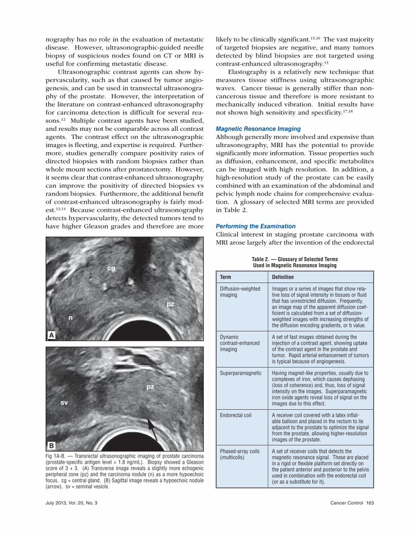
compared with the central gland. Prostate carcinoma
typically presents as a hypoechoic area within the
peripheral zone (Fig 1). However, transrectal ultra-
sonography is not highly sensitive or specific for the
detection of prostate carcinoma.8 Color Doppler and
power Doppler imaging do not substantially add ac-
curacy to the technique.9 However, vessel density as
shown on color Doppler and power Doppler imaging
may have prognostic importance, with high vessel
densities predicting a slower rate of decline of PSA
with radiation treatment.10 Similarly, transrectal ultra-
sonography has limited accuracy for the detection of
extraprostatic extent of tumor.11 In general, ultraso-
Table 1. — Selected NCCN Clinical Practice Guidelines for Prostate Cancer
Clinical Group Indicated Imaging
New diagnosis: > 5-yr life expectancy
Bone scan if:
T1 and PSA > 20
T2 and PSA > 10
Gleason score 8
T3, T4, or symptomatic
Pelvic CT or MRI if:T3, T4, or T1–T2 and nomogram indicate probability of lymph node involvement > 10%a
Rise in PSA following prostatectomy
± CT
± MRI
± Bone scan
Rise in PSA following radiation
± Abdomen/pelvis CT or MRI± Endorectal MRI± MR spectroscopyBone scan
a Staging studies may not be cost effective until the likelihood of lymph node positivity reaches 45%.
CT = computed tomography; MR = magnetic resonance; MRI = magnetic resonance imaging; NCCN = National Comprehensive Cancer Network; PSA = prostate-specific antigen; T = tumor.
Reproduced/adapted with permission from the NCCN Clinical Practice Guidelines in Oncology (NCCN Guidelines®) for Prostate Cancer. V.2.2013. © 2013 National Comprehensive Cancer Network, Inc. All rights reserved. The NCCN Guidelines® and illustrations herein may not be reproduced in any form for any purpose without the express written permission of the NCCN. To view the most recent and com-plete version of the NCCN Guidelines, go online to www.NCCN.org. NATIONAL COMPREHENSIVE CANCER NETWORK®, NCCN®, NCCN GUIDELINES™, and all other NCCN Content are trademarks owned by the National Comprehensive Cancer Network, Inc.
July 2013, Vol. 20, No. 3 Cancer Control 163
nography has no role in the evaluation of metastatic
disease. However, ultrasonographic-guided needle
biopsy of suspicious nodes found on CT or MRI is
useful for confirming metastatic disease.
Ultrasonographic contrast agents can show hy-
pervascularity, such as that caused by tumor angio-
genesis, and can be used in transrectal ultrasonogra-
phy of the prostate. However, the interpretation of
the literature on contrast-enhanced ultrasonography
for carcinoma detection is difficult for several rea-
sons.12 Multiple contrast agents have been studied,
and results may not be comparable across all contrast
agents. The contrast effect on the ultrasonographic
images is fleeting, and expertise is required. Further-
more, studies generally compare positivity rates of
directed biopsies with random biopsies rather than
whole mount sections after prostatectomy. However,
it seems clear that contrast-enhanced ultrasonography
can improve the positivity of directed biopsies vs
random biopsies. Furthermore, the additional benefit
of contrast-enhanced ultrasonography is fairly mod-
est.13,14 Because contrast-enhanced ultrasonography
detects hypervascularity, the detected tumors tend to
have higher Gleason grades and therefore are more
likely to be clinically significant.15,16 The vast majority
of targeted biopsies are negative, and many tumors
detected by blind biopsies are not targeted using
contrast-enhanced ultrasonography.13
Elastography is a relatively new technique that
measures tissue stiffness using ultrasonographic
waves. Cancer tissue is generally stiffer than non-
cancerous tissue and therefore is more resistant to
mechanically induced vibration. Initial results have
not shown high sensitivity and specificity.17,18
Magnetic Resonance Imaging
Although generally more involved and expensive than
ultrasonography, MRI has the potential to provide
significantly more information. Tissue properties such
as diffusion, enhancement, and specific metabolites
can be imaged with high resolution. In addition, a
high-resolution study of the prostate can be easily
combined with an examination of the abdominal and
pelvic lymph node chains for comprehensive evalua-
tion. A glossary of selected MRI terms are provided
in Table 2.
Performing the Examination
Clinical interest in staging prostate carcinoma with
MRI arose largely after the invention of the endorectal
Fig 1A-B. — Transrectal ultrasonographic imaging of prostate carcinoma (prostate-specific antigen level = 1.8 ng/mL). Biopsy showed a Gleason score of 3 + 3. (A) Transverse image reveals a slightly more echogenic peripheral zone (pz) and the carcinoma nodule (n) as a more hypoechoic focus. cg = central gland. (B) Sagittal image reveals a hypoechoic nodule (arrow). sv = seminal vesicle.
A
B
Table 2. — Glossary of Selected Terms Used in Magnetic Resonance Imaging
Term Definition
Diffusion-weighted imaging
Images or a series of images that show rela-tive loss of signal intensity in tissues or fluid that has unrestricted diffusion. Frequently, an image map of the apparent diffusion coef-ficient is calculated from a set of diffusion-weighted images with increasing strengths of the diffusion encoding gradients, or b value.
Dynamic contrast-enhanced imaging
A set of fast images obtained during the injection of a contrast agent, showing uptake of the contrast agent in the prostate and tumor. Rapid arterial enhancement of tumors is typical because of angiogenesis.
Superparamagnetic Having magnet-like properties, usually due to complexes of iron, which causes dephasing (loss of coherence) and, thus, loss of signal intensity on the images. Superparamagnetic iron oxide agents reveal loss of signal on the images due to this effect.
Endorectal coil A receiver coil covered with a latex inflat-able balloon and placed in the rectum to lie adjacent to the prostate to optimize the signal from the prostate, allowing higher-resolution images of the prostate.
Phased-array coils (multicoils)
A set of receiver coils that detects the magnetic resonance signal. These are placed in a rigid or flexible platform set directly on the patient anterior and posterior to the pelvis used in combination with the endorectal coil (or as a substitute for it).

164 Cancer Control July 2013, Vol. 20, No. 3
coil, which permitted high-resolution images of the
prostate.19 Higher signal-to-noise images, high resolu-
tion images, or both can be obtained with the use of
the endorectal coil, phased-array coils, or high-field
imaging with 3 Tesla magnets, or any combination
of these (Fig 2). Imaging without the endorectal coil
will not provide the best possible examination for
staging purposes, but it may be suitable for certain
clinical situations.
To perform a standard examination, the patient
is placed on his side on the MRI table, and the en-
dorectal coil is slid into place in the rectum. The
coil is anteriorly oriented to receive a signal from the
prostate. The patient is turned on his back, and an
antiperistaltic agent such as glucagon is often intra-
venously administered. Phased-array coils are placed
against the skin anterior and posterior to the patient,
and the table is slid into the MRI magnet.
Multiple images of different types, or series, are
obtained on multiple planes. These typically include
T1-weighted images of the abdomen and pelvis for
lymph node disease. Smaller field-of-view (higher res-
olution) T1-weighted images, and T2-weighted images
on multiple planes (coronal, axial, and sagittal) are also
obtained. Additional sequences can include diffusion-
weighted imaging, dynamic contrast-enhanced (DCE)
imaging, and magnetic resonance spectroscopy.
Diffusion-weighted imaging is typically performed
using echoplanar fast imaging with diffusion-encoding
gradients in three directions. These gradients degrade
the signal intensity of water moving in the direction of
the gradient, and using gradients in three directions
diminishes the signal in any direction. Therefore, the
tissue has a lower signal from water motion due to
perfusion and diffusion. Acquiring two or more sets
of images with increasing gradient strength allows a
calculated apparent diffusion coefficient (ADC) map;
by necessity, these images are low resolution.
DCE images are fast, fat-saturated, T1-weighted
images obtained during and after the administration
of an intravenous contrast agent (eg, gadopentetate).
These rapid images reveal the arrival of the contrast
to the vessels and tissues as increased signal inten-
sity, which will sequentially reflect the arterial arrival
of the contrast, venous perfusion, uptake in the tis-
sue interstitium, and the subsequent washout from
these compartments. Such images may be acquired
at several time points for qualitative assessment and
at many time points for quantitative analysis. Addi-
tional acquisitions may also include magnetic reso-
nance spectroscopy. A complete examination usually
takes 45 minutes.
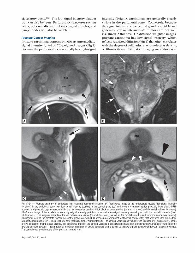
Anatomy of the Prostate
The most important anatomical features of the pros-
tate are shown in T2-weighted images (Fig 3). The
central gland of the prostate includes the periurethral
and transitional zones, generally with lower signal
intensity than the peripheral zone.20 The signal in-
tensity of the peripheral zone is generally high on
T2-weighted images largely due to glandular fluid;
by contrast, the signal intensity of the central gland
is variable and highly dependent on the amount and
characteristics of any present benign prostatic hyper-
trophy.20 The anterior central gland appears as low
signal intensity, the anterior fibromuscular stroma. A
low signal intensity line, the prostatic capsule, limits
the peripheral zone. Neurovascular bundles can be
identified as a group of vessels posterolateral to the
prostatic capsule on either side.
Seminal vesicles appear as high-signal-intensity,
grape-like clusters of tubules that reveal low-signal
intensity smooth muscle walls (Fig 3).20 This appear-
ance is due to the high-signal-intensity folded mucosa
filling the lumen with pockets of fluid. The ampullae
of the vas deferens can be identified as well as the
Fig 2A-B. — Imaging of the prostate with and without endorectal coil. (A) Transverse image of the prostate performed with phased-array coils with-out an endorectal coil reveals adequate differentiation of the central gland (cg), peripheral zone (pz), and the tumor nodule (n). However, visualization of the prostatic capsule near the tumor is poor (arrow). (B) Transverse image of the prostate with the endorectal coil shows the central gland (cg), peripheral zone (pz), and tumor nodule (n), with better visualization of the prostatic capsule near the tumor (arrow).
A
B
July 2013, Vol. 20, No. 3 Cancer Control 165
ejaculatory ducts.20,21 The low-signal intensity bladder
wall can also be seen. Periprostatic structures such as
veins, puborectalis and pubococcygeal muscles, and
lymph nodes will also be visible.21
Prostate Cancer Imaging
Prostate carcinoma appears on MRI as intermediate-
signal intensity (gray) on T2-weighted images (Fig 2).
Because the peripheral zone normally has high-signal
intensity (bright), carcinomas are generally clearly
visible in the peripheral zone. Conversely, because
the signal intensity of the central gland is variable and
generally low or intermediate, tumors are not well
visualized in this area. On diffusion-weighted images,
prostate carcinoma has low-signal intensity, which
reflects restricted diffusion (Fig 4) that often correlates
with the degree of cellularity, macromolecular density,
or fibrous tissue. Diffusion imaging may also assist
Fig 3A-D. — Prostate anatomy on endorectal coil magnetic resonance imaging. (A) Transverse image at the midprostate reveals high-signal intensity (brighter) in the peripheral zone (pz), low-signal intensity (darker) in the central gland (cg) with several scattered benign prostatic hyperplasia (BPH)nodules, and prostatic capsule (arrowhead), the neurovascular bundles (thick black arrows), urethra (thin black arrow) and the rectal wall (white arrow). (B) Coronal image of the prostate shows a high-signal intensity peripheral zone and a low-signal intensity central gland with the prostatic capsule (thick white arrows). The irregular ampulla of the vas deferens are visible (thin white arrows), as well as the prostatic urethra and verumontanum (black arrow). (C) Sagittal view of the prostate reveals the central gland (cg) with BPH producing a prominent subtrigonal nodule (stn) that protrudes into the bladder, a variant appearance of BPH. The peripheral zone (pz) has a higher signal intensity. The seminal vesicles and vas deferens lie superiorly (black arrow). White arrows denote the membranous urethra. (D) Transverse image of the seminal vesicles (black arrows) shows high-signal intensity lumens surrounded by the low-signal intensity walls. The ampullae of the vas deferens (white arrowheads) are visible as well as the low-signal intensity bladder wall (black arrowhead). The central subtrigonal nodule of the prostate is noted (stn).
A B
C D

166 Cancer Control July 2013, Vol. 20, No. 3
in differentiating between hemorrhage and tumor in
the peripheral zone.22 The enhancement of prostatic
tumors is somewhat variable; however, tumors typi-
cally enhance more rapidly and to a greater degree
than the peripheral zone normal tissue. Therefore,
they may be best displayed on an early arterial phase
image (Fig 5). Increased or more rapid contrast en-
hancement correlates with microvascular density in
the prostate tumor.23
Extracapsular extension, when gross, is mani-
fested by protrusion of the intermediate-signal in-
tensity tumors through the prostate capsule. More
subtle capsular penetration may be manifested by a
tumor that abuts the capsule over a wide area, capsu-
lar thickening, nodularity or bulging of the capsule,
or irregularity (Fig 6).24 Seminal vesicle invasion is
manifested by a low-signal intensity tumor, replacing
the high-signal intensity seminal vesicle lumens, the
mucosa, or both.25 Similarly, bladder wall invasion
will be manifested by intermediate-signal intensity on
T2-weighted images or enhancing tissue interrupting
the low-signal intensity bladder wall.
Magnetic Resonance Spectroscopy
Spectroscopy is a magnetic resonance technique to
assess the presence of metabolites in tissue. Proton
spectroscopy can detect certain hydrogen-containing
metabolites in the prostate such as choline, creatinine,
and citrate. The normal prostate gland produces high
levels of citrate and low levels of choline. Because of
phospholipid metabolism and higher cell membrane
turnover, prostate cancer has higher levels of choline.
Fig 4A-D. — Typical appearance of prostate carcinoma on magnetic resonance imaging. Subsequent biopsy revealed a Gleason score of 4 + 4. (A) T1-weighted image shows homogeneous low signal of the prostate (p), with no discrimination of the central and peripheral gland. The tumor nodule is not seen because no tissue contrast is present between the tumor and peripheral zone. The neurovascular bundles (black arrows) are seen laterally. (B) T2-weighted image shows the lower-signal intensity tumor (n) compared to the curve from zone on either side. (C) Coronal image shows the tumor nodule (n) with the adjoining apical prostatic capsule shown (arrow). (D) Early-phase gadolinium chelate-enhanced sections from a fast 3-dimensional gradient echo sequence show rapid intense enhancement of the tumor nodule (n), manifested by brighter signal intensity in the rest of the prostate, with tumor extending laterally to greater extent than is apparent on the T2-weighted image.
A B
C D

July 2013, Vol. 20, No. 3 Cancer Control 167
Fig 5A-C. — Prostatic carcinoma on magnetic resonance imaging with dif-fusion-weighted imaging of a patient with a prostate-specific antigen level of 3.1 ng/mL. Subsequent biopsy revealed a Gleason score of 3 + 4 with extracapsular extension. (A) T2-weighted and rectal coil image reveals a tumor nodule (n) contrasted with a higher-signal intensity peripheral zone. The central gland (cg) is expanded by benign prostatis hyperplasia, which has a lower signal intensity. (B) Apparent diffusion coefficient (ADC) map of the prostate reveals that the tumor nodule (n) has a lower signal intensity than the peripheral zone or the central gland (cg). Low signal intensity indi-cates that the ADC is lower than that of water and the water diffusion within the tumor is restricted. The ADC map is calculated from a set of three images at the same level (not shown) and performed with three different magnitudes of strength of diffusion-encoding gradients. (C) Early-phase gadolinium chelate-enhanced slice from a fast 3-dimensional gradient-echo sequence reveals rapid, intense enhancement of the tumor nodule (n). Portions of the central gland (cg) also reveal rapid enhancement. Note: Multiparametric imaging refers to the characteristics of abnormalities on all three sets of images.
A
B
C
Fig 6A-C. — Typical enhancement characteristics of a tumor on dynamic contrast-enhanced images in a patient with a prostate-specific antigen level of 15 ng/mL. Biopsy showed a Gleason score of 4 + 4. (A) Transverse T2-weighted image reveals a tumor nodule (n) involving both the central gland and the peripheral zone on the right side of the prostate. The arrow points to the capsular involvement on the right. (B) Early-phase gadolinium chelate-enhanced slice from a fast 3-dimensional gradient-echo sequence reveals a rapid, intense enhancement of the tumor nodule (n) on the right side of the prostate. (C) Late-phase enhanced T1-weighted image reveals the tumor nodule (n) with a lower signal intensity, indicating washout of the contrast compared with the rest of the prostate. Typically, a tumor dem-onstrates early enhancement and early washout (as shown in this case).
A
B
C

168 Cancer Control July 2013, Vol. 20, No. 3
The ratio of choline to citrate is increased in patients
with cancer. At 1.5 Tesla, the spectroscopic peaks of
choline overlap with creatinine; however, these can
be resolved at 3 Tesla.
Magnetic resonance spectroscopy of the prostate
can be performed with a 3-dimensional (3D) water
and lipid suppressed, double-spin-echo point-resolved
spectroscopy sequence; however, 3D magnetic reso-
nance spectroscopy is time consuming and technically
challenging. Adequate water and lipids suppression
is necessary to exclude contamination from tissue
outside the prostate. Data sets are typically acquired
with 16 × 8 × 8 phase-encoded spectral arrays. The
magnetic resonance spectra can be displayed as an
rectangular array from the individual voxels.
Multiple studies have suggested improved detec-
tion, localization, and staging with magnetic resonance
spectroscopy.6,26-28 However, a multisite prospective
trial showed no benefit of magnetic resonance spec-
troscopy compared with MRI alone for tumor identifi-
cation and compared with sextant localization on the
prostatectomy specimens.29 It is not clear whether
the results of this study represent inherent limitations
of magnetic resonance spectroscopy for the localiza-
tion of prostate cancer or difficulties in a technically
demanding technique with a multicenter trial design.
Accuracy of Staging
The reported sensitivity and specificity for prostate
carcinoma staging vary from high to low, and it is
difficult to determine the most appropriate figures to
use.30-32 Studies commonly vary in patient population,
particularly regarding the Gleason grading spectrum
of patients or PSA levels, as well as in techniques or
pulse sequences used (eg, diffusion imaging, dynamic-
enhanced imaging), without standardization for coils
(eg, endorectal or body coils), or field strength. Fur-
thermore, the vast majority are single institutional and
retrospective studies, which are prone to bias and can
be insufficiently powered. Finally, methodological
problems exist with mapping tumors or sites of cap-
sular penetration to precise sites on any prostatectomy
specimens to determine imaging accuracy.5
Results from a multicenter prospective trial dem-
onstrated limited accuracy of MRI in locating the pros-
tate tumor.29 One would expect that the accuracy for
staging would be even lower. Several generalities
can be stated regarding the accuracy of MR staging.
First, the accuracy for extracapsular extension is prob-
ably between 70% and 80%, while the identification
of seminal vesicle invasion is more accurate.24,30,33-36
Second, MRI at 3 Tesla is probably superior to MRI
at 1.5 Tesla.33,37 Third, diffusion-weighted and DCE
imaging may provide small incremental increases in
accuracy.6,7,33 Fourth, MRI probably provides a small
incremental increase in accuracy over clinical nomo-
grams based on PSA, digital rectal examination, and
Gleason grade, among others.38 Fifth, MRI is more
accurate in patients at intermediate or high risk, but
it is less accurate in patients with low-volume tumors
and low Gleason grades.39 The detection rate of tu-
mors is also dependent on size, with tumors smaller
than 2 cm unlikely to be detected.40
Multiparametric imaging, or the integration of
various sequences such as diffusion, DCE, and T2-
weighted imaging, may provide higher accuracy.6,33
However, how these varying data are combined for
optimal accuracy is not clear.
Evaluation of Prostate Cancer Metastases Patients with prostate carcinoma are evaluated for
possible metastatic disease if they fall into a high-
risk group with a new diagnosis or after biochemical
failure following prostatectomy, radiation treatment,
or local therapy.
Computed Tomography
CT is an important part of the management of prostate
carcinoma. CT scanning provides a quick evalua-
tion for metastases in the chest, abdomen, or pelvis.
It is efficient at identifying enlarged lymph nodes,
although lymph node enlargement is not highly sen-
sitive and not entirely specific for lymph node me-
tastases.41,42 Since CT provides poor tissue contrast
within the prostate itself, evaluation of the intrapros-
tatic tumor is limited.
CT scanning can detect bone metastases, particu-
larly those that are osteoblastic; therefore, it is useful
in patients at high risk of metastatic spread on initial
evaluation or with suspected progression after pros-
tatectomy or radiation treatment. It is also useful in
patients with identified metastases to monitor the
effectiveness of therapy.
Because lymph node size is not sensitive or spe-
cific for metastasis in patients with prostate carci-
noma, there is no one size criterion that is highly ac-
curate. A commonly used criterion is a pelvic lymph
node smaller than 10 mm but 8 mm or larger in size (if
round in shape) or 10 mm or larger in size (if oval in
shape). Using this criterion with contemporary scan-
ners, one study found a sensitivity rate of 34% and a
specificity rate of 97% for the detection of lymph node
metastases.43 If CT is used to evaluate for lymph node
metastases, suspicious nodes are generally subjected
to fine-needle aspiration for confirmation, which can
produce false-negative results. For these reasons, CT
is recommended with a very high pretest probability
of metastatic disease (Table 1).1
Magnetic Resonance Imaging
MRI can survey for metastatic disease similar to CT of
the lymph nodes and skeletal structures. MRI of the
July 2013, Vol. 20, No. 3 Cancer Control 169
abdomen and pelvis is used to evaluate for lymphade-
nopathy, bone metastases, and metastases elsewhere,
but it is generally more time consuming and more
expensive than CT scanning while providing similar
diagnostic information.
Recent developments exist to provide a more spe-
cific evaluation of lymph node metastases than an
assessment based solely on size. Diffusion-weighted
imaging of lymph nodes may be a more specific as-
sessment than size criteria.44 Metastatic lymph nodes
demonstrate a lower apparent ADC than benign lymph
nodes,14,15 presumably reflecting greater cell density in
lymph nodes with metastatic tumor. DCE properties
of lymph nodes differ between normal and metastatic
lymph nodes in that lymph nodes with metastases
show stronger and more rapid enhancement.45
Another innovation in lymph node imaging us-
ing MRI has been the development of superparamag-
netic contrast agents with preferential lymph node
uptake. These agents accumulate in normal lymph
nodes, causing losses of signal intensity within these
lymph nodes on imaging. Conversely, lymph nodes
with metastases within them do not accumulate the
agents in the metastatic portion and do not lose signal
intensity, or, conversely, they will lose signal intensity
in the normal part of the lymph node. A large trial of
men with intermediate or high risk of having nodal
metastases compared this MR contrast agent with CT
showed 82% sensitivity for lymph node metastases vs
34% for CT.38 This could obviate the need for lymph
node dissection in patients with a negative MRI.43
Because an MRI examination using a lympho-
tropic agent reveals all the lymph nodes in the pelvis,
it can reveal metastatic lymph nodes outside the nor-
mal range of the pelvic lymph node dissection chains.
For example, in a study of 296 men with prostate
cancer, MRI with a superparamagnetic agent revealed
lymph nodes outside of the pelvic lymph node dis-
section in 18 of the 44 patients (44%) with positive
lymph node metastases.46 Currently, no approved
superparamagnetic contrast agent exists in the United
States for lymph node imaging. An off-label use of
ferumoxytol, an agent indicated for iron deficiency
anemia, is being investigated for lymph node imaging
in prostate cancer, as it has similar properties to pre-
viously investigated contrast agents (NCT01296139).
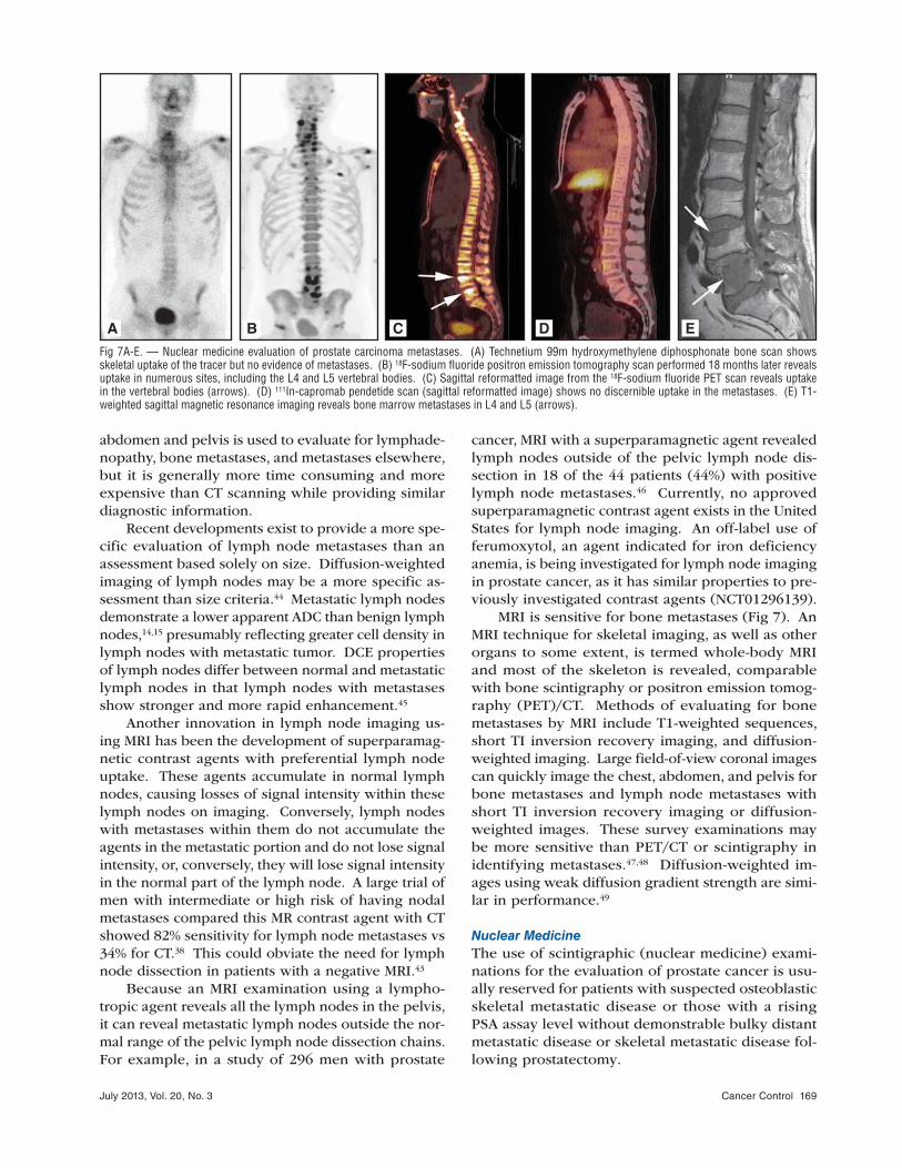
MRI is sensitive for bone metastases (Fig 7). An
MRI technique for skeletal imaging, as well as other
organs to some extent, is termed whole-body MRI
and most of the skeleton is revealed, comparable
with bone scintigraphy or positron emission tomog-
raphy (PET)/CT. Methods of evaluating for bone
metastases by MRI include T1-weighted sequences,
short TI inversion recovery imaging, and diffusion-
weighted imaging. Large field-of-view coronal images
can quickly image the chest, abdomen, and pelvis for
bone metastases and lymph node metastases with
short TI inversion recovery imaging or diffusion-
weighted images. These survey examinations may
be more sensitive than PET/CT or scintigraphy in
identifying metastases.47,48 Diffusion-weighted im-
ages using weak diffusion gradient strength are simi-
lar in performance.49
Nuclear Medicine
The use of scintigraphic (nuclear medicine) exami-
nations for the evaluation of prostate cancer is usu-
ally reserved for patients with suspected osteoblastic
skeletal metastatic disease or those with a rising
PSA assay level without demonstrable bulky distant
metastatic disease or skeletal metastatic disease fol-
lowing prostatectomy.
Fig 7A-E. — Nuclear medicine evaluation of prostate carcinoma metastases. (A) Technetium 99m hydroxymethylene diphosphonate bone scan shows skeletal uptake of the tracer but no evidence of metastases. (B) 18F-sodium fluoride positron emission tomography scan performed 18 months later reveals uptake in numerous sites, including the L4 and L5 vertebral bodies. (C) Sagittal reformatted image from the 18F-sodium fluoride PET scan reveals uptake in the vertebral bodies (arrows). (D) 111In-capromab pendetide scan (sagittal reformatted image) shows no discernible uptake in the metastases. (E) T1-weighted sagittal magnetic resonance imaging reveals bone marrow metastases in L4 and L5 (arrows).
A B C D E

170 Cancer Control July 2013, Vol. 20, No. 3
In patients with suspected osteoblastic skeletal
metastatic disease, nuclear medicine currently offers
the option of gamma emitters, such as technetium-
99m (99mTc) linked to a radioligand — in this case, a
“bone-seeking” agent, such as methylene diphospho-
nate or hydroxymethylene diphosphonate. Imaging of
such radiotracers is performed using gamma cameras
in the single detector (head), dual-head, or triple-head
configuration. In the last 10 years, hybrid nuclear
medicine–CT camera systems have become available
that combine a diagnostic quality CT gantry with a
dual-headed gamma camera system. These systems
provide the option of obtaining a nuclear medicine
tomographic examination (single-photon emission
computed tomography [SPECT]) registered to a co-
acquired low (nondiagnostic) or high radiographic
dose (diagnostic) CT for localization purposes.
The major drawback of the radiotracers used for
skeletal scintigraphy (bone scan) is low specificity.
Inflammatory processes such as degenerative joint
disease and osteoarthritis can demonstrate signifi-
cant uptake. Certainly other active inflammatory pro-
cesses, such as autoimmune processes, can also have
increased uptake. Infectious or reparative processes
involving the bone can also have increased uptake.
The above-discussed radiotracer primarily depos-
its within the cortical bone through chemoabsorption.
This radiotracer deposits in areas of bone deposition;
therefore, these examinations demonstrate areas of
osteoblastic response to tumoral deposits in bone,
not deposition within the tumor tissue. Sensitivity for
detection of osteoblastic activity related to metastatic
prostate cancer can reach a rate of 95% in patients
with PSA levels above 20 ng/mL.50-52 In cases where
a rising PSA level is present following prostatectomy
and negative skeletal scintigraphic findings, the likeli-
hood of soft-tissue metastatic disease increases.
Skeletal scintigraphy is mainly reserved for pa-
tients with high-risk cancer, elevated serum alkaline
phosphatase levels, bone pain, or equivocal osseous
lesions on other imaging modalities.53,54
Prior studies have demonstrated the relationship
of increased incidence of skeletal metastatic disease
with elevated PSA levels.50-52 The flare phenomenon,
which refers to evidence of increasing osteoblastic
metastatic disease (worsening of bone scan appear-
ance) following therapy, with decreasing symptom-
atology has been previously described.54-58 It is vis-
ible in patients with prostate carcinoma metastatic
to the skeleton, observable in 20% to 25% of scans if
obtained soon after therapy has been instituted.56-58
Because of this paradoxical worsening of scintigraph-
ic appearance in patients with favorably responding
metastatic disease, skeletal scintigraphy is not the
only test for the evaluation of such patients. Bone
scans are useful for the evaluation of patients with
diagnosed prostate cancer with a rising or elevated
PSA level, patients with normal PSA levels following
hormonal therapy, or patients who have a normal PSA
level but are symptomatic.
Bone scans using planar and tomographic tech-
niques have adequate sensitivity and specificity rates
and have been the main tool utilized for many years.
However, tumor detection using novel radiotracers and
newer imaging techniques may prove advantageous in
the future. Novel radiotracers such as positron emit-
ters (eg, 18F-fluoride [NaF]), as well as PET or PET/CT
and PET/MRI techniques, may prove indispensable in
the evaluation of metastatic skeletal disease. In malig-
nant bone lesions, NaF reflects the increase in regional
blood flow and skeletal turnover associated with meta-
static osteoblastic deposits (Fig 7). NaF PET/CT has
been shown to be more sensitive for the detection of
metastases than 99mTc methylene diphosphonate bone
scans. The use of hybrid imaging techniques such
as PET/CT reduces the risk of false-positive findings
and improves specificity of abnormalities detected by
virtue of the imaging capabilities for lesion-by-lesion
characterization using CT (Fig 7).59
In a recent study that reviewed the use of 99mTc
methylene diphosphonate and NaF, a direct compari-
son was made using whole-body planar images and
tomographic (SPECT) bone scans as well as PET alone
and PET/CT NaF for the detection and characterization
of skeletal metastases in 44 patients with high-risk
prostate cancer.59 In this study, whole-body planar
imaging had sensitivity and specificity rates of 57%.
Conversely, whole-body planar plus SPECT was 78%
sensitive and 67% specific. 18F PET was 100% sensi-
tive and 62% specific, whereas 18F PET/CT was 100%
sensitive and 100% specific. Conversely, fludeoxyglu-
cose 18F PET for the detection of prostate carcinoma
skeletal metastases had a low yield with a sensitivity
of less than 20% compared with skeletal scintigraphy.60
Soft-Tissue/Lymph Node Metastatic Disease Evaluation
Molecular imaging techniques, such as the use of 111In capromab pendetide, allow for the evaluation
of suspected metastatic prostatic carcinoma outside
of the prostate bed, particularly the pelvic sidewall
and retroperitoneal lymph nodes. 111In capromab
pendetide consists of a murine monoclonal antibody
(7D11-C5.3) covalently jointed to a liner-chelator mol-
ecule. It is directed against the intracellular domain of
prostate-specific membrane antigen (PSMA) expressed
on the surface of prostate cancer metastases as well
as the normal prostatic tissue. It is also expressed in
other tissue types, such as the kidney, liver, bowel,
and brain. PSMA is upregulated in hormone-resistant
states and in metastatic disease, which is why it is
used for the detection of extraprostatic, bone-scan-
negative metastatic disease.

July 2013, Vol. 20, No. 3 Cancer Control 171
These molecular imaging techniques involve mul-
tiday examinations, and time is allowed for the ra-
diopharmaceutical to circulate in the blood pool and
migrate to the targeted metastatic foci. Usual imaging
protocols call for dosing on day 0, with imaging per-
formed on day 4, day 5, or both (96 and/or 120 hours
after injection). Imaging techniques available include
the use of planar gamma camera images, evaluating the
whole body from an anterior and posterior projection,
as well as tomographic images obtained using SPECT
or SPECT/CT camera systems. However, with all the
advanced imaging systems used, the sensitivity rate
ranges from 62% to 75% (Fig 7).61-63 This technique
is also limited by nonspecific uptake in bowel, vas-
culature, bone marrow, and normal prostatic tissues,
and it has a low sensitivity rate for low-tumor-burden
prostate fossa disease as well as small metastases.
Novel Radiotracer
Novel radiotracers in different stages of research eval-
uation include positron emitters such as 11C-acetate
and 11C-choline.
Malignant transformation within the prostate cells
leads to a change in intracellular metabolic processes,
which result in a change from citrate-producing nor-
mal cells to citrate-oxidizing neoplastic cells, thereby
leading to an increased acetate turnover with increased
expression of fatty acid synthase. Hence, increased
anabolic metabolism is associated with cytoplasmic
lipid synthesis. Imaging with acetate-linked radiotrac-
ers benefits from this process.
Early studies using dedicated PET imaging sys-
tems comparing the use of fludeoxyglucose 18F and 11C-acetate for the evaluation of recurrent prostate
carcinoma showed promise when used to detect
metastatic disease over the use of fludeoxyglucose 18F alone, which had limited sensitivity and specific-
ity rates (Fig 8). However, a study by Oyama et al64
showed a statistical difference in the detection of re-
current disease in patients with elevated PSA levels
above 3 ng/mL but limited results in patients with
PSA levels of 3 ng/mL or below. 11C-acetate PET was
shown to have a sensitivity rate of 59% compared with
a sensitivity rate of 17% for fludeoxyglucose 18F PET.
Fig 8A-B. — Comparison of fludeoxyglucose 18F and 18F-acetate positron emission tomography scans. (A) Normal scan. (B) 18F-acetate examination of the same patient reveals prostate carcinoma metastases within the pelvic nodal stations. From Yu EY, Muzi M, Hackenbracht JA, et al. C11-acetate and F-18 FDG PET for men with prostate cancer bone metastases: relative findings and response to therapy. Clin Nucl Med. 2011;36(3):192-198. Reprinted with permission by Wolters Kluwer Health.
A
Pre-RX
PSA 432
B
Post-RX
PSA < 1
FDG PET Acetate PETBone Scan
172 Cancer Control July 2013, Vol. 20, No. 3
Of note, this study used stand-alone PET imaging
systems. In one report, Fricke et al65 noted a higher
rate of overall lesion detection with acetate (83%)
compared with fludeoxyglucose 18F (75%).
In a recent pilot study by Yu et al66 involving 8
participants, conventional skeletal scintigraphy was
compared with 99mTc methydiphosphonate, fludeoxy-
glucose 18F, and 11C-acetate in the detection of skel-
etal metastases and follow-up after therapy. Acetate
PET generally detected more metastases and had a
higher ratio of tumor-to-normal uptake. These results
indicate that acetate PET holds promise for response
assessment of prostate cancer skeletal metastases; it
was also complementary to fludeoxyglucose 18F in the
detection of bone metastasis.11C- and 18F-labeled choline derivatives have been
used to detect primary or relapsing prostate carci-
noma based on increased levels of phosphorylcholine,
upregulated enzymes of choline metabolism, choline
kinase with increased phosphatidylcholine turn-
over, and the metabolic flux of radiolabeled choline
through phospholipid biosynthesis and degradation
in prostate carcinoma.67 This uptake is unrelated to
proliferation (Ki67). Several authors68-70 have shown
that 11C-choline is taken up by prostate carcinoma and
its nodal and distant metastases (Fig 9).
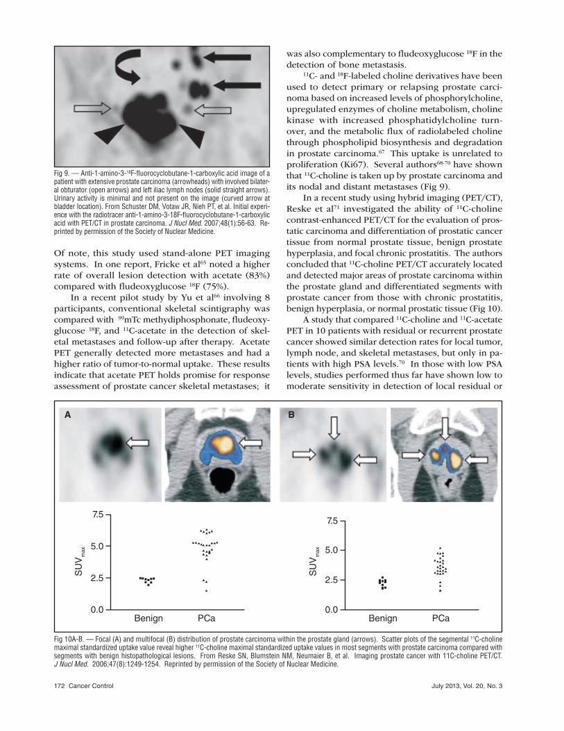
In a recent study using hybrid imaging (PET/CT),
Reske et al71 investigated the ability of 11C-choline
contrast-enhanced PET/CT for the evaluation of pros-
tatic carcinoma and differentiation of prostatic cancer
tissue from normal prostate tissue, benign prostate
hyperplasia, and focal chronic prostatitis. The authors
concluded that 11C-choline PET/CT accurately located
and detected major areas of prostate carcinoma within
the prostate gland and differentiated segments with
prostate cancer from those with chronic prostatitis,
benign hyperplasia, or normal prostatic tissue (Fig 10).
A study that compared 11C-choline and 11C-acetate
PET in 10 patients with residual or recurrent prostate
cancer showed similar detection rates for local tumor,
lymph node, and skeletal metastases, but only in pa-
tients with high PSA levels.70 In those with low PSA
levels, studies performed thus far have shown low to
moderate sensitivity in detection of local residual or
Fig 9. — Anti-1-amino-3-18F-fluorocyclobutane-1-carboxylic acid image of a patient with extensive prostate carcinoma (arrowheads) with involved bilater-al obturator (open arrows) and left iliac lymph nodes (solid straight arrows). Urinary activity is minimal and not present on the image (curved arrow at bladder location). From Schuster DM, Votaw JR, Nieh PT, et al. Initial experi-ence with the radiotracer anti-1-amino-3-18F-fluorocyclobutane-1-carboxylic acid with PET/CT in prostate carcinoma. J Nucl Med. 2007;48(1):56-63. Re-printed by permission of the Society of Nuclear Medicine.
Fig 10A-B. — Focal (A) and multifocal (B) distribution of prostate carcinoma within the prostate gland (arrows). Scatter plots of the segmental 11C-choline maximal standardized uptake value reveal higher 11C-choline maximal standardized uptake values in most segments with prostate carcinoma compared with segments with benign histopathological lesions. From Reske SN, Blumstein NM, Neumaier B, et al. Imaging prostate cancer with 11C-choline PET/CT. J Nucl Med. 2006;47(8):1249-1254. Reprinted by permission of the Society of Nuclear Medicine.
7.5
5.0
0.0Benign
SU
Vm
ax
PCa
2.5
7.5
5.0
0.0Benign
SU
Vm
ax
PCa
2.5
A B
July 2013, Vol. 20, No. 3 Cancer Control 173
recurrent tumor. Similarly, these studies have shown
moderate sensitivity in the detection of distant meta-
static disease.72,73 In a recent study, Vees et al74 evalu-
ated 20 patients with early-stage prostate carcinoma
staging after prostatectomy with low PSA levels (< 1
ng/mL) for the presence of local or metastatic prostate
carcinoma with 18F-choline, 11C-acetate, or both. Two
of the 20 patients were evaluated with both radiotrac-
ers. Both radiopharmaceuticals detected the presence
of neoplastic disease in 50% of the participants.
Other radiopharmaceuticals being evaluated in-
clude 11C-methionine, 18F-fluoro-5-dihydrotestosterone,
and anti-1-amino-3-18F-fluorocyclobutane-1-carboxylic
acid (Fig 9).75
Evaluation of the Prostate or Surgical Site Following TherapyMRI and sonography are two methods used to evalu-
ate for local recurrence following local therapy or
prostatectomy. PET and CT scanning are not accurate
methods for identifying recurrent tumor.76,77
Ultrasonography
Following prostatectomy in the surgical bed, transrec-
tal ultrasonography may detect recurrence of pros-
tate carcinoma.78 Recurrences appear as ill-defined
hypoechoic lesions in the bladder neck or at the
anastomosis.79 Power Doppler imaging and contrast
enhancement may improve the sensitivity of ultraso-
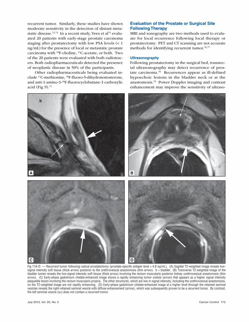
Fig 11A-D. — Recurrent tumor following radical prostatectomy (prostate-specific antigen level = 4.8 ng/mL). (A) Sagittal T2-weighted image reveals low-signal intensity soft tissue (thick arrow) posterior to the urethrovesical anastomosis (thin arrow). b = bladder. (B) Transverse T2-weighted image of the bladder lumen reveals the low-signal intensity soft tissue (thick arrow) involving the rectum muscularis posterior kidney urethrovesical anastomosis (thin arrow). (C) Early-phase gadolinium chelate-enhanced image shows a rapidly enhancing tumor nodule (arrow) that appears as a higher signal intensity plaquelike lesion involving the rectum muscularis propria. The other structures, which are low in signal intensity, including the urethrovesical anastomosis, on the T2-weighted image are not rapidly enhancing. (D) Early-phase gadolinium chelate-enhanced image at a higher level through the retained seminal vesicles reveals the right-retained seminal vesicle with diffuse enhancement (arrow), which was subsequently proven to be a recurrent tumor. By contrast, the left seminal vesicle (sv) does not contain a recurrent tumor.
A B
C D

174 Cancer Control July 2013, Vol. 20, No. 3
nography in identifying recurrent tumor.80 However,
biopsies of the surgical site are still more sensitive
than ultrasonographic imaging.81
Ultrasonography and Doppler imaging of the
prostate following local therapy (eg, cryosurgery,
Fig 12A-C. — Recurrent tumor with magnetic resonance imaging follow-ing brachytherapy with radiation seed implants (prostate-specific antigen level = 1.2 ng/mL). (A) T1-weighted image with a rectal coil through the prostate that has numerous low-signal intensity foci representing an artifact arising from the individual radiation seeds. (B) T2-weighted image reveals diffuse low-signal intensity throughout the prostate with poor discrimina-tion between the central gland and peripheral zone. No definite tumor can be identified, although the central gland is slightly higher in signal intensity. The radiation seeds identified are less apparent because fewer artifacts are visible on the image. (C) An early dynamic contrast-enhanced study reveals focal enhancement within the central gland (between arrows) that was sub-sequently demonstrated to represent recurrent tumor.
A
B
C
brachytherapy) may have little role in identifying re-
sidual tumor. Contrast-enhanced ultrasonography
may have increased sensitivity and specificity and
may help in evaluating the degree of ablation in local
therapies by distinguishing between vascularized and
nonvascularized (ie, ablated) tissue.82
Magnetic Resonance Imaging
Following total prostatectomy, MRI can detect recur-
rence in the prostatic bed. The usual appearance
after radical prostatectomy includes a descended
bladder neck in the prostatectomy space with a small
amount of fibrotic tissue around the urethrovesical
anastomosis. Linear scarring may be present at the
expected former site of the seminal vesicles.83 T2-
weighted images can detect recurrence as a higher
signal intensity nodule compared with the surround-
ing fibrosis and smooth muscle of the bladder neck,
urethra, and urethrovesical anastomosis.84 Sensitivity
rates of 95% and specificity rates of 100% have been
reported for MRI.84 DCE images may increase sen-
sitivity and identify enhancing nodules of recurrent
tumor (Fig 11).85,86
Following hormonal deprivation therapy, the
prostate becomes smaller in size with diminished
signal intensity of the peripheral zone, so tumors are
more difficult to localize on T2-weighted images.87 As
a result of apoptosis, androgen-deprivation therapies
may cause atrophy of the prostatic epithelium.26
The histologic changes in the prostate caused by
external beam irradiation include glandular atrophy,
inflammation, prostate shrinkage, and fibrosis.88 After
external beam radiation therapy, the peripheral zone
generally loses signal intensity on T2-weighted imag-
ing,89 which can effectively render most carcinomas
difficult or impossible to identify on standard MRI
following radiation therapy. Similar effects are vis-
ible using brachytherapy with radiation seeds. Diffu-
sion-weighted imaging or DCE imaging may increase
sensitivity in the irradiated prostate for the presence
of cancer.90 On diffusion-weighted or ADC imaging,
carcinoma generally appears as a more restricted dif-
fusion than the remainder of the prostate gland. MRI
may detect residual or recurrent tumor in patients
treated with high-intensity focused ultrasonography.91
DCE images are more sensitive than T2-weighted im-
ages to tumor recurrence (Fig 12).91,92
Similar to the effects on T2-weighted images
with external beam therapy, spectroscopic results
also change with radiation. The citrate levels tend
to decrease after radiation in nontumorous prostate
and choline levels increase, causing confusion with a
tumor. Nonetheless, spectroscopy can identify some
patients with residual tumor after radiation, although
reported accuracies are not as high as those seen in
patients with untreated prostate cancer.27,93

July 2013, Vol. 20, No. 3 Cancer Control 175
ConclusionsLocal staging of the prostate tumor is limited in ultra-
sonography and magnetic resonance imaging; both
techniques provide suboptimal sensitivity and speci-
ficity for tumor identification and capsular penetra-
tion. Additional magnetic resonance imaging tech-
niques such as dynamic contrast-enhanced imaging,
diffusion imaging, and spectroscopy may provide in-
cremental benefit; however, none of these are highly
sensitive. Computed tomography and bone scans are
used to assess metastatic disease, but both techniques
are limited by the poor sensitivity of lymph node size
as a criterion for detecting metastases.
Accepted methodologies for evaluating metastatic
prostate carcinoma to the skeletal system and soft
tissues include skeletal scintigraphy (bone scan) and
monoclonal antibody imaging that targets prostate-
specific membrane antigen. However, novel imaging
techniques such as hybrid imaging devices in the
form of single-photon emission computed tomogra-
phy/computed tomographic gamma cameras, posi-
tron emission/computed tomographic cameras, and,
in the near future, positron emission tomography/
magnetic resonance imaging combined with tumor-
specific imaging radiotracers may significantly and
favorably affect tumor staging, the accuracy of therapy
response, and patient care.
References
1. National Comprehensive Cancer Network. NCCN Clinical Practice Guidelines in Oncology: Prostate Cancer. Version 2.2013. http://www.nccn.org/professionals/physician_gls/pdf/prostate.pdf. Accessed March 15, 2013. 2. Plawker MW, Fleisher JM, Vapnek EM, et al. Current trends in pros-tate cancer diagnosis and staging among United States urologists. J Urol. 1997;158(5):1853-1858. 3. Schooler J. Unpublished results hide the decline effect. Nature. 2011; 470(7335):437. 4. Ioannidis JP. Why most published research findings are false. PLoS Med. 2005;2(8):e124. 5. Xiao G, Bloch BN, Chappelow J, et al. Determining histology-MRI slice correspondences for defining MRI-based disease signatures of prostate cancer. Comp Med Imaging Graph. 2011;35(7-8):568-578. 6. Turkbey B, Pinto PA, Mani H, et al. Prostate cancer: value of multipa-rametric MR imaging at 3 T for detection--histopathologic correlation. Radiol-ogy. 2010;255(1):89-99. 7. Turkbey B, Pinto PA, Choyke PL. Imaging techniques for prostate cancer: implications for focal therapy. Nat Rev Urol. 2009;6(4):191-203. 8. Heijmink S, van Moerkerk H, Kiemeney L, et al. A comparison of the diagnostic performance of systematic versus ultrasound-guided biopsies of prostate cancer. Eur Radiol. 2006;16(4):927-938. 9. Halpern EJ, Frauscher F, Strup SE, et al. Prostate: high-frequency Doppler US imaging for cancer detection. Radiology. 2002;225(1):71-77. 10. Sehgal CM, Arger PH, Holzer AC, et al. Correlation between Dop-pler vascular density and PSA response to radiation therapy in patients with localized prostate carcinoma. Acad Radiol. 2003;10(4):366-372. 11. Cornud F, Hamida K, Flam T, et al. Endorectal color doppler sonog-raphy and endorectal MR imaging features of nonpalpable prostate cancer: correlation with radical prostatectomy findings. AJR Am J Roentgenol. 2000;175(4):1161-1168. 12. Halpern EJ. Contrast-enhanced ultrasound imaging of prostate can-cer. Rev Urol. 2006;(8 suppl 1):S29-S37. 13. Halpern EJ, Ramey JR, Strup SE, et al. Detection of prostate carci-noma with contrast-enhanced sonography using intermittent harmonic imag-ing. Cancer. 2005;104(11):2373-2383. 14. Pelzer A, Bektic J, Berger AP, et al. Prostate cancer detection in men with prostate specific antigen 4 to 10 ng/ml using a combined approach of contrast enhanced color Doppler targeted and systematic biopsy. J Urol. 2005;173(6):1926-1929.
15. Halpern EJ, Rosenberg M, Gomella LG. Prostate cancer: contrast-enhanced us for detection. Radiology. 2001;219(1):219-225. 16. Mitterberger M, Pinggera GM, Horninger W, et al. Comparison of contrast enhanced color Doppler targeted biopsy to conventional systematic biopsy: impact on Gleason score. J Urol. 2007;178(2):464-468. 17. Salomon G, Köllerman J, Thederan I, et al. Evaluation of prostate cancer detection with ultrasound real-time elastography: a comparison with step section pathological analysis after radical prostatectomy. Eur Urol. 2008;54(6):1354-1362. 1 8. Curiel L, Souchon R, Rouviére O, et al. Elastography for the follow-up of high-intensity focused ultrasound prostate cancer treatment: initial com-parison with MRI. Ultrasound Med Biol. 2005;31(11):1461-1468. 19 . Schnall MD, Imai Y, Tomaszewski J, et al. Prostate cancer: local stag-ing with endorectal surface coil MR imaging. Radiology. 1991;178(3):797-802. 20 . Villeirs GM, Verstraete KL, De Neve WJ, et al. Magnetic resonance imaging anatomy of the prostate and periprostatic area: a guide for radio-therapists. Radiother Oncol. 2005;76(1):99-106. 21 . McLaughlin PW, Troyer S, Berri S, et al. Functional anatomy of the prostate: implications for treatment planning. Int J Radiat Oncol Biol Phys. 2005;63(2):479-491. 22 . Rosenkrantz AB, Kopec M, Kong X, et al. Prostate cancer vs. post-biopsy hemorrhage: diagnosis with T2- and diffusion-weighted imaging. J Magn Reson Imaging. 2010;31(6):1387-1394. 23 . Schlemmer HP, Merkle J, Grobholz R, et al. Can pre-operative con-trast-enhanced dynamic MR imaging for prostate cancer predict microvessel density in prostatectomy specimens? Eur Radiol. 2004;14(2):309-317. 24 . Outwater EK, Petersen RO, Siegelman ES, et al. Prostate carcinoma: assessment of diagnostic criteria for capsular penetration on endorectal coil MR images. Radiology. 1994;193(2):333-339. 25 . Sala E, Akin O, Moskowitz CS, et al. Endorectal MR imaging in the evaluation of seminal vesicle invasion: diagnostic accuracy and multivariate feature analysis. Radiology. 2006;238(3):929-937. 26 . Petraki CD, Sfikas CP. Histopathological changes induced by thera-pies in the benign prostate and prostate adenocarcinoma. Histol Histopathol. 2007;22(1):107-118. 27 . Pucar D, Shukla-Dave A, Hricak H, et al. Prostate cancer: correlation of MR imaging and MR spectroscopy with pathologic findings after radiation therapy-initial experience. Radiology. 2005;236(2):545-553. 28. Testa C, Schiavina R, Lodi R, et al. Prostate cancer: sextant localiza-tion with MR imaging, MR spectroscopy, and 11C-choline PET/CT. Radiol-ogy. 2007;244(3):797-806. 2 9. Weinreb JC, Blume JD, Coakley FV, et al. Prostate cancer: sextant localization at MR imaging and MR spectroscopic imaging before prosta-tectomy. Results of ACRIN prospective multi-institutional clinicopathologic study. Radiology. 2009;251(1):122-133. 30 . Sonnad SS, Langlotz CP, Schwartz JS. Accuracy of MR imaging for staging prostate cancer: a meta-analysis to examine the effect of technologic change. Acad Radiol. 2001;8(2):149-157. 31. Quinn SF, Franzini DA, Demlow TA, et al. MR imaging of prostate cancer with an endorectal surface coil technique: correlation with whole-mount specimens. Radiology. 1994;190(2):323-327. 32. Bloch BN, Furman-Haran E, Helbich TH, et al. Prostate cancer: ac-curate determination of extracapsular extension with high-spatial-resolution dynamic contrast-enhanced and T2-weighted MR imaging. Initial results. Radiology. 2007;245(1):176-185. 33 . Hoeks CM, Barentsz JO, Hambrock T, et al. Prostate cancer: multi-parametric MR imaging for detection, localization, and staging. Radiology. 2011;261(1):46-66. 34 . Yu KK, Hricak H, Alagappan R, et al. Detection of extracapsular ex-tension of prostate carcinoma with endorectal and phased-array coil MR im-aging: multivariate feature analysis. Radiology. 1997;202(3):697-702. 35 . Cornud F, Flam T, Chauveinc L, et al. Extraprostatic spread of clini-cally localized prostate cancer: factors predictive of pT3 tumor and of positive endorectal MR imaging examination results. Radiology. 2002;224(1):203-210. 36 . Mullerad M, Hricak H, Wang L, et al. Prostate cancer: detection of extracapsular extension by genitourinary and general body radiologists at MR imaging. Radiology. 2004;232(1):140-146. 37 . Heijmink SW, Fütterer JJ, Hambrock T, et al. Prostate cancer: body-array versus endorectal coil MR imaging at 3 T. Comparison of image quality, localization, and staging performance. Radiology. 2007;244(1):184-195. 38. Wang L, Mullerad M, Chen HN, et al. Prostate cancer: incremental value of endorectal MR imaging findings for prediction of extracapsular ex-tension. Radiology. 2004;232(1):133-139. 39. Wang L, Hricak H, Kattan MW, et al. Prediction of organ-confined prostate cancer: incremental value of MR imaging and MR spectroscopic imaging to staging nomograms. Radiology. 2006;238(2):597-603. 40. Roethke MC, Lichy MP, Jurgschat L, et al. Tumor size dependent detection rate of endorectal MRI of prostate cancer--a histopathologic cor-relation with whole-mount sections in 70 patients with prostate cancer. Eur J Radiol. 2011;79(2):189-195. 41. Tiguert R, Gheiler EL, Tefilli MV, et al. Lymph node size does not correlate with the presence of prostate cancer metastasis. Urology. 1999;53(2):367-371.

176 Cancer Control July 2013, Vol. 20, No. 3
42. Hövels AM, Heesakkers RA, Adang EM, et al. The diagnostic ac-curacy of CT and MRI in the staging of pelvic lymph nodes in patients with prostate cancer: a meta-analysis. Clin Radiol. 2008;63(4):387-395. 43. Heesakkers RA, Hövels AM, Jager GJ, et al. MRI with a lymph-node-specific contrast agent as an alternative to CT scan and lymph-node dissec-tion in patients with prostate cancer: a prospective multicohort study. Lancet Oncol. 2008;9(9):850-856. 44. E iber M, Beer AJ, Holzapfel K, et al. Preliminary results for charac-terization of pelvic lymph nodes in patients with prostate cancer by diffusion-weighted MR-imaging. Invest Radiol. 2010;45(1):15-23. 45. J ager GJ, Barentsz JO, Oosterhof GO, et al. Pelvic adenopathy in prostatic and urinary bladder carcinoma: MR imaging with a three-dimen-sional TI-weighted magnetization-prepared-rapid gradient-echo sequence. AJR Am J Roentgenol. 1996;167(6):1503-1507. 46. H eesakkers RA, Jager GJ, Hövels AM, et al. Prostate cancer: detec-tion of lymph node metastases outside the routine surgical area with feru-moxtran-10 enhanced MR imaging. Radiology. 2009;251(2):408-414. 47. La uenstein TC, Goehde SC, Herborn CU, et al. Whole-body MR imag-ing: evaluation of patients for metastases. Radiology. 2004;233(1):139-148. 48. Sc hmidt G, Schoenberg S, Schmid R, et al. Screening for bone me-tastases: whole-body MRI using a 32-channel system versus dual-modality PET-CT. Eur Radiol. 2007;17(4):939-949. 49. Lu boldt W, Küfer R, Blumstein N, et al. Prostate carcinoma: diffusion-weighted imaging as potential alternative to conventional MR and 11C-choline PET/CT for detection of bone metastases. Radiology. 2008;249(3):1017-1025. 50. Ja cobson AF. Association of prostate-specific antibody levels and patterns of benign and malignant uptake detected on bone scintigraphy in patients with newly diagnosed prostate cancer. Nucl Med Commun. 2000;21:617-622. 51. Mc Arthur C, McLaughlin G, Meddings RN. Changing the referral cri-teria for bone scan in newly diagnosed prostate cancer patients. Br J Radiol. 2012;85(1012):390-394. 52. Huncharek M, Muscat J. Serum prostate-specific antigen as a pre-dictor of radiographic staging studies in newly diagnosed prostate cancer. Cancer Invest. 1995;13(1):31-35. 53. Abuzallouf S, Dayes I, Lukka H. Baseline staging of newly diagnosed prostate cancer: a summary of the literature. J Urol. 2004;171(6 part 1): 2122-2127. 54. Wym enga LF, Boomsma JH, Groenier K, et al. Routine bone scans in patients with prostate cancer related to serum prostate-specific antigen and alkaline phosphatase. BJU Int. 2001;88(3):226-230. 55. Yap BK, Choo R, Deboer G, et al. Are serial bone scans useful for the follow-up of clinically localized, low to intermediate grade prostate cancer managed with watchful observation alone? BJU Int. 2003;91(7):613-617. 56. Pollen JJ, Witztum KF, Ashburn WL. The flare phenomenon on ra-dionuclide bone scan in metastatic prostate cancer. AJR Am J Roentgenol. 1984;142(4):773-776. 57. Love C, Din AS, Tomas MB, et al. Radionuclide bone imaging: an illustrative review. Radiographics. 2003;23(2):341-358. 58. Cook GJ, Venkitaraman R, Sohaib AS, et al. The diagnostic utility of the flare phenomenon on bone scintigraphy in staging prostate cancer. Eur J Nucl Med Mol Imaging. 2011;38(1):7-13. 59. Ev en-Sapir E, Metser U, Mishani E, et al. The detection of bone me-tastases in patients with high-risk prostate cancer: 99mTc-MDP Planar bone scintigraphy, single- and multi-field-of-view SPECT, 18F-fluoride PET, and 18F-fluoride PET/CT. J Nucl Med. 2006;47(2):287-297. 60. Yeh SD, Imbriaco M, Larson SM, et al. Detection of bony metasta-ses of androgen-independent prostate cancer by PET-FDG. Nucl Med Biol. 1996;23(6):693-697. 61. Bra ssell SA, Rosner IL, McLeod DG. Update on magnetic resonance imaging, ProstaScint, and novel imaging in prostate cancer. Curr Opin Urol. 2005;15(3):163-166. 62. Sel tzer MA, Barbaric Z, Belldegrun A, et al. Comparison of helical computerized tomography, positron emission tomography and monoclonal antibody scans for evaluation of lymph node metastases in patients with prostate specific antigen relapse after treatment for localized prostate cancer. J Urol. 1999;162(4):1322-1328. 63. Lan ge PH. PROSTASCINT scan for staging prostate cancer. Urol-ogy. 2001;57(3):402-406. 64. Oya ma N, Miller TR, Dehdashti F, et al. 11C-acetate PET imaging of prostate cancer: detection of recurrent disease at PSA relapse. J Nucl Med. 2003;44(4):549-555. 65. Fri cke E, Machtens S, Hofmann M, et al. Positron emission tomog-raphy with 11C-acetate and 18-FDG in prostate cancer patients. Eur J Nucl Med Mol Imaging. 2003;30(4):607-611. 66. Yu EY, Muzi M, Hackenbracht JA, et al. C11-acetate and F-18 FDG PET for men with prostate cancer bone metastases: relative findings and response to therapy. Clin Nucl Med. 2011;36(3):192-198. 67. Rod riguez-González A, Ramirez de Molina A, Benitez-Rajal J, et al. Phospholipase D and choline kinase: their role in cancer development and their potential as drug targets. Prog Cell Cycle Res. 2003;5:191-201. 68. de J ong IJ, Pruim J, Elsinga PH, et al. Visualization of prostate cancer with 11C-choline positron emission tomography. Eur Urol. 2002;42(1):18-23.
69. Hara T, Kosaka N, Kishi H. PET imaging of prostate cancer using carbon-11-choline. J Nucl Med. 1998;39(6):990-995. 70. Kotz erke J, Prang J, Neumaier B, et al. Experience with carbon-11 choline positron emission tomography in prostate carcinoma. Eur J Nucl Med Mol Imaging. 2000;27(9):1415-1419. 71. Resk e SN, Blumstein NM, Neumaier B, et al. Imaging prostate can-cer with 11C-choline PET/CT. J Nucl Med. 2006;47(8):1249-1254. 72. de J ong IJ, Pruim J, Elsinga PH, et al. 11C-choline positron emission tomography for the evaluation after treatment of localized prostate cancer. Eur Urol. 2003;44(1):32-39. 73. Schm id DT, John H, Zweifel R, et al. Fluorocholine PET/CT in patients with prostate cancer: initial experience. Radiology. 2005;235(2):623-628. 74. Vees H, Buchegger F, Albrecht S, et al. 18F-choline and/or 11C-acetate positron emission tomography: detection of residual or progressive subclinical disease at very low prostate-specific antigen values (<1 ng/mL) after radical prostatectomy. BJU Int. 2007;99(6):1415-1420. 75. Schu ster DM, Votaw JR, Nieh PT, et al. Initial experience with the radiotracer anti-1-amino-3-18F-fluorocyclobutane-1-carboxylic acid with PET/CT in prostate carcinoma. J Nucl Med. 2007;48(1):56-63. 76. Hofe r C, Laubenbacher C, Block T, et al. Fluorine-18-fluorodeoxy-glucose positron emission tomography is useless for the detection of local recurrence after radical prostatectomy. Eur Urol. 1999;36(1):31-35. 77. Kräm er S, Görich J, Gottfried HW, et al. Sensitivity of computed to-mography in detecting local recurrence of prostatic carcinoma following radi-cal prostatectomy. Br J Radiol. 1997;70(838):995-999. 78. Abi-Aa d AS, Macfarlane MT, Stein A, et al. Detection of local recur-rence after radical prostatectomy by prostate specific antigen and transrectal ultrasound. J Urol. 1992;147(3 pt 2):952-955. 79. Levent is AK, Shariat SF, Slawin KM. Local recurrence after radical prostatectomy: correlation of US features with prostatic fossa biopsy findings. Radiology. 2001;219(2):432-439. 80. Tamsel S, Killi R, Apaydin E, et al. The potential value of power Dop-pler ultrasound imaging compared with grey-scale ultrasound findings in the diagnosis of local recurrence after radical prostatectomy. Clin Radiol. 2006;61(4):325-330. 81. Scatto ni V, Roscigno M, Raber M, et al. Multiple vesico-urethral biop-sies following radical prostatectomy: the predictive roles of TRUS, DRE, PSA and the pathological stage. Eur Urol. 2003;44(4):407-414. 82. Rouviè re O, Glas L, Girouin N, et al. Prostate cancer ablation with transrectal high-intensity focused ultrasound: assessment of tissue destruc-tion with contrast-enhanced US. Radiology. 2011;259(2):583-591. 83. De Viss chere PJ, De Meerleer GO, Fütterer JJ, et al. Role of MRI in follow-up after focal therapy for prostate carcinoma. AJR Am J Roentgenol. 2010;194(6):1427-1433. 84. Sella T, Schwartz LH, Swindle PW, et al. Suspected local recur-rence after radical prostatectomy: endorectal coil MR imaging. Radiology. 2004;231(2):379-385. 85. Sciarra A, Panebianco V, Salciccia S, et al. Role of dynamic contrast-enhanced magnetic resonance (MR) imaging and proton MR spectroscopic imaging in the detection of local recurrence after radical prostatectomy for prostate cancer. Eur Urol. 2008;54(3):589-600. 86. Casciani E, Polettini E, Carmenini E, et al. Endorectal and eynamic contrast-enhanced MRI for detection of local recurrence after radical prosta-tectomy. AJR Am J Roentgenol. 2008;190(5):1187-1192. 87. Chen M, Hricak H, Kalbhen CL, et al. Hormonal ablation of prostatic cancer: effects on prostate morphology, tumor detection, and staging by en-dorectal coil MR imaging. AJR Am J Roentgenol. 1996;166(5):1157-1163. 88. Sheaff MT, Baithun SI. Effects of radiation on the normal prostate gland. Histopathology. 1997;30(4):341-348. 89. Coakley FV, Hricak H, Wefer AE, et al. Brachytherapy for prostate cancer: endorectal MR imaging of local treatment-related changes. Radiol-ogy. 2001;219(3):817-821. 90. Kim CK, Park BK, Lee HM. Prediction of locally recurrent prostate cancer after radiation therapy: incremental value of 3T diffusion-weighted MRI. J Magn Reson Imaging. 2009;29(2):391-397. 91. Rouvière O, Girouin N, Glas L, et al. Prostate cancer transrectal HIFU ablation: detection of local recurrences using T2-weighted and dynamic con-trast-enhanced MRI. Eur Radiol. 2010;20(1):48-55. 92. Kim CK, P ark BK, Park W, et al. Prostate MR imaging at 3T using a phased-arrayed coil in predicting locally recurrent prostate cancer after radia-tion therapy: preliminary experience. Abdom Imaging. 2009;35(2):246-252. 93. Westphale n AC, Coakley FV, Roach M III, et al. Locally recurrent prostate cancer after external beam radiation therapy: diagnostic perfor-mance of 1.5-T endorectal MR imaging and MR spectroscopic imaging for detection. Radiology. 2010;256(2):485-492.