Embed Size (px)
Citation preview

V BS 122 Physiology II Class of 2012 121
15. FERTILIZATION For fertilization to take place, a viable ovum and fertile sperm need to get together. Several conditions must be met by sperm cells before they can be considered fertile (Fig. 15-1).
SPERM REQUIREMENTS
Maturation
First, the sperm must undergo a period of maturation during which the swimming ability of the sperm is developed. This process takes place while the sperm travels through the epididymis, a process which may take from 10 to 15 days and involves the elimination of the cytoplasmatic droplet.
Capacitation
The second requirement is capacitation or the capacity of the sperm to fertilize the ovum. This process is attained by the residence of the sperm in the uterine cavity and oviducts. The following changes are involved in this process:
- removal of seminal plasma which takes place mainly in the cervix. - removal of cell surface components by uterine secretions. - removal of cholesterol and glycosaminoglycans within the oviduct.
Experimentally, capacitation can be reversed by incubation of sperm cells with seminal plasma.
Acrosome reaction
The final step which sperm must undergo before being able to fertilize is the acrosome reaction. Many of the requirements for successful acrosome reaction are related to the process of capacitation, such as, removal of surface material, cholesterol and glycosaminoglycans. The actual acrosome reaction consists of fusion of the sperm plasma membrane with the outer acrosome membrane resulting in formation of vesicles in the anterior portion of the acrosome (Figs. 15-2, 15-3).
SPERM REQUIREMENTS • Maturation
o Swimming ability • Capacitation (removal of)
o Seminal plasma o Cell surface components o Cholesterol and glycosaminoglycans
• Acrosome reaction o Fusion of outer acrosome membraneo Vesiculation of anterior acrosome
Figure 15-1. Stages followed by sperm cells towards acquiring the ability to fertilize
Figure 15-3. Changes observed in the acrosome as a consequence of the acrosomal reaction
Figure 15-2. Component of the sperm head.

V BS 122 Physiology II Class of 2012 122
FERTILIZATION STEPS
High rates of fertilization are obtained if the spermatozoa are in the oviduct shortly before the ovum arrives at the fertilization site.
For fertilization to take place, three events must occur during the encounter of the spermatozoa and the oocyte (Fig. 15-4):
-The sperm cells must migrate between any present cumulus cells and corona radiate cells. - The sperm cell must attach to and penetrate the zona pellucida. - Finally, the plasma membranes of the ovum and the sperm must fuse.
Cumulus and corona radiata penetration
To migrate successfully between the cumulus cells, the spermatozoa are equipped with different types of enzymes which disperse the cumulus cells (Fig. 15-5).
Bull sperm is equipped with hyaluronidase while boar sperm contains arylsulphatase. Physical penetration of the zona pellucida is also supported by the swimming activity of the sperm.
Attachment and penetration of the zona pellucid
Attachment to the zona pellucida seems dependent on receptor sites on the zona’s surface. These receptors appear to be species specific. Penetration takes 5 to 15 minutes after attachment and requires acrosome activation to be successful. During the acrosome reaction, the sperm exposes a lysin zone containing several enzymes which digests its way to the vitelline membrane (Figs. 15-6, 15-9, 15-10).
FERTILIZATION • Cumulus penetration
o Forward swimming o Enzyme dispersion
• Zona pelluzida penetrationo Acrosome activation
• Membrane fusion o Equatorial attachment
Figure 15-4. Sequential steps required to take place before fertilization can occur
Figure 15-5. Recently ovulated ova with the corona radiata and cumulus cell attached
Figure 15-6. Steps followed from zona penetration to syngamy
V BS 122 Physiology II Class of 2012 123
Membrane fusion
Attachment to the vitelline membrane occurs after the zona pellucida has been penetrated. The attachment takes place by the equatorial portion of the sperm’s head. Part of the sperm’s membrane fuses with
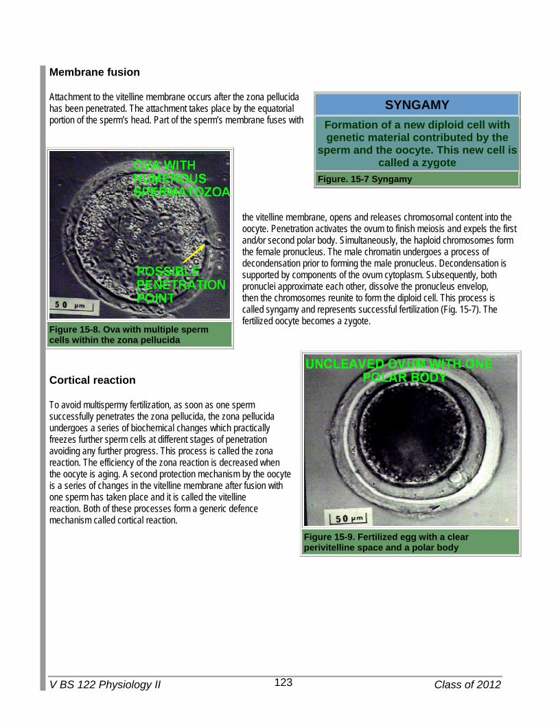
the vitelline membrane, opens and releases chromosomal content into the oocyte. Penetration activates the ovum to finish meiosis and expels the first and/or second polar body. Simultaneously, the haploid chromosomes form the female pronucleus. The male chromatin undergoes a process of decondensation prior to forming the male pronucleus. Decondensation is supported by components of the ovum cytoplasm. Subsequently, both pronuclei approximate each other, dissolve the pronucleus envelop, then the chromosomes reunite to form the diploid cell. This process is called syngamy and represents successful fertilization (Fig. 15-7). The fertilized oocyte becomes a zygote.
Cortical reaction
To avoid multispermy fertilization, as soon as one sperm successfully penetrates the zona pellucida, the zona pellucida undergoes a series of biochemical changes which practically freezes further sperm cells at different stages of penetration avoiding any further progress. This process is called the zona reaction. The efficiency of the zona reaction is decreased when the oocyte is aging. A second protection mechanism by the oocyte is a series of changes in the vitelline membrane after fusion with one sperm has taken place and it is called the vitelline reaction. Both of these processes form a generic defence mechanism called cortical reaction.
SYNGAMY Formation of a new diploid cell with genetic material contributed by the
sperm and the oocyte. This new cell is called a zygote
Figure. 15-7 Syngamy
Figure 15-8. Ova with multiple sperm cells within the zona pellucida
Figure 15-9. Fertilized egg with a clear perivitelline space and a polar body

V BS 122 Physiology II Class of 2012 124
CLEAVAGE AND IMPLANTATION
Cleavage
Immediately after fertilization, the zygote commences a process of cell division while travelling through the oviduct (Fig. 15-10).
This initial division process is called cleavage, because it takes place within the zona pellucida and it does not involve cell growth. Each daughter cell is called a blastomere (Fig. 15-14, 15-12) and they are supported by nutrients produced by uterine glands under the influence of P4. The uterine secretion is known as uterine milk or histotroph.
Once the embryo reaches 8-16 cell stage, (Fig. 15-10) it becomes a morula. At this stage, each blastomere secretes fluid and positions themselves in the outerpart of the spheric limitation imposed by the zona pellucida. The mitotic division of cleavage continues for about 4 to 8 days, a period in which the embryo becomes a blastocyst. At this stage, the embryo has a defined central cavity filled with fluid, called blastocoele, a peripheral single layer of cells called trophectoderm and a deeper concentration of cells in one area which is the inner cell mass (ICM) (Fig. 15-10).
Hatching
At this developmental point, the blastocyst starts expanding and is eventually released from the zona pellucida in a process called hatching. Subsequently, the blastocyst increases metabolic rate and commences hypertrophy and hyperplasia (Fig. 15-13). Depending on the species, the blastocyst enters the uterus from 46 to 160 hours after ovulation and may hatch between 6 to 12 days after fertilization.
Figure 15-10. Timeline for the displacement and cleavage of a blastocyst in the oviduct of a sow
Figure 15-11. Fertilized egg after the first cleavage showing two blastomeres
Figure 15-12. Four blastomere blastocyst

V BS 122 Physiology II Class of 2012 125
Positioning in the uterus
The place of attachment in the uterus varies with the species. In general, species which produce a single animal will have it implanted in the uterine horn at the same site of the ovary which ovulated as is the case in cattle and sheep. Horses tend to alternate attachment sites at each pregnancy, regardless the side of ovulation. When more than one ovulation occurs in an ovary, there is more chance of migration to the opposite horn by one of the embryos as in ewes. Litter bearing species have the embryos evenly distributed throughout both uterine horns implying significant transuterine embryonic migration. This phenomenon has been demonstrated in cats and pigs and serves the purpose of allocating even resources for fetal growth.
Initial attachment in all domestic species is in the endometrium of the uterine lumen. Before attachment commences there is a rapid growth of the tissues which will develop into the placenta, transforming a spherical blastocyst (Fig. 15-14) into a filamentous blastocyst (Fig. 15-15).
Apparently, in some species this is the mechanical form by which each conceptus attempts to secure as much uterine space as possible.
Figure 15-13. Hatched blastocyst after significant growth
Figure 15-14. Twelve day old pig embryo with a clear inner cell mass just before elongation
Figure 15-15. Filamentous blastocyst