Embed Size (px)
Citation preview
2 D to 3 D planning in Carcinoma of Oesophagus
Dr. R. K. VyasDirector
Gujarat Cancer & Research Institute, Ahmedabad, Gujarat


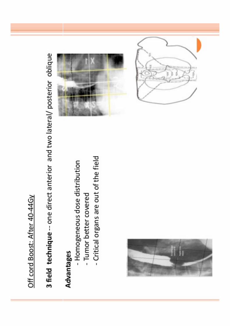
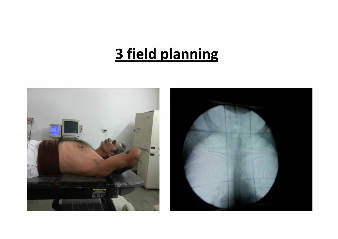
3 field planning
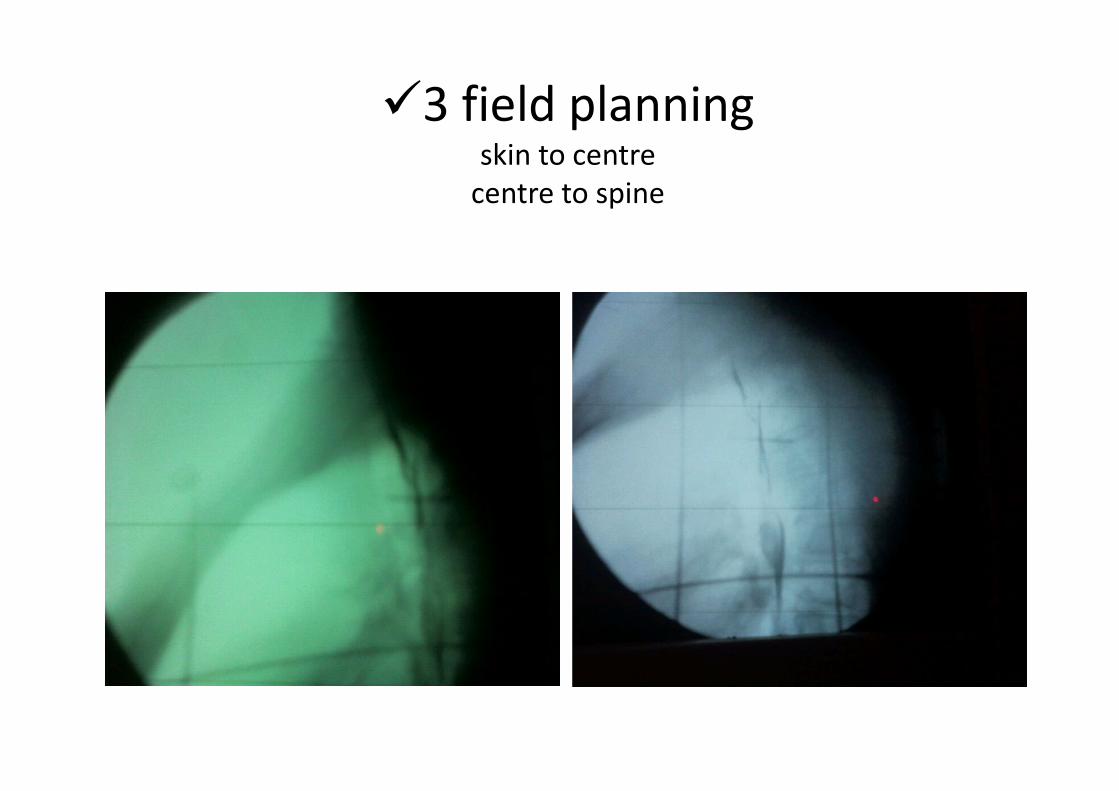
�3 field planningskin to centre
centre to spine


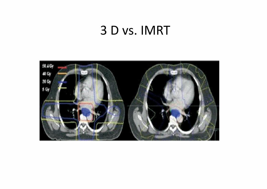
3 D vs. IMRT

IMRT

IMRT

IMRT

IMRT

IMRT• IMRT Dose Constraints
Organ Constraint
LungMean < 16 Gy
V20 < 30%*V5 < 60%
Cord 50 Gy max
Heart Mean < 30 Gy
Liver 30% < 30 Gy• *MD Anderson Data for ARDS; Wang et al. Int. J. Radiation Oncology Biol.
Phys., Vol. 64, No. 3, pp. 692–699, 2006

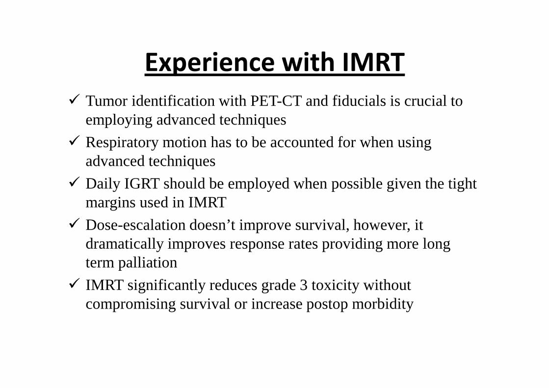
Experience with IMRT
� Tumor identification with PET-CT and fiducials is crucial to employing advanced techniques
� Respiratory motion has to be accounted for when using advanced techniques
� Daily IGRT should be employed when possible given the tight margins used in IMRT
� Dose-escalation doesn’t improve survival, however, it dramatically improves response rates providing more long term palliation
� IMRT significantly reduces grade 3 toxicity without compromising survival or increase postop morbidity