Embed Size (px)
Citation preview

15
Acute Phase Proteins in Cattle
Jawor Paulina and Stefaniak Tadeusz Wroclaw University of Environmental and Life Sciences,
Department of Immunology, Pathophysiology and Veterinary Prevention, Wroclaw
Poland
1. Introduction
The acute phase response (APR) is a reaction of an organism to homeostatic disturbances, that could be due to an infection, tissue injury, neoplastic growth or immunological disorders. At the site of injury there is a local reaction – the aggregation of platelets, clot formation, dilatation and leakage of blood vessels, and the accumulation and activation of granulocytes and mononuclear cells. Activated cells release acute phase cytokines including, interleukin-6 (IL-6), interleukin-1 beta (IL-1b) and tumor necrosis factor-alpha (TNF-a)in order of importance. Due to specific receptors present on different target cells, these cytokines may act a systemic reaction – fever, leucocytosis, an increase in erythrocyte sedimentation rate, an increase in secretion of ACTH and glucocorticoids, activation of complement and clotting cascades, a decrease in serum levels of iron and zinc, a negative nitrogen balance, and dramatic changes in the concentration of some plasma proteins called acute phase proteins (APPs) (Heinrich et. al., 1990). The APPs belong to the group of proteins that become increased in concentration by 25% or more during inflammation (Kushner 1982, as cited in Whicher et al., 1991). In cattle, inflammation is not always followed by a leukocyte population increase. The normally low neutrophil-to-lymphocyte ratio (0.5 in cows) compared with other species (3.5 in dogs, 1.8 in cats) may predispose ruminants to a delayed granulopoietic response because of a slower acceleration of myelopoiesis, preventing rapid leucocytosis (Taylor, 2006). Moreover, in cattle, the detection of inflammatory processes is more difficult to assess, because the symptoms of diseases are not easily detectable. Also, loose-housing systems make it even more difficult to observe individual cows and to recognize sick animals. The APPs are sensitive factors that allow the early and precise detection of inflammation in ruminants (Kent, 1992). The most frequently investigated proteins in cattle are: haptoglobin (Hp), serum amyloid A (SAA), fibrinogen (Fb), ceruloplasmin, alpha 1-antitrypsin and alpha 1-acid glycoprotein (1-AGP) (Conner et al., 1986; Dowling et al., 2002; Eckersall & Conner, 1988; Horadagoda et al., 1993). The liver is the major organ responsible for the synthesis of acute phase proteins (Miller et al., 1951). In cattle some proteins may be extra-hepatically synthesized, e.g. Hp and SAA may also be produced in the mammary gland (Hiss et al., 2004; Molenaar et al., 1999, as cited in Molenaar et al., 2005). Proteins that increase in concentration during the inflammatory process are positive reacting proteins (e.g. Hp, Fb and SAA) and proteins that decrease in concentration are negative reacting proteins (e.g.
www.intechopen.com
Acute Phase Proteins as Early Non-Specific Biomarkers of Human and Veterinary Diseases
382
albumin, transferrin). Changes in APP levels (Hp and Fb) are slower and more prolonged compared to other changes that occur during the acute phase response, such as fever development, white blood cell count and decreased zinc concentration (Godson et al., 1995). Serum amyloid A and Hp may be used to discriminate between acute and chronic inflammatory conditions and were found to be better discriminators than haematological tests e.g. number of leucocytes, percentage of neutrophils and band neutrophils (Horadagoda et al., 1999). A significantly higher proportion of cattle with acute inflammation were found to have higher values of SAA and Hp than those with chronic inflammation (Horadagoda et al., 1999). The possibility of using APPs as markers of inflammation has triggered broad research in terms of cattle diseases. The usefulness of acute phase proteins for diagnosis and prognosis during the course of the most frequently occurring disturbances in cows (dystocia, retained placenta, metritis, abdominal disorders, mastitis and limb diseases) and calves (respiratory tract and diarrhoea) and in herd health monitoring will be discussed.
2. Acute phase proteins in cattle diseases
2.1 Peripartum reproductive disorders (dystocia, retained placenta, metritis)
Transition from a dry period to lactation demands a tremendous effort from cows. Not all cows are capable of coping with this challenge and, as a result, they suffer from health problems. Normal calving induces an acute phase reaction. Parturition increases the Hp concentration but not the concentration of 1-acid glycoprotein in cows (Uchida et al. 1993). Contrary to findings by Uchida et al. (1993), Cairoli et al. (2006) found an increase 1-AGP concentrations between two weeks before and two weeks after parturition. The SAA concentration increases after delivery and the highest concentration was reached at 48 hours (Uchida et al., 1993). The increase in SAA concentration was related to tissue damage in the cow after passage of the calf through the cervix and expulsion of the placenta. Similar to Uchida et al. (1993), in a study by Chan et al. (2010), the SAA in healthy cows reached the highest concentration within 3 days after delivery (66±15 mg/l). Haptoglobin was detectable in the serum of all animals within 3 days after calving, and of these, 95.8% had concentrations higher than >130.9 mg/l. By the third week after parturition the serum Hp concentrations in healthy cows had decreased to 130.9 mg/l, while serum SAA concentrations decreased to 51.9 mg/l during the first week after parturition. The possibility that the synthesis of APPs may be influenced by hormones such as cortisol or prostaglandin cannot be excluded (Alsemgeest et al., 1993). In relation to peripartum reproductive disorders, acute phase proteins were examined as markers of the inflammatory process and predictors of the outcome of disease. It seemed that the APPs were not useful markers of predicting dystocia. In cows with dystocia followed by Caesarean section, the mean concentrations of Hp and Fb before surgery were within the reference ranges (0.07 and 6.3 g/l, respectively) (Hirvonen & Pyörälä, 1998). Also, in a study by Schönfelder et al. (2005), the Hp concentrations in cows with too large (relatively or absolutely) foetus, before Caesarean section, were similar to those in cows with regularly sized calves (median 0.1 vs. 0.009 g/l, respectively). It seems that conditions more serious than the foetus being too large must be present to induce increased Hp concentrations. In cows with torsio uteri, which led to a Caesarean section, the median Hp concentration before surgery was 2.1 g/l (Schönfelder et al., 2005).
www.intechopen.com
Acute Phase Proteins in Cattle
383
The serum Hp concentrations during other postpartum reproductive disorders were ambiguous in the literature. In a study by Skinner et al. (1991), the mean Hp serum concentration in cows with severe metritis was 1.04 g/l (range from 0.56 to 1.61 g/l). Cows with a retained placenta (RP) and without metritis had lower mean Hp concentrations 0.76 g/l (range 0.21-1.2 g/l). On the contrary, low Hp concentrations were found in cows with toxic puerperal metritis (Smith et al., 1998). The mean concentration on diagnosis day was 0.19±0.15 g/l. Because the cows were carefully watched for signs of metritis after parturition, it is possible that low serum Hp concentrations in some of the animals were due to early diagnosis of the disease, and blood samples were taken before Hp had increased. Hirvonen et al. (1999b) also showed that plasma Hp concentrations remained low or moderate in most cows with acute postpartum metritis, and that in one cow (out of 14) the Hp concentration was not even detectable. In RP cows, just 1 out of 5 showed an Hp concentration above 0.1 g/l. The 1-AGP levels were increased in cows with acute postpartum metritis, but their levels did not correlate with the severity of the disease. The highest concentrations of 1-AGP were noted in the plasma of cows with metritis and also dystocia and/or RP. In a study by Chan et al. (2010), cows with acute puerperal metritis had significantly higher Hp concentrations than those in the healthy group throughout the 6 months after delivery, but the higher concentrations of SAA in the metritis group were significantly different in a shorter period (between 4 days and 2 months postpartum). The highest Hp concentration was found in the period of 0-3 days postpartum (1.1 ±0.43 g/l), whereas for SAA it was 4-7 days postpartum (85±23 mg/l). The Hp concentration may be also used to evaluate the efficiency of therapy (Mordak, 2008). Cows with RP were divided into two groups with or without manual removal of the membrane. Cows with manual extraction were further divided into groups where the placenta had either been easily removed or there had been problems and remains of the placenta were left in the uterus. Cows without placenta removal were divided into groups where the placenta had been expelled within 4 days or after 4 days. Ten days after calving the highest Hp concentration was found in the group where the placenta had been expelled after 4 days (2.22±0.36g/l), and the lowest was found in cows where the placenta had been easily removed manually (0.9±0.3 g/l). For cows where placental removal was not complete or where they had been expelled within 4 days, the Hp concentrations were not statistically different (1.83±0.2 vs. 1.53±0.27 g/l, respectively). Regarding Hp concentration on the tenth day, these results suggest that situations where the membranes are easily removed manually are the most beneficial for cows, but Hp concentrations at that time were still higher than those in cows serum without RP (0.46±0.19 g/l). In spite of the ambiguous results for APP concentrations during postpartum health disturbances, it can clearly be seen that high APP concentrations postpartum are negatively associated with health. Cows exhibiting Hp serum concentrations higher than 1 g/l at 15 days of lactation had an increased risk of early culling (Bertoni et al., 1997). Also Hirvonen et al. (1999b) found that cows with Hp concentrations >0.7 g/l were later culled, due to poor body condition and low fertility. In recent years, it has become more popular to use APPs as predictors or risk factors for diseases. In heifers with RP, Hp was detectable in the last week before delivery and the mean Hp concentration was 0.9 ± 0.01 g/l. The mean SAA concentration was 20.3 ± 8.86 mg/l. The differences in Hp and SAA levels between heifers with and without RP were statistically significant (Krakowski & Zdzisinska, 2007). Huzzey et al. (2009a) showed that
www.intechopen.com

Acute Phase Proteins as Early Non-Specific Biomarkers of Human and Veterinary Diseases
384
the Hp concentration might be used as an early predictor of metritis. Cows with Hp concentrations ≥1 g/l at day 3 postpartum were 6.7 times more likely to develop severe or mild metritis. Also, Dubuc et al. (2010) showed that Hp concentration 0.8 g/l in the first week postpartum are a risk factor not only for metritis, but also for purulent vaginal discharge and cytological endometritis, as well. The determination of APPs, especially haptoglobin, seems to be very useful during the peripartum period. Early detection of animals at risk and monitoring the efficiency of therapy and prognosis are very crucial, not only for economic reasons, but also with regard to animal welfare issues.
2.2 The most common abdominal disorders and surgical interventions
The concentrations of APPs have been described in cases of traumatic pericarditis and reticuloperitonitis, abomasal displacement (DA), abomasal volvulus and caesarean sections. McSherry et al. (1970) examined the plasma concentrations of fibrinogen in 160 sick cows. Many diseases were found to be associated with elevated plasma Fb levels, but pericarditis and peritonitis tended to produce the most consistent changes. Pericarditis, which was traumatic in origin, was detected in eight cows where the Fb concentrations were in the range of 9.25-16.75 g/l (McSherry et al., 1970). Also, Makimura and Usui (1990), in cattle with traumatic pericarditis, examined the concentrations of Hp, mucoprotein and sialic acid. Although in most cases the Hp concentration was increased at the same time as the other APPs, in animals with concurrent Theileria sergenti parasitic infection, the Hp concentration was lower than expected. This showed that in the cases of latent or apparent haemolytic disorders, diagnosis of inflammatory disorders with Hp estimation may be difficult. In the diagnosis of traumatic pericarditis, albumins also seem to be very useful. In a study by Yoshida (1991), cows with pericarditis had decreased levels of serum albumins (21.9 ±3.8 g/l), and the lowest concentration was found in cases with purulent type of traumatic pericarditis. In a study on plasma Fb concentrations (McSherry et al., 1970) among 44 cows with peritonitis, 34 had Fb above 8.0 g/l (range 8.4-31.25 g/l). In 70% of these animals Fb exceeded 10 g/l. In 30 cows peritonitis was due to traumatic reticulo-peritonitis. Hirvonen and Pyörälä (1998) reported that cows with traumatic reticulo-peritonitis had high Fb concentrations. The mean Fb concentration in eleven cows was 11.6 g/l and in ten exceeded 7 g/l. All cows with traumatic reticulo-peritonitis had preoperative Hp concentrations >0.55 g/l and the mean Hp concentration was 1.07 g/l. The fact to find both Fb and Hp high concentrations in traumatic reticulo-peritonitis, as well as significantly higher concentrations than in cows with other surgically-treated abdominal disorders (e.g. abomasal displacement, caesarean section), makes them an attractive parameters for evaluation in diagnostics of traumatic reticulo-peritonitis. After surgery, cows with traumatic reticulo-peritonitis showed only a small increase in Hp and Fb concentrations, followed by a steady decrease during the late hospitalization phase. Displacement of the abomasum (DA) does not usually induce a significant Fb response. The Fb concentration was found to be in the range of 9.0-10.25 g/l in only three out of nine cows, and in the remaining cows it was between 3.1-8 g/l (McSherry et al., 1970). In a study by Hirvonen and Pyörälä (1998), in cows with abomasal displacement or volvulus, the mean plasma Fb concentration before surgery was 6.2 g/l. Fibrinogen was also found to be within the reference range in a study by Irmak and Turgut (2005). Cows with left DA had a mean
www.intechopen.com

Acute Phase Proteins in Cattle
385
Fb concentration 3.8 g/l and cows with right DA had 3.7 g/l. Similar to the results of Hirvonen and Pyörälä (1998), in a study by Jawor et al. (2009) the mean Fb concentration in cows with DA was within the reference range (Table 1). In this study, blood samples were taken at three times. The first time (I) at cow arrival to the clinic (where the surgery was to be performed); the second (II) was at the third day of hospitalization; and the third sample (III) was taken just before the animal was returned to the owner, mostly on the seventh day. A right paralumbar fossa omentopexy was performed. Levels of Fb, Hp, SAA and albumin levels were also determined. In contrast to findings reported by Hirvonen and Pyörälä (1998), in our experience, the mean Hp concentration before surgery was higher (0,15 vs. 1.21 g/l respectively). The mean Hp concentration was elevated in all blood collections. Fibrinogen remained low throughout the whole observation period. Only the changes in SAA during post-operative monitoring were significant, specifically between blood collections I and III (Table 1).
APP concentrations I n=8
II n=8
III n=8
Hp g/l
Mean (SD) 1.21 (1.08) 1.10 (0.63) 1.14 (1.28) Median 0.81 1.17 0.79
Fb g/l
Mean (SD) 6.4 (2.0) 5.95 (1.08) 5.23 (1.32) Median 6.24 5.95 5.48
SAA mg/l
Mean (SD) 138.59 (34.7) a 122.86 (38.6) 96.77 (38.9) b
Median 130.92 142.08 103.99
Table 1. Concentrations of Hp, Fb and SAA in cows with displaced abomasum (adapted from Jawor et al., 2009). I, II, III – blood-sampling. Values followed by different letters are significantly different at P ≤ 0.05.
Abdominal surgery in cows induces an increase in Hp. In the study done by Hirvonen & Pyörälä, (1998) on abdominal surgery (DA, caesarean section, explorative laparotomy) a moderate Hp response, that peaked 2-3 days after surgery, was reported. In our study mean Hp and SAA concentration was lower after surgery. These differences in Hp changes might result from different initial concentrations – contrary to our results, the initial Hp concentration in study Hirvonen and Pyörälä (1998) in cows with abomasal pathology was low and APR driven by surgery is not major. A low concentration of albumin was detected in cows during the entire period of observation (I-26.5±6.9; II-24.9±2.6, III-25.0±4.6). Low concentrations of albumins together with high Hp and SAA levels confirmed a severe inflammatory state in the cows. The relatively quick change in SAA concentrations contrary to Hp and albumin, showed that during the monitoring of recovery process in cows with DA, the most accurate APP marker for post-surgery recovery process is SAA. Fibrinogen did not seem to be efficient enough for monitoring the cases of DA. However, Hirvonen and Pyörälä (1998) pointed out that Fb concentrations might be helpful in the postoperative monitoring of infectious complications, such as peritonitis. In terms of DA, the Hp level determination before parturition may be a useful diagnostic tool to assess the risk of this disease (Huzzey et al., 2009b). During the week before parturition, the Hp serum concentration in cows developing DA postpartum was significantly higher as compared with healthy cows (0.5±0.02 vs. 0.2±0.02 g/l, respectively).
www.intechopen.com
Acute Phase Proteins as Early Non-Specific Biomarkers of Human and Veterinary Diseases
386
The odds for developing DA were 3.1-fold higher in cows with Hp concentration ≥0.8 g/l during week before parturition. The determination of Hp may also be useful for caesarean sections (Balbierz et al., 1977). In uncomplicated cases, Hp appeared in the serum on the second day after surgery and remained elevated for 8-9 days. When complications occurred after surgery, the Hp concentration remained elevated for the whole observation period (18 days). The determination of Hp concentrations after surgery may be also used as a predictor of regenerative potential in cases of caesarean section after torsio uteri (Schönfelder et al., 2005). Cows that were fertile later had the highest Hp concentration on day 3 (median 3.21 g/l), which later decreased. In cows that did not become pregnant later, the Hp concentration increased from day 2 (median 0.74 g/l) and demonstrated prolonged inflammatory states, which probably caused infertility. The determination of APPs in surgical procedures is a valuable tool for veterinary surgeons in monitoring the recovery process and the early detection of inflammatory complications. However, it must be remembered that surgical intervention induces a short increase in Hp and SAA levels, but not Fb levels.
2.3 Mastitis
Mastitis is recognized as being one of the most important reasons for culling dairy cows (Esslemont & Kossaibati 1997; Whitaker et al., 2000). The significant impact on health and milk production aroused researcher into APPs and mastitis. Up to now the evaluation of somatic cell count (SCC) has remained as the gold standard for determining udder health. However, research results have suggested that APPs might be interesting as either a valuable supplement even as an alternative to SCC. Changes in concentrations of Hp, Fb, ceruloplasmin, -1 antitrypsin, acid soluble glycoproteins, 1-proteinase inhibitor and SAA were investigated under field conditions and in experimentally induced mastitis.
2.3.1 APPs in serum
Haptoglobin, known as the haemoglobin reactive protein (HRP), in cows with mastitis was investigated by Spooner and Miller (1971). Out of 26 cows, Hp was detected in the sera of 24 cows with clinical summer mastitis (septic mastitis). Later, Conner et al. (1986) found that all cows with summer mastitis had significantly elevated concentrations of -1 antitrypsin and ceruloplasmin in comparison to normal cows. In all cows with mastitis, Hp was elevated, whereas in healthy cows Hp was undetectable. Skinner et al. (1991) concluded that a haptoglobin concentration >0.4 g/l indicates significant infection and that a concentration of 0.2 g/l and above may indicate early or mild infection. Eckersall et al. (1988, as cited in Skinner et al., 1991) suggested that the prognosis for cattle with an Hp concentration > 1 g/l is poor, but a later study could not confirm this (Skinner et al., 1991). The experimental induction of mastitis explained some more details about the usefulness of APPs and characterized the APR during mastitis. After experimental induction of mastitis in 10 pregnant heifers with Actinomyces pyogenes, Fusoacerium necrophorum and Peptostreptococcus
indolicus, changes in plasma Fb, serum Hp, acid-soluble glycoproteins (ASG) and the 1-proteinase inhibitor (1-PI) were analysed (Hirvonen et al., 1996). Fibrinogen was found to be a reliable indicator for detecting the presence of bacterial infections in all heifers, but not as a prognostic indicator for mastitis, as there was no difference in the Fb response between animals who recovered and those who did not. Haptoglobin and ASG were most effective in
www.intechopen.com

Acute Phase Proteins in Cattle
387
indicating the severity of infection and in predicting the final outcome of the disease in the heifers. The Hp response was different between moderate and severely affected heifers. The maximum Hp response was reached in 2-3 days. In severely infected heifers, Hp was four times higher than in the moderately affected heifers. The Hp levels in severely infected heifers remained elevated 2 weeks after bacterial inoculation, whereas in the moderately infected animals Hp returned to normal 5 days after the bacterial challenge. The maximum values for ASG were reached within 3-5 days. The ASG values in severely infected animals remained high for 2 weeks after inoculation. Serum 1-PI levels increased in severely affected heifers (maximum activities were reached in 3 days), but in moderately affected animals there was no clear response by this APP after the challenge. The WBC response was similar in all animals and had no predictive value for the severity of the disease in this study. These differences were not so evident in later studies. Hirvonen et al. (1999a) found that the differences between severely versus moderately or mildly affected cows with E. coli were present for SAA (which is more sensitive) but not for haptoglobin. Eckersall et al. (2001) showed that although serum Hp concentrations were higher in cows suffering from mastitis compared to healthy cows, no difference was observed between the cows suffering from mild and moderate mastitis. These differences might have been due to the different pathogens used and the numbers of bacteria inoculated, isolated from udders in these studies or individual ability for APPs synthesis (Jacobsen et al., 2004). Ohtsuka et al. (2001) showed that in naturally occurring coliform mastitis the Hp concentration was different in cows with severe (n=7) and mild mastitis (n=11) on days 3 and 9 after diagnosis, but not on day 6. The concentrations of Hp and SAA from different studies are compared in Table 2.
Eckersall et al. (2001)
Grönlund et al. (2003)
Nielsen et al. (2004)
(n=10)
Jawor (2007)
(n=11)
mild (n=16)
moderate (n=13)
acute (n=6)
chronic (n=5)
Hp (mg/ml) 0.47 0.74 0.965 0.07 0.79 1.27 SAA (µg/ml) 13.8 29.9 262 7.2 752*(739)** 69.86
Table 2. Median Hp and SAA serum concentrations in mastitis (*mean, **standard deviation).
2.3.2 APPs in milk
Milk seems to be a better than serum material for testing APPs during mastitis. It is easier and quicker to obtain a large number of milk samples without stressing the animals. Mazur (1980), using agar electrophoresis, showed the presence of HRP in cows’ whey. The highest frequency of HRP was detected in the whey obtained from quarters with progressed inflammatory lesions and a significantly increased number of somatic cells (Mazur A. & Mazur O., 1981). Increased milk concentrations of Hp and SAA in cows with mastitis were shown by Eckersall et al. (2001). In cows’ milk from infected quarters with moderate mastitis, concentrations of SAA but not Hp were significantly greater than in cows with mild mastitis. In milk SAA seems to have a greater potential for the detection of mastitis than Hp since it had higher sensitivity, specificity and efficiency in differentiating between cows with mastitis and healthy cows (Eckersall et al., 2001). Since the concentrations of APPs in milk also significantly increased with increasing somatic cell count, this suggests
www.intechopen.com
Acute Phase Proteins as Early Non-Specific Biomarkers of Human and Veterinary Diseases
388
that milk APPs may indicate the severity of an infection (Nielsen et al., 2004). In cases of chronic subclinical mastitis, only milk SAA levels were significantly higher than pre-infection and healthy control quarters (Grönlund et al. 2003). Later studies showed that whilst increased concentrations of APPs in milk were noticed in cows with chronic subclinical mastitis, the contents of Hp and SAA varied markedly (Grönlund et al., 2005). Haptoglobin and/or SAA were detected in 83% of the examined composite milk samples (pooled milk from quarters). Since cows had to have detectable levels of Hp or SAA in at least two udder quarters for elevated levels to be found in the composite samples, analysis at the quarter level is preferable. The important findings were that almost all udder quarter samples from healthy control cows had undetectable levels of Hp and SAA, and that Hp and/or SAA concentrations above 0.3 and 0.9 mg/l (the detection limit in this study), respectively, indicated an abnormal udder quarter. Although methods for determining levels of Hp in milk have been described (Eckersall et al., 2001; Grönlund et al., 2003; Hiss et al., 2004) tests which allow a quick and simple determination are necessary. Akerstedt et al. (2006) described an optical biosensor for Hp determination in milk. It was based on the strong interaction between Hp and haemoglobin. This assay has the potential to be useful in screening for Hp in milk samples. It could be used for the semi-quantitative determination in milk from cows with no observable signs of mastitis. The local production of Hp and SAA in the udder during acute mastitis makes these APPs sensitive and rapid markers of acute inflammation. Up till now there are no commercially available methods for their rapid testing. For broad milk testing for APPs presence at cow-side tests are necessary.
2.4 Lameness and limb diseases
Limb pathology in dairy herds is a serious problem. With increasing milk production the problem might become even more significant as high-yield milk cows are more predisposed to lameness (Green et al., 2002). The economic losses from lameness are due to the higher incidence of culling, the lower likelihood of pregnancy (Bicalho et al., 2007; Mendelez et al., 2003) and the lower milk production (Green et al., 2002; Rajala-Schultz et al., 1999). In order to limit the losses related to lameness, it is important to detect lame cows as early as possible. A few papers were recently published on the estimation of APPs in lameness, and the efficiency of trimming and treatment. Kujala et al. (2010) investigated concentrations of Hp and SAA cows with sole ulcers and/or white line abscesses, without any other clinical symptoms. The blood from lame and healthy cows was taken on day 0 (diagnosis of lameness), and again from lame cows on days: 4, 7 or 8 and 14. No antibiotic treatment was used. The SAA concentrations were significantly elevated from day 0 until 7/8. There was no difference between SAA concentrations in the samples taken on day 14 from lame cows and healthy cows, and the cows were no longer lame as well. No significant differences were observed in the serum Hp concentrations between the groups. Serum amyloid A is known to be more sensitive APP in cattle than Hp (Heegard et al., 2000) and, according to the authors cited above, the local changes in the hooves were large enough to induce a general SAA response but not a response by Hp. Higher concentrations of haptoglobin were detected among 60 dairy cattle diagnosed with pododermatitis septica (PS), pododermatitis circumscripta (PC), interdigital necrobacillosis (IN) and papillomatous digital dermatitis (PPD) (Smith et al., 2010). Haptoglobin was measured after the initial diagnosis and on days 3 and 5 afterwards. Elevated levels (>0.37g/l) were found on day 1 in cows with PS, PC, IN
www.intechopen.com

Acute Phase Proteins in Cattle
389
and PDD: 65.9%, 37.5%, 71.4% and 25.0% of the cows, respectively. Although the number of animals with an increased Hp concentration decreased after 5 days, still 14.3-50% of the cows with PS, PC and PPD had elevated Hp level in last blood sample. The decreasing Hp concentrations in cows with PS, IN and PDD indicated the effective treatment for these disorders (Smith et al., 2010). An investigation of albumin, Fb, Hp, seromucoid and ceruloplasmin levels in first lactation heifers was carried out to determine the presence of any relationships with the development of hoof horn haemorrhaging (Laven et al., 2004). None of seven examined proteins showed any relationship between changes and the development of hoof horn haemorrhaging after calving. It seems that more serious pathologies in hoofs such as - sole ulcers and interdigital necrobacillosis must be present to induce general APR. The estimation of concentrations and the dynamics of changes in APP levels can be a valuable tool, supplementing the clinical assessment during treatment (Jawor et al., 2008). In our study we compared concentrations of Hp, SAA and Fb in healthy cows and cows with limb diseases. The cows were retrospectively divided into groups I – with systematic decreases in APP levels in successive blood collections (n=6) and II – with an increase in the concentration of one or more APPs in the second or third blood collection (n=8). Group III comprised healthy cows at different stages of lactation and dry cows (n=10). From groups I and II blood was taken for the first time (1) on the day of their arrival at the clinic, for the second time (2) between the third and sixth day of stay, and for the third time (3) when the animal was healthy enough to be sent back to its owner. Blood from healthy cows was only taken once. The mean initial concentrations and changes in levels during treatment are shown in Table 3.
1 2 3
I II III* I II III* I II III*
Hp g/l
2.4 ±0.6 a
1.2 ±0.7
0.1 ±0.1b
0.8 ±0.4
0.9 ±0.5
0.1 ±0.1
0.1 ±0.1
0.3 ±0.2
0.1 ±0.1
SAA mg/l
219.7 ±30.4 A
153.7 ±24.3 A
24.4 ±7.8 B
124.5 ±6.5 a
127.1 ±29.0 Aa
24.4 ±7.8 Bb
79.1 ±22.4
86.1 ±17.8 a
24.4 ±7.8 b
Fb g/l
12.2 ±1.3 A
8.0 ±0.6 Bc
5.0 ±0.2 Bb
9.9 ±0.9 A
6.9 ±0.5 Bc
5.0 ±0.2 Bb
7.2 ±1.2
6.6 ±0.8
5.0 ±0.2
1,2,3 blood samples, I,II,III experimental groups. Values followed by different letters within the same blood collection were statistically different: small letters P ≤ 0.05, capital letters P ≤ 0.01. *cows from healthy group were sampled once
Table 3. Mean (±SEM) concentrations of haptoglobin, serum amyloid A and fibrinogen during the treatment of limb diseases (adapted from Jawor et al., 2008).
The highest concentrations of all investigated proteins were recorded at the beginning of the treatment. As the concentrations of APPs depend on the extent of underlying tissue damage (Heegaard et al., 2000), the very high concentrations of Hp, Fb, SAA at the beginning in group I suggested a very severe, acute inflammatory process. High, gradual decrease in this group shows that treatment process went without complications. An initial decrease in the concentrations of Hp, SAA and Fb between the first and second blood collections from five cows in Group II was also noticed. This proved that the treatment applied was appropriate and that it contributed towards reducing the inflammatory process in the cows, but, in cases
www.intechopen.com
Acute Phase Proteins as Early Non-Specific Biomarkers of Human and Veterinary Diseases
390
of further complications (e.g. wound infections, bronchitis, the occurrence of other inflammatory states of the limbs), we noticed increases in one or two of the APPs under examination at the next blood collection. Although the APR is not specific and the problem cannot be recognized based only an increase in APPs, this may actually be advantageous to a veterinarian as a valuable indicator that the recovery process is not going correctly. The triple determination of APPs during treatment seems to be a valuable supplement to clinical findings. For practical purposes, we recommend at least two APP estimations. The first one should be performed at the beginning of treatment to evaluate the severity (chronic/acute) of the process and the extent of tissue damage. The second should be performed immediately before returning the cow to its owner. This estimation, in addition to other clinical parameters (such as a decrease in lameness, an increase in appetite and milk yield), is a good measure of the efficiency of the therapy. The additional estimation should be performed during treatment in following cases: there are doubts regarding the correctness of the treatment, when there is very little or no clinical improvement, or before making critical decisions, such as whether or not to amputate claw. Acute phase protein determination is also useful during the recovery period because the time between dressing changes after claw resection is gradually prolonged. During this period, apart from assessing how a cow is coping with putting weight onto an affected leg and her general behaviour under field conditions, there is no possibility of assessing the healing process.
2.5 Respiratory diseases in calves
In addition to diarrhea, respiratory tract diseases are one of the main health problems afflicting young dairy calves (Sivula et al., 1996; Svensson et al., 2003). To prevent disease outbreaks, early detection, isolation and treatment of diseased animals is important. Results from several different studies have shown that estimation of APPs is useful for detecting and monitoring respiratory diseases. In calves with respiratory tract diseases, the following APPs were examined: Hp, Fb, SAA, transferrin (Tf), lipopolysaccharide binding protein (LBP), α1-acid glycoprotein (AGP), α1-antitripsin (α1-AT), seromucoid (Sm), ceruloplasmin (Cp), albumin, α1-antichymotrypsin and α2-macroglobulin.
2.5.1 APPs during experimental infections
Most of results regarding APPs in respiratory infections were obtained from experimental infections. Although scientists are not in agreement as to whether bacterial or viral infections mount a higher response, it has been shown that APR is induced faster after bacterial infections (Table 4). After intra-tracheal aerosol inoculation with Mannheimia
(Pasteurella) haemolytica, the earliest detectable rise in Hp, Sm and α1-AT in calves was after 24 h (Conner et al., 1989). However, bacterial infection did not cause a rise in ceruloplasmin levels. The highest concentrations of Hp, Sm and α1-AT were reached on day 3 (1.0 g/l; 1.91 g/l; 1090 iu/l respectively). In this study, α1-anitichymotripsin and α2-macroglobulin were also shown to be acute phase reactants in calves. Horadagoda and Eckresall (1994) also inoculated calves intra-tracheally with M. haemolytica serotype A1. Within 10 hours post-inoculation, there was a small, insignificant increase in Hp. In contrast, SAA showed more dynamic changes: there was a progressive, linear change in SAA, and the mean concentration increased from close to the detection limit (3 mg/l) at inoculation, to 18 mg/l by 10 hours post-inoculation. These results indicate that SAA is more rapid than Hp in
www.intechopen.com
Acute Phase Proteins in Cattle
391
calves in response to infection with M. haemolytica. To assess the acute phase response in bovine respiratory disease (BRD), infection with Bovine Herpes Virus-1 (BHV-1) and P.
haemolytica was performed on 49 animals (Godson et al., 1996). Calves were challenged on day 0 with BHV-1 and with M. haemolytica on day 4. Over the first 4 days post BHV-1 infection, only 10% of the animals developed Hp concentrations >0.1 g/l. On day 5 (24h post bacterial challenge) 43% of the calves displayed elevated Hp levels. The highest Hp concentrations (Table 4) were observed on day 8 (4 days post bacterial challenge), where 84% of the animals had Hp concentrations >0.1 g/l. Increased Hp concentrations were significantly associated with disease severity (e.g. fever, increased sickness index and weight loss). Godson et al. (1996) showed that Hp concentration measurements may serve as prognostic indicators, to assist determination of the severity of disease. Animals that subsequently died had significantly higher Hp concentrations on day 8, compared to those that recovered. Further examination of the acute phase response of calves to viral infection was performed by Heegaard et al. (2000). After experimental BRSV infection, most calves displayed elevated SAA concentrations, which were detectable at day 5 p.i and peaked at approximately day 5-8 p.i. The highest SAA concentrations ranged from 60-80 mg/l: 5-7 times the normal SAA concentrations in control animals. Hp showed a similar pattern of changes. The maximum Hp response was observed on day 6-7 p.i., and reached 8-10 g/l: approximately 500 times the detection limit of the assay (18 mg/l). Severe BRSV infections were found to correlate with haptoglobin concentrations > 1 g/l on day 7 p.i., and SAA concentrations ≥ 60 mg/l. In this study, the acute phase response induced by BRSV was generally the same or higher than previously reported for bacterial infections in calves. Serum amyloid A responded more rapidly to infection, but Hp concentrations correlated better with disease severity (fever and extent of lung consolidation) (Heegaard et al., 2000). In a study by Ganheim et al. (2003), calves were infected BVDV and/or M. haemolytica. In the group challenged with mixed infection, animals were first inoculated with virus, followed by bacteria 5 days later. After BVDV infection, concentrations of Hp, SAA and Fb reached a maximum level 8-9 days p.i. After inoculation with bacterial agent, the highest APP concentrations were reached 1-2.5 days p.i. In the BVDV/M. haemolytica infected group, APP response patterns were more complicated, with larger individual differences. In general, the magnitude of the response was similar, but the duration of elevated APP concentrations were longest in the mixed infection group, reflecting the duration of clinical symptoms. In a study by Grell et al. (2005), calves experimentally infected with BRSV displayed the highest Hp concentrations at approximately 7-9 days post-inoculation. Calves presenting with the most severe clinical symptoms (respiratory rate, rectal temperature) had the highest Hp levels, while calves presenting with the mildest clinical symptoms displayed the lowest levels of acute phase reaction; however, no statistical correlation was found, probably due to the small number of animals (n=6). APPs were shown to be valuable proteins for the evaluation of a P. multocida A3 experimental infection model in calves (Dowling et al., 2002). In calves challenged with greater volumes (300 vs. 60 ml), plasma Hp levels were found to be significantly increased, regardless of the number bacteria (109 vs. 1010 cfu). It is possible that increased volume challenges affect a greater area of the lungs, especially if the initially slow response in Hp production provides more time for bacterial proliferation. Dowling et al. (2002) showed that α1-AGP might be particularly valuable protein; examination of α1-AGP concentrations for all treatments indicated that large volume challenges were associated with a greater increase
www.intechopen.com

Acute Phase Proteins as Early Non-Specific Biomarkers of Human and Veterinary Diseases
392
from basal levels, than low volume challenges. Other experimental studies examined less popular APPs, such as LBP or transferrin (Tf). Single intratracheal inoculation of Pasteurella haemolytica A1 showed increased lipopolysaccharide binding protein (LBP) activity in calves (Horadagoda et al., 1995). Lipopolysaccharide binding protein activity increased after 12 hours, and by 36 h, there was a 4-fold increase in serum concentrations. Early reaction of LBP was reported in a study by Schroedl et al. (2001). Concentrations of LBP in experimental calves, that were intra-tracheally infected with Mannheimia haemolytica, increased 6 hours post-infection, with a peak response at 24 hours post-infection. Haptoglobin reactions were slower, with a significant increase occurring 12 hours post-infection, and a maximal response at approximately 48h post-infection. In terms of response time, LBP appears to be superior to Hp as an early marker of infection; however, its concentration only rose 7-fold, while, in contrast, Hp rose 300-fold. In calves, which died following infection, LBP levels were more elevated than in survivors; unfortunately, the number of animals was too small to draw any conclusions (Schroedl et al., 2001). A study by McNair et al. (1998) demonstrated that, the negative APP, Tf) could also be useful in calves. Although serum Tf levels remained within the reference range, in calves that were experimentally infected with Histophilus somni, its concentration was associated with lung lesions. In fact, the lowest Tf concentrations were measured in calves with extensive lesions, and there were significant differences between mean values (on days 1-6) between the group with no lesions and the group with extensive lesions.
Study Infectious agent
Time to reach maximum level
Hp (mean or max
range g/l)
Hp determination
method
Conner et al. (1989)
M. haemolytica 3d 1,0 haemoglobin binding capacity
Godson et al. (1996)
BHV-1 and M. haemolytica
8d (4d after bacterial challenge)
1,1 ELISA
Heegard et al. (2000)
BRSV 6-7d 8-10* ELISA
Schroedl et al. (2001)
M. haemolytica 2d >4.5** ELISA
Ganheim et al. (2003)
BVD and/or M. haemolytica
8.5-9.5 d (BVDV) 2-2.5 (M. haemolytica) 7- 10 (both agents)**
0.89-1.77* 1.1-2.0*
0.65-2.24*
measure of peroxidase
activity Hp-Hb
Table 4. Comparison of maximal Hp concentrations and the number of days required to reach maximum Hp levels across several studies. *Highest individual concentrations; **approximate results read off from line graphs.
2.5.2 APPs in field studies
Studies on acute phase reactions in field conditions are more complicated, because the timing of initial infection is often unknown, and it is difficult to determine the phase of infection. Nikunen et al. (2007) studied concentrations of Fb, Hp, SAA, LBP and AGP in 84 calves suffering from respiratory disease. Among isolated pathogens from tracheobronchial lavage (including Mycoplasma dispar and other mycoplasmas, but not M. bovis, P. multocida,
www.intechopen.com

Acute Phase Proteins in Cattle
393
Pasteurella sp., F. necrophorum and A. pyogenes) and seroconverted bovine viruses (e.g. adenovirus-7, adenovirus-3, coronavirus and parainfluenza virus-3), only Pasteurella multocida was associated with increased concentrations of all tested APPs. The higher concentration of APPs observed in calves with P. multocida suggests a strong pathogenic role for this bacteria. Although it was shown that APPs increase rapidly in experimentally infected cattle, this increase coincided with clinical signs, and the relative usefulness of APPs in actually predicting respiratory disease during the incubation period is not satisfactory. Svensson et al. (2007) showed that in calves, the discriminative ability of serum Hp levels for indicating the presence of clinical respiratory-tract diseases was poor overall, and no better than rectal temperature. Furthermore, Hp levels were significantly affected by sex and rectal temperature. When Hp and fever were combined, and either increased Hp or fever (>39.5 ºC) was used for detecting respiratory disease, the best test performance was achieved in heifer calves at serum Hp concentrations >0.15 g/L, with a sensitivity of 72% and a specificity of 59%. Therefore serum Hp levels, when combined with rectal temperature, may be a valuable parameter in herd-level diagnostics, at least in heifer calves. APPs may also be useful for determining the response to therapy and making the right treatment decisions. Carter et al. (2002) found that serum Hp concentrations in transported feedlot cattle were greater in calves treated more than once, than in calves not treated or treated only once. At the beginning of treatment, Hp concentrations were higher in calves requiring more than one treatment compared to calves with one treatment (0.76 vs. 0.55 g/l). In addition, Berry et al. (2004) showed that Hp concentrations are a useful tool for predicting the number of antimicrobial treatments required in newly received feedlot calves. Hp concentrations on day 0 and 7 were positively correlated with the number of treatments during the trial. Hp concentrations on those days increased with increasing numbers of antimicrobial treatments. Retrospective analysis of APPs concentrations in growing calves suffering from bronchopneumonia, showed that, both Hp and Fb could be used for identification of calves requiring an anti-inflammatory treatment (Humblet et al., 2004). Animals in this study were classified into two categories: animals requiring either no-treatment or antibiotics alone; and animals requiring both antibiotics and anti-inflammatory drugs (AI). Treatments were administered according to clinical symptoms. In blood samples taken before clinical examination, elevated APPs levels were observed. Hp and Fb thresholds for medical decisions (i.e. to use AI or not) were 25 mg/l and 2.7 g/l respectively. Hp and Fb were useful predictors of inflammation severity; most of the animals (80%) that received anti-inflammatory treatment after clinical examination presented pathological Hp and/or Fb values upon onset of disease. Hp alone was able to confirm> 75% of case decisions (i.e. whether diseased calves were treated or not). APP concentrations may be useful for monitoring the treatment of respiratory disease in calves. In our study (Jawor and Stefaniak, 2006), blood was sampled 3 times: first (I), when the owner either sent the calf to the clinic or reported signs of disease; second (II), between 3 and 6 days later; and third (III), 13-17 days after the first sample was collected. In one case, a 3rd sample was not collected because of euthanasia. Blood samples were also collected once from clinically healthy calves. Changes in APPs concentrations are shown in Table 5. Mean Hp concentrations were only increased in the beginning, while concentrations of SAA (sample I) and Fp (samples I and II) were significantly different from healthy controls. Hp was undetectable in all calves in samples II and III, with one exception: increased Hp and SAA concentrations were found in one calf in the last blood sample collection (III). An increase in Hp and SAA levels following an earlier decrease is characteristic of secondary
www.intechopen.com
Acute Phase Proteins as Early Non-Specific Biomarkers of Human and Veterinary Diseases
394
infection (Ganheim et al., 2003). Thus, the observed increase in Hp and SAA in the above mentioned calf was probably due to the presence of a secondary infection. Later, this calf was unsuccessfully treated several times and eventually died. After an initial decrease in SAA concentrations, an increase in SAA levels was observed in the serum of 3 calves in the last blood sample collection; although a simultaneously increase in Hp and Fb was observed in only one calf. Because SAA is known to be a more sensitive protein, it is hard to explain if this increase observed in 2 calves was caused by secondary infection or other factors, such as stress (Alsemgeest et al., 1995a). In 3 out of 7 calves from the healthy control group, SAA concentrations were higher than the 25.6 mg/l, basal level established by Ganheim et al. (2003). This may imply that, even in the absence of clinical symptoms, these calves were not perfectly healthy.
Calves with bronchopneumonia Control group (n=7) I (n=8) II (n=8) III (n=7)
Hp g/l 0.3 (±0.4) 0.0 (±0.0) 0.0 (±0.1) 0.0 (±0.0) SAA mg/l 95.7 (±46.1)a 62.0 (±42.5) 75.9 (±36.9) 31.3 (±20.9)b Fb g/l 6.54 (±2.08)a 5.85 (±1.24)a 4.94 (±1.21) 3.13* (±0.37)b
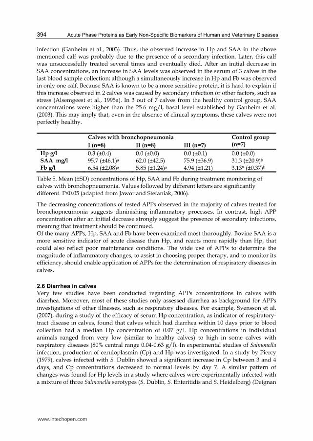
Table 5. Mean (±SD) concentrations of Hp, SAA and Fb during treatment monitoring of calves with bronchopneumonia. Values followed by different letters are significantly different. P≤0.05 (adapted from Jawor and Stefaniak, 2006).
The decreasing concentrations of tested APPs observed in the majority of calves treated for bronchopneumonia suggests diminishing inflammatory processes. In contrast, high APP concentration after an initial decrease strongly suggest the presence of secondary infections, meaning that treatment should be continued. Of the many APPs, Hp, SAA and Fb have been examined most thoroughly. Bovine SAA is a more sensitive indicator of acute disease than Hp, and reacts more rapidly than Hp, that could also reflect poor maintenance conditions. The wide use of APPs to determine the magnitude of inflammatory changes, to assist in choosing proper therapy, and to monitor its efficiency, should enable application of APPs for the determination of respiratory diseases in calves.
2.6 Diarrhea in calves
Very few studies have been conducted regarding APPs concentrations in calves with diarrhea. Moreover, most of these studies only assessed diarrhea as background for APPs investigations of other illnesses, such as respiratory diseases. For example, Svensson et al. (2007), during a study of the efficacy of serum Hp concentration, as indicator of respiratory-tract disease in calves, found that calves which had diarrhea within 10 days prior to blood collection had a median Hp concentration of 0.07 g/l. Hp concentrations in individual animals ranged from very low (similar to healthy calves) to high in some calves with respiratory diseases (80% central range 0.04-0.63 g/l). In experimental studies of Salmonella infection, production of ceruloplasmin (Cp) and Hp was investigated. In a study by Piercy (1979), calves infected with S. Dublin showed a significant increase in Cp between 3 and 4 days, and Cp concentrations decreased to normal levels by day 7. A similar pattern of changes was found for Hp levels in a study where calves were experimentally infected with a mixture of three Salmonella serotypes (S. Dublin, S. Enteritidis and S. Heidelberg) (Deignan
www.intechopen.com
Acute Phase Proteins in Cattle
395
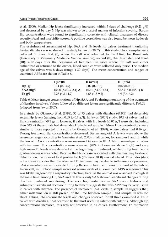
et al., 2000). Median Hp levels significantly increased within 3 days of challenge (0.21 g/l) and decreased by day 5. Hp was shown to be a useful marker of infection severity. Serum Hp concentrations were found to significantly correlate with clinical measures of disease severity: fecal and morbidity scores. A positive correlation was also found between hp levels and body temperature. The usefulness of assessment of Hp, SAA and Fb levels for calves treatment monitoring having diarrhea was evaluated in a study by Jawor (2007). In this study, blood samples were collected 3 times: first (I), when the calf was admitted to the Clinic for Ruminants (University of Veterinary Medicine Vienna, Austria); second (II), 3-4 days later; and third (III), 7-10 days after the beginning of treatment. In cases where the calf was either euthanized or returned to the owner, blood samples were collected only twice. The median age of the calves was 8 days (range 1-30 days). The mean concentration and ranges of examined APPs are shown in Table 6.
I (n=10) II (n=10) III (n=6)
Hp g/l 0.14 (0-0.49) 0.02 (0-0.18) 0.02 (0-0.13) SAA mg/l 156.8 (53.0-302.4) A 102.1 (54.6-142.1) 53.5 (15.0-105.1) B Fb g/l 7.28 (4.3-14.7) 6.68 (4.8-9.2) 4.9 (3.4-6.2)
Table 6. Mean (range) concentrations of Hp, SAA and Fb during monitoring of the treatment of diarrhea in calves. Values followed by different letters are significantly different. P≤0.01 (adapted from Jawor 2007).
In a study by Okamoto et al. (1998), 35 of 73 calves with diarrhea (47.9%) had detectable serum Hp levels (ranging from 0.05 to 0.7 g/l). In Jawor (2007) study, 40% of calves had an Hp concentration >0.1 g/l. However, if calves with Hp levels ≥0.05 g/l were also included, then 60% of the animals had detectable Hp in blood sample I. Mean Hp concentrations were similar to those reported in a study by Okamoto et al. (1998), where calves had 0.16 g/l. During treatment, Hp concentrations decreased. Serum amyloid A levels were above the reference range (according to Ganheim et al., 2003) in all calves, for samples I and II, while the lowest SAA concentrations were measured in sample III. A high percentage of calves with increased Fb concentrations were observed (70% in I samples above 5 g/l) and very high mean Fb levels were detected at the beginning of treatment, while during treatment a gradual decrease was noted. Because the Fb increase associated with diarrhea may be due to dehydration, the index of total protein to Fb (Thomas, 2000) was calculated. This index (data not shown) indicates that the observed Fb increase may be due to inflammatory processes. SAA concentrations were elevated during the entire treatment period for most of the calves. In one calf, in III blood sample increased serum levels of all examined APPs was found. This was likely triggered by a respiratory infection, because the animal was observed to cough at the same time. Among Hp, SAA and Fb levels, only SAA showed significant changes during diarrhea treatment monitoring. The very high initial serum SAA concentrations and subsequent significant decrease during treatment suggests that this APP may be very useful in calves with diarrhea. The presence of increased SAA levels in sample III suggests that, either inflammation is still present or the time between sample I and sample III was too short. Taking into account the levels and changes observed for all three examined APPs in calves with diarrhea, SAA seems to be the most useful in calves with enteritis. Although Hp concentrations increased, this was not observed in all calves. Furthermore, Fb estimation
www.intechopen.com
Acute Phase Proteins as Early Non-Specific Biomarkers of Human and Veterinary Diseases
396
required an additional determination of plasma proteins, to distinguish between a relative increase in hemo-concentration and an absolute Fb increase during inflammation.
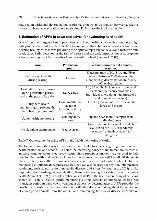
3. Estimation of APPs in cows and calves for evaluating herd health
One of the main targets of milk producers is to keep healthy cows with a long-term high milk production. Herd health protection has not only ethical but also economic significance. Keeping healthy cows means providing their optimal reproduction levels and therefore milk production. Early detection of the risk of disease and the early introduction of appropriate actions should protect the majority of animals within a herd (Stefaniak, 2003).
Aim Production group
Parameters/number of animals examined
Evaluation of health during rearing
Calves
Determination of Hp, SAA and Fb in 2nd and between 21-28 days of life,
along with Ig determination in 25-30% of newborn calves
Evaluation of risk in cows during transition period
and at the peak of lactation Dairy cows
Hp, SAA, Fb (% of cows with elevated levels and their concentrations in
individual cows, along with metabolic and mineral parameters)
Dairy herd health monitoring/improving the
herd health programme
Cows in different stages of
lactation and dry cows
Hp, Fb (% of animals with elevated levels and mean)
Udder health monitoring Lactating dairy cows
Hp and SAA in milk samples from individual cows
Pre-slaughter examination Feedlot steers
Confirmation of normal Hp and Fb levels in all of 5-10% of randomly
examined animals assigned to slaughter
Table 7. Opportunity for using APPs in the health monitoring of cattle.
The two most important ways to achieve this are: first – to supervising programmes of herd health protection, and second – to detect the increasing danger of inflammation/diseases at an early stage or before they occur. Acute phase protein estimation may be used to help monitor the health and welfare of production animals on farms (Eckersall, 2000). Acute phase proteins in cattle are valuable tools since they are not only applicable for the monitoring of inflammatory processes, but they can also be used for any non-inflammatory conditions, such as parturition, metabolic diseases and stress (Murata et al., 2004), or for improving the pre-slaughter examination, thereby improving the safety of food for public health (Saini et al., 1998). Possible applications of APPs in the health monitoring of cattle are shown in Table 7. Cattle health monitoring during periods of increased disease risk (transition period in dairy cows, weaning of calves) by determination of APPs provides the possibility of early disturbance detection, facilitating decision making about the separation of endangered animals from the others, and minimizing the risk of disease transmission
www.intechopen.com

Acute Phase Proteins in Cattle
397
(Stefaniak et al., 2003). The APPs are more useful for monitoring health than proinflammatory cytokines, because after the inflammatory stimulus, their concentrations remain elevated for longer time (Gruys et al., 2006). Therefore, the possibility of detecting their elevated levels in representative groups of clinically healthy cattle is more probable due to the typical dynamics of APPs during the acute phase response (Conner & Eckresall, 1988).
3.1 APP utilization for pre-slaughter cattle examination
Acute phase proteins may be used to monitor health of slaughter animals. In slaughtered cattle without evident disease activity Hp-values were unmeasurable, whereas in most cases with disease activity Hp values were elevated. Serum amyloid A values were found to be elevated in animals with overt lesions, but Hp appears to be a useful variable to discern cattle without disease activity from those with lesions (Gruys et al., 1993). Saini et al. (1998) proposed that a potential application of APPs might be in screening and separating healthy from diseased animals. In a group of 57 retained carcasses of ante-mortem normal cattle, 56% had elevated Hp levels. Standard pre-slaughter examination is sometimes unreliable. Over a 3-year period, the carcasses of 7.7% of ante-mortem clinically normal steers and 20.6% of culled normal cows were retained due to post-mortem found changes (Saini et al., 1998). In emergency slaughtered cattle it was found that Hp and AGP levels were raised, indicating that examination of the APPs could help in improving food safety (Hirvonen et al., 1997). Haptoglobin, SAA and AGP were estimated in the plasma of 81 cattle of which a detailed post-mortem examination confirmed the presence of inflammation, and the APP concentrations were compared between the cases classified as acute and chronic inflammatory lesions (Horadagoda et al., 1999). In the 81 animals with inflammation, it was apparent that SAA and Hp levels were raised in acute rather than chronic cases, whereas in the latter group AGP was more likely to be elevated. The author suggested that Hp and SAA have a high value for detecting acute inflammatory conditions, whereas AGP was a better marker for chronic inflammation in cattle (Horadagoda et al., 1999). Measurement of APPs in meat inspection has good perspectives but more rapid detection methods have to be developed.
3.2 APPs in evaluating herd health status
Estimation of APPs in calves and cows may be very helpful in herd health monitoring. In 175 heifer calves born within one year on a dairy farm of 390 cows, the mean Fb concentration in the blood plasma was 4.0 g/l at the 48th hour of life, and increased to 4.8 g/l by the 25th day. At the 48th hour of life, all of the calves with Ig levels greater than 15 g/l showed normal Fb concentrations, but in the other groups (Ig levels below 5 g/l, between 5-10 g/l and 10-15 g/l), few individuals had elevated Fb levels (4.54%, 1.20% and 3.64% respectively). By the 25th day of life the mean Fb levels were within reference range in all of the groups examined and the rates of calves showing elevated Fb levels were 18.2%, 12%, 3.6% and 13.3% in the respective groups divided according to the Ig level at the 48th hour of life (Furman et al., 2011). On the same farm, increased Hp concentrations (>0.1 g/l) were found in the serum of 32% of calves at the 48th hour of life. By the 25th day of life, 31% of the calves showed elevated Hp levels. Because Hp is not commonly detected in healthy cattle (Eckersall & Conner, 1988), it was surprising that a high rate of calves showed elevated Hp levels at the 48th hour and the 25th day of life. It was concluded that the calves had been in
www.intechopen.com

Acute Phase Proteins as Early Non-Specific Biomarkers of Human and Veterinary Diseases
398
risk of an inflammatory process from first few hours of life, since the increase in Hp concentrations after infection with the bacterial agent occurs within 24 hours (Ganheim et al., 2003). The highest Hp levels, as well as the highest individual differences, occurred in the calves of the group showing failure of passive transfer (Ig levels below 5 g/l at the 48th hour of life). In the same calves at the 25th day of the rate of elevated Hp levels as well as the mean Hp level had increased. In contrast, in properly colostrum-protected calves (over 10 g Ig/l), the mean level and the rate of elevated serum Hp concentrations had decreased. This observation agreed with the higher intensity and frequency of respiratory and gastrointestinal tract morbidity in the calves poorly protected with colostrum. In study by Alsemgeest et al. (1995b) the Hp and SAA levels were normal in newborn calves. Diseases that appeared before the fourth day of life induced an increase in SAA levels in all diseased calves, but Hp levels were only raised in two out of eight individuals. The authors indicated that because the SAA concentration at birth is very low and increases rapidly in diseased calves, it might be used as marker of infection during the perinatal period. Stefaniak et al. (2003), during an examination of 75 clinically healthy calves aged 1-3 days from two dairy herds, found elevated Hp levels in two calves. Both calves had failure of passive transfer (serum IgG level below 5 g/l) and were from the same farm in which only 31% of the calves achieved serum IgG levels > 10g/l. The authors considered two probable reasons for this – either the inflammation occurred very early (possibly before birth), which might have caused the poor transfer of colostrum Ig, or a poor immune protection enabled early disease appearance. The results showed that in problem herds or in herds with low levels of colostrum protection the probability of a rise in APP levels occurred more frequently (Stefaniak et al., 2003). Blood sampling during the first weeks after arrival in the herd would give an indication of the pressure of infections and/or stress during transportation and mixing and so they may not give a fair picture of the management conditions of the actual farm (Ganheim et al. 2007). Sampling at monthly intervals during the middle and later stages of the rearing period may be more relevant for determining the conditions on a specific farm. After experimental infection calves with BVDV and/or M. haemolytica basal levels of serum concentrations for haptoglobin, SAA and fibrinogen were established and may be used for evaluating calf health in herds (Ganheim et al., 2003). For Hp, SAA and Fb were 0.13 g/l, 25.6 mg/l. 6.45 g/l respectively. They suggested that such values might be useful as a diagnostic tool when screening herds of calves for subclinical or clinical disease to evaluate animal health. Detection of animals with supra-normal APPs values could be useful to identify animals that are, or have recently been, clinically or sub-clinically diseased. Estimation of APP might be used as a tool for controlling the effectiveness of vaccination program. In a study by Stefaniak et al. (1997), nine calves were immunized with Somnuvac® vaccine at the fourth and eighth weeks of life. The calves were on a farm of 400 dairy cows with a high incidence of respiratory tract infection. Blood samples were taken at 4, 5, 8, 9, and 12 weeks of life. No elevated (over 0.2 g/l) Hp levels were observed and no clinical signs of disease occurred in the experimental group. Among nine control, non-immunized calves at 8, 9 and 12 weeks of life, every time one (but not the same) animal had increased Hp levels, and in two of them coughing and elevated body temperatures were found (>39.5 °C). The determination of Hp levels confirmed the protective influence of vaccination against respiratory tract inflammation in the endangered herds.
www.intechopen.com
Acute Phase Proteins in Cattle
399
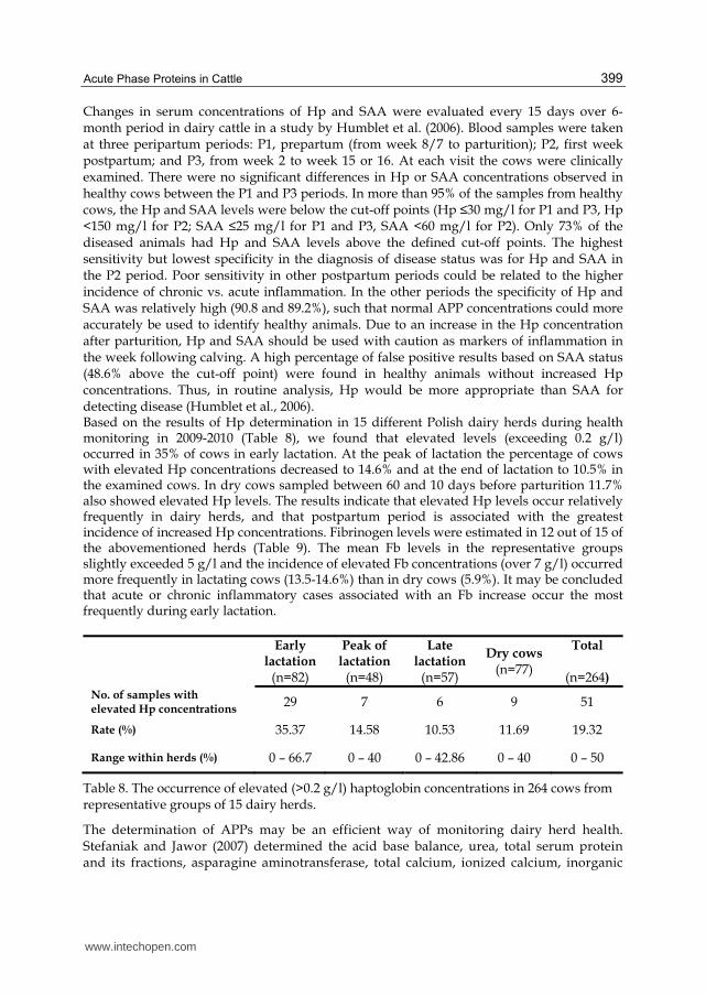
Changes in serum concentrations of Hp and SAA were evaluated every 15 days over 6-month period in dairy cattle in a study by Humblet et al. (2006). Blood samples were taken at three peripartum periods: P1, prepartum (from week 8/7 to parturition); P2, first week postpartum; and P3, from week 2 to week 15 or 16. At each visit the cows were clinically examined. There were no significant differences in Hp or SAA concentrations observed in healthy cows between the P1 and P3 periods. In more than 95% of the samples from healthy cows, the Hp and SAA levels were below the cut-off points (Hp ≤30 mg/l for P1 and P3, Hp <150 mg/l for P2; SAA ≤25 mg/l for P1 and P3, SAA <60 mg/l for P2). Only 73% of the diseased animals had Hp and SAA levels above the defined cut-off points. The highest sensitivity but lowest specificity in the diagnosis of disease status was for Hp and SAA in the P2 period. Poor sensitivity in other postpartum periods could be related to the higher incidence of chronic vs. acute inflammation. In the other periods the specificity of Hp and SAA was relatively high (90.8 and 89.2%), such that normal APP concentrations could more accurately be used to identify healthy animals. Due to an increase in the Hp concentration after parturition, Hp and SAA should be used with caution as markers of inflammation in the week following calving. A high percentage of false positive results based on SAA status (48.6% above the cut-off point) were found in healthy animals without increased Hp concentrations. Thus, in routine analysis, Hp would be more appropriate than SAA for detecting disease (Humblet et al., 2006). Based on the results of Hp determination in 15 different Polish dairy herds during health monitoring in 2009-2010 (Table 8), we found that elevated levels (exceeding 0.2 g/l) occurred in 35% of cows in early lactation. At the peak of lactation the percentage of cows with elevated Hp concentrations decreased to 14.6% and at the end of lactation to 10.5% in the examined cows. In dry cows sampled between 60 and 10 days before parturition 11.7% also showed elevated Hp levels. The results indicate that elevated Hp levels occur relatively frequently in dairy herds, and that postpartum period is associated with the greatest incidence of increased Hp concentrations. Fibrinogen levels were estimated in 12 out of 15 of the abovementioned herds (Table 9). The mean Fb levels in the representative groups slightly exceeded 5 g/l and the incidence of elevated Fb concentrations (over 7 g/l) occurred more frequently in lactating cows (13.5-14.6%) than in dry cows (5.9%). It may be concluded that acute or chronic inflammatory cases associated with an Fb increase occur the most frequently during early lactation.
Early
lactation (n=82)
Peak of lactation
(n=48)
Late lactation
(n=57)
Dry cows (n=77)
Total
(n=264)
No. of samples with elevated Hp concentrations
29 7 6 9 51
Rate (%) 35.37 14.58 10.53 11.69 19.32
Range within herds (%) 0 – 66.7 0 – 40 0 – 42.86 0 – 40 0 – 50
Table 8. The occurrence of elevated (>0.2 g/l) haptoglobin concentrations in 264 cows from representative groups of 15 dairy herds.
The determination of APPs may be an efficient way of monitoring dairy herd health. Stefaniak and Jawor (2007) determined the acid base balance, urea, total serum protein and its fractions, asparagine aminotransferase, total calcium, ionized calcium, inorganic
www.intechopen.com

Acute Phase Proteins as Early Non-Specific Biomarkers of Human and Veterinary Diseases
400
phosphate, magnesium and Hp and Fb levels in representative groups of cows from two dairy farms with a loose-housing system. Farm A (600 Polish Holstein-Fresian cows; milk yield 10,700 kg) and farm B (170 Polish black and white and Polish red and white × Holstein-Fresian cows; milk yield 6500 kg) were sampled six times at two monthly intervals. Blood samples were taken from groups of cows: group I – 10 cows between 14 and 150 days in milk (DIM), group II – 10 cows more than 150 DIM, group III – 10 dry cows. After each monitoring period the results were discussed with the owner and a local veterinarian and recommendations were proposed. Repeated laboratory monitoring helped to normalize the analysed parameters. The course of Hp and Fb levels and the decreasing frequency of elevated concentrations indicated improvements in the cows’ health. Any disturbances that occurred during herd monitoring mainly came from the owners’ or farm workers’ mistakes or a negligence at project completion. Herd health monitoring using APP measurement needs a close cooperation between the veterinarian and the owner.
Group Mean % (range)
Mean concentration
(g/l)
<3 g/l 3-5 g/l 5-7 g/l >7 g/l
Early lactation (n=74)
2.7% (0-20)
31.08% (0-77.8)
52.7% (0-100)
13.51% (0-33.3)
5.53
Lactation peak (n=57)
3.51% (0-33.3)
31.58% (0-66.7)
50.88% (0-100)
14.04% (0-66.7)
5.32
Late lactation (n=55)
5.45% (0-33.3)
52.73% (0-100)
27.27% (0-100)
14.55% (0-66.7%)
5.16
Dry cows (n=68)
7.35% (0-50%)
45.59% (0-100)
41.18% (0-80)
5.88% (0-33,3)
5.01
Total (n=254)
4.74% (0-29.17)
37.94% (6.67-55.17)
44.66% (12.5-80)
12.65% (0-42.86)
5.26
Table 9. Fibrinogen concentrations in 254 representative cows from 12 dairy farms.
The determination of APP levels seems to be one of better methods allowing the health and welfare of animals in large herds, based on the examination of blood or milk samples obtained from representative groups of animals (Nikołajczuk & Molenda 2000). When the results from representative groups are within the reference range, they may provide a basis for considering the herd health status as “good”. In our practice we simultaneously utilize Hp, Fb and albumin levels, and with this combination we can detect more cases of subclinical acute and chronic inflammation. For many years the estimation of APPs has provided an important source of knowledge about herd health. The determination of APPs along with other parameters during the metabolic monitoring of herds may provide a complete view of herd health.
4. Conclusions
Sick cattle have lower milk yields/body growth rates, causing significant direct and indirect economical losses. Because of the group housing systems, it is difficult to early diagnose an
www.intechopen.com
Acute Phase Proteins in Cattle
401
inflammation in individual animals. Despite these difficulties, handling strategies should focus on herd health protection and the early detection of threats, as well as the introduction of improved therapeutic procedures before animals become ill. The determination of APPs levels in blood and milk samples from representative groups of cows is an attractive method, that also enables determination of other biochemical parameters, providing an useful prognostic tool of the cows’ health status and the degree of possible threats. Multiple estimations of such parameters in the same herd can provide information, which could be used to improve animal handling. Among the many available APPs for cattle diagnostics, the most commonly used and the most readily available are Hp, SAA and Fb. In addition to a rise in APP concentration, the degree of increase can be used to evaluate herd health. The finding that the concentration of some APPs, such as SAA and Hp, increase in stressful conditions, as well as in fatty liver syndrome, broaden the possible diagnostic applications of APPs in cattle. A future diagnostic application of APPs is their use as part of pre-slaughter examination of slaughter animals. Such procedures should significantly improve the detection rate of animals suffering from subclinical inflammatory processes; which lead to significant losses because of confiscation of whole carcasses, or parts, showing signs of pathology/inflammation during postmortem/postslaughter examination. Implementation of such procedures on farms, before animals are transported to the slaughterhouse, would enable detection of ill animals for treatment, allowing them to be sent for slaughter after recovery. In spite of the time required, the determination of APPs has potential importance when deciding whether to treat ill animals or send them for sanitary slaughter to rescue the meat value. The presence of strongly elevated APP concentrations before treatment, combined with poor prognosis, may be helpful in making decision about therapy i.e. do not start. Moreover, if treatment has begun, the determination of APPs might help in monitoring the course of the disease and the efficacy of the chosen therapy. The results of our studies indicate that appropriate time points for collecting blood samples for monitoring most of diseases in cattle are: immediately before starting treatment; and after 3 to 7 days of treatment. A stepwise decrease in APP concentrations, and a return to normal (or near normal) levels, confirms the positive effect of treatment. There is a broad spectrum of possible applications of APP-based diagnostics for use with cattle. It is necessary to develop and optimise rapid field tests that allow determination of APPs in a short time period, after collection of blood or milk samples from animals.
5. References
Åkerstedt, M.; Björck, L.; Persson Waller, K. & Sternesjö, Å. (2006). Biosensor assay for determination of haptoglobin in bovine milk. Journal of Dairy Research, Vol.73, No.3, (August 2006), pp. 299-305
Alsemgeest, S.P.M.; Taverne, M.A.M.; Boosman, R.; Weyden, B.C. & Gruys, E. (1993). Peripartum acute-phase protein serum amyloid-A concentration in plasma of cows and fetuses. American Journal of Veterinary Research, Vol.54, No.1, (January 1993), pp.164-167
Alsemgeest, S.P.M.; Lambooy, I.E.; Wierenga, H.K.; Dieleman, S.J.; Meerkerk, B.; van Ederen, A.M. & Niewold, T.A. (1995a). Influence of physical stress on the plasma
www.intechopen.com
Acute Phase Proteins as Early Non-Specific Biomarkers of Human and Veterinary Diseases
402
concentration of serum amyloid-A (SAA) and haptoglobin (Hp) in calves. Veterinary Quarterly, Vol.17, No.1, (March 1995) pp. 9-12
Alsemgeest, S.P.M.; Jonker, F.H.; Taverne, M.A.M.; Kalsbeek, H.C.; Wensing, T.& Gruys, E. (1995b). Serum amyloid-A (SAA) and haptoglobin (Hp) plasma concentrations in newborn calves. Theriogenology, Vol.43, No.2, (January 1995), pp. 381-387
Balbierz, H.; Nowacki, W.; & Russ, T. (1977). Some properties of the HRP factor (Hemoglobin Reacive Protein) in cattle (in Polish, abstract in English). Polskie Archiwum Weterynaryjne Vol.20, No.1, pp. 87-95
Bertoni, G.; Calamari, L.; Maianti, M.G. & Cappa, V. (1997). Factors other than milk yield that affect reproductive traits of dairy cows. Livestock Production Science, Vol.50, no.1, (October 1997), pp. 99-100
Berry, B.A.; Confer, A.W; Krehbiel, C.R.; Gill, D.R.; Smith ,R.A. & Montelongo, M. (2004). Effects of dietary energy and starch concentrations for newly received feedlot calves: II. Acute-phase protein response. Journal of Animal Science, Vol.82, No.3, (March 2004), pp. 845-850
Bicalho, R.C.; Vokey, F.; Erb, H.N. & Guard, C.L. (2007). Visual locomotion scoring in the first seventy days in milk: impact on pregnancy and survival. Journal of Dairy Science, Vol.90, No.10, (October 2007), pp. 4586-4591
Cairoli, F.; Battocchio, M.; Veronesi, M.C.; Brambilla, D.; Conserva, F.; Eberini, I.; Wait, R. & Gianazza, E. (2006). Serum protein pattern during cow pregnancy: acute-phase proteins increase in the peripartum period. Electrophoresis, Vol.27, No.8, (April 2006), pp. 1617-1625
Carter, J.N.; Meredith, G.L.; Montelongo, M.; Gill, D.R.; Krehbiel, C.R.; Payton, M.E. & Confer, A.W. (2002). Relationship of vitamin E supplementation and antimirobial treatment with acute-phase protein responses in cattle affected by naturally acquired respiratory tract disease. American Journal of Veterinary Research, Vol.63, No.8, (August 2002), pp. 1111-1117
Chan, J.P-W.; Chang, C-C.; Hsu, W-I.; Liu, W-B. & Chen, T-H. (2010). Association of increased serum acute-phase protein concentrations with reproductive performance in dairy cows with postpartum metritis. Veterinary Clinical Pathology, Vol.39, No.1, (March 2010), pp. 72-78
Conner, J.G;. Eckersall, P.D.; Doherty, M. & Douglas, T.A. (1986). Acute phase response and mastitis in the cow. Research in Veterinary Science, Vol.41, No.1, (July 1986) pp. 126-128
Conner, J.G. & Eckersall, P.D. (1988). Bovine acute phase response following turpentine injection. Research in Veterinary Science, Vol.44, No.1, (January 1988), pp. 82-88
Conner, J.G.; Eckersall, P.D.; Wiseman, A.; Bain, R.K. & Douglas, T.A. (1989). Acute phase response in calves following infection with Pasteurella haemolytica, Ostertagia ostertagi and endotoxin administration. Research in Veterinary Science, Vol.47, No.2, (September 1989), pp. 203-207
Deignan, T.; Alwan, A.; Kelly, J.; McNair, J.; Warren, T. & O’Farrelly, C. (2000). Serum haptoglobin: an objective indicator of experimentally-induced Salmonella infection in calves. Research in Veterinary Science, Vol.69, No.2, (October 2000), pp. 153-158
Dowling, A.; Hodgson, J.C.; Schock, A.; Donachie, W.; Eckersall, P.D. & McKendrick, I.J. (2002). Experimental induction of pneumonic pasteurellosis in calves by
www.intechopen.com
Acute Phase Proteins in Cattle
403
intratracheal infection with Pasteurella multocida biotype A:3. Research in Veterinary Science, Vol.73, No.1, (August 2002), pp. 37-44
Dubuc, J.; Duffield, T.F.; Leslie, K.E.; Walton, J.S. & Leblanc, S.J. (2010). Risk factors for postpartum uterine diseases in dairy cows. Journal of Dairy Science, Vol.93, No.12, (December 2010), pp. 5764-5771
Eckersall, P.D. & Conner, J.G. (1988). Bovine and canine acute phase proteins. Veterinary Research Communications, Vol.12, No.2-3, pp. 169-178
Eckersall, P. D. (2000). Recent Advances and Future Prospects for the Use of Acute Phase Proteins as Markers of Disease in Animals. Revue de Médecine Vétérinaire, Vol.151, No.7, pp. 577-584
Eckersall, P.D.; Young, F.J.; McComb, C.; Hogarth, C.J.; Safi, S.; Weber, A.; McDonald, T.; Nolan, A.M. & Fitzpatrick, J.L. (2001). Acute phase proteins in serum and milk from dairy cows with clinical mastitis. The Veterinary Record, Vol. 148, No.2, (January 2001), pp. 35-41
Esslemont, R.J. & Kossaibati, M.A. (1997). Culling in 50 dairy herds in England. The Veterinary Record, Vol.140, No.2 (January 1997), pp. 36-39
Furman-Fratczak, K.; Rzasa, A. & Stefaniak T. (2011). The influence of colostral immunoglobulin concentration in heifer calves’ serum on their health and growth. Journal of Dairy Science (accepted for publication)
Ganheim, C.; Hulten, C.; Carlsson, U.; Kindahl, H.; Niskanen, R. & Waller, K.P. (2003). The acute phase response in calves experimentally infected with Bovine Viral Diarrhoea Virus and/or Mannheimia Haemolytica. Journal of Veterinary Medicine B, Vol.50, No.4, (May 2003), pp. 183 -190
Ganheim, C.; Alenius, S. & Persson Waller K. (2007). Acute phase proteins as indicators of calf herd health. The Veterinary Journal, Vol.173, No.3, (May 2007) pp. 645-651
Godson, D.L.; Baca-Estrada, M.E.; Van Kessel, A.G.; Hughes, H.P.; Morsy, M.A.; Van Donkersgoed, J.; Harland, R.J.; Shuster, D.E.; Daley, M.J. & Babiuk, L.A. (1995). Regulation of bovine acute phase responses by recombinant interleukin-1 beta. Canadian Journal of Veterinary Research, Vol.59, No.4, (October 1995), pp. 249-255
Godson, D.L; Campos, M.; Attah-Poku, S.K.; Redmond, M.J.; Cordeiro, D.M.; Sethi, M.S.; Harland, R.J. & Babiuk, L.A. (1996). Serum haptoglobin as an indicator of the acute phase response in bovine respiratory disease. Veterinary Immunology and Immunopatholology, Vol.51, No.3-4, (June 1996), pp. 277-292
Green, L.E.; Hedges, V.J.; Schukken, Y.H.; Biowey, R.W. & Packington, A.J. (2002). The impact of Clinical Lameness on Milk Yield of Dairy Cows. Journal of Dairy Science, Vol.85, No.9, (September 2002), pp. 2250-2256
Grell, N.S.; Tjørnehøj, K.; Larsen, L.E. & Heegard, P.M.H. (2005). Marked induction of IL-6, haptoglobin and INF-γ following experimental BRSV infection in young claves. Veterinary Immunology and Immunopathology, Vol.103, No.3-4, (February 2005), pp. 235-245
Grönlund, U.; Hulten, C.; Eckersall, P.D. & Hogarth, C. (2003). Haptoglobin and serum amyloid A in milk and serum during acute and chronic experimentally induced Staphylococcus aureus mastitis. Journal of Dairy Research, Vol.70, No.4, (November 2003), pp. 739-386
www.intechopen.com

Acute Phase Proteins as Early Non-Specific Biomarkers of Human and Veterinary Diseases
404
Grönlund, U.; Hallen Sandgren, C. & Persson Waller, K. (2005). Haptoglobin and serum amyloid A in milk from dairy cows with chronic sub-clinical mastitis. Veterinary Research, Vol.36, No.2, (March-April), pp.191-198
Gruys, E.; Ederen, A.M.; Alsemgeest, S.P.M.; Kalsbeek, H.C. & Wensing, T. (1993). Acute phase protein values in blood of cattle as indicator of animals with pathological process. Archiv für Lebensmittelhygiene, Vol.44, No.5, (September/October), pp.107-111
Gruys, E.; Toussaint, M.J.M.; Niewold, T.A.; Koopmans, S.J.; van Dijk, E. & Meloen, R.H. (2006). Monitoring health by values of acute phase proteins. Acta Histochemica, Vol.108, No.3, (May 2006), pp. 229—232
Heegaard, P.M.H.; Godson, D.L.; Toussaintc, M. J.M.; Tjùrnehùj, K.; Larsen, L. E.; Viuff, B. & Rùnsholt, L. (2000). The acute phase response of haptoglobin and serum amyloid A (SAA) in cattle undergoing experimental infection with bovine respiratory syncytial virus. Veterinary Immunology and Immunopathology, Vol.77, No.1-2, (November 2000), pp. 151-159
Heinrich, P.C.; Castell, J.V. & Andus, T. (1990). Interleukin-6 and the acute phase response. Biochemistry Journal, Vol.265, No.3, (February 1990), pp. 621-626
Hirvonen, J.; Pyörälä, S. & Jousimies-Somer, H. 1996. Acute phase response in heifers with experimentally induced mastitis. Journal of Dairy Research, Vol.63, No.3, (August 1996), pp. 351-360
Hirvonen, J.; Hietakorpi, S. & Saloniemi, H. (1997). Acute phase response in emergency slaughtered dairy cows. Meat Science, Vol.46, No.3, (July 1997), pp. 29-257
Hirvonen, J. & Pyörälä, S. (1998). Acute-phase response in dairy cows with surgically-treated abdominal disorders. The Veterinary Journal, Vol.155, No.1, (January 1998) pp. 53-61
Hirvonen, J.; Eklund, K.; Teppo, A.M.; Huszenicza, G.; Kulcsar, M.; Saloniemi, H. & Pyörälä, S. (1999a). Acute phase response in dairy cows with experimentally induced Escherichia coli mastitis. Acta Vererinaria Scandinavica, Vol. 40, No.1, pp. 35-46
Hirvonen, J.; Huszenicza, G.; Kulcsar, M. & Pyörälä, S. (1999b). Acute-phase response in dairy cows with acute postpartum metritis. Theriogenology, Vol.51, No.6, (April 1999) pp. 1071-1083
Hiss, S.; Mielenz, M.; Bruckmaier, R.M. & Sauerwein, H. (2004). Haptoglobin concentrations in blood and milk after endotoxin challenge and quantification of mammary hp mRNA expression. Journal of Dairy Science, Vol.87, No.11, (November 2004), pp. 3778-3784
Horadagoda, A.; Eckersall, P.D.; Alsemgeest, S.P.M. & Gibbs, H. A. (1993). Purification and quatnitative measurment of bovine serum amyloid-A. Research in Veterinary Science, Vol.55, No.3, (November 1993), pp. 317-325
Horadagoda, A. & Eckersall, P.D. (1994). Immediate responses in serum TNFα and acute phase protein concentrations to infection with Pasteurella haemolytica A1 in calves. Research in Veterinary Science, Vol.57, No.1, (July 1994), pp. 129-132
Horadagoda, N.U.; Eckersall, P.D.; Andrew, L.; Gallay, P.; Heumann, D. & Gibbs, H.A. (1995). Characterisation of bovine lipopolysaccharide binding protein and the in vivo acute phase response to Pasteurella haemolytica Type A. Veterinary Immunology and Immunopathology, Vol.49, No. 1-2 (November 1995), pp. 61-74
www.intechopen.com

Acute Phase Proteins in Cattle
405
Horadagoda, N.U.; Knox, K.M.G.; Gibbs, H.A.; Reid, S.W.J.; Horadagoda, A.; Edwards, S.E.R. & Eckersall, P.D. (1999). Acute phase proteins in cattle: discrimination between acute and chronic inflammation. The Veterinary Record, Vo.144, No.16, (April 1999), pp. 437-441
Humblet, M.F.; Coghe, J.; Lekeux, P. & Godeau, J.M. (2004). Acute phase proteins assessment for an early selection of treatments in growing calves suffering from bronchopneumonia under field conditions. Research in Veterinary Science, Vol.77, No.1, (August 2004), pp. 41–47
Humblet, M.F.; Guyot, H.; Boudry, B.; Mbayahi, F.; Hanzen, C.; Rollin, F. & Godeau, J.M. (2006). Relationship between haptoglobin, serum amyloid A, and clinical status in a survey of dairy herds during a 6-month period. Veterinary Clinical Pathology, Vol.35, No.2, (June 2006), pp. 188-193
Huzzey, J.M.; Duffield, T.F.; LeBlanc, S.J.; Veira, D.M.; Weary, D.M. & Keyserlingk, M.A.G. (2009a). Haptoglobin as an early indicator of metritis. Journal of Dairy Science, Vol.92, No.2, (February 2009), pp. 621-625
Huzzey, J.M.; Overton, T.R.; Nydam, D.V. & Grant, R.J. (2009b). Peripartum measures of stress, inflammation and energy status as predictors for postpartum health disorders in transition dairy cows. Journal of Animal Science, 87, E-Supplement 2, 120. Journal of Dairy Science, 92, E-Supplelent 1, 120
Irmak, K. & Turgut, K. (2005). Disseminated intravascular coagulation in cattle with abomasal displacement. Veterinary Research Communications, Vol.29, No.1, (January 2005), pp. 61-68
Jacobsen, S.; Andersen, P.H.; Toelboell T. & Heegard, P.M.H. (2004). Dose dependency and individual variability of the lipopolysaccharide-induced bovine acute phase protein response. Journal Dairy Science, Vol.87, No.10, (October 2004), pp. 3330-3339
Jawor, P. & Stefaniak, T. (2006). Acute phase proteins in treatment of calves with respiratory tract inflammation (in Polish, English abstract). Folia Universitatis Agriculturae Stetinensis, Zootechnica Vol.48, No.250, pp. 51-56
Jawor, P. (2007). Determination of haptoglobin and fibrinogen during monitoring herd health and selected inflammatory states in cattle. Ph. D thesis (in Polish), Wroclaw University of Environmental and Life Sciences
Jawor, P.; Hauser, S.; Baumgartner, W.; Stefaniak, T. & Rzasa, A. (2008). Determination of selected acute phase proteins during the treatment of limb diseases in dairy cows. Veterinarni Medicina, Vol.53, No.4, (April 2008), pp.173-183
Jawor, P.; Stefaniak, T.; Steiner, S. & Baumgarnter, W. (2009). Dynamics of Selected Acute Phase Proteins In Surgical Abomasum Reposition In Cows. Folia Veterinaria, Vol.53, Supplementum LIII, (June 2009), pp.18-21
Kent J. (1992). Acute phase proteins: their use in veterinary diagnosis. British Veterinary Journal, Vol.148, No.4, (July-August 1992), pp. 279-282
Krakowski, L. & Zdzisińska, B. (2007). Selected cytokines and acute phase proteins in heifers during the ovarian cycle course and in different pregnancy periods. Bulletin of the Veterinary Institute in Pulawy, Vol.51, No.1, pp.31-36
Kujala, M.; Orro, T. & Soveri, T. (2010). Serum acute phase proteins as a marker of infammation in dairy cattle with hoof diseases. The Veterinary Record, Vol.166, No.8, (February 2010), pp. 240-241
www.intechopen.com
Acute Phase Proteins as Early Non-Specific Biomarkers of Human and Veterinary Diseases
406
Laven, R.A.; Livesey, C.T. & May, S.A. (2004). Relationship between acute phase proteins and hoof horn haemorrhages in postpartum first-lactation heifers. The Veterinary Record, Vol.154, No.13, (March 2004), pp. 389-395
Makimura, S. & Usui, M. (1990). Correlation between haptoglobin and sialic acid or mucoprotein in diseased bovine serum. Japan Journal of Veterinary Medical Science, Vol.52, No.6, (December 1990), pp. 1245-1250
Mazur, A. (1980). Determination of HRP (Hemoglobin Reactive Protein) in sera and whey of cows by simple agar-electrophoresis (in Polish, English abstract). Medycyna Weterynaryjna, Vol.36, No.11, pp. 699-700
Mazur, A. & Mazur, O. (1981). Hemoglobin Reactive Protein (HRP) in case of udder inflammation in cows (in Polish, English abstract). Medycyna Weterynaryjna, Vol.37, No.5, pp. 262-264
McNair, J.; Elliott, C.; Bryson, D.G. & Mackie, D.P. (1998). Bovine serum transferrin concentration during acute infection with Haemophilus somnus. The Veterinary Journal, Vol.155, No.3, (May 1998), pp. 251-255
McSherry, B.J.; Horney, F.D. & deGroot, J.J. (1970). Plasma fibrinogen levels in normal and sick cows. Canadian Journal of comparative Medicine, Vol.34, No.3, (July 1970), pp. 191-197
Melendez, P.; Bartolome, J.; Archbald, L.F. & Donovan, A. (2003). The association between lameness, ovarian cysts and fertility in lactating dairy cows. Theriogenology Vol.59, No.3-4, (February 2003), pp. 927-937
Miller, L.L.; Bly, C.G.; Watson, M.L. & Bale, W.F. (1951). The dominant role of the liver in plasma protein synthesis; a direct study of the isolated perfused rat liver with the aid of lysine-epislon-C14. The Journal of Experimental Medicine, Vol.94, No.5, (November 1951), pp. 431-453
Molenaar, A.; Harris, P.; Green, J.; Callaghan, M.; Kim, K.; Wieliczko, R.; Johannessen, K.; Dines, R.; Watson, R.; Arkus, V.; Dalziel, J.; Singh, K.; Rajan, G.; Pearson, M.; Farr, V.; Miles, M.; Oden, K.; Good, L.; McLaren, R.; Prosser, C.; Grigor, M.; Davis, S. & Stelwagen, K. (2005). Mammary serum amyloid A3, progress toward determining structure and function. Proceedings of 5th International Colloquium on Animal Acute Phase Proteins. Dublin, March 2005
Mordak, R. (2008). Usefulness of haptoglobin for monitoring the efficiency of therapy of fetal membrane retention in cows (in Polish, English abstract). Medycyna Weterynaryjna Vol.64, No.4A (April 2008), pp. 434-437
Murata, H.; Shimada, N. & Yoshioka M. (2004). Current research on acute phase proteins in veterinary diagnosis: an overview. The Veterinary Journal, Vol.168, No.1 (July 2004) pp. 28–40
Nielsen, B.H.; Jacobsen, S.; Andersen, P.H.; Niewold, T.A. & Heegaard, P.M.H. (2004). Acute phase protein concentrations in serum and milk from healthy cows, cows with clinical mastitis and cows with extramammary inflammatory conditions. The Veterinary Record, Vol.154, No.12 (March 2004), pp. 361-365
Nikołajczuk, M., Molenda. (2000) Application of the acute phase proteins in veterinary diagnostics (in Polish, English abstract). Zeszyty Naukowe Akademii Rolniczej we Wrocławiu. Vol.29, No.390 (October 2000), pp. 9-14
Nikunen, S.; Härtel, H.; Orro, T.; Neuvonen, E.; Tanskanen, R.; Kivelä, S.L.; Sankari, S. Aho, P.; Pyörälä, S.; Saloniemi, H. & Soveri, T. (2007). Association of bovine respiratory
www.intechopen.com
Acute Phase Proteins in Cattle
407
disease with clinical status and acute phase proteins in calves. Comparative Immunology, Microbiology and Infectious Diseases, Vol.30, No.3, (May 2007), pp. 143-151
Ohtsuka, H.; Kudo, K.; Mori, K.; Nagai, F.; Hatsugaya, A.; Tajima, M.; Tamura, K.; Hoshi, F.; Koiwa, M. & Kawamura, S. (2001). Acute phase response in naturally occurring coliform mastitis. Journal of Veterinary Medical Science, Vol.63, No.6 (June 2001), pp. 675-678
Okamoto, K.; Harada, M.; Akuzawa, M. & Deguchi, E. (1998). Peripheral blood lymphocyte blastogenesis and concentrations of serum haptoglobin and alpha 1-acid glycoprotein in normal calves and calves on the first onset of diarrhea. Animal Science and Technology, Vol.69, No.7 (July 1998), pp. 683-689
Piercy, D.W.T. (1979). Acute phase responses to experimental salmonellosis in calves and colibacillosis in chickens: serum iron and caeruloplasmin. Journal of Comparative Pathology, Vol.89, No.3, (July 1979) pp. 309-319
Rajala-Schultz, P.J.; Gröhn, Y.T. & McCulloch, C.E. (1999). Effects of Milk Fever, Ketosis and Lameness on Milk Yield in Dairy Cows. Journal of Dairy Science, Vol.82, No.2, (February 1999), pp. 288-294
Saini, P.K.; Riaz, M.; Webert, D.W.; Eckersall, P.D.; Young, C.R.; Stanker, L.H.; Chakrabarti, E. & Judkins J.C. (1998). Development of a simple enzyme immunoassay for blood haptoglobin concentration in cattle and its application in improving food safety. American Journal of Veterinary Research, Vol.59, No.9, (September 1998), pp. 1101-1107
Schönfelder, A.; Schrödl, W.; Krüger, M.; Richter, A. & Sobiraj, A. (2005). The change of acute phase protein haptoglobin in cattle during spontaneous labor and Caesarean section with or without torsio uteri intrapartum (in German). Berliner und Munchener Tierarztliche Wochenschrift, Vol.118, No.5-6, (May-June 2005), pp.240-246
Schroedl, W.; Fuerll, B.; Reinhold, P.; Krueger, M. & Schuett, C. (2001). A novel acute phase marker in cattle: lipopolisacharide binding protein (LBP). Journal of Endotoxin Research, Vol.7, No.1, pp. 49-52
Sivula, N.J.; Ames, T.R.; Marsh, W.E. & Werdin, R.E. (1996). Descriptive epidemiology of morbidity and mortality in Minnesota dairy heifer calves. Preventive Veterinary Medicine, Vol.27, No.3-4, (July 1996), pp. 155-171
Skinner, J.G.; Brown, R.A.L. & Roberts, L. (1991). Bovine haptoglobin response in clinically defined filed conditions. The Veterinary Record, Vol.128, No.7, (February 1991), pp. 147-149
Smith, B.I.; Donovan, G.A.; Risco, C.A.; Young, C.R. & Stanker, L.R. (1998). Serum haptoglobin concentrations in Holstein dairy cattle with toxic puerperal metritis. The Veterinary Record, Vol.142, No.4, (January 1998) pp. 83-85
Smith, B.; Kauffold, J. & Sherman, L. (2010). Serum haptoglobin concentrations in dairy cattle with lameness due to claw disorders. The Veterinary Journal, Vol.186, No.2, (November 2010) pp. 162-165
Spooner, R.L. & Miller, J.K. (1971). The measurement of haemoglobin reactive protein in ruminants as an aid to the diagnosis of acute inflammation. The Veterinary Record, Vol.88, No.1, (January 1971) pp. 2-4
Stefaniak, T.; Nowacki, W.; Molenda, J.; Sobiech, E. & Nikołajczuk M. (1997). Monitoring selected blood parameters in calves immunized with Somnuvac® vaccine in field
www.intechopen.com
Acute Phase Proteins as Early Non-Specific Biomarkers of Human and Veterinary Diseases
408
conditions (in Polish, English abstract). Medycyna Weterynaryjna, Vol.53, No.11 (November 1997) pp. 668-673
Stefaniak, T. (2003). Acute phase proteins as a markers of animal welfare (in Polish). In: Acute phase proteins in Animals, Kostro K. & Gliński Z. (Ed.), 247-249, Wydawnictwo Akademii Rolniczej w Lublinie, Lublin, Poland
Stefaniak T., Galli J., Borkowski J., Jawor P. (2003). Utilization of acute phase proteins in health monitoring of young ruminants (in Polish, English abstract). Folia Univsitatis Agriculturae Stetinensis, 233, Zootechnica 45, pp. 81-86
Stefaniak, T.& Jawor, P. (2007). Large dairy herds health monitoring. Proceedings of 13th International Conference “Production Diseases In Farm Animals”, p. 351, Leipzig, Germany, July 29th – August 4th 2007
Svensson, C.; Lundborg, K.; Emanuelson, U. & Olsson S.O. (2003). Morbidity in Swedish dairy calves from birth to 90 days of age and individual calf-level risk factors for infectious diseases. Preventive Veterinary Medicine, Vol.58, No.3-4, (May 2003) pp. 179–197
Svensson, C.; Liberg, P. & Hultgren, J. (2007). Evaluating the efficacy of serum haptoglobin concentration as indicator of respiratory-tract disease in dairy calves. The Veterinary Journal, Vol.174, No.2, (September 2007) pp. 288-294
Taylor, J.A. (2006). Leukocyte Responses in Ruminants, In: Schalm’s Veterinary Hematology Ed.Feldman A.B.F., Zinkl J.G., Jain N.C. pp. 391-404 Ed. Blackwell Publishing Professional, Iowa, USA
Toussaint, M.J.M.; van Ederen, A.M. & Gruys, E. (1995). Implication of clinical pathology in assessment of animal health and in animal production and meat inspection. Comparative Haematology International, Vol.5, No.3, pp. 149-157
Tomas J.S. (2000). Overview of plasma proteins, In: Schalm’s Veterinary Hematology Ed.Feldman A.B.F., Zinkl J.G., Jain N.C. pp. 891-898 Ed. Lippincott Williams & Wilkins, Canada
Uchida, E.; Katoh, N. & Takahashi, K. (1993). Appearance of haptoglobin in serum from cows at parturition. Journal of Veterinary Medical Science, Vol.55, No.5, (October 1993) pp. 893-894
Wither, T.; Bienvenu, J. & Price, C.P. (1991). Molecular biology, measurement and clinical utility of the acute phase proteins. Pure and Applied Chemistry, Vol.63, No.8, (October 1991) pp. 1111-1116
Yoshida, Y. (1991). Electrophoretic studies on serum proteins in cows with traumatic pericarditis. Journal of Veterinary Medical Science, Vol.53, No.1, (February 1991) pp. 5-11
www.intechopen.com

Acute Phase Proteins as Early Non-Specific Biomarkers of Humanand Veterinary DiseasesEdited by Prof. Francisco Veas
ISBN 978-953-307-873-1Hard cover, 408 pagesPublisher InTechPublished online 10, October, 2011Published in print edition October, 2011
InTech EuropeUniversity Campus STeP Ri Slavka Krautzeka 83/A 51000 Rijeka, Croatia Phone: +385 (51) 770 447 Fax: +385 (51) 686 166www.intechopen.com
InTech ChinaUnit 405, Office Block, Hotel Equatorial Shanghai No.65, Yan An Road (West), Shanghai, 200040, China
Phone: +86-21-62489820 Fax: +86-21-62489821
The two volumes of Acute Phase Proteins book consist of chapters that give a large panel of fundamental andapplied knowledge on one of the major elements of the inflammatory process during the acute phaseresponse, i.e., the acute phase proteins expression and functions that regulate homeostasis. We haveorganized this book in two volumes - the first volume, mainly containing chapters on structure, biology andfunctions of APP, the second volume discussing different uses of APP as diagnostic tools in human andveterinary medicine.
How to referenceIn order to correctly reference this scholarly work, feel free to copy and paste the following:
Jawor Paulina and Stefaniak Tadeusz (2011). Acute Phase Proteins in Cattle, Acute Phase Proteins as EarlyNon-Specific Biomarkers of Human and Veterinary Diseases, Prof. Francisco Veas (Ed.), ISBN: 978-953-307-873-1, InTech, Available from: http://www.intechopen.com/books/acute-phase-proteins-as-early-non-specific-biomarkers-of-human-and-veterinary-diseases/acute-phase-proteins-in-cattle