Embed Size (px)
Citation preview
7/26/2019 14. INFECTED Odontogenic Keratocyst.20141212075824
http://slidepdf.com/reader/full/14-infected-odontogenic-keratocyst20141212075824 1/4
Kang GS et al. Infected Odontogenic Keratocyst.
85Journal of Advanced Medical and Dental Sciences Research |Vol. 2|Issue 4| October-December 2014
INFECTED ODONTOGENIC KERATOCYST- A CASE REPORT
Gurbinder S Kang BDS (Intern), Sri Guru Ram Das Institute of Dental sciences & Research,Amritsar, Punjab, Dilawarjit Kaur, BDS
Corresponding Author: Dr. Gurbinder S Kang BDS (Intern), Sri Guru Ram Das Institute of
Dental sciences & Research, Amritsar, Punjab.
This article may be cited as: Kang GS, Kaur D. Infected Odontogenic Keratocyst- A Case
Report. J Adv Med Dent Scie Res 2014;2(4):85-88.
IntroductionThe term ‘odontogenic keratocyst’ was
introduced by Philipsen in 1956. It has a
bimodal age of occurrence, first peak in
second decade and the second peak in fifth
decade.1
Odontogenic keratocyst (OKC)
is a developmental odontogenic cyst,characterized by unique and distincthistopathologic characteristics and an
aggressive biologic behavior.2-6
Theaggressive clinical behavior and frequent
recurrence following curettage has been the
focus of several studies, which indicatedthat the OKC epithelial lining may have
some intrinsic growth potential.7-12
The epithelial lining is a regular continuouslayer of stratified epithelium, usually 6-8
cells thick with prominent basal cells which
are columnar and have palisaded nuclei.
Suprabasal ecells frequently show
intracellular edema and often an abrupt
transition between them and the surface
layer of parakeratin.14
The keratinized
epithelial lining can change into non-
keratinized epithelium because of
inflammation.5,13
It is possible that
inflammation may alter not only themorphology but also the proliferativepotential of the epithelial lining. The
capsule is of thin fibrous tissue which isusually free from inflammatory cell
infiltrate except for occasional focal
accumulations.
Case Report
A 18years old female patient reported to the
college with complaint of pain and pusdischarge from lower intraoral back regionsince one and a half months. Extraoral
features showed presence of minimal
Case Report
Abstract:Odontogenic keratocyst (OKC) is a developmental odontogenic cyst, characterized by unique
and distinct histopathologic characteristics and an aggressive biologic behavior. The aggressiveclinical behavior and frequent recurrence following curettage has been the focus of severalstudies, which indicated that the OKC epithelial lining may have some intrinsic growth
potential. Histopathologically the epithelial lining is a regular continuous layer of keratinized
stratified epithelium, usually 6-8 cells thick. The keratinized epithelial lining can change intonon-keratinized epithelium because of inflammation. It is possible that inflammation may alter
not only the morphology but also the proliferative potential of the epithelial lining. Here wepresent a case report of a 18 year female who was diagnosed with an infected odontogenickeratocyst.
Ke words: Inflammation, odonto enic keratoc st.
7/26/2019 14. INFECTED Odontogenic Keratocyst.20141212075824
http://slidepdf.com/reader/full/14-infected-odontogenic-keratocyst20141212075824 2/4
Kang GS et al. Infected Odontogenic Keratocyst.
86Journal of Advanced Medical and Dental Sciences Research |Vol. 2|Issue 4| October-December 2014
swelling on the left side of the face inmandibular body and ramus region.
Intraorally, a swelling measuring 1x1cm
was seen in the retromolar and extending to
37 & 38 region. The swelling was showing
smooth surface, normal colour & hard inconsistency. Patient gave a history of
extraction of 37 and 38 and had undergone
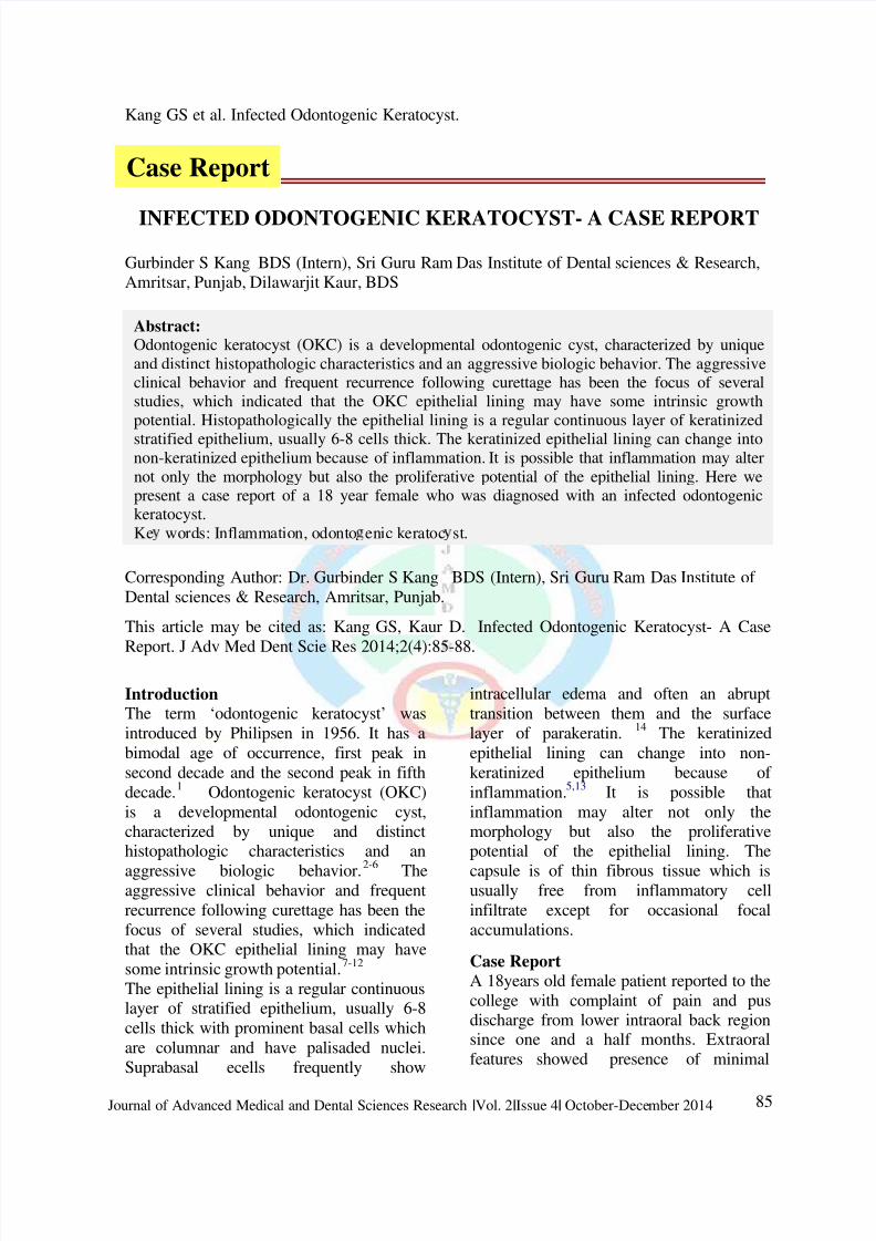
marsupialization in the same region. OPG
revealed multilocular radiolucency
extending from distal of 36 upto the angle
of the mandible with involvement of the
whole ramus along with the coronoid
process of mandible (Figure 1).
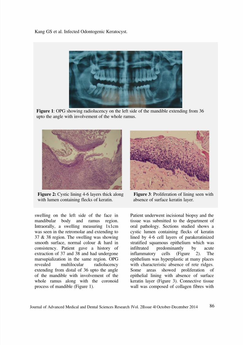
Patient underwent incisional biopsy and thetissue was submitted to the department of
oral pathology. Sections studied shows a
cystic lumen containing flecks of keratin
lined by 4-6 cell layers of parakeratinized
stratified squamous epithelium which wasinfiltrated predominantly by acute
inflammatory cells (Figure 2). The
epithelium was hyperplastic at many places
with characteristic absence of rete ridges.
Some areas showed proliferation of
epithelial lining with absence of surface
keratin layer (Figure 3). Connective tissue
wall was composed of collagen fibres with
Figure 1: OPG showing radiolucency on the left side of the mandible extending from 36
upto the angle with involvement of the whole ramus.
Figure 2: Cystic lining 4-6 layers thick along Figure 3: Proliferation of lining seen with
with lumen containing flecks of keratin. absence of surface keratin layer.
7/26/2019 14. INFECTED Odontogenic Keratocyst.20141212075824
http://slidepdf.com/reader/full/14-infected-odontogenic-keratocyst20141212075824 3/4
Kang GS et al. Infected Odontogenic Keratocyst.
87Journal of Advanced Medical and Dental Sciences Research |Vol. 2|Issue 4| October-December 2014
dense infiltration of both acute and chronic
inflammatory cells.
DiscussionAlthough OKC is classified as a
developmental cyst, inflammation in the
connective tissue wall of OKC has been
found in almost 75% of the cases reported
in the literature.5,13
In the present case,
certain degree of inflammation was seenwhich correlates with the histopathological
appearance of infected OKC.
Transformation of the keratinized epithelial
lining to non-keratinized epithelium is
common in OKC, and inflammation hasbeen suggested to be responsible.5,13
The
morphologic alterations in the epithelial
lining of OKC in the presence ofinflammation may also be associated withchanges in the proliferative potential, thus
affecting its biologic behavior. Source of
inflammation in the present case can be
ascribed to the infection with respect to 37
and 38 for which they had been extracted.
The presence of inflammation may partly
be attributed to possible communicationswith the oral mucosa via perforations of thecortical bone, which have been documented
in up to 39% of OKC.15
Inflammation may
also be introduced via the periodontal
ligament in cases located close to adjacent
teeth.15
Inflammation has a puzzling effect on theepithelial lining of different origins. In
several pathologic conditions, inflammationresults in epithelial hyperplasia and
metaplasia, e.g., radicular cysts, gastric
epithelial-cell proliferation related to
mucosal inflammation, prostatic
hyperplasia, metaplasia in the nasal
epithelium, and metaplastic polyp of the
colon.15
Several recent studies have found a
direct influence of the inflammation onepithelial cells, either through direct
adhesion of the inflammatory cells, or
through an indirect response to a series of
chemokines produced by inflammatory
cells.15
In the presence of inflammatory
reaction in an odontogenic keratocyst, thedegree of keratinization over these areas
would be altered and this was likely toincrease the permeability of the lining and
result in a soluble protein level in the fluid
higher than in the uninflammed keratinizing
cysts. As a result of inflammation various
cytokines such as IL2, IL3 will be
overexpressed which will further lead to the
expression of IL2β, β convertase,
prostaglandins E2 and coactivatecompliment. This in turn, would lead to
vascular permeability and leukotactic
response. In odontogenic keratocyst theinflammation may result in the changes in
the epithelium from keratinized to nonkeratinized epithelium.16
Conclusion
As reported in the previous literature and as
seen in the present case, the presence ofinflammation can obscure the characteristichistological appearance of the odontogenic
keratocyst lining by causing a transition
from keratinized to non keratinizedepithelium. Such cases can prove to be a
diagnostic dilemma, hence they should be
evaluated very cautiously by correlating the
clinical, radiographic and histological
features.
References1.
Browne RM. The odontogenic
keratocyst. Brit Dent J. 1970; 3:225-
238. 2. Kramer IRH, Pindborg JJ, Shear M.
Histologic typing of odontogenictumours. Berlin:Springer Verlag; 1992.
3. Shear M. Cysts of the oral region. 3rd
ed. Oxford: Wright, ButterworthHeinemann; 1992.
4.
Shear M. Developmental odontogenic
cysts. An update. J Oral Pathol Med
1994;23:1–11.
5. El-Hajj G, Anneroth G. Odontogenic
keratocysts––a retrospective clinical
7/26/2019 14. INFECTED Odontogenic Keratocyst.20141212075824
http://slidepdf.com/reader/full/14-infected-odontogenic-keratocyst20141212075824 4/4
Kang GS et al. Infected Odontogenic Keratocyst.
88Journal of Advanced Medical and Dental Sciences Research |Vol. 2|Issue 4| October-December 2014
and histologic study. Int J Oral
Maxillofac Surg 1996;25: 124–9.6. Stoelinga PJ. Long-term follow-up on
keratocysts treated according to adefined protocol. Int J Oral Maxillofac
Surg 2001;30:14–25.
7.
Li T-J, Browne RM, Mathews JB.
Epithelial cell proliferation in
odontogenic keratocysts: a comparative
immunocytochemical study of Ki-67 in
simple, recurrent and basal cell naevus
syndrome (BCNS)-associated lesions. JOral Pathol Med 1995;24:221.
8.
Li T-J, Browne RM, Mathews JB.
Quantification of PCNA positive cellswithin odontogenic jaw cyst
epithelium. J Oral Pathol Med1994;23:184–9.
9. Ogden GR, Chisholm DM, Kiddie RA,
Lane DP. p53 protein in odontogenic
cysts: increased expression in someodontogenic keratocysts. J Clin Pathol
1992;45:1007–10.Shear M. The
aggressive nature of the odontogenic
keratocyst: is it a benign cystic
neoplasm? Part I. Clinical and earlyexperimental evidence of aggressive
behavior. Oral Oncol 2002;38:219–26.
10. Shear M. The aggressive nature of the
odontogenic keratocyst: is it a benigncystic neoplasm? Part II. Proliferation
and genetic studies. Oral Oncol2002;38:323–31.
11.
Shear M. The aggressive nature of the
odontogenic keratocyst: is it a benign
cystic neoplasm? Part III.
Immunocytochemistry of cytokeratin
and other epithelial cell markers. Oral
Oncol 2002;38:407–15.
12. Rodu B, Tate AL, Martinez Jr MG. Theimplication of inflammation in
odontogenic keratocysts. J Oral Pathol
1987;16:518–21.13. Browne RM. The odontogenic
keratocyst. histological features andtheir correlation with clinical
behaviour. Brit Dent J. 1971;131:249-
59.
14. Kaplan, Hirshberg. The correlationbetween epithelial cell proliferation and
inflammation in odontogenic
keratocyst. Oral Oncol 2004;40:985-
91.
15.
Shear M. Cysts of the oral region.3rd ed. Oxford: Wright, Butterworth-
Heinemann; 1992.
Source of support: NilConflict of interest: None declared