Embed Size (px)
Citation preview
MAGNETIC RESONANCE IN MEDICINE 12,400-406 ( 1989)
I3C Spectroscopic Imaging. A Simple Approach to in ~ i v o 13C Investigations *
s. MULLER AND N. BECKMANN
MR Center of the University, Klingelbergstrasse SO, CH-4031 Basel, Switzerland
Received June 20, 1989; revised September 15, 1989
I3C spectroscopic imaging (SI) is implemented and tested on a Siemens whole-body MR instrument for in vivo localized "C spectroscopy. The method provides spectro- scopic maps of extended regions of interest. It is found that 13C SI is an ideal field of application for the spectroscopic imaging technique since most other localization meth- ods suffer from chemical-shift or offset artifacts for this nucleus. Applications of 13C SI with a single "C rfchannel as well as in combination with polarization transfer experi- ments and heteronuclear broadband decoupling are shown. o 1989 Academic hess , Inc.
INTRODUCTION
In vivo 13C magnetic resonance spectroscopy (MRS) is a fascinating tool for the noninvasive determination of metabolic functions. For several years whole-body MR units which allow I3C spectroscopy experiments on humans have been available (see ( I ) and references therein). It is interesting to note that most previous workers using I3C localization techniques have relied on the surface coil approach for excitation and detection of the I3C signals. This technique is straightforward to implement, is sensitive, and allows the acquisition of high-resolution spectra averaged over the whole measurement volume of the coil. In some cases the localization was improved with pulse sequences which accentuate the E l field distribution of the surface coil (e.g., depth pulses ( 2 ) ) . As a result the sensitive volume of the rf coil is further re- stricted to regions characterized by specific B1 field intensities.
For many situations, however, it would be advantageous to apply more versatile localization techniques. For 31P and 'H spectroscopy a number of powerful methods exist (ISIS ( 3 ) , VEST/STEAM ( 4 ) , rotating-frame spectroscopy ( 5 ) ) which allow the image-guided selection of a volume of interest for localized spectroscopy. Most of these techniques however cannot be applied for 13C due to the broad chemical- shift range of this nucleus: frequency-selective 13C rf pulses in combination with Bo gradients lead to a strong chemical-shift dispersion in the spatial dimension such that each chemically shifted species has a different spatial origin ( 6 ) . Other techniques which make use of rf excitation with a narrow excitation bandwidth (e.g., rotating- frame spectroscopy ( 5 ) ) lead to offset truncation in the spectra.
* A preliminary account of this work was given in "Proceedings, Eighth Annual Meeting, Society of Magnetic Resonance in Medicine, Amsterdam, 1989," p. 657.
400 0740-3 194/89 $3.00 Copyright 0 1989 by Academic Press, Inc. All rights of reproduction in any form reserved.

COMMUNICATIONS 40 1
We have recently proposed to perform volume selection in proton space and trans- fer the localized magnetization to 13C via polarization transfer ( 7). This technique strongly reduces the chemical-shift artifact. In addition the sensitivity of the 13C spec- tra is increased by approximately a factor of four and editing of the spectra is possible. We have successfully implemented this technique on our whole-body instrument (8); nevertheless, the method has two general drawbacks: ( 1 ) Two separate rf chan- nels are required for ‘H and I3C which are not yet standard equipment for most whole-body scanners. (2) Despite the possibility of shaping the volume according to the desired geometry, the acquired signal still represents an integrated response from the whole sample. This comes from the fact that the volume selection is only intro- duced during the excitation period of the experiment while the acquisition of the signal is not local ( 9 ) . Therefore if signal contributions from outside the selected region persist, they would inevitably mix with the desired signals and remain unsepa- rable from the spectrum of interest.
Here we present a different approach to in vivo 13C spectroscopy. It is based on spectroscopic imaging (SI) first described by Brown et al. in 1982 (10) . In spectro- scopic imaging the spatial resolution is introduced via pulsed Bo gradients which en- code positional information into the phase of the MR signal. Similar to the phase encoding in MR imaging a sequence of gradient values generates a complete set of nuclear signals which can be spatially and spectroscopically resolved after multidi- mensional Fourier transform. The result is an n + 1 -dimensional map with n spatial dimensions and one spectroscopic axis.
Spectroscopic imaging applied to I3C has the following advantages:
1. The phase encoding with pulsed Bo gradients does not introduce any chemical- shift dispersion in the spatial dimension; spatial information is completely separated from the spectroscopic information.
2. Since the spatial resolution is only introduced via the Bo gradients the Bl field does not interfere with the localization mechanism. Therefore local coils can be used which are optimized for sensitivity.
3. The low sensitivity of 13C usually requires extensive signal averaging anyway. Therefore the phase encoding does not prolong the measurement time and spatial mapping is obtained “free of charge.”
RESULTS
Spectroscopic imaging is a multidimensional experiment providing spatial resolu- tion in up to three dimensions. Here we simplify the experiment by reducing the dimensionality to one spatial and one spectroscopic axis. In this case high-resolution spectra are acquired along a single phase-encoding direction; i.e., each spectrum has its origin in a plane orthogonal to the phase-encoding gradient. This is a realistic situation in many in vivo experiments where a surface coil is positioned parallel to the periphery of the body. A first plane close to the surface coil may consist of adipose tissue while the planes behind the fat layer could be the regions of interest.
We first apply this type of one-dimensional spectroscopic imaging to a phantom consisting oftwo compartments: a first layer (thickness: 2.5 cm) which contains vege- table oil and is located close to the surface coil and an adjacent layer of ethanol

402
1 3
COMMUNICATIONS
Acquisition a
90" 180" e (Waltz-8) n
FIG. 1. (a). Simplest pulse sequence for I3C spectroscopic imaging using only a single I3C rfchannel: a pulse-acquire experiment is modified with a short phase-encoding period prior to data acquisition. The phase-encoding gradient encodes the position of the spins along the gradient direction into the phase of the MR signal. A two-dimensional Fourier transformation recovers spectroscopic and positional informa- tion from the acquisition matrix. (b) Pulse sequence for "C spectroscopic imaging combined with DEPT polarization transfer and optional Waltz-8 broadband heterodecoupling. Interpulse delay, 3.5 ms; duration of 'H rfpulses, 500 and 1000 ps, respectively; duration of I3C rf pulses, 100 and 200 ps.
(thickness: 6 cm) lying behind the oil compartment. Excitation and detection of the MR signals are accomplished with a homebuilt concentric surface coil system with diameters of 8 and 13 cm for 13C and 'H, respectively. All I3C results are natural abundance spectra. The MR unit is a Siemens Helicon system operating at 1.5 T and modified with an additional rf channel tuned to the I3C frequency. The pulse se- quences are displayed in Fig. 1; the results, shown in Figs. 2-4, are presented as stacked two-dimensional absolute value spectra with one spatial and one spectro- scopic dimension, The surface coil is located at the origin of the spatial coordinates in Figs. 2-4.
In the first experiment (Fig. 2 ) a 13C FID sequence is combined with SI. For this purpose the acquisition period of a conventional pulse-acquire experiment was de- layed by 3.25 ms during which the phase-encoding gradient was applied (Fig. la) . In Fig. 2 the ethanol region (in front) and the vegetable oil region are clearly visible. This kind of experiment can be obtained with a single I3C rf channel. In a next step
COMMUNICATIONS 403
Cpm 150 100 50 0
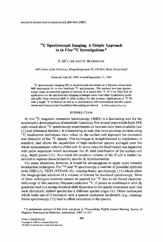
FIG. 2. Natural abundance I3C spectroscopic map of a two-compartment phantom consisting of a 2.5- ern vegetable oil layer close to the surface coil and a 6-cm ethanol section. In the absolute value spectra both regions are clearly visible. The spectroscopic map was obtained from a I3C FID sequence with a phase- encoding period prior to data acquisition (Fig. la). This type of experiment requires only a "C rfchannel; i.e., it can be applied also for single-channel MR equipment tuned to I3C frequency. Acquisition time, 17 min; TR, I s; 64 phase-encoding steps (-6.5 to 6.5 mT/m); 16 acquisitions.
(Fig. 3) DEPT polarization transfer and 'H broadband decoupling are combined with phase encoding (sequence in Fig. 1 b). DEPT increases the sensitivity of the 13C signals by about a factor of four and allows the editing of the spectra. The gain in SNR is clearly visible (a factor of 3.8 for the actual experimental conditions). Note the lack of the carbonyl signals at 180 ppm. This comes from the fact that the carbonyl group has no protons which could transfer magnetization to I3C. Note in addition that the phase cycling of the 'H refocusing pulses narrows the spatial extent of the excited region. This kind of additional localization effect was extensively described in ( I 1 ). DEPT combined with Waltz-8 broadband heterodecoupling results in spec-
- < , , , , ppm 150 100 50 0
FIG. 3. DEFT and 'H broadband (Waltz-8) decoupling combined with SI (sequence shown in Fig. Ib). Same experimental setup as in Fig. 2. Note the increase in SNR compared to Fig. 2 due to the polarization transfer. The 'H refocusing pulses were phase cycled through all four phases. This phase cycling narrows the spatial extent of the excited region, which results in an additional localization effect. Note the spatially homogeneous decoupling in the whole sample. Acquisition time, 17.1 min; TR, 1 s; 64 phase-encoding steps (-6.5 to 6.5 mT/m); four averages per phase-cycling step.
404 COMMUNICATIONS
< / , , , ppm 150 100 50 0
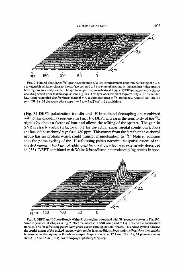
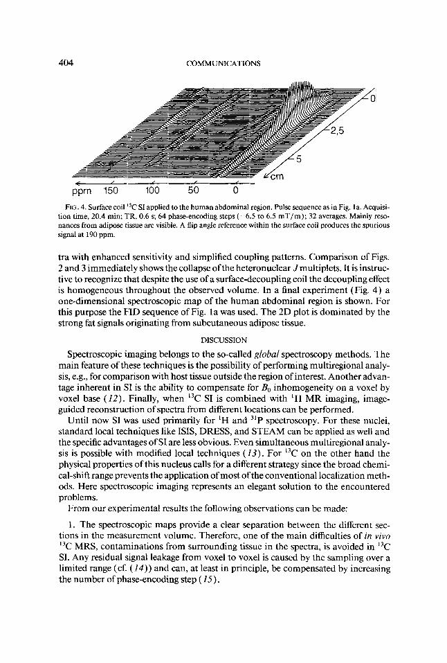
FIG. 4. Surface coil "C SI applied to the human abdominal region. Pulse sequence as in Fig. la. Acquisi- tion time, 20.4 min; TR, 0.6 s; 64 phase-encoding steps (-6.5 to 6.5 mT/m); 32 averages. Mainly reso- nances from adipose tissue are visible. A flip angle reference within the surface coil produces the spurious signal at 190 ppm.
tra with enhanced sensitivity and simplified coupling patterns. Comparison of Figs. 2 and 3 immediately shows the collapse ofthe heteronuclear Jmultiplets. It is instruc- tive to recognize that despite the use of a surface-decoupling coil the decoupling effect is homogeneous throughout the observed volume. In a final experiment (Fig. 4) a one-dimensional spectroscopic map of the human abdominal region is shown. For this purpose the FID sequence of Fig. la was used. The 2D plot is dominated by the strong fat signals originating from subcutaneous adipose tissue.
DISCUSSION
Spectroscopic imaging belongs to the so-called global spectroscopy methods. The main feature of these techniques is the possibility of performing multiregional analy- sis, e.g., for comparison with host tissue outside the region of interest. Another advan- tage inherent in SI is the ability to compensate for Bo inhomogeneity on a voxel by voxel base (12). Finally, when I3C SI is combined with 'H MR imaging, image- guided reconstruction of spectra from different locations can be performed.
Until now SI was used primarily for 'H and 3'P spectroscopy. For these nuclei, standard local techniques like ISIS, DRESS, and STEAM can be applied as well and the specific advantages of SI are less obvious. Even simultaneous multiregional analy- sis is possible with modified local techniques ( 1 3 ) . For 13C on the other hand the physical properties of this nucleus calls for a different strategy since the broad chemi- cal-shift range prevents the application of most of the conventional localization meth- ods. Here spectroscopic imaging represents an elegant solution to the encountered problems.
From our experimental results the following observations can be made:
1. The spectroscopic maps provide a clear separation between the different sec- tions in the measurement volume. Therefore, one of the main difficulties of in viva 13C MRS, contaminations from surrounding tissue in the spectra, is avoided in 13C SJ. Any residual signal leakage from voxel to voxel is caused by the sampling over a limited range (cf. (14 ) ) and can, at least in principle, be compensated by increasing the number of phase-encoding step ( 2 5 ) .

COMMUNICATIONS 405
2. Since the localization process is introduced only via a gradient-encoding period, SI can be combined with a variety of sophisticated experiments and rf configurations. In consequence, e.g., the sensitivity improvements and editing capabilities of polar- ization transfer experiments are available and rf coils based on SNR optimization can be used.
3. Neither offset nor chemical-shift artifacts are introduced by the phase encoding. Thus the limitations imposed by standard localization methods when applied to 13C are not present with I3C SI.
When SI is compared with local techniques (techniques which select/excite/invert a single volume element for the acquisition of the spectroscopic information) one recognizes as a drawback of SI the relatively long minimum measurement time. Mainly for 'H and 31P an interesting experimental aspect is therefore lost since the acquisition of spectra within minutes or seconds is impossible. This drawback, how- ever, is of minor importance for I3C since the low sensitivity of this nucleus calls for long measurement times anyway. Therefore the averaging times can be used for phase encoding of the signals, introducing the spatial resolution basically without any loss.
I3C Sl is also a useful technique for spatially resolving less abundant metabolites like glycogen in muscle or liver. Our preliminary results indicate that by increasing the acquisition time and decreasing the spatial resolution, sufficient sensitivity per voxel is achieved to detect these metabolites. Special care is necessary for resonances with T; in the range of a few milliseconds because a significant part of the nuclear signal may be lost due to the dead time between excitation and acquisition of the data. In these cases time-domain analysis of the data (e.g., LPSVD ( 1 6 ) ) may be applied to retrieve the full information. These techniques have already been success- fully applied to 31P Sl ( Z7) and are currently implemented in our 13C SI experiments.
In order to generally validate the proposed method for in vivo applications, it can be noted that for relatively unlocalized disorders or if the chemical components under study are clearly separated in the 13C spectrum, surface coils or other local rf coils with minimal localization may be sufficient. However, as soon as heterogeneous tissues are involved and/ or if quantitative information should be extracted from the spectra more refined spatial localization is crucial. In these cases the local technique as pro- posed in ( 7) or 13C spectroscopic imaging as described above is the method of choice for unambiguous results.
In conclusion 13C spectroscopic imaging was implemented and tested on a whole- body MR unit. It was found that this technique provides an elegant and simple tool for image-related 13C spectroscopy. The method can be used for systems with single or double rf channel equipment. It allows the acquisition of spectroscopic informa- tion from multiple VOls over a wide field of view without introducing chemical-shift or offset artifacts.
ACKNOWLEDGMENTS
The authors thank J. Seelig for valuable discussions. This work has been supported by the Swiss National Science Foundation Grant 4.889.085.18 and the Komission zur Forderung der wissenschaftlichen For- schung, Projekt 1462.
406 COMMUNICATIONS
REFERENCES
1. A. HEERSCHAP, P. R. LUYTEN, J. I. VAN DER HEYDEN, L. J. M. P. OOSTERWAAL, AND J. A. DEN
2. M. R. BENDALL AND R. E. GORDON, J. Magn. Reson. 53,365 ( 1983). 3. R. J. ORDIEGE, A. CONNELLY, AND J. A. B. LOHMAN, J. Magn. Reson. 66,283 ( 1986). 4. J. GRANOT, J. Mugn. Reson. 70, 488 (1986); J. FRAHM, K-D. MERBOLDT, AND W. HANICKE, J.
5. S. J. Cox AND P. STYLES, J. Mugn. Reson. 40,209 ( 1980).
HOLLANDER, NMR Biomed. 2(3), 1 (1989).
Magn. Reson. 72,502 (1987).
6. R. G. WEISS, P. A. BOTTOMLEY, c. J. HARDY, V. P. CHACKO, J. D. GLICKSON, AND G. GERSTEN- BLITH, in “Proceedings, Seventh Annual Meeting, Society of Magnetic Resonance in Medicine, San Francisco, 1988,” p. 672.
7. w. P. AUE, s. MULLER, ANDJ. SEELIG, J. Magn. Reson. 61,392 (1985). 8. N. BECKMANN, S. MULLER, AND J. SEELIG, in “Proceedings, Eighth Annual Meeting, Society of Mag-
9. S. MULLER, in “Proceedings, 10th Ampere Summer School and Symposium, Portoroz, 1988,” p. 22. netic Resonance in Medicine, Amsterdam, 1989,” p. 250.
10. T. R. BROWN, B. M. KINCAID, ANDK. UGURBIL, Proc. Natl. Acad. Sci. USA 79,3523 (1982). Il. M. R. BENDALLANDD. T. PEW, J. Mugn. Reson. 63,494( 1985). 12. A. A. MAUDSLEY AND S. K. HILAL, Magn. Reson. Med. 2,2 18 ( 1985). 13. S. MULLER, R. SAUTER, H. WEBER, ANDJ. SEELIG, J. Mugn. Reson. 76,155 ( 1988). 14. H. R. BROOKER, T. H. MARECI, AND J. MAO, Mugn. Reson. Med. 5,417 ( 1987). 15. P. A. NARAYANA, E. F. JACKSON, AND W. A. KURDLE, “Abstracts 7th Annual Meeting SMRI 1989,”
16. H. BARKHUIJSEN, R. DE BEER, W. M. M. J. BOV~E, AND D. VAN ORMONDT, J . Mugn. Reson. 61, P69, p. 145.
465 (1985).
WEINER, in “Proceedings, Eighth Annual Meeting, Society of Magnetic Resonance in Medicine, Amsterdam, 1989,” p. 252.
17. A. A. MAUDSLEY, D. B. TWIEG, M. D. BOSKA, D. SAPPEY-MARINIER, B. HUBESCH, AND M. W,