Embed Size (px)
Citation preview

2
136 Asian Journal of Multidisciplinary Studies, 6(2) February, 2018
Insomnia and Sleep Deficiency in Pregnant Women:
A Potential Study with Two Contrasts
Hina Mushtaq1, Naveed Jabbar
2 and Aqsa Aqeel Rao
3
1University of Health Sciences, Lahore, Pakistan
Email: [email protected]
PMDC# 81607-P 2Fatima Jinnah Medical University, Lahore, Pakistan
Email: [email protected]
PMDC# 85634-P 3Quaid-e-Azam Medical College, Bahawalpur, Pakistan
Email: [email protected]
PMDC# 83723-P
Abstract
Insomnia in pregnancy is associated with depression,
preeclampsia, gestational diabetes, and preterm labor. In
normal pregnancies, maternal plasma melatonin levels
increase significantly as pregnancy proceeds, reaching its peak near term. However, the role of melatonin in
insomnia during pregnancy is not known. The objective
of this study was to measure nocturnal saliva melatonin
levels in pregnant women with and without insomnia.
Results did not show a significant difference in melatonin levels between insomniac (treated and
untreated) and healthy pregnant women in all trimesters.
However, sub-group analysis showed significantly lower
melatonin levels in untreated insomniac pregnancies
compared to healthy pregnancies and those treated with sleep medications. Results of this study confirmed lower
levels of nocturnal melatonin in untreated pregnancies
with insomnia. Future research is needed to investigate
the safety and efficacy of melatonin supplementation for
the treatment of insomnia in pregnancy, replacing psychotropic drugs.
Keywords: Insomnia depression, preeclampsia,
gestational diabetes, preterm labor
Introduction
Circadian System
The circadian system is located both in the central
nervous system in the suprachiasmatic nucleus
(SCN) of the hypothalamus, and peripheral organs
such as the heart and liver.1
The system plays an
important role in different physiological
arrangements and behavior. It is the biological
clock of the body managing both night and day
behaviors. The system functions not only
independent of external stimuli such as ambient
brightness, but also in a manner dependent on
external factors such as food intake.2
Any
disruption of the system leads to conditions such as
sleep disorder, glucose intolerance (diabetes),
obesity, and ultimately decreased life expectancy.3
Suprachiasmatic Nucleus
SCN is located in the anterior hypothalamus on top
of the optic chiasm, next to the third ventricle with
50,000 neurons.4
It is a bilateral structure and is
activated by light. In other words, it regulates the
circadian rhythm. Although it is now known that
circadian rhythm exists in peripheral organs
independent of the SCN, their function is lost
without input from SCN. For this reason, SCN is
called the master circadian clock, or the
coordinator of the clocks. A circadian clock has a
24-hour cycle and its most potent stimulus is light
that is directly projected from the retina to SCN
through the retino-hypothalamic tract (RHT). The
indirect pathway of receiving light by the SCN is
through the intergeniculate tract (GHT).
Malfunctioning of any of the above pathways
impairs the phase-shifting and light-dark cycle of
the circadian system. For example, the light-dark
cycle is impaired by the RHT pathway damage,
and phase-shifting is delayed by damage to GHT
pathway.1
Research also shows that the circadian
clock can shift a phase independent of light with
other stimuli such as exogenous melatonin,
temperature, exercise, and food. For example,
melatonin supplementation in blind people can
regulate the circadian clock.5
Core body
temperature also affects the peripheral circadian
clock but not the SCN.6
There are 2 pathways by
which the SCN manages the circadian clock
complementing each other: endocrine and neural
pathways. Circadian clock controls the endocrine
system including melatonin, cortisol from the
hypothalamic-pituitary- adrenal (HPA) axis, the
hypothalamic-pituitary-thyroid (HPT) axis, and
epinephrine.7
The neural pathway includes
projections from the SCN to the pineal gland
(Figure 1).
brought to you by COREView metadata, citation and similar papers at core.ac.uk
provided by Asian Journal of Multidisciplinary Studies (AJMS)

2
137 Asian Journal of Multidisciplinary Studies, 6(2) February, 2018
Figure 1: The neural pathway between the SCN and pineal gland in rats. Reprinted with permission from
Springer.46
Pineal Gland
Description
The pineal gland had several names over-time
given its anatomy and function in different
animals. It was first identified as a different
cerebral organ a few centuries BC, and was
initially named “Konareion” for its pine shape,
similar to lymph nodes. It was then named
“Glandula Pinealis”, pineal. Later Rene Descartes
called the gland the “seat of the soul”, coordinator
of psychophysiological functions of the body.
Bioassay advances resulted in the discovery of
pineal “extracts” responsible for lightening frog’s
skin, which lead to the isolation of melatonin in
1958. Development of fluorescent techniques
allowed the measurement of melatonin and
serotonin, their light-dark cycles and their
concentrations.8
Structure
The location and structure of the pineal gland is
species specific and responds to environmental
stimuli. The human pineal gland is located at the
posterior of the diencephalon. It develops in the
second month of gestation as an invagination of the
ependymal lining of the diencephalic third
ventricle, between the habenular and posterior
commissure. The “pineal stalk” consists of a rostral
and a caudal lamina surrounded by a pial layer, and
is suspended in the CSF- filled pineal recess below
the splenium. Average adult dimensions are 5-
9mm in length, 1-5mm in width, and 3-5mm in
thickness and the average weight of 100-180mg
varies in different ages and genders.8
Its structure
is described as two parts. A central core of lobules
and peripheral neurons along with pinalocytes are
the main cell types of human pineal gland.
Pinalocytes are granular shape, and have a nucleus
and cytoplasmic processes that end in capillaries,
indicative of an endocrine gland. Neuroglia are
distributed unevenly in the periphery. Calcareous
deposits are characteristic of the pineal deposits as
calcium and magnesium salts on both parenchymal
and intracellular tissue. Calcareous deposits are
present from birth and increase in concentration by
age, starting to decrease in young adulthood. A
negative correlation was found between the density
of calcification, age and chronic sleepiness. The
pineal gland has a rich vascular system receiving
blood from posterior choroidal arteries deriving
from cerebral arteries. It lacks an endothelial
blood-brain barrier, and is sensitive to drugs. The
gland is innervated with fibres from sympathetic,
parasympathetic, and central nervous systems. The
most important pathway is the noradrenergic
sympathetic pathway neurons which receive input
from the suprachiasmatic nucleus (SCN) of the
hypothalamus, which itself receives input from
retinal ganglion cells.8
Neural Pathway
The neural pathway regulating the production of
melatonin in the pineal gland consists of an input
from the GABAergic system from SCN to a
subdivision of paraventricular nucleus (PVN),
which in turn projects to the intermediolateral
(IML) cell column and, by pre- ganglionic
adrenergic fibres, to the superior cervical ganglion
(SCG). The input then reaches
the pineal gland from SCG by post ganglionic
adrenergic fibres and releases norepinephrine
(NE).35,36
In the pineal gland, beta adrenergic
receptors of pinalocytes are stimulated by a subunit
of G protein coupled receptors that increase
adenylate cyclase. Ca++
and protein kinase C
(PKC) are involved in increased beta adrenergic
activity and stimulation of cAMP potentiated by
Alpha adrenergic receptors.37
The synergistic
activity of alpha and beta adrenergic receptors
increases cAMP and production of NAT38
(Figure
2). The rate of melatonin secretion is negatively
correlated with age 39
, and dim light melatonin
onset (DLMO) may not reach the threshold
concentration, 4 pg/ml, in the elderly.40
However,
concentration is significantly higher in younger
people.41
There are also inter-individual
differences in timing, duration, and the amount of
nocturnal hormone secretion42
, but stability within
individuals.43
,44
For example, mean melatonin
production is estimated to be higher in men than
women at night, 60.7± 24.6 pg/ml and 29.3± 21.8
pg/ml respectively, p<0.01.45
DLMO also occurs
later in women than in men.41
Inter-individual
differences in melatonin production may be due to
different metabolism rates.46

2
138 Asian Journal of Multidisciplinary Studies, 6(2) February, 2018
Figure 2: Adrenergic pathway of melatonin biosynthesis. Reprinted with permission from Elsevier. 47
Day length affects melatonin secretion as well, so
there are seasonal variations in the rate of
nocturnal melatonin, which affects the physiology,
behavior, and reproduction of some species.9
The
use of artificial light or artificial shortening of
summer days has been shown to affect the
secretion of the hormone.48,49
Evidence shows
that the onset of “seasonal affective disorder” is
related to shorter days and longer nights in fall and
winter.50
Evidence of melatonin as the circadian phase
maker comes from studies showing shifts in body
temperature and sleep timing following melatonin
administration. Therefore, supplementation with
melatonin at the right time can adjust circadian
rhythm after a phase-shift due to shift work, jetlag,
blindness, or sleep disorders.51
Evidence shows
strong association between melatonin, sleep, and
body temperature, energy, activity, and mental
ability. The daytime low level of melatonin and
high level of cortisol mediate daytime activities.
Sleep and the dark-light cycle are also mediated by
changes in melatonin and cortisol levels.52
The
rise of melatonin two hours before bed allows for
sleep onset, which in turn decreases the nocturnal
body temperature.17
Studies show that blocking
the beta adrenergic receptors suppresses nocturnal
melatonin, and increases body temperature. This
can be reversed by administering 5 mg melatonin.
This further confirms the role of melatonin in body
temperature regulation.53
Melatonin levels
suppressed either pathologically or artificially by
light lead to insomnia (initiation, maintenance,
sleep quality).54
Figure 3, below shows the
immediate effect of light exposure on nocturnal
melatonin levels.55
Figure 3: Effect of light exposure on nocturnal melatonin concentration. Normal pattern of melatonin synthesis
in dark environment is shown in grey area. Reprinted with permission from Elsevier.55

2
139 Asian Journal of Multidisciplinary Studies, 6(2) February, 2018
Sleep
Sleep is defined as a state characterized by a
reduction in voluntary movement, reduced
response to stimuli and, ultimately, loss of
awareness.52
Sleep is divided into two parts: non-
rapid eye movement (NREM) and rapid eye
movement (REM). NREM itself is divided into
three stages, which progress to the deeper sleep
phase, or REM.56
Sleep restriction which reduces
sleep duration affects the structure of sleep.57
Regulation of sleep in the brain is a complicated
process involving several growth factors
synthesized in response to neural activity. These
growth factors also affect synaptic efficacy.
Interlukin-1 (IL-1) and tumor necrosis factor
(TNF) are the two most important factors in the
sleep regulation process, released by the
production of ATP after neurotransmission. They
increase NREM sleep. Inhibition of these two
compromises spontaneous sleep and rebound sleep
after an episode of sleep deprivation. After sleep
deprivation, IL-1 and TNF concentrations rise.
However, their over-activation, due to the
activation of innate immune response, inhibits
sleep.58
Other factors leading to this up regulation
are light and excessive food intake. IL-1 leads to
production of corticotropin releasing hormone
(CRH) which, in turn, inhibits sleep dependent IL-
1 inhibition. CRH also inhibits IL-1 which causes
sleep inhibition. Other growth factors involved in
sleep regulation are nitrous oxide (NO), growth
hormone releasing hormone (GHRH) and nerve
growth factor. Nuclear factor kappa B (NFkB),
adenosine, and prostaglandin. NFkB is activated by
sleep deprivation. Activation of NFkB leads to the
activation of adenosine receptor, COX2, and NO
synthase. COX2 leads to the synthesis of sleep
promoting prostaglandin D2 (PGD2) and sleep
inhibiting E2 (PGE2) that inhibits IL-1. Therefore,
several positive and negative feedback loops are
involved in sleep regulation or dysregulation. The
neural system defined above affects the synaptic
efficacy and downstream pathways that lead to
wakefulness and consolidation of memory, or
sleepiness.59
Insomnia
Insomnia, a form of sleep restriction, is an
important public health issue.60
A large
population- based study calculated the prevalence
of insomnia to be 6 - 48% after all diagnostic
criteria were brought into consideration.61
It is
defined as an inability to initiate sleep, waking up
during the night, difficulty falling back to sleep,
and non-restorative sleep.62
Delayed sleep phase
disorder (DSPD) is a disorder of circadian rhythm
and insomnia, in which the patient has difficulty
initiating sleep at a normal time, therefore the
circadian rhythm is delayed when compared to
healthy individuals.41
DSPD patients are able to
maintain sleep if they go to bed in their favourite
time, but sleep initiation is delayed. This type of
insomnia is associated with daytime sleepiness and
many physiological dysfunctions. Ultimately long
term DSPD insomnia is associated with
psychological disorders, substance and alcohol
abuse, and other neurobehavioral and physiological
disorders. The prevalence of DSPD is 7-16% in the
general population.63
To further understand the
etiology of DSPD, nocturnal melatonin levels were
measured in patients with this disorder and were
compared with normal sleepers. Results showed
that DSPD patients have significantly delayed dim
light melatonin onset (DLMO) by three hours
when compared with healthy sleepers (Figure 4).
There was no difference in the duration of
melatonin secretion but levels did not change
significantly from DLMO to acrophase in DSPD
patients.64
DLMO is considered an approximate
night-time threshold of melatonin synthesis and
secretion. DLMO makes an assessment of the
circadian rhythm disorder in patients with
insomnia easier than a provisional assessment. 10
pg/ml is considered the threshold for plasma
DLMO and 3 pg/ml for saliva (Figure 6).65
Melatonin secretion starts at darkness and is
suppressed by light, so it is very important to
measure hormone levels in dim light to determine
its onset time (DLMO) and to assess circadian
rhythm in advanced sleep phase disorder (ASPD)
or delayed in DSPD41
. DLMO can be measured in
both blood and saliva. Although DLMO
measurement is currently used in research, there is
no approved assay or standards in clinical
settings.66

2
140 Asian Journal of Multidisciplinary Studies, 6(2) February, 2018
Figure 4: Pattern of melatonin secretion in a normal (solid line), ASPS (dashed line), and DSPS (dotted line)
subject. Reprinted with permission from Elsevier.41
Figure 5: DLMO levels at 3 pg/ml in saliva. Reprinted with permission from the Journal of Clinical Sleep Medicine.65
Physiological Impacts of Insomnia
Chronic medical conditions were reported in 76 -
83% of the population of different ethnic groups
due to poor sleep pattern.67
Adverse psychological
and physiological effects of insomnia have a major
impact on the quality of life of affected
individuals. Recent experimental and
epidemiological data have associated insomnia
with multiple daytime symptoms and chronic
conditions. Among physiological impacts of sleep
disorders are metabolic disorders leading to obesity
and type 2 diabetes.68
Insufficient sleep
contributes to weight gain via excessive food
intake associated with increased energy
expenditure. Studies show that sleep restriction
reduces daytime leptin while increasing daytime
ghrelin. These hormonal changes lead to increased
hunger and appetite.69
Increased food intake is
beyond the energy needed leading to weight
gain.70
Glucose intolerance increases and its
clearance decreases upon sleep loss due to an
impaired metabolism system.71
Studies suggest
the potential benefit of recovery sleep.70

2
141 Asian Journal of Multidisciplinary Studies, 6(2) February, 2018
Hypertension is among other physiological side
effects of sleep restriction. It is shown in several
studies that insomnia and short sleep duration is
associated with hypertension. Duration of sleep is a
predictor of the severity of hypertension.72
When
adrenocorticotropic hormone (ACTH) and cortisol
levels were compared in insomniac patients and
normal sleepers in a lab environment, results were
elevated nocturnal levels of those two hormones
leading to hyper-activation of hypothalamic-
pituitary-adrenal axis. This, in turn, causes several
pathological consequences, such as
hypertension.73
Obesity, increased insulin
resistance and cardiovascular diseases are
associated with elevated levels of ACTH and
cortisol. Irwin et al. showed an increase in
nocturnal norepinephrine in insomniac patients
when compared to depressed and healthy
individuals. This may be an indication of increased
sympathetic activity, exhibited by increased heart
rate, body temperature and metabolic rate.74
Other
studies also showed coronary artery disease
mortality after chronic insomnia in middle-aged
men and women.75
Impacts of Insomnia in Pregnancy
There are physiological and psychological
consequences following insomnia in pregnancy.
Glucose intolerance and gestational diabetes
mellitus (GDM) are amongst the physiological
impacts of insomnia in pregnancy. The prevalence
of GDM in the US is approximately 7%, affecting
200,000 women every year. Risk increases by
obesity and/or type 2 diabetes (T2D).40
Earlier it
was mentioned that insomnia is a risk factor for
type 2 diabetes. As a result, insomnia and T2D
may increase the risk of a pregnancy to develop
GDM. In a cohort study following up pregnant
women from early pregnancy with their sleep
duration, it was found that women with shorter
sleep duration are at 1.86 fold increased risk for
development of gestational diabetes.41
GDM
adversely affects both maternal and fetal health and
pregnancy outcomes. Mothers with GDM are at
increased risk for cardiovascular disease and
metabolic dysfunction.42
GDM put the pregnancy
at risk for pre-eclampsia, preterm labor and
caesarean section due to the accumulation of
excessive amniotic fluid, and infection.43
In
addition to maternal risks associated with GDM,
children of diabetic mothers are at increased risk
for macrosomia, related birth injuries, respiratory
problems, neonatal hypoglycemia, and jaundice.44
Hypertension is one of the other physiological
impacts of insomnia in individuals. In a pregnancy
prospective cohort study, mean diastolic, systolic
and mean arterial pressures were measured. It was
found that systolic blood pressure is highest in
those who sleep less than six hours at night in both
first trimester and third trimester, a 1.62 and 4.41
fold increase respectively. Also, diastolic blood
pressure blood pressure was highest in short time
sleepers in both first and third trimesters, 1.85 and
3.52 fold increase respectively. Mean arterial
pressure was significantly higher in both short and
long sleep durations. 45
In the previous chapter
mechanisms responsible for hypertension
associated with insomnia have been explained.
Changes in body’s hemodynamics such as
temperature, heart rate and hyper-activation of the
HPA axis are all factors in increased risk for
hypertension.
Circadian system dysregulation and insomnia is
now known to be a risk factor in depression and
mood disorders in pregnancy. In line with this,
studies investigated plasma melatonin levels in
pregnant women with and without depression.
Levels were measured at baseline, synthesis onset,
and offset times. It was found that daytime
melatonin levels are significantly higher in
depressed pregnant women, while night time levels
were significantly lower when compared to healthy
pregnancies. Melatonin synthesis onset time and
offset time were also significantly earlier in
depressed pregnant women. Second finding of this
study was that the number of previous episodes of
depression was negatively correlated with
melatonin offset time r=-0.66.
Therefore, previous episodes of depression may be
a risk factors in earlier onset and offset times of the
hormone production and its low levels in
pregnancy. Depression disorders in pregnancy are
explained by loss of sensitivity of melatonin
receptor to estradiol and progesterone levels
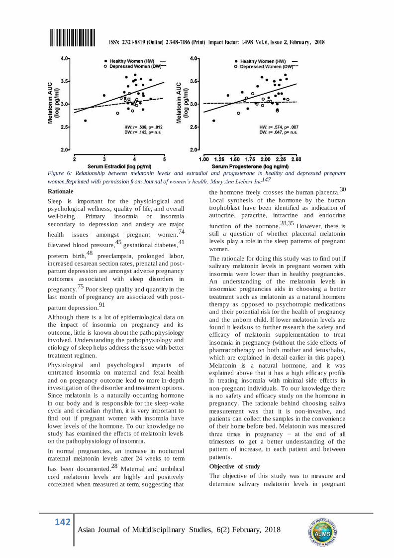
(Figure 6). Therefore, rise in gonadal hormone in
healthy pregnancy results in an increase in
melatonin levels but not in depressed
pregnancies.46
2
142 Asian Journal of Multidisciplinary Studies, 6(2) February, 2018
Figure 6: Relationship between melatonin levels and estradiol and progesterone in healthy and depressed pregnant
women.Reprinted with permission from Journal of women’s health, Mary Ann Liebert Inc147
Rationale
Sleep is important for the physiological and
psychological wellness, quality of life, and overall
well-being. Primary insomnia or insomnia
secondary to depression and anxiety are major
health issues amongst pregnant women.74
Elevated blood pressure,45
gestational diabetes,41
preterm birth,48
preeclampsia, prolonged labor,
increased cesarean section rates, prenatal and post-
partum depression are amongst adverse pregnancy
outcomes associated with sleep disorders in
pregnancy.75
Poor sleep quality and quantity in the
last month of pregnancy are associated with post-
partum depression.91
Although there is a lot of epidemiological data on
the impact of insomnia on pregnancy and its
outcome, little is known about the pathophysiology
involved. Understanding the pathophysiology and
etiology of sleep helps address the issue with better
treatment regimen.
Physiological and psychological impacts of
untreated insomnia on maternal and fetal health
and on pregnancy outcome lead to more in-depth
investigation of the disorder and treatment options.
Since melatonin is a naturally occurring hormone
in our body and is responsible for the sleep-wake
cycle and circadian rhythm, it is very important to
find out if pregnant women with insomnia have
lower levels of the hormone. To our knowledge no
study has examined the effects of melatonin levels
on the pathophysiology of insomnia.
In normal pregnancies, an increase in nocturnal
maternal melatonin levels after 24 weeks to term
has been documented.28
Maternal and umbilical
cord melatonin levels are highly and positively
correlated when measured at term, suggesting that
the hormone freely crosses the human placenta.30
Local synthesis of the hormone by the human
trophoblast have been identified as indication of
autocrine, paracrine, intracrine and endocrine
function of the hormone.28,35
However, there is
still a question of whether placental melatonin
levels play a role in the sleep patterns of pregnant
women.
The rationale for doing this study was to find out if
salivary melatonin levels in pregnant women with
insomnia were lower than in healthy pregnancies.
An understanding of the melatonin levels in
insomniac pregnancies aids in choosing a better
treatment such as melatonin as a natural hormone
therapy as opposed to psychotropic medications
and their potential risk for the health of pregnancy
and the unborn child. If lower melatonin levels are
found it leads us to further research the safety and
efficacy of melatonin supplementation to treat
insomnia in pregnancy (without the side effects of
pharmacotherapy on both mother and fetus/baby,
which are explained in detail earlier in this paper).
Melatonin is a natural hormone, and it was
explained above that it has a high efficacy profile
in treating insomnia with minimal side effects in
non-pregnant individuals. To our knowledge there
is no safety and efficacy study on the hormone in
pregnancy. The rationale behind choosing saliva
measurement was that it is non-invasive, and
patients can collect the samples in the convenience
of their home before bed. Melatonin was measured
three times in pregnancy − at the end of all
trimesters to get a better understanding of the
pattern of increase, in each patient and between
patients.
Objective of study
The objective of this study was to measure and
determine salivary melatonin levels in pregnant

2
143 Asian Journal of Multidisciplinary Studies, 6(2) February, 2018
women with insomnia, treated or untreated, and to
compare their levels with healthy pregnant women
in all trimesters of pregnancy.
Methods
Measuring Melatonin in Saliva
Saliva
Healthy individuals produce 500-1500 ml of saliva
per day, or 0.5 ml/minute salivary output, and
composition may be different in terms of viscosity,
ions and protein concentrations. The flow of the
saliva depends on autonomic stimulation.
Parasympathetic stimulation results in a large flow,
with low levels of inorganic and non-protein
organic compounds, such as cholesterol, uric acid,
glucose, bilirubin, and creatinine. Sympathetic
stimulation results in a small flow high in
inorganic compounds such as K+
and proteins.
Compounds are released into saliva by different
means, such as passive diffusion for lipophilic
compounds, active transport and ultrafilteration
through gap junctions.76
Saliva Melatonin and its Interactions
Melatonin transfers into saliva by passive
diffusion.76
Composition of the saliva is affected
by food intake, increasing the release of total
proteins. Caffeine should be avoided 12-24 hours
before saliva sampling because it stimulates the
activity of CYP 1A2.77
Banana with low
melatonin content is shown to increase urinary
melatonin metabolite after consumption. This can
be explained by the rate of absorption or
metabolism of melatonin due to CYP1A2
polymorphism. Chocolate increases melatonin
concentration by its flavonoid content.178
Some
drugs such as NSAIDS (ibuprofen, naproxen,
diclofenac, ketorolac, etc.) may also reduce
melatonin concentrations by inhibiting
prostaglandin and COX-2.79
Therefore, in the
current study, patients were instructed to avoid the
above mentioned to prevent any interaction with
saliva melatonin levels.
Measuring Melatonin in Saliva
Salivary measurement of melatonin is now a
practical and reliable tool for diagnosis of the
condition and research purposes. Saliva melatonin
as a circadian phase maker is validated and
compared to plasma levels. There is a significant
positive correlation between saliva and plasma
melatonin onset r=0.64, and saliva and plasma
acrophase concentrations, r=0.83 with saliva levels
being one-third of plasma’s (Figure 7). 80
Saliva
sampling should be done in dim light and taken
every 30-60 minutes for at least one hour to
determine its onset.81
Reliability of saliva
melatonin measurement to determine dim light
melatonin onset (DLMO) was assessed. Results
showed that saliva measurement of melatonin
levels is valid, and it is a reliable tool for the
measurement of hormones.81
This was further
investigated by comparing in-home methods to
laboratory measurements. Patients with insomnia
were instructed to collect saliva samples at home
and the following evening saliva samples were
collected in the lab under lab conditions. It was
found that at-home saliva melatonin levels are
positively correlated with in-lab measurements
r=0.85. However, a delay of approximately forty
minutes was seen with the in- home method. This
study confirmed the previous work that in-home
saliva melatonin measurement is practical and
valid to determine DLMO and to assess circadian
rhythm (Figure 7).66
Figure 7: Relationship between plasma and saliva melatonin levels r=0.64. Reprinted with permission from SAGE
publications.180

2
144 Asian Journal of Multidisciplinary Studies, 6(2) February, 2018
Figure 8: Relationship between in-home saliva melatonin and in-lab measurement, r=0.8. Reprinted with
permission from Elsvier Limited.66
Study Methodology
Saliva collection was done three times during
pregnancy; at 12 -14 weeks, 24-26 weeks, and 34-
36 weeks. I was interested to know the pattern of
melatonin rise from the beginning of pregnancy in
all three groups described above. Plasma melatonin
levels were measured in all trimesters29
.
Participants collected saliva samples 3 times every
30-60 minutes, in dim light, starting at least 1 hour
prior to bedtime. Therefore, in total there were 9
saliva samples from each participant. No chocolate
or bananas, alcohol, caffeine, or drinks with
artificial colorants should have been taken on the
day of sampling. No aspirin or medicines
containing ibuprofen, would be taken on the day of
sampling. The participant needed to remain in dim
light during the sampling hours with a night light
or a low wattage lamp. They should not have sat
closer than 6 feet to a TV; and if using a
computer/laptop/tablet, they should have adjusted
the contrast to low. To avoid contamination with
food, participants needed to finish their main meal
at least 30 minutes before sampling time and to
brush their teeth without toothpaste then rinse with
water 10 minutes before sampling.80
For saliva
sampling a cotton swab was used, which was
chewed or held in the mouth for 1-2 minutes, and
then placed in a vial when soaked enough.
Participants documented each sample time on a log
provided to them along with the duration of staying
in dim light. Participants were instructed to
refrigerate or freeze their saliva samples within 30
minutes of sampling and mail them back in a cold
box within 1-3 days. All storage and shipment
accessories were provided to participants. The
saliva samples were tested for melatonin levels
using the enzyme-linked immunosorbent assay
(ELISA) method in the Motherisk lab.
Statistical Analysis
All statistical analysis was done with student SPSS
version 23. All data is presented in mean ± SEM.
In order to compare melatonin concentrations
between pregnant women with insomnia either
treated or untreated (combined in one group) with
healthy pregnant women a t-test was used. The
null-hypothesis was that there was no difference in
melatonin levels in insomniac pregnant women and
healthy pregnancies.
For sub-group analysis, one-way ANOVA was
used. Subgroups included pregnant women with
treated insomnia, pregnant women with untreated
insomnia and healthy pregnant women.In the
general linear model, repeated measure analysis
was used to compare the pattern of increase of
melatonin levels in all two groups of untreated
insomniacs, treated insomniacs and healthy
subjects. Alpha was set at p<0.05 in all statistical
analysis.
Results
In the first group, Exposed, a total of 24 people
were recruited. 13 were lost to follow up. They did
not respond to emails or phone calls after the ICF

2
145 Asian Journal of Multidisciplinary Studies, 6(2) February, 2018
was signed. Two had miscarriages and
automatically were excluded from the study. One
patient provided a very small amount of saliva
sample that was not measurable. One patient’s
saliva melatonin measure was an outlier, and was
deleted from the analysis. In the first trimester
seven samples were used in (T1) melatonin
measurement. In the second trimester (T2). This is
while one patient withdrew after T1, but one
patient was recruited in the second trimester due to
her severe insomnia symptoms. In the third
trimester (T3) measurement, there were five
samples because two patients withdrew after T2.
In the second group, Disease-match, 19 people
were recruited. Six were lost to follow up, and 13
samples were received for the T1 measurement.
For the T2 measurement there were 12 samples, as
one patient withdrew after T1. For the T3
measurement there were 9 samples because one
patient withdrew. Two other patients had sampling
dates much later than the expected end date of
study.
In the third group, Healthy, a total of 28 people
were recruited. 7 were lost to follow up, two had
miscarriages and one provided small amount of
saliva that was not measurable. A total of 18
samples were used for the T1 measurement. For
the T2 there were 11 samples. There were 4
withdrawals, 2 miscarriages, and one was lost to
follow up in T2. In T3, there were 12 samples,
because one patient that was lost to follow up in T2
came back and continued the study (Figure 9).
Figure 9: Study population saliva sample size per group per trimester.
Mean (SD) age was not significantly different
among the groups, 35.43±3.7, 35.54±4.4,
33.73±3.7, respectively. Ethnic background,
marital status, education, occupational status,
number of pregnancies (gravidity), number of live
children (parity), spontaneous abortions (SA),
therapeutic abortion (TA), ectopic pregnancies
(Ect), pre-pregnancy BMI, third trimester BMI,
and total weight gain.
Disease Characteristics of the Insomnia Group
Five patients in the Exposed group suffered from
insomnia only. One patient had insomnia,
depression, anxiety and attention deficit disorder
(ADD). One patient had insomnia, is bipolar, and
had mania. One patient had insomnia and anxiety,
and one patient suffered from insomnia and
depression.
In the Disease-match group, eight patients suffered
from insomnia only. One patient had insomnia,
depression, and anxiety. One patient had insomnia
and anxiety, two patients had insomnia and
depression.
All nine patients in the Exposed (treated) group
took medications for either sleep issues or
comorbid symptoms such as depression and
anxiety. Five patients took one or combinations of
zopiclone, quetiapine, lorazepam, dimenhydrinate,
and progesterone as a sleep aid. One patient took a
combination of bupropion, amitriptyline,
lisdexamphetamine, and clonazepam for insomnia,
depression/anxiety, and ADD. Trazodone and
aripiprazole were taken by another patient for
bipolar disorder, mania and insomnia. Zopiclone,
duloxetine and trazodone were used in
combination for insomnia and comorbid anxiety. A combination of bupropion and quetiapine was taken for
insomnia and comorbid depression (Table 1).
Exposed:
Lost F/U (13)
Miscarriages (2)
Small amnt (1)
Final Exposed Gr. Subjects T1: N=7 T2: N=7
T3: N=5
Disease-Match:
Lost F/U (6)
Final Disease-match Gr. Subjects
T1: N=13
T2: N=12 T3: N=9
Healthy:
Lost F/U (7) Miscarriages (2)
Small amount (1)
Final Healthy Gr. Subjects T1: N=18
T2: N=11 T3: N=12

2
146 Asian Journal of Multidisciplinary Studies, 6(2) February, 2018
Condition Exposed
Group N=9
Disease-Match
Group N=12
Insomnia Only 5(55%) 8(67%)
Insomnia with
other psychiatric
comorbidities
4(44%) 4(33%)
Table 1: Disease composition of exposed and
disease-match group.
T-tests compared insomniac (exposed and disease-
match) pregnant women (N=20) with healthy
pregnant women. It was found that mean melatonin
levels in all three trimesters were not significantly
different between the two groups, p=0.4, p=0.9,
p=0.1 for T1, T2, and T3 respectively (Table2)
(Figure 10).
Table 2: Descriptive statistics and t-test results. No significant difference found between the groups in all
trimesters. T1: first trimester, T2: second trimester, T3: third trimester, Insom: insomnia, H: healthy
Figure 10: Comparison of melatonin levels in both groups in all trimesters.
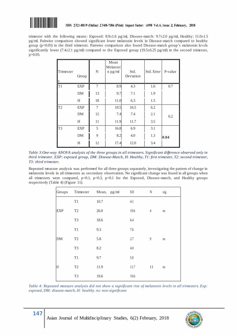
Sub-group analysis by one-way ANOVA (Table 3) showed that Disease-match (untreated) insomniac pregnant
women have significantly lower melatonin levels (p=0.04) in the third trimester (mean (pg/ml)=8.2±1.33 SEM),
when compared to the treated (mean (pg/ml) = 16.8±3.1 SEM) and Healthy pregnant women (mean
(pg/ml)=17.4 ±3.5 SEM). No significant difference was found in melatonin levels among the groups in the first
Groups
N
Mean melatonin
pg/ml
Std.
Deviation
Std. Error
Mean
P-value
T1 Insom. H
20 18
9.4 11.0
6.1 6.3
1.3 1.5
0.4
T2 Insom. H
19 11
11.9 11.9
12.6 11.7
2.9 3.5
0.9
T3
Insom. H
14 12
11.3 17.4
6.5 12.0
1.7 3.4
0.1
2
147 Asian Journal of Multidisciplinary Studies, 6(2) February, 2018
trimester with the following means: Exposed: 8.9±1.6 pg/ml, Disease-match: 9.7±2.0 pg/ml, Healthy: 11.0±1.5
pg/ml. Pairwise comparison showed significant lower melatonin levels in Disease-match compared to healthy
group (p=0.03) in the third trimester. Pairwise comparison also found Disease-match group’s melatonin levels
significantly lower (7.4±2.1 pg/ml) compared to the Exposed group (19.5±6.25 pg/ml) in the second trimester,
p=0.03.
Trimester
Group
s
N
Mean
Melatoni
n pg/ml
Std.
Deviation
Std. Error
P-value
T1 EXP 7 8.9 4.3 1.6 0.7
DM 13 9.7 7.1 1.9
H 18 11.0 6.3 1.5
T2 EXP 7 19.5 16.5 6.2
0.2 DM 12 7.4 7.4 2.1
H 11 11.9 11.7 3.5
T3 EXP 5 16.8 6.9 3.1
0.04 DM 9 8.2 4.0 1.3
H 12 17.4 12.0 3.4
Table 3:One-way ANOVA analysis of the three groups in all trimesters. Significant difference observed only in
third trimester. EXP: exposed group, DM: Disease-Match, H: Healthy, T1: first trimester, T2: second trimester,
T3: third trimester.
Repeated measure analysis was performed for all three groups separately, investigating the pattern of change in
melatonin levels in all trimesters as secondary observation. No significant change was found in all groups when
all trimesters were compared, p=0.1, p=0.3, p=0.1 for the Exposed, Disease-match, and Healthy groups
respectively (Table 4) (Figure 11).
Groups Trimester Mean, pg/ml SD N sig.
T1 10.7 4.1
EXP T2 26.0 19.6 4 ns
T3 18.6 6.4
T1 9.3 7.6
DM T2 5.8 2.7 9 ns
T3 8.2 4.0
T1 9.7 5.0
H T2 11.9 11.7 11 ns
T3 19.6 19.6
Table 4: Repeated measure analysis did not show a significant rise of melatonin levels in all trimesters. Exp:
exposed, DM: disease-match, H: healthy. ns: non-significant

2
148 Asian Journal of Multidisciplinary Studies, 6(2) February, 2018
Figure 11: Dot plot of repeated measure analysis of melatonin levels in all trimesters. EXP: exposed, DM:
disease-match, H: healthy. T1: first trimester, T2: second trimester, T3: third trimester.
Discussion
Both groups with insomnia were associated with
comorbid mental disorders.
Melatonin levels in treated women with
psychotropic drugs were similar to healthy
controls. A non-significant difference in melatonin
levels in insomniac pregnancies (Exposed and
Disease- match combined group) compared with
Healthy subjects is indicative of higher melatonin
level in the exposed group due to the effects of
sleep medications. All nine patients in the Exposed
(treated) group took medications for either only
sleep issues or comorbid symptoms such as
depression and anxiety. Five patients took one or
combinations of zopiclone, quetiapine, lorazepam,
dimenhydrinate, and progesterone as a sleep aid.
One patient took a combination of bupropion,
amitriptyline, lisdexamphetamine, and clonazepam
for insomnia, depression/anxiety and ADD.
Trazodone and aripiprazole were taken by another
patient for bipolar, mania, and insomnia.
Zopiclone, duloxetine and trazodone were used in
combination for insomnia and comorbid anxiety. A
combination of bupropion and quetiapine was
taken for insomnia and comorbid depression.
Amitriptyline decreases sleep latency while
treating depression symptoms. It also increases
melatonin synthesis by increasing NAS (N-acetyl
transferase) in the pineal gland.84
SSRIs cause
increased sleep latency, REM suppression and
frequent waking up during the night. As a result,
pharmacotherapy addressing the treatment of
insomnia should be considered with SSRIs.85
This
is in conflict with a clinical study where duloxetine
was shown to increase 6- sulphatoxymelatonin
(aMT6s) levels after treatment.19
Duloxetine was
used in one patient for anxiety and trazodone and
zopiclone were used concomitantly to improve
sleep. Zopiclone has no effect on melatonin
concentration when compared to zalplona placebo
and a non- benzodiazepine hypnotic in rats.20
Similar results have been found in healthy
volunteers following acute and subchronic
administration of zopiclone.121
A sleep-inducing
effect may be due to its hypnotic properties.
Increased melatonin levels and improved sleep
with trazodone has been documented in clinical
studies.24
,25
Bupropion is associated with
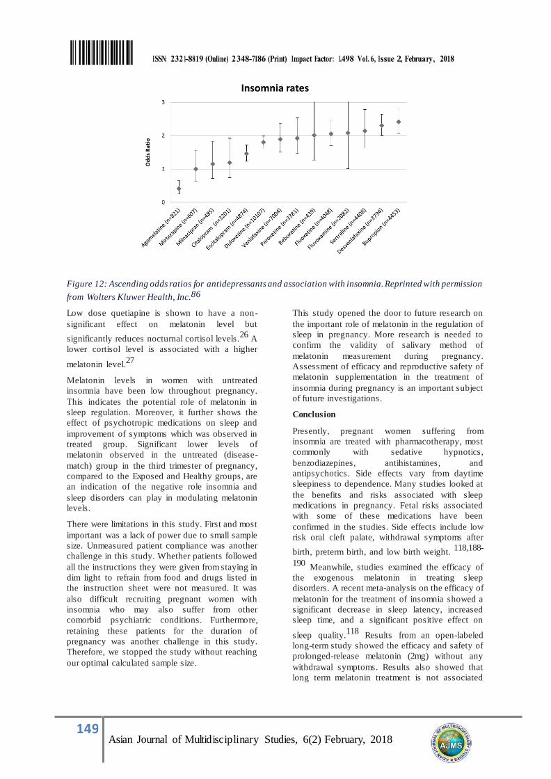
increased REM (rapid eye movement) sleep,85
however, a meta-analysis found bupropion was one
of the antidepressants that caused insomnia the
most often (Figure 12).86
2
149 Asian Journal of Multidisciplinary Studies, 6(2) February, 2018
Figure 12: Ascending odds ratios for antidepressants and association with insomnia. Reprinted with permission
from Wolters Kluwer Health, Inc.86
Low dose quetiapine is shown to have a non-
significant effect on melatonin level but
significantly reduces nocturnal cortisol levels.26
A
lower cortisol level is associated with a higher
melatonin level.27
Melatonin levels in women with untreated
insomnia have been low throughout pregnancy.
This indicates the potential role of melatonin in
sleep regulation. Moreover, it further shows the
effect of psychotropic medications on sleep and
improvement of symptoms which was observed in
treated group. Significant lower levels of
melatonin observed in the untreated (disease-
match) group in the third trimester of pregnancy,
compared to the Exposed and Healthy groups, are
an indication of the negative role insomnia and
sleep disorders can play in modulating melatonin
levels.
There were limitations in this study. First and most
important was a lack of power due to small sample
size. Unmeasured patient compliance was another
challenge in this study. Whether patients followed
all the instructions they were given from staying in
dim light to refrain from food and drugs listed in
the instruction sheet were not measured. It was
also difficult recruiting pregnant women with
insomnia who may also suffer from other
comorbid psychiatric conditions. Furthermore,
retaining these patients for the duration of
pregnancy was another challenge in this study.
Therefore, we stopped the study without reaching
our optimal calculated sample size.
This study opened the door to future research on
the important role of melatonin in the regulation of
sleep in pregnancy. More research is needed to
confirm the validity of salivary method of
melatonin measurement during pregnancy.
Assessment of efficacy and reproductive safety of
melatonin supplementation in the treatment of
insomnia during pregnancy is an important subject
of future investigations.
Conclusion
Presently, pregnant women suffering from
insomnia are treated with pharmacotherapy, most
commonly with sedative hypnotics,
benzodiazepines, antihistamines, and
antipsychotics. Side effects vary from daytime
sleepiness to dependence. Many studies looked at
the benefits and risks associated with sleep
medications in pregnancy. Fetal risks associated
with some of these medications have been
confirmed in the studies. Side effects include low
risk oral cleft palate, withdrawal symptoms after
birth, preterm birth, and low birth weight. 118,188-
190 Meanwhile, studies examined the efficacy of
the exogenous melatonin in treating sleep
disorders. A recent meta-analysis on the efficacy of
melatonin for the treatment of insomnia showed a
significant decrease in sleep latency, increased
sleep time, and a significant positive effect on
sleep quality.118
Results from an open-labeled
long-term study showed the efficacy and safety of
prolonged-release melatonin (2mg) without any
withdrawal symptoms. Results also showed that
long term melatonin treatment is not associated
2
150 Asian Journal of Multidisciplinary Studies, 6(2) February, 2018
with suppression of endogenous secretion of the
hormone as levels measured in urine two weeks
after discontinuation. No dependence, relapse of
symptoms, or withdrawal symptoms have been
reported after discontinuation of treatment.91
Pilot
studies at this time are investigating the role of
antenatal melatonin administration as an
antioxidant in late pregnancies to prevent or treat
IUGR and preeclampsia.92, 93
The prospect of
treating pregnant women with insomnia with the
natural sleep hormone melatonin has received very
little attention.
This study confirmed the lower saliva levels of
melatonin in pregnant women with untreated
insomnia. Therefore, melatonin supplementation
may be considered an intervention for insomnia in
pregnancy. Melatonin is inexpensive, is available
over-the counter and it has a low side effect
profile. This study confirmed a need for future
research to introduce melatonin as a natural
hormone therapy for insomnia. The results of this
study may have a significant positive effect on the
quality of life and health of many pregnant women
worldwide currently on sleep medication who have
no choice but to continue pharmacotherapy to
maintain a stable mental and physical health during
pregnancy.
References
1. Morris CJ, Aeschbach D, Scheer FA. Circadian system, sleep and endocrinology. Mol Cell Endocrinol
2012;349:91-104.
2. Yamazaki S, Numano R, Abe M, et al. Resetting central and peripheral circadian oscillators in transgenic
rats. Science 2000;288:682-5.
3. Turek FW, Joshu C, Kohsaka A, et al. Obesity and metabolic syndrome in circadian Clock mutant mice.
Science 2005;308:1043-5.
4. Van den Pol AN. The hypothalamic suprachiasmatic nucleus of rat: intrinsic anatomy. J Comp Neurol
1980;191:661-702.
5. Sack RL, Brandes RW, Kendall AR, Lewy AJ. Entrainment of free-running circadian rhythms by melatonin
in blind people. N Engl J Med 2000;343:1070-7.
6. Buhr ED, Yoo SH, Takahashi JS. Temperature as a universal resetting cue for mammalian circadian
oscillators. Science 2010;330:379-85.
7. Scheer FA, Hu K, Evoniuk H, et al. Impact of the human circadian system, exercise, and their interaction on
cardiovascular function. Proc Natl Acad Sci U S A 2010;107:20541-6.
8. Macchi MM, Bruce JN. Human pineal physiology and functional significance of melatonin. Front
Neuroendocrinol 2004;25:177-95.
9. Arendt J. Melatonin and mammalian pineal gland. 1995.
10. Cardinali DP, Lynch HJ, Wurtman RJ. Binding of melatonin to human and rat plasma proteins.
Endocrinology 1972;91:1213-8.
11. Mallo C,Zaidan R., Galy G., Vermeulen E., Brun J., Chazot G., Claustrat B. Pharmacokinetics of melatonin
in man after intravenous infusion and bolus injection. Eur J Clin Pharmacol 1990;38:297-301.
12. Lane EA, Moss HB. Pharmacokinetics of melatonin in man: first pass hepatic metabolism. J Clin Endocrinol
Metab 1985;61:1214-6.
13. Viljoen M, Steyn ME, van Rensburg BW, Reinach SG. Melatonin in chronic renal failure. Nephron
1992;60:138-43.
14. Cebrian-Perez JA, Casao A, Gonzalez-Arto M, dos Santos Hamilton TR, Perez-Pe R, Muino-Blanco
15. T. Melatonin in sperm biology: breaking paradigms. Reprod Domest Anim 2014;49 Suppl 4:11-21.
16. Kivela A, Kauppila A, Leppaluoto J, Vakkuri O. Serum and amniotic fluid melatonin during human labor. J
Clin Endocrinol Metab 1989;69:1065-8.
17. Cohen Engler A, Hadash A, Shehadeh N, Pillar G. Breastfeeding may improve nocturnal s leep and reduce
2
151 Asian Journal of Multidisciplinary Studies, 6(2) February, 2018
infantile colic: potential role of breast milk melatonin. Eur J Pediatr 2012;171:729-32.
18. Cagnacci A. Melatonin in relation to physiology in adult humans. J Pineal Res 1996;21:200-13.
19. Buxton OM, L'Hermite-Baleriaux M, Hirschfeld U, Cauter E. Acute and delayed effects of exercise on
human melatonin secretion. J Biol Rhythms 1997;12:568-74.
20. Nelson RJ, Drazen DL. Melatonin mediates seasonal adjustments in immune function. Reprod Nutr Dev
1999;39:383-98.
21. Reppert SM, Godson C, Mahle CD, Weaver DR, Slaugenhaupt SA, Gusella JF. Molecular characterization
of a second melatonin receptor expressed in human retina and brain: the Mel1b melatonin receptor. Proc
Natl Acad Sci U S A 1995;92:8734-8.
22. Reppert SM, Weaver DR, Rivkees SA, Stopa EG. Putative melatonin receptors in a human biological clock.
Science 1988;242:78-81.
23. Konakchieva R, Kyurkchiev S, Kehayov I, Taushanova P, Kanchev L. Selective effect of methoxyindoles on
the lymphocyte proliferation and melatonin binding to activated human lymphoid cells. J Neuroimmunol
1995;63:125-32.
24. Lopez-Gonzalez MA, Calvo JR, Osuna C, Guerrero JM. Interaction of melatonin with human lymphocytes:
evidence for binding sites coupled to potentiation of cyclic AMP stimulated by vasoactive intestinal peptide
and activation of cyclic GMP. J Pineal Res 1992;12:97-104.
25. Gilad E, Laudon M, Matzkin H, et al. Functional melatonin receptors in human prostate epithelial cells.
Endocrinology 1996;137:1412-7.
26. Yie SM, Niles LP, Younglai EV. Melatonin receptors on human granulosa cell membranes. J Clin
Endocrinol Metab 1995;80:1747-9.
27. van Vuuren RJ, Pitout MJ, van Aswegen CH, Theron JJ. Putative melatonin receptor in human spermatozoa.
Clin Biochem 1992;25:125-7.
28. Vacas MI, Del Zar MM, Martinuzzo M, Cardinali DP. Binding sites for [3H]-melatonin in human platelets. J
Pineal Res 1992;13:60-5.
29. Lanoix D, Beghdadi H, Lafond J, Vaillancourt C. Human placental trophoblasts synthesize melatonin and
express its receptors. J Pineal Res 2008;45:50-60.
30. Sugden D. Melatonin biosynthesis in the mammalian pineal gland. Experientia 1989;45:922-32.
31. Maronde E SJH. The Mamalian Pineal Gland: known facts, unknown facets. Trends Endocrinol Metab
2007;18:142-9.
32. Meijer JH, Rietveld WJ. Neurophysiology of the suprachiasmatic circadian pacemaker in rodents. Physiol
Rev 1989;69:671-707.
33. Berson DM. Strange vision: ganglion cells as circadian photoreceptors. Trends Neurosci 2003;26:314-20.
34. Brainard GC, Lewy AJ, Menaker M, et al. Dose-response relationship between light irradiance and the
suppression of plasma melatonin in human volunteers. Brain Res 1988;454:212-8.
35. Brainard GC,Hanifin J.P., Greeson J.M., Byrne B., Glickman G., Gerner E., Rollag M.D. Action spectrum
for melatonin regulation in humans: evidence for a novel circadian photoreceptor. J Neurosci
2001;21:6405-12.
36. Moore RY. Neural control of the pineal gland. Behav Brain Res 1996;73:125-30.
37. Moore RY, Sibony P. Enkephalin-like immunoreactivity in neurons in the human pineal gland. Brain Res
1988;457:395-8.
38. Ho AK, Chik CL, Klein DC. Protein kinase C is involved in adrenergic stimulation of pineal cGMP
accumulation. J Biol Chem 1987;262:10059-64.
39. Klein DC. Photoneural regulation of the mammalian pineal gland. Ciba Found Symp 1985;117:38- 56.
40. Wetterberg L, Bratlid T, von Knorring L, Eberhard G, Yuwiler A. A multination al study of the relationships

2
152 Asian Journal of Multidisciplinary Studies, 6(2) February, 2018
between nighttime urinary melatonin production, age, gender, body size, and latitude. Eur Arch Psychiatry
Clin Neurosci 1999;249:256-62.
41. Haimov I, Laudon M, Zisapel N, et al. Sleep disorders and melatonin rhythms in elderly people. BMJ
1994;309:167.
42. Keijzer H, Smits MG, Duffy JF, Curfs LM. Why the dim light melatonin onset (DLMO) should be measured
before treatment of patients with circadian rhythm sleep disorders. Sleep Med Rev 2014;18:333-9.
43. Bergiannaki JD,Paparrigopoulos T.J., Syrengela M., Stefanis C.N. Low and high melatonin excretors among
healthy individuals. J Pineal Res 1995;18:159-64.
44. Lushington K,Dawson D., Encel N., Lack L. Urinary 6-sulfatoxymelatonin cycle-to-cycle variability.
Chronobiol Int 1996;13:411-21.
45. Kripke DF,Garfinkel L., Wingard D.L., Klauber M.R., Marler M.R. Mortality associated with sleep duration
and insomnia. Arch Gen.Psychiatr. 2002;59:131-6.
46. Geoffriau M,Claustrat B., Veldhuis J. Estimation if nocturnal melatonin production in humans by
deconvolution analysis.in: Y. Touitou (Ed), biological Clocks,mechanism and applications. 1998:325-8.
47. Braam W, van Geijlswijk I, Keijzer H, Smits MG, Didden R, Curfs LM. Loss of response to melatonin
treatment is associated with slow melatonin metabolism. J Intellect Disabil Res 2010;54:547- 55.
48. Takahashi JS. Circadian rhythms. ICER is nicer at night (sir!). Curr Biol 1994;4:165-8.
49. Vondrasova D, Hajek I, Illnerova H. Exposure to long summer days affects the human melatonin and
cortisol rhythms. Brain Res 1997;759:166-70.
50. Wehr TA. The durations of human melatonin secretion and sleep respond to changes in daylength
(photoperiod). J Clin Endocrinol Metab 1991;73:1276-80.
51. Rosenthal NE, Sack DA, Gillin JC, et al. Seasonal affective disorder. A description of th e syndrome and
preliminary findings with light therapy. Arch Gen Psychiatry 1984;41:72-80.
52. Arendt J, Skene DJ, Middleton B, Lockley SW, Deacon S. Efficacy of melatonin treatment in jet lag, shift
work, and blindness. J Biol Rhythms 1997;12:604-17.
53. Fuller PM,Gooley J.J., Saper C.B. Neurobiology of the sleep-wake cycle;sleep architecture, circadian
regulation, and regulatory feedback. J Biol Rhythms 2006;21:482-93.
54. Van Den Heuvel CJ, Reid KJ, Dawson D. Effect of atenolol on nocturnal sleep and temperature in young
men: reversal by pharmacological doses of melatonin. Physiol Behav 1997;61:795-802.
55. Hajak G, Rodenbeck A, Staedt J, Bandelow B, Huether G, Ruther E. Nocturnal plasma melatonin levels in
patients suffering from chronic primary insomnia. J Pineal Res 1995;19:116-22.
56. Kalsbeek A, Cutrera RA, Van Heerikhuize JJ, Van Der Vliet J, Buijs RM. GABA release from
suprachiasmatic nucleus terminals is necessary for the light-induced inhibition of nocturnal melatonin
release in the rat. Neuroscience 1999;91:453-61.
57. McCarley RW. Neurobiology of REM and NREM sleep. Sleep Med 2007;8:302-30.
58. Banks S, Dinges DF. Behavioral and physiological consequences of sleep restriction. J Clin Sleep Med
2007;3:519-28.
59. da Silveira Cruz-Machado S, Carvalho-Sousa CE, Tamura EK, et al. TLR4 and CD14 receptors expressed in
rat pineal gland trigger NFKB pathway. J Pineal Res 2010;49:183-92.
60. Krueger JM. The role of cytokines in sleep regulation. Curr Pharm Des 2008;14:3408-16.
61. Banks S DDF. Behavioural and Physiological consequences of sleep Restriction. J Clin Sleep Med
2007;3:519-28.
62. Ohayon MM. Epidemiology of insomnia: what we know and what we still need to learn. Sleep Med Rev
2002;6:97-111.
63. Morphy H, Dunn KM, Lewis M, Boardman HF, Croft PR. Epidemiology of insomnia: a longitudinal study
2
153 Asian Journal of Multidisciplinary Studies, 6(2) February, 2018
in a UK population. Sleep 2007;30:274-80.
64. Barion A, Zee PC. A clinical approach to circadian rhythm sleep disorders. Sleep Med 2007;8:566- 77.
65. Micic G, Lovato N, Gradisar M, et al. Nocturnal Melatonin Profiles in Patients with Delayed Sleep - Wake
Phase Disorder and Control Sleepers. J Biol Rhythms 2015.
66. Benloucif S, Burgess HJ, Klerman EB, et al. Measuring melatonin in humans. J Clin Sleep Med 2008;4:66-
9.
67. Pullman RE, Roepke SE, Duffy JF. Laboratory validation of an in-home method for assessing circadian
phase using dim light melatonin onset (DLMO). Sleep Med 2012;13:703-6.
68. National Sleep Foundation. Sleep in America. 2010.
69. Depner CM, Stothard ER, Wright KP,Jr. Metabolic consequences of sleep and circadian disorders. Curr
Diab Rep 2014;14:507,014-0507-z.
70. Spiegel K, Leproult R, L'hermite-Baleriaux M, Copinschi G, Penev PD, Van Cauter E. Leptin levels are
dependent on sleep duration: relationships with sympathovagal balance, carbohydrate regulation, cortisol,
and thyrotropin. J Clin Endocrinol Metab 2004;89:5762-71.
71. Markwald RR, Melanson EL, Smith MR, et al. Impact of insufficient sleep on total daily energy
expenditure, food intake, and weight gain. Proc Natl Acad Sci U S A 2013;110:5695-700.
72. Spiegel K, Leproult R, Van Cauter E. Impact of sleep debt on metabolic and endocrine function. Lancet
1999;354:1435-9.
73. Fernandez-Mendoza J, Vgontzas AN, Liao D, et al. Insomnia with objective short sleep duration and
incident hypertension: the Penn State Cohort. Hypertension 2012;60:929-35.
74. Vgontzas AN, Bixler EO, Lin HM, et al. Chronic insomnia is associated with nyctohemeral activation of the
hypothalamic-pituitary-adrenal axis: clinical implications. J Clin Endocrinol Metab 2001;86:3787-94.
75. Irwin M, Clark C, Kennedy B, Christian Gillin J, Ziegler M. Nocturnal catecholamines and immune function
in insomniacs, depressed patients, and control subjects. Brain Behav Immun 2003;17:365-72.
76. Mallon L, Broman JE, Hetta J. Relationship between insomnia, depression, and mortality: a 12-year follow-
up of older adults in the community. Int Psychogeriatr 2000;12:295-306.
77. Baglioni C,Spiegelhalder K., Lombardo C., Riemann D. Sleep and emotions:a focus on insomnia. Sleep
Med Rev 2010;14:227-38.
78. Belenky G, Wesensten NJ, Thorne DR, et al. Patterns of performance degradation and rest oration during
sleep restriction and subsequent recovery: a sleep dose-response study. J Sleep Res 2003;12:1- 12.
79. Huang ZL, Zhang Z, Qu WM. Roles of adenosine and its receptors in sleep -wake regulation. Int Rev
Neurobiol 2014;119:349-71.
80. Stutts JC, Wilkins JW, Scott Osberg J, Vaughn BV. Driver risk factors for sleep-related crashes. Accid Anal
Prev 2003;35:321-31.
81. Fortier-Brochu E,Beaulieu-Bonneau S., Ivers H., Morin C.M. Relations between sleep, fatigue, and health -
related quality of life in individuals with insomnia. J Psychosom Res 2010;69:475-83.
82. Kling RN,McLeod C.B., Koehoorn M. Sleep problems and workplace injuries in Canada. Sleep
2010;33:611-8.
83. Oomen CA, Bekinschtein P, Kent BA, Saksida LM, Bussey TJ. Adult hippocampal neurogenesis and its role
in cognition. Wiley Interdiscip Rev Cogn Sci 2014;5:573-87.
84. Riemann D, Voderholzer U, Spiegelhalder K, et al. Chronic insomnia and MRI-measured hippocampal
volumes: a pilot study. Sleep 2007;30:955-8.
85. Noh HJ, Joo EY, Kim ST, et al. The relationship between hippocampal volume and cognition in patients
with chronic primary insomnia. J Clin Neurol 2012;8:130-8.
86. Fernandez-Mendoza J, Calhoun SL, Bixler EO, et al. Sleep misperception and chronic insomnia in the

2
154 Asian Journal of Multidisciplinary Studies, 6(2) February, 2018
general population: role of objective sleep duration and psychological profiles. Psychosom Med
2011;73:88-97.
87. Baglioni C, Battagliese G, Feige B, et al. Insomnia as a predictor of depression: a meta -analytic evaluation
of longitudinal epidemiological studies. J Affect Disord 2011;135:10-9.
88. Riemann D, Spiegelhalder K, Feige B, et al. The hyperarousal model of insomnia: a review of the concept
and its evidence. Sleep Med Rev 2010;14:19-31.
89. Buckley TM, Schatzberg AF. A pilot study of the phase angle between cortisol and melatonin in major
depression - a potential biomarker? J Psychiatr Res 2010;44:69-74.
90. Pinho M, Sehmbi M, Cudney LE, et al. The association between biological rhythms, depression, and
functioning in bipolar disorder: a large multi-center study. Acta Psychiatr Scand 2015.