Embed Size (px)
Citation preview

Dental Research Journal
Dental Research Journal / Dec 2012 / Vol 9 / Issue 8 (Supplement Issue 2)S202
Original ArticleMicroflora and periodontal diseaseLuca Scapoli1, Ambra Girardi1, Annalisa Palmieri2, Tiziano Testori3, Francesco Zuffetti3, Riccardo Monguzzi4, Dorina Lauritano5, Francesco Carinci2
1Departments of Histology, Embryology and Applied Biology, Centre of Molecular Genetics, CARISBO Foundation, University of Bologna, Bologna, 2Department of Medical-Surgical Sciences of Communication and Behavior, Section of Maxillofacial and Plastic Surgery, University of Ferrara, Ferrara, 3Dentistry, Galeazzi Hospital, Milan, Italy, 4Private Practice, Dentist, Milan, Italy, 5Neurosciences and Biomedical Technologies, University Milano Bicocca, Italy
ABSTRACT
Background: Periodontitis is a disease that affects and destroys the tissues that support teeth. Tissue damage results from a prolonged inflammatory response to an ecological shift in the composition of subgingival biofilms. Three bacterial species that constitute the red complex group, Porphyromonas gingivalis, Tannerella forsythia, and Treponema denticola, are considered the main pathogens involved in periodontitis.Materials and Methods: In the present study, a real‑time polymerase chain reaction bases assay was designed to detect and quantify red complex species, then used to investigate 307 periodontal pocket samples from 127 periodontitis patients and 180 controls.Results: Significant higher prevalence of red complex species and increased amount of P. gingivalis and T. denticola were detected in periodontal pocket of periodontitis patients.Conclusions: Results demonstrated that the test is a valuable tool to improve diagnosis of periodontal disease.
Key Words: Bone, diseases, inflammation, ligament, periodontal, resorption, tooth
INTRODUCTION
Periodontitis represents a destructive chronic inflammatory disease with a bacterial infection resulting from the complex actions of a small subset of periodontal pathogens.[1]
From a pathological point of view, periodontitis can be defined as the presence of gingival inflammation at sites where there has been a pathological detachment of collagen fibers from the cementum and the junctional epithelium has migrated apically.[2,3] The inflammatory response of the periodontal tissues to infection is influenced by environmental factors as well
as by genetic factors.[4] The primary microbial factor contributing to periodontitis is a shift in the content of the oral microflora, while the primary immunological factor is the destructive host inflammatory response.[5,6]
The microbiota associated with periodontal health and disease has been intensely studied for well over a century by several generations of skilled scientists and clinicians.[7,8] Oral microbiota is an enormously complex and dynamic entity that is profoundly affected by perpetually changing local environments and host‑mediated selective pressures.[9] The presence of a commensal microbiota, including potential pathogens, is essential for the proper development of mucosal immunity.[10]
The normal oral flora is hence in a balance between pathogens and commensals that requires regular cleaning to be maintained. A decrease in oral hygiene is quickly followed by the build‑up of oral biofilms on tooth surfaces and, if left untreated, will progress to gingival inflammation and possibly periodontitis,
Received: August 2012Accepted: November 2012
Address for correspondence: Prof. Francesco Carinci, Department of D.M.C.C.C., Section of Maxillofacial and Plastic Surgery, University of Ferrara, Corso Giovecca 203, Ferrara, Italy.E‑mail: [email protected]
Access this article online
Website: www.drj.ir
www.mui.ac.ir

Scapoli, et al.: Periodontitis
Dental Research Journal / Dec 2012 / Vol 9 / Issue 8 (Supplement Issue 2) S203
alveolar bone loss, and loss of teeth. It is likely that differences in host‑defense mechanisms, including antimicrobial protein profiles, determine whether bacterial colonization progresses to overt disease.[11]
Recent data estimate that the oral cavity may contain up to 19 000 bacterial phylotypes,[12] but each individual will only have a rate of the total numbers of pathogens. Indeed, there is a substantial diversity in the content of the microflora between individuals[13] and between different oral sites within the same individual.[14,15] Research has indicated that dietary changes combined with poor hygiene can cause a shift in the composition of the oral bacteria.[15,16] Moreover, some evidence in recent studies suggests that the oral microbiome changes as human beings age and the dysbiosis in the oral cavity can lead to periodontitis.[5]
Several methods have been used for microbiological testing in periodontitis.[17] However, many techniques have not been fully accepted due to low sensitivity or specificity; moreover, sometimes they are slow, expensive, and laborious. In our laboratory (LAB SRL, Ferrara, Italy), we developed a rapid and sensitive test to detect and quantify the three bacterial species more involved in periodontitis that constituted the red complex group: Porphyromonas gingivalis, Tannerella forsythia, and Treponema denticola. Both P. gingivalis and T. denticola occur concomitantly with the clinical signs of periodontal destruction. They appear closely “linked” topologically in the developing biofilm, shown an in vitro ability to produce a number of outer membrane‑associated proteinases, and are considered the first pathogens involved in the clinical destruction of periodontal tissues. Moreover, both of them and T. forsythia show an higher prevalence in disease than in health, suggesting that these bacterial are associated with the local development of periodontitis.[18]
The presence and the level of these pathogens can be effectively revealed by real‑time polymerase chain reaction (PCR) analysis using bacterial species‑specific primers and probes.
Our findings support the hypothesis that detection and quantification of red complex bacteria in crevicular fluid could be an appropriate tool for diagnosis and prognosis of periodontitis.
MATERIALS AND METHODS
A total of 307 individuals participated in the study,
127 were affected by chronic periodontitis, while 180 constituted the control group. Controls include 66 healthy individuals and 114 affected by a moderate gingivitis. Table 1 summarizes principal characteristics of the two groups.
A sample of the periodontal pocket microbiota was obtained from a single site by a paper probe. DNA was extracted and purified using standard protocols that include two consecutive incubations with lysozyme and proteinase K, followed by spin‑column purification.
Real‑time polymerase chain reactionPrimers and probes oligonucleotides were designed basing on 16S rRNA gene sequences of the Human Oral Microbiome Database (HOMD 16S rRNA RefSeq Version 10.1) counting 845 entries. All the sequences were aligned in order to find either consensus sequence or less conservate spots. Two real‑time PCR runs were performed for each sample. The first reaction quantified the total amount of bacteria using two degenerate primers and a single probe matching a highly conservated sequence of the 16S ribosomal RNA gene. The second reaction detected and quantified the three red complex bacteria, i.e., P. gingivalis, T. forsythia, and T. denticola, in a multiplex PCR. This reaction included a total of six primers and three probes that were highly specific for each species. Oligonucleotide concentrations and PCR conditions were optimized to ensure sensitivity, specificity, and no inhibitions in case of unbalanced target amounts. Absolute quantification assays were performed using the Applied Biosystems 7500 Sequence Detection System. The amplification profile was initiated by a 10‑minute incubation period at 95°C to activate polymerase, followed by a two‑step amplification of 15 s at 95°C and 60 s at 57°C for 40 cycles. All these experiments were performed including non‑template controls to exclude reagents contamination.
Plasmids containing synthetic DNA target sequences (Eurofin MWG Operon, Ebersberg Germany) were
Table 1: Sample studySample study features
Totals Health Gingivitis Periodontitis
Subjects (n) 307 66 114 127Male (n) 124 26 42 56Female (n) 183 40 72 71Age (mean years±SD)
39.8±18.9 31.6±18.6 34.3±15.4 48.9±18.2
Sampling depth (mm±SD)
3.9±1.7 2.6±0.6 3.2±1.0 5.0±1.7
www.mui.ac.ir
Scapoli, et al.: Periodontitis
Dental Research Journal / Dec 2012 / Vol 9 / Issue 8 (Supplement Issue 2)S204
used as standard for the quantitative analysis. Standard curves for each target were constructed in a triplex reaction, by using a mix of the same amount of plasmids, in serial dilutions ranging from 101 to 107 copies. There was a linear relationship between the threshold cycle values plotted against the log of the copy number over the entire range of dilutions (data not shown). The copy numbers for individual plasmid preparations were estimated using the Thermo NanoDrop spectrophotometer.
The absolute quantification of total bacterial genome copies in samples allowed for the calculation of relative amount of red complex species. To prevent samples and PCR contamination, plasmid purification and handling was performed in a separate laboratory with dedicated pipettes.
Statistical analysisDescriptive statistics was performed using Microsoft Excel spreadsheets. The Freeman‑Halton extension of Fisher’s exact test was used to compute the (two‑tailed) probability of obtaining a distribution of values in a 2 × 3 contingency table, given the number of observations in each cell. Odds ratio calculation was performed online at the OpenEpi web site (www.openepi.com).
Absolute bacteria amount were normalized against the total bacterial load, obtaining the relative bacteria amount (RBA). The one‑way analysis of variance (ANOVA) was used to determine whether there were any significant differences between the mean RBA value of three patients group, i.e., healthy, gingivitis, and periodontitis.
RESULTS
Occurrence and amount of red complex bacteria from crevicular fluid were evaluated in 307 individuals.
A single specimen from each patient was analyzed by quantitative real‑time PCR, obtaining measures of total bacteria load and of three species involved in periodontitis, i.e., P. gingivalis, T. forsythia, and T. denticola. Here, we report a preliminary study focused mainly on prevalence of these three species among groups of patients with different diagnosis—regardless of different clinical aspects that may describe severity of the disease—in order to understand whether the presence of the red complex species and their relative amount may be considered predictive factors of periodontitis.
Prevalence of the three investigated species among healthy, gingivitis, and periodontitis patients is shown in Figure 1. Each species was common among healthy patients; however, the prevalence was roughly double in periodontitis group. Intermediate values, but closer to healthy individuals, were observed among patients affected by gingivitis.
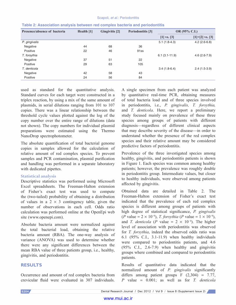
Obtained data are detailed in Table 2. The Freeman‑Halton extension of Fisher’s exact test indicated that the prevalence of each red complex species is different among groups of patients with high degree of statistical significance, P. gingivalis (P value = 2 × 10⁻⁸), T. forsythia (P value = 1 × 10⁻⁸), and T. denticola (P value = 2 × 10⁻⁴). The higher level of association with periodontitis was observed for T. forsythia, indeed the observed odds ratio was 6.1 (95% C.I., 3.1‑11.9) when healthy individuals were compared to periodontitis patients, and 4.6 (95% C.I., 2.6‑7.9) when healthy and gingivitis groups where combined and compared to periodontitis patients.
Results of quantitative data indicated that the normalized amount of P. gingivalis significantly differs among patient groups F (2,304) = 7.77, P value = 0.001; as well as for T. denticola
Table 2: Association analysis between red complex bacteria and periodontitisPresence/absence of bacteria Health [1] Gingivitis [2] Periodontitis [3] OR (95% C.I.)
[1] vs. [3] [1]+[2] vs. [3]P. gingivalis 5.1 (1.8‑4.3) 4.2 (2.6‑6.8)
Negative 44 68 36Positive 22 46 91ss
T. forsythia 6.1 (3.1‑11.9) 4.6 (2.6‑7.9)Negative 37 51 22Positive 29 63 105
T. denticola 3.4 (1.8‑6.4) 2.4 (1.5‑3.9)Negative 42 58 43Positive 24 56 84
www.mui.ac.ir

Scapoli, et al.: Periodontitis
Dental Research Journal / Dec 2012 / Vol 9 / Issue 8 (Supplement Issue 2) S205
F (2,304) = 7.47, P value = 0.001. On the contrary, it did not vary for T. forsythia F (2,304) = 1.41, P value = 0.25. The calculated mean values are plotted in Figure 2.
DISCUSSION
The PCR is the most sensitive and rapid method to detect microbial pathogens in clinical specimens. In particular, the diagnostic value of PCR is significantly higher when specific pathogens that are difficult to culture in vitro or require a long cultivation period such as for anaerobic bacteria species are involved in periodontitis onset. A recent improvement of this technique is the real‑time PCR that allow for quantitation of DNA target using fluorogenic probes in a close setup. Besides the opportunity to quantify target, the advantage to perform the assay is a closed system, in which the reaction tube is never opened after amplification, is of great value to prevent laboratory contamination and false‑positive results. In addition, the need of a probe, in addition to the two
PCR primers, further increases the specificity of the reaction.
In the present investigation, we designed and tested the performance of a real‑time PCR‑based assay to detect and quantify the red complex bacteria involved in periodontal disease. In particular, we found that P. gingivalis, T. forsythia, and T. denticola were strongly related to periodontitis because their prevalence was higher among periodontitis patient. The presence of these bacterial species can significantly increase the risk to develop periodontitis, the OR being comprised between 6.1 (T. forsythia) and 3.4 (T. denticola). The results of quantitative data analysis indicated that the relative amount of P. gingivalis and T. denticola in periodontal pocket was sensibly higher in affected patients. This indicated that both the presence and relative amount of red complex bacteria is relevant data in periodontal disease diagnosis.
CONCLUSION
Molecular analysis of periodontal pocket microflora by real‑time PCR represents an effective inexpensive method to rapidly detect and quantify red complex bacterial species. This test was performed in a large patient sample and results demonstrated that the test is a valuable tool to improve diagnosis of periodontal disease.
ACKNOWLEDGEMENTS
This work was supported by FAR from the University of Ferrara (FC), Ferrara, Italy, and LAB® s.r.l, Ferrara, Italy.
REFERENCES
1. Kuboniwa M, Inaba H, Amano A. Genotyping to distinguish microbial pathogenicity in periodontitis. Periodontol
Figure 1: Prevalence of red complex bacterial species in healthy (green), gingivitis (yellow), and periodontitis (red) patients
Figure 2: Plots represent the relative amount of each red complex bacterial species in the different group of patients
www.mui.ac.ir

Scapoli, et al.: Periodontitis
Dental Research Journal / Dec 2012 / Vol 9 / Issue 8 (Supplement Issue 2)S206
2000 2010;54:136‑59.2. Savage A, Eaton KA, Moles DR, Needleman I. A systematic
review of definitions of periodontitis and methods that have been used to identify this disease. J Clin Periodontol 2009;36:458‑67.
3. Gersdorff N, Miró X, Roediger M, Geffers R, Toepfer T, Huels A, et al. Gene expression analysis of chronically inflamed and healthy human periodontal ligament cells in vivo. Dent Res J (Isfahan) 2008;5:5‑11.
4. Kinane DF, Peterson M, Stathopoulou PG. Environmental and other modifying factors of the periodontal diseases. Periodontol 2000 2006;40:107‑19.
5. Berezow AB, Darveau RP. Microbial shift and periodontitis. Periodontol 2000 2011;55:36‑47.
6. Kanaparthy A, Kanaparthy R, Niranjan N. Evaluation of serum C‑reactive protein levels in subjects with aggressive and chronic periodontitis and comparison with healthy controls. Dent Res J (Isfahan) 2012;9:261‑5.
7. Tanner AC. Tannerella forsythia, a periodontal pathogen entering the genomic era. Periodontol 2000 2006;42:88‑113.
8. Teles RP, Haffajee AD, Socransky SS. Microbial goals of periodontal therapy. Periodontol 2000 2006;42:180‑218.
9. Armitage GC. Comparison of the microbiological features of chronic and aggressive periodontitis. Periodontol 2000,2010;53:70‑88.
10. Davey ME, O’Toole GA. Microbial biofilms: From ecology to molecular genetics. Microbiol Mol Biol Rev 2000;64:847‑67.
11. Gorr SU, Abdolhosseini M. Antimicrobial peptides and periodontal disease. J Clin Periodontol 2011;38:126‑41.
12. Slots J. Update on actinobacillus actinomycetemcomitans and Porphyromonas gingivalis in human periodontal disease. J Int Acad Periodontol 1999;1:121‑6.
13. Nasidze I, Li J, Quinque D, Tang K, Stoneking M. Global diversity in the human salivary microbiome. Genome Res 2009;19:636‑43.
14. Aas JA, Paster BJ, Stokes LN, Olsen I, Dewhirst FE. Defining the normal bacterial flora of the oral cavity. J Clin Microbiol 2005;43:5721‑32.
15. Avila M, Ojcius DM, Yilmaz O. The oral microbiota: Living with a permanent guest. DNA Cell Biol 2009;28:405‑11.
16. Al‑Ahmad A, Roth D, Wolkewitz M, Wiedmann‑Al‑Ahmad M, Follo M, Ratka‑Kruger P, et al. Change in diet and oral hygiene over an 8‑week period: Effects on oral health and oral biofilm. Clin Oral Investig 2010;14:391‑6.
17. Loomer PM. Microbiological diagnostic testing in the treatment of periodontal diseases. Periodontol 2000 2004;34:49‑56.
18. Mineoka T, Awano S, Rikimaru T, Kurata H, Yoshida A, Ansai T, et al. Site‑specific development of periodontal disease is associated with increased levels of Porphyromonas gingivalis, Treponema denticola, and Tannerella forsythia in subgingival plaque. J Periodontol 2008;79:670‑6.
How to cite this article: Scapoli L, Girardi A, Palmieri A, Testori T, Zuffetti F, Monguzzi R, et al. Microflora and periodontal disease. Dent Res J 2012;9:S202‑6.Source of Support: This work was supported by FAR from the University of Ferrara (FC), Ferrara, Italy, and LAB® s.r.l, Ferrara, Italy. Conflict of Interest: None declared.
www.mui.ac.ir