Embed Size (px)
Citation preview
13 . The Eye and Related Drugs:
Topical Medications:
For topical medications installed in the eye, you may need to use diagnostic
drugs to perform a complete ocular examination, and to aid in the proper
diagnosis of a certain ocular condition, or you may apply local therapeutic agents
to treat a specific pathology.
Proper technique to instill eyedrops
1. Wash your hands; wear disposable gloves if desired.
2. Instruct the seated patient to tilt the head back and to look up.
3. Expose the palpebral conjunctiva by gently pulling downward on the
skin over the cheekbone. Avoid direct pressure on the eyeball.
4. Instill the correct amount of medication into the lower conjunctival fornix.
Avoid applying drops directly to the cornea, which is the most sensitive
part of the eye, and avoid touching the tip of the applicator to the patient's
lids or eye.
5. Instruct the patient to close both eyes gently for a few seconds. Wipe
any excess medication from the patient's skin with a tissue.
Objectives:
The undergraduate student should be aware of:
Ocular effects, proper dosages, local and systemic complications of
the commonly used eye drops.
Ocular complications of systemically prescribed drugs and how to
recognize and prevent.
To know and practice the proper technique of instilling eye drops in
the eye

Intact corneal sensation is one of the protective mechanisms of the eye, so patients should
be warned against self medication with topical anaesthetics . They are ONLY to be used by
doctors in the office.
Topical Diagnostic Drugs:
1. Fluorescein Dye:
Sodium fluorescein is a water-soluble, non-irritant orange-yellow dye. It turns into
green when diluted. When blue light is used, the green color becomes more
brilliant (fluoresces).
It is helpful in:
− detecting corneal epithelial abrasions and corneal ulcers as fluorescein
stains the spots denuded from epithelium.
− diagnosis of dry eye (tear break-up time)
− In measuring the IOP by applanation
− clinical evaluation of the lacrimal drainage system. Normally, if the dye is
instilled in the conjunctival sac it passes to the nose.
Side effects:
To avoid discoloration, contact lenses should be removed before the fluorescein
is instilled. Freshly prepared dye or sterile strips should be used to avoid
contamination with bacteria especially pseudomonas.
2. Topical Anesthetics:
Topical anesthetic drops (proparacaine hydrochloride 0.5% and tetracaine
0.5%) are useful to make surface manipulations painless such as when we need
to remove a superficial corneal foreign body or perform tonometry.
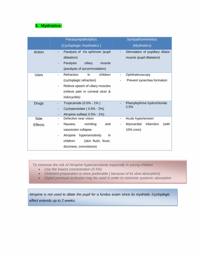
3. Mydriatics:
Parasympatholytics
(Cycloplegic /mydriatics )
Sympathomimetics
(Mydriatics)
Action - Paralysis of iris sphincter (pupil
dilatation)
- Paralysis ciliary muscle
(paralysis of accommodation)
- Stimulation of pupillary dilator
muscle (pupil dilatation)
Uses - Refraction in children
(cycloplegic refraction)
- Relieve spasm of ciliary muscles
(relieve pain in corneal ulcer &
iridocyclitis)
- Ophthalmoscopy
- Prevent synechea formation
Drugs - Tropicamide (0.5% - 1% )
- Cyclopentolate ( 0.5% - 2%)
- Atropine sulfate( 0.5% - 1%)
- Phenylephrine hydrochloride 2.5%
Side
Effects
- Defective near vision
- Nausea, vomiting and
vasomotor collapse.
- Atropine hypersensitivity in
children (skin flush, fever,
dizziness, convulsions)
- Acute hypertension
- Myocardial infarction (with
10% conc)
Atropine is not used to dilate the pupil for a fundus exam since its mydriatic /cycloplegic
effect extends up to 2 weeks.
To minimize the risk of Atropine hypersensitivity especially in young children
Use the lowest concentration (0.5%)
Ointment preparation is more preferable ( because of its slow absorption)
Digital punctual occlusion may be used in order to minimize systemic absorption
Topical Therapeutic Drugs:
A. Decongestants:
Decongestants are most commonly used eye drops. They produce
vasoconstriction that reduces hyperaemia in the eye due to minor eye
irritations caused by smoke, dust, wind, glare, swimming, contact lenses, or
fatigue.
Decongestants are weak concentrations of sympathomimetic or alpha-
adrenergic agonists that include adrenaline (epinephrine), naphazoline
hydrochloride, and phenylephrine hydrochloride.
The most frequent complication of ocular decongestants arises from
overuse, with rebound vasodilation of conjunctival vessels. In rare
instances, acute angle-closure glaucoma may be precipitated in
susceptible eyes by the use of sympathomimetic drugs because they can
dilate the pupil.
B. Tear substitutes:
Treatment of dry eye is usually with lubricating eyedrops or gels.
- 1) The methyl and hydroxpropyl cellulose derivatives are widely used in
artificial tear formulations. They work on the simple principle of lubricating
the ocular surface in order to promote its integrity.
- 2) Some brands that include sodium hyaluronate as an inactive
ingredient aids in a gradual release of water molecules, which increases
the duration of wettability.
- 3) Polyvinyl alcohol (PVA) based artificial tears is a hypotonic solution
which helps to counteract the hyperosmolarity seen in dry eyes,
- 4) Oil based brands of tear substitutes use oils in their composition.
These has been shown to increase the thickness of the lipid layer of the
tear film.
C. Topical antibiotics: (see chapter 7)
Topical antibiotics are often used to treat common bacterial conjunctivitis.
The dosage and duration of treatment are variable according to the severity as
well as the active drug ingredient. Risks of topical allergic sensitivity and
development of antibiotic resistant organisms is significant with long-term and
inappropriate antibiotic use.
D. Topical antiviral agents: (see chapter 7)
Topical antivirals are very effective in treating ophthalmic herpes viral
infections. The most preferred topical medications are acyclovir ointment (
commonly used in Europe and in Egypt) and ganciclovir gel ( commonly used in
the U.S). Both are selective antivirals which only attack the virus infected cells.
Their toxicity is very low.
Other non selective topical antivirus medications such as trifluorothymidine
(TFT), iododuoxy uridine ( IDU) and vidarabine are almost equally effective but
long term use can result in toxicity in the form of corneal epithelial punctate
keratitis or conjunctival congestion and dryness.
- Many tear substitutes are available in Gel-forms which increases
the retention time of the artificial tear drops.
- Preservatives used in eye drops can increase the eye dryness.
Therefore, preservative-free forms are preferred for the long term
treatment characteristic of the dry eye disease.
- The choice of a specific type of tear substitute depends on the cause
of eye dryness and the subjective comfort of the patient
E. Topical Corticosteroids:
Topical corticosteroids preparations are very useful as anti-
inflammatory and antiallergic in the management of various ocular situations as
conjunctival allergic reaction, uveitis, scleritis and post-surgical inflammatory
reaction. However, these can induce serious ocular complications;
i. Activation of viral, bacterial, and fungal infections.
ii. Complicated cataract.
iii. Secondary open angle glaucoma
Nonsteroidal anti-inflammatory agents do not potentiate these
complications but alone are generally not potent enough to control
significant intraocular inflammation, however. They are also used for other
specific indications, such as ocular itching, macular edema, or prevention
of miosis during cataract surgery or when steroids are contraindicated.
Never use or prescribe a topical ocular corticosteroid unless you have a precise
diagnosis for which the drug is specifically indicated.
You must be prepared to monitor the patient for serious side effects, such as
glaucoma, cataract, or infection.
F. Topical anti-glaucoma drugs: (see chapter 5)
The topically administered anti-glaucoma drugs may have local or, most
seriously, systemic side effects. The systemic side effects may be more
In genetically predisposed individuals, the use of topical corticosteroids preparation can induce
acute rise of intraocular pressure, i.e. steroid responders
prominent in the elderly, many of whom have multiple systemic conditions and
are taking multiple other medications.
a) ß-Adrenergic Antagonists (timolol, levobunolol, metipranolol, carbachol)
These are nonselective ß-adrenergic antagonists (beta-blockers) that reduce the
formation of aqueous humor by the ciliary body and thereby reduce IOP. They
are highly effective and widely used in the management of open angle glaucoma.
These drugs induce bronchospasm. Therefore, they are contraindicated in
patients with asthma or chronic obstructive pulmonary disease. They may also
precipitate or worsen cardiac failure and must be used with caution if bradycardia
or systemic hypotension .
b) Cholinergic-Stimulating Drugs (Topical pilocarpine)
Pilocarpine lowers IOP by increasing aqueous outflow through the trabecular
rneshwork. Local side effects, include diminished vision due to induced myopia
and headaches from ciliary muscle spasm. Systemic side effects are rare in the
form of lacrimation, salivation, perspiration, nausea, vomiting, and diarrhea may
occasionally occur, especially with overdosage.
c) α2-Adrenoceptor Agonists:
i. Topical brimonidine: is a relatively selective α2 agonist that lowers IOP by a
dual mechanism of decreased aqueous production and increased uveoscleral
aqueous outflow. It may cause a local allergic reaction. Systemic side effects
Topical betaxolol, a cardio-selective ß-adrenergic antagonist, was developed to
avoid the cardiac complications of timolol.
Still caution should be used when this drug is employed in patients with excessive
impairment of pulmonary function due to its pulmonary effects.
Pilocarpine systemic toxicity occurs only at 5 to 10 times the usual ocular dosage.
Brimonidine must not be used in infants due to risk of severe hypotension and apnea
include oral dryness, headache, fatigue, and drowsiness as it is lipid soluble
and crosses the blood-brain barrier.
ii. Topical apraclonidine decreases aqueous formation and increases
uveoscleral outflow. It can cause local sensitivity reaction and contact
dermatitis of the lids and conjunctiva. Systemic side effects include promotion
of orthostatic hypotension and vasovagal episodes.
d) Prostaglandin Analogues: (Topical latanoprost, bimatoprost, travoprost)
Lowers the IOP through increasing uveoscleral aqueous outflow, with no
major systemic toxic effects. They have the advantage of once daily
application which increases the patient compliance. However, ocular
effects in the form of; darkening of the iris, eyelid skin hyper-pigmentation,
lengthening and thickening of the eyelashes may occur after several
months of therapy (figure 1). Moreover, the use of these topical
preparations may promote ocular and periocular inflammation (uveitis,
macular edema) especially in those predisposed to this condition after
cataract surgery or following vascular disease in the eye (as central retinal
vein occlusion).
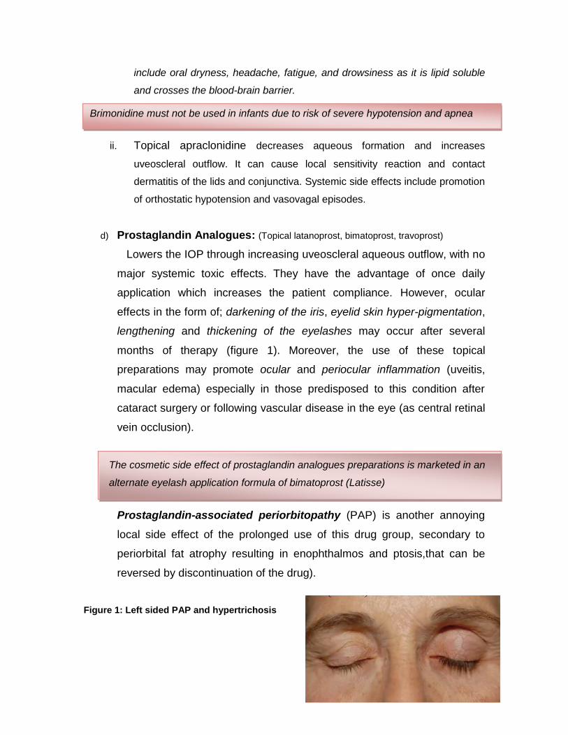
Prostaglandin-associated periorbitopathy (PAP) is another annoying
local side effect of the prolonged use of this drug group, secondary to
periorbital fat atrophy resulting in enophthalmos and ptosis,that can be
reversed by discontinuation of the drug).
Figure 1: Left sided PAP and hypertrichosis
The cosmetic side effect of prostaglandin analogues preparations is marketed in an
alternate eyelash application formula of bimatoprost (Latisse)
e) Carbonic Anhydrase Inhibitors (Oral acetazolamide, methazolamide,
dichlorphenamide).
The prolonged use of this drug group may cause systemic side effects,
which include paresthesias, anorexia, gastrointestinal disturbances,
headaches, altered taste and smell, sodium and potassium depletion, and
a predisposition to form renal calculi, and rarely, bone marrow
suppression.
In order to minimize the side effect of oral carbonic anhydrase inhibitors, Potassium
containing supplements are prescribed.
Ocular Side Effects of Systemic Drugs
The drugs covered in this section are systemically administered medications
that may have profound ocular side effects.
Amiodarone
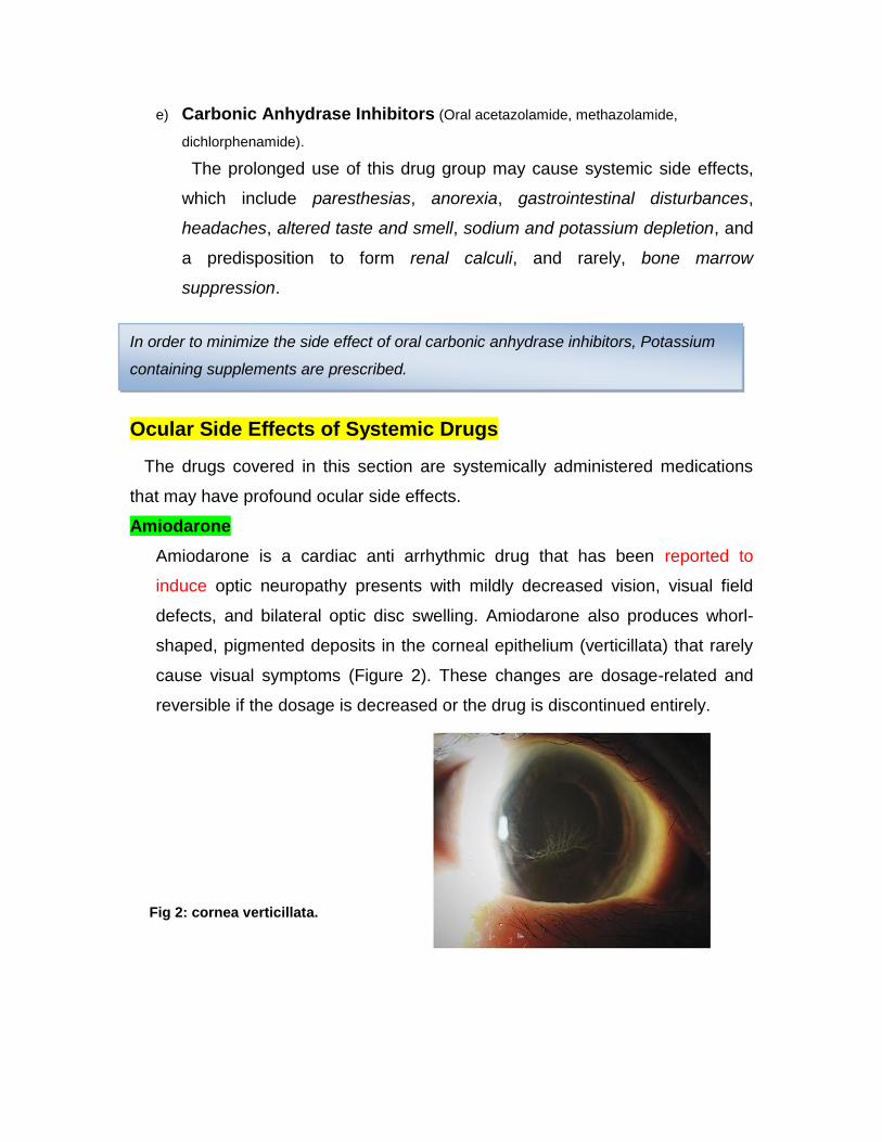
Amiodarone is a cardiac anti arrhythmic drug that has been reported to
induce optic neuropathy presents with mildly decreased vision, visual field
defects, and bilateral optic disc swelling. Amiodarone also produces whorl-
shaped, pigmented deposits in the corneal epithelium (verticillata) that rarely
cause visual symptoms (Figure 2). These changes are dosage-related and
reversible if the dosage is decreased or the drug is discontinued entirely.
Fig 2: cornea verticillata.
Systemic steroids may also precipitate or aggravate acute or chronic central serous retinopathy, a condition resulting in pooling of fluid under the macula
Corticosteroids:
The long-term oral systemic corticosteroids given in moderate dosage, may
produce posterior subcapsular cataracts. Moreover, the use of systemic or
inhaled corticosteroids is associated with elevated IOP (steroid-induced
glaucoma) in susceptible individuals.
Chloroquines:
Chloroquine and hydroxychloroquine (Plaquenil) are used to treat malaria,
rheumatoid arthritis, lupus erythematosus and other autoimmune disorders.
Chloroquines can produce asymptomatic corneal deposits, which can cause
glare and photophobia, and usually regress when the drug is discontinued.
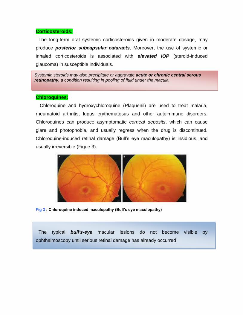
Chloroquine-induced retinal damage (Bull’s eye maculopathy) is insidious, and
usually irreversible (Figue 3).
Fig 3 : Chloroquine induced maculopathy (Bull’s eye maculopathy)
The typical bull's-eye macular lesions do not become visible by
ophthalmoscopy until serious retinal damage has already occurred
Interferon
Interferon is used in the treatment of hepatitis C, leukaemia and lymphoma.
Drug-induced retinopathy (cotton wool spots, intraretinal haemorrhages) can
develop in some patients on high-dose therapy, which usually resolves
spontaneously with cessation of therapy.
Ethambutol:
Ethambutol is a useful chemotherapeutic for tuberculosis. As a side effect,
ethambutol produces a dosage-related optic neuropathy, with the onset of
vision loss may be within 1 month of starting the drug.
Recovery usually occurs when the drug is stopped, but it may take months;
although, vision loss may occasionally be permanent.
At dosages of 15 mg/kg/day, optic neuropathy occurs in less than 1% of patients,
but it increases to 5%of patients receiving 25 mg/kg/day and to 15% receiving 50
mg/kg/day.
Sildenafil (Viagra):
Sildenafil (Viagra) and its related compounds is used primarily for the
treatment of men with erectile dysfunction and Raynaud’s phenomena. Patients
may experience transient, mild impairment of color discrimination, often noted as
blue color tinge of vision. The ocular effects of sildenafil are not reported,
despite a few case reports of nonarteritic ischemic optic neuropathy and
central serous retinopathy,
All patients beginning hydroxychloroquine therapy should have a complete ophthalmic examination
including visual acuity, color vision, Amsler grid, visual field testing, fundus photography and OCT.
This examination is repeated with follow up.
Low-risk patients may be followed at a minimum of 2 years from baseline, patients at higher risk
should be screened at intervals determined by their level of risk based on the patient's age,
physique, drug dosage and duration of use, and any presence of renal or liver disease.
Patients on hydroxychloroquine with visual complaints should be referred to an ophthalmologist for
ocular examination.
Topiramate (Topamax):
Topiramate, a drug used for the treatment of seizure disorder and has been
shown to induce acute bilateral angle-closure glaucoma.
Digitalis:
Intoxication with this widely used cardiovascular drug almost always produces
blurred vision or abnormally colored vision (ie, chromatopsia). Classically,
normal objects appear yellow with the overdosage of digitalis.