Embed Size (px)
Citation preview
Egypt. J. Rad. Sci. Applic., Vol. 30, No.2 pp.131- 143 (2017)13
Corresponding auther :[email protected] :10.21608/ejrsa.2017.1523.1017©2017 National Information and Documentation Center (NIDOC)
Radioiodination, Molecular Modelling and Biological Evaluation of Aniracetam as a Tracer for Brain Imaging
M. H. Sanad1, A. B. Farag2 , Dina H. Salama3
1Labeled Compounds Department, Hot Labs Center, Atomic Energy Authority, P.O. Box 13759, Cairo, 2Pharmaceutical Chemistry Department, Faculty of Pharmacy, Ahram Canadian University, Giza, 3 Health Radiation Research Department (Radiodiagnosis Unit), National Center for Radiation Research and Technology, Atomic Energy Authority, Cairo, Egypt
THE PRESENT study focused on synthesis of radioiodinated aniracetam for a potential brain imaging. Aniracetam (AN) has been labeled using [125I] with chloramine-T (Ch-T)
as an oxidizing agent. The key effective factors such as amount of oxidizing agent, amount of substrate, pH, reaction temperature and reaction time, have been systematically studied to get high radiochemical yield of the radioiodinated aniracetam. The obtained results show a high radiochemical yield of iodoaniracetam that reached 98%. The labeled compound was separated and purified using thin layer chromatography (TLC), paper electrophoreses and high performance liquid chromatography (HPLC). Docking and modeling with AMPA receptors, gamma camera and drug inhibition were studied. In vivo biodistribution of radioiodinated aniracetam was evaluated in Swiss albino mice. The biodistribution results show a brain uptake that reached 9.50 ± 0.17 %ID/g at 15 min post injection (p.i.). Based on these results, it can be stated that radioiodinated aniracetam could be efficiently used as a potential radiotracer drug for brain imaging.
Keywords: Radioiodinated Aniracetam, Brain Imaging, Gamma Camera, Molecular Modeling and Docking
Introduction There are many techniques that are used in
brain imaging or neuro imaging (Thomas et al., 1984; Heilbrun et al., 1987; Levivier etal., 2004; Eggebrecht et al., 2012) et al., 2012; Silberstein, 2000; Farid et al., 2012; Bateman et al., 2012). Iodine -123 is the isotope of choice for medical imaging 10 with a half-life of 13.22 h. and decays by emission of gamma radiation with energy of 159 keV. However, its short half-life and high expenseslead to using iodine-125 to develop the labeling procedure and evaluate the biodistribution in mice(Bruno and Nicolaus, 1982; Hupf et al., 1968; Venkat et al., 1992).Therefore, iodine-131,(Simon et al., 2006) a radioactive isotope is considered more suitable than iodine-123 in imaging process, due to its low expenses compared to others.Brain imaging diseases require the selection of certain compounds with high binding affinity to selective receptors such as AMPA receptor in case of aniracetam (Maina
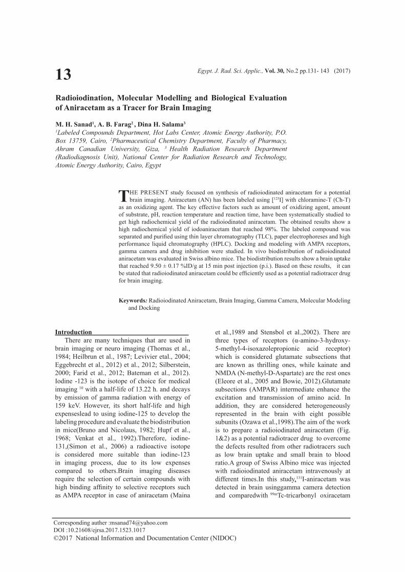
et al.,1989 and Stensbol et al.,2002). There are three types of receptors (α-amino-3-hydroxy-5-methyl-4-isoxazolepropionic acid receptor) which is considered glutamate subsections that are known as thrilling ones, while kainate and NMDA (N-methyl-D-Aspartate) are the rest ones (Eleore et al., 2005 and Bowie, 2012).Glutamate subsections (AMPAR) intermediate enhance the excitation and transmission of amino acid. In addition, they are considered heterogeneously represented in the brain with eight possible subunits (Ozawa et al.,1998).The aim of the work is to prepare a radioiodinated aniracetam (Fig. 1&2) as a potential radiotracer drug to overcome the defects resulted from other radiotracers such as low brain uptake and small brain to blood ratio.A group of Swiss Albino mice was injected with radioiodinated aniracetam intravenously at different times.In this study,131I-aniracetam was detected in brain usinggamma camera detection and comparedwith 99mTc-tricarbonyl oxiracetam
132
Egypt. J. Rad. Sci. Applic., Vol. 30, No.1(2017)
M. H. SANAD et al.
complex,99mTc(CO)3-5HTIDA,99mTc (CO)3-DEDT,99mTc(CO)3-OH-PP-CS2 ,99mTc-ECD, and 99mTc-HMPAO.
ExperimentalMaterials and Methods Reagents
Sodium iodide radiotracer Na125I (185 MBq/50 μL) was purchased from the institute of isotopes, Budapest, Hungary. It was diluted into 0.04M NaOH, pH 9-11. In addition sodium [131I] iodide (3.8 GBq/mL) diluted in 0.05 M NaOH, pH 8 to 11 for radiolabeling was given as a gift from RPF, Atomic Energy Authority, Egypt. Aniracetam, Chloramine-T [N-chloro-p-toluene sulfonamide salt (Ch-T)], ethanol, Tetrahydrofuran and methanol were purchased from Sigma-Aldrich. Thin layer chromatography (TLC) aluminum sheets (20 × 25 cm) SG-60F254 were supplied by Merck. Whatman paper number (PC) 1, was supplied from Whatman International Ltd, Maidstone, Kent, UK. All chemicals were of analytical or clinical grade and were used directly without further purification unless otherwise stated. Double distilled water was used for the preparation of solutions, dilution and washing purposes.
ApparatusAll reactions were followed by TLC (Silica
gel, Aluminum Sheets 60 GF254, Merck), and were detected with a NM UV lamp. A well-type NaI scintillation γ-Counter model Scalar Ratemeter SR7 (Nuclear Enterprises Ltd., USA) was used for radioactive measurement; pH meter: model 601. A digital ion analyzer (Orion Research, USA); ionization chamber: model CRC-15R (Capintec, USA); precision electronic balance: model HA 120 (MAD Company Ltd., Japan); stirring hot plate:model 210 T Thermix (Fisher, USA); electrophoresis apparatus: E.C. Corporation (Albany, OR, USA). The mixture was completely purified usinghigh performance liquid chromatography (HPLC) with the column (RP-C18-250 mm x4.6 mm, 5μm, LiChrosorb) using a Shimadzu model detector SpD-6A model which consists of pumps LC-9A, Rheodyne injector and UV spectrophotometer detector at 285 nm wavelength. Methanol: water (60:40) was used as mobile phase with a 1.0 mL min-1 flow rate (Kamlesh et al.,2016).The Dose mice Calibrator was used for measuring the radioactive dose administered to the mice by a Well Ion Chamber Detector, manufactured by Capintec Company, USA. The gamma camera with a 5-mm pinhole
collimator, window setting of 190 keV, and 20 % width, Dual-Head Variable Angle Gamma from Siemens, in the Nuclear Medicine Unit of the National Cancer. Institute, Cairo University, Egypt, was used in the present work.
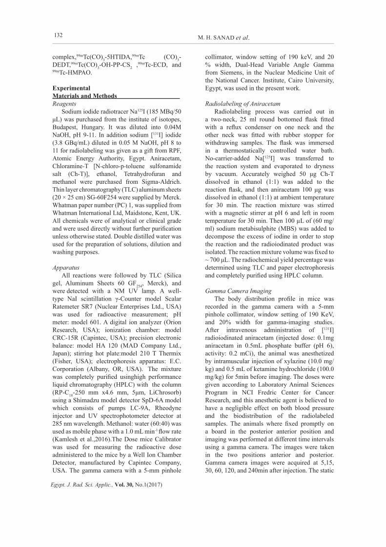
Radiolabeling of AniracetamRadiolabeling process was carried out in
a two-neck, 25 ml round bottomed flask fitted with a reflux condenser on one neck and the other neck was fitted with rubber stopper for withdrawing samples. The flask was immersed in a thermostatically controlled water bath. No-carrier-added Na[125I] was transferred to the reaction system and evaporated to dryness by vacuum. Accurately weighed 50 μg Ch-T dissolved in ethanol (1:1) was added to the reaction flask, and then aniracetam 100 μg was dissolved in ethanol (1:1) at ambient temperature for 30 min. The reaction mixture was stirred with a magnetic stirrer at pH 6 and left in room temperature for 30 min. Then 100 µL of (60 mg/ml) sodium metabisulphite (MBS) was added to decompose the excess of iodine in order to stop the reaction and the radioiodinated product was isolated. The reaction mixture volume was fixed to ~ 700 μL. The radiochemical yield percentage was determined using TLC and paper electrophoresis and completely purified using HPLC column.
Gamma Camera ImagingThe body distribution profile in mice was
recorded in the gamma camera with a 5-mm pinhole collimator, window setting of 190 KeV, and 20% width for gamma-imaging studies. After intravenous administration of [131I] radioiodinated aniracetam (injected dose: 0.1mg aniracetam in 0.5mL phosphate buffer (pH 6), activity: 0.2 mCi), the animal was anesthetized by intramuscular injection of xylazine (10.0 mg/kg) and 0.5 mL of ketamine hydrochloride (100.0 mg/kg) for 5min before imaging. The doses were given according to Laboratory Animal Sciences Program in NCI Fredric Center for Cancer Research, and this anesthetic agent is believed to have a negligible effect on both blood pressure and the biodistribution of the radiolabeled samples. The animals where fixed promptly on a board in the posterior anterior position and imaging was performed at different time intervals using a gamma camera. The images were taken in the two positions anterior and posterior. Gamma camera images were acquired at 5,15, 30, 60, 120, and 240min after injection. The static
133
Egypt. J. Rad. Sci. Applic., Vol. 30, No.1(2017)
RADIOIODINATION MOLECULAR MODELLING AND BIOLOGICAL ...
images werestored in a 512x512 matrix size and acquisition times were 300s. Scintigraphy was the diagnostic nuclear medicine test used in this study, where [131I]iodoaniracetam was administered intravenously and the emitted gamma radiation captured by the gamma camera (Philips axis gamma 2) to form two-dimensional images.
Different Analytical TechniquesA thin layer chromatography was used to
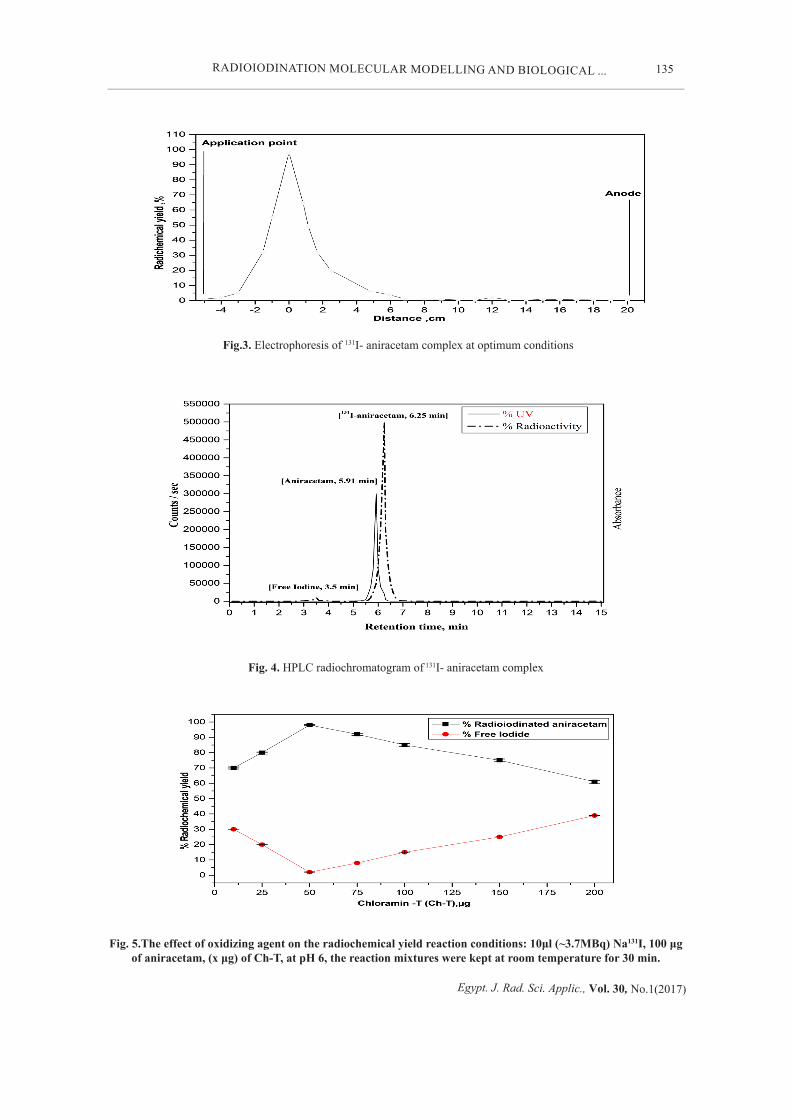
separate the free iodide from the labeled compound to get high radiochemical yield. Using TLC-SG60 F254, the sheets were marked at 2 cm from the base with 1 cm lining fragments up to 16 cm using non-pointed pencil 2-5 µL. The reaction mixture was spotted using micropipette at the origin then the thin layer chromatography sheets were developed using a fresh mixture of chloroform: ethanol (9:1v/v) as described by author name. Another separation process was performed bypaper electrophoresis analysis according to Sanad etal.,(2016). The analysis shows that labeled
compound remained at zero point indicating its neutrally charged nature while, free radioiodide (I-) moved towards the positive electrode (+) with 12 cm distance (Fig.3). Finally,HPLCwas used,in which 10μL aliquot of the radioiodinated aniracetam mixture was injected into the column using the flow rate and the mobile phase mentioned above. Hence, fractions of one mL were collected separately using a fraction collector up to 15 mL and counted in a well-type- γ-scintillation Counter. The data obtained from HPLC analysis show that the retention time Rt of free iodide and radioiodinated aniracetamwere found at 3.5 and 8 min, respectively, and UV of aniracetam was given at 5.91 min. (Fig.4). Differences in the retention times (Rt) between UV of aniracetam and radioiodinated aniracetammay be due to increasing of the hydrophobicity of labeled compound upon incorporation of iodine in aromatic rings which leads to thesedifferences in Rt (Sanad et al., 2017 and Kamlesh et al., 2016).
Fig.1.The chemical structure of aniracetam
Fig.2. The proposed structure of radioiodinated aniracetam
134
Egypt. J. Rad. Sci. Applic., Vol. 30, No.1(2017)
M. H. SANAD et al.
Animal and biodistribution studies of the [125I]aniracetam.
This procedureoccurred and set out according to animal ethics committee approved at Labeled Compounds Department, Egyptian Atomic Energy Authority. Normal Swiss Albino mice (30-40 g) wereintravenously injected in tail, with 0.1 µL (7.2 MBq) of purified radiotracer 125I-AN. They kept alive in metabolic cages,under normal conditions, for different periods of time. In addition, the quantitative determination of organ distribution, tissue samples for 5 animals at each post-injection time points were weighed and counted.Quantitative determination of organ distribution can be approved using, five mice for each experiment and the mice were sacrificed at different time intervals post-injection ( p.i.) at (5, 15, 30 min, 1, 2 and 4 hr). Then, different organs werefirst removed then counted. The count of the total injected volume of radiolabeled compound was compared to the count of a standard solution of 125I-AN.It was approved that the following organs such as bone, blood, and muscles were supposed to be 10, 7, and 40 %, respectively, of the total body weight. The samples were compiled in pre-weighed vials and counted. The rest organs then, were removed counted and compared to a standard solution of the complex. Finally, the uptake of each organ can be calculated as a percentage of the injected dose per gram (% ID/gram ± SD). The data were estimated with one way ANOVA test. Results for P were reported and all the outcomes were given as mean ± SD. The level of importance was set at P < 0.05 ( Sanad et al., 2016).
Determination of the Partition Coefficient for the [125I]Aniracetam
The octanol/water partition coefficient of [125I] aniracetam was determined at pH value of 7.4 by measuring its distribution in n-octanol and phosphate buffered saline (PBS), respectively. A sample of 100 μL was added to an immiscible liquid containing PBS (900 μL; pH 7.4) and n-octanol (1 mL), then after 5 minutes vigorous vortex, the mixture was incubated for 30 minutes at room temperature. Centrifugation at 5000rpm for 5 minutes ensured complete separation of the organic and the aqueous layers. An aliquot (100 μL) from each layer was measured usinga γ counter. The partition coefficient value can be expressed as log Po/w values and repeated for five times.
Stability in Two Different MediaThe stability in two different media such
as human serum and saline has been evaluated as follows: In serum, a purified radioiodinated
aniracetamwas determined by mixing 0.2 ml of radiotracer solution with 0.8 ml serum and kept at ambient temperature. In addition, radioiodinated aniracetam [5 μL (3.20 MBq)] was checked in saline. At time intervals, the stability of radioiodinated aniracetamin two different media was assayed using TLC or HPLC technique, and counted in a well-type γ-scintillation counter (Sanad et al., 2016).
Drug Inhibition Study of Labeled Compound Different amounts of unlabeled aniracetam
were used in the range of (0–1000 μg), after that the animals were injected, 15 min prior to the injection of radioiodinated aniracetam, then the percentage of brain uptake was evaluated at 15 min post-injection of radioiodinated aniracetam (n=5) (Sanad et al.,2016 and Amin et al., 2013).
Molecular ModelingThe docking emulation can be achieved using
the x-ray crystallographic composition ofAMPA (PDB code: 4UQK) bound to the inhibitor QUS (s)-2-Amino-3-(3,5-dioxo-[1,2,4]oxadiazolidin). The PDB file was restored from Protein Data Bank.Chains B, C and D were deleted and structure of chain A was processed using the Structure Preparation application in Molecular Operating Environment (MOE), 2008.10. Subsequently, the protonate 3D application of MOE was used to add the missing hydrogens and properly assign the ionization states(Molecular Operating Environment (MOE), 2008; Labute, 2008; Naïm et al., 2007).The resultant model was further refined by energy minimization to a gradient of 0.01 kcal mol A 2 keeping atoms tethered within 0.5 Å from their crystal structure positions. The default procedure in the MOEDock application was used to find the favorable binding configurations of the studied ligands. Initial placement poses generated by the Alpha Triangle matcher were rescored and filtered using the London dG Scoring method to pick those exhibiting maximal hydrophobic, ionic, and hydrogen-bond contacts to the protein. This was followed by a refinement stage. The generated poses were energy minimized using the MMFF94x force field. Finally, the optimized poses were ranked using the GBVI/WSA DG free-energy estimates as shown in Table 1.(Naïm et al., 2007). Docking poses were visually inspected and interactions with binding pocket residues were analyzed.
135
Egypt. J. Rad. Sci. Applic., Vol. 30, No.1(2017)
RADIOIODINATION MOLECULAR MODELLING AND BIOLOGICAL ...
Fig.3. Electrophoresis of 131I- aniracetam complex at optimum conditions
Fig. 4. HPLC radiochromatogram of 131I- aniracetam complex
Fig. 5.The effect of oxidizing agent on the radiochemical yield reaction conditions: 10μl (~3.7MBq) Na131I, 100 μg of aniracetam, (x μg) of Ch-T, at pH 6, the reaction mixtures were kept at room temperature for 30 min.
136
Egypt. J. Rad. Sci. Applic., Vol. 30, No.1(2017)
M. H. SANAD et al.
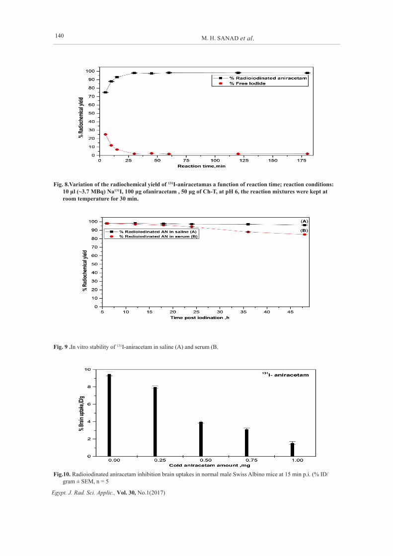
Results The pH of the reaction mixture, amount of the
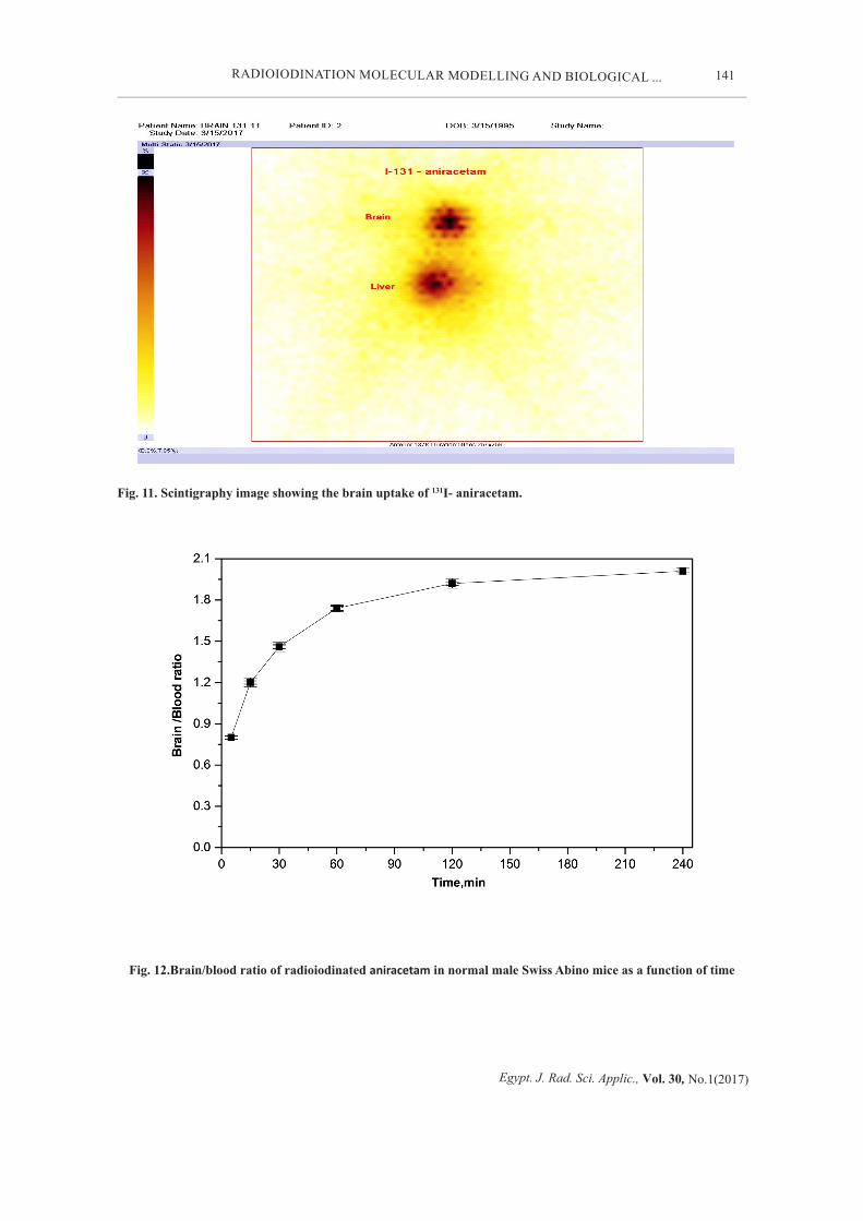
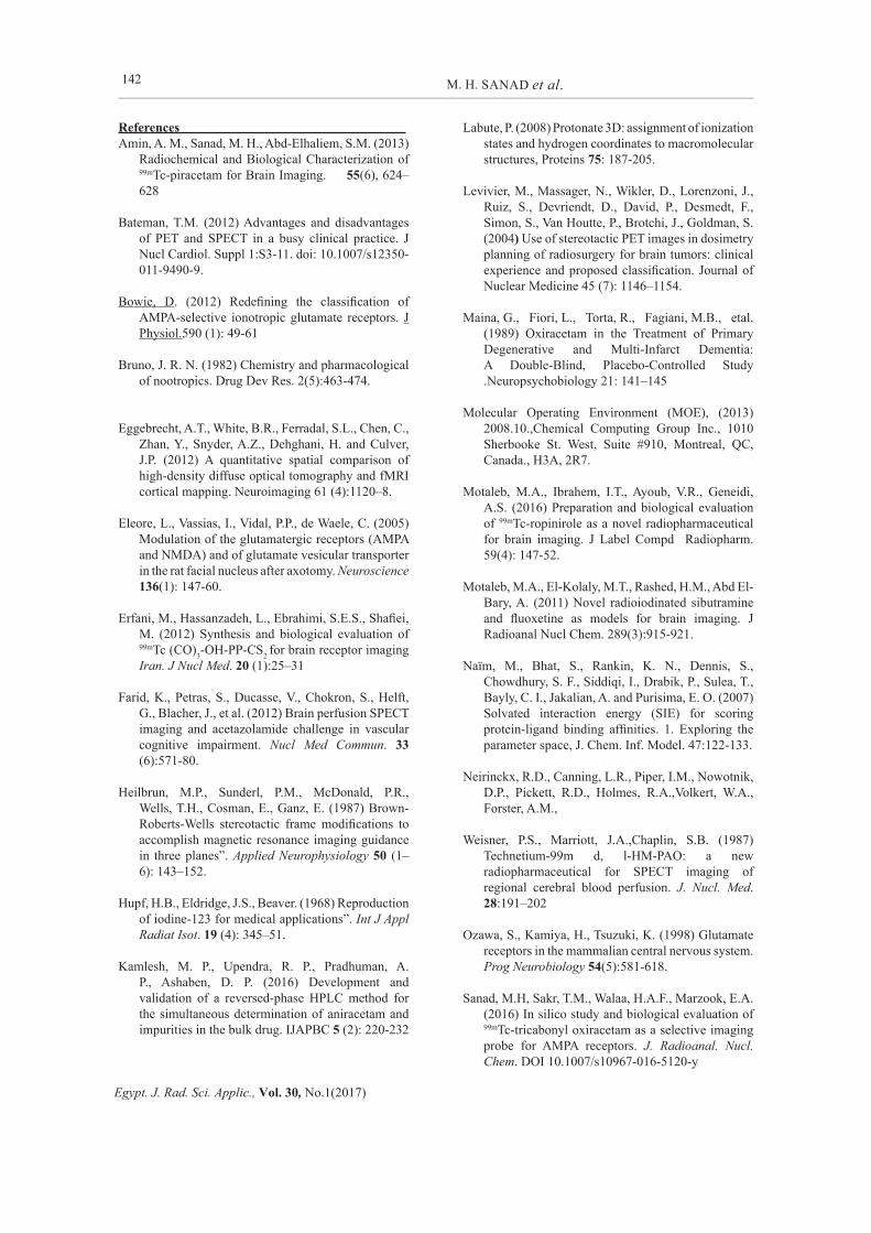
oxidizing agent (chloramine-T (Ch-T)), amount of the ligand, and reaction time of the mixture were optimized at ambient temperature.Figure 5shows that the conversion to radioiodinated aniracetam increased up to a ceiling of 50 μg of Ch-T giving an optimum radiochemical conversion of 98.0%,other reaction parameters were kept constant. Excess Ch-T may lead to the formation of undesirable oxidative by-products such as chlorination and polymerization. Hence 50 μg of Ch-T (~1.5eq.) gave the optimum conversion to iodo aniracetam (Sanad etal., 2016).The effect of changing substrate to iodide ratio is shown in figure 6 which reveals that the optimum radiochemical conversion to radioiodinated aniracetam (98.0%) at 100 μg of aniracetam and [125I] Na (7.5MBq). In addition, pH is an important factor in radio-iodination (see Figure7) which needs to be controlled, pH 6 proved to beoptimal which be may reflected in part the stability of radioiodinated aniracetam (Sanad et al., 2016).The effect of reaction time was also studied giving a maximum conversion at 30 min. (Figure 8).The in-vitro stability of iodonizatidine (Fig.9) was studied in two different media. It was found to be stable in saline for up to 48 hours. In contrast, in serum after 24 hours, the purity dropped to 94.0 % then decreased to 94 % at 48 h.The logarithm of the partition coefficient (log p) value of [125I]aniracetam was 2.11 ± 0.01 that demonstrated its ability to cross the blood brain barrier. Finally, Drug inhibition studyindicated that the different amounts of unlabelled aniracetam used, 15 min before the injection of [125I]aniracetam decreased the brain uptake ratio from 9.50 to 1.55 % ID/g organ at 15 min p.i.(Fig. 10). This result indicates that [125I] aniracetam binds selectively to AMPA receptors located in the brain.
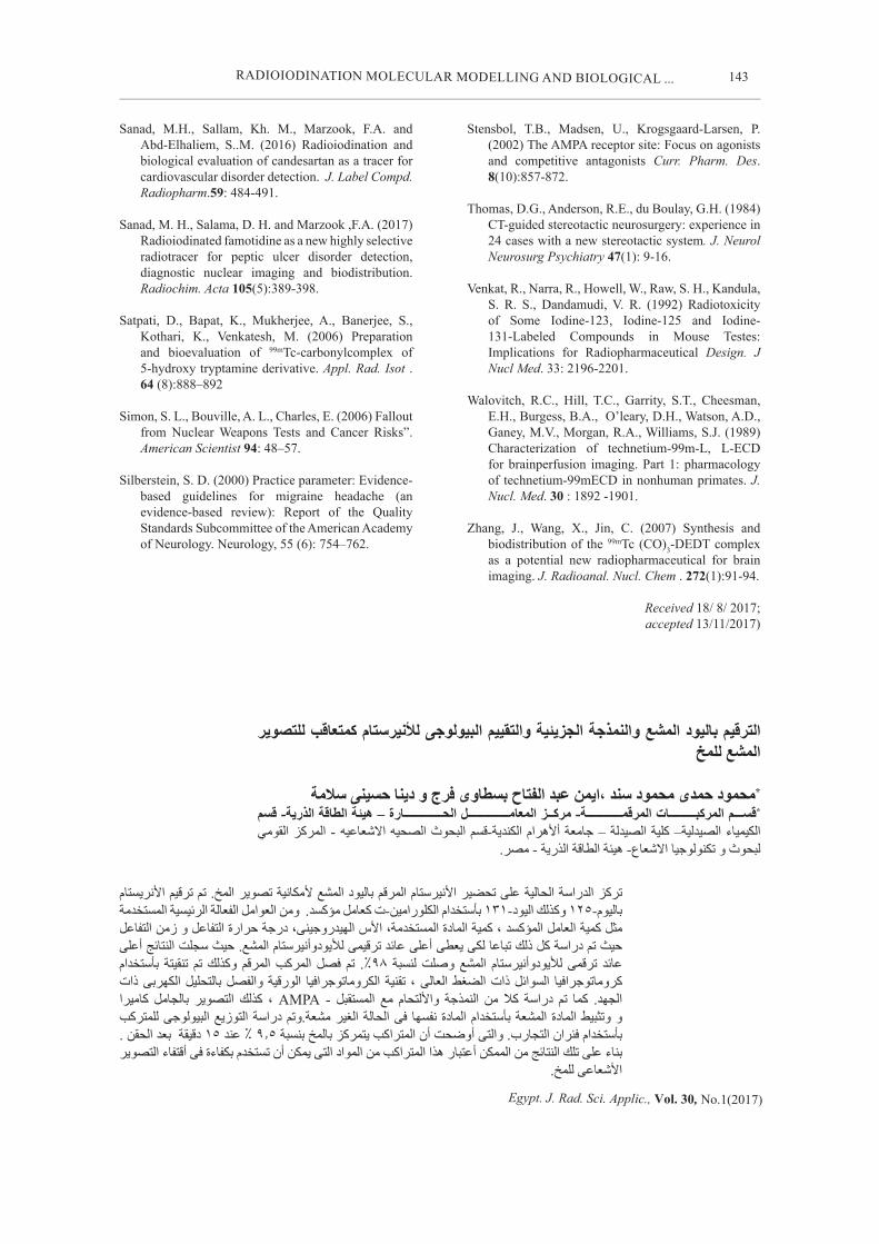
Gamma scintigraphy imaging results, as shown in Fig. 11, the biodistribution of [131I] aniracetam accumulated into the brain of mice reached its maximum in 15 minutes p.i..
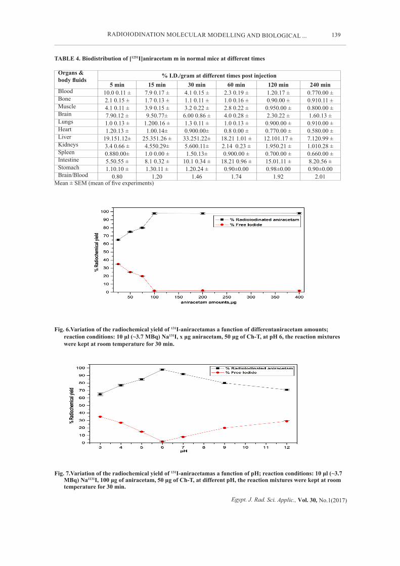
The biodistribution of radioiodinated aniracetamas shown in Table 4, demonstrated thatimportant body organs and fluids were represented as percent-injected dose per gram ±S.E.M. (%ID/gram). The radioactivity accumulated in the thyroid should be referred to the in vivo instability of radioiodinated aniracetam. Therefore low activity uptakes in thyroid indicated that this radiotracer was
considered stable in vivo. The liver up takes increased from 19.15 at 5 min post injection (p.i.) to 33.25 at 30 min post injection and declined at 4 hours to give 7.12(%ID/gram),therefore, the labeled compound was excreted through hepatobiliary pass way and it was considered lipophilic radiotracer(Zhang etal ., 2007).Kidneys uptakeincreased from 3.4 % at 5 min post injection to 5.60 % at 30 minpost injection that declined to 1.01% at 4 h post injection.The brain uptake ofradioiodinated aniracetam was 7.9,9.5, 6.0,4, 2.3and 1.6 at 5,15,30 min, 1h, 2h and 4h(%ID/gram) respectively. The high uptake of the radiotracer at 15 min can be noticed in gamma scintigraphy images (Fig.11). Additionally, the brain/blood ratios were 0.80 at 5 min to 2.01 at 4 h which was a good ratio to image brain compared to other radiotracers (Fig.12.).
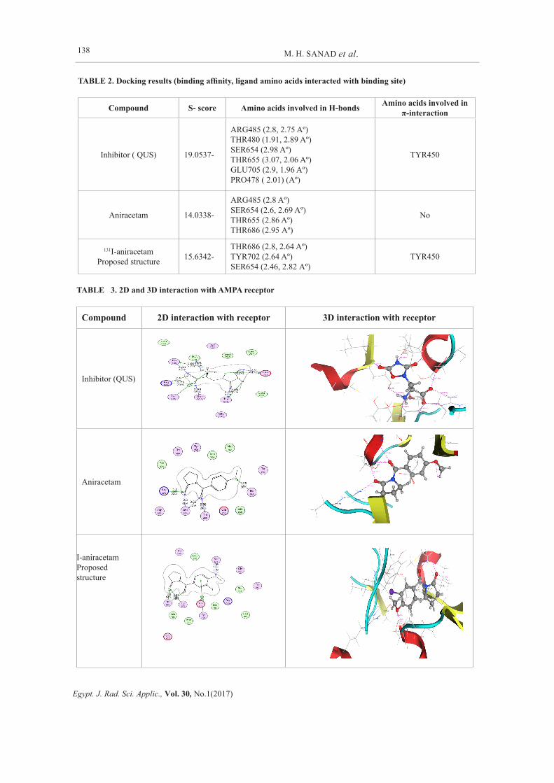
Molecular modeling studies were advocated by self-docking in the AMPAcrystal structure (PDB code 4UQK), by eliminating the bound ligand, QUS, from the radiotracer after that docking it back into the binding site and the main interactions showed that it has ten hydrogen bonds with the key amino acids ARG485, THR480, SER654, THR655, GLU705 and PRO478 also it has a π-interaction with TYR450 as shown in Table 2. Furthermore, the top ranked pose exhibited heavy-atom root-mean-square deviation (RMSD) value of 0.346 Å from the experimental crystal structure.
Discussion Reaction optimization of [125I] aniracetam
gave a high radiochemical yield, high stability in two different media , and also[125I]aniracetam binds selectively to AMPA receptors located in the brain that was indicated by Molecular Operating Environment (MOE) docking that can reliably predict docking poses for the studied compounds to AMPA. The binding affinity of the ligand and the tested compounds were evaluated with S- score. The minimum the docking score is the higher the binding affinity is to the enzyme complex. Interactive docking using MOE protocol was carried out between the predicted structures and the prepared AMPA receptor. Each proposed structure gave 10possible docked poses. The mainone of each molecule can be elected by the uniformity of its binding format in the binding site to that of the ligand QUS. The results of the docking study including S-score, interacted amino acids and the length of H-bonds formed of all the

137
Egypt. J. Rad. Sci. Applic., Vol. 30, No.1(2017)
RADIOIODINATION MOLECULAR MODELLING AND BIOLOGICAL ...
compounds and the reference ligand are listed in Table 3.Generally, MOE docking gave the top-ranked poses indicated that both aniracetam and [125I]aniracetam can interact with 4UQK in a very similar manner like QUS as shown in Table 2, 3.The docking and binding affinity of aniracetam to the receptor pocket has S-score of -14.0338 kcal/mol and exhibited five hydrogen bonds: one with ARG485 of distance (2.8 Aº), two with SER654 of distance (2.6, 2.69 Aº), one with THR655 of distance (2.86 Aº) and last with THR686 of distance (2.95 Aº). While, the docking and binding affinity of 125I-aniracetamto the receptor pocket has S-score of -15.6342 kcal/mol indicating that the binding of [125I]aniracetam is better than aniracetam. Moreover, [125I]aniracetam exhibited five hydrogen bonds: Two with THR686 of distance (2.8, 2.64Aº), two with SER654 of distance (2.46, 2.82 Aº), one with TYR702 of distance (2.64 Aº) and also it has a π-interaction with TYR450. Conclusively, introduction of heavy iodine atoms improved binding of aniracetam to its target receptor (4UQK). From biodistribution study, radioiodinated aniracetam was considered more than other complexes used such as 99mTc-piracetam that gave 4.9 ± 0.1 % ID/organ (Amin etal., 2013). In addition,
99mTc-ropinirole,125I-fluoxetine, 125I-sibutramine,
99mTc(CO)3-5HTIDA and 99mTc (CO)3-DEDT that showed 4.7, 1.29, 3.19,0.25 and 1.41%ID/g at 30 min post injection in mice, respectively. Also, agents such as 99mTc-HMPAO and 99mTc(CO)3-
OH-PP-CS2 that showed 2.25 and 0.32 %ID/g at 30 min post injection in rats, respectively. Finally radioiodinated aniracetam higher than that
99mTc-tricarbonyl oxiracetam complex (Sanad., etal 2017) and 99mTc-ECD complex that showed 7.5 ± 0.11% ID/g at 5 min post injection in mice and 4.7 %ID/g at 24 hr post injection in monkey respectively (Motaleb etal 2016; Motaleb etal., 2011; Satpati etal., 2006; Zhang etal., 2007; Neirinckx etal., 2012; Erfani etal., 2012; Walovitch etal., 1989). From above data, it was concluded that radioiodinated aniracetam has brain uptake at 15 min p.i. more than all above complexes. Therefore, radioiodinated aniracetam could be used to brain imaging than others according to its receptors (AMPA).
Conclusion An optimized protocol for the synthesis of
radioiodinated aniracetam in high yield has been elaborated. Biodistribution studies indicated that radioiodinated aniracetam has a high brain uptake of 9.5 % ID/g at 15 min. that was confirmed by gamma camera and binding with AMPA receptors. The radiotracer radioiodinated aniracetam considers more better than recently discovered agents such as [99mTc] piracetam, [99mTc]tricarbonyl oxiracetam complex, [99mTc](CO)3-5HTIDA,[99mTc](CO)3DEDTand [99mTc](CO)3-OH-PP-CS2 , [
99mTc]ropinirole, [99mTc]ECD and [99mTc]HMPAO complexes have.
TABLE 1. Aniracetam and 125I-aniracetam proposed structure after energy minimization
Compound 2D format 3D after energy minimization
Aniracetam NO
OO
131I-aniracetamProposed structure NO
OO
I
138
Egypt. J. Rad. Sci. Applic., Vol. 30, No.1(2017)
M. H. SANAD et al.
TABLE 2. Docking results (binding affinity, ligand amino acids interacted with binding site)
Compound S- score Amino acids involved in H-bonds Amino acids involved in π-interaction
Inhibitor ( QUS) 19.0537-
ARG485 (2.8, 2.75 Aº)THR480 (1.91, 2.89 Aº)SER654 (2.98 Aº)THR655 (3.07, 2.06 Aº)GLU705 (2.9, 1.96 Aº)PRO478 ( 2.01) (Aº)
TYR450
Aniracetam 14.0338-
ARG485 (2.8 Aº)SER654 (2.6, 2.69 Aº)THR655 (2.86 Aº)THR686 (2.95 Aº)
No
131I-aniracetamProposed structure 15.6342-
THR686 (2.8, 2.64 Aº)TYR702 (2.64 Aº)SER654 (2.46, 2.82 Aº)
TYR450
TABLE 3. 2D and 3D interaction with AMPA receptor
Compound 2D interaction with receptor 3D interaction with receptor
Inhibitor (QUS)
Aniracetam
I-aniracetam Proposed structure
139
Egypt. J. Rad. Sci. Applic., Vol. 30, No.1(2017)
RADIOIODINATION MOLECULAR MODELLING AND BIOLOGICAL ...
TABLE 4. Biodistribution of [1251I]aniracetam m in normal mice at different times
Organs & body fluids
% I.D./gram at different times post injection5 min 15 min 30 min 60 min 120 min 240 min
Blood 10.0 0.11 ± 7.9 0.17 ± 4.1 0.15 ± 2.3 0.19 ± 1.20.17 ± 0.770.00 ±Bone 2.1 0.15 ± 1.7 0.13 ± 1.1 0.11 ± 1.0 0.16 ± 0.90.00 ± 0.910.11 ±Muscle 4.1 0.11 ± 3.9 0.15 ± 3.2 0.22 ± 2.8 0.22 ± 0.950.00 ± 0.800.00 ±Brain 7.90.12 ± 9.50.77± 6.00 0.86 ± 4.0 0.28 ± 2.30.22 ± 1.60.13 ±Lungs 1.0 0.13 ± 1.200.16 ± 1.3 0.11 ± 1.0 0.13 ± 0.900.00 ± 0.910.00 ±Heart 1.20.13 ± 1.00.14± 0.900.00± 0.8 0.00 ± 0.770.00 ± 0.580.00 ±Liver 19.151.12± 25.351.26 ± 33.251.22± 18.21 1.01 ± 12.101.17 ± 7.120.99 ±Kidneys 3.4 0.66 ± 4.550.29± 5.600.11± 2.14 0.23 ± 1.950.21 ± 1.010.28 ±Spleen 0.880.00± 1.0 0.00 ± 1.50.13± 0.900.00 ± 0.700.00 ± 0.660.00 ±Intestine 5.50.55 ± 8.1 0.32 ± 10.1 0.34 ± 18.21 0.96 ± 15.01.11 ± 8.20.56 ±Stomach 1.10.10 ± 1.30.11 ± 1.20.24 ± 0.90±0.00 0.98±0.00 0.90±0.00Brain/Blood 0.80 1.20 1.46 1.74 1.92 2.01
Mean ± SEM (mean of five experiments)
Fig. 6.Variation of the radiochemical yield of 131I-aniracetamas a function of differentaniracetam amounts; reaction conditions: 10 μl (~3.7 MBq) Na131I, x μg aniracetam, 50 μg of Ch-T, at pH 6, the reaction mixtures were kept at room temperature for 30 min.
Fig. 7.Variation of the radiochemical yield of 131I-aniracetamas a function of pH; reaction conditions: 10 μl (~3.7 MBq) Na1131I, 100 μg of aniracetam, 50 μg of Ch-T, at different pH, the reaction mixtures were kept at room temperature for 30 min.
140
Egypt. J. Rad. Sci. Applic., Vol. 30, No.1(2017)
M. H. SANAD et al.
Fig. 8.Variation of the radiochemical yield of 131I-aniracetamas a function of reaction time; reaction conditions: 10 μl (~3.7 MBq) Na131I, 100 μg ofaniracetam , 50 μg of Ch-T, at pH 6, the reaction mixtures were kept at room temperature for 30 min.
Fig. 9 .In vitro stability of 131I-aniracetam in saline (A) and serum (B.
Fig.10. Radioiodinated aniracetam inhibition brain uptakes in normal male Swiss Albino mice at 15 min p.i. (% ID/gram ± SEM, n = 5
141
Egypt. J. Rad. Sci. Applic., Vol. 30, No.1(2017)
RADIOIODINATION MOLECULAR MODELLING AND BIOLOGICAL ...
Fig. 12.Brain/blood ratio of radioiodinated aniracetam in normal male Swiss Abino mice as a function of time
Fig. 11. Scintigraphy image showing the brain uptake of 131I- aniracetam.
142
Egypt. J. Rad. Sci. Applic., Vol. 30, No.1(2017)
M. H. SANAD et al.
References Amin, A. M., Sanad, M. H., Abd-Elhaliem, S.M. (2013)
Radiochemical and Biological Characterization of 99mTc-piracetam for Brain Imaging. 55(6), 624–628
Bateman, T.M. (2012) Advantages and disadvantages of PET and SPECT in a busy clinical practice. J Nucl Cardiol. Suppl 1:S3-11. doi: 10.1007/s12350-011-9490-9.
Bowie, D. (2012) Redefining the classification of AMPA-selective ionotropic glutamate receptors. J Physiol.590 (1): 49-61
Bruno, J. R. N. (1982) Chemistry and pharmacological of nootropics. Drug Dev Res. 2(5):463-474.
Eggebrecht, A.T., White, B.R., Ferradal, S.L., Chen, C., Zhan, Y., Snyder, A.Z., Dehghani, H. and Culver, J.P. (2012) A quantitative spatial comparison of high-density diffuse optical tomography and fMRI cortical mapping. Neuroimaging 61 (4):1120–8.
Eleore, L., Vassias, I., Vidal, P.P., de Waele, C. (2005) Modulation of the glutamatergic receptors (AMPA and NMDA) and of glutamate vesicular transporter in the rat facial nucleus after axotomy. Neuroscience 136(1): 147-60.
Erfani, M., Hassanzadeh, L., Ebrahimi, S.E.S., Shafiei, M. (2012) Synthesis and biological evaluation of 99mTc (CO)3-OH-PP-CS2 for brain receptor imaging Iran. J Nucl Med. 20 (1):25–31
Farid, K., Petras, S., Ducasse, V., Chokron, S., Helft, G., Blacher, J., et al. (2012) Brain perfusion SPECT imaging and acetazolamide challenge in vascular cognitive impairment. Nucl Med Commun. 33 (6):571-80.
Heilbrun, M.P., Sunderl, P.M., McDonald, P.R., Wells, T.H., Cosman, E., Ganz, E. (1987) Brown-Roberts-Wells stereotactic frame modifications to accomplish magnetic resonance imaging guidance in three planes”. Applied Neurophysiology 50 (1–6): 143–152.
Hupf, H.B., Eldridge, J.S., Beaver. (1968) Reproduction of iodine-123 for medical applications”. Int J Appl Radiat Isot. 19 (4): 345–51.
Kamlesh, M. P., Upendra, R. P., Pradhuman, A. P., Ashaben, D. P. (2016) Development and validation of a reversed-phase HPLC method for the simultaneous determination of aniracetam and impurities in the bulk drug. IJAPBC 5 (2): 220-232
Labute, P. (2008) Protonate 3D: assignment of ionization states and hydrogen coordinates to macromolecular structures, Proteins 75: 187-205.
Levivier, M., Massager, N., Wikler, D., Lorenzoni, J., Ruiz, S., Devriendt, D., David, P., Desmedt, F., Simon, S., Van Houtte, P., Brotchi, J., Goldman, S. (2004) Use of stereotactic PET images in dosimetry planning of radiosurgery for brain tumors: clinical experience and proposed classification. Journal of Nuclear Medicine 45 (7): 1146–1154.
Maina, G., Fiori, L., Torta, R., Fagiani, M.B., etal. (1989) Oxiracetam in the Treatment of Primary Degenerative and Multi-Infarct Dementia: A Double-Blind, Placebo-Controlled Study .Neuropsychobiology 21: 141–145
Molecular Operating Environment (MOE), (2013) 2008.10.,Chemical Computing Group Inc., 1010 Sherbooke St. West, Suite #910, Montreal, QC, Canada., H3A, 2R7.
Motaleb, M.A., Ibrahem, I.T., Ayoub, V.R., Geneidi, A.S. (2016) Preparation and biological evaluation of 99mTc-ropinirole as a novel radiopharmaceutical for brain imaging. J Label Compd Radiopharm. 59(4): 147-52.
Motaleb, M.A., El-Kolaly, M.T., Rashed, H.M., Abd El-Bary, A. (2011) Novel radioiodinated sibutramine and fluoxetine as models for brain imaging. J Radioanal Nucl Chem. 289(3):915-921.
Naïm, M., Bhat, S., Rankin, K. N., Dennis, S., Chowdhury, S. F., Siddiqi, I., Drabik, P., Sulea, T., Bayly, C. I., Jakalian, A. and Purisima, E. O. (2007) Solvated interaction energy (SIE) for scoring protein-ligand binding affinities. 1. Exploring the parameter space, J. Chem. Inf. Model. 47:122-133.
Neirinckx, R.D., Canning, L.R., Piper, I.M., Nowotnik, D.P., Pickett, R.D., Holmes, R.A.,Volkert, W.A., Forster, A.M.,
Weisner, P.S., Marriott, J.A.,Chaplin, S.B. (1987) Technetium-99m d, l-HM-PAO: a new radiopharmaceutical for SPECT imaging of regional cerebral blood perfusion. J. Nucl. Med. 28:191–202
Ozawa, S., Kamiya, H., Tsuzuki, K. (1998) Glutamate receptors in the mammalian central nervous system. Prog Neurobiology 54(5):581-618.
Sanad, M.H, Sakr, T.M., Walaa, H.A.F., Marzook, E.A. (2016) In silico study and biological evaluation of 99mTc-tricabonyl oxiracetam as a selective imaging probe for AMPA receptors. J. Radioanal. Nucl. Chem. DOI 10.1007/s10967-016-5120-y
143
Egypt. J. Rad. Sci. Applic., Vol. 30, No.1(2017)
RADIOIODINATION MOLECULAR MODELLING AND BIOLOGICAL ...
Sanad, M.H., Sallam, Kh. M., Marzook, F.A. and Abd-Elhaliem, S..M. (2016) Radioiodination and biological evaluation of candesartan as a tracer for cardiovascular disorder detection. J. Label Compd. Radiopharm.59: 484-491.
Sanad, M. H., Salama, D. H. and Marzook ,F.A. (2017) Radioiodinated famotidine as a new highly selective radiotracer for peptic ulcer disorder detection, diagnostic nuclear imaging and biodistribution. Radiochim. Acta 105(5):389-398.
Satpati, D., Bapat, K., Mukherjee, A., Banerjee, S., Kothari, K., Venkatesh, M. (2006) Preparation and bioevaluation of 99mTc-carbonylcomplex of 5-hydroxy tryptamine derivative. Appl. Rad. Isot . 64 (8):888–892
Simon, S. L., Bouville, A. L., Charles, E. (2006) Fallout from Nuclear Weapons Tests and Cancer Risks”. American Scientist 94: 48–57.
Silberstein, S. D. (2000) Practice parameter: Evidence-based guidelines for migraine headache (an evidence-based review): Report of the Quality Standards Subcommittee of the American Academy of Neurology. Neurology, 55 (6): 754–762.
Stensbol, T.B., Madsen, U., Krogsgaard-Larsen, P. (2002) The AMPA receptor site: Focus on agonists and competitive antagonists Curr. Pharm. Des. 8(10):857-872.
Thomas, D.G., Anderson, R.E., du Boulay, G.H. (1984) CT-guided stereotactic neurosurgery: experience in 24 cases with a new stereotactic system. J. Neurol Neurosurg Psychiatry 47(1): 9-16.
Venkat, R., Narra, R., Howell, W., Raw, S. H., Kandula, S. R. S., Dandamudi, V. R. (1992) Radiotoxicity of Some Iodine-123, Iodine-125 and Iodine- 131-Labeled Compounds in Mouse Testes: Implications for Radiopharmaceutical Design. J Nucl Med. 33: 2196-2201.
Walovitch, R.C., Hill, T.C., Garrity, S.T., Cheesman, E.H., Burgess, B.A., O’leary, D.H., Watson, A.D., Ganey, M.V., Morgan, R.A., Williams, S.J. (1989) Characterization of technetium-99m-L, L-ECD for brainperfusion imaging. Part 1: pharmacology of technetium-99mECD in nonhuman primates. J. Nucl. Med. 30 : 1892 -1901.
Zhang, J., Wang, X., Jin, C. (2007) Synthesis and biodistribution of the 99mTc (CO)3-DEDT complex as a potential new radiopharmaceutical for brain imaging. J. Radioanal. Nucl. Chem . 272(1):91-94.
Received 18/ 8/ 2017; accepted 13/11/2017)
البيولوجى للأنيرستام كمتعاقب للتصوير الترقيم باليود المشع والنمذجة الجزيئية والتقييم المشع للمخ
*محمود حمدى محمود سند ،ايمن عبد الفتاح بسطاوى فرج و دينا حسينى سلامة
*قســـم المركبـــــــــات المرقمــــــــــــة- مركــز المعامــــــــــــــل الحـــــــــــــارة – هيئة الطاقة الذرية- قسم
القومي المركز - البحوث الصحيه الاشعاعيه الكندية-قسم الصيدلة – جامعة ألأهرام الصيدلية– كلية الكيمياء لبحوث و تكنولوجيا الاشعاع- هيئة الطاقة الذرية - مصر.
تركز الدراسة الحالية على تحضير الأنيرستام المرقم باليود المشع لأمكانية تصوير المخ. تم ترقيم الأنريستام باليوم-125 وكذلك اليود-131 بأستخدام الكلورامين-ت كعامل مؤكسد. ومن العوامل الفعالة الرئيسية المستخدمة مثل كمية العامل المؤكسد ، كمية المادة المستخدمة، الأس الهيدروجينى، درجة حرارة التفاعل و زمن التفاعل حيث تم دراسة كل ذلك تباعا لكى يعطى أعلى عائد ترقيمى للأيودوأنيرستام المشع. حيث سجلت النتائج أعلى بأستخدام تنقيتة تم وكذلك المرقم المركب فصل تم .%98 لنسبة وصلت المشع للأيودوأنيرستام ترقمى عائد ذات الكهربى بالتحليل والفصل الورقية الكروماتوجرافيا تقنية ، العالى الضغط ذات السوائل كروماتوجرافيا كاميرا بالجامل التصوير كذلك ، AMPA - المستقبل مع والألتحام النمذجة من كلا دراسة تم كما الجهد. للمتركب البيولوجى التوزيع الغير مشعة.وتم دراسة الحالة فى نفسها المادة بأستخدام المشعة المادة وتثبيط و بأستخدام فئران التجارب. والتى أوضحت أن المتراكب يتمركز بالمخ بنسبة 9.5 % عند 15 دقيقة بعد الحقن . بناء على تلك النتائج من الممكن أعتبار هذا المتراكب من المواد التى يمكن أن تستخدم بكفاءة فى أقتفاء التصوير
الأشعاعى للمخ.