Embed Size (px)
Citation preview
Page1
Lecture 13 Anatomy د. احمد فاضل
Triangles of the neck ANTERIOR TRIANGLE
Borders of the anterior triangle:
● posteriorly: Anterior border of the sternocleidomastoid
●superiorly: Inferior border of the mandible
●anteriorly: Midline of the neck
Using the hyoid as a keystone, the omohyoid and digastric muscles subdivide the
anterior triangle into:
● Submental triangle
● Submandibular triangle
● Carotid triangle
● Muscular triangle
All of the triangles within the anterior triangle are paired except for the submental
triangle, which spans the right and the left sides of the neck
1. SUBMENTAL TRIANGLE Borders of the submental triangle:
●inferiorly: Body of hyoid
● laterally: Anterior digastric on right and left
● anteriorly: midline of the neck
Floor of the triangle is composed of the:
● Mylohyoid
Roof is made of the:
● Skin
● Superficial fascia with platysma
● Deep cervical fascia
Page2
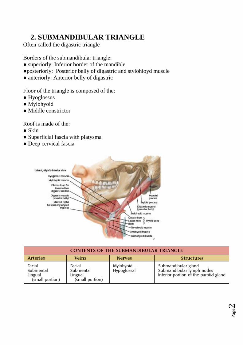
2. SUBMANDIBULAR TRIANGLE Often called the digastric triangle
Borders of the submandibular triangle:
● superiorly: Inferior border of the mandible
●posteriorly: Posterior belly of digastric and stylohioyd muscle
● anteriorly: Anterior belly of digastric
Floor of the triangle is composed of the:
● Hyoglossus
● Mylohyoid
● Middle constrictor
Roof is made of the:
● Skin
● Superficial fascia with platysma
● Deep cervical fascia
Page3
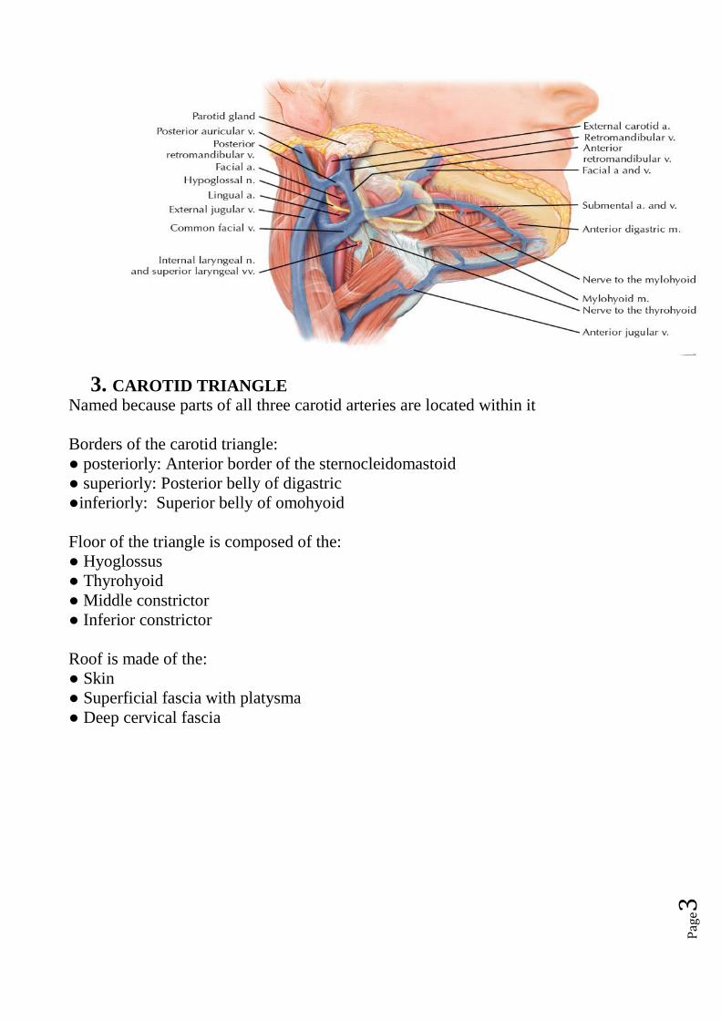
3. CAROTID TRIANGLE
Named because parts of all three carotid arteries are located within it
Borders of the carotid triangle:
● posteriorly: Anterior border of the sternocleidomastoid
● superiorly: Posterior belly of digastric
●inferiorly: Superior belly of omohyoid
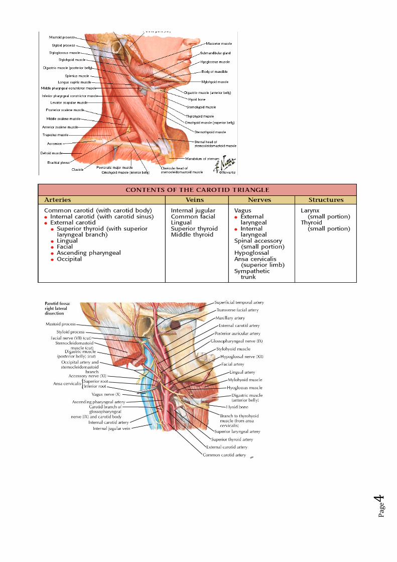
Floor of the triangle is composed of the:
● Hyoglossus
● Thyrohyoid
● Middle constrictor
● Inferior constrictor
Roof is made of the:
● Skin
● Superficial fascia with platysma
● Deep cervical fascia
Page4
Page5
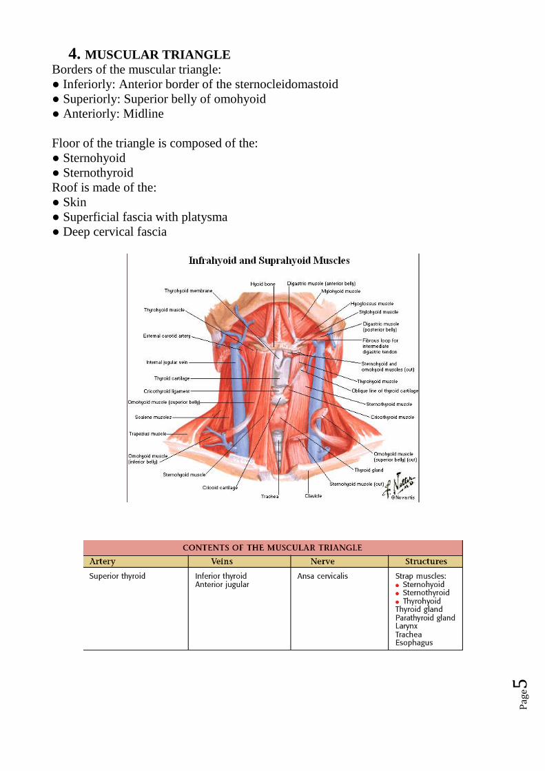
4. MUSCULAR TRIANGLE
Borders of the muscular triangle:
● Inferiorly: Anterior border of the sternocleidomastoid
● Superiorly: Superior belly of omohyoid
● Anteriorly: Midline
Floor of the triangle is composed of the:
● Sternohyoid
● Sternothyroid
Roof is made of the:
● Skin
● Superficial fascia with platysma
● Deep cervical fascia
Page6
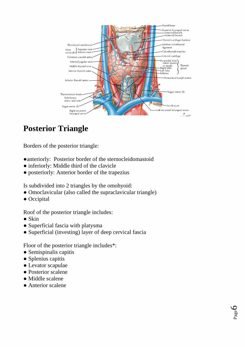
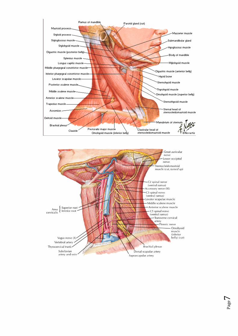
Posterior Triangle
Borders of the posterior triangle:
●anteriorly: Posterior border of the sternocleidomastoid
● inferiorly: Middle third of the clavicle
● posteriorly: Anterior border of the trapezius
Is subdivided into 2 triangles by the omohyoid:
● Omoclavicular (also called the supraclavicular triangle)
● Occipital
Roof of the posterior triangle includes:
● Skin
● Superficial fascia with platysma
● Superficial (investing) layer of deep cervical fascia
Floor of the posterior triangle includes*:
● Semispinalis capitis
● Splenius capitis
● Levator scapulae
● Posterior scalene
● Middle scalene
● Anterior scalene
Page7
Page8
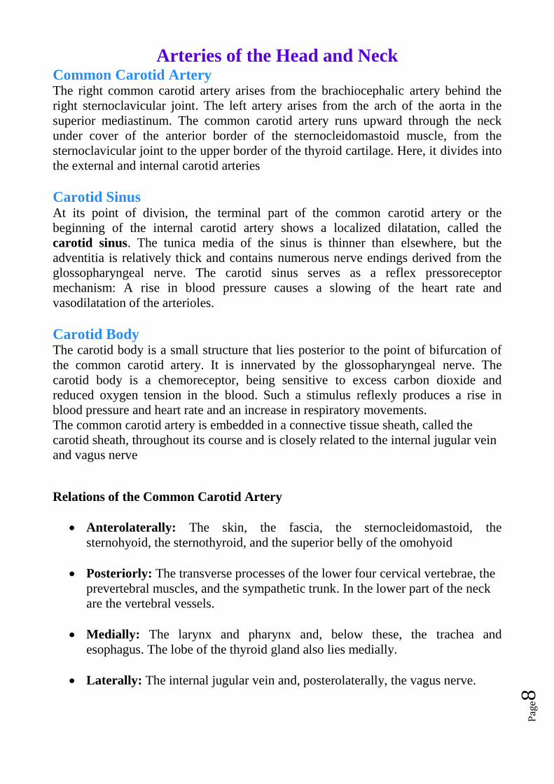
Arteries of the Head and Neck Common Carotid Artery
The right common carotid artery arises from the brachiocephalic artery behind the
right sternoclavicular joint. The left artery arises from the arch of the aorta in the
superior mediastinum. The common carotid artery runs upward through the neck
under cover of the anterior border of the sternocleidomastoid muscle, from the
sternoclavicular joint to the upper border of the thyroid cartilage. Here, it divides into
the external and internal carotid arteries
Carotid Sinus At its point of division, the terminal part of the common carotid artery or the
beginning of the internal carotid artery shows a localized dilatation, called the
carotid sinus. The tunica media of the sinus is thinner than elsewhere, but the
adventitia is relatively thick and contains numerous nerve endings derived from the
glossopharyngeal nerve. The carotid sinus serves as a reflex pressoreceptor
mechanism: A rise in blood pressure causes a slowing of the heart rate and
vasodilatation of the arterioles.
Carotid Body The carotid body is a small structure that lies posterior to the point of bifurcation of
the common carotid artery. It is innervated by the glossopharyngeal nerve. The
carotid body is a chemoreceptor, being sensitive to excess carbon dioxide and
reduced oxygen tension in the blood. Such a stimulus reflexly produces a rise in
blood pressure and heart rate and an increase in respiratory movements.
The common carotid artery is embedded in a connective tissue sheath, called the
carotid sheath, throughout its course and is closely related to the internal jugular vein
and vagus nerve
Relations of the Common Carotid Artery
Anterolaterally: The skin, the fascia, the sternocleidomastoid, the
sternohyoid, the sternothyroid, and the superior belly of the omohyoid
Posteriorly: The transverse processes of the lower four cervical vertebrae, the
prevertebral muscles, and the sympathetic trunk. In the lower part of the neck
are the vertebral vessels.
Medially: The larynx and pharynx and, below these, the trachea and
esophagus. The lobe of the thyroid gland also lies medially.
Laterally: The internal jugular vein and, posterolaterally, the vagus nerve.
Page9
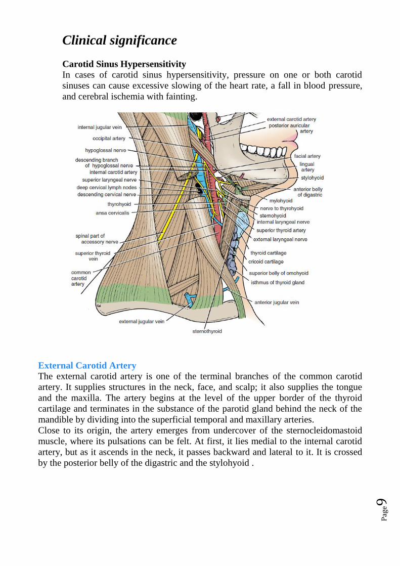
Clinical significance
Carotid Sinus Hypersensitivity
In cases of carotid sinus hypersensitivity, pressure on one or both carotid
sinuses can cause excessive slowing of the heart rate, a fall in blood pressure,
and cerebral ischemia with fainting.
C
External Carotid Artery
The external carotid artery is one of the terminal branches of the common carotid
artery. It supplies structures in the neck, face, and scalp; it also supplies the tongue
and the maxilla. The artery begins at the level of the upper border of the thyroid
cartilage and terminates in the substance of the parotid gland behind the neck of the
mandible by dividing into the superficial temporal and maxillary arteries.
Close to its origin, the artery emerges from undercover of the sternocleidomastoid
muscle, where its pulsations can be felt. At first, it lies medial to the internal carotid
artery, but as it ascends in the neck, it passes backward and lateral to it. It is crossed
by the posterior belly of the digastric and the stylohyoid .
Page10
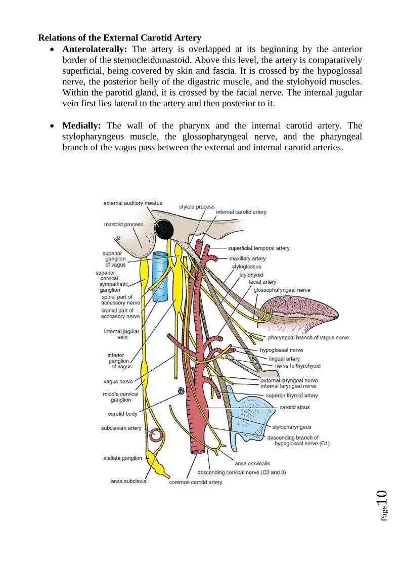
Relations of the External Carotid Artery
Anterolaterally: The artery is overlapped at its beginning by the anterior
border of the sternocleidomastoid. Above this level, the artery is comparatively
superficial, being covered by skin and fascia. It is crossed by the hypoglossal
nerve, the posterior belly of the digastric muscle, and the stylohyoid muscles.
Within the parotid gland, it is crossed by the facial nerve. The internal jugular
vein first lies lateral to the artery and then posterior to it.
Medially: The wall of the pharynx and the internal carotid artery. The
stylopharyngeus muscle, the glossopharyngeal nerve, and the pharyngeal
branch of the vagus pass between the external and internal carotid arteries.
Page11
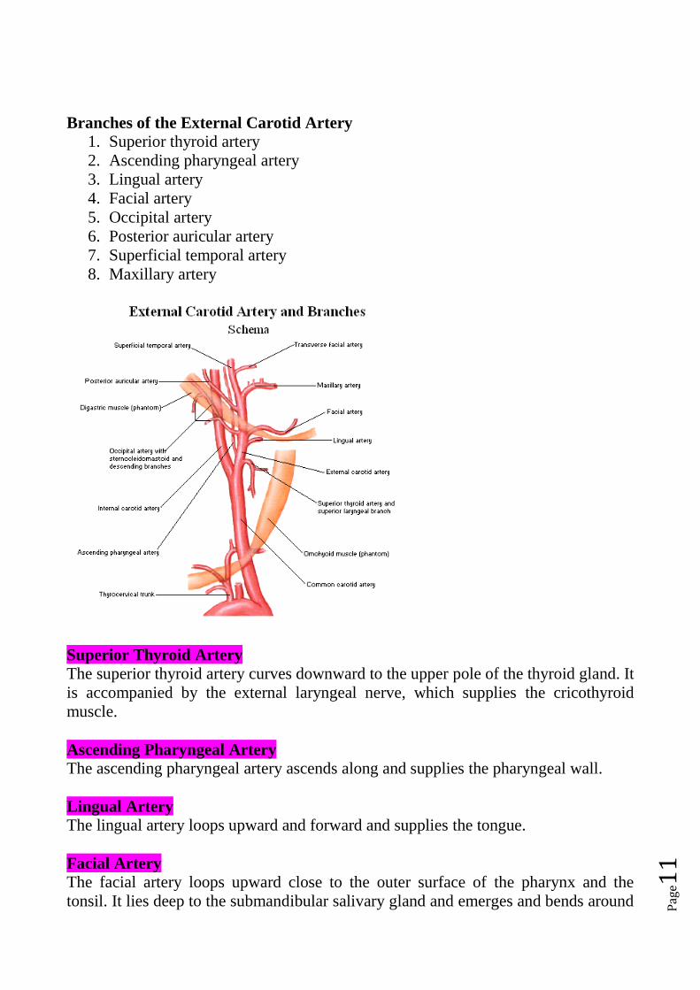
Branches of the External Carotid Artery
1. Superior thyroid artery
2. Ascending pharyngeal artery
3. Lingual artery
4. Facial artery
5. Occipital artery
6. Posterior auricular artery
7. Superficial temporal artery
8. Maxillary artery
Superior Thyroid Artery
The superior thyroid artery curves downward to the upper pole of the thyroid gland. It
is accompanied by the external laryngeal nerve, which supplies the cricothyroid
muscle.
Ascending Pharyngeal Artery
The ascending pharyngeal artery ascends along and supplies the pharyngeal wall.
Lingual Artery
The lingual artery loops upward and forward and supplies the tongue.
Facial Artery
The facial artery loops upward close to the outer surface of the pharynx and the
tonsil. It lies deep to the submandibular salivary gland and emerges and bends around
Page12
the lower border of the mandible. It then ascends over the face close to the anterior
border of the masseter muscle. The artery then ascends around the lateral margin of
the mouth and terminates at the medial angle of the eye. Branches of the facial artery
supply the tonsil, the submandibular salivary gland, and the muscles and the skin of
the face.
Occipital Artery
The artery supplies the back of the scalp.
Posterior Auricular Artery
The posterior auricular artery supplies the auricle and the scalp.
Superficial Temporal Artery
The superficial temporal artery ascends over the Zygomatic arch, where it may be
palpated just in front of the auricle. It is accompanied by the auriculotemporal nerve,
and it supplies the scalp.
Maxillary Artery
The maxillary artery runs forward medial to the neck of the mandible and enters the
pterygopalatine fossa of the skull.