Embed Size (px)
Citation preview

Neuroscience DayMAY
13,
2010
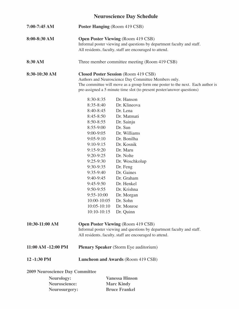
Neuroscience Day Schedule
7:00-7:45 AM Poster Hanging (Room 419 CSB)
8:00-8:30 AM Open Poster Viewing (Room 419 CSB)Informal poster viewing and questions by department faculty and staff.All residents, faculty, staff are encouraged to attend.
8:30 AM Three member committee meeting (Room 419 CSB)
8:30-10:30 AM Closed Poster Session (Room 419 CSB)Authors and Neuroscience Day Committee Members only. The committee will move as a group form one poster to the next. Each author is pre-assigned a 5 minute time slot (to present poster/answer questions)
8:30-8:35 8:35-8:40 8:40-8:45 8:45-8:50 8:50-8:55 8:55-9:00 9:00-9:05 9:05-9:10 9:10-9:15 9:15-9:20 9:20-9:25 9:25-9:30 9:30-9:35 9:35-9:40 9:40-9:45 9:45-9:50 9:50-9:55 9:55-10:0010:00-10:0510:05-10:1010:10-10:15
Dr. HansonDr. KlineovaDr. LenaDr. MatmatiDr. SainjuDr. SunDr. WilliamsDr. BonilhaDr. KosnikDr. MaruDr. NolteDr. WoschkolupDr. FengDr. GainesDr. GrahamDr. HenkelDr. KrishnaDr. MorganDr. SohnDr. MonroeDr. Quinn
10:30-11:00 AM Open Poster Viewing (Room 419 CSB)Informal poster viewing and questions by department faculty and staff.All residents, faculty, staff are encouraged to attend.
11:00 AM -12:00 PM Plenary Speaker (Storm Eye auditorium)
12 -1:30 PM Luncheon and Awards (Room 419 CSB)
2009 Neuroscience Day CommitteeNeurology:Neuroscience:Neurosurgery:
Vanessa HinsonMarc KindyBruce Frankel

MUSC Residents’ Neuroscience Day 2010
ABSTRACTS

MUSC Residents’ Neuroscience Day 2010
Resident Name: Jarom E. Hanson PGY: 2Abstract Title: Methamphetamine-induced dopaminergic deficits and refractoriness to subsequent
treatment
Co-Investigator: Elisabeth Birdsall, Kristi S. Seferian, Marcus A. Crosby, Kristen A. Keefe, James W. Gibb, Glen R. Hanson, and Annette E. Fleckenstein
Faculty Mentor:
ABSTRACTAbstract: Repeated high-dose methamphetamine administrations can cause persistent dopaminergic
deficits. As individuals abusing methamphetamine are often exposed to recurrent high-dose administration, the impact of its repeated exposure merits investigation. Accordingly, rats were pretreated with repeated high-dose injections of methamphetamine, and subsequently “challenged” with the same neurotoxic regimen 7 or 30 days later. Results revealed that the initial methamphetamine treatment caused persistent deficits in striatal dopamine levels, dopa-mine transporter function, and vesicular monoamine transporter-2 function. The subsequent methamphetamine challenge treatment was without further persistent effects on these param-eters, as assessed 7 days after the challenge, regardless of the interval (7 or 30 days) between the initial and challenge drug exposures. Similarly, a methamphetamine challenge treatment administered 7 days after the initial drug treatment was without further acute effect on dopa-mine transporter or VMAT-2 function, as assessed 1 hour later. Thus, this study describes a model of resistance, possibly explained by: 1) the existence of dopaminergic neurons that are a priori refractory to deficits caused by methamphetamine; 2) the existence of dopaminergic neurons made persistently resistant consequent to a neurotoxic methamphetamine exposure; and/or 3) altered activation of post-synaptic basal ganglia systems necessary for the elabora-tion of methamphetamine-induced dopamine neurotoxicity.

MUSC Residents’ Neuroscience Day 2010
Resident Name: Sylvia Klineova PGY: 2Abstract Title: Paraneoplastic Encephalitis Associated with Ovarian Teratoma in a Teenage Female
Co-Investigator:Faculty Mentor:
ABSTRACTAbstract: NMDA receptor associated paraneoplastic encephalitis is an infrequently diagnosed entity in
pediatric population. Due to presenting symptoms this potentially treatable condition repre-sents a diagnostic challenge for clinicians. A fifteen year old Hispanic female presented with sudden onset of insomnia, visual and audi-tory hallucinations, seizures, and dyskinesias. Neurological exam was dominated by delirium and dyskinesias of extremities, tongue, and jaw. Signs of autonomic instability with hyperten-sion, frequent episodes of diaphoresis, and hyperthermia were present. Diagnostic evaluation for infectious, inflammatory, autoimmune and toxic causes, including MRI and EEG, was negative.CT of the pelvis showed an ovarian teratoma of 4x 6 cm. Patient underwent teratoma removal and treatment with IVIg and steroids with complete resolution of her presenting symptoms within one month. Serum and CSF testing showed NMDA antibodies and teratoma pathol-ogy showed mature cerebral and choroid tissue. MMSE score at 3 months post operation was 28/30 but testing showed somewhat poor concentration and planning.Successful treatment outcome in this disorder is determined by prompt and accurate diag-nosis, with removal of the teratoma and immunotherapy, being crucial for full recovery and prevention of relapse.

MUSC Residents’ Neuroscience Day 2010
Resident Name: Jonathan Lena PGY: 2Abstract Title: CNS Choristoma: A Report of Two Cases
Co-Investigator:Faculty Mentor: Dr.Vandergrift
ABSTRACTBackground: Choristomas rarely involve the central nervous system. These lesions are nonneoplastic in
nature and typically composed of any combination of glial cells, smooth or skeletal muscle, adipose cells, and glandular cells.
Methods: The records of two patients found to have a CNS choristoma were reviewed. A search and review of the literature was then performed.
Results: Choristomas typically occur outside the nervous system. Few cases of choristomas involving the central nervous system have been reported. Those involving the optic nerve often pres-ent with gradually worsening visual symptoms over the course of several years. Choristomas involving the pituitary may present with any of the symptoms typical for pituitary tumors including endocrinopathies, visual disturbances, and headaches. Surgery is needed to confirm the diagnosis and may be curative in cases where the entire lesion is able to be removed.
Conclusions: Choristoma of the central nervous system is a rare lesion. Symptoms often develop insidi-ously over the course of several years. Surgery is often necessary to confirm the diagnosis as well as definitely treat the lesion.

MUSC Residents’ Neuroscience Day 2010
Resident Name: Kelly Matmati PGY: 1Abstract Title: Dural MALT Lymphoma presenting with disseminated disease: a unique case and
review of the literature
Co-Investigator: Nabil Matmati, Yusuf Hannun, Zoran Rumboldt, John Lazarchick, Robert Stuart, Pierre Giglio
Faculty Mentor: Pierre Giglio
ABSTRACTAbstract: Central nervous system (CNS) lymphoma involving the dura mater is very rare and histologi-
cally is usually Mucosa-Associated Lymphoid Tissue (MALT) lymphoma. Treatment of dural MALT lymphoma is not well established because these tumors are more indolent than other CNS lymphomas. We present a case of a 46 year old woman with MALT lymphoma involv-ing the dura, lacrimal gland, inguinal lymph nodes and bone marrow. Magnetic resonance imaging of the brain showed an extraxial enhancing mass approximately 2 cm in maximum diameter along the right frontotemporal convexity. Histopathology of the dural mass showed MALT lymphoma that was CD20, CD52, CD19, and CD38 positive. Molecular studies of the B-cell receptor heavy chain demonstrated monoclonality. The patient was treated with four cycles of fludarabine, mitoxantrone, and rituximab with complete remission. She had recurrence in the subcutaneous tissue of the back at 12 months but remains free of intracranial disease at 31 months. A review of the literature reveals 57 cases of dural MALT lymphoma. Only 2 were disseminated at presentation, and only 2 had local recurrence of the dural tumor. Because of the indolent behavior of this tumor, the intracranial portion can be treated conser-vatively, deferring radiation and providing close follow-up.

MUSC Residents’ Neuroscience Day 2010
Resident Name: Rup Sainju PGY: 2Abstract Title: Stroke due to cardiac myxoma in a young woman with remote history of neuroblas-
toma
Co-Investigator: Feng. Wuwei, Linsheng ZhangFaculty Mentor: Papamitsakis, Nikolaos I.H.
ABSTRACTAbstract: 22 year old Caucasian female, treated for neuroblastoma during infancy, who presented with
2 weeks history of intermittent neurological symptoms. MRI brain showed embolic strokes in various arterial territories. Echocardiogram disclosed an echogenic mass attached to the pos-terior mitral leaflet, further characterized by cardiac CT and MRI. The mass was surgically removed and histopathology was consistent with cellular cardiac myxoma. To our knowledge this is second reported case of cardiac myxoma in a patient previously treated for neuroblas-toma.
Methods: 22 year old Caucasian female presented with transient slurred speech, blurred vision and right facial droop which resolved in a few hours. She had a remote history of neuroblastoma treat-ed with chemotherapy (Adriamycin and Cytoxan), along with extensive abdominal resection of the tumor, with pelvic floor repair, during infancy. CT Head in local ED was normal. She also complained of memory problems, frequently losing her “train of thought”, difficulty with writing and impaired fine movements of her right hand. Brain MRI done after 2 weeks of ini-tial symptoms showed areas of restricted diffusion in the left caudate, putamen, anterior limb of internal capsule, left mid brain and right frontal region. She denied any prodromal consti-tutional symptoms. Her clinical examination was remarkable for loss of fine movements in her right hand. Follow-up brain MRI was consistent with subacute infarcts versus infective or metabolic changes in the basal ganglia. Her CSF examination was normal: no white or red cells identified, protein and glucose were normal, VDRL was nonreactive, HSV I/II PCR was negative, cytology and flow cytometry were negative for malignant cells. In addition four sets of blood cultures and urine culture were negative. Her electrolytes and liver enzymes were within normal limits. Head and neck MRA was normal. Transthoracic echocardiogra-phy revealed 1.5x1.5 cm echogenic mass attached to posterior mitral leaflet, with differential diagnosis of myoma, fibroelastoma, thrombus and endocarditis. Cardiac CT and MRI helped further characterizing the mass. CT abdomen and pelvis were negative for recurrence of neuroblastoma, presence of other tumors or metastasis. Patient underwent surgical removal of the mass without significant mitral valve regurgitation. Pathological study confirmed that the mass was consistent with a cellular myxoma.
Discussion: Cardiac myxoma is a rare but important cause of stroke in young patients. One or more of a classical triad of symptoms can be present: a) obstructive cardiac b) embolic, most commonly ischemic stroke and c) constitutional. Most cases are sporadic, but at least 7% are familial including the so-called Carney complex. There is a paucity of information in medical litera-ture on the causal relationship between cardiac myxomas and other tumors, cancer therapy or immunocompromised state. However, there are case reports of possible temporal relationship between myxoma and immunocompromised state, as post cardiac transplant, bone marrow transplant in patients with sickle cell disease, as well as with other tumors, as colorectal carci-noma and neuroblastoma. Our case is 2nd of its kind. It raises the question whether there is a

MUSC Residents’ Neuroscience Day 2010
temporal association of development of cardiac myxoma along with or after a diagnosis of neuroblastoma or whether it could be related to patient’s previous chemotherapy treatment and possible immunocompromised state. It also underscores the importance of echocardiogra-phy as part of the stoke work up especially in young patients, as cardiac myxomas are surgi-cally curable in most cases.

MUSC Residents’ Neuroscience Day 2010
Resident Name: Xiaoyan Sun PGY: 2Abstract Title: A case report of Creutzfldt-Jakob disease presenting with severe peripheral polyneu-
ropathy
Co-Investigator:Faculty Mentor: Dr. David Bachman
ABSTRACTBackground: Creutzfeldt-Jakob disease (CJD) is a rare neurodegenerative disease characterized by spongi-
form changes, neuronal loss and reactive astrocytosis in the central nervous system. Involve-ment of the peripheral nervous system is reported. Clinical presentation and evidence of the peripheral nerve involvement are limited.
Methods: In this presentation, one case of clinical diagnosed CJD is reported. The clinical presentation and clinical findings of MRI of brain, EEG, neuropsychological testing, CSF study, and EMG are described.
Results: 1. The patient had signs of central nervous system damage typical of sporadic CJD includ-ing progressive cognitive decline and ataxia. In addition, the patient showed progressive peripheral neuropathy presenting with stocking-glove sensory loss.
2. Brain image study showed hyper-intensity in the caudate head, putamen and thalamus regions by DIW image commonly seen in the patient with CJD.
3. Neuropsychological testing demonstrated moderate to severe impairment of multiple cognitive domains.
4. CSF study had positive 14-3-3 protein and significant elevated total tau protein.5. EMG/NCS study showed moderate to severe, axonal, sensory and motor polyneuropa-
thy involved in right peroneal, tibial, superficial radial nerves and right superficial radial nerve
Conclusions: In this case report, the patient with clinical diagnosed CJD had clinical presentation of the involvement of the central and peripheral nervous system. MRI of brain and CSF studies are proved to be a value tool for the diagnosis of CJD. Severe poly neuropathy of unknown etiol-ogy as a part of early presentation supports the notion that the involvement of the peripheral nervous system is an integral component of the disease.

MUSC Residents’ Neuroscience Day 2010
Resident Name: Nolan Williams PGY: 2Abstract Title: The Relationship between relative intake of omega 3 to omega 6 fatty acids and the
conversion of LDL to oxidized LDL in patients with intra-& extra-cranial atheroscle-rosis.
Co-Investigator: Nolan WilliamsFaculty Mentor: Tanya Turan
ABSTRACTBackground: Atherosclerotic stenosis of the major extra- and intracranial arteries is a major cause of stroke
worldwide yet the pathophysiology leading to the development of these disease processes has yet to be fully elucidated. It is speculated that ox-LDL (oxidized LDL) promotes the develop-ment of atherosclerosis through a process where it causes endothelial cells to secrete VCAM-1 (vascular cell adhesion molecule-1), which stimulates these monocytes to penetrate be-tween the endothelial cells and promote endothelial cell death. It is further speculated that the development of ox-LDL may be the result of high intake of dietary omega 6 as well as a low relative level of omega 3 intake producing a pro-inflammatory/pro-oxidative state. Several recent studies such as the Lyon Heart Study have shown that by modifying the dietary intake of omega 3/6, one can decrease incidence of a second vascular event.
Methods: Inclusion criteria: Angiographic evidence of intra or extra cranial atherosclerosis, but without tandem intra/extracranial atherosclerosis and without evidence of coronary artery disease. Methods: Participants will be admitted to the MUSC GCRC where fasting plasma is collected followed by MacDonald #2 Breakfast followed by blood collection (10cc each) at 1, 2, 3, 4, 6, and 8 hours. During this time, the Willet Food Frequency Questionaire (FFQ) will be administered which measures dietary omega 3/6 intake. The aforementioned plasma samples will be sent for quantitation of oxLDL using commercially available ELISA kits. The Willet FFQ will then be sent to the Harvard/Willet FFQ scoring center. The total omega 6 as well as the omega 3/6 ratio will be compared to the proportion of LDL oxidation as well as the sever-ity and type (intra-versus extra-cranial) of stenosis.
Results: pending
Conclusions: pending

MUSC Residents’ Neuroscience Day 2010
Resident Name: Leonardo Bonilha PGY: 3Abstract Title: Hippocampal atrophy in patients with medial temporal lobe epilepsy: differences
between the ‘generator’ and the ‘receiver’.
Co-Investigator: Paul Morgan PhD, Jonathan J Halford MD, Jonathan C Edwards MDFaculty Mentor:
ABSTRACTRationale: Hippocampal sclerosis (HS) is the most common histological abnormality observed in pa-
tients with medial temporal lobe epilepsy (MTLE). HS is traditionally considered to be the cause of seizures; hence the resection of the sclerotic hippocampus yields seizure-freedom in the majority of operated MTLE patients. HS can be observed with routine brain MRI, under the form of hippocampal atrophy. Nonetheless many patients with MTLE may display MRI features of HS in both hippocampi, even when seizures only originate in one hippocampus. The explanation for this discrepancy lies on the fact that the atrophy of the hippocampus that generates seizures (the ‘generator’) is pathologically distinct from the atrophy of the hippocampus that receives seizures (the receiver). Atrophy in the hippocampus with HS is considered to be a consequence of developmental gliosis and cell death with the formation of aberrant epileptogenic neuronal circuitry, whilst the contralateral hippocampus is atrophied as a consequence of the excitotoxic effects of seizures without epileptogenic reorganization. To date, there are not well-defined parameters to distinguish between the epileptogenic hippo-campal atrophy of HS (the ‘generator’), versus the non-epileptogenic atrophy (the ‘receiver). This distinction has a crucial clinical importance as it may enable the correct identification of the seizure onset site and lead to better surgical results. Objective: This study aimed to evaluate the tridimensional distribution of hippocampal atrophy in patients with medication refractory MTLE. We hypothesized that the hippocampal atrophy in the epileptogenic hippocampus is spatially distinct from the non-epileptogenic hip-pocampus.
Methods: We studied 14 patients with MTLE according to the parameters defined by the ILAE. All patients had unilateral seizure onset documented by video-EEG corresponding to the side of hippocampal atrophy. Six patients had right MTLE and eight had left MTLE. All patients were submitted to unilateral hippocampal resection and had hippocampal sclerosis confirmed by histology. Importantly, all patients were seizure free at least 12 months after surgery. We also studied a control group of 34 healthy individuals. We obtained a T1-weighted image with 1mm isotropic voxels acquired in the sagittal plane with the following parameters: TR=8.1ms, TE=3.7ms, flip angle = 8°, FOV = 256x256mm from all subjects. All images were then submitted to voxel-based morphometry preprocessing, including iterative spatial normalization to the stereotaxic space, bias field correction and tissue segmentation based on ICBM a priori templates (affine and16 iteration non-linear transformations). We focused the analyses on the hippocampus, through regions of interest (ROIs), defined based on cytoarchi-tectonic and functional data, which were employed as masks constraining further statistical analyses to the voxels contained within these regions. Patients and controls were compared employing a non-parametric voxel-wise Wilcoxon test. The left hippocampus of patients was compared to the left hippocampus of controls, and the right hippocampus was compared to the right hippocampus. Results were corrected for multiple comparisons with the application of an FDR corrected threshold for q<0.05.

MUSC Residents’ Neuroscience Day 2010
Results: Patients with left MTLE showed atrophy involving the ipsilateral hippocampus and the con-tralateral hippocampus. There was diffuse atrophy within both hippocampi, but, as expected, the atrophy within the ipsilateral hippocampus (both for left and right MTLE) was more intense and more widely distributed within the hippocampus (Figure 1). On the contralateral hippocampus, there was also significant atrophy, but less intense and less pervasive. Interest-ingly, the anterior–inferior aspect of the head of the hippocampus was the site of more sig-nificant damaged in the ipsilateral hippocampus (Figures 1 and 2) (site of maximal atrophy: Left MTLE, left hippocampus x=-28, y=-16, z=-24, Z=3.6; Right MTLE, right hippocampus x=22, y=-11, z=-27, Z=2.9). On the contralateral hippocampus, the atrophy was more notice-able in the posterior head and body areas (site of maximal atrophy: Left MTLE, right hip-pocampus x=25, y=-17, z=-21, Z=2.1; Right MTLE, left hippocampus x=-27, y=-19, z=-17, Z=2.1). This pattern was very similar between left MTLE and right MTLE data. We also ob-served a more pronounced atrophy of the hippocampal tail in the ipsilateral hippocampus in patients with left MTLE (site of maximal atrophy: Left MTLE, left hippocampus x=-16, y=-35, z=1, Z=2.6), but this was not observed in the hippocampi of patients with right MTLE. There were no areas of increased gray matter volume in patients compared with controls.
Discussion: In this study, we demonstrated that the hippocampal atrophy observed in the hippocampus that generates seizures (the ‘generator’) has an anatomically distinct pattern compared to the atrophy of the contralateral hippocampus (the ‘receiver’). This was demonstrated on patients who achieved seizure freedom with surgery, thereby eliminating the possibility that the con-tralateral hippocampus may also be a site of epileptogenesis. This finding will benefit from further refinement by a study of a larger sample size, also to include patients with hippocam-pal atrophy as a consequence of extra-temporal epilepsy, nevertheless it has the potential to directly improve the pre-surgical analysis of MTLE.

MUSC Residents’ Neuroscience Day 2010
Resident Name: Libby Kosnik PGY: 3Abstract Title: Subfrontal Trans-Lamina Terminalis approach to third ventricle utilizing eyebrow
incision
Co-Investigator: Krishna, VFaculty Mentor: Vandergrift, A
ABSTRACTBackground: Third ventricular lesions pose a challenge because of their deep location and proximity to vi-
tal neural and vascular structures. Various modifications of trans-lamina terminalis approach have been described. We report a novel minimally invasive approach for third ventricular lesions via supra-orbital craniotomy.
Methods: We will review three cases in which we preformed a supra-orbital (eyebrow) craniotomy for resection of third ventricular lesions.
Results: Each patient’s history and pre-operative images will be reviewed, followed by intraoperative descriptions of the procedures. Next the post operative course and pathology will be dis-cussed, and advantages of this technique will be highlighted.
Conclusions: Subfrontal trans-lamina terminalis approach is safe and effective for resection of lesions contained within the third ventricle. Endoscopic assistance can enhance visualization and broaden field of view during this operation.

MUSC Residents’ Neuroscience Day 2010
Resident Name: Neal Maru PGY: 3Abstract Title: “Is There a Relationship between Allergies/Medication Intolerances and Nonepileptic
Events?: A Retrospective Analysis.”
Co-Investigator:Faculty Mentor: Jonathan Halford MD
ABSTRACTAbstract: Non-epileptic events (NEE), previously referred to as psychogenic non-epileptic seizures or
pseudoseizures, are one of many somatoform disorders. In clinical practice, we have noticed that patients with NEE tend to report multiple allergies and medication intolerances. This may be a characteristic of patients with somatoform or conversion disorders. However, al-lergies to anti-epileptic drugs (AED) are responsible for a large percentage of newly reported allergies, and the NEE population is subject to AED exposures. In this study, we aim to eval-uate whether multiple allergies and/or medication intolerances are correlated with a diagnosis of NEE in the inpatient adult epilepsy monitoring unit (EMU). In this study we will perform a restrospective review of electronic medical records of patients admitted to EMU. Informa-tion we will collect will be de-identified. Information collected will be aspects of general past medical history, antiepileptic medication history, medication allergies, and results of epilepsy diagnostic testing. The data will be used to determine a relationship between the presence or absence of NEE and multiple allergies and medication intolerances.

MUSC Residents’ Neuroscience Day 2010
Resident Name: Justin Nolte PGY: 3Abstract Title: VZV Encephalitis/Vasculitis Presenting as New Onset Seizure
Co-Investigator:Faculty Mentor:
ABSTRACTBackground: Varicella Zoster (VZ) is a relatively common infection, seen predominantly in childhood
manifesting clinically as the typical “chicken pox” rash. This condition is relatively benign, however not all infections from the VZ virus (VZV) are so benign. Reactivation of the virus can be potentially deadly; especially when reactivation occurs in patient’s who are immuno-compromised.
Methods: Case report of a patient recently admitted to the general neurology service, with subsequent review of the literature for VZV infection and clinical manifestations, diagnostic evaluation including typical radiographic findings and finally post-mortem pathologic findings.
Results: Patient SA was a 47 year old African American male with past medical history of HIV diag-nosed in 1999, alcohol and polysubstance abuse and depression vs. bipolar disease who was found to be confused with incontinence of stool and urine with dysarthria and a right facial droop. He was brought to MUSC for evaluation for possible stroke, but CTA did not reveal any vessel occlusions nor did the CTP show any areas of restricted perfusion. He underwent an MRI brain which was negative for acute infarct, but did reveal multiple areas of abnormal signal on Flair consistent with ADEM, PML or viral encephalitis. His rou-tine EEG was showed diffuse, 2-4Hz generalized slowing with occasional triphasic waves, without epileptic abnormalities. He was continued on dilantin after being loaded in the ER with a diagnosis of presumed new-onset seizure with postictal Todd’s paralysis An LP was done on 2 of admission, revealing protein 83, glucose 38, 4 RBCs and 7 WBCs (85% lymphocytes). No xanthochromia was noted on initial LP; however the initial quantity of CSF removed was not sufficient for a full malignant workup, so repeat LP was done on day 5 of admission, which did reveal xanthochromia. A stat head CT revealed a lobar ICH in the right, frontal lobe, with areas of subarachnoid hemorrhage located in the right, anterior frontal lobe and left, superior frontal lobe. A conventional angiogram was done the following day which showed vasculitic changes in right MCA and L ACA distributions, without evidence for aneurysm or other vascular malfor-mations. His VZV PCR came back positive on day 4 of admission, so a diagnosis of probable VZV vasculitis was quickly made and he was started on treatment dose acyclovir 10mg/kg q8h immediately.
Discussion: Reactivation of VZV can manifest clinically in multiple different ways, including the most benign dermatologic presentation shingles, a painful neuritis, myelitis, encephalitis, small, medium and large vessel vasculitis and periventriculitis. The degree of severity of reactiva-tion is primarily related to the patients’ immune status, so SA had a more severe clinical presentation due to his underlying HIV with CD4 count of 74. He clinically exhibited en-cephalitis with subsequent seizure disorder, small vessel vasculitis with white matter changes on MRI/lobar hemorrhage and large vessel vasculitis, exhibited on conventional angiogram

MUSC Residents’ Neuroscience Day 2010
with subsequent subarachnoid hemorrhage. The presence of vasculitic changes in SA is a bit atypical, as VZV typically causes medium and large vessel damage in the MCA and PCA distributions, he vascular irregularities were appreciated in his left ACA distribution. Pt SA’s diagnosis of VZV vasculitis was made by positive VZV PCR in his CSF. While definitive diagnosis is typically made with pathology review from biopsy, studies have shown that the sensitivity and specificity for detection of VZV in the CSF with PCR to be >95%, and given the lack of other positive cultures in the CSF, including JC Virus, HSV, EBV, CMV, Toxoplasmosis, VDRL, ACE, cytopathology/flow cytometry and AFB, we felt confi-dent in our final diagnosis.

MUSC Residents’ Neuroscience Day 2010
Resident Name: Wayne Feng PGY: 4Abstract Title: Short and Long Term Outcome of Hospitalized Intracerebral Hemorrhagic Patients in
South Carolina
Co-Investigator: Justin Nolte, Rup Sainju, Robert HendryFaculty Mentor: Dr. Angela Hays and Dr. Robert Adams
ABSTRACTObjective: This study is to assess the short and long term outcome of hospitalized intracerebral hemor-
rhagic stroke patients in South Carolina.
Methods: Patients with a primary diagnosis of incerebral hemorrhage (ICD9 code=431) discharged from the year 2002 were identified from the South Carolina state hospital discharge data-base. Kaplan-Meier estimates of recurrent stroke, MI, vascular death, all-cause death, and composite events were calculated at 1 month, 6 months, 1 year, 2 years, 3 years, and 4 years. Log rank test is applied to test the difference between Caucasian and African American for composite events (including recurrent stroke, MI or vascular death).
Results: 893 intracerebral hemorrhagic patients were identified in the database in 2002. The Kaplan-Meier estimate of cumulative risk at 1 month, 6 months, 1 year, 2 years, 3 years, and 4 years for recurrent stroke is 1.5%, 4.1%, 7.6%, 11.3%, 14.8% and 18.6%; for MI is 0.2%,0.8%, 1.0%, 2.0%, 2.7% and 3.5%; for vascular death is 37.5%, 40.8%, 42.1%, 45.0%, 47.2% and 49.4%, for all-cause death is 41.9%, 47.5%, 50.2%, 54.5%, 58.1%, 61.6%; for composite events (recurrent stroke, MI, or vascular death) is 38.7%, 42.9%, 46.1%, 50.7%, 54.4%, 57.9%; for composite events (recurrent stroke, MI, or all-cause death) is 42.8%, 48.5%, 53.8%, 58.7%, 63.0%, 67.3%.
Conclusions: The outcome for hospitalized patients with intracerebral hemorrhage is poor. There is a sig-nificant room for improvement for both treatment and secondary prevention in South Caro-lina to alleviate the burden.

MUSC Residents’ Neuroscience Day 2010
Resident Name: Kathryn Gaines PGY: 4Abstract Title: Preliminary Results of A Descriptive Comparison of MUSC Parkinson’s Disease
Patients
Co-Investigator: Vanessa Hinson, MDFaculty Mentor: Vanessa Hinson, MD
ABSTRACTBackground: Parkinson’s disease (PD) is felt to be a disease affecting white men more than women and
more than any other race, regardless of gender. In an MUSC study by Bergmann et al., comparisons of disease prevalence across South Carolina Medicare recipients indeed found a difference among prevalence rates of blacks and whites. However, little is known about the differences in severity of disease and treatment efficacy between gender and race.
Objective: To analyze MUSC PD population differences among race and gender noting specifically dif-ferences in disease severity, symptom onset, time between symptom onset and diagnosis and treatment choice at beginning of diagnosis as well at most current clinic visit.
Methods: Using our MUSC PD database, a retrospective review of Practice Partner records was per-formed. Fifty six charts were hand reviewed, detailing age, gender, age of symptom onset and diagnosis as well as severity of disease using Hoehn and Yahr (H/Y) staging at initial diagnosis and most current clinic visit. Additionally noted was medication choice at initial symptom or diagnosis as well as most current clinic visit.
Results: All MUSC patients included in the PD database of non-white origin were reviewed, total-ing 18 patients. Nine of these were black females, 7 were black males, one was an Asian male and one was a Hispanic male. Chart review of 36 white patients was also completed, of which 16 were female. Due to the small number of Asian and Hispanic data points, these data were discarded for the purposes of this study. Average current age for white males at time of chart review was 69 yrs; 73 years for white females, 67 years for black males and 69 years for black females. The average number of years between symptom onset and diagnosis for white males was 2.25 years, 3.42 years for black females, 4 years for black males and 5 years for white females. At initial diagnosis, a higher percentage of blacks were diagnosed with more advanced stage, especially black females with an average H/Y of Stage 3 com-pared to white males and females at H/Y Stage 1. Once more, at most current clinic visit, only blacks made up the most advanced stage of disease (Stage 5), again notably the black females. Regarding medication, at initial diagnosis, both white men and black men were started on dopamine agonists more than any other medication even though black males did not keep company with white males in regards to the mild H/Y of Stage 1. In fact, average initial H/Y stage for black males was between Stage 2 and Stage 3. Evaluation of current clinic visit medication found all groups were maintained on levodopa. Deep brain stimulation was more common among whites and almost non-existent in the black MUSC population.
Conclusions: It was difficult to gather all specified data points for each patient due to medical records that did not document specific interests concerning this study. It seems from analysis of results that white men were diagnosed earlier than white women, possibly due to an increased rate of comorbities among white males compared to white females, leading to an earlier

MUSC Residents’ Neuroscience Day 2010
manifestation of parkinsonian symptoms in white males. Additionally, black females were diagnosed, on average with a higher severity of H/Y stage. It could be that black females tend to have a more aggressive disease course naturally or tend to present more commonly with abnormal postural reflexes, necessitating a higher H/Y at initial diagnosis. Finally, medication differences among groups showed a difference between all females compared to all males initially for unclear reasons, however, at later stages of disease and as noted on the most current clinic visit, whites were found to have a higher percentage of DBS surgery as opposed to blacks. This may be explained by cultural differences.

MUSC Residents’ Neuroscience Day 2010
Resident Name: Patricia Graham PGY: 4Abstract Title: Clinical Applications of Functional Magnetic Resonance Imaging in Epilepsy
Co-Investigator: Leo Bonilha, MD; Paul Morgan, PhD; Maria Spampinato, MDFaculty Mentor: Jonathan Edwards, MD
ABSTRACTBackground: In recent years, functional MRI (fMRI) has become one of the most widely used neuroimag-
ing techniques for mapping regional brain activity. Its applications are widespread and it is fast becoming a preferred method of imaging because it is both noninvasive and does not ex-pose patients to ionizing radiation. Functional MRI can be used as a clinical tool in the pre-surgical evaluation of medically refractory epilepsy patients. Many institutions are currently using fMRI as a supplement to or replacement for WADA testing to effectively determine laterality for functions such as language and memory. The Comprehensive Epilepsy Center at MUSC is working to establish fMRI as a noninvasive test to help predict postoperative outcomes of surgical epilepsy patients.
Methods: Using a 3T functional MRI equipped with a Multihance head coil and a TR of three seconds, three healthy, adult volunteer subjects were scanned and underwent testing to assess for ver-bal memory. The paradigm for verbal memory that was used involved showing each subject 15 medium frequency words over one minute. Afterwards, each subject was asked to recall as many of the listed words as possible. Each subject was given one minute to recall the words. This process was repeated four more times. As a control, each subject was presented with a list of 15 nonsense words over one minute. Next, each subject was asked to recite the alphabet for one minute. Again, this control test was repeated four more times.
Results: Final results of this study are still being calculated. Since the individuals that volunteered for this study were healthy adults, the expected outcome when testing verbal memory would be an increased pattern of activation seen in the dominant medial temporal lobe (the left side for most individuals). We also expect to see the level of activation higher during the first half of testing when subjects are exposed to sense words. The level of activation should be some-what decreased when the nonsense words are displayed. There will also likely be a differ-ence in degree of reliability between this group of control subjects and epilepsy patients given the fact that epilepsy patients may have one or more dysfunctional temporal lobes.
Conclusions: The main objective of this study is to develop a successful paradigm for testing verbal mem-ory in medically refractory epilepsy patients undergoing presurgical evaluation. Once we have created an effective and consistent model for verbal memory testing, other paradigms for functions such as visual memory and language will need to be established. Over the next year, we hope to begin incorporating fMRI into our battery of tests used to predict favorable outcomes in surgical epilepsy patients.

MUSC Residents’ Neuroscience Day 2010
Resident Name: Brody L. Henkel PGY: 4Abstract Title: Copper deficiency myeloneuropathy and anemia due to excessive denture cream use
Co-Investigator: David E. Stickler, MDFaculty Mentor: David E. Stickler, MD
ABSTRACTBackground: Copper deficiency secondary to excessive zinc intake has been associated with bone marrow
suppression, combined degeneration of the spinal cord, and polyneuropathy. We describe a case of painful neuropathy, lower extremity weakness, and sensory ataxia secondary to over-use of denture cream that was refractory to oral copper supplementation.
Case History: A 37-year-old female initially complained of neurological symptoms 2 years prior to our evaluation. Two years ago she developed numbness and tingling in her bilateral feet. The paresthesias became painful and progressed up to her knees, extending to both hands over the next 6 months. She then developed left lower extremity weakness along with gait instability resulting in several falls, prompting referral to an academic medical center for evaluation. Neuroimaging was unremarkable at that time, and initial EMG/NCS revealed a mild axonal sensorimotor polyneuropathy. Right gastrocnemius muscle biopsy was unrevealing, and sural nerve biopsy revealed severe, chronic, active neuropathy. In the course of evalua-tion for a normocytic anemia, bone marrow biopsy revealed pancytopenia. The serum copper was low (<10 mcg/dL, normal 80-155 mcg/dL), and she was started on oral copper gluconate therapy. The anemia improved, but her neurological condition continued to deteriorate and she was referred for another evaluation. Nerve conduction studies revealed a severe primary axonal neuropathy. Electromyography showed signs of denervation, with fibrillations and increased insertional activity in all distal muscles tested. There was also evidence of chronic changes, with large, occasionally complex MUP’s with a decreased recruitment and interference pattern. MRI of both the cervical and thoracic spine showed subtle T2 hyperintensity along the posterior columns. The patient later revealed that she had been using 2 tubes of Super Poli-Grip denture cream per week for the past four years due to poorly fitting dentures. Serum copper (55 mcg/dL, 80-155mcg/dL) and ceruloplasmin (14mg/dL, 17-54mg/dL) were both low despite treat-ment with oral copper gluconate 10mg per day. Serum zinc was elevated (138 mcg/dL, nor-mal 60-120 mcg/dL). She was diagnosed with copper deficiency myeloneuropathy secondary to hyperzincemia. Despite cessation of denture cream use and copper therapy, the patient’s weakness and painful neuropathy have improved little over the past two years.
Discussion: This patient developed a significant myelopathy and painful neuropathy due to copper deficiency that resulted from overuse of denture cream. Zinc concentrations of 17,000 to 34,000 mcg/g have been found in commonly used denture creams, with about 80 grams per tube. Zinc is thought to upregulate metallothionen, an intracellular metal binding protein, within enterocytes. Copper binds to metallothionen with a higher affinity than zinc, resulting in reduced copper uptake and increased fecal loss. EMG/NCS often reveals a mild to moderate distal, axonal, sensorimotor peripheral neu-ropathy. Spine MRI may reveal increased T2 signal in the dorsal columns, commonly

MUSC Residents’ Neuroscience Day 2010
involving the cervical cord. Most often patients develop a microcytic anemia, but macrocytic and normocytic anemias are also reported, as well as pancytopenia Hypocupremia secondary to hyperzincemia from denture creams is likely more prevalent than first recognized. One recent series of 11 patients with previously unexplained hyperz-incinemia and copper deficiency found denture cream as the source of excessive zinc in all 11 patients. Discontinuation of excessive zinc intake and copper supplementation promptly resolves the hematological abnormalities in most cases, but neurological recovery is variable, with subjective improvement of sensory symptoms most frequent. The patient in this case was initially diagnosed with a copper deficiency of unknown etiology but continued to use excessive amounts of denture cream, and thus exhibited neurological decline despite supple-mentation with high dose oral copper. This highlights the importance of questioning patients with unexplained signs of both polyneuropathy and myelopathy about denture cream use.
MUSC Residents’ Neuroscience Day 2010
Resident Name: Vibhor Krishna PGY: 4Abstract Title: Cervical disc arthroplasty compared to anterior cervical fusion – a meta-analysis
Co-Investigator: Varma, A; Warmath, W.Faculty Mentor: Varma, A.
ABSTRACTBackground: Anterior cervical discectomy and fusion (ACDF) is the most commonly performed proce-
dure for cervical radiculopathy or myelopathy. ACDF restricts motion and can alter cervical dynamics leading to increased risk of adjacent level disc degeneration. Cervical disc arthro-plasty preserves motion and thus prevents adjacent level disc degeneration.
Methods: Literature search was performed to identify randomized controlled trials comparing cervical disc arthroplasty to anterior cervical discectomy and fusion. Six randomized controlled trials with eight publications were identified. Data was extracted using standardized data extrac-tion sheets. Statistical analysis was carried out using SAS® to calculate the pooled effect estimates for improvement in pain, disability, range of motion and overall outcome. Available data regarding transitional changes was also collected.
Results: Five randomized controlled trials evaluated cervical arthroplasty for single level disease. The implants used were, Bryan arthroplasty, Prestige arthroplasty and Prodisc-C. One trial evalu-ated disc replacement for disc degeneration at two levels. Most studies report a follow up of two to three years with a total of 2741 person year data. The data shows that disc arthroplasty offers better resolution of neck pain, arm pain and pain disability. The mean range of motion on dynamic imaging is preserved long term. Two of these trials indicate that arthroplasty prevents transitional changes.
Conclusions: Cervical disc replacement is superior to cervical fusion for resolution of pain and decreased disability. Range of motion is preserved after several years of implantation. Limited data is available to evaluate risk of adjacent level disc degeneration.

MUSC Residents’ Neuroscience Day 2010
Resident Name: Mimi Sohn PGY: 5Abstract Title: Spinal Neurosarcoidosis: A Case Series of 12 Patients
Co-Investigator: Dr. Marc JudsonFaculty Mentor: Dr. Marc Judson, Dr. Kenkichi Nozaki
ABSTRACTBackground: Neurosarcoidosis, with the involvement of central and peripheral nervous system, is esti-
mated to occur in approximately 5-10% of total sarcoidosis patients. Spinal neurosarcoid-osis is rare with an incidence estimated at 0.43-1%. Spinal neurosarcoidosis is problematic to diagnose since it tends to mimic other inflammatory neurologic diseases such as multiple sclerosis (MS) or neuromyelitis optica. We report 12 cases of spinal neurosarcoidosis with a special focus on the magnetic resonance imaging (MRI) and laboratory findings. The goal of this study is to clarify the clinical manifestations of spinal neurosarcoidosis.
Methods: We reviewed electronic medical records of sarcoidosis patients who were identified from a MUSC-approved clinical sarcoidosis database. We identified patients who met the diagnostic criteria of definite neurosarcoidosis (Judson, MA et al., 1999) that had spinal cord involve-ment with sarcoidosis. Data collected included sex, race, and the age of onset, nature of neurological symptoms, biopsy results, laboratory findings, anatomic-radiologic location, and distribution of the lesions with characteristic MRI findings.
Results: We identified 12 sarcoidosis patients with definite sarcoidosis who has spinal cord involve-ment. The clinical presentation, laboratory and the MRI findings of the patients reviewed are summarized in Table 1. Seven of 12 (58%) patients were women. The group comprised 12(100%) African Americans. The age range was from 34-63years, with a mean 44years. The most common neurologic symptoms are extremity (leg>arm) weakness (8/12, 67%), pares-thesia (5/12, 42%) and back or neck pain (4/12, 33%). The MRI characteristics consisted of multiple, diffuse or nodular enhancing intramedullary lesions (6/12, 50%), leptomeningeal (5/12, 42%) and vertebral body (2/12, 17%) enhancement. The most commonly involved site being at the thoracic levels (7/12, 58%), then cervical levels (6/12, 50%), less commonly, conus medullalris /cauda equina (3/12, 25%).The distribution of involved vertebral body segments ranged from 1~10, with a mean of 4.4 vertebral body segments. Ten of 12(83%) patients involved over ≥3 vertebral body segments. Histological diagnosis was established in all subjects: 2/12 (17%) from the spinal cord, 1/12 (8%) by autopsy and 8/12 (67%) from extra-neural organs. Cerebrospinal fluid (CSF) studies were performed in 7 patients. CSF an-giotensin converting enzyme (ACE) level was elevated in only 1/7 (14%), while oligoclonal bands (OCB) were positive in 3/4 (75%) patients.
Conclusions: We found spinal neurosarcoidosis typically extends over 3 or more spinal segments. The common spinal cord manifestations are multiple, diffuse or nodular enhancing intramedullary lesions and leptomeningeal enhancement. These MRI findings are in contrast to multiple sclerosis is which rarely extends more than 2 vertebral body segments. The MRI finding of abnormalities extending over 3 or more vertebral bodies should prompt the clinician to seri-ously consider other etiologies including neurosarcoidosis as a possible cause along with

MUSC Residents’ Neuroscience Day 2010
multiple sclerosis. In most subjects, diagnosis was established with histological evidence obtained from extra-neural organs. Regarding CSF studies, measuring ACE level and OCB do not help to diagnose spinal neurosarcoidosis or to differentiate it from multiple sclerosis.

MUSC Residents’ Neuroscience Day 2010
Resident Name: Tanya Quinn PGY: 7Abstract Title: Decompressive Hemicraniectomy in the Management of Severe Traumatic Brain
Injury
Co-Investigator: Dr. Ian F. Dunn, Mr. Joseph Taylor, Mr. Jordan Magarik, Prof. Mark Kindy Faculty Mentor:
ABSTRACTAbstract: Decompressive craniectomy is a neurosurgical technique in which a portion of the skull is
removed in order to reduce intracranial pressure. The rationale for this procedure is based on the Monro-Kellie Doctrine; increasing the volume of intracranial space reduces intracranial pressure and thus increases compliance in the event of worsening cerebral edema following traumatic brain injury. There is significant debate over the efficacy of decompressive craniec-tomy despite its sound rationale and historical significance. Considerable variation in the em-ployment of decompressive craniectomy, particularly for secondary brain injury, explains the inconsistent results and mixed opinions of this potentially valuable technique. Critical analy-ses of decompressive craniectomy need to replace the compilation of case studies, anecdotal observations, and empirical hunches that currently dictates its indication and employment. The purpose of this study is to review the rationale, technique, and recent clinical evidence for use of decompressive craniectomy in patients with traumatic brain injury.