Embed Size (px)
Citation preview
VOL. XL NO. 5 THE JOURNAL OF ANTIBIOTICS 575
TIACUMICINS, A NOVEL COMPLEX OF 18-MEMBERED
MACROLIDES
II. ISOLATION AND STRUCTURE DETERMINATION
J. E. Hochlowski, S. J. Swanson, L. M. Ranfranz, D. N. Whittern,A. M. Buko and J. B. McAlpine
Abbott Laboratories,North Chicago, Illinois 60064, U.S.A.
(Received for publication November 7, 1986)
A novel complex of Gram-positive antibiotics has been isolated from the fermentationbroth and myceliumof Dactylosporangiumaurantiacum subsp. hamdenensissubsp. nov. Thestructures of these six compoundswere deduced employing UV,MS, IR, and extensive IDand 2Dhomonuclear and heteronuclear NMRexperiments. Each componentcontaineda highly unsaturated 18-memberedmacrolide ring. Componentsdiffered from one anotherby minor structural variations in the macrolide ring and by the numberand esterificationpattern of glycosidically bound sugars.
In the course of screening microorganisms for the production of antibiotics a Dactylosporangiumspecies was discovered which produced a novel complex of 18-membered macrolides. The companionpaperx) describes the taxonomy and fermentation of this organism and the biological activity of theindividual antibiotics. This paper will describe the isolation of these components and the elucidationof their structures.
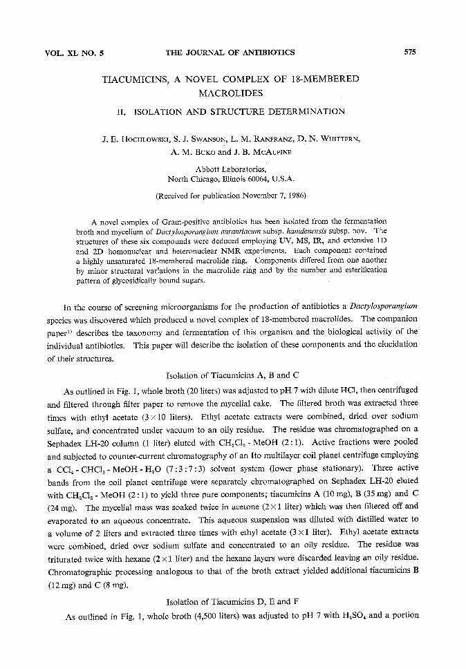
Isolation of Tiacumicins A, B and CAs outlined in Fig. 1, whole broth (20 liters) was adjusted to pH 7 with dilute HC1, then centrifuged
and filtered through filter paper to remove the mycelial cake. The filtered broth was extracted threetimes with ethyl acetate (3 x 10 liters). Ethyl acetate extracts were combined, dried over sodiumsulfate, and concentrated under vacuumto an oily residue. The residue was chromatographed on aSephadex LH-20 column (1 liter) eluted with CH2C12 - MeOH(2 : 1). Active fractions were pooledand subjected to counter-current chromatography of an Ito multilayer coil planet centrifuge employinga CC14- CHC13 - MeOH-H2O(7:3 :7:3) solvent system (lower phase stationary). Three activebands from the coil planet centrifuge were separately chromatographed on Sephadex LH-20 elutedwith CH2C12 - MeOH(2 : 1) to yield three pure components; tiacumicins A (10 mg), B (35 mg) and C(24 mg). The mycelial mass was soaked twice in acetone (2 x 1 liter) which was then filtered off andevaporated to an aqueous concentrate. This aqueous suspension was diluted with distilled water toa volume of 2 liters and extracted three times with ethyl acetate (3 x 1 liter). Ethyl acetate extractswere combined, dried over sodium sulfate and concentrated to an oily residue. The residue wastriturated twice with hexane (2 x 1 liter) and the hexane layers were discarded leaving an oily residue.Chromatographic processing analogous to that of the broth extract yielded additional tiacumicins B(12mg) and C (8 mg).
Isolation of Tiacumicins D, E and FAs outlined in Fig. 1, whole broth (4,500 liters) was adjusted to pH 7 with H2SO4and a portion
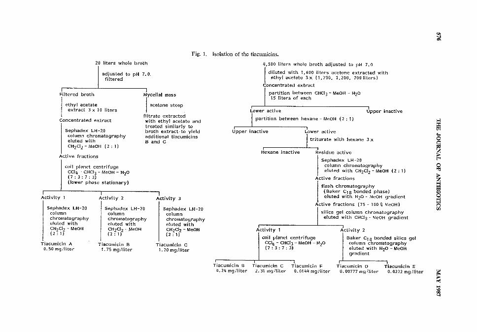
Fig. 1. Isolation of the tiacumicins.
3
g
3
Q
VOL. XL NO. 5 THE JOURNAL OF ANTIBIOTICS 577
of the insoluble material removed by centrifugation. The remaining broth and mycelial mass, dilutedwith acetone (1,600 liters) to lyse the mycelium, was extracted three times with ethyl acetate (1,700,1,200 and 700 liters). Extracts were combined and concentrated under reduced pressure to an oilyresidue. The residue was partitioned between CHC13- MeOH- H2O(15 liters of each) and the upperlayer discarded. The lower layer was concentrated to an oil and the CHC13- MeOH- H2Opartitioningrepeated twice. The final lower layer residue was partitioned between MeOHand hexane (6 liters ofeach). The upper layer was discarded and the lower layer concentrated to a residue. This residuewas triturated with hexane four times (4 x 6 liters) and the hexane was discarded to leave a solid (200 g).A portion (40 g) was chromatographed over a Sephadex LH-20 column (7.5x90 cm) eluted withCH2C12- MeOH(2 : 1). Active fractions were combined and subjected to flash chromatography ina Baker C18 column eluted with a step gradient ranging from H2Oto MeOHin 25 % increments. Ac-tive fractions (75 % and 100% MeOHeluates) were combined and concentrated by evaporation underreduced pressure followed by lyophilization to yield a solid residue (10 g). This was chromatographedon a silica gel column (7.5x100 cm) eluted with a step gradient of CHC13to CHC13- 50%MeOH.Active fractions were combined into two pools each of which was concentrated to a residue (12.5 gand 0.2 g, respectively). The former residue was chromatographed on an Ito multilayer coil planetcentrifuge employing a CC14 - CHC13- MeOH- H2O(7 : 3 : 7 : 3) solvent system (lower phase stationary)in 10 batches to yield pure tiacumicins B (3.82 g), C (2.08 g) and F (13 mg). The latter residue waschromatographed on Baker C18 bonded phase silica gel to yield pure tiacumicins D (7 mg) and
E(20mg).
Structure Determination of Tiacumicin BThe structure of tiacumicin B, the major antibiotic component produced by this culture will be
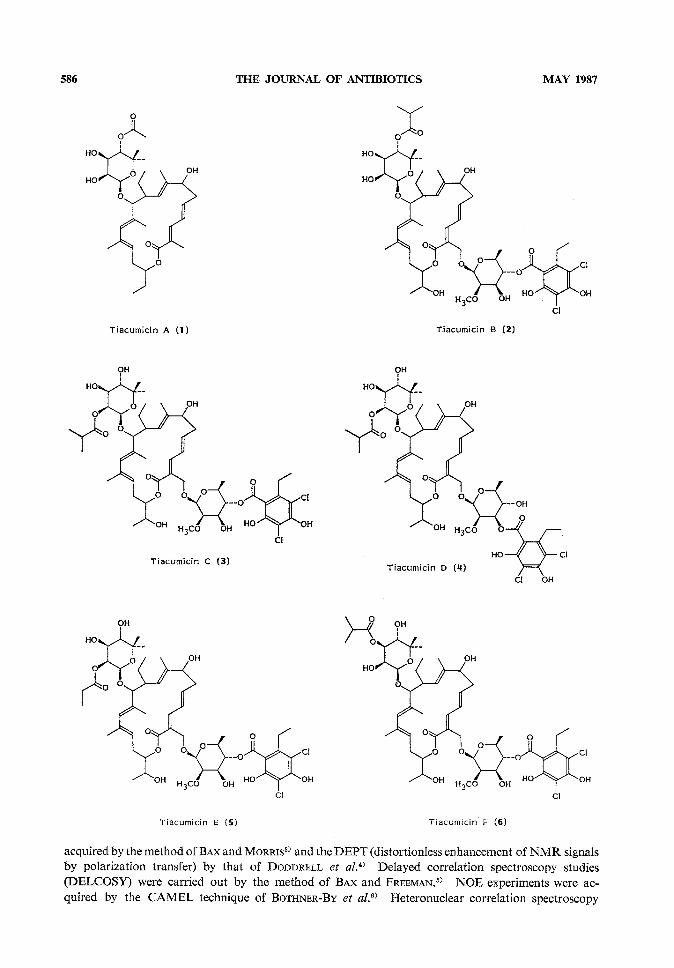
discussed first. For ease of data presentation, the full structure of tiacumicin B (2) is given at theonset of this discussion.
Fast atom bombardment mass spectrometry (FAB-MS)in both the negative and positive ion modesestablished the molecular weight of the lowest abundant isotope of tiacumicin B as m/z 1,056. Theisotopic distribution pattern of the molecular ioncluster indicated the presence of two chlorineatoms. Proton decoupled and distortionlessenhancement by polarization transfer (DEPT)
13C NMRspectra indicated the presence of 52carbon atoms with 67 attached protons (seeTable 1). Although no mass match was possibleon the parent ion cluster, mass matching of
several fragments suggested a molecular formulaof C52H74O18Cl2 indicating in tiacumicin B 15units of unsaturation and seven exchangeableprotons.
A chemical shift correlation map (CSCM)helped to identify many functional groups withintiacumicin B (see Fig. 2 and compiled data inTable 1). In particular, two protons at d 5.08

578 THE JOURNAL OF ANTIBIOTICS
Table 1. Proton and carbon NMRassignments (in CD3OD)for tiacumicin B.
MAY 1987
Carbon Carbonchemical Proton(s)chemicalshift (5)number shift (d)
9
10ll12
1314
1516
17
181920
21
2223
24251'
2'
169.1 (Q)125.6 (Q)146.3 (CH)
128.5 (CH)
143.7 (CH)37.3 (CH2)
72.8 (CH)
137.0 (Q)124.6 (CH)
42.5 (CH)
94.3 (CH)
136.9 (Q)134.6 (CH)
136.3 (Q)126.8 (CH)
28.4 (CH2)78.6 (CH)
68.3 (CH)14.6 (CH8)
63.9 (CH2)
15.4 (CH8)26.9 (CH2)
ll.3 (CHs)13.9 (CH8)
17.5 (CH8)102.2 (CH)
71.6 (CH)2'-QCH3
62.2(CH3)
3'
4'
5'
6'
1"
2"
3"
4"
5"
6"
7"
1'"
2///
3//,
4///
5"'
6"'
r,/8///
9///
y<>'
2""
I""
A'»>
73.2 (CH)76.8 (CH)82.5 (CH)18.1 (CH3)97.1 (CH)73.5 (CH)70.5 (CH)75.9 (CH)74.5 (Q)28.7 (CH3)
18.2 (CH3)170.1 (Q)110.7 (Q)155.4 (Q)*108.9 (Q)155.8 (Q)*115.7 (Q)141.9 (Q)
26.5 (CH2)20.2 (CH3)
178.4 (Q)35.4 (CH)19.1 (CH3)**
19.5 (CH3)**
7.21 (d, 7=11.4Hz)6.58 (dd, 7=14.5, ll.4Hz)
5.94 (ddd, 7=14.5, 9.3, 4.9Hz)2.7 (m), 2.48 (ddd, 7=9.3, 7.2, 4.5Hz)
4.21 (m)
5.13 (br d, 7=10.0Hz)2.7 (m)
3.69 (d, 7=9.7Hz)
5.82 (br s)
5.56 (br t, 7=7.9Hz)2.7 (m), 2.42 (ddd, 7=13.9, 7.9, 4.5Hz)4.72 (ddd, 7=6.4, 4.8, 4.5Hz)4.01 (pentet, 7=6.4 Hz)1.15 (d, 7=6.8Hz)4.42 (d,7=11.5Hz), 4.60 (d, 7=11.5Hz)
1.64 (br s)1.25 (m), 2.00 (m)0.87 (t, 7=7.4Hz)
1.80 (br s)1.75 (br s)4.62 (br s)
3.52 (br d, 7=3.2Hz)3.55 (s)3.73 (dd, 7=9.8, 3.2Hz)
5.08 (t, 7=9.8 Hz)3.54 (dq, 7=9.8, 6.2Hz)1.33 (d, 7=6.2Hz)
4.70 (br s)3.92 (br d, 7=3.4Hz)3.71 (dd, 7=10.2, 3.4Hz)5.01 (d, 7=10.2Hz)
1.14 (s)1.ll (s)
2.95 (m)1.19 (t, /=7.3Hz)
2.60 (heptet, /=7.0 Hz)1.17 (d>/=7Hz)1.18 (d,/=7Hz)
* and ** represent unresolved carbons.Q: Quaternary.
VOL. XL NO. 5 THE JOURNAL OF ANTIBIOTICS 579
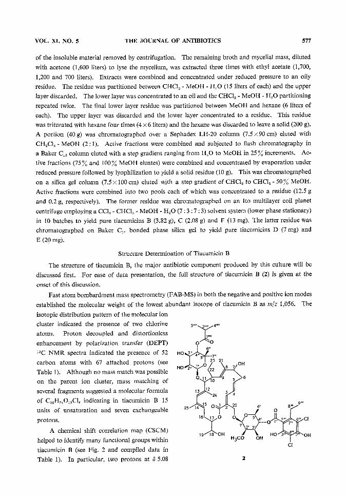
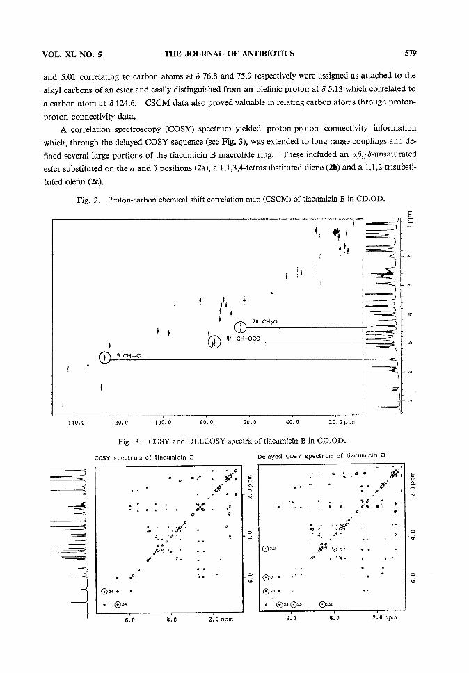
and 5.01 correlating to carbon atoms at 3 76.8 and 75.9 respectively were assigned as attached to thealkyl carbons of an ester and easily distinguished from an olefinic proton at 3 5.13 which correlated toa carbon atom at 3 124.6. CSCMdata also proved valuable in relating carbon atoms through proton-
proton connectivity data.A correlation spectroscopy (COSY)spectrum yielded proton-proton connectivity information
which, through the delayed COSYsequence (see Fig. 3), was extended to long range couplings and de-fined several large portions of the tiacumicin B macrolide ring. These included an ^,^^-unsaturatedester substituted on the a and 3 positions (2a), a 1,1,3,4-tetrasubstituted diene (2b) and a 1,1,2-trisubsti-tuted olefin (2c).
Fig. 2. Proton-carbon chemical shift correlation map (CSCM)of tiacumicin B in CD3OD.
Fig. 3. COSYand DELCOSYspectra of tiacumicin B in CD3OD.

580 THE JOURNAL OF ANTIBIOTICS MAY 1987
A 13C NMRspectrum of tiacumicin B contained signals attributable to three ester carbonyl car-bons at 5 178.4, 170.1 and 169.1. Intense bands in the IR spectrum at 1735, 1695 and 1665 cm"1 sup-ported the presence of three ester functionalities associated with varying degrees of unsaturation.A heteronuclear COSYexperiment (see Fig. 4) was employed to define coupling between these ester
Fig. 4. Heteronuclear COSYspectrum of tiacumicin B in CD3OD.

VOL. XL NO. 5 THE JOURNAL OF ANTIBIOTICS 581
carbonyls and protonated portions of the struc-ture. The carbon signal at d 178.4 was coupledto the protons of two methyl groups defined by
decoupling experiments as part of an isolated
isopropyl group. A positive ion mode FAB-MSfragment of m\z 231 and a negative ion mode
fragment at m/z 247 together with COSYinformation suggested an isobutyrate of a 7 carbon sugar(2d).
Mass spectral fragments of m/z 409 in the negative ion mode and m\z 393 and m/z 249 in thepositive ion mode FAB-MSwere mass matched to C16H19O8C12, C16H19O7C12and C9H7O4C12respec-tively. These fragments were attributed to a 6-deoxy hexose esterified with a dichloro substituted
aromatic acid isomeric with everninic acid (2e). The ester linkage is further supported and the aromaticacid implicated as one having a phenolic hydroxyl group ortho to the carbonyl by the 1665 cm"1 ab-sorption in the IR spectrum. In addition, a coupling of the proton at position 4 of the 6-deoxy sugar
was observed to the carbonyl carbon of <5 170.1 in the heteronuclear COSYspectrum.Quaternary carbon atoms in the 13C NMRspectrum oftiacumicin B at 3 110.7, 156.4, 108.9, 155.8,
115.7 and 141.9 were in good agreement with calculated values2) of 3 112.8, 156.4, 107.9, 162.1, 115.7and 146.5 for carbons 2'" through 1'".
Finally, the heteronuclear COSYspectrum showed a connectivity for the remaining carbonylcarbon signal at 3 7.21 previously defined as part of the a/3, f<5-unsaturated ester system (2c). This
same carbon was also coupled to an isolated CH2Ogroup. Further, heteronuclear couplings fromfully substituted oleflnic carbon atoms and supported by nuclear Overhauser experiments (to be dis-
cussed later) allowed the 3 major fragments of the aglycone to be connected.The positions of the free hydroxyl groups and, by difference the site of glycosidic attachment ofthe sugars was determined by taking advantage of the fact that a carbon atom attached to a hydroxylgroup will resonate at a slightly different chemical shift when the proton is replaced by a deuterium.
13C NMRspectra of tiacumicin B were therefore acquired in CD3ODand CH3OHseparately. Byobserving which signals shift slightly between the two spectra the carbons bearing hydroxyl groupswere distinguished from ether carbons. In this experiment, carbon signals of 3 68.3 (C18) and 72.8(C7) on the macrolide ring were established as hydroxyl bearing carbons and thus carbons ll and 20must be the sites of attachment of the two sugars. Through similar arguments, the methoxyl groupwhose presence was deduced from signals at 3 62.2 and 3.55 in the 13C NMRand XHNMRspectrarespectively was placed at position 2 of the 6-deoxy sugar.
Nuclear Overhauser effect (NOE) experiments showed dipolar coupling between the anomeric
proton at 3 4.70 (carbon 1") and a proton at 3 3.69 (carbon ll) placing the 7 carbon sugar at carbon

582 THE JOURNAL OF ANTIBIOTICS MAY 1987
ll of the macrolide and, by default, the 6-deoxy sugar at carbon 20. In addition, NOEdata alsoestablished the stereochemistry about the olefinic systems of the macrolide ring. Dipolar couplingswere observed between the olefinic protons on carbons 3 and 5, and between the proton on carbon 4and those on 20 establishing a trans-trans diene stereochemistry for this unsaturated ester. Similarly,strong NOE's were observed between protons on carbon atoms 13 and 25, and 24 and 15 establishingan singlet-cw configuration for this diene. The absence of an NOEbetween protons on carbons 9and 21 although negative evidence suggests that the stereochemistry about this olefin is trans.
Structure Determination of Tiacumicins A, C, D, E and FTiacumicins C (3) and F (6) each had the same molecular weight as tiacumicin B. Analysis of
the homonuclear and heteronuclear carbon and proton data (see Tables 2 and 3) in conjunction withmass spectral data indicated that these compounds differed from tiacumicin B only in the position ofbutyrate esterification on the 7 carbon sugar. These changes are best demonstrated in the XHNMRspectra. The chemical shifts of protons on carbon atoms 2", 3" and 4" in the spectrum of tiacumicinB were changed from 3 3.92, 3.71, and 5.01 to 5.34, 3.72, and 3.44 respectively for tiacumicin C andto 4.01, 4.77, and 3.74 respectively for tiacumicin F with only minor changes in the values of theircoupling constants.
Tiacumicin D (4), also isomeric with tiacumicin B, differed from B by the position of esterificationon the 6-deoxy sugar. Protons on carbon atoms 3' and 4' in tiacumicin B had chemical shifts of3 3.73 and 5.08. These occurred at 4.99 and 3.60 respectively in tiacumicin D. Thus the everninicacid isomer on carbon 4' in tiacumicin B is attached to carbon 3' in tiacumicin D.
Tiacumicin E (5) had a molecular weight of m/z 1,042, or 14 mass units lower than that of tiacu-micin B. NMRspectra revealed that the structure of tiacumicin E was identical to that of tiacumicinC except for the ester moiety attached to carbon 2". The isobutyrate in tiacumicin C with protonsignals at 3 2.65 (heptet, 1H, /=7.0 Hz) and 1.18 (d, 3H, /=7.0 Hz) has been replaced in tiacumicinE by a propionate ester moiety as evidenced by two signals at 3 2.46 (q, 2H, J=7.6Hz) and 1.16(t, 3H, /-7.6 Hz).
The molecular formula of tiacumicin A (1) was established as C34H52O9by mass matching of theelectron impact mass spectral ion at m\z 604.3611 (calcd 604.3597). All signals for the aromatic
ester and 6-deoxy sugar were absent from the 13C NMRand XHNMRspectra (see Tables 2 and 3).The esterification pattern in the 7 carbon sugar was the same as that for tiacumicin B, but the ester isan acetate in tiacumicin A. Minorstructural changes were also observed in the macrolide ring. Twosites of oxygenation in tiacumicin B appear to be reduced in the tiacumicin A aglycone. Carbonatoms 18 and 20 whose signals appear at 3 68.3 (CH) and 63.9 (CH2) in tiacumicin B are assigned tosignals at 3 26.1 (CH2) and ll.7 (CH3) in tiacumicin A. Associated changes in the proton signalassignments (Table 1) are consistent with the formula for tiacumicin A.
Experimenta l
General ProceduresOptical rotations were measured in 1 dm tubes on a Perkin Elmer Model 241 polarimeter. Melting
points were recorded on a Hoover Unimelt and are uncorrected. IR spectra were recorded on a PerkinElmer 683 dual beam dispersive instrument and UVspectra on a Perkin Elmer Lambda 3B ultra-violet-visible spectrophotometer. NMRspectra were measured on either a General Electric GN300or GN500spectrometer with 5 mmprobes. The chemical shift correlation maps (CSCM)were
VOL. XL NO. 5 THE JOURNAL OF ANTIBIOTICS 583
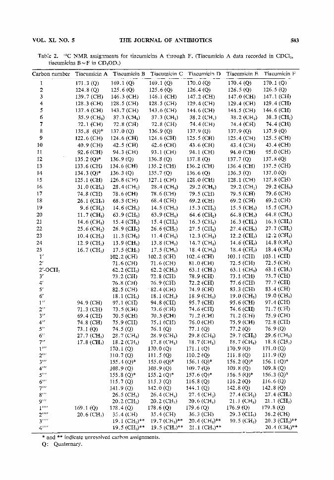
Table 2. 13C NMRassignments for tiacumicins A through F. (Tiacumicin A data recorded in CDC13tiacumicins B~ F in CD3OD.)
Carbon number Tiacumicin A Tiacumicin B Tiacumicin C Tiacumicin D Tiacumicin E Tiacumicin F1
2
3
4
5
6
7
8
9
10
ll
1213
1415
1617
18
1920
2122
23
2425
V2'
2/-OCH3
3'
4'
5'
6'
1"
2"
3"
4"
5"
6"
7"
r//
2///
3/7/
4>"
5r//
6//r
T"%'"
9'"
V"
1""
y,,r
4""
171.3 (Q)124.8 (Q)139.7 (CH)128.3 (CH)137.4 (CH)
35.9 (CH2)
169.1 (Q)
125.6 (Q)146.3 (CH)128.5 (CH)
143.7 (CH)37.3 (CH8)
72.1 (CH)135.8 (Q)*122.6 (CH)
40.9 (CH)92.6 (CH)
135.2 (Q)*133.6 (CH)134.3 (Q)*
72.8 (CH)137.0 (Q)124.6 (CH)42.5 (CH)94.3 (CH)136.9 (Q)134.6 (CH)136.3 (Q)
169.1 (Q)125.6 (Q)
146.1 (CH)170.0 (Q)126.4 (Q)
147.2 (CH)128.5 (CH)
143.6 (CH)37.3 (CH2)
72.8 (CH)136.9 (Q)
124.6 (CH)
42.6 (CH)
129.4 (CH)
144.6 (CH)38.2 (CH2)
74.4 (CH)137.9 (Q)125.5 (CH)
43.6 (CH)93.1 (CH)
136.8 (Q)135.2 (CH)135.7 (Q)
94.1 (CH)137.8 (Q)136.2 (CH)136.6 (Q)
125.1 (CH) 126.8(CH) 127.1 (CH) 128.0(CH)
31.0(CH2) 28.4(CH2) 28.4 (CH2) 29.2 (CH2)74.8 (CH) 78.6 (CH) 78.6 (CH)
26.1 (CH2) 68.3(CH) 68.4(CH)
9.6(CH3) 14.6(CH8) 14.5(CH8)ll.7(CH8) 63.9(CH2)
14.6(CH8) 15.4(CH8)25.6(CH2) 26.9(CH2)
10.4(CH8) ll.3(CH8)
12.9(CH8) 13.9(CH8)16.7 (CH8)
94.9 (CH)71.3 (CH)69.4 (CH)74.8 (CH)73.1 (Q)27.7 (CH8)17.8 (CHS)
169.1 (Q)
17.5 (CH8)
102.2 (CH)71.6 (CH)
62.2 (CH3)73.2 (CH)76.8 (CH)82.5 (CH)18.1 (CH8)97.1 (CH)73.5 (CH)70.5 (CH)75.9 (CH)74.5 (Q)
28.7 (CH8)18.2 (CH8)
170.1 (Q)110.7 (Q)155.4 (Q)*108.9 (Q)155.8 (Q)*115.7 (Q)
141.9 (Q)26.5 (CH2)
20.2 (CH3)
178.4 (Q)20.6(CH3) 35.4(CH)
63,9 (CH2)
15.4 (CH3)26.6 (CH2)
ll.4 (CH3)13.8 (CH3)17.5 (CH3)
102.2 (CH)71.6 (CH)62.2 (CH3)72.8 (CH)76.9 (CH)82.4 (CH)18.1 (CH3)
94.8 (CH)73.6 (CH)70.3 (CH)
75.1 (CH)76.1 (Q)
28.9 (CH3)17.8 (CH8)
170.0 (Q)111.5(Q)155.0 (Q)*108.9 (Q)155.2 (Q)*115.3 (Q)142.0 (Q)
26.4 (CH2)20.2 (CH3)
178.6 (Q)
35.4 (CH)
79.5 (CH)69.2 (CH)
15.3 (CH3)64.6 (CH2)
16.3 (CH3)27.5 (CH2)
12.3 (CH8)
14.7 (CH8)18.4 (CH8)
102.4 (CH)81.0 (CH)63.1 (CH8)78.9 (CH)72.2 (CH)74.9 (CH)18.9 (CH8)95.7 (CH)74.6 (CH)71.2 (CH)
76.0 (CH)77.1 (Q)29.8 (CH8)18.7 (CH8)
171.1 (Q)110.2 (Q)156.1 (Q)*109.7 (Q)157.6 (Q)*116.8 (Q)144.1 (Q)
27.4 (CH2)20.6 (CH8)
179.6 (Q)36.3 (CH)
19.1 (CH3)** 19.7(CH8)** 20.4(CH3)**
19.5(CH3)** 19.5(CH3)** 21.1 (CH3)**
170.4 (Q)126.5 (Q)
147.0 (CH)129.4 (CH)144.5 (CH)
38.2 (CH2)
74.4 (CH)
170.1 (Q)126.5 (Q)
147.1 (CH)129.4 (CH)
144.6 (CH)38.3 (CH2)
74.4 (CH)137.9 (Q)125.4 (CH)
43.4 (CH)94.0 (CH)
137.7 (Q)136.4 (CH)136.3 (Q)128.1 (CH)
29.2 (CH2)79.5 (CH)69.2 (CH)15.5 (CH3)64.8 (CH2)16.3 (CH3)27.4 (CH2)12.2 (CH3)14.6 (CH3)
18.4 (CH3)103.1 (CH)
72.5 (CH)63.1 (CH3)73.1 (CH)77.6 (CH)83.3 (CH)19.0 (CH3)95.6 (CH)74.6 (CH)71.2 (CH)75.9 (CH)77.2 (Q)29.7 (CH3)18.7 (CH8)
170.9 (Q)111.8 (Q)156.2 (Q)*109.8 (Q)156.5 (Q)*116.2 (Q)142.8 (Q)
27.4 (CH2)21.1 (CH3)
176.9 (Q)29.3 (CH2)10.5 (CH3)
137.9 (Q)125.5 (CH)43.4 (CH)95.0 (CH)137.8 (Q)137.5 (CH)137.0 (Q)127.8 (CH)29.2 (CH2)79.6 (CH)69.2 (CH)15.5 (CH3)64.8 (CH2)16.3 (CH3)27.7 (CH2)12.2 (CH3)14.8 (CH3)18.4 (CH3)103.1 (CH)72.5 (CH)63.1 (CH3)73.7 (CH)77.7 (CH)83.4 (CH)19.0 (CH3)97.4 (CH)71.7 (CH)75.9 (CH)72.8 (CH)76.9 (Q)29.6 (CH3)18.8 (CH3)171.0 (Q)111.9(Q)156.1 (Q)*109.8 (Q)156.3 (Q)*116.6 (Q)142.8 (Q)27.4 (CH2)21.1 (CH3)179.8 (Q)36.2 (CH)20.3 (CH3)**
20.4 (CH3)**
* and ** indicate unresolved carbon assignments.Q: Quaternary.
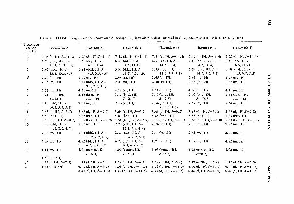
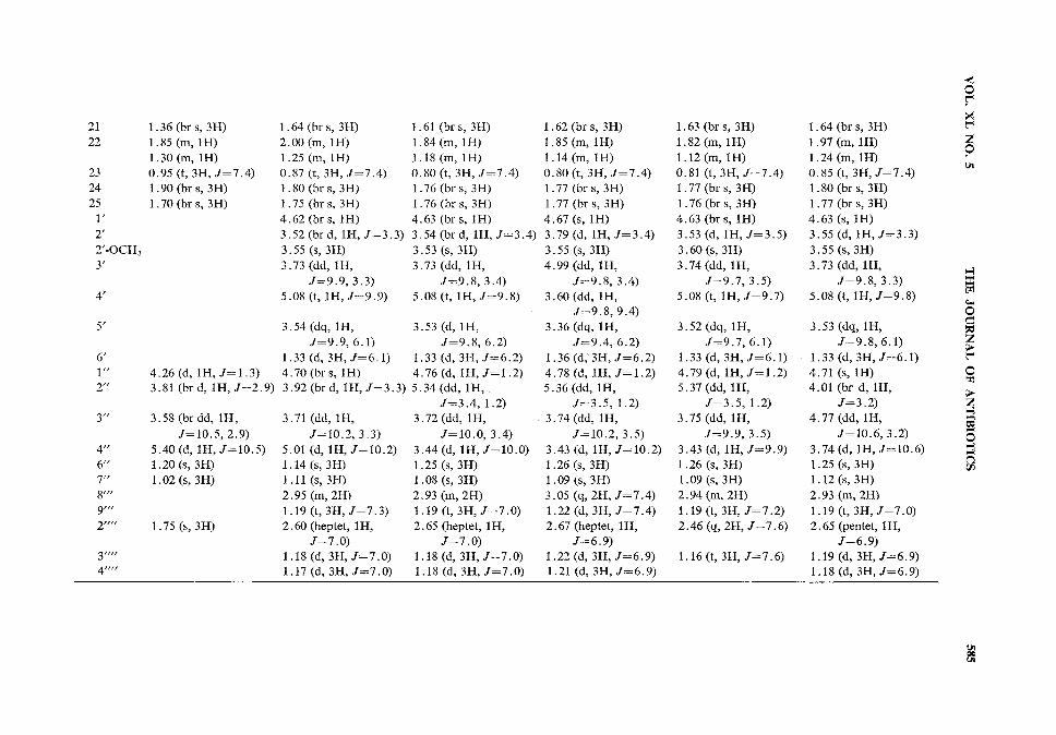
Table 3. XH NMRassignments for tiacumicins A through F. (Tiacumicin A data recorded in C6D6, tiacumicins B~F in CD3OD,/; Hz.)Protons oncarbon Tiacumicin A Tiacumicin B Tiacumicin C Tiacumicin D Tiacumicin E Tiacumicin Fnumber
ll
1315
16
7.25 (d, 1H,7=11.3)
6.25 (ddd, 1H, 7=
15.1,ll.3,1.1)
5.67 (ddd, 1H, 7=15.1, 10.3,4.7)
2.34 (m, 1H)2.15 (m, 1H)
3.97 (m, 1H)
5.21 (brd, 1H,7=10.3)
2.66 (ddd, 1H, 7=10.3, 9.7, 5.7)
3.45 (d, 1H,7=9.7)
5.58 (brs, 1H)
5.53 (brt, 1H,7=8.2)
2.48 (ddd, 1H, 7=ll.1,8.2,4.5)
2.18 (m, lH)
4.99 (m, 1H)
1.85 (m, 1H)
1.58 (m, lH)
0.82 (t, 3H, 7=7.4)1.95 (brs, 3H)
7.21 (d, 1H,/=11.4)
6.58 (dd, 1H, J=
14.5, ll.4)
5.94 (ddd, 1H, 7=
14.5, 9.3,4.9)
2.70 (m, 1H)2.48 (ddd, 1H, 7=
9.3,7.2,3.5)
4.21 (m, lH)
5.13 (brd, 1H,/=10.0)
2.70 (m, 1H)
3.69 (d, 1H,"7=9.7)
5.82(brs, 1H)5.56(brt, 1H,7=7.9)
2.70 (m, 1H)
2.42 (ddd, 1H, J=13.9, 7.9,4.5)
4.72 (ddd, 1H, 7=6.4,4.8,4.5)
4.01 (pentet, 1H,7=6.4).
1.15 (d, 1H, 7=6.4)
4.62 (d, 1H, 7=11.5)
4.42(d, 1H,7=11.5)
7.18 (d, 1H,/=11.4)
6.57 (dd, 1H, /=14.5, ll.4)
5.91 (ddd, 1H, /=14.5, 9.3,4.9)
2.64 (m, 1H)
2.47 (m, 1H)
4.19 (m, 1H)
5.10(brd, 1H,7=10.0)
2.54 (m, lH)
3.64 (d, 1H, 7=9.7)5.83 (brs, 1H)
5.56 (brt, lH,/=7.9)2.72 (ddd, 1H, J=
12.2, 7.9, 4.8)
2.43 (ddd, 1H, 7=12.2, 7.9, 4.4)
4.71 (ddd, 1H, 7=6.4,4.8,4.4)
4.03 (pentet, 1H,7=6.4)
1.18 (d, 3H, 7=6.4)
4.59 (d, lH, 7=11.5)
4.42 (d, lH, /=11.5)
7.21 (d, 1H, J=11A)
6.57 (dd, 1H, /=
14.5, ll.4)
5.93 (ddd, 1H, /=
14.5,9.9, 5.1)
2.66 (m, 1H)
2.46 (m, lH)
4.21 (m, 1H)5.10(brd, 1H,
/-9.8)
2.54 (qd, 1H,.7=9.8, 2.1)
3.66(d, 1H, /=9.8)5.85 (brs, 1H)
5.58 (brt, 1H,/=8.1)2.74 (m, 1H)
2.44 (m, lH)
4.21 (m, 1H)
4.03 (pentet, 1H,J=6A)
7.19 (d, 1H, /-ll.4)
6.58 (dd, 1H, J=14.5, ll.4)
5.92 (ddd, 1H, /=
14.5, 9.7, 5.3)2.67 (m, 1H)
2.45 (m, 1H)
4.20 (m, lH)5.10 (brd, 1H,
/=10.6)
2.57 (m, lH)
3.67 (d, 1H, /=9.8)5.85 (brs, 1H)
5.58 (brt, 1H,/=8.O)2.73 (m, 1H)
2.45 (m, 1H)
4.72 (m, lH)
4.03 (pentet, 1H,/=6.3)
1.18 (d, 3H, /=6.4) 1.17 (d, 3H, /=7.4)
7.20 (d, lH, /==l1.4)6.58 (dd, 1H,J=
14.5, ll.4)
5.94 (ddd, 1H, /=14.5,9.8, 5.2)
2.67 (m, 1H)
2.48 (m, 1H)
4.21 (m, 1H)
5.12(brd, 1H,/=10.6)
2.69 (m, lH)
3.69 (d, lH,/-9.8)5.85 (brs, 1H)
5.58 (brt, 1H,/-8.1)2.72 (m, 1H)
2.43 (m, 1H)
4.72 (m, lH)
4.02 (m, 1H)
1.17 (d, 3H, /=7.0)4.59(d,1H,/==11.5) 4.60(d,1H,/=11.5) 4.61 (d,1H,/=11.5)4.43(d,1H,/=11.5) 4.42(d,1H,/=11.5) 4.42(d,1H,/=11.5)
o
>
o
i
o
3
21 1.36 (brs, 3H)22 1.85 (m, lH)
1.30(m, 1H)
23 0.95 (t, 3H, /=7.4)24 1.90 (brs, 3H)
25 1.70 (brs, 3H)V
2'
2/-OCH3
3'
4.26(d, 1H,/=1.3)
3.81 (brd, 1H,/=2.9)
3 "3.58 (brdd, 1H,
4"
6"
7"
9'"
2""
3""
4-
/=10.5, 2.9)5.40(d, 1H, /=10.5)
1.20 (s, 3H)
1.02 (s, 3H)
1.75 (s, 3H)
1.64(brs, 3H) 1.61 (brs, 3H)
2.00 (m, 1H) 1.84 (m, 1H)
1.25 (m, 1H) 1.18 (m, 1H)
0.87 (t, 3H, /=7.4) 0.80 (t, 3H, J=lA)
1.80 (brs, 3H) 1.76 (brs, 3H)
1.75 (brs, 3H) 1.76 (brs, 3H)
4.62 (brs, 1H) 4.63 (brs, 1H)
3.52(brd, lH,/=3.3) 3.54(brd, 1H,/=3.4)
3.55 (s, 3H) 3.53 (s, 3H)
3.73 (dd, 1H, 3.73 (dd, 1H,
/=9.9, 3.3) /=9.8, 3.4)
5.08 (t, 1H,/=9.9) 5.08 (t, 1H,/=9.8)
3.54 (dq, 1H, 3.53 (d, lH,
/=9.9, 6.1) /=9.8, 6.2)
1.33 (d, 3H,/=6.1) 1.33 (d, 3H,/=6.2)
4.70 (brs, 1H) 4.76 (d, 1H, /=1.2)
3.92(brd, lH,/=3.3) 5.34(dd, 1H,
/=3.4,1.2)
3.71 (dd, 1H, 3.72(dd, 1H,
/=10.2, 3.3)
5.01 (d, 1H,/=1O.2)1.14(s, 3H)1.ll (s,3H)
2.95 (m, 2H)
1.19 (t, 3H,/=7.3)
2.60 (heptet, 1H,/=7.0)
1.18 (d, 3H, /=7.0)
1.17 (d, 3H, J=1.Q)
/=10.0, 3.4)
3.44 (d, 1H, /=10.0)
1.25 (s, 3H)
1.08 (s, 3H)
2.93 (m, 2H)
1.19 (t, 3H,/=7.0)
2.65 (heptet, 1H,/=7.0)
1.18 (d, 3H,/=7.0)
1.18 (d, 3H,/=7.0)
1.62 (brs, 3H)
1.85 (m, 1H)1.14(m, 1H)
0.80 (t, 3H, /=7.4)1.77 (brs, 3H)
1.77 (brs, 3H)
4.67 (s, 1H)3.79 (d, 1H,/=3.4)
3.55 (s, 3H)
4.99 (dd, 1H,
1.63 (brs, 3H)1.82(m, 1H)
1.12(m, 1H)
0.81 (t, 3H, /=7.4)1.77 (brs, 3H)
1.76 (brs, 3H)4.63 (brs, 1H)
3.53 (d, 1H,/=3.5)
3.60 (s, 3H)3.74 (dd, 1H,
J =S ,3.4)
/=9.7, 3.5)
3.60 (dd, 1H,
/=9.8, 9.4)
3.36 (dq, 1H,
/=9.4, 6.2)
1.36 (d, 3H, J=6.2)
4.78 (d, 1H, /=1.2)
5.36 (dd, 1H,
/=3.5, 1.2)
3.74 (dd, 1H,
/-10.2, 3.5)
3.43 (d, 1H, /=10.2)
1.26 (s, 3H)
1.09 (s, 3H)
3.05 (q, 2H, /=7.4)
1.22 (d, 3H, /-7.4)
2.67 (heptet, 1H,/=6.9)
1.22 (d, 3H, /=6.9)
1.21 (d, 3H,/-6.9)
5.08 (t, 1H, J=9.1)
3.52 (dq, 1H,
/=9.7, 6.1)
1.33 (d, 3H,J=6A)
4.79 (d, 1H,/=1.2)
5.37 (dd, 1H,
/=3.5, 1.2)
3.75 (dd, 1H,
/=9.9, 3.5)
3.43 (d, lH, /=9.9)
1.26 (s, 3H)
1.09 (s, 3H)
2.94 (m, 2H)
1.19 (ti 3H, /=7.2)
2.46 (q, 2H, /=7.6)
1.16 (t, 3H, J=1.6)
1.64 (brs, 3H)1.97 (m, 1H)
1.24 (m, 1H)
0.85 (t, 3H, /=7.4)1.80(brs, 3H)
1.77 (brs, 3H)4.63 (s, 1H)
3.55 (d, lH,/=3.3)
3.55 (s, 3H)3.73 (dd, 1H,
/=9.8, 3.3)
5.08 (t, lH,/=9.8)
3.53 (dq, 1H,7=9.8,6.1)
1.33 (d, 3H,/=6.1)4.71 (s, 1H)
4.01 (br d, 1H,/=3.2)
4.77 (dd, 1H,
/=10.6, 3.2)3.74 (d, 1H, /-10.6)
1.25 (s, 3H)1.12(s, 3H)
2.93 (m, 2H)
1.19 (t, 3H,/=7.0)2.65 (pentet, 1H,
.7=6.9)1.19 (d, 3H,/=6.9)
1.18 (d, 3H,/=6.9)
i
>
i
§
n
586 THE JOURNAL OF ANTIBIOTICS MAY 1987
acquired by the method of Bax and Morris3) and the DEPT(distortionless enhancement of NMRsignalsby polarization transfer) by that of Doddrell et alS* Delayed correlation spectroscopy studies(DELCOSY)were carried out by the method of Bax and Freeman.5) NOEexperiments were ac-quired by the CAMELtechnique of Bothner-By et al.6) Heteronuclear correlation spectroscopy
VOL. XL NO. 5 THE JOURNAL OF ANTIBIOTICS 587
experiments (FUCOUP)were performed employing a modification of the method of Bodenhausenand Freeman.70 Mass spectra were measured on a Kratos MS-50spectrometer.
Tiacumicin A: |>]2D5 +41° (c 0.10, MeOH) was a clear oil. A molecular weight of m/z 604.3611was established by electron impact mass spectroscopy. An UVspectrum in MeOHcontained bandsat ;max nm (e) 203 (6,785), 235 (6,410), 266 (4,960). These bands were unchanged upon addition of acidor base. An IR spectrum measured neat contained bands at: 3480, 2930, 1735, 1700, 1460, 1370,1240, 1150, 1115, 1080, 1040cm"1. See tables for *H and 13C NMRdata.
Tiacumicin B: |>]2D5 -6.5° (c 8.2, MeOH) had a melting point of 143~145°C. A molecularweight of m/z 1,056 was established by FABpositive and negative ion mass spectrometry. An UVspectrum in MeOH contained bands ^max nm (s) 206 (13,940), 229 (17,430), 266 (9,300), 314 (2,965), inMeOH - HC1 at ^max nm (e) 206 (14,815), 229 (17,778), 266 (9,630), 314 (870) and in MeOH -NaOHat ;max nm (e) 204 (32,500), 232 (17,110), 235 (sh), 255 (sh), 270 (sh), 314 (8,220) (see Fig. 4). AnIRspectrum measured in KBr contained bands at: 3565, 3502, 2978, 2935, 2880, 1735, 1695, 1665, 1590,1465, 1455, 1410, 1402, 1380, 1370, 1320, 1310, 1240, 1215, 1195, 1140, 1110, 1065, 1020cm"1. See
tables for *H and 13C NMRdata.Tiacumicin C: [a]2D5 -8.6° (c 15.8, MeOH) had a melting point of 142~143°C. A molecular
weight of m/z 1,056 was established by FAB positive ion mass spectrometry. An UV spectrum inMeOH contained bands at ^max nm (e) 206 (25,096), 228 (29,630), 267 (15,410), 315 (6,300), in MeOH -HC1 at >*max nm (e) 206 (25,740), 228 (29,630), 267 (15,555), 315 (1,222), and in MeOH - NaOH at ^maxnm (e) 203 (29,815), 234 (26,500), 240 (sh), 255 (sh), 276 (sh), 315 (ll,555). An IR spectrum measuredin KBr contained bands at: 3665, 2990, 2945, 2880, 1735, 1695, 1650, 1595, 1470, 1455, 1415, 1405,1390, 1370, 1310, 1250, 1230, 1200, 1165, 1145, 1115, 1090, 1070, 1025cm"1. See tables for XH and
13CNMRdata.Tiacumicin D: [a]2D5 -5.6° (c 0.47, MeOH) had a melting point of 141~145°C. A molecularweight of m/z 1,056 was established by FABpositive ion mass spectrometry. An UVspectrum inMeOH contained bands at ^max nm (s) 205 (sh), 229 (15,957), 266 (8,342),3 14 (4,464), in MeOH - HC1at ^max nm (e) 205 (sh), 229 (17,200), 266 (9,440), 314 (1,050), and in MeOH - NaOH at ^max nm (e) 203(sh), 233 (13,878), 242 (sh), 269 (sh), 314 (5,456) (see Fig. 10). An IR spectrum measured in KBrcontained bands at: 3436, 2974, 2933, 2874, 1716, 1700, 1640, 1590, 1508, 1456, 1383, 1308, 1251, 1210,1071 cm"1. See tables for *H and 13C NMRdata.
Tiacumicin E: [a]2D5 -3.2° (c 1.3, MeOH) had a melting point of 138~ 141°C. A molecular weightof m\z 1,042 was established by FAB positive ion mass spectrometry. An UV spectrum in MeOHcontained bands at ^max nm (e) 205 (sh), 228 (39,444), 266 (24,853), 315 (10,170), in MeOH - HC1 at^max nm (e) 205 (40,700), 228 (48,470), 266 (25,730), 315 (1,795), and in MeOH - NaOH at ;max nm (e)206 (sh), 234 (43,680), 240 (sh), 255 (sh), 270 (sh), 315 (16,750). An IR spectrum measured in KBrcontained bands at: 3444, 2976, 2935, 2876, 1712, 1700, 1640, 1590, 1456, 1380, 1314, 1247, 1209,
1200, 1087, 1068, 1026 cm"1. See tables for XH and 13CNMRdata.Tiacumicin F: [a]2D5 +5.8° (c 0.66, MeOH) had a melting point of 141~143°C. A molecular
weight of m/z 1,056 was established by FAB positive ion mass spectrometry. An UV spectrum inMeOH contained bands at ^max nm (e) 204 (16,245), 228 (20,675), 266 (10,127), 315 (4,747), in MeOH -HC1 at ^max nm (e) 204 (17,510), 228 (21,097), 266 (ll,392), 315 (1,055) and in MeOH - NaOHat^maxnm (e) 202 (sh), 234 (19,304), 240 (sh), 255 (sh), 315 (7,226). An IR spectrum measured in CDC13 con-tained bands at: 3479, 2975, 2934, 2875, 1709, 1696, 1641, 1588, 1515, 1456, 1381, 1314, 1249, 1200,1144, 1067, 1022, 908 cm"1. See tables for XH and 13C NMRdata.
Addendum in Proof
The tiacumicins from Dactylosporangium aurantiacum appear to have membersin commonwithantibiotics from two other acinomycetes. The clostomicins from Micromonosporaechinospora subsp.armeniaca were discovered at the Kitasato Institute. Their report,8) published at the same time aswe presented this work at the 26th ICAACMeeting,9>10) describes the production, isolation, and spec-tral characteristics of five membersof the clostomicin complex. The published spectra would indicatethat clostomicin A is identical to tiacumicin F and clostomicin B2 to tiacumicin C. Clostomicins C
588 THE JOURNAL OF ANTIBIOTICS MAY 1987
and D have no known counterpart nor do tiacumicins A, D and E. Clostomicin Bx and tiacumicin Bare identical to the Actinoplanes deccanensis produced antibiotic, lipiarmycin, reported earlier byLepetit scientists.n~15)
References
1) Theriault, R. J.; J. P. Karwowski, M. Jackson, R. L. Girolami, G. N. Sunga, C. M. Vojtko & L.J. Coen: Tiacumicins, a novel complex of 18-membered macrolide antibiotics. I. Taxonomy, fermenta-tion and antibacterial activity. J. Antibiotics 40: 567- 574, 1987
2) Wehrli, F. W. & T. Wirthlin: Interpretation of Carbon-13 NMRspectra. Heyden, London, 19783) Bax, A. & G. A. Morris: An improved method for heteronuclear chemical shift correlation by two-
dimensional NMR. J. Mag. Res. 42: 501-505, 19814) Doddrell, D. M.; D. T. Pegg & M. R. Bendall: Distortionless enhancement of NMRsignals by
polarization transfer. J. Mag. Res. 48: 323-327, 19825) Bax, A. & R. Freeman: Investigation of complex networks of spin-spin coupling by two-dimensional
NMR. J. Mag. Res. 44: 542-561, 1981
6) Bothner-By, A. A.; R. L. Stephens & J. Lee: Structure determination of a tetrasaccharide: Transientnuclear Overhauser effects in the rotating frame. J. Am. Chem. Soc. 106: 811 - 813, 1984
7) Bodenhausen, G. & R. Freeman: Correlation of proton and carbon-13 NMRspectra by heteronucleartwo-dimensional spectroscopy. J. Mag. Res. 28: 471 -476, 1977
8) Omura, S.; N. Imamura, R. Oiwa, H. Kuga, R. Iwata, R. Masuma & Y. Iwai: Clostomicins, newantibiotics produced by Micromonospora echinospora subsp. armeniaca subsp. nov. I. Production, isola-tion, and physico-chemical and biological properties. J. Antibiotics 39: 1407- 1412, 1986
9) Theriault, R.J.; J.P. Karwowski, M. Jackson, R.L. Girolami, G.N. Sunga & C M. Vojtko:Tiacumicins, a novel series of 18-membered macrolide antibiotics. I. Discovery, taxonomy, fermentationand antibacterial activity. Program and Abstracts of the 26th Intersci. Conf. on Antimicrob. AgentsChemother., No. 936, p. 268, New Orleans, Sept. 28-Oct. 1, 1986
10) Hochlowski, J. E.; S. J. Swanson, D. N. Whittern, A. M. Buko & J. B. McAlpine: Tiacumicins, anovel series of 18-membered macrolide antibiotics. II. Isolation and elucidation of structures. Programand Abstracts of the 26th Intersci. Conf. on Antimicrob. Agents Chemother., No. 937, p. 268, New Orleans,
Sept. 28-Oct. 1, 1986
ll) Coronelli, C. ; F. Parenti, R. White & H. Pagani (Gruppo Lepetit S.p.A.) : Lipiarmycin and its prepara-tion. U.S. 3,978,211, Aug. 31, 1976
12) Parenti, F.; H. Pagani & G. Beretta: Lipiarmycin, a new antibiotic from Actinoplanes. I. Descriptionof the producer strain and fermentation studies. J. Antibiotics 28 : 247-252, 1975
13) Coronelli, C; R. J. White, G. C. Lancini & F. Parenti: Lipiarmycin, a new antibiotic from Actino-planes. II. Isolation, chemical, biological and biochemical characterization. J. Antibiotics 28 : 253-259,
1975
14) Sergio, S.; G. Pirali, R. White & F. Parenti: Lipiarmycin, a new antibiotic from Actinoplanes. III.Mechanism of action. J. Antibiotics 28: 543-549, 1975
15) Martinelli, E.; L. Faniuolo, G. Tuan, G. G. Gallo & B. Cavalleri: Structural studies on lipiar-mycin. I. Characterization by XH and 13C NMRspectroscopy and isolation of methyl 2-0-methyl-4-<9-homodichloroorsellinate-/3-rhamnoside. J. Antibiotics 36: 1312- 1322, 1983