Embed Size (px)
Citation preview
12.6 Signaling in Microorganisms and PlantsMuch of what we have said here about signaling relatesto mammalian tissues or cultured cells from such tis-sues. Bacteria, eukaryotic microorganisms, and vascu-lar plants must also respond to a variety of external sig-nals, such as O2, nutrients, light, noxious chemicals, andso on. We turn here to a brief consideration of the kindsof signaling machinery used by microorganisms andplants.
Bacterial Signaling Entails Phosphorylation in a Two-Component System
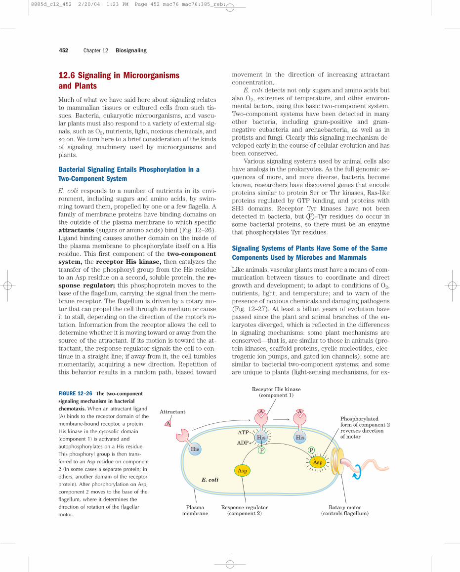
E. coli responds to a number of nutrients in its envi-ronment, including sugars and amino acids, by swim-ming toward them, propelled by one or a few flagella. Afamily of membrane proteins have binding domains onthe outside of the plasma membrane to which specificattractants (sugars or amino acids) bind (Fig. 12–26).Ligand binding causes another domain on the inside ofthe plasma membrane to phosphorylate itself on a Hisresidue. This first component of the two-component
system, the receptor His kinase, then catalyzes thetransfer of the phosphoryl group from the His residueto an Asp residue on a second, soluble protein, the re-
sponse regulator; this phosphoprotein moves to thebase of the flagellum, carrying the signal from the mem-brane receptor. The flagellum is driven by a rotary mo-tor that can propel the cell through its medium or causeit to stall, depending on the direction of the motor’s ro-tation. Information from the receptor allows the cell todetermine whether it is moving toward or away from thesource of the attractant. If its motion is toward the at-tractant, the response regulator signals the cell to con-tinue in a straight line; if away from it, the cell tumblesmomentarily, acquiring a new direction. Repetition ofthis behavior results in a random path, biased toward
movement in the direction of increasing attractantconcentration.
E. coli detects not only sugars and amino acids butalso O2, extremes of temperature, and other environ-mental factors, using this basic two-component system.Two-component systems have been detected in manyother bacteria, including gram-positive and gram-negative eubacteria and archaebacteria, as well as inprotists and fungi. Clearly this signaling mechanism de-veloped early in the course of cellular evolution and hasbeen conserved.
Various signaling systems used by animal cells alsohave analogs in the prokaryotes. As the full genomic se-quences of more, and more diverse, bacteria becomeknown, researchers have discovered genes that encodeproteins similar to protein Ser or Thr kinases, Ras-likeproteins regulated by GTP binding, and proteins withSH3 domains. Receptor Tyr kinases have not beendetected in bacteria, but P –Tyr residues do occur insome bacterial proteins, so there must be an enzymethat phosphorylates Tyr residues.
Signaling Systems of Plants Have Some of the SameComponents Used by Microbes and Mammals
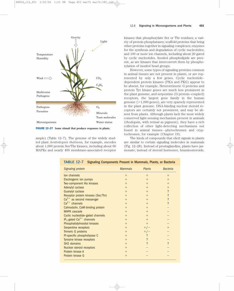
Like animals, vascular plants must have a means of com-munication between tissues to coordinate and directgrowth and development; to adapt to conditions of O2,nutrients, light, and temperature; and to warn of thepresence of noxious chemicals and damaging pathogens(Fig. 12–27). At least a billion years of evolution havepassed since the plant and animal branches of the eu-karyotes diverged, which is reflected in the differencesin signaling mechanisms: some plant mechanisms areconserved—that is, are similar to those in animals (pro-tein kinases, scaffold proteins, cyclic nucleotides, elec-trogenic ion pumps, and gated ion channels); some aresimilar to bacterial two-component systems; and someare unique to plants (light-sensing mechanisms, for ex-
Chapter 12 Biosignaling452
AAttractant
Receptor His kinase(component 1)
A
His
A
His
His
ATP
ADP
E. coli
Response regulator(component 2)
Plasmamembrane
Rotary motor(controls flagellum)
Phosphorylatedform of component 2reverses directionof motor
Asp
Asp
PP
FIGURE 12–26 The two-componentsignaling mechanism in bacterialchemotaxis. When an attractant ligand(A) binds to the receptor domain of themembrane-bound receptor, a proteinHis kinase in the cytosolic domain(component 1) is activated andautophosphorylates on a His residue.This phosphoryl group is then trans-ferred to an Asp residue on component2 (in some cases a separate protein; inothers, another domain of the receptorprotein). After phosphorylation on Asp,component 2 moves to the base of theflagellum, where it determines thedirection of rotation of the flagellarmotor.
8885d_c12_452 2/20/04 1:23 PM Page 452 mac76 mac76:385_reb:
ample) (Table 12–7). The genome of the widely stud-ied plant Arabidopsis thaliana, for example, encodesabout 1,000 protein Ser/Thr kinases, including about 60MAPKs and nearly 400 membrane-associated receptor
kinases that phosphorylate Ser or Thr residues; a vari-ety of protein phosphatases; scaffold proteins that bringother proteins together in signaling complexes; enzymesfor the synthesis and degradation of cyclic nucleotides;and 100 or more ion channels, including about 20 gatedby cyclic nucleotides. Inositol phospholipids are pres-ent, as are kinases that interconvert them by phospho-rylation of inositol head groups.
However, some types of signaling proteins commonin animal tissues are not present in plants, or are rep-resented by only a few genes. Cyclic nucleotide–dependent protein kinases (PKA and PKG) appear tobe absent, for example. Heterotrimeric G proteins andprotein Tyr kinase genes are much less prominent inthe plant genome, and serpentine (G protein–coupled)receptors, the largest gene family in the humangenome (�1,000 genes), are very sparsely representedin the plant genome. DNA-binding nuclear steroid re-ceptors are certainly not prominent, and may be ab-sent from plants. Although plants lack the most widelyconserved light-sensing mechanism present in animals(rhodopsin, with retinal as pigment), they have a richcollection of other light-detecting mechanisms notfound in animal tissues—phytochromes and cryp-tochromes, for example (Chapter 19).
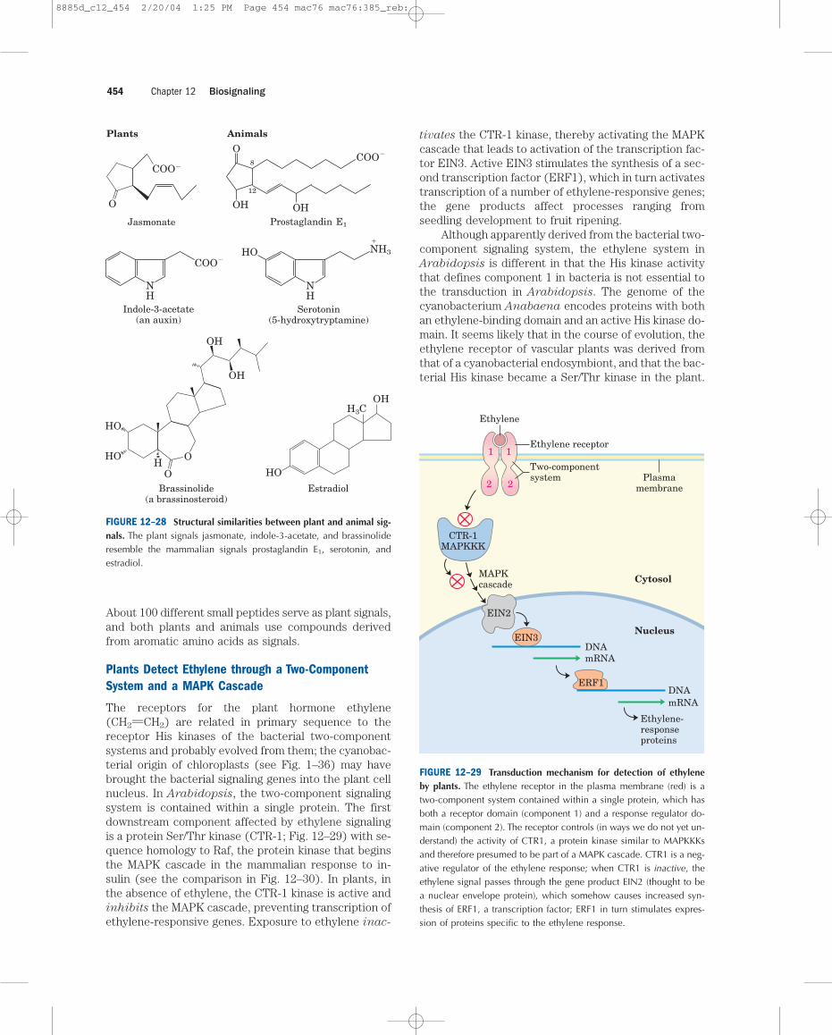
The kinds of compounds that elicit signals in plantsare similar to certain signaling molecules in mammals(Fig. 12–28). Instead of prostaglandins, plants have jas-monate; instead of steroid hormones, brassinosteroids.
12.6 Signaling in Microorganisms and Plants 453
GravityLight
HumidityTemperature
Wind
HerbivoresPathogens
Pathogens
Parasites
Microorganisms
O2
Minerals
Toxic molecules
Water status
CO2
C2H4
FIGURE 12–27 Some stimuli that produce responses in plants.
TABLE 12–7 Signaling Components Present in Mammals, Plants, or Bacteria
Signaling protein Mammals Plants Bacteria
Ion channels � � �Electrogenic ion pumps � � �Two-component His kinases � � �Adenylyl cyclase � � �Guanylyl cyclase � � ?Receptor protein kinases (Ser/Thr) � � ?Ca2� as second messenger � � ?Ca2� channels � � ?Calmodulin, CaM-binding protein � � �MAPK cascade � � �Cyclic nucleotide–gated channels � � �IP3-gated Ca2� channels � � �Phosphatidylinositol kinases � � �Serpentine receptors � �/� �Trimeric G proteins � �/� �PI-specific phospholipase C � ? �Tyrosine kinase receptors � ? �SH2 domains � ? ?Nuclear steroid receptors � � �Protein kinase A � � �Protein kinase G � � �
8885d_c12_453 2/20/04 1:23 PM Page 453 mac76 mac76:385_reb:
About 100 different small peptides serve as plant signals,and both plants and animals use compounds derivedfrom aromatic amino acids as signals.
Plants Detect Ethylene through a Two-ComponentSystem and a MAPK Cascade
The receptors for the plant hormone ethylene(CH2UCH2) are related in primary sequence to thereceptor His kinases of the bacterial two-componentsystems and probably evolved from them; the cyanobac-terial origin of chloroplasts (see Fig. 1–36) may havebrought the bacterial signaling genes into the plant cellnucleus. In Arabidopsis, the two-component signalingsystem is contained within a single protein. The firstdownstream component affected by ethylene signalingis a protein Ser/Thr kinase (CTR-1; Fig. 12–29) with se-quence homology to Raf, the protein kinase that beginsthe MAPK cascade in the mammalian response to in-sulin (see the comparison in Fig. 12–30). In plants, inthe absence of ethylene, the CTR-1 kinase is active andinhibits the MAPK cascade, preventing transcription ofethylene-responsive genes. Exposure to ethylene inac-
tivates the CTR-1 kinase, thereby activating the MAPKcascade that leads to activation of the transcription fac-tor EIN3. Active EIN3 stimulates the synthesis of a sec-ond transcription factor (ERF1), which in turn activatestranscription of a number of ethylene-responsive genes;the gene products affect processes ranging fromseedling development to fruit ripening.
Although apparently derived from the bacterial two-component signaling system, the ethylene system inArabidopsis is different in that the His kinase activitythat defines component 1 in bacteria is not essential tothe transduction in Arabidopsis. The genome of thecyanobacterium Anabaena encodes proteins with bothan ethylene-binding domain and an active His kinase do-main. It seems likely that in the course of evolution, theethylene receptor of vascular plants was derived fromthat of a cyanobacterial endosymbiont, and that the bac-terial His kinase became a Ser/Thr kinase in the plant.
Chapter 12 Biosignaling454
O
COO�
Jasmonate
Plants Animals
OCOO�
COO�
Prostaglandin E1
OH HO
8
12
OH
Estradiol
H3
HO
C
Brassinolide(a brassinosteroid)
HOH
O
O
HO
OH
OH
Serotonin(5-hydroxytryptamine)
HO
NH
Indole-3-acetate(an auxin)
NH
�
NH3
Cytosol
Nucleus
MAPKcascade
DNAmRNA
DNAmRNA
Ethylene-responseproteins
Ethylene
Ethylene receptor
Two-componentsystem Plasma
membrane
CTR-1MAPKKK
EIN2
EIN3
1 1
2 2
ERF1
FIGURE 12–28 Structural similarities between plant and animal sig-nals. The plant signals jasmonate, indole-3-acetate, and brassinolideresemble the mammalian signals prostaglandin E1, serotonin, andestradiol.
FIGURE 12–29 Transduction mechanism for detection of ethyleneby plants. The ethylene receptor in the plasma membrane (red) is atwo-component system contained within a single protein, which hasboth a receptor domain (component 1) and a response regulator do-main (component 2). The receptor controls (in ways we do not yet un-derstand) the activity of CTR1, a protein kinase similar to MAPKKKsand therefore presumed to be part of a MAPK cascade. CTR1 is a neg-ative regulator of the ethylene response; when CTR1 is inactive, theethylene signal passes through the gene product EIN2 (thought to bea nuclear envelope protein), which somehow causes increased syn-thesis of ERF1, a transcription factor; ERF1 in turn stimulates expres-sion of proteins specific to the ethylene response.
8885d_c12_454 2/20/04 1:25 PM Page 454 mac76 mac76:385_reb:
Receptorlike Protein Kinases Transduce Signals fromPeptides and Brassinosteroids
One common motif in plant signaling involves recep-
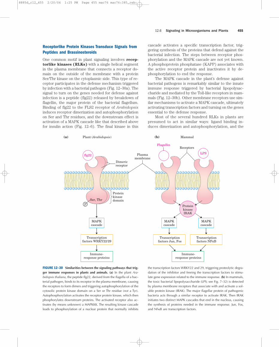
torlike kinases (RLKs) with a single helical segmentin the plasma membrane that connects a receptor do-main on the outside of the membrane with a proteinSer/Thr kinase on the cytoplasmic side. This type of re-ceptor participates in the defense mechanism triggeredby infection with a bacterial pathogen (Fig. 12–30a). Thesignal to turn on the genes needed for defense againstinfection is a peptide (flg22) released by breakdown offlagellin, the major protein of the bacterial flagellum.Binding of flg22 to the FLS2 receptor of Arabidopsis
induces receptor dimerization and autophosphorylationon Ser and Thr residues, and the downstream effect isactivation of a MAPK cascade like that described abovefor insulin action (Fig. 12–6). The final kinase in this
cascade activates a specific transcription factor, trig-gering synthesis of the proteins that defend against thebacterial infection. The steps between receptor phos-phorylation and the MAPK cascade are not yet known.A phosphoprotein phosphatase (KAPP) associates withthe active receptor protein and inactivates it by de-phosphorylation to end the response.
The MAPK cascade in the plant’s defense againstbacterial pathogens is remarkably similar to the innateimmune response triggered by bacterial lipopolysac-charide and mediated by the Toll-like receptors in mam-mals (Fig. 12–30b). Other membrane receptors use sim-ilar mechanisms to activate a MAPK cascade, ultimatelyactivating transcription factors and turning on the genesessential to the defense response.
Most of the several hundred RLKs in plants are presumed to act in similar ways: ligand binding in-duces dimerization and autophosphorylation, and the
12.6 Signaling in Microorganisms and Plants 455
(a) (b)Plant (Arabidopsis) Mammal
Dimericreceptor
flg22
Proteinkinasedomain
Transcriptionfactors WRKY22/29
Immune-response proteins
Transcriptionfactors Jun, Fos
Transcriptionfactors NFkB
Immune-response proteins
MAPKcascade
ProteinkinaseIRAK
LPS
FlagellinReceptors
Plasmamembrane
Ser, Thr
MAPKcascade
MAPKcascade
P
FIGURE 12–30 Similarities between the signaling pathways that trig-ger immune responses in plants and animals. (a) In the plant Ara-bidopsis thaliana, the peptide flg22, derived from the flagella of a bac-terial pathogen, binds to its receptor in the plasma membrane, causingthe receptors to form dimers and triggering autophosphorylation of thecytosolic protein kinase domain on a Ser or Thr residue (not a Tyr).Autophosphorylation activates the receptor protein kinase, which thenphosphorylates downstream proteins. The activated receptor also ac-tivates (by means unknown) a MAPKKK. The resulting kinase cascadeleads to phosphorylation of a nuclear protein that normally inhibits
the transcription factors WRKY22 and 29, triggering proteolytic degra-dation of the inhibitor and freeing the transcription factors to stimu-late gene expression related to the immune response. (b) In mammals,the toxic bacterial lipopolysaccharide (LPS; see Fig. 7–32) is detectedby plasma membrane receptors that associate with and activate a sol-uble protein kinase (IRAK). The major flagellar protein of pathogenicbacteria acts through a similar receptor to activate IRAK. Then IRAKinitiates two distinct MAPK cascades that end in the nucleus, causingthe synthesis of proteins needed in the immune response. Jun, Fos,and NF�B are transcription factors.
8885d_c12_455 2/20/04 1:25 PM Page 455 mac76 mac76:385_reb:
activated receptor kinase triggers downstream responsesby phosphorylating key proteins at Ser or Thr residues.The ligands for these kinases have been identified inonly a few cases: brassinosteroids, the peptide triggerfor the self-incompatibility response that prevents self-pollination, and CLV1 peptide, a factor involved in reg-ulating the fate of stem cells (undifferentiated cells) inplant development.
SUMMARY 12.6 Signaling in Microorganisms and Plants
■ Bacteria and unicellular eukaryotes have avariety of sensory systems that allow them tosample and respond to their environment. Inthe two-component system, a receptor Hiskinase senses the signal and autophosphory-lates a His residue, then phosphorylates theresponse regulator on an Asp residue.
■ Plants respond to many environmental stimuli,and employ hormones and growth factors tocoordinate the development and metabolicactivities of their tissues. Plant genomesencode hundreds of signaling proteins,including some very similar to those used insignal transductions in mammalian cells.
■ Two-component signaling mechanisms commonin bacteria have been acquired in altered formsby plants. Cyanobacteria use typical two-component systems in the detection ofchemical signals and light; plants use relatedproteins—which autophosphorylate on Ser/Thr,not His, residues—to detect ethylene.
■ Plant receptorlike kinases (RLKs), with anextracellular ligand-binding domain, a singletransmembrane segment, and a cytosolicprotein kinase domain, participate in detectinga wide variety of stimuli, including peptidesthat originate from pathogens, brassinosteroidhormones, self-incompatible pollen, anddevelopmental signals. RLKs autophosphorylateSer/Thr residues, then activate downstreamproteins that in some cases are MAPKcascades. The end result of many such signalsis increased transcription of specific genes.
12.7 Sensory Transduction in Vision,Olfaction, and GustationThe detection of light, smells, and tastes (vision, olfac-tion, and gustation, respectively) in animals is accom-plished by specialized sensory neurons that use signal-transduction mechanisms fundamentally similar to thosethat detect hormones, neurotransmitters, and growth
factors. An initial sensory signal is amplified greatly bymechanisms that include gated ion channels and intra-cellular second messengers; the system adapts to con-tinued stimulation by changing its sensitivity to thestimulus (desensitization); and sensory input from sev-eral receptors is integrated before the final signal goesto the brain.
Light Hyperpolarizes Rod and Cone Cells of theVertebrate Eye
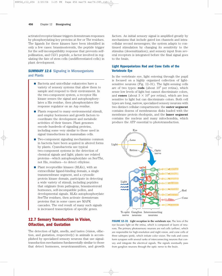
In the vertebrate eye, light entering through the pupilis focused on a highly organized collection of light-sensitive neurons (Fig. 12–31). The light-sensing cellsare of two types: rods (about 109 per retina), whichsense low levels of light but cannot discriminate colors,and cones (about 3 � 106 per retina), which are lesssensitive to light but can discriminate colors. Both celltypes are long, narrow, specialized sensory neurons withtwo distinct cellular compartments: the outer segment
contains dozens of membranous disks loaded with themembrane protein rhodopsin, and the inner segment
contains the nucleus and many mitochondria, whichproduce the ATP essential to phototransduction.
Chapter 12 Biosignaling456
Light
To optic nerve
Ganglionneurons
Interconnectingneurons
Rod
Cone
Light
Lens
Eye
Retina Opticnerve
FIGURE 12–31 Light reception in the vertebrate eye. The lens of theeye focuses light on the retina, which is composed of layers of neu-rons. The primary photosensory neurons are rod cells (yellow), whichare responsible for high-resolution and night vision, and cone cells ofthree subtypes (pink), which initiate color vision. The rods and conesform synapses with several ranks of interconnecting neurons that con-vey and integrate the electrical signals. The signals eventually passfrom ganglion neurons through the optic nerve to the brain.
8885d_c12_456 2/20/04 1:25 PM Page 456 mac76 mac76:385_reb:
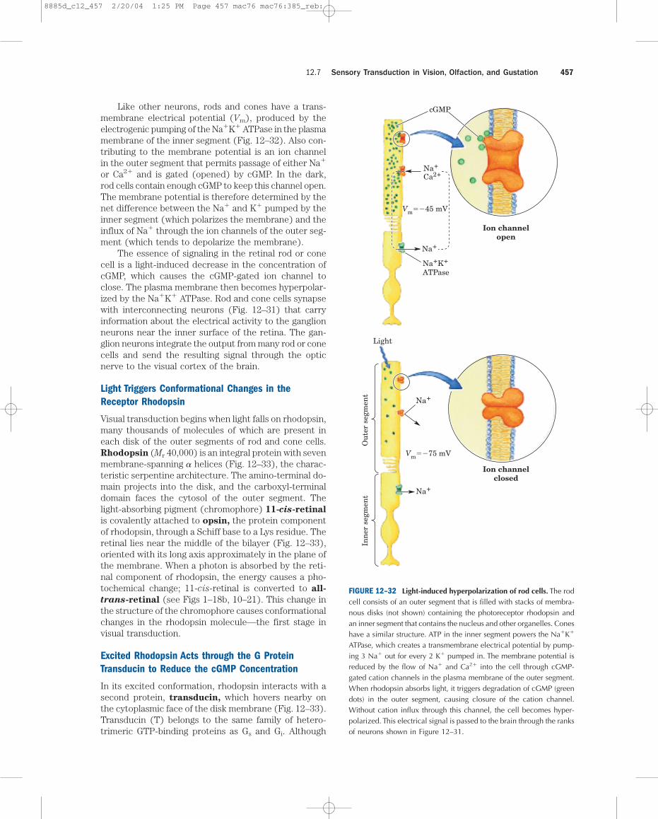
Like other neurons, rods and cones have a trans-membrane electrical potential (Vm), produced by theelectrogenic pumping of the Na�K� ATPase in the plasmamembrane of the inner segment (Fig. 12–32). Also con-tributing to the membrane potential is an ion channelin the outer segment that permits passage of either Na�
or Ca2� and is gated (opened) by cGMP. In the dark,rod cells contain enough cGMP to keep this channel open.The membrane potential is therefore determined by thenet difference between the Na� and K� pumped by theinner segment (which polarizes the membrane) and theinflux of Na� through the ion channels of the outer seg-ment (which tends to depolarize the membrane).
The essence of signaling in the retinal rod or conecell is a light-induced decrease in the concentration ofcGMP, which causes the cGMP-gated ion channel toclose. The plasma membrane then becomes hyperpolar-ized by the Na�K� ATPase. Rod and cone cells synapsewith interconnecting neurons (Fig. 12–31) that carryinformation about the electrical activity to the ganglionneurons near the inner surface of the retina. The gan-glion neurons integrate the output from many rod or conecells and send the resulting signal through the opticnerve to the visual cortex of the brain.
Light Triggers Conformational Changes in theReceptor Rhodopsin
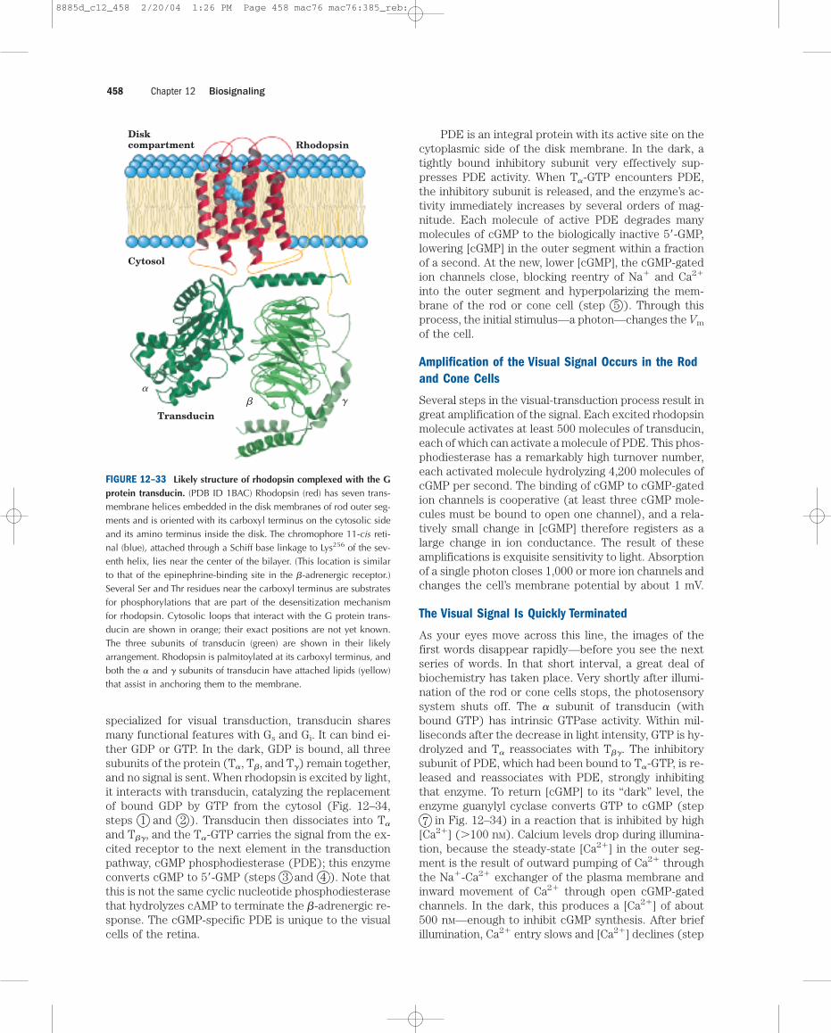
Visual transduction begins when light falls on rhodopsin,many thousands of molecules of which are present ineach disk of the outer segments of rod and cone cells.Rhodopsin (Mr 40,000) is an integral protein with sevenmembrane-spanning � helices (Fig. 12–33), the charac-teristic serpentine architecture. The amino-terminal do-main projects into the disk, and the carboxyl-terminaldomain faces the cytosol of the outer segment. Thelight-absorbing pigment (chromophore) 11-cis-retinal
is covalently attached to opsin, the protein componentof rhodopsin, through a Schiff base to a Lys residue. Theretinal lies near the middle of the bilayer (Fig. 12–33),oriented with its long axis approximately in the plane ofthe membrane. When a photon is absorbed by the reti-nal component of rhodopsin, the energy causes a pho-tochemical change; 11-cis-retinal is converted to all-
trans-retinal (see Figs 1–18b, 10–21). This change inthe structure of the chromophore causes conformationalchanges in the rhodopsin molecule—the first stage invisual transduction.
Excited Rhodopsin Acts through the G ProteinTransducin to Reduce the cGMP Concentration
In its excited conformation, rhodopsin interacts with asecond protein, transducin, which hovers nearby onthe cytoplasmic face of the disk membrane (Fig. 12–33).Transducin (T) belongs to the same family of hetero-trimeric GTP-binding proteins as Gs and Gi. Although
12.7 Sensory Transduction in Vision, Olfaction, and Gustation 457
Inn
er s
egm
ent
Ou
ter
segm
ent
Na�K�
ATPase
Ion channelopen
Ion channelclosed
Light
Na�
Na�
Na�
Na�
Ca2�
Vm��45 mV
Vm��75 mV
cGMP
FIGURE 12–32 Light-induced hyperpolarization of rod cells. The rodcell consists of an outer segment that is filled with stacks of membra-nous disks (not shown) containing the photoreceptor rhodopsin andan inner segment that contains the nucleus and other organelles. Coneshave a similar structure. ATP in the inner segment powers the Na�K�
ATPase, which creates a transmembrane electrical potential by pump-ing 3 Na� out for every 2 K� pumped in. The membrane potential isreduced by the flow of Na� and Ca2� into the cell through cGMP-gated cation channels in the plasma membrane of the outer segment.When rhodopsin absorbs light, it triggers degradation of cGMP (greendots) in the outer segment, causing closure of the cation channel.Without cation influx through this channel, the cell becomes hyper-polarized. This electrical signal is passed to the brain through the ranksof neurons shown in Figure 12–31.
8885d_c12_457 2/20/04 1:25 PM Page 457 mac76 mac76:385_reb:
specialized for visual transduction, transducin sharesmany functional features with Gs and Gi. It can bind ei-ther GDP or GTP. In the dark, GDP is bound, all threesubunits of the protein (T�, T�, and T�) remain together,and no signal is sent. When rhodopsin is excited by light,it interacts with transducin, catalyzing the replacementof bound GDP by GTP from the cytosol (Fig. 12–34,steps 1 and 2 ). Transducin then dissociates into T�
and T��, and the T�-GTP carries the signal from the ex-cited receptor to the next element in the transductionpathway, cGMP phosphodiesterase (PDE); this enzymeconverts cGMP to 5�-GMP (steps 3 and 4 ). Note thatthis is not the same cyclic nucleotide phosphodiesterasethat hydrolyzes cAMP to terminate the �-adrenergic re-sponse. The cGMP-specific PDE is unique to the visualcells of the retina.
PDE is an integral protein with its active site on thecytoplasmic side of the disk membrane. In the dark, atightly bound inhibitory subunit very effectively sup-presses PDE activity. When T�-GTP encounters PDE,the inhibitory subunit is released, and the enzyme’s ac-tivity immediately increases by several orders of mag-nitude. Each molecule of active PDE degrades manymolecules of cGMP to the biologically inactive 5�-GMP,lowering [cGMP] in the outer segment within a fractionof a second. At the new, lower [cGMP], the cGMP-gatedion channels close, blocking reentry of Na� and Ca2�
into the outer segment and hyperpolarizing the mem-brane of the rod or cone cell (step 5 ). Through thisprocess, the initial stimulus—a photon—changes the Vm
of the cell.
Amplification of the Visual Signal Occurs in the Rodand Cone Cells
Several steps in the visual-transduction process result ingreat amplification of the signal. Each excited rhodopsinmolecule activates at least 500 molecules of transducin,each of which can activate a molecule of PDE. This phos-phodiesterase has a remarkably high turnover number,each activated molecule hydrolyzing 4,200 molecules ofcGMP per second. The binding of cGMP to cGMP-gatedion channels is cooperative (at least three cGMP mole-cules must be bound to open one channel), and a rela-tively small change in [cGMP] therefore registers as alarge change in ion conductance. The result of theseamplifications is exquisite sensitivity to light. Absorptionof a single photon closes 1,000 or more ion channels andchanges the cell’s membrane potential by about 1 mV.
The Visual Signal Is Quickly Terminated
As your eyes move across this line, the images of thefirst words disappear rapidly—before you see the nextseries of words. In that short interval, a great deal ofbiochemistry has taken place. Very shortly after illumi-nation of the rod or cone cells stops, the photosensorysystem shuts off. The � subunit of transducin (withbound GTP) has intrinsic GTPase activity. Within mil-liseconds after the decrease in light intensity, GTP is hy-drolyzed and T� reassociates with T��. The inhibitorysubunit of PDE, which had been bound to T�-GTP, is re-leased and reassociates with PDE, strongly inhibitingthat enzyme. To return [cGMP] to its “dark” level, theenzyme guanylyl cyclase converts GTP to cGMP (step7 in Fig. 12–34) in a reaction that is inhibited by high
[Ca2�] (�100 nM). Calcium levels drop during illumina-tion, because the steady-state [Ca2�] in the outer seg-ment is the result of outward pumping of Ca2� throughthe Na�-Ca2� exchanger of the plasma membrane andinward movement of Ca2� through open cGMP-gatedchannels. In the dark, this produces a [Ca2�] of about500 nM—enough to inhibit cGMP synthesis. After briefillumination, Ca2� entry slows and [Ca2�] declines (step
Chapter 12 Biosignaling458
�� �
Cytosol
Transducin
Diskcompartment Rhodopsin
FIGURE 12–33 Likely structure of rhodopsin complexed with the Gprotein transducin. (PDB ID 1BAC) Rhodopsin (red) has seven trans-membrane helices embedded in the disk membranes of rod outer seg-ments and is oriented with its carboxyl terminus on the cytosolic sideand its amino terminus inside the disk. The chromophore 11-cis reti-nal (blue), attached through a Schiff base linkage to Lys256 of the sev-enth helix, lies near the center of the bilayer. (This location is similarto that of the epinephrine-binding site in the �-adrenergic receptor.)Several Ser and Thr residues near the carboxyl terminus are substratesfor phosphorylations that are part of the desensitization mechanismfor rhodopsin. Cytosolic loops that interact with the G protein trans-ducin are shown in orange; their exact positions are not yet known.The three subunits of transducin (green) are shown in their likelyarrangement. Rhodopsin is palmitoylated at its carboxyl terminus, andboth the � and � subunits of transducin have attached lipids (yellow)that assist in anchoring them to the membrane.
8885d_c12_458 2/20/04 1:26 PM Page 458 mac76 mac76:385_reb:
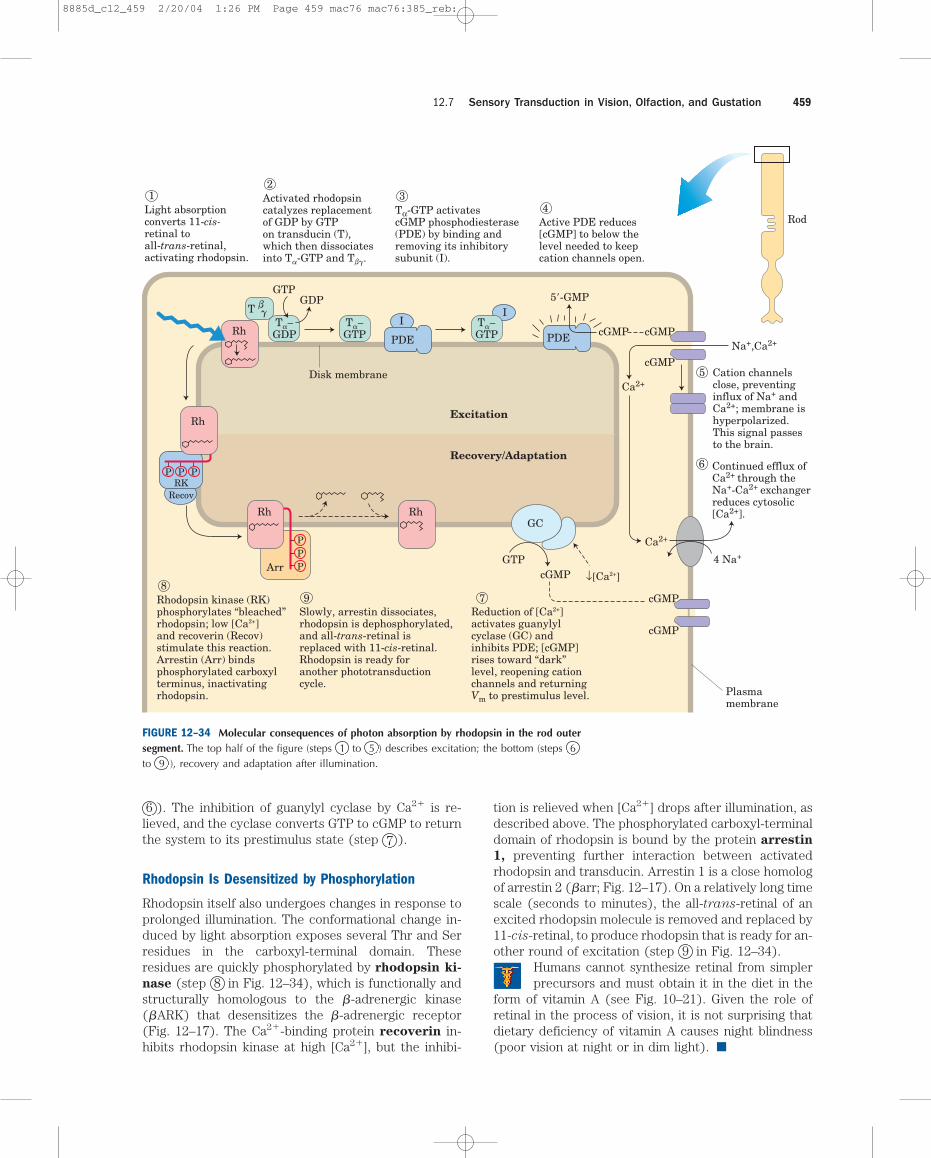
6 ). The inhibition of guanylyl cyclase by Ca2� is re-lieved, and the cyclase converts GTP to cGMP to returnthe system to its prestimulus state (step 7 ).
Rhodopsin Is Desensitized by Phosphorylation
Rhodopsin itself also undergoes changes in response toprolonged illumination. The conformational change in-duced by light absorption exposes several Thr and Serresidues in the carboxyl-terminal domain. Theseresidues are quickly phosphorylated by rhodopsin ki-
nase (step 8 in Fig. 12–34), which is functionally andstructurally homologous to the �-adrenergic kinase(�ARK) that desensitizes the �-adrenergic receptor(Fig. 12–17). The Ca2�-binding protein recoverin in-hibits rhodopsin kinase at high [Ca2�], but the inhibi-
tion is relieved when [Ca2�] drops after illumination, asdescribed above. The phosphorylated carboxyl-terminaldomain of rhodopsin is bound by the protein arrestin
1, preventing further interaction between activatedrhodopsin and transducin. Arrestin 1 is a close homologof arrestin 2 (�arr; Fig. 12–17). On a relatively long timescale (seconds to minutes), the all-trans-retinal of anexcited rhodopsin molecule is removed and replaced by11-cis-retinal, to produce rhodopsin that is ready for an-other round of excitation (step 9 in Fig. 12–34).
Humans cannot synthesize retinal from simplerprecursors and must obtain it in the diet in the
form of vitamin A (see Fig. 10–21). Given the role ofretinal in the process of vision, it is not surprising thatdietary deficiency of vitamin A causes night blindness(poor vision at night or in dim light). ■
12.7 Sensory Transduction in Vision, Olfaction, and Gustation 459
Ca2+
Na+,Ca2+
4 Na+
Ca2+
Light absorptionconverts 11-cis-retinal toall-trans-retinal,activating rhodopsin.
1 Activated rhodopsincatalyzes replacementof GDP by GTPon transducin (T),which then dissociatesinto Ta-GTP and Tbg.
2
Tα-GTP activatescGMP phosphodiesterase(PDE) by binding andremoving its inhibitorysubunit (I).
3
Active PDE reduces[cGMP] to below thelevel needed to keepcation channels open.
4
Reduction of [Ca2+]activates guanylylcyclase (GC) andinhibits PDE; [cGMP]rises toward “dark”level, reopening cationchannels and returningVm to prestimulus level.
7Slowly, arrestin dissociates,rhodopsin is dephosphorylated,and all-trans-retinal isreplaced with 11-cis-retinal.Rhodopsin is ready foranother phototransductioncycle.
9Rhodopsin kinase (RK)phosphorylates “bleached”rhodopsin; low [Ca2+]and recoverin (Recov)stimulate this reaction.Arrestin (Arr) bindsphosphorylated carboxylterminus, inactivatingrhodopsin.
8
Cation channelsclose, preventinginflux of Na+ andCa2+; membrane ishyperpolarized.This signal passesto the brain.
Plasmamembrane
Disk membrane 5
Continued efflux ofCa2+ through theNa+-Ca2+ exchangerreduces cytosolic[Ca2+].
6P
PPP
PRK
Recov
P
GDP
GTPcGMP ↓[Ca2+]
Recovery/Adaptation
Excitation
Rod
IbgT
Rh
Rh
Rh
Arr
RhGC
PDE PDE cGMP
5�-GMP
cGMP
cGMP
cGMP
cGMP
ITa−GTP
Ta−GTP
Ta−GDP
GTP
FIGURE 12–34 Molecular consequences of photon absorption by rhodopsin in the rod outersegment. The top half of the figure (steps 1 to 5 ) describes excitation; the bottom (steps 6to 9 ), recovery and adaptation after illumination.
8885d_c12_459 2/20/04 1:26 PM Page 459 mac76 mac76:385_reb:
Cone Cells Specialize in Color Vision
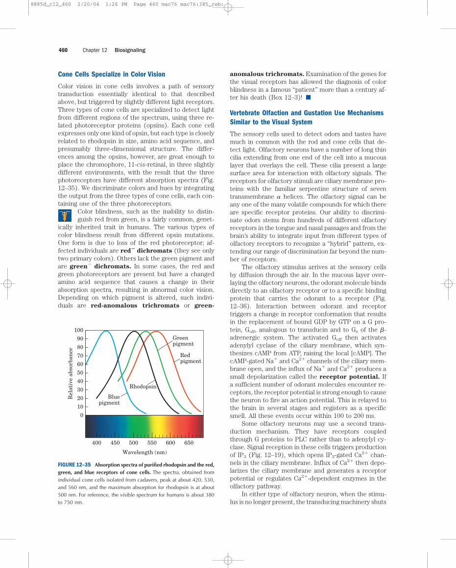
Color vision in cone cells involves a path of sensorytransduction essentially identical to that describedabove, but triggered by slightly different light receptors.Three types of cone cells are specialized to detect lightfrom different regions of the spectrum, using three re-lated photoreceptor proteins (opsins). Each cone cellexpresses only one kind of opsin, but each type is closelyrelated to rhodopsin in size, amino acid sequence, andpresumably three-dimensional structure. The differ-ences among the opsins, however, are great enough toplace the chromophore, 11-cis-retinal, in three slightlydifferent environments, with the result that the threephotoreceptors have different absorption spectra (Fig.12–35). We discriminate colors and hues by integratingthe output from the three types of cone cells, each con-taining one of the three photoreceptors.
Color blindness, such as the inability to distin-guish red from green, is a fairly common, genet-
ically inherited trait in humans. The various types ofcolor blindness result from different opsin mutations.One form is due to loss of the red photoreceptor; af-fected individuals are red
�dichromats (they see only
two primary colors). Others lack the green pigment andare green
�dichromats. In some cases, the red and
green photoreceptors are present but have a changedamino acid sequence that causes a change in their absorption spectra, resulting in abnormal color vision.Depending on which pigment is altered, such indivi-duals are red-anomalous trichromats or green-
anomalous trichromats. Examination of the genes forthe visual receptors has allowed the diagnosis of colorblindness in a famous “patient” more than a century af-ter his death (Box 12–3)! ■
Vertebrate Olfaction and Gustation Use MechanismsSimilar to the Visual System
The sensory cells used to detect odors and tastes havemuch in common with the rod and cone cells that de-tect light. Olfactory neurons have a number of long thincilia extending from one end of the cell into a mucouslayer that overlays the cell. These cilia present a largesurface area for interaction with olfactory signals. Thereceptors for olfactory stimuli are ciliary membrane pro-teins with the familiar serpentine structure of seventransmembrane � helices. The olfactory signal can beany one of the many volatile compounds for which thereare specific receptor proteins. Our ability to discrimi-nate odors stems from hundreds of different olfactoryreceptors in the tongue and nasal passages and from thebrain’s ability to integrate input from different types ofolfactory receptors to recognize a “hybrid” pattern, ex-tending our range of discrimination far beyond the num-ber of receptors.
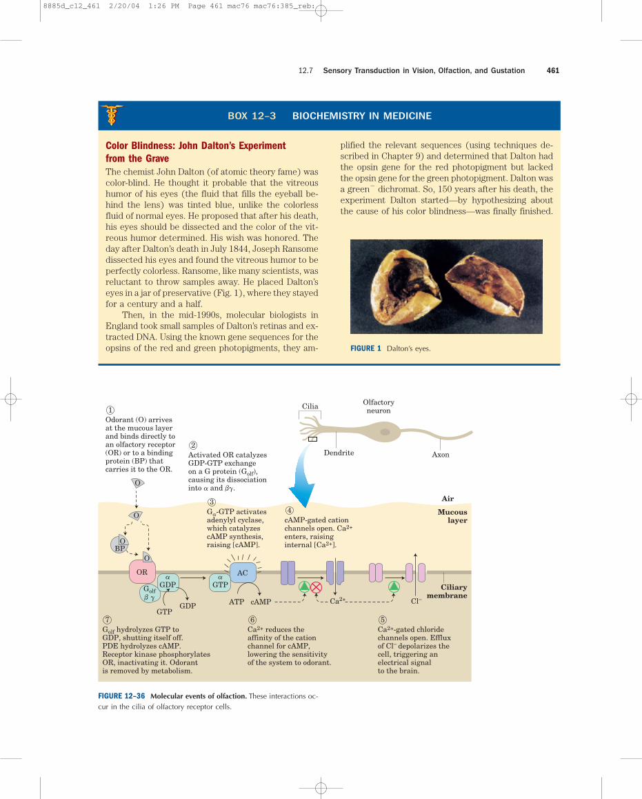
The olfactory stimulus arrives at the sensory cellsby diffusion through the air. In the mucous layer over-laying the olfactory neurons, the odorant molecule bindsdirectly to an olfactory receptor or to a specific bindingprotein that carries the odorant to a receptor (Fig.12–36). Interaction between odorant and receptortriggers a change in receptor conformation that resultsin the replacement of bound GDP by GTP on a G pro-tein, Golf, analogous to transducin and to Gs of the �-adrenergic system. The activated Golf then activatesadenylyl cyclase of the ciliary membrane, which syn-thesizes cAMP from ATP, raising the local [cAMP]. ThecAMP-gated Na� and Ca2� channels of the ciliary mem-brane open, and the influx of Na� and Ca2� produces asmall depolarization called the receptor potential. Ifa sufficient number of odorant molecules encounter re-ceptors, the receptor potential is strong enough to causethe neuron to fire an action potential. This is relayed tothe brain in several stages and registers as a specificsmell. All these events occur within 100 to 200 ms.
Some olfactory neurons may use a second trans-duction mechanism. They have receptors coupledthrough G proteins to PLC rather than to adenylyl cy-clase. Signal reception in these cells triggers productionof IP3 (Fig. 12–19), which opens IP3-gated Ca2� chan-nels in the ciliary membrane. Influx of Ca2� then depo-larizes the ciliary membrane and generates a receptorpotential or regulates Ca2�-dependent enzymes in theolfactory pathway.
In either type of olfactory neuron, when the stimu-lus is no longer present, the transducing machinery shuts
Chapter 12 Biosignaling460
Blue pigment
Rhodopsin
Red pigment
100
90
80
70
60
50
40
30
20
10
0
Rel
ativ
e ab
sorb
ance
400 450 500 550 600 650
Greenpigment
Wavelength (nm)
FIGURE 12–35 Absorption spectra of purified rhodopsin and the red,green, and blue receptors of cone cells. The spectra, obtained fromindividual cone cells isolated from cadavers, peak at about 420, 530,and 560 nm, and the maximum absorption for rhodopsin is at about500 nm. For reference, the visible spectrum for humans is about 380to 750 nm.
8885d_c12_460 2/20/04 1:26 PM Page 460 mac76 mac76:385_reb:
12.7 Sensory Transduction in Vision, Olfaction, and Gustation 461
Ciliarymembrane
Mucouslayer
Air
cAMPATPGTP
GDP
Olfactoryneuron
Dendrite Axon
Cilia
AC
Ca2+ Cl–Golfb g
O
OBP
O
Odorant (O) arrivesat the mucous layerand binds directly toan olfactory receptor(OR) or to a bindingprotein (BP) thatcarries it to the OR.
1
Activated OR catalyzesGDP-GTP exchangeon a G protein (Golf),causing its dissociationinto a and bg.
2
Ca2+ reduces theaffinity of the cationchannel for cAMP,lowering the sensitivityof the system to odorant.
6Golf hydrolyzes GTP toGDP, shutting itself off. PDE hydrolyzes cAMP.Receptor kinase phosphorylatesOR, inactivating it. Odorantis removed by metabolism.
7Ca2+-gated chloridechannels open. Effluxof Cl– depolarizes thecell, triggering anelectrical signalto the brain.
5
Ga-GTP activatesadenylyl cyclase,which catalyzescAMP synthesis,raising [cAMP].
3
cAMP-gated cationchannels open. Ca2+
enters, raisinginternal [Ca2+].
4
OR
O
a
GDPa
GTP
FIGURE 12–36 Molecular events of olfaction. These interactions oc-cur in the cilia of olfactory receptor cells.
FIGURE 7 (a)�?/Au� (b)�?/Au�BOX 12–3 BIOCHEMISTRY IN MEDICINE
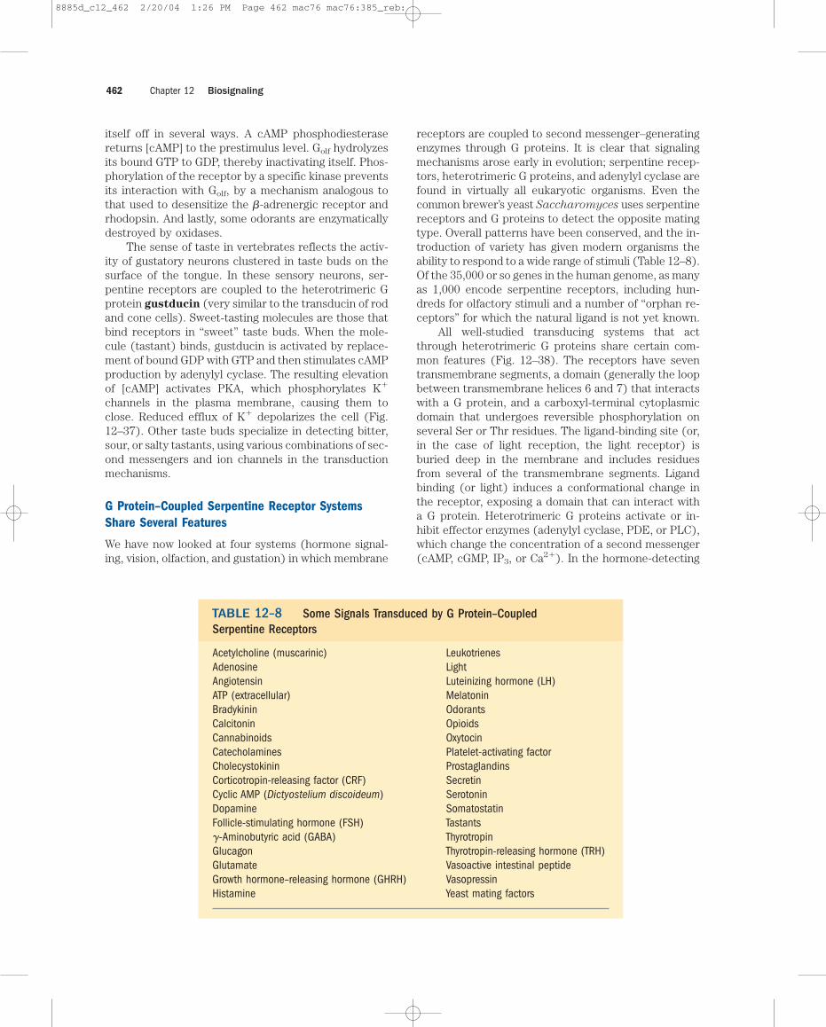
Color Blindness: John Dalton’s Experiment from the GraveThe chemist John Dalton (of atomic theory fame) wascolor-blind. He thought it probable that the vitreoushumor of his eyes (the fluid that fills the eyeball be-hind the lens) was tinted blue, unlike the colorlessfluid of normal eyes. He proposed that after his death,his eyes should be dissected and the color of the vit-reous humor determined. His wish was honored. Theday after Dalton’s death in July 1844, Joseph Ransomedissected his eyes and found the vitreous humor to beperfectly colorless. Ransome, like many scientists, wasreluctant to throw samples away. He placed Dalton’seyes in a jar of preservative (Fig. 1), where they stayedfor a century and a half.
Then, in the mid-1990s, molecular biologists inEngland took small samples of Dalton’s retinas and ex-tracted DNA. Using the known gene sequences for theopsins of the red and green photopigments, they am-
plified the relevant sequences (using techniques de-scribed in Chapter 9) and determined that Dalton hadthe opsin gene for the red photopigment but lackedthe opsin gene for the green photopigment. Dalton wasa green� dichromat. So, 150 years after his death, theexperiment Dalton started—by hypothesizing aboutthe cause of his color blindness—was finally finished.
FIGURE 1 Dalton’s eyes.
8885d_c12_461 2/20/04 1:26 PM Page 461 mac76 mac76:385_reb:
itself off in several ways. A cAMP phosphodiesterasereturns [cAMP] to the prestimulus level. Golf hydrolyzesits bound GTP to GDP, thereby inactivating itself. Phos-phorylation of the receptor by a specific kinase preventsits interaction with Golf, by a mechanism analogous tothat used to desensitize the �-adrenergic receptor andrhodopsin. And lastly, some odorants are enzymaticallydestroyed by oxidases.
The sense of taste in vertebrates reflects the activ-ity of gustatory neurons clustered in taste buds on thesurface of the tongue. In these sensory neurons, ser-pentine receptors are coupled to the heterotrimeric Gprotein gustducin (very similar to the transducin of rodand cone cells). Sweet-tasting molecules are those thatbind receptors in “sweet” taste buds. When the mole-cule (tastant) binds, gustducin is activated by replace-ment of bound GDP with GTP and then stimulates cAMPproduction by adenylyl cyclase. The resulting elevationof [cAMP] activates PKA, which phosphorylates K�
channels in the plasma membrane, causing them toclose. Reduced efflux of K� depolarizes the cell (Fig.12–37). Other taste buds specialize in detecting bitter,sour, or salty tastants, using various combinations of sec-ond messengers and ion channels in the transductionmechanisms.
G Protein–Coupled Serpentine Receptor SystemsShare Several Features
We have now looked at four systems (hormone signal-ing, vision, olfaction, and gustation) in which membrane
receptors are coupled to second messenger–generatingenzymes through G proteins. It is clear that signalingmechanisms arose early in evolution; serpentine recep-tors, heterotrimeric G proteins, and adenylyl cyclase arefound in virtually all eukaryotic organisms. Even thecommon brewer’s yeast Saccharomyces uses serpentinereceptors and G proteins to detect the opposite matingtype. Overall patterns have been conserved, and the in-troduction of variety has given modern organisms theability to respond to a wide range of stimuli (Table 12–8).Of the 35,000 or so genes in the human genome, as manyas 1,000 encode serpentine receptors, including hun-dreds for olfactory stimuli and a number of “orphan re-ceptors” for which the natural ligand is not yet known.
All well-studied transducing systems that actthrough heterotrimeric G proteins share certain com-mon features (Fig. 12–38). The receptors have seventransmembrane segments, a domain (generally the loopbetween transmembrane helices 6 and 7) that interactswith a G protein, and a carboxyl-terminal cytoplasmicdomain that undergoes reversible phosphorylation onseveral Ser or Thr residues. The ligand-binding site (or,in the case of light reception, the light receptor) isburied deep in the membrane and includes residuesfrom several of the transmembrane segments. Ligandbinding (or light) induces a conformational change inthe receptor, exposing a domain that can interact witha G protein. Heterotrimeric G proteins activate or in-hibit effector enzymes (adenylyl cyclase, PDE, or PLC),which change the concentration of a second messenger(cAMP, cGMP, IP3, or Ca2�). In the hormone-detecting
Chapter 12 Biosignaling462
Acetylcholine (muscarinic)AdenosineAngiotensinATP (extracellular)BradykininCalcitoninCannabinoidsCatecholaminesCholecystokininCorticotropin-releasing factor (CRF)Cyclic AMP (Dictyostelium discoideum)DopamineFollicle-stimulating hormone (FSH)�-Aminobutyric acid (GABA)GlucagonGlutamate Growth hormone–releasing hormone (GHRH)Histamine
LeukotrienesLightLuteinizing hormone (LH)MelatoninOdorantsOpioidsOxytocinPlatelet-activating factorProstaglandinsSecretinSerotoninSomatostatinTastantsThyrotropinThyrotropin-releasing hormone (TRH)Vasoactive intestinal peptideVasopressinYeast mating factors
Some Signals Transduced by G Protein–Coupled Serpentine ReceptorsTABLE 12–8
8885d_c12_462 2/20/04 1:26 PM Page 462 mac76 mac76:385_reb:
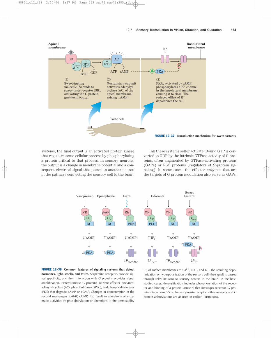
systems, the final output is an activated protein kinasethat regulates some cellular process by phosphorylatinga protein critical to that process. In sensory neurons,the output is a change in membrane potential and a con-sequent electrical signal that passes to another neuronin the pathway connecting the sensory cell to the brain.
All these systems self-inactivate. Bound GTP is con-verted to GDP by the intrinsic GTPase activity of G pro-teins, often augmented by GTPase-activating proteins(GAPs) or RGS proteins (regulators of G-protein sig-naling). In some cases, the effector enzymes that arethe targets of G protein modulation also serve as GAPs.
12.7 Sensory Transduction in Vision, Olfaction, and Gustation 463
Basolateralmembrane
Apicalmembrane
cAMPATPGTP
Olfactoryneuron
AC
K�
Ggust� �
SR
Sweet-tastingmolecule (S) binds tosweet-taste receptor (SR),activating the G proteingustducin (Ggust).
1Gustducin subunitactivates adenylylcyclase (AC) of theapical membrane,raising [cAMP].
�
Taste cell
2PKA, activated by cAMP,phosphorylates a K� channelin the basolateral membrane,causing it to close. Thereduced efflux of K�
depolarizes the cell.
3
P
S
GTPGDP
GDP PKA
� �
Vasopressin Epinephrine Light OdorantsSweettastant
↓[cAMP]
↓
↑[cAMP]
↑P
↓[cGMP] ↑[IP3] ↑[cAMP] ↑[cAMP]
↓PCa2�,Na� ↑PCa2� ↑PCa2�,Na� ↓PK�
VR
Gi
AC
PKA
-AR
Gs
AC
PKA
↑ PKA
Rh
T
PDE
OR1
Golf
PLC
OR2
Golf
AC
SR
Ggust
AC
�
FIGURE 12–38 Common features of signaling systems that detecthormones, light, smells, and tastes. Serpentine receptors provide sig-nal specificity, and their interaction with G proteins provides signalamplification. Heterotrimeric G proteins activate effector enzymes:adenylyl cyclase (AC), phospholipase C (PLC), and phosphodiesterases(PDE) that degrade cAMP or cGMP. Changes in concentration of thesecond messengers (cAMP, cGMP, IP3) result in alterations of enzy-matic activities by phosphorylation or alterations in the permeability
(P) of surface membranes to Ca2�, Na�, and K�. The resulting depo-larization or hyperpolarization of the sensory cell (the signal) is passedthrough relay neurons to sensory centers in the brain. In the best-studied cases, desensitization includes phosphorylation of the recep-tor and binding of a protein (arrestin) that interrupts receptor–G pro-tein interactions. VR is the vasopressin receptor; other receptor and Gprotein abbreviations are as used in earlier illustrations.
FIGURE 12–37 Transduction mechanism for sweet tastants.
8885d_c12_463 2/20/04 1:27 PM Page 463 mac76 mac76:385_reb:
Disruption of G-Protein Signaling Causes Disease
Biochemical studies of signal transductions haveled to an improved understanding of the patho-
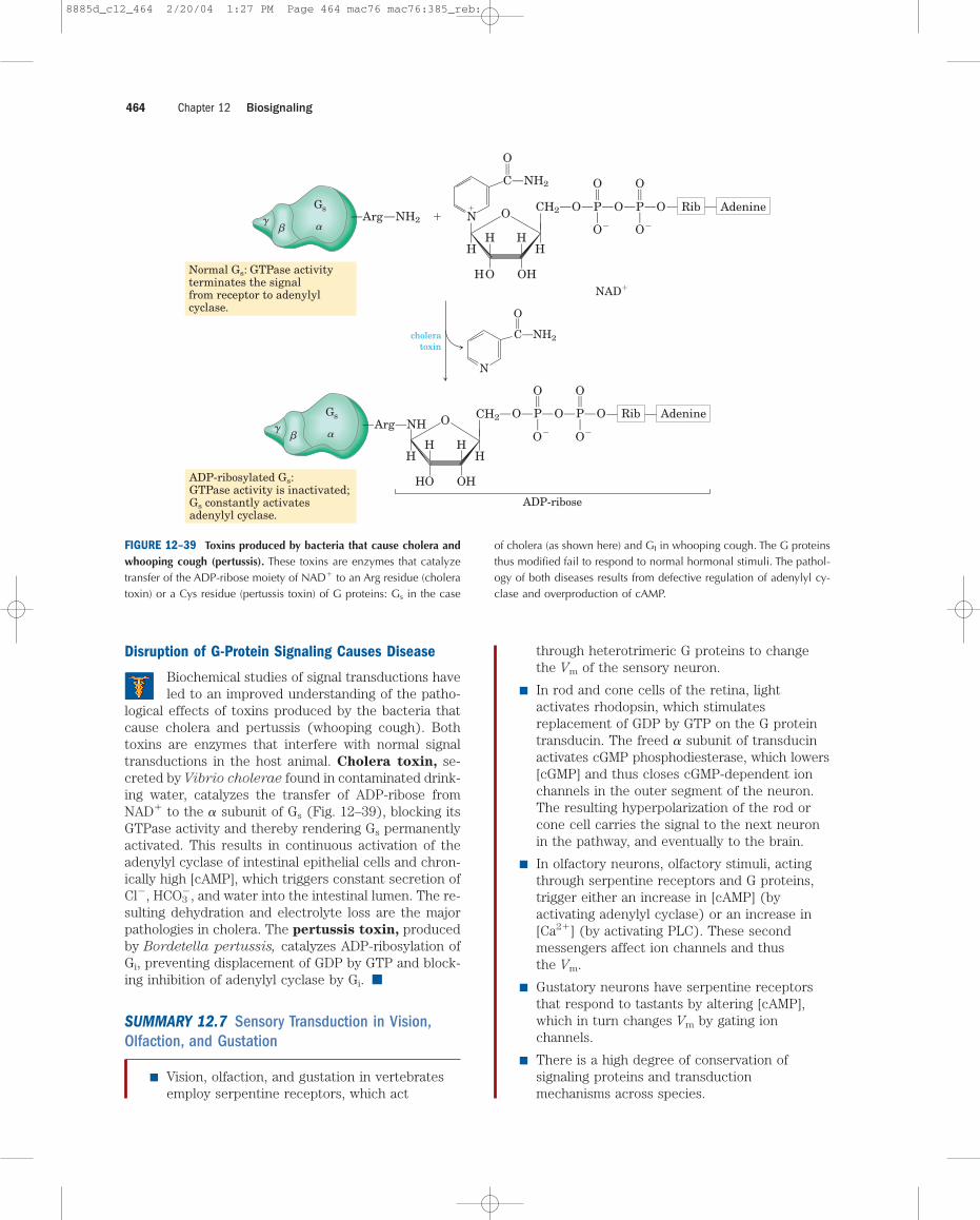
logical effects of toxins produced by the bacteria thatcause cholera and pertussis (whooping cough). Bothtoxins are enzymes that interfere with normal signaltransductions in the host animal. Cholera toxin, se-creted by Vibrio cholerae found in contaminated drink-ing water, catalyzes the transfer of ADP-ribose fromNAD� to the � subunit of Gs (Fig. 12–39), blocking itsGTPase activity and thereby rendering Gs permanentlyactivated. This results in continuous activation of theadenylyl cyclase of intestinal epithelial cells and chron-ically high [cAMP], which triggers constant secretion ofCl�, HCO3
�, and water into the intestinal lumen. The re-sulting dehydration and electrolyte loss are the majorpathologies in cholera. The pertussis toxin, producedby Bordetella pertussis, catalyzes ADP-ribosylation ofGi, preventing displacement of GDP by GTP and block-ing inhibition of adenylyl cyclase by Gi. ■
SUMMARY 12.7 Sensory Transduction in Vision,Olfaction, and Gustation
■ Vision, olfaction, and gustation in vertebratesemploy serpentine receptors, which act
through heterotrimeric G proteins to changethe Vm of the sensory neuron.
■ In rod and cone cells of the retina, lightactivates rhodopsin, which stimulatesreplacement of GDP by GTP on the G proteintransducin. The freed � subunit of transducinactivates cGMP phosphodiesterase, which lowers[cGMP] and thus closes cGMP-dependent ionchannels in the outer segment of the neuron.The resulting hyperpolarization of the rod orcone cell carries the signal to the next neuronin the pathway, and eventually to the brain.
■ In olfactory neurons, olfactory stimuli, actingthrough serpentine receptors and G proteins,trigger either an increase in [cAMP] (byactivating adenylyl cyclase) or an increase in[Ca2�] (by activating PLC). These secondmessengers affect ion channels and thusthe Vm.
■ Gustatory neurons have serpentine receptorsthat respond to tastants by altering [cAMP],which in turn changes Vm by gating ionchannels.
■ There is a high degree of conservation ofsignaling proteins and transductionmechanisms across species.
Chapter 12 Biosignaling464
��
�
Gs
��
�
Gs CH2
O
OH
P
O�
H
N
HHH
NH2
O
O
H
O O
C
O
2
P
O�
O
O
Rib Adenine
�
O
NH2C
NAD�
�
choleratoxin
CH2
OH
P
O�
H
HHH
O
O
H
O O
O
P
O�
O
O
Rib AdenineN
Arg NH
ADP-ribose
Normal Gs: GTPase activityterminates the signalfrom receptor to adenylylcyclase.
ADP-ribosylated Gs:GTPase activity is inactivated;Gs constantly activatesadenylyl cyclase.
NHArg
FIGURE 12–39 Toxins produced by bacteria that cause cholera andwhooping cough (pertussis). These toxins are enzymes that catalyzetransfer of the ADP-ribose moiety of NAD� to an Arg residue (choleratoxin) or a Cys residue (pertussis toxin) of G proteins: Gs in the case
of cholera (as shown here) and GI in whooping cough. The G proteinsthus modified fail to respond to normal hormonal stimuli. The pathol-ogy of both diseases results from defective regulation of adenylyl cy-clase and overproduction of cAMP.
8885d_c12_464 2/20/04 1:27 PM Page 464 mac76 mac76:385_reb:
12.8 Regulation of Transcription by Steroid HormonesThe large group of steroid, retinoic acid (retinoid), andthyroid hormones exert at least part of their effects bya mechanism fundamentally different from that of otherhormones: they act in the nucleus to alter gene expres-sion. We therefore discuss their mode of action in detailin Chapter 28, along with other mechanisms for regu-lating gene expression. Here we give a brief overview.
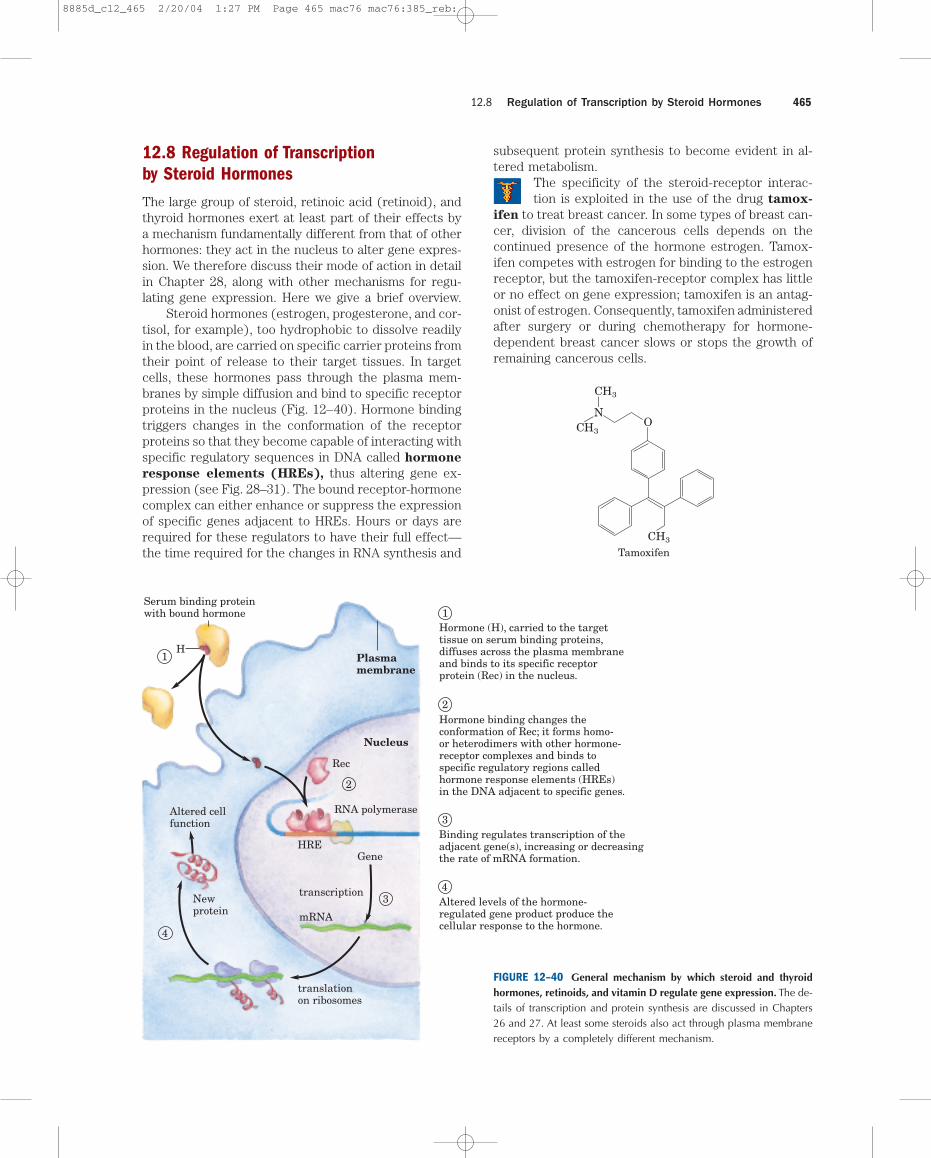
Steroid hormones (estrogen, progesterone, and cor-tisol, for example), too hydrophobic to dissolve readilyin the blood, are carried on specific carrier proteins fromtheir point of release to their target tissues. In targetcells, these hormones pass through the plasma mem-branes by simple diffusion and bind to specific receptorproteins in the nucleus (Fig. 12–40). Hormone bindingtriggers changes in the conformation of the receptorproteins so that they become capable of interacting withspecific regulatory sequences in DNA called hormone
response elements (HREs), thus altering gene ex-pression (see Fig. 28–31). The bound receptor-hormonecomplex can either enhance or suppress the expressionof specific genes adjacent to HREs. Hours or days arerequired for these regulators to have their full effect—the time required for the changes in RNA synthesis and
subsequent protein synthesis to become evident in al-tered metabolism.
The specificity of the steroid-receptor interac-tion is exploited in the use of the drug tamox-
ifen to treat breast cancer. In some types of breast can-cer, division of the cancerous cells depends on thecontinued presence of the hormone estrogen. Tamox-ifen competes with estrogen for binding to the estrogenreceptor, but the tamoxifen-receptor complex has littleor no effect on gene expression; tamoxifen is an antag-onist of estrogen. Consequently, tamoxifen administeredafter surgery or during chemotherapy for hormone-dependent breast cancer slows or stops the growth ofremaining cancerous cells.
12.8 Regulation of Transcription by Steroid Hormones 465
1
1Hormone (H), carried to the target tissue on serum binding proteins, diffuses across the plasma membrane and binds to its specific receptor protein (Rec) in the nucleus.
2
3
3Binding regulates transcription of the adjacent gene(s), increasing or decreasing the rate of mRNA formation.
4
4Altered levels of the hormone-regulated gene product produce the cellular response to the hormone.
Plasmamembrane
Nucleus
Rec
RNA polymerase
HREGene
transcription
mRNA
translationon ribosomes
Newprotein
Altered cellfunction
H
Serum binding proteinwith bound hormone
2Hormone binding changes the conformation of Rec; it forms homo- or heterodimers with other hormone-receptor complexes and binds to specific regulatory regions called hormone response elements (HREs) in the DNA adjacent to specific genes.
FIGURE 12–40 General mechanism by which steroid and thyroidhormones, retinoids, and vitamin D regulate gene expression. The de-tails of transcription and protein synthesis are discussed in Chapters26 and 27. At least some steroids also act through plasma membranereceptors by a completely different mechanism.
O
CH3
NCH3
CH3
Tamoxifen
8885d_c12_465 2/20/04 1:27 PM Page 465 mac76 mac76:385_reb:
Another steroid analog, the drug RU486, is used to ter-minate early (preimplantation) pregnancies. An antag-onist of the hormone progesterone, RU486 binds to theprogesterone receptor and blocks hormone actions es-sential to implantation of the fertilized ovum in theuterus. ■
The classic mechanism for steroid hormone actionthrough nuclear receptors does not explain certain ef-fects of steroids that are too fast to be the result of al-tered protein synthesis. For example, the estrogen-mediated dilation of blood vessels is known to beindependent of gene transcription or protein synthesis,as is the steroid-induced decrease in cellular [cAMP].Another transduction mechanism is probably responsi-ble for some of these effects. A plasma membrane pro-tein predicted to have seven transmembrane helical seg-ments binds progesterone with very high affinity andmediates the inhibition of adenylyl cyclase by that hor-mone, accounting for the decrease in [cAMP]. A secondnonclassical mechanism involves the rapid activation ofthe MAPK cascade by progesterone, acting through thesoluble progesterone receptor. This is the same recep-tor that, in the nucleus, causes the much slower changesin gene expression that constitute the classic mecha-nism of progesterone action. How the MAPK cascade isactivated is not yet clear.
SUMMARY 12.8 Regulation of Transcription by Steroid Hormones
■ Steroid hormones enter cells and bind tospecific receptor proteins.
■ The hormone-receptor complex binds specificregions of DNA, the hormone responseelements, and regulates the expression ofnearby genes by interacting with transcriptionfactors.
■ Two other, faster-acting mechanisms producesome of the effects of steroids. Progesteronetriggers a rapid drop in [cAMP], mediated by aplasma membrane receptor, and binding ofprogesterone to the classic soluble steroidreceptor activates a MAPK cascade.
12.9 Regulation of the Cell Cycle by Protein KinasesOne of the most dramatic roles for protein phosphory-lation is the regulation of the eukaryotic cell cycle. Dur-ing embryonic growth and later development, cell divi-sion occurs in virtually every tissue. In the adultorganism most tissues become quiescent. A cell’s “deci-sion” to divide or not is of crucial importance to the or-ganism. When the regulatory mechanisms that limit celldivision are defective and cells undergo unregulateddivision, the result is catastrophic—cancer. Proper celldivision requires a precisely ordered sequence of bio-chemical events that assures every daughter cell a fullcomplement of the molecules required for life. Investi-gations into the control of cell division in diverse eu-karyotic cells have revealed universal regulatory mech-anisms. Protein kinases and protein phosphorylation arecentral to the timing mechanism that determines entryinto cell division and ensures orderly passage throughthese events.
The Cell Cycle Has Four Stages
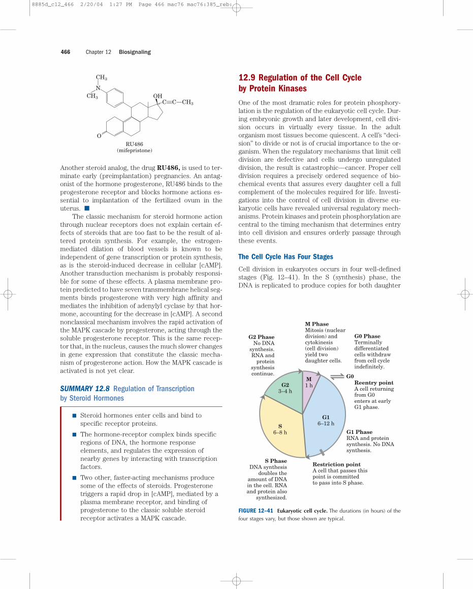
Cell division in eukaryotes occurs in four well-definedstages (Fig. 12–41). In the S (synthesis) phase, theDNA is replicated to produce copies for both daughter
Chapter 12 Biosignaling466
O
NCH3
CH3
RU486(mifepristone)
OHCC
CH3
G16–12 hS
6–8 h
G23–4 h
M1 h
G1 PhaseRNA and proteinsynthesis. No DNAsynthesis.
Restriction pointA cell that passes thispoint is committedto pass into S phase.
Reentry pointA cell returningfrom G0enters at earlyG1 phase.
G0 PhaseTerminallydifferentiatedcells withdrawfrom cell cycleindefinitely.
M PhaseMitosis (nucleardivision) andcytokinesis(cell division)yield twodaughter cells.
G2 PhaseNo DNA
synthesis.RNA and
proteinsynthesiscontinue.
S PhaseDNA synthesis
doubles theamount of DNAin the cell. RNAand protein also
synthesized.
G0
FIGURE 12–41 Eukaryotic cell cycle. The durations (in hours) of thefour stages vary, but those shown are typical.
8885d_c12_466 2/20/04 1:27 PM Page 466 mac76 mac76:385_reb:
cells. In the G2 phase (G indicates the gap betweendivisions), new proteins are synthesized and the cellapproximately doubles in size. In the M phase (mitosis),the maternal nuclear envelope breaks down, matchingchromosomes are pulled to opposite poles of the cell,each set of daughter chromosomes is surrounded by anewly formed nuclear envelope, and cytokinesis pinchesthe cell in half, producing two daughter cells. In em-bryonic or rapidly proliferating tissue, each daughtercell divides again, but only after a waiting period (G1).In cultured animal cells the entire process takes about24 hours.
After passing through mitosis and into G1, a cell ei-ther continues through another division or ceases to di-vide, entering a quiescent phase (G0) that may lasthours, days, or the lifetime of the cell. When a cell inG0 begins to divide again, it reenters the division cyclethrough the G1 phase. Differentiated cells such as he-patocytes or adipocytes have acquired their specializedfunction and form; they remain in the G0 phase.
Levels of Cyclin-Dependent Protein Kinases Oscillate
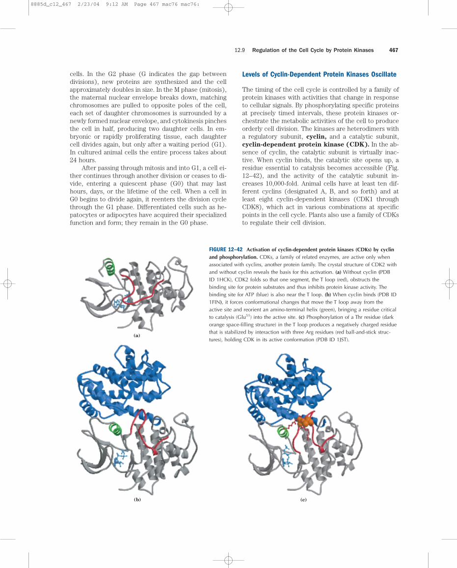
The timing of the cell cycle is controlled by a family ofprotein kinases with activities that change in responseto cellular signals. By phosphorylating specific proteinsat precisely timed intervals, these protein kinases or-chestrate the metabolic activities of the cell to produceorderly cell division. The kinases are heterodimers witha regulatory subunit, cyclin, and a catalytic subunit,cyclin-dependent protein kinase (CDK). In the ab-sence of cyclin, the catalytic subunit is virtually inac-tive. When cyclin binds, the catalytic site opens up, aresidue essential to catalysis becomes accessible (Fig.12–42), and the activity of the catalytic subunit in-creases 10,000-fold. Animal cells have at least ten dif-ferent cyclins (designated A, B, and so forth) and atleast eight cyclin-dependent kinases (CDK1 throughCDK8), which act in various combinations at specificpoints in the cell cycle. Plants also use a family of CDKsto regulate their cell division.
12.9 Regulation of the Cell Cycle by Protein Kinases 467
(b)
(a)
(c)
FIGURE 12–42 Activation of cyclin-dependent protein kinases (CDKs) by cyclinand phosphorylation. CDKs, a family of related enzymes, are active only whenassociated with cyclins, another protein family. The crystal structure of CDK2 withand without cyclin reveals the basis for this activation. (a) Without cyclin (PDBID 1HCK), CDK2 folds so that one segment, the T loop (red), obstructs thebinding site for protein substrates and thus inhibits protein kinase activity. Thebinding site for ATP (blue) is also near the T loop. (b) When cyclin binds (PDB ID1FIN), it forces conformational changes that move the T loop away from theactive site and reorient an amino-terminal helix (green), bringing a residue criticalto catalysis (Glu51) into the active site. (c) Phosphorylation of a Thr residue (darkorange space-filling structure) in the T loop produces a negatively charged residuethat is stabilized by interaction with three Arg residues (red ball-and-stick struc-tures), holding CDK in its active conformation (PDB ID 1JST).
8885d_c12_467 2/23/04 9:12 AM Page 467 mac76 mac76:
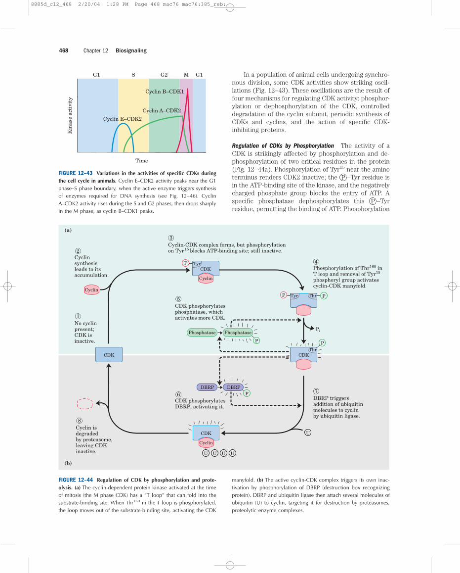
In a population of animal cells undergoing synchro-nous division, some CDK activities show striking oscil-lations (Fig. 12–43). These oscillations are the result offour mechanisms for regulating CDK activity: phosphor-ylation or dephosphorylation of the CDK, controlleddegradation of the cyclin subunit, periodic synthesis ofCDKs and cyclins, and the action of specific CDK-inhibiting proteins.
Regulation of CDKs by Phosphorylation The activity of aCDK is strikingly affected by phosphorylation and de-phosphorylation of two critical residues in the protein(Fig. 12–44a). Phosphorylation of Tyr15 near the aminoterminus renders CDK2 inactive; the P –Tyr residue isin the ATP-binding site of the kinase, and the negativelycharged phosphate group blocks the entry of ATP. Aspecific phosphatase dephosphorylates this P –Tyrresidue, permitting the binding of ATP. Phosphorylation
Chapter 12 Biosignaling468
Kin
ase
acti
vity
Time
G1 S G2 M
Cyclin E–CDK2
Cyclin A–CDK2
Cyclin B–CDK1
G1
FIGURE 12–43 Variations in the activities of specific CDKs duringthe cell cycle in animals. Cyclin E–CDK2 activity peaks near the G1phase–S phase boundary, when the active enzyme triggers synthesisof enzymes required for DNA synthesis (see Fig. 12–46). CyclinA–CDK2 activity rises during the S and G2 phases, then drops sharplyin the M phase, as cyclin B–CDK1 peaks.
U
Cyclin
U U U U
Cyclin
Tyr
No cyclinpresent;CDK isinactive.
2Cyclinsynthesisleads to itsaccumulation.
Cyclin-CDK complex forms, but phosphorylationon Tyr15 blocks ATP-binding site; still inactive.
3
Phosphorylation of Thr160 inT loop and removal of Tyr15
phosphoryl group activatescyclin-CDK manyfold.
4
1
Cyclin isdegradedby proteasome,leaving CDKinactive.
8
CDK phosphorylatesphosphatase, whichactivates more CDK.
5
CDK phosphorylatesDBRP, activating it.
6DBRP triggersaddition of ubiquitinmolecules to cyclinby ubiquitin ligase.
7
Pi
CDKCDK
CDK
Tyr Thr
Thr
Cyclin
CDK
PhosphatasePhosphatase
DBRPDBRP
P
P
P
P
P
P
(a)
(b)
FIGURE 12–44 Regulation of CDK by phosphorylation and prote-olysis. (a) The cyclin-dependent protein kinase activated at the timeof mitosis (the M phase CDK) has a “T loop” that can fold into thesubstrate-binding site. When Thr160 in the T loop is phosphorylated,the loop moves out of the substrate-binding site, activating the CDK
manyfold. (b) The active cyclin-CDK complex triggers its own inac-tivation by phosphorylation of DBRP (destruction box recognizingprotein). DBRP and ubiquitin ligase then attach several molecules ofubiquitin (U) to cyclin, targeting it for destruction by proteasomes,proteolytic enzyme complexes.
8885d_c12_468 2/20/04 1:28 PM Page 468 mac76 mac76:385_reb:
of Thr160 in the “T loop” of CDK, catalyzed by the CDK-activating kinase, forces the T loop out of the substrate-binding cleft, permitting substrate binding and catalyticactivity.
One circumstance that triggers this control mecha-nism is the presence of single-strand breaks in DNA,which leads to arrest of the cell cycle in G2. A specificprotein kinase (called Rad3 in yeast), which is activatedby single-strand breaks, triggers a cascade leading to theinactivation of the phosphatase that dephosphorylatesTyr15 of CDK. The CDK remains inactive and the cell isarrested in G2. The cell will not divide until the DNA isrepaired and the effects of the cascade are reversed.
Controlled Degradation of Cyclin Highly specific and pre-cisely timed proteolytic breakdown of mitotic cyclinsregulates CDK activity throughout the cell cycle.Progress through mitosis requires first the activationthen the destruction of cyclins A and B, which activatethe catalytic subunit of the M-phase CDK. These cyclinscontain near their amino terminus the sequenceArg–Thr–Ala–Leu–Gly–Asp–Ile–Gly–Asn, the “destruc-tion box,” which targets them for degradation. (This us-age of “box” derives from the common practice, in dia-gramming the sequence of a nucleic acid or protein, ofenclosing within a box a short sequence of nucleotideor amino acid residues with some specific function. Itdoes not imply any three-dimensional structure.) Theprotein DBRP (destruction box recognizing protein)recognizes this sequence and initiates the process of cy-clin degradation by bringing together the cyclin and an-other protein, ubiquitin. Cyclin and activated ubiqui-tin are covalently joined by the enzyme ubiquitin ligase(Fig. 12–44b). Several more ubiquitin molecules arethen appended, providing the signal for a proteolytic en-zyme complex, or proteasome, to degrade cyclin.
What controls the timing of cyclin breakdown? Afeedback loop occurs in the overall process shown inFigure 12–44. Increased CDK activity activates cyclinproteolysis. Newly synthesized cyclin associates withand activates CDK, which phosphorylates and activatesDBRP. Active DBRP then causes proteolysis of cyclin.Lowered [cyclin] causes a decline in CDK activity, andthe activity of DBRP also drops through slow, constantdephosphorylation and inactivation by a DBRP phos-phatase. The cyclin level is ultimately restored by syn-thesis of new cyclin molecules.
The role of ubiquitin and proteasomes is not limitedto the regulation of cyclin; as we shall see in Chapter 27,both also take part in the turnover of cellular proteins,a process fundamental to cellular housekeeping.
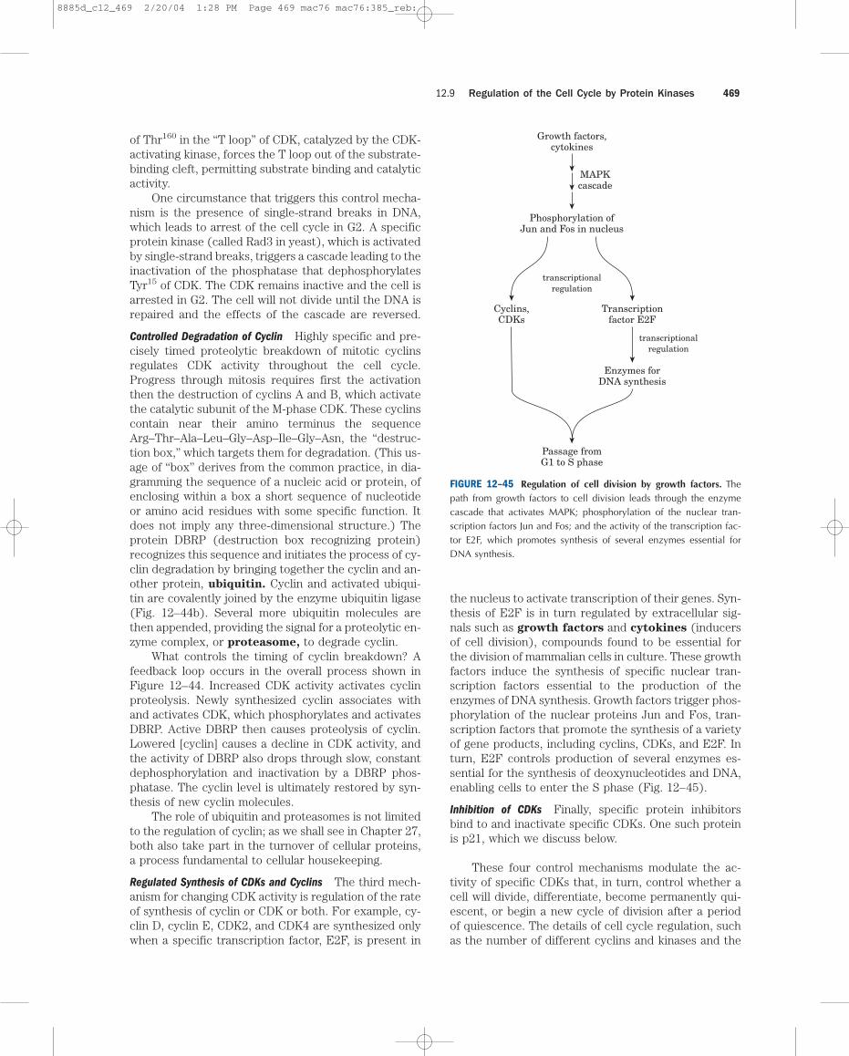
Regulated Synthesis of CDKs and Cyclins The third mech-anism for changing CDK activity is regulation of the rateof synthesis of cyclin or CDK or both. For example, cy-clin D, cyclin E, CDK2, and CDK4 are synthesized onlywhen a specific transcription factor, E2F, is present in
the nucleus to activate transcription of their genes. Syn-thesis of E2F is in turn regulated by extracellular sig-nals such as growth factors and cytokines (inducersof cell division), compounds found to be essential forthe division of mammalian cells in culture. These growthfactors induce the synthesis of specific nuclear tran-scription factors essential to the production of the enzymes of DNA synthesis. Growth factors trigger phos-phorylation of the nuclear proteins Jun and Fos, tran-scription factors that promote the synthesis of a varietyof gene products, including cyclins, CDKs, and E2F. Inturn, E2F controls production of several enzymes es-sential for the synthesis of deoxynucleotides and DNA,enabling cells to enter the S phase (Fig. 12–45).
Inhibition of CDKs Finally, specific protein inhibitorsbind to and inactivate specific CDKs. One such proteinis p21, which we discuss below.
These four control mechanisms modulate the ac-tivity of specific CDKs that, in turn, control whether acell will divide, differentiate, become permanently qui-escent, or begin a new cycle of division after a periodof quiescence. The details of cell cycle regulation, suchas the number of different cyclins and kinases and the
12.9 Regulation of the Cell Cycle by Protein Kinases 469
Growth factors,cytokines
Phosphorylation ofJun and Fos in nucleus
transcriptionalregulation
Passage fromG1 to S phase
MAPKcascade
Transcriptionfactor E2F
transcriptionalregulation
Cyclins,CDKs
Enzymes forDNA synthesis
FIGURE 12–45 Regulation of cell division by growth factors. The path from growth factors to cell division leads through the enzymecascade that activates MAPK; phosphorylation of the nuclear tran-scription factors Jun and Fos; and the activity of the transcription fac-tor E2F, which promotes synthesis of several enzymes essential forDNA synthesis.
8885d_c12_469 2/20/04 1:28 PM Page 469 mac76 mac76:385_reb:
combinations in which they act, differ from species tospecies, but the basic mechanism has been conservedin the evolution of all eukaryotic cells.
CDKs Regulate Cell Division by PhosphorylatingCritical Proteins
We have examined how cells maintain close control ofCDK activity, but how does the activity of CDK controlthe cell cycle? The list of target proteins that CDKs areknown to act upon continues to grow, and much remainsto be learned. But we can see a general pattern behindCDK regulation by inspecting the effect of CDKs on thestructures of laminin and myosin and on the activity ofretinoblastoma protein.
The structure of the nuclear envelope is maintainedin part by highly organized meshworks of intermediatefilaments composed of the protein laminin. Breakdownof the nuclear envelope before segregation of the sisterchromatids in mitosis is partly due to the phosphoryla-tion of laminin by a CDK, which causes laminin filamentsto depolymerize.
A second kinase target is the ATP-driven actin-myosin contractile machinery that pinches a dividingcell into two equal parts during cytokinesis. After thedivision, CDK phosphorylates a small regulatory subunitof myosin, causing dissociation of myosin from actin fil-aments and inactivating the contractile machinery. Sub-sequent dephosphorylation allows reassembly of thecontractile apparatus for the next round of cytokinesis.
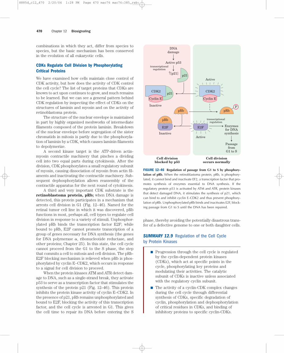
A third and very important CDK substrate is theretinoblastoma protein, pRb; when DNA damage isdetected, this protein participates in a mechanism thatarrests cell division in G1 (Fig. 12–46). Named for theretinal tumor cell line in which it was discovered, pRbfunctions in most, perhaps all, cell types to regulate celldivision in response to a variety of stimuli. Unphosphor-ylated pRb binds the transcription factor E2F; whilebound to pRb, E2F cannot promote transcription of agroup of genes necessary for DNA synthesis (the genesfor DNA polymerase �, ribonucleotide reductase, andother proteins; Chapter 25). In this state, the cell cyclecannot proceed from the G1 to the S phase, the stepthat commits a cell to mitosis and cell division. The pRb-E2F blocking mechanism is relieved when pRb is phos-phorylated by cyclin E–CDK2, which occurs in responseto a signal for cell division to proceed.
When the protein kinases ATM and ATR detect dam-age to DNA, such as a single-strand break, they activatep53 to serve as a transcription factor that stimulates thesynthesis of the protein p21 (Fig. 12–46). This proteininhibits the protein kinase activity of cyclin E–CDK2. Inthe presence of p21, pRb remains unphosphorylated andbound to E2F, blocking the activity of this transcriptionfactor, and the cell cycle is arrested in G1. This givesthe cell time to repair its DNA before entering the S
phase, thereby avoiding the potentially disastrous trans-fer of a defective genome to one or both daughter cells.
SUMMARY 12.9 Regulation of the Cell Cycle by Protein Kinases
■ Progression through the cell cycle is regulatedby the cyclin-dependent protein kinases(CDKs), which act at specific points in thecycle, phosphorylating key proteins andmodulating their activities. The catalyticsubunit of CDKs is inactive unless associatedwith the regulatory cyclin subunit.
■ The activity of a cyclin-CDK complex changesduring the cell cycle through differentialsynthesis of CDKs, specific degradation ofcyclin, phosphorylation and dephosphorylationof critical residues in CDKs, and binding ofinhibitory proteins to specific cyclin-CDKs.
Chapter 12 Biosignaling470
Cell divisionblocked by p53
Cell divisionoccurs normally
CDK2
Inactive
CDK2
Active
Cyclin E
Active p53
DNAdamage
↑[p21]
p21
p21
pRbP
pRb
pRb
E2F
Inactive
E2F Enzymesfor DNAsynthesis
transcriptionalregulation
transcriptionalregulation
Passagefrom
G1 to S
Active
Cyclin E
FIGURE 12–46 Regulation of passage from G1 to S by phosphory-lation of pRb. When the retinoblastoma protein, pRb, is phosphory-lated, it cannot bind and inactivate EF2, a transcription factor that pro-motes synthesis of enzymes essential to DNA synthesis. If theregulatory protein p53 is activated by ATM and ATR, protein kinasesthat detect damaged DNA, it stimulates the synthesis of p21, whichcan bind to and inhibit cyclin E–CDK2 and thus prevent phosphory-lation of pRb. Unphosphorylated pRb binds and inactivates E2F, block-ing passage from G1 to S until the DNA has been repaired.
8885d_c12_470 2/20/04 1:28 PM Page 470 mac76 mac76:385_reb:
12.10 Oncogenes, Tumor Suppressor Genes,and Programmed Cell DeathTumors and cancer are the result of uncontrolled celldivision. Normally, cell division is regulated by a familyof extracellular growth factors, proteins that cause rest-ing cells to divide and, in some cases, differentiate. Defects in the synthesis, regulation, or recognition ofgrowth factors can lead to cancer.
Oncogenes Are Mutant Forms of the Genes for Proteins That Regulate the Cell Cycle
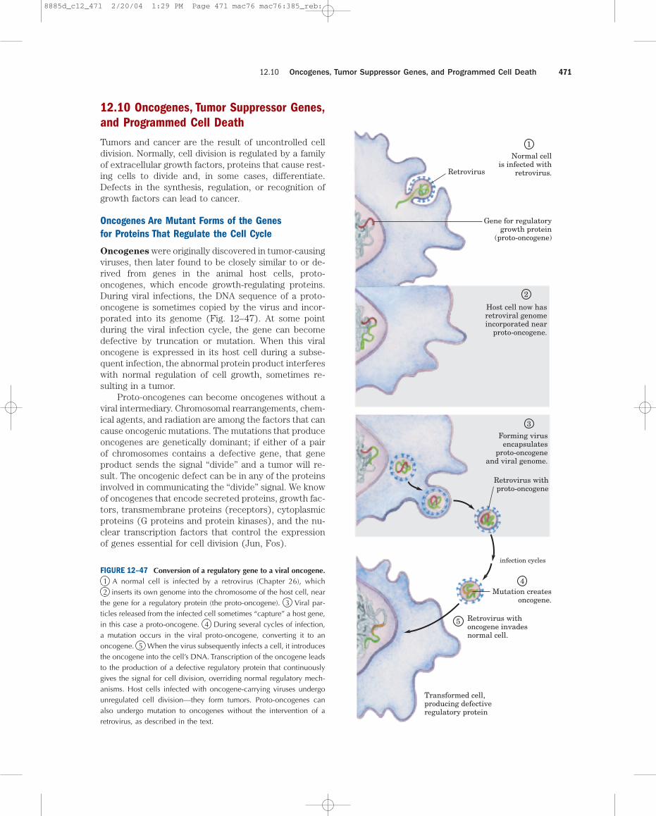
Oncogenes were originally discovered in tumor-causingviruses, then later found to be closely similar to or de-rived from genes in the animal host cells, proto-oncogenes, which encode growth-regulating proteins.During viral infections, the DNA sequence of a proto-oncogene is sometimes copied by the virus and incor-porated into its genome (Fig. 12–47). At some pointduring the viral infection cycle, the gene can becomedefective by truncation or mutation. When this viraloncogene is expressed in its host cell during a subse-quent infection, the abnormal protein product interfereswith normal regulation of cell growth, sometimes re-sulting in a tumor.
Proto-oncogenes can become oncogenes without aviral intermediary. Chromosomal rearrangements, chem-ical agents, and radiation are among the factors that cancause oncogenic mutations. The mutations that produceoncogenes are genetically dominant; if either of a pairof chromosomes contains a defective gene, that geneproduct sends the signal “divide” and a tumor will re-sult. The oncogenic defect can be in any of the proteinsinvolved in communicating the “divide” signal. We knowof oncogenes that encode secreted proteins, growth fac-tors, transmembrane proteins (receptors), cytoplasmicproteins (G proteins and protein kinases), and the nu-clear transcription factors that control the expressionof genes essential for cell division (Jun, Fos).
12.10 Oncogenes, Tumor Suppressor Genes, and Programmed Cell Death 471
Normal cellis infected with
retrovirus.Retrovirus
Gene for regulatorygrowth protein
(proto-oncogene)
Host cell now hasretroviral genomeincorporated near
proto-oncogene.
Forming virusencapsulates
proto-oncogeneand viral genome.
Retrovirus withproto-oncogene
infection cycles
Mutation createsoncogene.
Retrovirus withoncogene invadesnormal cell.
Transformed cell,producing defectiveregulatory protein
3
4
5
2
1
FIGURE 12–47 Conversion of a regulatory gene to a viral oncogene.1 A normal cell is infected by a retrovirus (Chapter 26), which2 inserts its own genome into the chromosome of the host cell, near
the gene for a regulatory protein (the proto-oncogene). 3 Viral par-ticles released from the infected cell sometimes “capture” a host gene,in this case a proto-oncogene. 4 During several cycles of infection,a mutation occurs in the viral proto-oncogene, converting it to anoncogene. 5 When the virus subsequently infects a cell, it introducesthe oncogene into the cell’s DNA. Transcription of the oncogene leadsto the production of a defective regulatory protein that continuouslygives the signal for cell division, overriding normal regulatory mech-anisms. Host cells infected with oncogene-carrying viruses undergounregulated cell division—they form tumors. Proto-oncogenes canalso undergo mutation to oncogenes without the intervention of aretrovirus, as described in the text.
8885d_c12_471 2/20/04 1:29 PM Page 471 mac76 mac76:385_reb:
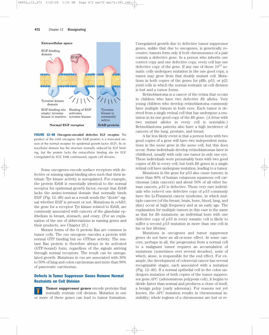
Some oncogenes encode surface receptors with de-fective or missing signal-binding sites such that their in-trinsic Tyr kinase activity is unregulated. For example,the protein ErbB is essentially identical to the normalreceptor for epidermal growth factor, except that ErbBlacks the amino-terminal domain that normally bindsEGF (Fig. 12–48) and as a result sends the “divide” sig-nal whether EGF is present or not. Mutations in erbB2,the gene for a receptor Tyr kinase related to ErbB, arecommonly associated with cancers of the glandular ep-ithelium in breast, stomach, and ovary. (For an expla-nation of the use of abbreviations in naming genes andtheir products, see Chapter 25.)
Mutant forms of the G protein Ras are common intumor cells. The ras oncogene encodes a protein withnormal GTP binding but no GTPase activity. The mu-tant Ras protein is therefore always in its activated(GTP-bound) form, regardless of the signals arrivingthrough normal receptors. The result can be unregu-lated growth. Mutations in ras are associated with 30%to 50% of lung and colon carcinomas and more than 90%of pancreatic carcinomas.
Defects in Tumor Suppressor Genes Remove NormalRestraints on Cell Division
Tumor suppressor genes encode proteins thatnormally restrain cell division. Mutation in one
or more of these genes can lead to tumor formation.
Unregulated growth due to defective tumor suppressorgenes, unlike that due to oncogenes, is genetically re-cessive; tumors form only if both chromosomes of a paircontain a defective gene. In a person who inherits onecorrect copy and one defective copy, every cell has onedefective copy of the gene. If any one of those 1012 so-matic cells undergoes mutation in the one good copy, atumor may grow from that doubly mutant cell. Muta-tions in both copies of the genes for pRb, p53, or p21yield cells in which the normal restraint on cell divisionis lost and a tumor forms.
Retinoblastoma is a cancer of the retina that occursin children who have two defective Rb alleles. Veryyoung children who develop retinoblastoma commonlyhave multiple tumors in both eyes. Each tumor is de-rived from a single retinal cell that has undergone a mu-tation in its one good copy of the Rb gene. (A fetus withtwo mutant alleles in every cell is nonviable.)Retinoblastoma patients also have a high incidence ofcancers of the lung, prostate, and breast.
A far less likely event is that a person born with twogood copies of a gene will have two independent muta-tions in the same gene in the same cell, but this doesoccur. Some individuals develop retinoblastomas later inchildhood, usually with only one tumor in only one eye.These individuals were presumably born with two goodcopies of Rb in every cell, but both Rb genes in a singleretinal cell have undergone mutation, leading to a tumor.
Mutations in the gene for p53 also cause tumors; inmore than 90% of human cutaneous squamous cell car-cinomas (skin cancers) and about 50% of all other hu-man cancers, p53 is defective. Those very rare individ-uals who inherit one defective copy of p53 commonlyhave the Li-Fraumeni cancer syndrome, in which mul-tiple cancers (of the breast, brain, bone, blood, lung, andskin) occur at high frequency and at an early age. Theexplanation for multiple tumors in this case is the sameas that for Rb mutations: an individual born with onedefective copy of p53 in every somatic cell is likely tosuffer a second p53 mutation in more than one cell inhis or her lifetime.
Mutations in oncogenes and tumor suppressorgenes do not have an all-or-none effect. In some can-cers, perhaps in all, the progression from a normal cellto a malignant tumor requires an accumulation ofmutations (sometimes over several decades), none ofwhich, alone, is responsible for the end effect. For ex-ample, the development of colorectal cancer has severalrecognizable stages, each associated with a mutation(Fig. 12–49). If a normal epithelial cell in the colon un-dergoes mutation of both copies of the tumor suppres-sor gene APC (adenomatous polyposis coli), it begins todivide faster than normal and produces a clone of itself,a benign polyp (early adenoma). For reasons not yetknown, the APC mutation results in chromosomal in-stability; whole regions of a chromosome are lost or re-
Chapter 12 Biosignaling472
EGF-bindingdomain
EGF-binding siteempty; tyrosinekinase is inactive.
Binding of EGFactivatestyrosine kinase.
Tyrosinekinase isconstantlyactive.
Tyrosine kinasedomain
EGF
Extracellular space
Normal EGF receptor ErbB protein
FIGURE 12–48 Oncogene-encoded defective EGF receptor. Theproduct of the erbB oncogene (the ErbB protein) is a truncated ver-sion of the normal receptor for epidermal growth factor (EGF). Its in-tracellular domain has the structure normally induced by EGF bind-ing, but the protein lacks the extracellular binding site for EGF.Unregulated by EGF, ErbB continuously signals cell division.
8885d_c12_472 2/20/04 1:30 PM Page 472 mac76 mac76:385_reb:
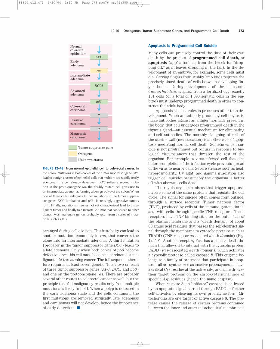
arranged during cell division. This instability can lead toanother mutation, commonly in ras, that converts theclone into an intermediate adenoma. A third mutation(probably in the tumor suppressor gene DCC) leads toa late adenoma. Only when both copies of p53 becomedefective does this cell mass become a carcinoma, a ma-lignant, life-threatening cancer. The full sequence there-fore requires at least seven genetic “hits”: two on eachof three tumor suppressor genes (APC, DCC, and p53)and one on the protooncogene ras. There are probablyseveral other routes to colorectal cancer as well, but theprinciple that full malignancy results only from multiplemutations is likely to hold. When a polyp is detected inthe early adenoma stage and the cells containing thefirst mutations are removed surgically, late adenomasand carcinomas will not develop; hence the importanceof early detection. ■
Apoptosis Is Programmed Cell Suicide
Many cells can precisely control the time of their owndeath by the process of programmed cell death, orapoptosis (app�-a-toe�-sis; from the Greek for “drop-ping off,” as in leaves dropping in the fall). In the de-velopment of an embryo, for example, some cells mustdie. Carving fingers from stubby limb buds requires theprecisely timed death of cells between developing fin-ger bones. During development of the nematodeCaenorhabditis elegans from a fertilized egg, exactly131 cells (of a total of 1,090 somatic cells in the em-bryo) must undergo programmed death in order to con-struct the adult body.
Apoptosis also has roles in processes other than de-velopment. When an antibody-producing cell begins tomake antibodies against an antigen normally present inthe body, that cell undergoes programmed death in thethymus gland—an essential mechanism for eliminatinganti-self antibodies. The monthly sloughing of cells ofthe uterine wall (menstruation) is another case of apop-tosis mediating normal cell death. Sometimes cell sui-cide is not programmed but occurs in response to bio-logical circumstances that threaten the rest of theorganism. For example, a virus-infected cell that diesbefore completion of the infection cycle prevents spreadof the virus to nearby cells. Severe stresses such as heat,hyperosmolarity, UV light, and gamma irradiation alsotrigger cell suicide; presumably the organism is betteroff with aberrant cells dead.
The regulatory mechanisms that trigger apoptosisinvolve some of the same proteins that regulate the cellcycle. The signal for suicide often comes from outside,through a surface receptor. Tumor necrosis factor(TNF), produced by cells of the immune system, inter-acts with cells through specific TNF receptors. Thesereceptors have TNF-binding sites on the outer face ofthe plasma membrane and a “death domain” of about80 amino acid residues that passes the self-destruct sig-nal through the membrane to cytosolic proteins such asTRADD (TNF receptor-associated death domain) (Fig.12–50). Another receptor, Fas, has a similar death do-main that allows it to interact with the cytosolic proteinFADD (Fas-associated death domain), which activatesa cytosolic protease called caspase 8. This enzyme be-longs to a family of proteases that participate in apop-tosis; all are synthesized as inactive proenzymes, all havea critical Cys residue at the active site, and all hydrolyzetheir target proteins on the carboxyl-terminal side ofspecific Asp residues (hence the name caspase).
When caspase 8, an “initiator” caspase, is activatedby an apoptotic signal carried through FADD, it furtherself-activates by cleaving its own proenzyme form. Mi-tochondria are one target of active caspase 8. The pro-tease causes the release of certain proteins containedbetween the inner and outer mitochondrial membranes:
12.10 Oncogenes, Tumor Suppressor Genes, and Programmed Cell Death 473
APC
ras
DCC?
p53
?
?
Normalcolorectalepithelium
Earlyadenoma
Intermediateadenoma
Advancedadenoma
Colorectalcarcinoma
Invasivecarcinoma
Metastaticcarcinoma
Tumor suppressor gene
Oncogene
Unknown status
FIGURE 12–49 From normal epithelial cell to colorectal cancer. Inthe colon, mutations in both copies of the tumor suppressor gene APClead to benign clusters of epithelial cells that multiply too rapidly (earlyadenoma). If a cell already defective in APC suffers a second muta-tion in the proto-oncogene ras, the doubly mutant cell gives rise toan intermediate adenoma, forming a benign polyp of the colon. Whenone of these cells undergoes further mutations in the tumor suppres-sor genes DCC (probably) and p53, increasingly aggressive tumorsform. Finally, mutations in genes not yet characterized lead to a ma-lignant tumor and finally to a metastatic tumor that can spread to othertissues. Most malignant tumors probably result from a series of muta-tions such as this.
8885d_c12_473 2/20/04 1:30 PM Page 473 mac76 mac76:385_reb:
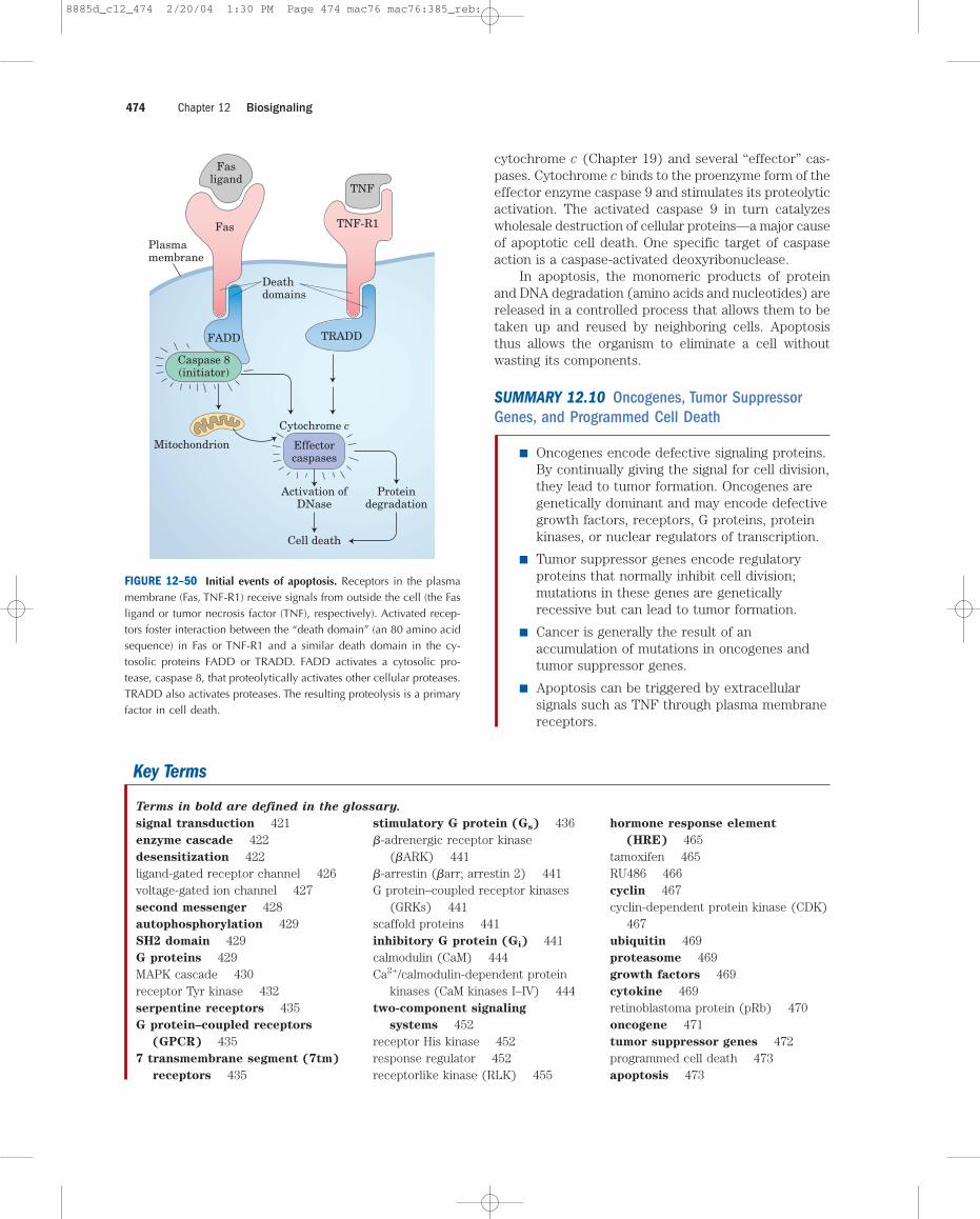
cytochrome c (Chapter 19) and several “effector” cas-pases. Cytochrome c binds to the proenzyme form of theeffector enzyme caspase 9 and stimulates its proteolyticactivation. The activated caspase 9 in turn catalyzeswholesale destruction of cellular proteins—a major causeof apoptotic cell death. One specific target of caspaseaction is a caspase-activated deoxyribonuclease.
In apoptosis, the monomeric products of proteinand DNA degradation (amino acids and nucleotides) arereleased in a controlled process that allows them to betaken up and reused by neighboring cells. Apoptosisthus allows the organism to eliminate a cell withoutwasting its components.
SUMMARY 12.10 Oncogenes, Tumor SuppressorGenes, and Programmed Cell Death
■ Oncogenes encode defective signaling proteins.By continually giving the signal for cell division,they lead to tumor formation. Oncogenes aregenetically dominant and may encode defectivegrowth factors, receptors, G proteins, proteinkinases, or nuclear regulators of transcription.
■ Tumor suppressor genes encode regulatoryproteins that normally inhibit cell division;mutations in these genes are geneticallyrecessive but can lead to tumor formation.
■ Cancer is generally the result of anaccumulation of mutations in oncogenes andtumor suppressor genes.
■ Apoptosis can be triggered by extracellularsignals such as TNF through plasma membranereceptors.
Chapter 12 Biosignaling474
FIGURE 12–50 Initial events of apoptosis. Receptors in the plasmamembrane (Fas, TNF-R1) receive signals from outside the cell (the Fasligand or tumor necrosis factor (TNF), respectively). Activated recep-tors foster interaction between the “death domain” (an 80 amino acidsequence) in Fas or TNF-R1 and a similar death domain in the cy-tosolic proteins FADD or TRADD. FADD activates a cytosolic pro-tease, caspase 8, that proteolytically activates other cellular proteases.TRADD also activates proteases. The resulting proteolysis is a primaryfactor in cell death.
Fas
Fasligand
FADD
TNF-R1
TNF
TRADD
Mitochondrion
Plasmamembrane
Effectorcaspases
Activation ofDNase
Cytochrome c
Deathdomains
Proteindegradation
Cell death
Caspase 8(initiator)
Key Terms
signal transduction 421enzyme cascade 422desensitization 422ligand-gated receptor channel 426voltage-gated ion channel 427second messenger 428autophosphorylation 429SH2 domain 429G proteins 429MAPK cascade 430receptor Tyr kinase 432serpentine receptors 435G protein–coupled receptors
(GPCR) 4357 transmembrane segment (7tm)
receptors 435
stimulatory G protein (Gs) 436�-adrenergic receptor kinase
(�ARK) 441�-arrestin (�arr; arrestin 2) 441G protein–coupled receptor kinases
(GRKs) 441scaffold proteins 441inhibitory G protein (Gi) 441calmodulin (CaM) 444Ca2+/calmodulin-dependent protein
kinases (CaM kinases I–IV) 444two-component signaling
systems 452receptor His kinase 452response regulator 452receptorlike kinase (RLK) 455
hormone response element
(HRE) 465tamoxifen 465RU486 466cyclin 467cyclin-dependent protein kinase (CDK)
467ubiquitin 469proteasome 469growth factors 469cytokine 469retinoblastoma protein (pRb) 470oncogene 471tumor suppressor genes 472programmed cell death 473apoptosis 473
Terms in bold are defined in the glossary.
8885d_c12_474 2/20/04 1:30 PM Page 474 mac76 mac76:385_reb:
Chapter 12 Further Reading 475
Further Reading
GeneralCohen, P. (2001) The role of protein phosphorylation in humanhealth and disease: the Sir Hans Krebs Medal Lecture. Eur. J.
Biochem. 268, 5001–5010.An intermediate-level review of the role of protein kinases, theiralteration in disease, and the drugs that affect their activity.
Cohen, P. (2000) The regulation of protein function by multisitephosphorylation—a 25 year update. Trends Biochem. Sci. 25,
596–601.Historical account of the developments in proteinphosphorylation.
Heilmeyer, L. & Friedrich, P. (eds) (2001) Protein Modules in
Cellular Signalling, IOS Press, Washington, DC.
Receptor Ion ChannelsSee also Chapter 11, Further Reading, Ion Channels.
Aidley, D.J. & Stanfield, P.R. (1996) Ion Channels: Molecules
in Action, Cambridge University Press, Cambridge. Clear, concise introduction to the physics, chemistry, andmolecular biology used in research on ion channels; emphasis ison molecular approaches.
Lehmann-Horn, F. & Jurkat-Rott, K. (1999) Voltage-gated ionchannels and hereditary disease. Physiol. Rev. 79, 1317–1372.
Advanced review of the structure and function of ion channels,with special emphasis on cases in which ion-channel defectsproduce human diseases.
Receptor EnzymesFoster, D.C., Wedel, B.J., Robinson, S.W., & Garbers, D.L.
(1999) Mechanisms of regulation and functions of guanylylcyclases. Rev. Physiol. Biochem. Pharmacol. 135, 1–39.
Advanced review of the structure and function of signal-transducing guanylyl cyclases.
Saltiel, A.R. & Pessin, J.E. (2002) Insulin signaling pathways intime and space. Trends Biochem Sci. 12, 65–71.
Short, intermediate-level review.
Schaeffer, H.J. & Weber, M.J. (1999) Mitogen-activated proteinkinases: specific messages from ubiquitous messengers. Mol. Cell.
Biol. 19, 2435–2444.Intermediate-level review of MAPKs and the basis for specificsignaling through these general signaling proteins.
Schlessinger, J. (2000) Cell signaling by receptor tyrosinekinases. Cell 103, 211–225.
Shepherd, P.R., Withers, D.J., & Siddle, K. (1998) Phospho-inositide 3-kinase: the key switch mechanism in insulin signalling.Biochem. J. 333, 471–490.
Intermediate-level review of the importance of PKB and PI-3Kin metabolic regulation by insulin.
Shields, J.M., Pruitt, K., McFall, A., Shaub, A., & Der, C.J.
(2000) Understanding Ras: it ain’t over ’til it’s over. Trends Cell
Biol. 10, 147–154.Intermediate-level review of the monomeric G protein Ras.
Widmann, C., Gibson, S., Jarpe, M.B., & Johnson, G.L.
(1999) Mitogen-activated protein kinase: conservation of a three-kinase module from yeast to human. Physiol. Rev. 79, 143–180.
Advanced review of the roles of MAPKs in diverse organisms,from yeast, slime mold, and nematode to vertebrates and plants.
Zajchowski, L.D. & Robbins, S.M. (2002) Lipid rafts and littlecaves: compartmentalized signalling in membrane microdomains.Eur. J. Biochem. 269, 737–752.
Serpentine ReceptorsHahn, K. & Toutchkine, A. (2002) Live-cell fluorescent biosen-sors for activated signaling proteins. Curr. Opin. Cell Biol. 14,
167–172.Brief, intermediate-level review.
Hamm, H.E. (1998) The many faces of G protein signaling. J. Biol. Chem. 273, 669–672.
Introduction to a series of short reviews on G proteins.
Lodowski, D.T., Pitcher, J.A., Capel, W.D., Lefkowitz, R.J., &
Tesmer, J.J.G. (2003) Keeping G proteins at bay: a complex be-tween G protein–coupled receptor kinase 2 and G��. Science 300,
1256–1262.
Martin, T.F.J. (1998) Phosphoinositide lipids as signaling mole-cules. Annu. Rev. Cell Dev. Biol. 14, 231–264.
Discussion of the roles of phosphatidylinositol derivatives insignal transduction, cytoskeletal regulation, and membranetrafficking.
Perry, S.J. & Lefkowitz, R.J. (2002) Arresting developments inheptahelical receptor signaling and regulation. Trends Cell Biol.12, 130–138.
Role of arrestins in serpentine receptor adaptation.
Pinna, L.A. & Ruzzene, M. (1996) How do protein kinases rec-ognize their substrates? Biochim. Biophys. Acta 1314, 191–225.
Advanced review of the factors, including consensus sequences,that give protein kinases their specificity.
Roach, P.J. (1991) Multisite and hierarchal protein phosphoryla-tion. J. Biol. Chem. 266, 14,139–14,142.
Report on the importance of multiple phosphorylation sites inthe fine regulation of protein function.
Skiba, N.P. & Hamm, H.E. (1998) How Gs� activates adenylylcyclase. Nat. Struct. Biol. 5, 88–92.
Intermediate-level review of the mechanism of G-protein action,based on structural studies.
Zaccolo, M., De Giorgi, F., Cho, C.Y., Feng, L., Knapp, T.,
Negulescu, P.A., Taylor, S.S., Tsien, R.Y., & Pozzan, T. (2000)A genetically encoded, fluorescent indicator for cyclic AMP in liv-ing cells. Nat. Cell Biol. 2, 25–29.
Describes the technique shown in Figure 5 of Box 12–2.
Zhang, J., Campbell, R.E., Ting, A.Y., & Tsien, R.Y. (2002)Creating new fluorescent probes for cell biology. Nat. Rev. Molec.
Cell Biol. 3, 906–918.The basis for techniques such as those described in Box 12–2.
Scaffold Proteins and Membrane Rafts See also Chapter 11, Further Reading, Membrane Dynamics.
Filipek, S., Stenkamp, R.E., Teller, D.C., & Palczewski, K.
(2003) G protein-coupled receptor rhodopsin: A prospectus.Annu. Rev. Physiol. 65, 851–879.
Advanced review.
8885d_c12_475 2/20/04 1:30 PM Page 475 mac76 mac76:385_reb:
Chapter 12 Biosignaling476
Lim, W.A. (2002) The modular logic of signaling proteins: buildingallosteric switches from simple binding domains. Curr. Opin.
Struct. Biol. 12, 61–68.
Michel, J.J.C. & Scott, J.D. (2002) AKAP mediated signaltransduction. Annu. Rev. Pharmacol. Toxicol. 42, 235–257.
Advanced review of the proteins that target PKA to subcellularcompartments.
Neel, B.G., Gu, H., & Pao, L. (2003) The “Shp”ing news: SH2domain–containing tyrosine phosphatases in cell signaling. Trends
Biochem. Sci. 28, 284–293.
Pawson, T. & Nash, P. (2003) Assembly of cell regulatory systemsthrough protein interaction domains. Science 300, 445–452.
Intermediate-level review of multivalent proteins and the com-plexes they form.
Saltiel, A.R. & Pessin, J.E. (2002) Insulin signaling pathways intime and space. Trends Cell Biol. 12, 65–71.
Siegal, G. (1999) The surprisingly flexible PTB domain. Nat.
Struct. Biol. 6, 7–10.
Tsui-Pierchala, B.A., Encinas, M., Milbrandt, J., & Johnson,
E.M., Jr. (2002) Lipid rafts in neuronal signaling and function.Trends Neurosci. 25, 412–417.
Yaffe, M.D. & Elia, A.E.H. (2001) Phosphoserine/threonine-binding domains. Curr. Opin. Cell Biol. 13, 131–138.
Calcium Ions in SignalingBerridge, M.J. (1993) Inositol triphosphate and calcium sig-nalling. Nature 361, 315–325.
Classic description of the IP3 signaling system.
Berridge, M.J., Lipp, P., & Bootman, M.D. (2000) The versatil-ity and universality of calcium signaling. Nat. Rev. Molec. Cell
Biol. 1, 11–21.Intermediate review.
Chin, D. & Means, A.R. (2000) Calmodulin: a prototypicalcalcium receptor. Trends Cell Biol. 10, 322–328.
Nowycky, M.C. & Thomas, A.P. (2002) Intracellular calciumsignaling. J. Cell Sci. 115, 3715–3716.
A comprehensive poster of all elements of Ca2� signaling.
Takahashi, A., Camacho, P., Lechleiter, J.D., & Herman, B.
(1999) Measurement of intracellular calcium. Physiol. Rev. 79,
1089–1125.Advanced review of methods for estimating intracellular Ca2�
levels in real time.
Thomas, A.P., Bird, G.S.J., Hajnoczky, G., Robb-Gaspers,
L.D., & Putney, J.W., Jr. (1996) Spatial and temporal aspects ofcellular calcium signaling. FASEB J. 10, 1505–1517.
Signaling in Plants and BacteriaBakal, C.J. & Davies, J.E. (2000) No longer an exclusive club:eukaryotic signaling domains in bacteria. Trends Cell Biol. 10,
32–38.Intermediate review.
Becraft, P.W. (2002) Receptor kinase signaling in plant develop-ment. Annu. Rev. Cell Dev. Biol. 18, 163–192.
Advanced review.
Chang, C. & Stadler, R. (2001) Ethylene hormone receptoraction in Arabidopsis. Bioessays 23, 619–627.
Cock, J.M., Vanoosthuyse, V., & Gaude, T. (2002) Receptorkinase signalling in plants and animals: distinct molecular systemswith mechanistic similarities. Curr. Opin. Cell Biol. 14, 230–236.
Hwang, I., Chen, H.-C., & Sheen, J. (2002) Two-componentcircuitry in Arabidopsis cytokinin signal transduction. Nature
413, 383–389.
Jones, A.M. (2002) G-protein–coupled signaling in Arabidopsis.
Curr. Opin. Plant Biol. 5, 402–407.
Matsubayashi, Y., Yang, H., & Sakagami, Y. (2001) Peptide signalsand their receptors in higher plants. Trends Plant Sci. 6, 573–577.
McCarty, D.R. & Chory, J. (2000) Conservation and innovationin plant signaling pathways. Cell 103, 201–209.
Meijer, H.J.G. & Munnik, T. (2003) Phospholipid-based signalingin plants. Annu. Rev. Plant Biol. 54, 265–306.
Advanced review.
Ouaked, F., Rozhon, W., Lecourieus, D., & Hirt, H. (2003) AMAPK pathway mediates ethylene signaling in plants. EMBO J. 22,
1282–1288.
Talke, I.N., Blaudez, D., Maathuis, F.J.M., & Sanders, D.
(2003) CNGCs: prime targets of plant cyclic nucleotide signalling?Trends Plant Sci. 8, 286–293.
Tichtinsky, G., Vanoosthuyse, V., Cock, J.M., & Gaude, T.
(2003) Making inroads into plant receptor kinase signalling path-ways. Trends Plant Sci. 8, 231–237.
Vision, Olfaction, and GustationBaylor, D. (1996) How photons start vision. Proc. Natl. Acad.
Sci. USA 93, 560–565.One of six short reviews on vision in this journal issue.
Margolskee, R.F. (2002) Molecular mechanisms of bitter andsweet taste transduction. J. Biol. Chem. 277, 1–4.
Menon, S.T., Han, M., & Sakmar, T.P. (2001) Rhodopsin: struc-tural basis of molecular physiology. Physiol. Rev. 81, 1659–1688.
Advanced review.
Mombaerts, P. (2001) The human repertoire of odorant receptorgenes and pseudogenes. Annu. Rev. Genomics Hum. Genet. 2,
493–510.Advanced review.
Nathans, J. (1989) The genes for color vision. Sci. Am. 260
(February), 42–49.
Ronnett, G.V. & Moon, C. (2002) G proteins and olfactory signaltransduction. Annu. Rev. Physiol. 64, 189–222.
Advanced review.
Scott, K. & Zuker, C. (1997) Lights out: deactivation of the pho-totransduction cascade. Trends Biochem. Sci. 22, 350–354.
Steroid Hormone Receptors and ActionCarson-Jurica, M.A., Schrader, W.T., & O’Malley, B.W. (1990)Steroid receptor family—structure and functions. Endocr. Rev. 11,
201–220.Advanced discussion of the structure of nuclear hormonereceptors and the mechanisms of their action.
Hall, J.M., Couse, J.F., & Korach, K.S. (2001) The multifac-eted mechanisms of estradiol and estrogen receptor signaling. J. Biol. Chem. 276, 36,869–36,872.
Brief, intermediate-level review.
8885d_c12_476 2/20/04 1:30 PM Page 476 mac76 mac76:385_reb:
Chapter 12 Problems 477
1. Therapeutic Effects of Albuterol The respi-ratory symptoms of asthma result from constriction
of the bronchi and bronchioles of the lungs due to contrac-tion of the smooth muscle of their walls. This constriction canbe reversed by raising the [cAMP] in the smooth muscle. Ex-plain the therapeutic effects of albuterol, a �-adrenergic ag-onist taken (by inhalation) for asthma. Would you expect thisdrug to have any side effects? How might one design a bet-ter drug that did not have these effects?
2. Amplification of Hormonal Signals Describe all thesources of amplification in the insulin receptor system.
3. Termination of Hormonal Signals Signals carried byhormones must eventually be terminated. Describe severaldifferent mechanisms for signal termination.
4. Specificity of a Signal for a Single Cell Type Dis-cuss the validity of the following proposition. A signalingmolecule (hormone, growth factor, or neurotransmitter) elic-its identical responses in different types of target cells if theycontain identical receptors.
5. Resting Membrane Potential A variety of unusual in-vertebrates, including giant clams, mussels, and polychaeteworms, live on the fringes of hydrothermal vents on the oceanbottom, where the temperature is 60 �C.
(a) The adductor muscle of a deep-sea giant clam has aresting membrane potential of �95 mV. Given the intracellu-lar and extracellular ionic compositions shown below, wouldyou have predicted this membrane potential? Why or why not?
(b) Assume that the adductor muscle membrane is per-meable to only one of the ions listed above. Which ion coulddetermine the Vm?
Concentration (mM)
Ion Intracellular Extracellular
Na� 50 440K� 400 20Cl� 21 560Ca2� 0.4 10
Problems
Jordan, V.C. (1998) Designer estrogens. Sci. Am. 279 (October),60–67.
Introductory review of the mechanism of estrogen action andthe effects of estrogenlike compounds in medicine.
Lösel, R.M., Falkenstein, E., Feuring, M., Schultz, A.,
Tillmann, H.-C., Rossol-Haseroth, K., & Wehling, M. (2003)Nongenomic steroid action: controversies, questions, and answers.Physiol. Rev. 83, 965–1016.
Detailed review of the evidence for steroid hormone actionthrough plasma membrane receptors.
Cell Cycle and CancerCavenee, W.K. & White, R.L. (1995) The genetic basis of cancer.Sci. Am. 272 (March), 72–79.
Chau, B.N. & Wang, J.Y.J. (2003) Coordinated regulation of lifeand death by RB. Nat. Rev. Cancer 3, 130–138.
Fearon, E.R. (1997) Human cancer syndromes—clues to the ori-gin and nature of cancer. Science 278, 1043–1050.
Intermediate-level review of the role of inherited mutations inthe development of cancer.
Herwig, S. & Strauss, M. (1997) The retinoblastoma protein: amaster regulator of cell cycle, differentiation and apoptosis. Eur. J.
Biochem. 246, 581–601.
Hunt, M. & Hunt, T. (1993) The Cell Cycle: An Introduction,
W. H. Freeman and Company/Oxford University Press, New York/Oxford.
Kinzler, K.W. & Vogelstein, B. (1996) Lessons from hereditarycolorectal cancer. Cell 87, 159–170.
Evidence for multistep processes in the development of cancer.
Levine, A.J. (1997) p53, the cellular gatekeeper for growth anddivision. Cell 88, 323–331.
Intermediate coverage of the function of protein p53 in thenormal cell cycle and in cancer.
Morgan, D.O. (1997) Cyclin-dependent kinases: engines, clocks,and microprocessors. Annu. Rev. Cell Dev. Biol. 13, 261–291.
Advanced review.
Obaya, A.J. & Sedivy, J.M. (2002) Regulation of cyclin-Cdkactivity in mammalian cells. Cell. Mol. Life Sci. 59, 126–142.
Rajagopalan, H., Nowak, M.A., Vogelstein, B., & Lengauer,
C. (2003) The significance of unstable chromosomes in colorectalcancer. Nat. Rev. Cancer 3, 695–701.
Sherr, C.J. & McCormick, F. (2002) The RB and p53 pathwaysin cancer. Cancer Cell 2, 102–112.
Weinberg, R.A. (1996) How cancer arises. Sci. Am. 275 (Septem-ber), 62–70.
Yamasaki, L. (2003) Role of the RB tumor suppressor in cancer.Cancer Treatment Res. 115, 209–239.
Advanced review.
ApoptosisAnderson, P. (1997) Kinase cascades regulating entry intoapoptosis. Microbiol. Mol. Biol. Rev. 61, 33–46.
Ashkenazi, A. & Dixit, V.M. (1998) Death receptors: signalingand modulation. Science 281, 1305–1308.
This and the papers by Green and Reed and by Thornberry andLazebnik (below) are in an issue of Science devoted to apoptosis.
Duke, R.C., Ojcius, D.M., & Young, J.D.-E. (1996) Cell suicidein health and disease. Sci. Am. 275 (December), 80–87.
Green, D.R. & Reed, J.C. (1998) Mitochondria and apoptosis.Science 281, 1309–1312.
Jacobson, M.D., Weil, M., & Raff, M.C. (1997) Programmedcell death in animal development. Cell 88, 347–354.
Lawen, A. (2003) Apoptosis—an introduction. Bioessays 25,
888–896.
Thornberry, N.A. & Lazebnik, Y. (1998) Caspases: enemieswithin. Science 281, 1312.
8885d_c12_477 2/20/04 1:31 PM Page 477 mac76 mac76:385_reb:
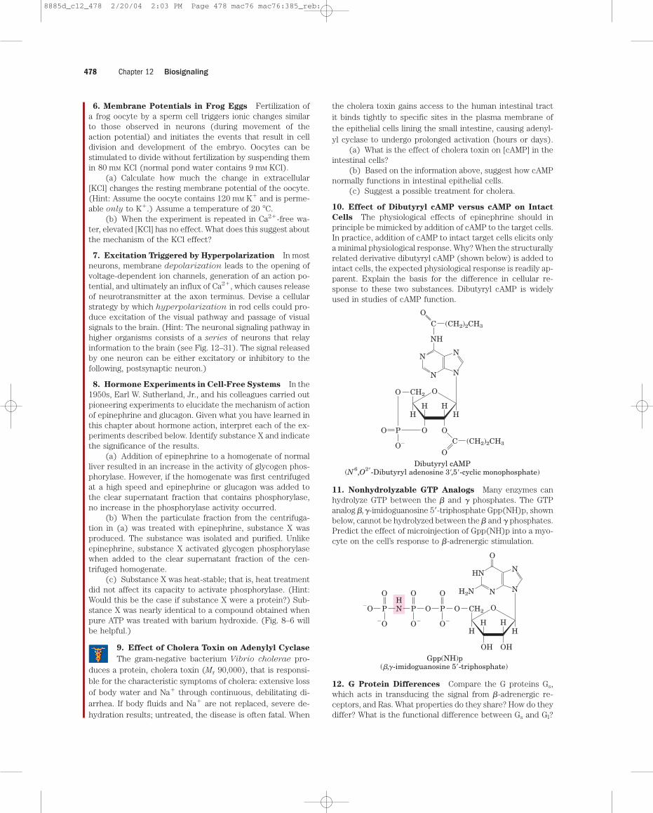
Chapter 12 Biosignaling478
6. Membrane Potentials in Frog Eggs Fertilization ofa frog oocyte by a sperm cell triggers ionic changes similarto those observed in neurons (during movement of the action potential) and initiates the events that result in cell division and development of the embryo. Oocytes can be stimulated to divide without fertilization by suspending themin 80 mM KCl (normal pond water contains 9 mM KCl).
(a) Calculate how much the change in extracellular[KCl] changes the resting membrane potential of the oocyte.(Hint: Assume the oocyte contains 120 mM K� and is perme-able only to K�.) Assume a temperature of 20 �C.
(b) When the experiment is repeated in Ca2�-free wa-ter, elevated [KCl] has no effect. What does this suggest aboutthe mechanism of the KCl effect?
7. Excitation Triggered by Hyperpolarization In mostneurons, membrane depolarization leads to the opening ofvoltage-dependent ion channels, generation of an action po-tential, and ultimately an influx of Ca2�, which causes releaseof neurotransmitter at the axon terminus. Devise a cellularstrategy by which hyperpolarization in rod cells could pro-duce excitation of the visual pathway and passage of visualsignals to the brain. (Hint: The neuronal signaling pathway inhigher organisms consists of a series of neurons that relayinformation to the brain (see Fig. 12–31). The signal releasedby one neuron can be either excitatory or inhibitory to thefollowing, postsynaptic neuron.)
8. Hormone Experiments in Cell-Free Systems In the1950s, Earl W. Sutherland, Jr., and his colleagues carried outpioneering experiments to elucidate the mechanism of actionof epinephrine and glucagon. Given what you have learned inthis chapter about hormone action, interpret each of the ex-periments described below. Identify substance X and indicatethe significance of the results.
(a) Addition of epinephrine to a homogenate of normalliver resulted in an increase in the activity of glycogen phos-phorylase. However, if the homogenate was first centrifugedat a high speed and epinephrine or glucagon was added tothe clear supernatant fraction that contains phosphorylase,no increase in the phosphorylase activity occurred.
(b) When the particulate fraction from the centrifuga-tion in (a) was treated with epinephrine, substance X wasproduced. The substance was isolated and purified. Unlikeepinephrine, substance X activated glycogen phosphorylasewhen added to the clear supernatant fraction of the cen-trifuged homogenate.
(c) Substance X was heat-stable; that is, heat treatmentdid not affect its capacity to activate phosphorylase. (Hint:Would this be the case if substance X were a protein?) Sub-stance X was nearly identical to a compound obtained whenpure ATP was treated with barium hydroxide. (Fig. 8–6 willbe helpful.)
9. Effect of Cholera Toxin on Adenylyl Cyclase
The gram-negative bacterium Vibrio cholerae pro-duces a protein, cholera toxin (Mr 90,000), that is responsi-ble for the characteristic symptoms of cholera: extensive lossof body water and Na� through continuous, debilitating di-arrhea. If body fluids and Na� are not replaced, severe de-hydration results; untreated, the disease is often fatal. When
the cholera toxin gains access to the human intestinal tractit binds tightly to specific sites in the plasma membrane ofthe epithelial cells lining the small intestine, causing adenyl-yl cyclase to undergo prolonged activation (hours or days).
(a) What is the effect of cholera toxin on [cAMP] in theintestinal cells?
(b) Based on the information above, suggest how cAMPnormally functions in intestinal epithelial cells.
(c) Suggest a possible treatment for cholera.
10. Effect of Dibutyryl cAMP versus cAMP on Intact
Cells The physiological effects of epinephrine should inprinciple be mimicked by addition of cAMP to the target cells.In practice, addition of cAMP to intact target cells elicits onlya minimal physiological response. Why? When the structurallyrelated derivative dibutyryl cAMP (shown below) is added tointact cells, the expected physiological response is readily ap-parent. Explain the basis for the difference in cellular re-sponse to these two substances. Dibutyryl cAMP is widelyused in studies of cAMP function.
11. Nonhydrolyzable GTP Analogs Many enzymes canhydrolyze GTP between the � and � phosphates. The GTPanalog �,�-imidoguanosine 5�-triphosphate Gpp(NH)p, shownbelow, cannot be hydrolyzed between the � and � phosphates.Predict the effect of microinjection of Gpp(NH)p into a myo-cyte on the cell’s response to �-adrenergic stimulation.
12. G Protein Differences Compare the G proteins Gs,which acts in transducing the signal from �-adrenergic re-ceptors, and Ras. What properties do they share? How do theydiffer? What is the functional difference between Gs and GI?
Gpp(NH)p
CH2O O
OH
��( , -imidoguanosine 5�-triphosphate)
OH
HHH
H
N
O
N
HN
H2N
N
O�
P
O
O
O�
P
O
�O
P
O
�OHN
Dibutyryl cAMP(N6,O2�-Dibutyryl adenosine 3�,5�-cyclic monophosphate)
(CH2)2CH3
CH2
C
(CH2)2CH3C
O
OO O
O
HHH
H
N
NH
N
N N
O
P
O
O
�
8885d_c12_478 2/20/04 2:03 PM Page 478 mac76 mac76:385_reb:
Chapter 12 Problems 479
13. EGTA Injection EGTA (ethylene glycol-bis(�-amino-ethyl ether)-N,N,N�,N�-tetraacetic acid) is a chelating agentwith high affinity and specificity for Ca2�. By microinjectinga cell with an appropriate Ca2�-EDTA solution, an experi-menter can prevent cytosolic [Ca2�] from rising above 10�7
M.How would EGTA microinjection affect a cell’s response tovasopressin (see Table 12–5)? To glucagon?
14. Visual Desensitization Oguchi’s disease is aninherited form of night blindness. Affected individu-
als are slow to recover vision after a flash of bright lightagainst a dark background, such as the headlights of a car onthe freeway. Suggest what the molecular defect(s) might bein Oguchi’s disease. Explain in molecular terms how this de-fect accounts for the night blindness.
15. Mutations in PKA Explain how mutations in the R orC subunit of cAMP-dependent protein kinase (PKA) mightlead to (a) a constantly active PKA or (b) a constantly inac-tive PKA.
16. Mechanisms for Regulating Protein Kinases Iden-tify eight general types of protein kinases found in eukary-otic cells, and explain what factor is directly responsible foractivating each type.
17. Mutations in Tumor Suppressor Genes and Onco-
genes Explain why mutations in tumor suppressor genesare recessive (both copies of the gene must be defective forthe regulation of cell division to be defective) whereas mu-tations in oncogenes are dominant.
18. Retinoblastoma in Children Explain whysome children with retinoblastoma develop multiple
tumors of the retina in both eyes, whereas others have a sin-gle tumor in only one eye.
19. Mutations in ras How does a mutation in the ras
gene that leads to formation of a Ras protein with no GTPaseactivity affect a cell’s response to insulin?
8885d_c12_479 2/20/04 1:31 PM Page 479 mac76 mac76:385_reb: