Embed Size (px)
Citation preview

1,2,3W.F. Sensakovic, PhD, DABR
1Florida Hospital 2Florida State University, 3University of Central Florida

• Does not include
– In-depth discussions (see references on each slide)
– PET/MRI
– Radiotracers other than F-18 FDG
– Segmentation Methods
– Registration and Respiration Control Methods
– QA/QC
– Reimbursement and Regulation
– Use in specific tumor types

1. Positron emission
2. Annihilation
3
+
Images: William F. Sensakovic, PhD

• 0.97 Positron (β+) per Disintegration
– 634keV Maximum
– 250keV Mean
• 0.03 Electron Capture per Disintegration
Energy (keV) 0 100 200 300 400 500 600
Nu
mb
er
of
Po
sitr
on
s
F18
9
O18
8
EC1.022 MeV
• T1/2=110min • Range (adds ~0.2mm blur)
₋ 2.3mm mean in water ₋ 0.6mm max in water ₋ 2m max in air
(Contamination)
Images: William F. Sensakovic, PhD

• Positron slows to stop and forms positronium
• Annihilates creating two photons at 180o and 511keV
– ~2% of the time non-zero Ke+
• Not equal energy or angle
• ~±0.25o angular variation for F-18 (adds 1.75-2mm blur)
• Can have more than 2 photons, but it is rare
– Three-photon: 0.2%
– More than 3 photons: ~0.00015%

6
• CT – Similar to diagnostic – Defer description to talk on
CT simulation
• PET – 2D being phased out – Bore: 70-80cm
» Scan FOV ~10cm less
– 170-200cm scan length – Scan time: 30s-3min per bed
position » Most are step-shoot
Siemens is an exception
http://www3.gehealthcare.com/en/Education/Product_Education_Clinical/TiP_Applications/PET_Onsite/Discovery_LS IAEA Training Materials Radiation Protection in PET/CT
PET CT

7
• Rings
– Varies: 4-8 Block Rings
• 15-22cm axial coverage
• Detector elements
– 12,000-30,000
http://www3.gehealthcare.com/en/Education/Product_Education_Clinical/TiP_Applications/PET_Onsite/Discovery_LS IAEA Training Materials Radiation Protection in PET/CT

8
• ~50 blocks per ring
– 4 PMT
– 1 Scintillator
» Cut into multiple channels (~10x10)
• Anger logic
Photo: IAEA Training Materials Part 7. Medical Exposure Diagnostic Procedures
~4mmx4mmx20mm
A B
A
C
DCBA
DCBA
DCBA
CADB
SSSS
SSSSY
SSSS
SSSSX
X
Y

9
• Cut to allow linear sharing of light between PMT
– Reflective coating in channels
• Results in unique light patterns for each channel
Figure: William F. Sensakovic
Image: IAEA Training Materials Part 7. Medical Exposure Diagnostic Procedures
~4mmx4mmx20mm

10
• Most are LSO/LYSO, though BGO also is used
• Testing and correction of LSO can be difficult due to natural radioactivity of Lu-176
• 420keV β-, and 3 γ (308keV, 202keV, and 88keV)
Density (g/cc) Z
Decay Time (ns)
Light yield (%NaI)
NaI (Tl) 3.7 51 230 100
BGO 7.1 75 300 15
LSO (Ce) 7.4 66 45 80
% PE
18
34
44

11
• Line of Response
– No need for collimation
– Coincident detection
» 5-12ns
Direct Plane
Oblique Plane Images: William F. Sensakovic, PhD

• Time-of-Flight (TOF)
– VUE point FX, TF, UltraHD
– ~0.5ns coincidence timing
» ~7-9cm uncertainty
– Increases SNR
» Better for big patient D is patient size, x is signal
position
Photo: PET/CT Atlas on Quality Control and Image Artefacts, © IAEA, 2014, pg. 11 Ref: Physica Medica (2009) 25, 1-21
TOF
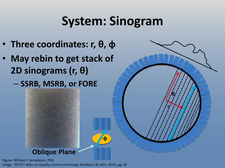
• Three coordinates: r, θ, φ
• May rebin to get stack of 2D sinograms (r, θ)
– SSRB, MSRB, or FORE
r
θ
Oblique Plane
φ
FIgure: William F. Sensakovic, PhD Image: PET/CT Atlas on Quality Control and Image Artefacts, © IAEA, 2014, pg. 26

• HVL in crystal ~1cm
• System displaces LOR
– 10cm off-center: 40% worse resolution
• Center target in scanner
• Correction
– Layered detector to determine depth of interaction (not clinically available)
– PSF reconstruction
Images: William F. Sensakovic, PhD

• Random
– Random Fraction: 35-65%
• Corrections
– Delayed coincidence subtraction
• Best for 2D, worst SNR (high noise)
– Delayed subtraction with smoothing
– Singles estimate for each LOR
•
– Reduce Tcoinc.
– Note: No perceptual difference between correction methods
21. ** SingleSingleCoincRandom RRTR
Images: William F. Sensakovic, PhD

• Scatter – 50% scatter in the scintillator
• Generally left uncorrected
– Scatter Fraction 30-70% (body)
– Most probable scatter is 35o (433keV) by Klein-Nishina
• Corrections – Energy Window
• 380-640keV, 400-600keV
– Single Scatter Simulation (standard)
– Tail fitting
– Monte Carlo Image: Watson, C.C.; Casey, M.E.; Michel, C.; Bendriem, B., "Advances in scatter correction for 3D PET/CT," Nuclear Science Symposium Conference Record, 2004 IEEE , vol.5, no., pp.3008,3012, 16-22 Oct. 2004 Figure: William F. Sensakovic, PhD

• Klein-Nishina Electronic Cross-section
–
• Klein-Nishina Form Factor (FKN)
•
• So,
0 0.1 0.2 0.3 0.4 0.5 0.6 0.7 0.8 0.9
1
0 20 40 60 80 100 120 140 160 180
Pro
po
rtio
n o
f Sc
atte
red
P
ho
ton
s
Scatter Angle (Degrees)
dd sin2
ref: Podgorsak, Radiation Physics for Medical Physicists 2nd ed. Springer Heidelberg, 2010 Figure: William F. Sensakovic, PhD

• Scattered photon energy
• Low energy thresh
– 380keV 0
0.1 0.2 0.3 0.4 0.5 0.6 0.7 0.8 0.9
1
0 20 40 60 80 100 120 140 160 180
Pro
po
rtio
n o
f Sc
atte
red
P
ho
ton
s
Scatter Angle (Degrees) ref: Podgorsak, Radiation Physics for Medical Physicists 2nd ed. Springer Heidelberg, 2010. Figures: William F. Sensakovic, PhD
0 50
100 150 200 250 300 350 400 450 500 550
0 20 40 60 80 100 120 140 160 180
Scat
tere
d P
ho
ton
En
erg
y
Scattered Angle (Degrees)
400keV accepts up to 44o scatter 380keV accepts up to 49o scatter
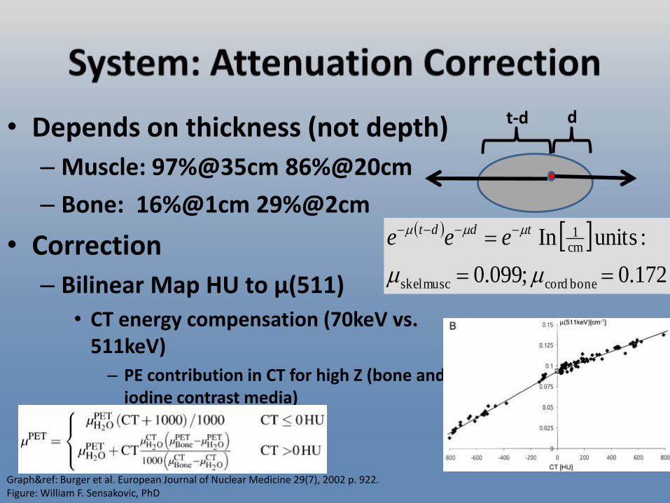
• Depends on thickness (not depth)
– Muscle: 97%@35cm 86%@20cm
– Bone: 16%@1cm 29%@2cm
• Correction
– Bilinear Map HU to µ(511)
• CT energy compensation (70keV vs. 511keV) – PE contribution in CT for high Z (bone and
iodine contrast media)
172.0 ;099.0
:units In
bone cordmusc skel
cm1
tddt eee
t-d d
Graph&ref: Burger et al. European Journal of Nuclear Medicine 29(7), 2002 p. 922. Figure: William F. Sensakovic, PhD

Image: William F. Sensakovic
Uncorrected Corrected

• Many Techniques
– 3DRP, OSEM, etc.
• OSEM widely used
– 2-5 iterations with 8-28 subsets
» Time vs. Accuracy
» Noise increases with effective iteration number
Tong et al. Image reconstruction for PET/CT scanners: past achievements and future challenges Imaging Med. 2010 October 1; 2(5): 529–545
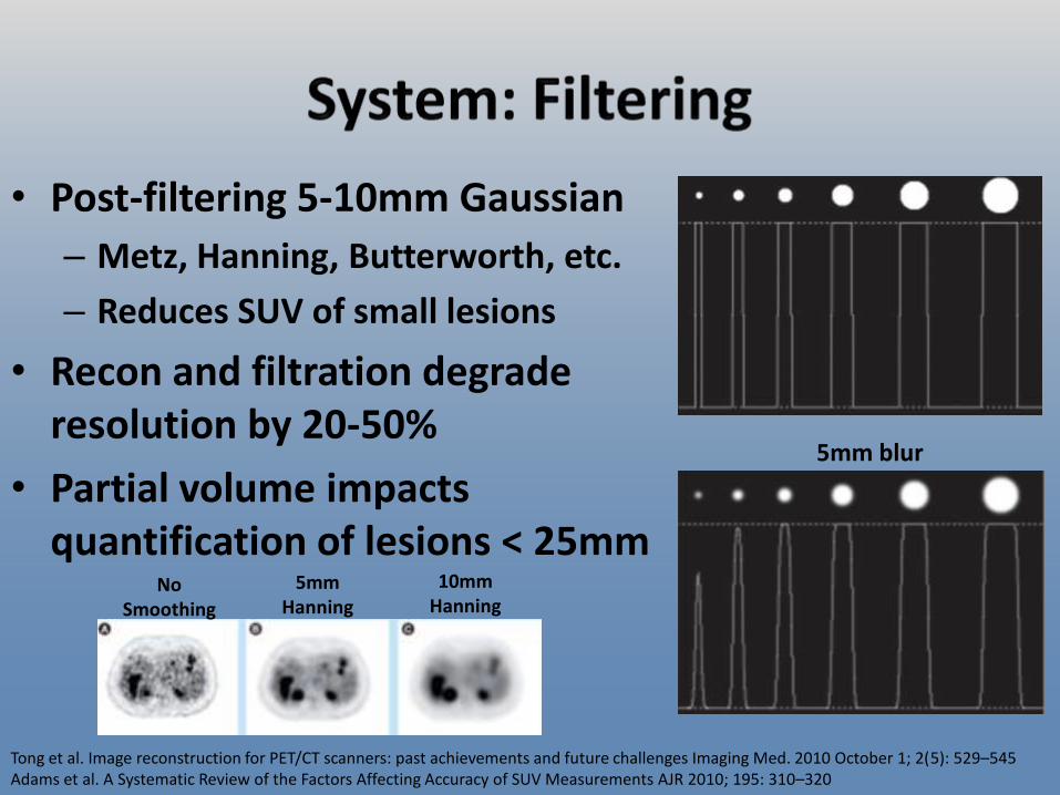
• Post-filtering 5-10mm Gaussian
– Metz, Hanning, Butterworth, etc.
– Reduces SUV of small lesions
• Recon and filtration degrade resolution by 20-50%
• Partial volume impacts quantification of lesions < 25mm
Tong et al. Image reconstruction for PET/CT scanners: past achievements and future challenges Imaging Med. 2010 October 1; 2(5): 529–545 Adams et al. A Systematic Review of the Factors Affecting Accuracy of SUV Measurements AJR 2010; 195: 310–320
5mm Hanning
No Smoothing
10mm Hanning
5mm blur

• PSF varies across FOV and out-of-plane
• Measure PSF through the volume and include in recon alg.
– HD, SharpIR, Astonish
• More effective for small objects
Photo: PET/CT Atlas on Quality Control and Image Artefacts, © IAEA, 2014, pg. 26 Figure: William F. Sensakovic, PhD

• Frame-mode – Traditionally used
– Accumulate counts in sinogram or projection matrix
– Fast calculation and easy reconstruction
– May be gated, static, or dynamic
• List-mode – Becoming standard (flexibility and information)
– List of event position, detection time, energy, etc.
– Engineering challenges with high data rate (MB/s)
– Can retrospectively resort data for dynamic, gated, etc.
– Facilitates advanced reconstruction with motion compensation

• Resolution
– Intrinsic: 2-3mm
– System (NEMA Measured): ~5mm
– Temporal: minutes per bed position, less for dynamic or gated
• Sensitivity
– To material: 10-15 mol of material
– System: 8% (0.08cps/Bq)

• Sensitivity increases with axial length of scanner
– More LOR
– 16cm increase to 22cm increases sensitivity 78%
Images: William F. Sensakovic, PhD ref: D W Townsend 2008 Phys. Med. Biol. 53 R1

• Glucose
– Essential carbon supplier for tissue creation
• Proliferation
– Fuel for cellular respiration
• Energy
• Fluorodeoxyglucose
– Analogue of glucose
Images: William F. Sensakovic, PhD
O
OH
OH OH
OH
OH
O
OH
OH OH
F
OH

• Glucose is too big to diffuse across membrane
• Facilitated Diffusion
– Rate increases linearly until carrier saturated (Fasting)
• Competition: Glucose vs. FDG
– Insulin increases rate 10-20x (Diabetes)
– Six transporters (GLUT-1, GLUT-2, etc.) differ in location, kinetics, and sugar specificity
• GLUT-1, 3, & 5 are overexpressed in tumors
• GLUT-4 is in brown fat (Artifact)
• GLUT-1 at Blood-Brain Barrier Images: Public Domain

• Facilitated Diffusion
– Glucose is phosporylated by hexokinase to glucose-6-phosphate glycolysis (creates energy)
– FDG fluorodeoxyglucose-6-phosphate cannot move on until F decays to O
• Picks up H from environment and moves on to glycolysis
Images: Public Domain

• Active transport
– Requires energy and a carrier to move against concentration gradient
– Sodium-dependent glucose transporter (SGLT) poor FDG binding
• Glomerular filtered glucose transported into blood in distal renal tubules
• FDG stays in urinary tract (poor recirculation) and moves to bladder

• Tumors use more glucose due to altered metabolism and proliferation (hyperglycolysis/Warburg effect) – Glycolytic capacity
proportional to differentiation (Grading/Staging)
• Increased transporter expression and activity
• Increased production and activity of hexokinase
• Absent or low levels of phosphatase (reverse hexokinase) ref: Elgazzar, The Pathophysiologic Basis of Nuclear Medicine 2nd ed. Springer 2006
Image: Vander Heiden et al. Science. May 22, 2009; 324(5930): 1029–1033

• Hypoxia
– FDG uptake increases in acute and chronic hypoxic cells
• Conflicting and variable results in human studies
• Increased GLUT-1 expression through HIF-1
– F-MISO, Cu-ASTM, F-nitroimidazoles are better agents
Images: Kashefi, et al. Molecular Imaging in Pulmonary Diseases , AJR 2011; 197:295–307

• Necrosis
– Lack of uptake by necrotic tissue
– Bright normoxic/hypoxic ring around hypointese necrosis
Images: Rakheja et al. Necrosis on FDG PET/CT Correlates With Prognosis and Mortality in Sarcomas AJR 2013; 201:170–177.

• Fast 6hrs (2-4hrs pediatric) – Glucose 80-120 mg/dL or 180-200 mg/dL for diabetic patients
(Muscle Uptake)
• Dress in gown and remove jewelry (Metal Artifact) • Questionnaire and Oral CT Contrast
– Pregnancy, breastfeeding, fasting compliance, prior surgery, therapy, conditions, hydration
• Start IV (22-24 gauge) contralateral to side of interest • Patient may rest for up to 15min before injection
– Especially important in brain
Fast Oral Contrast
and Questionnaire
Dress Start IV Rest

• Injection with FDG – 10-15mCi (0.081 to 0.14mCi/kg pediatric)
• Patient rests in warm calm environment (45-60min) – Warm room and blankets (Brown Fat)
– No motion, talking, eating, or other stimulation (Artifact)
• Void bladder just before scan (Dose and Reproducibility)
• Position patient with immobilization devices and in treatment position (Reproducibility) – If using as simulation as well
– Large bore size makes this easier
FDG Void Rest IV
Contrast Position

• Whole body PET protocol – 2mmx2mmx2mm voxel size
– 1-3min per bed position • 12min for 4D gated
– 5-9 bed positions (25-50% overlap)
– OSEM reconstruction with 5mm Gaussian filter
• CT – Simulation: 120-140kVp, 100-200mAs, pitch 1.1-1.4,
standard (non-enhancing) recon kernel, dose modulation (if patient is centered in bore)
– Non-simulation: reduce mAs ~50%
6min 1min
PET/CT Atlas on Quality Control and Image Artefacts, © IAEA, 2014, pg. 26

• Differential FDG uptake continues after injection
• Impacts SUV and apparent tumor size
– Serial scans should be made with same injection-scan interval to ensure accurate results
PET/CT Atlas on Quality Control and Image Artefacts, © IAEA, 2014, pg.80

Table: Standard Operating Procedures for PET/CT: A Practical Approach for Use in Adult Oncology, © IAEA, 2013, pg. 25
39
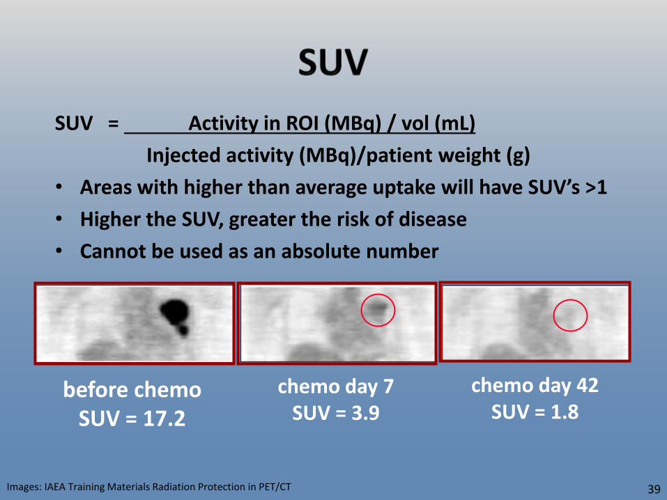
SUV = Activity in ROI (MBq) / vol (mL)
Injected activity (MBq)/patient weight (g)
• Areas with higher than average uptake will have SUV’s >1
• Higher the SUV, greater the risk of disease
• Cannot be used as an absolute number
before chemo SUV = 17.2
chemo day 7 SUV = 3.9
chemo day 42 SUV = 1.8
Images: IAEA Training Materials Radiation Protection in PET/CT

• Patient size – Overestimation of SUV due to fat on large patients or
patients whose weight changes » Use lean body mass or body surface area instead of weight
• Glucose level – High glucose level causes reduced target uptake
» Attempts at normalization are not recommended
• Protocol – Time from injection to scan, scanner, ROI selection,
postprocessing, contrast media, resolution, etc. » Keep consistent in longitudinal tracking of patient
Boellaard. Standards for PET Image Acquisition and Quantitative Data Analysis . J Nucl Med. 2009;50:11S-20S Adams et al. A Systematic Review of the Factors Affecting Accuracy of SUV Measurements AJR 2010; 195: 310–320

• CT:
– Breath hold
– Slow scan
– Gated
• PET
– Free breath
– Gated
– 4D
Images and ref: Bowen et al. Clinical and Translational Medicine 2012, 1:18

• Independent of SUV confounding factors
• Lesion definition
• Manual, automated, semi-automated
– Manual slower but correlates to pathologic size better
right images and ref: Nestle et al. J Nucl Med 2005;46:1342-1348. left images and ref: Wu et al. J Nucl Med 2010;51:1517-1523

• PET
– ICRP 106
– ImageWisely: 3.5-10.5mSv for 5-15mCi
• CT
– ImageWisely:
http://www.imagewisely.org/Imaging-Modalities/Nuclear-Medicine/Articles/CT-Protocol-Selection

• Breastfeeding
– Breast uptake due to active nursing
• Stimulates GLUT-1
– Expressed milk: 5.54-19.3 Bq/mL/MBq
• Uninterrupted feeding unlikely to cause dose in excess of regulatory limits
– Infant dose largely due to proximity to breast and not ingestion of isotope
• Delay feeding at least 4 hrs after injection
• Express milk and have another family member bottle feed
Hicks et al. Pattern of Uptake and Excretion of 18F-FDG in the Lactating Breast J Nucl Med 2001; 42:1238–1242

• FDG crosses placenta – Concentrates in the brain
– Excreted by the fetal kidneys
• As high as 0.04mGy/MBq – Photon dose 1/10th positron
– 1/4 of dose due to mother’s tissue
• Example: – 10mCi FDG gives 14.8mGy
– 10mSv for diagnostic CT
– ~25mSv total Zanotti-Fregonara et al. Absorbed 18F-FDG Dose to the Fetus During Early Pregnancy J Nucl Med. 2010;51:803-805.

• Typical technologist effective dose
– 6mSv/year
– 0.02 µSv/MBq per unit injected activity
– Largest dose from escorting injected patient to bathroom or scanner
• Typical technologist hand dose
– 1.4mSv/GBq hand dose
– 30cm forceps to reduce dose (distance)
– 5mm W syringe shield drops dose by factor of 10 (Shielding)
Carnicer et al. Hand exposure in diagnostic nuclear medicine with 18F- and 99mTc-labelled radiopharmaceuticals - Results of the ORAMED project Radiation Measurements, 46(11) 2011 Pgs. 1277-1282A. Benatar. Radiation dose rates from patients undergoing PET: implications for technologists and waiting areas. Eur J Nucl Med. 2000;27(5) Images: William F. Sensakovic

• Power injector
– 20% dose reduction (95% hand dose reduction)
– 18.5mSv/min finger dose from unshielded syringe (10mCi)
• Patient Bathroom
– 2mR/hr at surface just after patient void @60min post-injection
– Increases through the day
Covens et al. The introduction of automated dispensing and injection during PET procedures: a step in the optimisation of extremity doses and whole-body doses of nuclear medicine staff. Radiation Protection Dosimetry (2010), pp. 1–9

• Dose from patient
– 0.092 µSv-m2/MBq-h
– Could cause confusion after brachytherapy retraction if patient was not surveyed pre-therapy and underwent PET scan within a few hours
Image: Federspeil and Hogg. PETCT Radiotherapy Planning Pt.3 A Technologists Guide. Published by EANM 2012 AAPM TG108 P E T and PET/ CT S hie lding Re quirements

• 30cm from 1MBq point source
– 0.12mSv/hr skin dose from positrons
– 0.081mSv/hr deep tissue dose from Gamma
• 0.05 ml (0.001MBq) skin contact
– 0.79mSv/hr skin dose
– Skin dose due to positrons
Delacroix et al. Radionuclide and Radiation Protection Data Handbook 2nd ed. Nuclear Technology Publishing 2002


• Impacts visualization and SUV
• Test before scanning
• Fasting and insulin
Figure: Standard Operating Procedures for PET/CT: A Practical Approach for Use in Adult Oncology, © IAEA, 2013, pg. 33
High Glucose Fasting Glucose

• Impacts visualization and SUV
– Rest in quiet room with no talking or movement
• TV/calm music
– Beta blocker or sedative
Figure: Standard Operating Procedures for PET/CT: A Practical Approach for Use in Adult Oncology, © IAEA, 2013
Hyperventilation Talking

• Most likely in young adults
– Use blankets to keep patient warm
– Match uptake to CT fat regions
Image: William F. Sensakovic

PET/CT Atlas on Quality Control and Image Artefacts, © IAEA, 2014, pg. 26
• Checked in daily QA
• Smaller changes can impact SUV
• Large changes necessary to impact visualization

• 11% of PET scans have some level of extravasation
• Calculation: 135Gy if 10mCi contained in 1mL
• Reality: Spreads and is absorbed by the body – Does not usually cause skin
damage
– Does substantially alter SUV and visualization
Ref: Hoop The Inifitrated Radiopharmaceutical Injection: Risk Considerations The Journal of Nuclear Medicine 32(5) 1991 Ref: Osman et al. FDG dose extravasations in PET/CT: frequency and impact on SUV measurements Frontiers in Oncology Cancer Imaging and Diagnosis 1 Article 41 2011 Image: William F. Sensakovic

• Marrow and spleen demonstrate increased uptake
– Higher than liver is abnormal
• Ports may cause uptake error in AC corrected images
Extravasation Image: William F. Sensakovic
Port Image: PET/CT Atlas on Quality Control and Image Artefacts, © IAEA, 2014, pg. 52

• Motion control to help
PET/CT Atlas on Quality Control and Image Artefacts, © IAEA, 2014, pg. 58
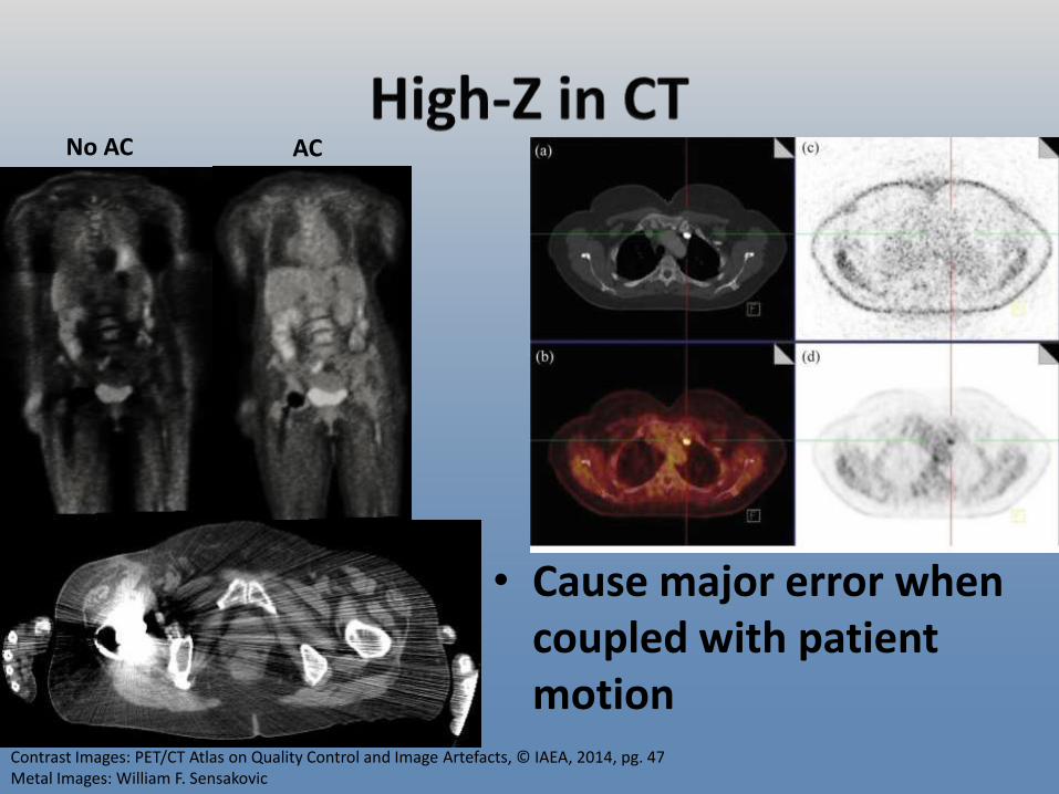
• Cause major error when coupled with patient motion
Contrast Images: PET/CT Atlas on Quality Control and Image Artefacts, © IAEA, 2014, pg. 47 Metal Images: William F. Sensakovic
AC No AC

Image: William F. Sensakovic

• SUV: 3.36.1
• Use extended FOV if possible
Image top: PET/CT Atlas on Quality Control and Image Artefacts, © IAEA, 2014, pg.63 Image bottom: William F. Sensakovic