Embed Size (px)
Citation preview

(12) United States Patent Kao et al.
US008574869B2
(10) Patent No.: US 8,574,869 B2 (45) Date of Patent: Nov. 5, 2013
(54) PREVENTION OF DISULFIDE BOND REDUCTION DURING RECOMBINANT PRODUCTION OF POLYPEPTIDES
(75) Inventors: Yung-Hsiang Kao, San Mateo, CA (US); Michael W. Laird, San Ramon, CA (US); Melody Trexler Schmidt, San Carlos, CA (US); Rita L. Wong, Redwood City, CA (US); Daniel P. Hewitt, Sunnyvale, CA (US)
(73) Assignee: Genentech, Inc., South San Francisco, CA (US) Subject to any disclaimer, the term of this patent is extended or adjusted under 35 U.S.C. 154(b) by 0 days.
(21) Appl. No.: 13/354,223
(*) Notice:
(22) Filed: Jan. 19, 2012 (65) Prior Publication Data
US 2013/OO17598A1 Jan. 17, 2013
Related U.S. Application Data (63) Continuation of application No. 12/217,745, filed on
Jul. 8, 2008, now abandoned. (60) Provisional application No. 60/948,677, filed on Jul.9,
2007.
(51) Int. Cl. CI2P I/O CI2N 5/02
(52) U.S. Cl. USPC ............................................. 435/41; 435/325
(58) Field of Classification Search None See application file for complete search history.
(2006.01) (2006.01)
(56) References Cited
U.S. PATENT DOCUMENTS
2004/OO29229 A1* 2004/O138424 A1 2006.0143549 A1 2007,0292411 A1 2009.0053786 A1
2, 2004 Reeves et al. ................ 435/69.1 7/2004 Takeda et al. 6/2006 Yasumoto et al. 12/2007 Salcedo et al. 2/2009 Kao et al.
FOREIGN PATENT DOCUMENTS
WO 97.26357 7/1997
OTHER PUBLICATIONS
Kocket al. (Biochem Soc Trans. Apr. 2004:32(Pt 2):273-5).* Christiansen et al. (Biochemistry 1998, 37, 12611-12623).* Bobovnikova et al., "Characterization of Soluble, Disulfide Bond Stabilized Prokaryotically Expressed Human Thryotropin Receptor Ectodomain' Endocrinology 138(2):588-593 (1997). Chaderjian et al., “Effect of Copper Sulfate on Performance of a Serum-Free CHO Cell Culture Process and the Level of FreeThiol in the Recombinant Antibody Expressed” Biotechnology Progress 21(2):550-553 (2005). Gromer et al., “The Thioredoxin System: From Science to Clinic' Medicubak Research Reviews 24(1):40-89 (Jan. 2004). Kao et al., “Mechanism of Antibody Reduction in Cell Culture Pro duction Processes' Biotechnology and Bioengineering 107(4):622 632 (Nov. 2010).
Kerblat et al., “Importance of Thioredoxin in the Proteolysis of an Immunoglobulin Gas Antigen by Lysosomal Cys-Proteases’ Immu nology 97(1):62-68 (1999). Li et al., “Low Level Formation of Potent Catalytic IgG Fragments Mediated by Disulfide Bond Instability” Molecular Immunology 33(7-8):593-600 (1996). Liu et al., “Study of Tioredoxin' Journal of Northeast Agricultural University 34(23):219-225 (Jun. 30, 2003). Mun et al., “BIOT 245-Air Sparging of Harvested Cell Culture Fluid (HCCF) to Prevent Antibody Disulfide Bond Reduction' Abstracts of Papers American Chemical Society 238:245 (Aug. 2009). Nordberg et al., “Reactive Oxygen Species, Antioxidants, and the Mammalian Thioredoxin System” Free Radical Biology & Medicine 31(11): 1287-1312 (2001). Powis et al., “Properties and Biological Activities of Thioredoxins' Annual Review of Pharmacology and Toxicology 41:261-295 (2001). Powis et al., “Thioredoxin Redox Control of Cell Growth and Death and the Effects of Inhibitors' Chemico-Biologicallnteractions 111 112:23-34 (1998). Salas-Solano et al., “Optimization and Validation of a Quantitative Capillary Electrophoresis Sodium Dodecyl Sulfate Method for Qual ity Control and Stability Monitoring of Monoclonal Antibodies' Analytical Chemistry 78(18):6583-6594 (2006). Smith et al., “Specific Cleavage of Immunoglobulin G by Copper Ions' International Journal of Peptide and Protein Research 48(1):49-55 (1996). Starks et al., “Atomic-Resolution Crystal Structure of Thioredoxin From the Acidophilic Bacterium Acetobacter Aceti” Protein Science 16(1):92-98 (Jan. 2007). Teilum et al., “Disulfide Bond Formation and Folding of Plant Peroxidases Expressed as Inclusion Body Protein in Escherichia coli Thioredoxin Reductase Negative Strains” Protein Expression and Purification Academic Press 15(1): 77-82 (1999). Trexler-Schmidt et al., “Identification and Prevention of Antibody Disulfide Bond Reduction During Cell Culture Manufacturing” Biotechnology and Bioengineering 160(3):452-461 (2006). Uriget al., “On the Potential of Thioredoxin Reductase Inhibitors for Cancer Therapy” Seminars in Cancer Biology 16(6):452-465 ( 2006). Wipf et al., “New Inhibitors of the Thioredoxin-Thioredoxin Reducatase System Based on a Naphthoquinone Spiroketal Natural Product Lead” Bioorganic & Medicinal Chemistry Letters(11):2637 2641 (2001). Zhang et al., “Free sulfhydryl in recombinant monoclonal antibod ies’ Biotechnol. Prog. 18:509-513 (2002). Lillig, C.H. etal. (Jan. 2007). “Thioredoxin and Related Molecules— From Biology to Health and Disease.” Antioxidants & Redox Signal ing 9(1):25-47. Lydersen, B.K. etal. (Nov. 1994). "Acid Precipitation of Mammalian Cell Fermentation Broth. Ann. N.Y. Acad. Sci. 745:222-231. Roman, B. etal. (Sep. 2005). “Development of a Robust Clarification Process for MAb Purification.” Case Study presented at the BioProcess International 2005 Conference & Exhibition, Sep. 19-22, 2005, Boston, MA, four pages particularly p. 4. Roush, D.J. etal. (2008). "Advances in Primary Recovery: Centrifu gation and Membrane Technology,” Biotechnol. Prog. 24(3):488 495.
* cited by examiner Primary Examiner — Suzanne M Noakes Assistant Examiner — Jae W Lee (74) Attorney, Agent, or Firm — Morrison & Foerster LLP (57) ABSTRACT Provided herein are methods for preventing the reduction of disulfide bonds during the recombinant production of disul fide-containing polypeptides. In particular, the invention con cerns the prevention of disulfide bond reduction during har vesting of disulfide-containing polypeptides, including antibodies, from recombinant host cell cultures.
10 Claims, 40 Drawing Sheets

U.S. Patent Nov. 5, 2013 Sheet 1 of 40 US 8,574,869 B2
(kDa)
Ladder OHOurS 1 Hours 3HOurS 21 Hours 25HOurS 29Hours
240 or memore root owe osmos o
150
63 - as
46- . . . .
Dialysis Experiment
FIG.

U.S. Patent Nov. 5, 2013 Sheet 2 of 40 US 8,574,869 B2
kDa
Ladder OHOurS 1 Hours 3Hours 21 Hours 25Hours 29Hours 48Hours
240
46 -
Dialysis Experiment
FIG 2

U.S. Patent Nov. 5, 2013 Sheet 3 of 40 US 8,574,869 B2
9000
800.0
700.0
6000
5000
4000 -0- Outside Dialysis Bag
300.0
2000
1OOO
O.O O 10 20 30 40 50
Time (hours)
Free Thiol Levels from Dialysis Experiment
FIG 3

U.S. Patent Nov. 5, 2013 Sheet 4 of 40 US 8,574,869 B2
Thioredoxin System S SH S
p? Trx1 ToRC NADPH+H" YS YSH S
SH S SH p? Trx TxRC NADP" Y SH NS SH
First Reaction in Pentose Phosphate Pathway
NADPH 6-Phosphogluconolactone
Glucose-6-phosphate dehydrogenase
NADP" Glucose-6-phosphate (Mg-ADP)' +H"
Hexokinase
Glucose (Mg-ADP)2"
First Reaction in Glycolysis
Thioredoxin System and Other Reactions involved in Antibody Reduction
FIG 4

U.S. Patent Nov. 5, 2013 Sheet 5 of 40 US 8,574,869 B2
(kDa)
Ladder OHOurS 0.5Hours 1 Hours 2HOurS 3Hours 21 Hours 23Hours
240 - coasia seasini seases snaissa sitesiasis osses assessesses to
150 - assassi.
95 - esses
63 six-se:cises::sissississ:
In Vitro Activity of Thioredoxin System FIG 5

U.S. Patent Nov. 5, 2013 Sheet 6 of 40 US 8,574,869 B2
kDa
Ladder OHOurS 0.5Hours 1 Hours 2Hours 3HOurS 21 Hours 23Hours
240 - so seas awarass assesses so seese souro sesses
150 - . . . . .
63 -
In Vitro Activity of Thioredoxin System Inhibited by Aurothioglucose
FIG. 6

U.S. Patent Nov. 5, 2013 Sheet 7 of 40 US 8,574,869 B2
(kDa)
Ladder OHOurS 0.5HOurS 1 Hours 2Hours 3Hours 21 Hours 23Hours
240 - essesses occessors so asso assessors ocess
63
46
In Vitro Activity Of Thioredoxin System Inhibited by Aurothiomalate
FIG. 7

U.S. Patent Nov. 5, 2013 Sheet 8 of 40 US 8,574,869 B2
(kDa)
Ladder OHOurS 0.5HOurS 1 Hours 2Hours 3Hours 21 Hours 23Hours
240 - seconomios sometre creasesses pesosier seases essesses esses eases
150 w &&...:::::::::::::::::::::::2&
In Vitro Activity of Thioredoxin System
FIG. 8

U.S. Patent Nov. 5, 2013 Sheet 9 of 40 US 8,574,869 B2
(kDa)
Ladder OHOurS 0.5HOurS 1 Hours 2Hours 3Hours 21 Hours 23Hours
240
63
In Vitro Activity of Thioredoxin System inhibited by CuSO4 FIG. 9

U.S. Patent Nov. 5, 2013 Sheet 10 of 40 US 8,574,869 B2
kDa
Ladder OHOurS 0.5Hours 1 Hours 2Hours 19Hours 21 Hours 23Hours
240 res 2&ixes & exississa::::::ss & sixás::::::::::::::::gs& :
95 - costics
OCrelizumab Reduction
FIG. O

U.S. Patent Nov. 5, 2013 Sheet 11 of 40 US 8,574,869 B2
kDa
Ladder OHOurS 0.5Hours 1 Hours 2Hours 19Hours 21 Hours 23Hours
240- essesses sessessessessesses assessessessessee
Inhibition of OCrelizumab Reduction in HCCF by Aurothioglucose FIG.

U.S. Patent Nov. 5, 2013 Sheet 12 of 40 US 8,574,869 B2
kDa
Ladder OHours 0.5Hours 1 Hours 2HOurs 19Hours 21 Hours 23Hours
240 && ::s:::::::::::::ss.xx x&:xxxxxx8&s &&sixx & & . . . . .
46 - -sesseries
15 - essessee
Inhibition of OCrelizumab Reduction in HCCF by Aurothiomalate
FIG 2

U.S. Patent Nov. 5, 2013 Sheet 13 of 40 US 8,574,869 B2
kDa
Ladder OHOurS 1 Hours 2Hours 4Hours 19Hours 21 Hours
95
63
LOsing Reduction Activity in HCCF
FIG. 13

U.S. Patent Nov. 5, 2013 Sheet 14 of 40 US 8,574,869 B2
kDa
Ladder OHours Hours 2HOurS 3Hours 4Hours 19Hours 21 Hours 23Hours
240 - onest-selors is essesses assessessines weaeacekeeps sesses seasies sails.
iiigise is
The Lost Reduction Activity in HCCF Restored by Addition of NADPH FIG. 4

U.S. Patent Nov. 5, 2013 Sheet 15 of 40 US 8,574,869 B2
kDa
Ladder OHOurs 1 Hours 2Hours 3HOurS 4Hours 19Hours 21 Hours 23HOurS
240- a.s.l.... assassical score--see". "
The Lost Reduction Activity in HCCF Restored by Addition of Glucose-6-Phosphate
FIG 15

U.S. Patent Nov. 5, 2013 Sheet 16 of 40 US 8,574,869 B2
kDa
Ladder OHOurS 1 HourS 2HOUrS 3HOurS 19HOUrS 21 Hours
150-sks
OCreizumab Reduction
FIG 16
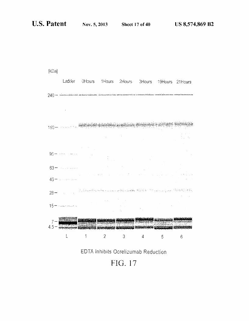
U.S. Patent Nov. 5, 2013 Sheet 17 of 40 US 8,574,869 B2
kDa
Ladder OHOurS 1 Hours 2Hours 3Hours 19Hours 21 Hours
240 - so ownesses writonealoes seasons uses worse assessio
150
EDTA inhibits OCreizumab Reduction
FIG. 7

U.S. Patent Nov. 5, 2013 Sheet 18 of 40 US 8,574,869 B2
kDa
Ladder OHOurS 1 Hours 2Hours 3Hours 19HOurS 21HOurS
240 - a. aesar seases essessee ocessee to esses series.
150
63
46
The Lost Reduction Activity in Run 8 HCCF Restored by Addition of Glucose-6-Phosphate but NO Inhibition of Reduction by EDTA
FIG. 18

U.S. Patent Nov. 5, 2013 Sheet 19 of 40 US 8,574,869 B2
-- 20 mM EDTA
|-A-30 mM CuSO4 -o-pH 5.5 (acetic acid) --NO additions | 50
---o-o-o-o-o-o-on
O 5 10 15 2O 25 3O 35 40
HCCF Hold Time (hr)
FIG. 19

U.S. Patent Nov. 5, 2013 Sheet 20 of 40 US 8,574,869 B2
8O -O-Air Sparged 70 -0-Nitrogen Sparged
to-o-o-
i 30
2. v 10 - . O O 5 10 15 20 25 30 35 40
HCCF Hold Time (hr)
FIG. 20

U.S. Patent Nov. 5, 2013 Sheet 21 of 40 US 8,574,869 B2
Light Chain
15 3 O 45 DIOMTOSPSSLS ASVG DRVTITCRASODVNTAVAWYOOKPGKAPK
4 6 6 O 75 9 O LLIY SAS FLYSGVPSRFSGSRSGTDFTL TISSLOPEDF ATYY COO
9 1 O 5 HYTTPPTFGOGTKVEIK
FIG 21
Heavy Chain
1. 5 3 O 45 EVOLVESGGGLVOPGGSLRLSCAA SGFNIKDTY IHWVROAPGKGL
4 6 6 O 75 9 O EWVARIYPTNGY TRY ADSVKGRFTISADTSKNTAYLOMNSLRAED
9 O 5 12 O TAVYYCSRWGGDGFY AMDYWGOGTLVTVSS
FIG. 22

US 8,574,869 B2 Sheet 22 of 40 Nov. 5, 2013 U.S. Patent


US 8,574,869 B2 Sheet 24 of 40 Nov. 5, 2013 U.S. Patent
97 – 96–

US 8,574,869 B2 Sheet 25 of 40 Nov. 5, 2013 U.S. Patent

US 8,574,869 B2 Sheet 26 of 40 Nov. 5, 2013 U.S. Patent
99– ~~ ~ £9

Sheet 27 of 40 Nov. 5, 2013 U.S. Patent
97 –


US 8,574,869 B2 Sheet 29 of 40 Nov. 5, 2013 U.S. Patent
sae – 8Z
96– 0

US 8,574,869 B2 Sheet 30 of 40 Nov. 5, 2013 U.S. Patent
97– 96– 07? --:

US 8,574,869 B2 Sheet 31 of 40 Nov. 5, 2013 U.S. Patent
97 –ae — 97

US 8,574,869 B2 Sheet 32 of 40 Nov. 5, 2013 U.S. Patent
€ £ (4) | H

US 8,574,869 B2
-
U.S. Patent

US 8,574,869 B2 Sheet 34 of 40 Nov. 5, 2013 U.S. Patent
s

US 8,574,869 B2 Sheet 35 of 40 Nov. 5, 2013 U.S. Patent
09 || — 07? --
0
Jappen {eq}}]

US 8,574,869 B2 Sheet 36 of 40 Nov. 5, 2013 U.S. Patent

US 8,574,869 B2 Sheet 37 of 40 Nov. 5, 2013 U.S. Patent
97 – 99– 96–


U.S. Patent Nov. 5, 2013 Sheet 40 of 40 US 8,574,869 B2
150
95
63
46

US 8,574,869 B2 1.
PREVENTION OF DISULFIDE BOND REDUCTION DURING RECOMBINANT PRODUCTION OF POLYPEPTIDES
CROSS REFERENCE TO RELATED APPLICATIONS
This application is a continuation of U.S. application Ser. No. 12/217,745, filed Jul. 8, 2008, which is a non-provisional application filed under 37 CFR 1.53(b)(1), claiming priority under 35 USC 119(e) to provisional Application No. 60/948, 677 filed Jul. 9, 2007, the contents of which are incorporated herein by reference.
FIELD OF THE INVENTION
The invention concerns methods and means for preventing the reduction of disulfide bonds during the recombinant pro duction of disulfide-containing polypeptides. In particular, the invention concerns the prevention of disulfide bond reduc tion during harvesting of disulfide-containing polypeptides, including antibodies, from recombinant host cell cultures.
BACKGROUND OF THE INVENTION
In the biotechnology industry, pharmaceutical applications require a variety of proteins produced using recombinant DNA techniques. Generally, recombinant proteins are pro duced by cell culture, using either eukaryotic cells, such as mammalian cells, or prokaryotic cells, such as bacterial cells, engineered to produce the protein of interest by insertion of a recombinant plasmid containing the nucleic acid encoding the desired protein. For a protein to remain biologically active, the conformation of the protein, including its tertiary structure, must be maintained during its purification and iso lation, and the protein's multiple functional groups must be protected from degradation.
Mammaliancells have become the dominant system for the production of mammalian proteins for clinical applications, primarily due to their ability to produce properly folded and assembled heterologous proteins, and their capacity for post translational modifications. Chinese hamster ovary (CHO) cells, and cell lines obtained from various other mammalian Sources, such as, for example, mouse myeloma (NSO), baby hamster kidney (BHK), human embryonic kidney (HEK 293) and human retinal cells, such as the PER.C6(R) cell line isolated from a human retinal cell, which provides human glycosylation characteristics, and is able to naturally produce antibodies that match human physiology, have been approved by regulatory agencies for the production of biopharmaceu tical products.
Usually, to begin the production cycle, a small number of transformed recombinant host cells are allowed to grow in culture for several days (see, e.g., FIG. 23). Once the cells have undergone several rounds of replication, they are trans ferred to a larger container where they are prepared to undergo fermentation. The media in which the cells are grown and the levels of oxygen, nitrogen and carbon dioxide that exist during the production cycle may have a significant impact on the production process. Growth parameters are determined specifically for each cell line and these param eters are measured frequently to assure optimal growth and production conditions. When the cells grow to sufficient numbers, they are trans
ferred to large-scale production tanks and grown for a longer period of time. At this point in the process, the recombinant protein can be harvested. Typically, the cells are engineered to
10
15
25
30
35
40
45
50
55
60
65
2 secrete the polypeptide into the cell culture media, so the first step in the purification process is to separate the cells from the media. Typically, harvesting includes centrifugation and fil tration to produce a Harvested Cell Culture Fluid (HCCF). The media is then subjected to several additional purification steps that remove any cellular debris, unwanted proteins, salts, minerals or other undesirable elements. At the end of the purification process, the recombinant protein is highly pure and is Suitable for human therapeutic use.
Although this process has been the Subject of much study and improvements over the past several decades, the produc tion of recombinant proteins is still not without difficulties. Thus, for example, during the recombinant production of polypeptides comprising disulfide bonds, especially multi chain polypeptides comprising inter-chain disulfide bonds Such as antibodies, it is essential to protect and retain the disulfide bonds throughout the manufacturing, recovery and purification process, in order to produce properly folded polypeptides with the requisite biological activity.
SUMMARY OF THE INVENTION
The instant invention generally relates to a method for preventing reduction of a disulfide bond in a polypeptide expressed in a recombinant host cell, comprising Supplement ing the pre-harvest or harvested culture fluid of the recombi nant host cell with an inhibitor of thioredoxin or a thiore doxin-like protein.
In one embodiment, the thioredoxin inhibitor is added to the pre-harvest culture fluid.
In another embodiment, the thioredoxin inhibitor is added to the harvested culture fluid.
In a further embodiment, the thioredoxin inhibitor is a direct inhibitor of thioredoxin.
In all embodiments, the thioredoxin inhibitor may, for example, be an alkyl-2-imidazolyl disulfide or a naphtho quinone spiroketal derivative.
In a further embodiment, the thioredoxin inhibitor is a specific inhibitor of thioredoxin reductase.
In a still further embodiment, the thioredoxin inhibitor is a gold complex, where the gold complex may, for example, be aurothioglucose (ATG) or aurothiomalate (ATM). While the effective inhibitory concentration may vary, it typically is between about 0.1 mM and 1 mM. Similarly, the minimum effective inhibitory concentration varies depending on the nature of the polypeptide and overall circumstances, and is typically reached when the ATG or ATG concentration is at least about four-times of thioreduxin concentration in the pre-harvest or harvested culture fluid.
In another embodiment of this aspect of the invention, the thioredoxin inhibitor is a metal ion, where the metal ion, without limitation, may be selected from the group consisting of Hg", Cut, Zn", Co, and Mn". When the metalion is added in the form of cupric sulfate, the effective inhibitory concentration generally is between about 5 uMandabout 100 uM, or between about 10 uM and about 80 uM, or between about 15 uM and about 50 uM. The minimum inhibitory concentration of cupric Sulfate also varies, but typically is reached when cupric Sulfate is added at a concentration at least about two-times of thioredoxin concentration in the pre-harves or harvested culture fluid.
In different embodiment, the thioredoxin inhibitor is an oxidizing agent, e.g., an inhibitor of G6PD, such as, for example, pyridoxal 5'-phosphate, 1 fluoro-2,4 dinitroben Zene, dehydroepiandrosterone (DHEA) or epiandrosterone (EA): cystine or cysteine. Typical effective inhibitor concen

US 8,574,869 B2 3
trations of DHEA are between about 0.05 mM and about 5 mM, or between about 0.1 mM and about 2.5 mM.
In a further embodiment, the thioredoxin inhibitor is an inhibitor of hexokinase activity, including, without limita tion, chelators of metal ions, such as, for example, ethylene diamine tetraacetic acid (EDTA). EDTA is typically added and effective at a concentration between about 5 mM and about 60 mM, or about 10 mM and about 50 mM, or about 20 mM and about 40 mM.
In other preferred embodiments, the inhibitor of hexoki nase activity is selected from the group consisting of Sorbose 1-phosphate, polyphosphates, 6-deoxy-6-fluoroglucose, 2-C-hydroxy-methylglucose, Xylose, and lyxose.
Other inhibitors include cystine, cysteine, and oxidized glutathione which are typically added at a concentration at least about 40-times of the concentration of the polypeptide in question in the pre-harvest or harvested culture fluid.
In a still further embodiment, the thioredoxin inhibitorisan siRNA, an antisense nucleotide, or an antibody specifically binding to a thioredoxin reductase.
In another embodiment, the thioredoxin inhibitor is a mea sure indirectly resulting in the inhibition of thioredoxin activ ity. This embodiment includes, for example, air sparging the harvested culture fluid of the recombinant host cell, and/or lowering the pH of the harvested culture fluid of the recom binant host cell.
In various embodiments, indirect means for inhibiting thioredoxin activity, Such as air sparging and/or lowering of the pH, can be combined with the use of direct thioredoxin inhibitors, such as those listed above.
In all embodiments, the polypeptide may, for example, be an antibody, or a biologically functional fragment of an anti body. Representative antibody fragments include Fab, Fab', F(ab'), scEv, (scEv), dAb, complementarity determining region (CDR) fragments, linear antibodies, single-chain anti body molecules, minibodies, diabodies, and multispecific antibodies formed from antibody fragments.
Therapeutic antibodies include, without limitation, anti HER2 antibodies anti-CD20 antibodies; anti-IL-8 antibodies; anti-VEGF antibodies; anti-CD40 antibodies, anti-CD11a antibodies; anti-CD18 antibodies; anti-IgE antibodies; anti Apo-2 receptor antibodies; anti-Tissue Factor (TF) antibod ies; anti-human C. B., integrin antibodies; anti-EGFR antibod ies; anti-CD3 antibodies; anti-CD25 antibodies; anti-CD4 antibodies; anti-CD52 antibodies; anti-Fc receptor antibod ies; anti-carcinoembryonic antigen (CEA) antibodies; anti bodies directed against breast epithelial cells; antibodies that bind to colon carcinoma cells; anti-CD38 antibodies; anti CD33 antibodies; anti-CD22 antibodies; anti-EpCAM anti bodies; anti-GpIIb/IIIa antibodies; anti-RSV antibodies; anti CMV antibodies; anti-HIV antibodies; anti-hepatitis antibodies; anti-CA 125 antibodies; anti-CVB3 antibodies: anti-human renal cell carcinoma antibodies; anti-human 17-1A antibodies; anti-human colorectal tumor antibodies; anti-human melanoma antibody R24 directed against GD3 ganglioside; anti-human squamous-cell carcinoma; and anti human leukocyte antigen (HLA) antibodies, and anti-HLA DR antibodies.
In other embodiments, the therapeutic antibody is an anti body binding to a HER receptor, VEGF, IgE, CD20, CD11a, CD40, or DR5.
In a further embodiment, the HER receptoris HER1 and/or HER2, preferably HER2. The HER2 antibody may, for example, comprise a heavy and/or light chain variable domain sequence selected from the group consisting of SEQ ID NO: 16, 17, 18, and 19.
10
15
25
30
35
40
45
50
55
60
65
4 In another embodiment, the therapeutic antibody is an
antibody that binds to CD20. The anti-CD20 antibody may, for example, comprise a heavy and/or light chain variable domain sequence selected from the group consisting of SEQ ID NOS: 1 through 15.
In yet another embodiment, the therapeutic antibody is an antibody that binds to VEGF. The anti-VEGF antibody may, for example, comprise a heavy and/or light chain variable domain sequence selected from the group consisting of SEQ ID NOS: 20 through 25.
In an additional embodiment, the therapeutic antibody is an antibody that binds CD11a. The anti-CD11a antibody may, for example, comprise a heavy and/or light chain variable domain sequence selected from the group consisting of SEQ ID NOS: 26 through 29.
In a further embodiment, the therapeutic antibody binds to a DR5 receptor. The anti-DR5 antibody may, for example, be selected from the group consisting of Apomabs 1.1, 2.1, 3.1, 4.1, 5.1, 5.2, 5.3, 6.1, 6.2, 6.3, 7.1, 7.2, 7.3, 8.1, 8.3, 9.1, 1.2, 2.2, 3.2, 4.2, 5.2, 6.2, 7.2, 8.2, 9.2, 1.3, 2.2, 3.3, 4.3, 5.3, 6.3, 7.3, 8.3, 9.3, and 25.3, and preferably is Apomab 8.3 or Apomab 7.3, and most preferably Apomab 7.3.
In other embodiments of the method of the present inven tion, the polypeptide expressed in the recombinant host cell is a therapeutic polypeptide. For example, the therapeutic polypeptide can be selected from the group consisting of a growth hormone, including human growth hormone and bovine growth hormone; growth hormone releasing factor; parathyroid hormone; thyroid stimulating hormone; lipopro teins; alpha-1-antitrypsin; insulin A-chain; insulin B-chain; proinsulin; follicle stimulating hormone; calcitonin; luteiniz ing hormone; glucagon; clotting factors such as factor VIIIC, factor IX, tissue factor, and von Willebrands factor; anti clotting factors such as Protein C; atrial natriuretic factor; lung Surfactant; a plasminogen activator, Such as urokinase or human urine or tissue-type plasminogen activator (t-PA); bombesin; thrombin; hemopoietic growth factor; tumor necrosis factor-alpha and -beta; enkephalinase; RANTES (regulated on activation normally T-cell expressed and secreted); human macrophage inflammatory protein (MIP-1- alpha); a serum albumin Such as human serum albumin; Muellerian-inhibiting Substance; relaxin A-chain; relaxin B-chain; prorelaxin, mouse gonadotropin-associated pep tide; a microbial protein, such as beta-lactamase; DNase; IgE: a cytotoxic T-lymphocyte associated antigen (CTLA). Such as CTLA-4; inhibin; activin; vascular endothelial growth factor (VEGF); receptors for hormones or growth factors: Protein A or D; rheumatoid factors; a neurotrophic factor Such as bone derived neurotrophic factor (BDNF), neurotrophin-3, -4, -5, or -6 (NT-3, NT-4, NT-5, or NT-6), or a nerve growth factor such as NGF-?3; platelet-derived growth factor (PDGF): fibro blast growth factor such as aFGF and bFGF; epidermal growth factor (EGF); transforming growth factor (TGF) such as TGF-alpha and TGF-beta, including TGF-31, TGF-32, TGF-B3, TGF-B4, or TGF-35; insulin-like growth factor-I and -II (IGF-I and IGF-II): des(1-3)-IGF-I (brain IGF-I), insulin-like growth factor binding proteins; CD proteins such as CD3, CD4, CD8, CD19, CD20, CD34, and CD40; eryth ropoietin, osteoinductive factors; immunotoxins; a bone mor phogenetic protein (BMP); an interferon such as interferon alpha, -beta, and -gamma; colony stimulating factors (CSFs), e.g., M-CSF, GM-CSF, and G-CSF; interleukins (ILs), e.g., IL-1 to IL-10; superoxide dismutase; T-cell receptors; surface membrane proteins; decay accelerating factor, viral antigen Such as, for example, a portion of the AIDS envelope; trans port proteins; homing receptors; addressins; regulatory pro teins; integrins such as CD11a, CD11b, CD11c, CD18, an

US 8,574,869 B2 5
ICAM, VLA-4 and VCAM: a tumor associated antigen such as HER2, HER3 or HER4 receptor; and fragments of said polypeptides.
In all embodiments, the recombinant host cell can be an eukaryotic host cell. Such as a mammalian host cell, includ ing, for example, Chinese Hamster Ovary (CHO) cells.
In all embodiments, the recombinant host cell can also be a prokaryotic host cell. Such as a bacterial cell, including, with out limitation, E. coli cells.
BRIEF DESCRIPTION OF THE DRAWINGS
FIG. 1. Dialysis Experiment: Digital gel-like imaging obtained from Bioanalyzer analysis (each lane representing a time point) demonstrating that ocrelizumab (rhuMAb 2H7— Variant A) inside the dialysis bag remained intact during the incubation period.
FIG. 2. Dialysis Experiment: Digital gel-like imaging obtained from Bioanalyzer analysis (each lane representing a time point) showing that ocrelizumab outside the dialysis bag was reduced during the incubation period. This is evidenced by the loss of intact antibody (~150 kDa) and the formation of antibody fragments depicted in the Figure. At the 48-hour time point (Lane 7), the reduced antibody appeared to be reoxidized, presumably as a result of loosing reduction activ ity in the Harvested Cell Culture Fluid (HCCF). The band appearing just above the 28 kDa marker arose from the light chain of antibody. There was a significant amount of free light already present in the HCCF before the incubation began. The presence of excess free light chain and dimers of light chain in the HCCF is typical for the cell line producing ocrelizumab.
FIG.3. FreeThiol Levels from Dialysis Experiment: Puri fied ocrelizumab in phosphate buffered saline (PBS) was inside the dialysis bag and HCCF containing ocrelizumab was outside the bag. Free thiols inside (boxes) and outside (diamonds) the dialysis bag reached comparable levels within a few hours, indicating a good exchange of Small molecule components in the HCCF between inside and outside the dialysis bag.
FIG. 4. Thioredoxin System and Other Reactions Involved in Antibody Reduction: The thioredoxin system, comprising thioredoxin (Trx), thioredoxin reductase (TrxR) and NADPH, functions as a hydrogen donor system for reduction of disulfide bonds in proteins. Trx is a small monomeric protein with a COX active site motif that catalyzes many redox reactions through thiol-disulfide exchange. The oxi dized Trx can be reduced by NADPH via TrxR. The reduced Trx is then able to catalyze the reduction of disulfides in proteins. The NADPH required for thioredoxin system is provided via reactions in pentose phosphate pathway and glycolysis.
FIG. 5. In Vitro Activity of Thioredoxin System: Digital gel-like image from Bioanalyzer analysis (each lane repre senting a time point) demonstrating that incubation of intact ocrelizumab (1 mg/mL) with 0.1 mM TrXR (rat liver), 5 mM Trx (human), and 1 mM NADPH in PBS resulted in the complete reduction of ocrelizumab; the ocrelizumab was completely reduced in less than 21 hours.
FIG. 6. In Vitro Activity of Thioredoxin System Inhibited by Aurothioglucose: The addition of aurothioglucose (ATG) to the same reaction mixture as described in the caption for FIG. 5, above, effectively inhibited the ocrelizumab reduc tion. This is seen by the digital gel-like image from Bioana lyZer analysis (each lane representing a time point).
FIG. 7. In vitro Activity of Thioredoxin System Inhibited by Aurothiomalate: The addition of aurothiomalate (ATM) at a concentration of 1 mM to the same reaction mixture as
10
15
25
30
35
40
45
50
55
60
65
6 described in the caption for FIG. 5, above, effectively inhib ited the ocrelizumab reduction. This is seen by the digital gel-like image from Bioanalyzer analysis (each lane repre senting a time point).
FIG. 8. In Vitro Activity of Thioredoxin System: Digital gel-like image from Bioanalyzer analysis (each lane repre senting a time point) showing that incubation of intact ocre lizumab (1 mg/mL) with 0.1 mM TrxR (rat liver), 5 mM Trx (human), and 1 mM NADPH in 10 mM histidine sulfate buffer resulted in the reduction of ocrelizumab in less than 1 hour.
FIG. 9. In vitro Activity of Thioredoxin System Inhibited by CuSO: The addition of CuSO at a concentration of 50 uM to the same reaction mixture as described in the caption for FIG. 8 effectively inhibited the ocrelizumab reduction as shown in the digital gel-like image from Bioanalyzer analysis (each lane representing a time point).
FIG. 10. Ocrelizumab Reduction: Digital gel-like image from Bioanalyzer analysis (each lane representing a time point) showing that ocrelizumab was reduced in an incuba tion experiment using HCCF from a homogenized CCF gen erated from a 3-L fermentor.
FIG. 11. Inhibition of Ocrelizumab Reduction. In HCCF by Aurothioglucose: Digital gel-like image from Bioanalyzer analysis (each lane representing a time point) showing that the addition of 1 mMaurothioglucose to the same HCCF as used for the incubation experiment as shown in FIG. 10 inhibited the reduction of ocrelizumab.
FIG. 12. Inhibition of Ocrelizumab Reduction. In HCCF by Aurothiomalate: Digital gel-like image from Bioanalyzer (each lane representing a time point) analysis indicating that the addition of 1 mMaurothiomalate to the same HCCF as used for the incubation experiment shown in FIG. 10 inhib ited the reduction of ocrelizumab.
FIG. 13. Losing Reduction Activity in HCCF: The HCCF from one of the large scale manufacturing runs for ocreli Zumab (the “beta' run) that was subject to several freeze/thaw cycles demonstrated no ocrelizumab reduction when used in an incubation experiment. This was shown by Bioanalyzer analysis (each lane representing a time point), and can be contrasted to the antibody reduction seen previously in the freshly thawed HCCF from the same fermentation batch.
FIG. 14. The Lost Reduction Activity in HCCF Restored by Addition of NADPH: The reduction of ocrelizumab was observed again in the Bioanalyzer assay (each lane represent ing a time point) after the addition of NADPH at a concen tration of 5 mM into the HCCF where the reduction activity has been eliminated under the conditions described above in FIG. 13.
FIG. 15. The Lost Reduction Activity in HCCF Restored by Addition of Glucose-6-Phosphate: The reduction of ocre lizumab was observed again in the Bioanalyzer assay (each lane representing a time point) after the addition of G6P at a concentration of 10 mM into the HCCF where the reduction activity has been eliminated due to the treatment described above in FIG. 13.
FIG. 16. Ocrelizumab Reduction: A digital gel-like image from Bioanalyzer analysis showing that ocrelizumab was reduced in an incubation experiment using a HCCF from a large scale manufacturing run (the “alpha' run).
FIG. 17. EDTA Inhibits Ocrelizumab Reduction: Digital gel-like image from Bioanalyzer analysis (each lane repre senting a time point) showing that the reduction of ocreli Zumab was inhibited in an incubation experiment using a HCCF from the alpha run with EDTA added at a concentra tion of 20 mM to the HCCF whose reducing activity is dem onstrated in FIG. 16.

US 8,574,869 B2 7
FIG. 18. The Lost Reduction Activity in “Beta Run” HCCF Restored by Addition of Glucose-6-Phosphate but No Inhi bition of Reduction by EDTA: The reduction of ocrelizumab was observed in the Bioanalyzer assay (each lane represent ing a time point) after the addition of G6P at a concentration of 5 mM and 20 mM EDTA into the HCCF whose reduction activity had been lost (see FIG. 13). In contrast to the results shown in FIG. 17, the presence of EDTA did not block the reduction of ocreliumab.
FIG. 19. Inhibition of Ocrelizumab Reduction: by (i) addi tion of EDTA, (ii) addition of CuSO, or (iii) adjustment of pH to 5.5. All three different methods, (1) addition of EDTA, (2) addition of CuSO, and (3) adjustment of pH to 5.5, used independently, were effective in inhibiting ocrelizumab reduction. This was demonstrated by the depicted quantita tive Bioanalyzer results that showed that nearly 100% intact (150 kDa) antibody remained in the protein A elution pools. In contrast, ocrelizumab was completely reduced in the con trol HCCF after 20 hours of HCCF hold time.
FIG. 20. Inhibition of Ocrelizumab Reduction by Air Sparging: Sparging the HCCF with air was effective in inhib iting ocrelizumab disulfide bond reduction. This was demon strated by the quantitative Bioanalyzer results showing that nearly 100% intact (150 kDa) antibody remained in the pro tein A elution pools. In contrast, ocrelizumab was almost completely reduced in the control HCCF after 5 hours of Sparging with nitrogen.
FIG. 21 shows the V, (SEQ ID NO. 24) amino acid sequence of an anti-Her2 antibody (Trastuzumab).
FIG. 22 shows the V (SEQ ID No. 25) amino acid sequence of an anti-Her2 antibody (Trastuzumab).
FIG. 23 is a schematic showing some steps of a typical large scale manufacturing process.
FIG.24 is a digital gel-like image from Bioanalyzer analy sis: 2H7 (Variant A)+1 mM NADPH--5uM thioredoxin--0.1 uMthioredoxin reductase (recombinant) in 10 mM histidine sulfate.
FIG.25 is a digital gel-like image from Bioanalyzer analy sis: 2H7 (Variant A)+1 mM NADPH--5uM thioredoxin--0.1 uM thioredoxin reductase (recombinant) in 1 mM histidine sulfate--1 mM ATG.
FIG. 26 is a digital gel-like image from Bioanalyzer analy sis: 2H7 (Variant A)+1 mM NADPH--5uM thioredoxin--0.1 uMthioredoxin reductase (recombinant) in 10 mM histidine sulfate+0.6 uMATG (6:1 ATG:TrxR).
FIG. 27 is a digital gel-like image from Bioanalyzer analy sis: 2H7 (Variant A)+1 mM NADPH--5uM thioredoxin--0.1 uMthioredoxin reductase (recombinant) in 10 mM histidine sulfate+0.4 uMATG (4:1 ATG:TrxR).
FIG. 28 is a digital gel-like image from Bioanalyzer analy sis: 2H7 (Variant A)+1 mM NADPH--5uM thioredoxin--0.1 uMthioredoxin reductase (recombinant) in 10 mM histidine sulfate+0.2 uMATG (2:1 ATG:TrxR).
FIG. 29 is a digital gel-like image from Bioanalyzer analy sis: 2H7 (Variant A)+1 mM NADPH--5uM thioredoxin--0.1 uMthioredoxin reductase (recombinant) in 10 mM histidine sulfate--0.1 mM autothiomalate (ATM).
FIG.30 is a digital gel-like image from Bioanalyzer analy sis: 2H7 (Variant A)+1 mM NADPH--5uM thioredoxin--0.1 uMthioredoxin reductase (recombinant) in 10 mM histidine sulfate--0.01 mM autothiomalate (ATM).
FIG.31 is a digital gel-like image from Bioanalyzer analy sis: 2H7 (Variant A)+1 mM NADPH--5uM thioredoxin--0.1 uMthioredoxin reductase (recombinant) in 10 mM histidine sulfate+20 uM CuSO (4:1 Cu":Trx).
FIG.32 is a digital gel-like image from Bioanalyzer analy sis: 2H7 (Variant A)+1 mM NADPH--5uM thioredoxin--0.1
5
10
15
25
30
35
40
45
50
55
60
65
8 uMthioredoxin reductase (recombinant) in 10 mM histidine sulfate+10 uM CuSO (2:1 Cut:Trx). FIG.33 is a digital gel-like image from Bioanalyzer analy
sis: 2H7 (Variant A)+1 mM NADPH--5uM thioredoxin--0.1 uMthioredoxin reductase (recombinant) in 10 mM histidine sulfate+5uM CuSO (1:1 Cu":Trx). FIG.34 is a digital gel-like image from Bioanalyzer analy
sis: 2H7 (Variant A)+1 mM NADPH--5uM thioredoxin--0.1 uMthioredoxin reductase (recombinant) in 10 mM histidine sulfate--532 uM cystamine (20:1 cystamine:2H7 disulfide). FIG.35 is a digital gel-like image from Bioanalyzer analy
sis: 2H7 (Variant A)+1 mM NADPH--5uM thioredoxin--0.1 uMthioredoxin reductase (recombinant) in 10 mM histidine sulfate--266 uM cystamine (10:1 cystamine:2H7 disulfide).
FIG. 36 is a digital gel-like image from Bioanalyzer analy sis: 2H7 (Variant A)+1 mM NADPH--5uM thioredoxin--0.1 uMthioredoxin reductase (recombinant) in 10 mM histidine sulfate-133 uM cystamine (5:1 cystamine:2H7 disulfide). FIG.37 is a digital gel-like image from Bioanalyzer analy
sis: 2H7 (Variant A)+1 mM NADPH--5uM thioredoxin--0.1 uMthioredoxin reductase (recombinant) in 10 mM histidine sulfate-26.6 uM cystamine (1:1 cystamine:2H7 disulfide). FIG.38 is a digital gel-like image from Bioanalyzer analy
sis: 2H7 (Variant A)+1 mM NADPH--5uM thioredoxin--0.1 uMthioredoxin reductase (recombinant) in 10 mM histidine sulfate (pH=7.6)+2.6 mM cystine.
FIG. 39 is a digital gel-like image from Bioanalyzer analy sis: 2H7 (Variant A)+1 mM NADPH--5uM thioredoxin--0.1 uMthioredoxin reductase (recombinant) in 10 mM histidine sulfate--2.6 mM GSSG (oxidized glutathione).
FIG. 40 Reconstructed enzymatic reduction system. 1 mg/ml 2H7 (Variant A)+10 g/mL hexokinase, 50 lug/mL glucose-6-phosphate dehydrogenase, 5 LM thioredoxin, 0.1 uMthioredoxin reductase, 2 mM glucose, 0.6 mM ATP 2 mM Mg", and 2 mM NADP in 50 mM histidine sulfate buffer at pH=7.38.
FIG. 41 The thioredoxin system requires NADPH. 1 mg/ml 2H7 (Variant A)+5 uM thioredoxin, 0.1 uM thioredoxin reductase, and 2 mMNADP in 50 mM histidine sulfate buffer at pH=7.38.
DETAILED DESCRIPTION OF THE PREFERRED EMBODIMENTS
I. Definitions
In the present invention, in the context of proteins, includ ing antibodies, in general, or with regard to any specific protein orantibody, the term “reduction' is used to refer to the reduction of one or more disulfide bonds of the protein or antibody. Thus, for example, the terms “ocrelizumab reduc tion' is used interchangeably with the term “ocrelizumab disulfide bond reduction' and the term “antibody (Ab) reduc tion' is used interchangeably with the term “antibody (Ab) disulfide bond reuction.” The terms “reduction' or “disulfide bond reduction' are
used in the broadest sense, and include complete and partial reduction and reduction of some or all of the disulfide bonds, interchain or intrachain, present in a protein such as an anti body. By “protein’ is meanta sequence of amino acids for which
the chain length is sufficient to produce the higher levels of tertiary and/or quaternary structure. This is to distinguish from "peptides” or other small molecular weight drugs that do not have such structure. Typically, the protein herein will have a molecular weight of at least about 15-20 kD. prefer ably at least about 20 kD. Examples of proteins encompassed

US 8,574,869 B2
within the definition herein include all mammalian proteins, in particular, therapeutic and diagnostic proteins, such as therapeutic and diagnostic antibodies, and, in general pro teins that contain one or more disulfide bonds, including multi-chain polypeptides comprising one or more inter- and/ or intrachain disulfide bonds. The term “therapeutic protein' or “therapeutic polypep
tide' refers to a protein that is used in the treatment of disease, regardless of its indication or mechanism of action. In order for therapeutic proteins to be useful in the clinic it must be manufactured in large quantities. “Manufacturing scale pro duction of therapeutic proteins, or other proteins, utilize cell cultures ranging from about 400 L to about 80,000 L, depend ing on the protein being produced and the need. Typically Such manufacturing scale production utilizes cell culture sizes from about 400 L to about 25,000 L. Within this range, specific cell culture sizes such as 4,000 L, about 6,000 L, about 8,000, about 10,000, about 12,000L, about 14,000 L, or about 16,000 L are utilized. The term “therapeutic antibody' refers to an antibody that
is used in the treatment of disease. A therapeutic antibody may have various mechanisms of action. A therapeutic anti body may bind and neutralize the normal function of a target associated with an antigen. For example, a monoclonal anti body that blocks the activity of the of protein needed for the survival of a cancer cell causes the cell's death. Another therapeutic monoclonal antibody may bind and activate the normal function of a target associated with an antigen. For example, a monoclonal antibody can bind to a protein on a cell and trigger an apoptosis signal. Yet another monoclonal antibody may bind to a target antigen expressed only on diseased tissue; conjugation of a toxic payload (effective agent). Such as a chemotherapeutic or radioactive agent, to the monoclonal antibody can create an agent for specific delivery of the toxic payload to the diseased tissue, reducing harm to healthy tissue. A “biologically functional fragment of a therapeutic antibody will exhibit at least one if not some or all of the biological functions attributed to the intact antibody, the function comprising at least specific binding to the target antigen. The term "diagnostic protein refers to a protein that is
used in the diagnosis of a disease. The term “diagnostic antibody” refers to an antibody that is
used as a diagnostic reagent for a disease. The diagnostic antibody may bind to a target antigen that is specifically associated with, or shows increased expressionin, aparticular disease. The diagnostic antibody may be used, for example, to detect a target in a biological sample from a patient, or in diagnostic imaging of disease sites, such as tumors, in a patient. A “biologically functional fragment of a diagnostic antibody will exhibit at least one if not some or all of the biological functions attributed to the intact antibody, the func tion comprising at least specific binding to the target antigen.
“Purified” means that a molecule is present in a sample at a concentration of at least 80-90% by weight of the sample in which it is contained.
The protein, including antibodies, which is purified is pref erably essentially pure and desirably essentially homoge neous (i.e. free from contaminating proteins etc.). An 'essentially pure' protein means a protein composition
comprising at least about 90% by weight of the protein, based on total weight of the composition, preferably at least about 95% by weight. An "essentially homogeneous” protein means a protein
composition comprising at least about 99% by weight of protein, based on total weight of the composition.
10
15
25
30
35
40
45
50
55
60
65
10 As noted above, in certain embodiments, the protein is an
antibody. 'Antibodies’ (Abs) and “immunoglobulins’ (Igs) are glycoproteins having the same structural characteristics. While antibodies exhibit binding specificity to a specific anti gen, immunoglobulins include both antibodies and other anti body-like molecules which generally lack antigen specificity. Polypeptides of the latter kind are, for example, produced at low levels by the lymph system and at increased levels by myelomas. The term “antibody” is used in the broadest sense and
specifically covers monoclonal antibodies (including full length antibodies which have an immunoglobulin Fc region), antibody compositions with polyepitopic specificity, bispe cific antibodies, diabodies, and single-chain molecules Such as ScPV molecules, as well as antibody fragments (e.g., Fab, F(ab'), and Fv). The term “monoclonal antibody” as used herein refers to
an antibody obtained from a population of Substantially homogeneous antibodies, i.e., the individual antibodies com prising the population are identical except for possible muta tions, e.g., naturally occurring mutations, that may be present in minor amounts. Thus, the modifier"monoclonal indicates the character of the antibody as not being a mixture of discrete antibodies. In certain embodiments, such a monoclonal anti body typically includes an antibody comprising a polypeptide sequence that binds a target, wherein the target-binding polypeptide sequence was obtained by a process that includes the selection of a single target binding polypeptide sequence from a plurality of polypeptide sequences. For example, the selection process can be the selection of a unique clone from a plurality of clones, such as a pool of hybridoma clones, phage clones, or recombinant DNA clones. It should be understood that a selected target binding sequence can be further altered, for example, to improve affinity for the target, to humanize the target binding sequence, to improve its pro duction in cell culture, to reduce its immunogenicity in vivo, to create a multispecific antibody, etc., and that an antibody comprising the altered target binding sequence is also a monoclonal antibody of this invention. In contrast to poly clonal antibody preparations which typically include differ ent antibodies directed against different determinants (epitopes), each monoclonal antibody of a monoclonal anti body preparation is directed against a single determinant on an antigen. In addition to their specificity, monoclonal anti body preparations are advantageous in that they are typically uncontaminated by other immunoglobulins. The modifier "monoclonal indicates the character of the
antibody as being obtained from a Substantially homoge neous population of antibodies, and is not to be construed as requiring production of the antibody by any particular method. For example, the monoclonal antibodies to be used in accordance with the present invention may be made by a variety of techniques, including, for example, the hybridoma method (e.g., Kohler et al., Nature, 256: 495 (1975); Harlow et al., Antibodies: A Laboratory Manual. (Cold Spring Har bor Laboratory Press, 2nd ed. 1988); Hammerling et al., in: Monoclonal Antibodies and T-Cell Hybridomas 563-681 (Elsevier, N.Y., 1981)), recombinant DNA methods (see, e.g., U.S. Pat. No. 4,816,567), phage display technologies (see, e.g., Clackson et al., Nature, 352: 624-628 (1991); Marks et al., J. Mol. Biol. 222:581-597 (1992); Sidhu et al., J. Mol. Biol. 338(2): 299-310 (2004); Lee et al., J. Mol. Biol. 340(5): 1073-1093 (2004): Fellouse, Proc. Natl. Acad. Sci. USA 101 (34): 12467-12472 (2004); and Lee et al., J. Immunol. Meth ods 284(1-2): 119-132 (2004), and technologies for produc ing human or human-like antibodies in animals that have parts or all of the human immunoglobulin loci or genes

US 8,574,869 B2 11
encoding human immunoglobulin sequences (see, e.g., WO98/24893; WO96/34096; WO96/33735; WO91/10741; Jakobovits et al., Proc. Natl. Acad. Sci. USA 90: 2551 (1993); Jakobovits et al., Nature 362:255-258 (1993); Bruggemann et al., Year in Immunol. 7:33 (1993); U.S. Pat. Nos. 5,545,807; 5,545,806; 5,569,825; 5,625,126; 5,633,425; 5,661,016: Marks et al., BioTechnology 10: 779-783 (1992); Lonberget al., Nature 368: 856-859 (1994); Morrison, Nature 368: 812 813 (1994); Fishwild et al., Nature Biotechnol. 14: 845-851 (1996); Neuberger, Nature Biotechnol. 14: 826 (1996) and Lonberg and Huszar, Intern. Rev. Immunol. 13: 65-93 (1995). The monoclonal antibodies herein specifically include
"chimeric' antibodies in which a portion of the heavy and/or light chain is identical with or homologous to corresponding sequences in antibodies derived from a particular species or belonging to a particular antibody class or Subclass, while the remainder of the chain(s) is identical with or homologous to corresponding sequences in antibodies derived from another species or belonging to another antibody class or Subclass, as well as fragments of such antibodies, so long as they exhibit the desired biological activity (U.S. Pat. No. 4,816,567; and Morrison et al., Proc. Natl. Acad. Sci. USA 81:6851-6855 (1984)). “Humanized forms of non-human (e.g., murine) antibod
ies are chimeric antibodies that contain minimal sequence derived from non-human immunoglobulin. In one embodi ment, a humanized antibody is a human immunoglobulin (recipient antibody) in which residues from a hypervariable region of the recipient are replaced by residues from a hyper variable region of a non-human species (donorantibody) Such as mouse, rat, rabbit, or nonhuman primate having the desired specificity, affinity, and/or capacity. In some instances, frame work region (FR) residues of the human immunoglobulin are replaced by corresponding non-human residues. Further more, humanized antibodies may comprise residues that are not found in the recipient antibody or in the donor antibody. These modifications may be made to further refine antibody performance. In general, a humanized antibody will comprise substantially all of at least one, and typically two, variable domains, in which all or substantially all of the hypervariable loops correspond to those of a non-human immunoglobulin, and all or substantially all the FRs are those of a human immunoglobulin sequence. The humanized antibody option ally will also comprise at least a portion of an immunoglobu lin constant region (Fc), typically that of a human immuno globulin. For further details, see Jones et al., Nature 321:522 525 (1986); Riechmann et al., Nature 332:323-329 (1988); and Presta, Curr: Op. Struct. Biol. 2:593-596 (1992). See also the following review articles and references cited therein: Vaswani and Hamilton, Ann. Allergy, Asthma & Immunol. 1:105-115 (1998); Harris, Biochem. Soc. Transactions 23:1035-1038 (1995); Hurle and Gross, Curr: Op. Biotech. 5:428-433 (1994). The humanized antibody includes a Pri matizedTM antibody wherein the antigen-binding region of the antibody is derived from an antibody produced by immu nizing macaque monkeys with the antigen of interest. A“human antibody' is one which possesses an amino acid
sequence which corresponds to that of an antibody produced by a human and/or has been made using any of the techniques for making human antibodies as disclosed herein. This defi nition of a human antibody specifically excludes a humanized antibody comprising non-human antigen-binding residues. An “affinity matured antibody is one with one or more
alterations in one or more CDRS/HVRs thereof which result in an improvement in the affinity of the antibody for antigen, compared to a parent antibody which does not possess those alteration(s). Preferred affinity matured antibodies will have
10
15
25
30
35
40
45
50
55
60
65
12 nanomolar or even picomolar affinities for the target antigen. Affinity matured antibodies are produced by procedures known in the art. Marks et al., Bio/Technology 10:779-783 (1992) describes affinity maturation by V, and V, domain shuffling. Random mutagenesis of CDR/HVR and/or frame work residues is described by: Barbas et al., Proc Nat. Acad. Sci. USA 91:3809-3813 (1994); Schier et al., Gene 169: 147 155 (1995); Yelton et al., J. Immunol. 155:1994-2004 (1995); Jackson et al., J. Immunol. 154(7):3310-9 (1995); and Hawk inset al., J. Mol. Biol. 226:889-896 (1992). The “variable region' or “variable domain of an antibody
refers to the amino-terminal domains of the heavy or light chain of the antibody. The variable domain of the heavy chain may be referred to as “V” The variable domain of the light chain may be referred to as “V,” These domains are gener ally the most variable parts of an antibody and contain the antigen-binding sites. The term “variable” refers to the fact that certain portions
of the variable domains differ extensively in sequence among antibodies and are used in the binding and specificity of each particular antibody for its particular antigen. However, the variability is not evenly distributed throughout the variable domains of antibodies. It is concentrated in three segments called complementarity-determining regions (CDRs) or hypervariable regions (HVRs) both in the light-chain and the heavy-chain variable domains. The more highly conserved portions of variable domains are called the framework regions (FR). The variable domains of native heavy and light chains each comprise four FR regions, largely adopting a beta-sheet configuration, connected by three CDRs, which form loops connecting, and in Some cases forming part of the beta-sheet structure. The CDRs in each chain are held together in close proximity by the FR regions and, with the CDRs from the other chain, contribute to the formation of the antigen-binding site of antibodies (see Kabat et al., Sequences of Proteins of Immunological Interest, Fifth Edition, National Institute of Health, Bethesda, Md. (1991)). The constant domains are not involved directly in the binding of an anti body to an antigen, but exhibit various effector functions, Such as participation of the antibody in antibody-dependent cellular toxicity. The “light chains of antibodies (immunoglobulins) from
any vertebrate species can be assigned to one of two clearly distinct types, called kappa (K) and lambda (W), based on the amino acid sequences of their constant domains.
Depending on the amino acid sequences of the constant domains of their heavy chains, antibodies (immunoglobulins) can be assigned to different classes. There are five major classes of immunoglobulins: IgA, Ig|D, IgE, IgG and IgM, and several of these may be further divided into subclasses (iso types), e.g., IgG, IgG, IgGs. IgG4, IgA1, and IgA2. The heavy chain constant domains that correspond to the different classes of immunoglobulins are called a, d, e. g., and m, respectively. The subunit structures and three-dimensional configurations of different classes of immunoglobulins are well known and described generally in, for example, Abbas et al., Cellular and Mol. Immunology, 4th ed. (2000). An anti body may be part of a larger fusion molecule, formed by covalent or non-covalent association of the antibody with one or more other proteins or peptides. The terms “full length antibody.” “intact antibody' and
“whole antibody are used herein interchangeably to refer to an antibody in its Substantially intact form, not antibody fragments as defined below. The terms particularly refer to an antibody with heavy chains that contain the Fc region.
"Antibody fragments’ comprise only a portion of an intact antibody, wherein the portion retains at least one, and as many

US 8,574,869 B2 13
as most or all, of the functions normally associated with that portion when present in an intact antibody. In one embodi ment, an antibody fragment comprises an antigenbinding site of the intact antibody and thus retains the ability to bind antigen. In another embodiment, an antibody fragment, for example one that comprises the Fc region, retains at least one of the biological functions normally associated with the Fc region when present in an intact antibody, Such as FcRn binding, antibody half life modulation, ADCC function and complement binding. In one embodiment, an antibody frag ment is a monovalent antibody that has an in vivo half life Substantially similar to an intact antibody. For example, Such an antibody fragment may comprise an antigen binding arm linked to an Fc sequence capable of conferring in vivo Stabil ity to the fragment.
Papain digestion of antibodies produces two identical anti gen-binding fragments, called “Fab' fragments, each with a single antigen-binding site, and a residual "Fo' fragment, whose name reflects its ability to crystallize readily. Pepsin treatment yields an F(ab')2 fragment that has two antigen combining sites and is still capable of cross-linking antigen. The Fab fragment contains the heavy- and light-chain vari
able domains and also contains the constant domain of the light chain and the first constant domain (CH1) of the heavy chain. Fab' fragments differ from Fab fragments by the addi tion of a few residues at the carboxy terminus of the heavy chain CH1 domain including one or more cysteines from the antibody hinge region. Fab'-SH is the designation herein for Fab' in which the cysteine residue(s) of the constant domains bear a free thiol group. F(ab')2 antibody fragments originally were produced as pairs of Fab' fragments which have hinge cysteines between them. Other chemical couplings of anti body fragments are also known.
“Fv' is the minimum antibody fragment which contains a complete antigen-binding site. In one embodiment, a two chain Fv species consists of a dimer of one heavy- and one light-chain variable domain in tight, non-covalent associa tion. In a single-chain FV (ScPv) species, one heavy- and one light-chain variable domain can be covalently linked by a flexible peptide linker such that the light and heavy chains can associate in a “dimeric' structure analogous to that in a two chain Fv species. It is in this configuration that the three CDRs of each variable domain interact to define an antigen binding site on the surface of the V-V, dimer. Collectively, the six CDRs confer antigen-binding specificity to the anti body. However, even a single variable domain (or half of an Fv comprising only three CDRS specific for an antigen) has the ability to recognize and bind antigen, although at a lower affinity than the entire binding site.
“Single-chain Fv' or “scFvantibody fragments comprise the VandV, domains of an antibody, whereinthese domains are present in a single polypeptide chain. Generally, the Sclv polypeptide further comprises a polypeptide linker between the V, and V, domains which enables the scEv to form the desired structure for antigen binding. For a review of sclv see Pluckthun, in The Pharmacology of Monoclonal Antibodies, vol. 113, Rosenburg and Moore eds. Springer-Verlag, New York, pp. 269-315 (1994).
The term "diabodies' refers to small antibody fragments with two antigen-binding sites, which fragments comprise a heavy-chain variable domain (V) connected to a light-chain variable domain (V) in the same polypeptide chain (V-V). By using a linker that is too short to allow pairing between the two domains on the same chain, the domains are forced to pair with the complementary domains of another chain and create two antigen-binding sites. Diabodies may be bivalent or bispecific. Diabodies are described more fully in, for
10
15
25
30
35
40
45
50
55
60
65
14 example, EP 404,097; WO93/1161; Hudson et al., (2003) Nat. Med. 9:129-134; and Hollinger et al., Proc. Natl. Acad. Sci. USA 90: 6444-6448 (1993). Triabodies and tetrabodies are also described in Hudson et al., (2003) Nat. Med. 9:129 134. The antibody may bind to any protein, including, without
limitation, a member of the HER receptor family, such as HER1 (EGFR), HER2, HER3 and HER4; CD proteins such as CD3, CD4, CD8, CD19, CD2O, CD21, CD22, and CD34: cell adhesion molecules such as LFA-1, Mol, p150, 95, VLA 4, ICAM-1, VCAM and av/p3 integrin including either C. or B or subunits thereof (e.g. anti-CD11a, anti-CD18 or anti CD11b antibodies); growth factors such as vascular endothe lial growth factor (VEGF); IgE: blood group antigens; flk2/ flt3 receptor, obesity (OB) receptor; and protein C. Other exemplary proteins include growth hormone (GH), including human growth hormone (hGH) and bovine growth hormone (bCH); growth hormone releasing factor; parathyroid hor mone; thyroid stimulating hormone; lipoproteins: C-1-antit rypsin; insulin A-chain; insulin B-chain; proinsulin; follicle stimulating hormone; calcitonin; luteinizing hormone; gluca gon; clotting factors such as factor VIIIC, factor, tissue factor, and von Willebrands factor; anti-clotting factors such as Pro tein C; atrial natriuretic factor, lung Surfactant; a plasminogen activator, such as urokinase or tissue-type plasminogen acti vator (t-PA); bombazine; thrombin; tumor necrosis factor-C. and -?; enkephalinase; RANTES (regulated on activation normally T-cell expressed and secreted); human macrophage inflammatory protein (MIP-1-C.); serum albumin such as human serum albumin (HSA); mullerian-inhibiting sub stance; relaxin A-chain; relaxin B-chain; prorelaxin; mouse gonadotropin-associated peptide; DNase: inhibin; activin; receptors for hormones or growth factors; an integrin: protein A or D, rheumatoid factors; a neurotrophic factor Such as bone-derived neurotrophic factor (BDNF), neurotrophin-3, -4, -5, or -6 (NT-3, NT-4, NT-5, or NT-6), or a nerve growth factor such as NGF-?3; platelet-derived growth factor (PDGF): fibroblast growth factor such as aFGF and bFGF; epidermal growth factor (EGF); transforming growth factor (TGF) such as TGF-C. and TGF-B, including TGF-B1, TGF B2. TGF-B3, TGF-B4, or TGF-35; insulin-like growth fac tor-I and -II (IGF-I and IGF-II): des(1-3)-IGF-I (brain IGF-I): insulin-like growth factor binding proteins (IGFBPs); eryth ropoietin (EPO); thrombopoietin (TPO); osteoinductive fac tors; immunotoxins; a bone morphogenetic protein (BMP); an interferon Such as interferon-C. -B, and -y, colony stimu lating factors (CSFs), e.g., M-CSF, GM-CSF, and G-CSF: interleukins (ILS), e.g., IL-1 to IL-10; Superoxide dismutase; T-cell receptors; Surface membrane proteins; decay acceler ating factor (DAF); a viral antigen Such as, for example, a portion of the AIDS envelope; transport proteins; homing receptors; addressins; regulatory proteins; immunoadhesins; antibodies; and biologically active fragments or variants of any of the above-listed polypeptides. Many other antibodies and/or other proteins may be used in accordance with the instant invention, and the above lists are not meant to be limiting. A “biologically functional fragment of an antibody com
prises only a portion of an intact antibody, wherein the portion retains at least one, and as many as most orall, of the functions normally associated with that portion when present in an intact antibody. In one embodiment, a biologically functional fragment of an antibody comprises an antigen binding site of the intactantibody and thus retains the ability to bind antigen. In another embodiment, a biologically functional fragment of an antibody, for example one that comprises the Fc region, retains at least one of the biological functions normally asso

US 8,574,869 B2 15
ciated with the Fc region when present in an intact antibody, such as FcRn binding, antibody half life modulation, ADCC function and complement binding. In one embodiment, a biologically functional fragment of an antibody is a monova lent antibody that has an in vivo half life substantially similar to an intact antibody. For example, Such a biologically func tional fragment of an antibody may comprise an antigen binding arm linked to an Fc sequence capable of conferring in vivo stability to the fragment. The terms “thioredoxin inhibitor and “Trx inhibitor are
used interchangeably, and include all agents and measures effective in inhibiting thioredoxin activity. Thus, thioredoxin (TrX) inhibitors include all agents and measures blocking any component of the Trx, G6PD and/or hexokinase enzyme sys tems. In this context, “inhibition' includes complete elimina tion (blocking) and reduction of thioredoxin activity, and, consequently, complete or partial elimination of disulfide bond reduction in a protein, such as an antibody. An "isolated antibody is one which has been identified
and separated and/or recovered from a component of its natu ral environment. Contaminant components of its natural envi ronment are materials which would interfere with research, diagnostic or therapeutic uses for the antibody, and may include enzymes, hormones, and other proteinaceous or non proteinaceous solutes. In some embodiments, an antibody is purified (1) to greater than 95% by weight of antibody as determined by, for example, the Lowry method, and in some embodiments, to greater than 99% by weight; (2) to a degree sufficient to obtain at least 15 residues of N-terminal or inter nal amino acid sequence by use of for example, a spinning cup sequenator, or (3) to homogeneity by SDS-PAGE under reducing or nonreducing conditions using, for example, Coo massie blue or silver stain. Isolated antibody includes the antibody in situ within recombinant cells since at least one component of the antibody's natural environment will not be present. Ordinarily, however, isolated antibody will be pre pared by at least one purification step. The terms “Protein A' and “ProA are used interchange
ably herein and encompasses Protein A recovered from a native source thereof. Protein A produced synthetically (e.g. by peptide synthesis or by recombinant techniques), and Vari ants thereof which retain the ability to bind proteins which have a C2/C3 region, Such as an Fc region. Protein A can be purchased commercially from Repligen, GE Healthcare and Fermatech. Protein A is generally immobilized on a solid phase support material. The term “ProA also refers to an affinity chromatography resin or column containing chro matographic Solid Support matrix to which is covalently attached Protein A. The term "chromatography” refers to the process by which
a solute of interest in a mixture is separated from other solutes in a mixture as a result of differences in rates at which the individual Solutes of the mixture migrate through a stationary medium under the influence of a moving phase, or in bind and elute processes. The term “affinity chromatography” and “protein affinity
chromatography are used interchangeably herein and refer to a protein separation technique in which a protein of interest or antibody of interest is reversibly and specifically bound to a biospecific ligand. Preferably, the biospecific ligand is covalently attached to a chromatographic Solid phase material and is accessible to the protein of interest in solution as the Solution contacts the chromatographic solid phase material. The protein of interest (e.g., antibody, enzyme, or receptor protein) retains its specific binding affinity for the biospecific ligand (antigen, Substrate, cofactor, or hormone, for example) during the chromatographic steps, while other Solutes and/or
10
15
25
30
35
40
45
50
55
60
65
16 proteins in the mixture do not bind appreciably or specifically to the ligand. Binding of the protein of interest to the immo bilized ligandallows contaminating proteins or protein impu rities to be passed through the chromatographic medium while the protein of interest remains specifically bound to the immobilized ligand on the Solid phase material. The specifi cally bound protein of interest is then removed in active form from the immobilized ligand with low pH, high pH, high salt, competing ligand, and the like, and passed through the chro matographic column with the elution buffer, free of the con taminating proteins or protein impurities that were earlier allowed to pass through the column. Any component can be used as a ligand for purifying its respective specific binding protein, e.g. antibody. The terms “non-affinity chromatography” and “non-affin
ity purification” refer to a purification process in which affin ity chromatography is not utilized. Non-affinity chromatog raphy includes chromatographic techniques that rely on non specific interactions between a molecule of interest (such as a protein, e.g. antibody) and a solid phase matrix. A “cation exchange resin” refers to a solid phase which is
negatively charged, and which thus has free cations for exchange with cations in an aqueous solution passed over or through the Solid phase. A negatively charged ligandattached to the Solid phase to form the cation exchange resin may, e.g., be a carboxylate or sulfonate. Commercially available cation exchange resins include carboxy-methyl-cellulose, Sulphop ropyl (SP) immobilized on agarose (e.g. SP-SEPHAROSE FAST FLOWTM or SP-SEPHAROSE HIGH PERFOR MANCETM, from GE Healthcare) and sulphonyl immobi lized on agarose (e.g. S-SEPHAROSE FAST FLOWTM from GE Healthcare). A "mixed mode ion exchange resin refers to a solid phase which is covalently modified with cationic, anionic, and hydrophobic moieties. A commercially available mixed mode ion exchange resin is BAKERBONDABXTM (J. T. Baker, Phillipsburg, N.J.) containing weak cation exchange groups, a low concentration of anion exchange groups, and hydrophobic ligands attached to a silica gel Solid phase Support matrix. The term “anion exchange resin' is used herein to refer to
a solid phase which is positively charged, e.g. having one or more positively charged ligands, such as quaternary amino groups, attached thereto. Commercially available anion exchange resins include DEAE cellulose, QAE SEPHA DEXTM and FASTQ SEPHAROSETM (GE Healthcare). A "buffer is a solution that resists changes in pH by the
action of its acid-base conjugate components. Various buffers which can be employed depending, for example, on the desired pH of the buffer are described in Buffers. A Guide for the Preparation and Use of Buffers in Biological Systems, Gueffroy, D., ed. Calbiochem Corporation (1975). In one embodiment, the buffer has a pH in the range from about 2 to about 9, alternatively from about 3 to about 8, alternatively from about 4 to about 7 alternatively from about 5 to about 7. Non-limiting examples of buffers that will control the pH in this range include MES, MOPS, MOPSO, Tris, HEPES, phosphate, acetate, citrate. Succinate, and ammonium buffers, as well as combinations of these. The “loading buffer is that which is used to load the
composition comprising the polypeptide molecule of interest and one or more impurities onto the ion exchange resin. The loading buffer has a conductivity and/or pH such that the polypeptide molecule of interest (and generally one or more impurities) is/are bound to the ion exchange resin or Such that the protein of interest flows through the column while the impurities bind to the resin.
US 8,574,869 B2 17
The “intermediate buffer is used to elute one or more impurities from the ion exchange resin, prior to eluting the polypeptide molecule of interest. The conductivity and/or pH of the intermediate buffer is/are such that one or more impu rity is eluted from the ion exchange resin, but not significant amounts of the polypeptide of interest. The term “wash buffer when used herein refers to a buffer
used to wash or re-equilibrate the ion exchange resin, prior to eluting the polypeptide molecule of interest. Conveniently, the wash buffer and loading buffer may be the same, but this is not required. The "elution buffer is used to elute the polypeptide of
interest from the solid phase. The conductivity and/or pH of the elution buffer is/are such that the polypeptide of interest is eluted from the ion exchange resin. A “regeneration buffer may be used to regenerate the ion
exchange resin Such that it can be re-used. The regeneration buffer has a conductivity and/or pH as required to remove substantially all impurities and the polypeptide of interest from the ion exchange resin. The term “substantially similar or “substantially the
same as used herein, denotes a Sufficiently high degree of similarity between two numeric values (for example, one associated with an antibody of the invention and the other associated with a reference/comparator antibody). Such that one of skill in the art would consider the difference between the two values to be of little or no biological and/or statistical significance within the context of the biological characteristic measured by said values (e.g., Kd values). The difference between said two values is, for example, less than about 50%, less than about 40%, less than about 30%, less than about 20%, and/or less than about 10% as a function of the refer ence/comparator value. The phrase “substantially reduced, or “substantially dif
ferent, as used herein with regard to amounts or numerical values (and not as reference to the chemical process of reduc tion), denotes a sufficiently high degree of difference between two numeric values (generally one associated with a molecule and the other associated with a reference/comparator mol ecule) such that one of skill in the art would consider the difference between the two values to be of statistical signifi cance within the context of the biological characteristic mea sured by said values (e.g., Kd values). The difference between said two values is, for example, greater than about 10%, greater than about 20%, greater than about 30%, greater than about 40%, and/or greater than about 50% as a function of the value for the reference/comparator molecule. The term “vector” as used herein, is intended to refer to a
nucleic acid molecule capable of transporting another nucleic acid to which it has been linked. One type of vector is a “plasmid, which refers to a circular double stranded DNA into which additional DNA segments may be ligated. Another type of vector is a phage vector. Another type of vector is a viral vector, wherein additional DNA segments may be ligated into the viral genome. Certain vectors are capable of autonomous replication in a host cell into which they are introduced (e.g., bacterial vectors having a bacterial origin of replication and episomal mammalian vectors). Other vectors (e.g., non-episomal mammalian vectors) can be integrated into the genome of a host cell upon introduction into the host cell, and thereby are replicated along with the host genome. Moreover, certain vectors are capable of directing the expres sion of genes to which they are operatively linked. Such vectors are referred to herein as “recombinant expression vectors, or simply, 'expression vectors. In general, expres sion vectors of utility in recombinant DNA techniques are often in the form of plasmids. In the present specification,
10
15
25
30
35
40
45
50
55
60
65
18 “plasmid' and “vector” may be used interchangeably as the plasmid is the most commonly used form of vector.
“Percent (%) amino acid sequence identity” with respect to a reference polypeptide sequence is defined as the percentage of amino acid residues in a candidate sequence that are iden tical with the amino acid residues in the reference polypeptide sequence, after aligning the sequences and introducing gaps, if necessary, to achieve the maximum percent sequence iden tity, and not considering any conservative Substitutions as part of the sequence identity. Alignment for purposes of determin ing percent amino acid sequence identity can be achieved in various ways that are within the skill in the art, for instance, using publicly available computer software such as BLAST, BLAST-2, ALIGN or Megalign (DNASTAR) software. Those skilled in the art can determine appropriate parameters for aligning sequences, including any algorithms needed to achieve maximal alignment over the full length of the sequences being compared. For purposes herein, however, 96 amino acid sequence identity values are generated using the sequence comparison computer program ALIGN-2. The ALIGN-2 sequence comparison computer program was authored by Genentech, Inc., and the source code has been filed with user documentation in the U.S. Copyright Office, Washington D.C., 20559, where it is registered under U.S. Copyright Registration No.TXU510087. The ALIGN-2 pro gram is publicly available from Genentech, Inc., South San Francisco, Calif., or may be compiled from the source code. The ALIGN-2 program should be compiled for use on a UNIX operating system, preferably digital UNIXV4.0D. All sequence comparison parameters are set by the ALIGN-2 program and do not vary.
In situations where ALIGN-2 is employed for amino acid sequence comparisons, the '% amino acid sequence identity of a given amino acid sequence A to, with, or against a given amino acid sequence B (which can alternatively be phrased as a given amino acid sequence A that has or comprises a certain % amino acid sequence identity to, with, or against a given amino acid sequence B) is calculated as follows:
100 times the fraction X/Y where X is the number of amino acid residues scored as
identical matches by the sequence alignment program ALIGN-2 in that programs alignment of A and B, and
where Y is the total number of amino acid residues in B. It will be appreciated that where the length of amino acid sequence A is not equal to the length of amino acid sequence B, the '% amino acid sequence identity of A to B will not equal the '% amino acid sequence identity of B to A. Unless specifi cally stated otherwise, all '% amino acid sequence identity values used herein are obtained as described in the immedi ately preceding paragraph using the ALIGN-2 computer pro gram.
“Percent (%) nucleic acid sequence identity” is defined as the percentage of nucleotides in a candidate sequence that are identical with the nucleotides in a reference Factor D-encod ing sequence, after aligning the sequences and introducing gaps, if necessary, to achieve the maximum percent sequence identity. Alignment for purposes of determining percent nucleic acid sequence identity can be achieved in various ways that are within the skill in the art, for instance, using publicly available computer software such as BLAST, BLAST-2, ALIGN or Megalign (DNASTAR) software. Those skilled in the art can determine appropriate parameters for measuring alignment, including any algorithms needed to achieve maximal alignment over the full length of the sequences being compared. Sequence identity is then calcu lated relative to the longer sequence, i.e. even if a shorter

US 8,574,869 B2 19
sequence shows 100% sequence identity with a portion of a longer sequence, the overall sequence identity will be less than 100%.
“Treatment” refers to both therapeutic treatment and pro phylactic or preventative measures. Those in need of treat ment include those already with the disorder as well as those in which the disorder is to be prevented. “Treatment herein encompasses alleviation of the disease and of the signs and symptoms of the particular disease. A “disorder is any condition that would benefit from
treatment with the protein. This includes chronic and acute disorders or diseases including those pathological conditions which predispose the mammal to the disorder in question. Non-limiting examples of disorders to be treated herein include carcinomas and allergies. “Mammal’ for purposes of treatment refers to any animal
classified as a mammal, including humans, non-human higher primates, other vertebrates, domestic and farm ani mals, and Zoo, sports, or pet animals, such as dogs, horses, cats, cows, etc. Preferably, the mammal is human. An “interfering RNA” or “small interfering RNA
(siRNA) is a double stranded RNA moleculeless than about 30 nucleotides in length that reduces expression of a target gene. Interfering RNAs may be identified and synthesized using known methods (Shi Y., Trends in Genetics 19(1): 9-12 (2003), WO/2003056012 and WO2003064621), and siRNA libraries are commercially available, for example from Dhar macon, Lafayette, Colo. Frequently, siRNAS can be success fully designed to target the 5' end of a gene.
II. Compositions and Methods of the Invention
The practice of the present invention will employ, unless otherwise indicated, conventional techniques of molecular biology and the like, which are within the skill of the art. Such techniques are explained fully in the literature. See e.g., Molecular Cloning. A Laboratory Manual, (J. Sambrook et al., Cold Spring Harbor Laboratory, Cold Spring Harbor, N.Y., 1989); Current Protocols in Molecular Biology (F. Ausubel et al., eds., 1987 updated); Essential Molecular Biol ogy (T. Browned. IRL Press 1991); Gene Expression Tech nology (Goeddel ed., Academic Press 1991); Methods for Cloning and Analysis of Eukaryotic Genes (A. Bothwell et al., eds., Bartlett Publ. 1990); Gene Transfer and Expression (M. Kriegler, Stockton Press 1990); Recombinant DNA Meth odology II (R. Wu et al., eds., Academic Press 1995); PCR: A Practical Approach (M. McPherson et al., IRL Press at Oxford University Press 1991); Oligonucleotide Synthesis (M. Gait ed., 1984); Cell Culture for Biochemists (R. Adams ed., Elsevier Science Publishers 1990); Gene Transfer Vectors for Mammalian Cells (J. Miller & M. Calos eds., 1987); Mammalian Cell Biotechnology (M. Butler ed., 1991); Ani mal Cell Culture (J. Pollard et al., eds., Humana Press 1990); Culture of Animal Cells, 2" Ed. (R. Freshney et al., eds. Alan R. Liss 1987); Flow Cytometry and Sorting (M. Melamed et al., eds., Wiley-Liss 1990); the series Methods in Enzymology (Academic Press, Inc.); Wirth M. and Hauser H. (1993); Immunochemistry in Practice, 3rd edition, A. Johnstone & R. Thorpe, Blackwell Science, Cambridge, Mass., 1996; Tech niques in Immunocytochemistry, (G. Bullock & P. Petrusz eds., Academic Press 1982, 1983, 1985, 1989); Handbook of Experimental Immunology, (D. Weir & C. Blackwell, eds.); Current Protocols in Immunology (J. Coligan et al., eds. 1991); Immunoassay (E.P. Diamandis & T. K. Christopoulos, eds. Academic Press, Inc., 1996); Goding (1986) Mono clonal Antibodies. Principles and Practice (2d ed) Academic Press, New York; Ed Harlow and David Lane, Antibodies A
5
10
15
25
30
35
40
45
50
55
60
65
20 laboratory Manual, Cold Spring Harbor Laboratory, Cold Spring Harbor, N.Y., 1988; Antibody Engineering, 2" edition (C. Borrebaeck, ed., Oxford University Press, 1995); and the series Annual Review of Immunology; the series Advances in Immunology.
1. Prevention of Disulfide Bond Reduction The present invention concerns methods for the prevention
of the reduction of disulfide bonds of proteins during recom binant production. In particular, the invention concerns meth ods for preventing the reduction of disulfide bonds of recom binant proteins during processing following fermentation. The methods of the invention are particularly valuable for large scale production of disulfide bond containing proteins, Such as at a manufacturing scale. In one embodiment, the methods of the invention are useful for large scale protein production at a scale of greater than 5,000 L.
It has been experimentally found that disulfide bond reduc tion occurs during processing of the Harvested Cell Culture Fluid (HCCF) produced during manufacturing of recombi nant proteins that contain disulfide bonds. Typically, this reduction is observed after cell lysis, especially mechanical cell lysis during harvest operations, when it reaches a certain threshold, such as, for example, from about 30% to about 70%, or from about 40% to about 60%, or from about 50% to about 60% total cell lysis. This threshold will vary, depending on the nature of the protein (e.g. antibody) produced, the recombinant host, the production system, production param eters used, and the like, and can be readily determined experi mentally.
Theoretically, such reduction might result from a variety of factors and conditions during the manufacturing process, and might be caused by a variety of reducing agents. The present invention is based, at least in part, on the recognition that the root cause of this reduction is an active thioredoxin (Trx) or thioredoxin-like system in the HCCF. The Trx enzyme system, composed of Trx, thioredoxin
reductase (TrxR) and NADPH, is a hydrogen donor system for reduction of disulfide bonds in proteins. Trx is a small monomeric protein with a COX active site motif that cata lyzes many redox reactions through thiol-disulfide exchange. The oxidized Trx can be reduced by NADPH via TrxR. The reduced Trx is thenable to catalyze the reduction of disulfides in proteins. The NADPH required for thioredoxin system is provided via reactions in pentose phosphate pathway and glycolysis. The results presented herein demonstrate that NADPH, which is required for activity of the Trx system is provided by glucose-6-phosphate dehyrogenase (G6PD) activity, which generates NADPH from glucose and ATP by hexokinase (see FIG. 4). These cellular enzymes (Trx system, G6PD, and hexokinase) along with their substrates are released into the CCF upon cell lysis, allowing reduction to occur. Accordingly, disulfide reduction can be prevented by inhibitors of the TrX enzyme system or upstream enzyme systems providing components for an active Trx system, Such as G6PD and hexokinase activity.
For further details of these enzyme systems, or regarding other details of protein production, see, for example: Babson, A. L. and Babson, S. R. (1973) Kinetic Colorimetric Mea surement of Serum Lactate Dehydrogenase Activity. Clin. Chem. 19: 766-769; Michael W. Laird et al., “Optimization of BLyS Production and Purification from Eschericia coli,' Pro tein Expression and Purification 39:237-246 (2005); John C. Joly et al., “Overexpression of Eschericia coli Oxidoreduc tases Increases Recombinant Insulin-like Growth Factor-I Accumulation. Proc. Natl. Acad. Sci. USA 95:2773-2777 (March 1998); Dana C. Andersen et al., “Production Tech nologies for Monoclonal Antibodies and Their Fragments.”

US 8,574,869 B2 21
Current Opinion in Biotechnology 15:456-462 (2004): Yariv Mazor et al., “Isolation of Engineered, Full-length Antibodies from Libraries Expressed in Escherichia coli,' Nature Bio tech. 25, 563-565 (1 Jun. 2007); Laura C. Simmons et al., “Expression of Full-length Immunoglobulins in Escherichia coli: Rapid and Efficient Production of Aglycosylated Anti bodies.” Journal of Immunological Methods 263:133-147 (2002); Paul H. Bessette et al., “Efficient Folding of Proteins with Multiple Disulfide Bonds in the Escherichia coli cyto plasm.” Proc. Natl. Acad. Sci. 96(24): 13703-08 (1999); Chaderjian, W. B., Chin, E.T., Harris, R.J., and Etcheverry, T. M., (2005) “Effect of copper sulfate on performance of a serum-free CHO cell culture process and the level of free thiol in the recombinant antibody expressed. Biotechnol. Prog. 21: 550-553; Gordon G., Mackow M. C., and Levy H. R., (1995) “On the mechanism of interaction of steroids with human glucose 6-phosphate dehydrogenase. Arch. Bio chem. Biophys. 318: 25-29; Gromer S., Urig. S., and Becker K., (2004) “TheTrx System. From Science to Clinic.” Medicinal Research Reviews, 24: 40-89; Hammes G. G. and Kochavi D., (1962a) “Studies of the Enzyme Hexokinase. I. Steady State Kinetics at pH 8.” J. Am. Chem. Soc. 84:2069 2073; Hammes G. G. and KochaviD., (1962b)“Studies of the Enzyme Hexokinase. III. The Role of the Metal Ion. J. Am. Chem. Soc. 84:2076-2079; Johansson C. Lillig C. H., and Holmgren A. (2004) “Human Mitochondrial Glutaredoxin Reduces S-Glutathionylated Proteins with High Affinity Accepting Electrons from Either Glutathione or Thioredoxin Reductase.” J. Biol. Chem. 279:7537-7543; Legrand, C., Bour, J. M., Jacob, C., Capiaumont J., Martial, A., Marc, A., Wudtke, M., Kretzmer, G., Demangel, C., Duval, D., and Hache J., (1992)“Lactate Dehydrogenase (LDH) Activity of the Number of Dead Cells in the Medium of Cultured Eukary otic Cells as Marker J. Biotechnol., 25: 231-243: McDonald, M. R., (1955) “Yeast Hexokinase: ATP+ Hexose->Hexose-6-phosphate-ADP Methods in Enzymol ogy, 1: 269-276, Academic Press, NY; Sols, A., DelaFuente, G., Villar-Palasi, C., and Asensio, C., (1958) "Substrate Specificity and Some Other Properties of Bakers' Yeast Hex okinase. Biochim Biophys Acta 30: 92-101; Kirkpatrick D. L., Kuperus M., Dowdeswell M. Potier N. Donald L. J., Kunkel M., Berggren M., Angulo M., and Powis G. (1998) “Mechanisms of inhibition of the Trx growth factor system by antitumor 2-imidazolyl disulfides. Biochem. Pharmacol. 55: 987-994; Kirkpatrick D. L., Watson S. Kunkel M., Fletcher S., Ulhaq S., and Powis G. (1999) “Parallel syntheses of disulfide inhibitors of the Trx redox system as potential anti tumor agents.” Anticancer Drug Des. 14: 421-432; Mil hausen, M., and Levy, H. R., (1975) “Evidence for an Essen tial Lysine in G6PD from Leuconostoc mesenteroides.' Eur. J. Biochem. 50: 453-461; Pleasants, J. C., Guo, W., and Raben stein, D. L., (1989) “A comparative study of the kinetics of selenol/diselenide and thiol/disulfide exchange reactions. J. Am. Chem. Soc. 111: 6553-6558: Whitesides, G.M., Lilburn, J. E., and Szajewski, R. P., (1977) “Rates of thioldisulfide interchange reactions between mono- and dithiols and Ell man's reagent.” J. Org. Chem. 42: 332-338; and Wipf P. Hopkins T. D., Jung J. K. Rodriguez, S., Birmingham A., Southwick E. C., Lazo J. S., and Powis G. (2001) “New inhibitors of the Trx-TrxR system based on a naphthoquinone spiroketal natural product lead. Bioorg. Med. Chem. Lett. 11: 2637-2641.
According to one aspect of the present invention, disulfide bond reduction can be prevented by blocking any component of the Trx, G6PD and hexokinase enzyme systems. Inhibitors of these enzyme systems are collectively referred to herein as “thioredoxin inhibitors, or “Trx inhibitors.” The Trx inhibi
10
15
25
30
35
40
45
50
55
60
65
22 tors are typically added to the cell culture fluid (CCF), which contains the recombinant host cells and the culture media, and/or to the harvested cell culture fluid (HCCF), which is obtained after harvesting by centrifugation, filtration, or simi lar separation methods. The HCCF lacks intact host cells but typically contains host cell proteins and other contaminants, including DNA, which are removed in Subsequent purifica tion steps. Thus, the Trx inhibitors may be added before harvest and/or during harvest, preferably before harvest.
Alternatively or in addition other, non-specific methods can also be used to prevent the reduction of disulfide bond reduction following fermentation during the recombinant production of recombinant proteins, such as air sparging or pH adjustment. Certain reduction inhibition methods con templated herein are listed in the following Table 1.
TABLE 1
Reduction Inhibition Methods
Method' Purpose
Addition of EDTA, EGTA, or To inhibit hexokinase citrate Addition of sorbose-1-phosphate, To inhibit hexokinase polyphosphates, 6-deoxy-6- fluoroglucose, 2-C-hydroxy methylglucose, xylose, or lyxose Addition of epiandrosterone or To inhibit G6PD dehydroepiandrosterone (DHEA) Addition of pyridoxal 5'-phosphate To inhibit G6PD or 1- -2,4-dinitrobenzene OO
metal ions such as Cu?", ZnHg2+, Co?", or Mn?"
alkyl-2-imidazolyl disulfides and related compounds
., 1 methylpropyl-2-imidazolyl disulfide’) or naphthoquinone spiroketal derivatives (e.g. palmarumycin CPI) Addition of aurothioglucose (ATG) or aurothiomalate (ATM)
To inhibit Trx system
To inhibit Trx
To inhibit TrxR
Air Sparging To deplete G6P and NADPH: oxidizing agent
Cystine Oxidizing agent Oxidized glutathione pH Adjustment to below 6.0
Oxidizing agents To reduce thiol-disulfide exchange rate and Trx system activity
Applied to CCF prior to harvest or in HCCF immediately after harvest, °Currently not available commercially,
“Trx inhibitors' for use in the methods of the present invention include, without limitation, (1) direct inhibitors of Trx, such as alkyl-2-imidazolyl disulfides and related com pounds (e.g., 1 methylpropyl-2-imidazolyl disulfide) (Kirk patrick et al., 1998 and 1999, Supra) and naphthoduinone spiroketal derivatives (e.g., palmarumycin CP) (Wipfet al., 2001, Supra); (2) specific inhibitors of TrxR, including gold complexes, such as aurothioglucose (ATG) and aurothioma late (ATM) (see, e.g., the review by Gromer et al., 2004), which are examples of irreversible inhibitors of TrxR; (3) metalions, such as Hg", Cu, Zn, Co.", and Mn, which can form readily complexes with thiols and selenols, and thus can be used in embodiments of the instant invention as inhibi tors of TrxR or Trx; (4) inhibitors of G6PD, such as, for example, pyridoxal 5'-phosphate and 1 fluoro-2,4 dinitroben Zene (Milhausen and Levy 1975, supra), certainsteroids, such as dehydroepiandrosterone (DHEA) and epiandrosterone (EA) (Gordon et al., 1995, supra); and (4) inhibitors of hex okinase activity (and thereby production of G6P for the G6PD), including chelators of metal ions, e.g. Mg", such as EDTA, and compounds that react with SH groups, sorbose 1-phosphate, polyphosphates, 6-deoxy-6-fluoroglucose,

US 8,574,869 B2 23
2-C-hydroxy-methylglucose, Xylose and lyxose (Sols et al., 1958, supra; McDonald, 1955, supra); further hexokinase inhibitors are disclosed in U.S. Pat. No. 5,854,067 entitled "Hexokinase Inhibitors.” It will be understood that these inhibitors are listed for illustration only. Other TrX inhibitors exists and can be used, alone or in various combinations, in the methods of the present invention.
“Trx inhibitors' for use in the methods of the present invention also include reagents whereby the reduction of recombinantly produced antibodies or proteins may be reduced or prevented by decreasing the levels of enzymes of the Trx system, the pentosephosphate pathway or hexokinase at various points during the production campaign. In some embodiments, this reduction of enzyme levels may be accom plished by the use of targeted siRNAs, antisense nucleotides, or antibodies. To design targeted siRNAS or antisense nucle otides to the genes as found in CHO cells, these gene sequences are available from public databases to select sequences for targeting enzymes in different organisms. See Example 9 below for examples of the genes of the E. coli and mouse Trx system.
In addition to using inhibitors discussed above, it is also possible in certain embodiments of the instant invention to prevent the reduction of a recombinant protein to be purified by sparging the HCCF with air to maintain an oxidizing redox potential in the HCCF. This is a non-directed measure that can deplete glucose, G6P and NADPH by continuously oxidizing the reduced forms of Trx and TrxR. Airsparging of the HCCF tank can be performed, for example, with an airflow of about 100 liters to about 200 liters, such as, for example, 150 liters per minutes. Air Sparging can be performed to reach an end point percentage of saturation; for example, air sparging can be continued until the HCCF is about 100% saturated with air, or it can be continued until the HCCR is about 30% saturated with air, or until it is between about 100% saturated to about 30% saturated with air. The minimum amount of dissolved oxygen (dO) required for the desired inhibitory effect also depends on the antibody or other recombinant protein pro duced. Thus, for example, about 10% dO (or about 10 sccm for continuous stream) will have the desired effect during the production of antibody 2H7 (VariantA), while Apomab might require a higher (about 30%) dO.
In further embodiments of the instant invention, another non-directed method usable to block the reduction of the recombinant protein is lowering the pH of the HCCF. This embodiment takes advantage of particularly slow thiol-disul fide exchange at lower pH values (Whitesides et al., 1977, supra; Pleasants et al., 1989, supra). Therefore, the activity of the Trx system is significantly lower at pH values below 6, and thus the reduction of the recombinant protein, such as ocre lizumab, can be inhibited. The non-directed approaches can also be combined with
each other and/or with the use of one or more Trx inhibitors. Disulfide bond reduction can be inhibited (i.e., partially or
fully blocked) by using one or more Trx inhibitors and/or applying non-directed approaches following completion of the cell culture process, preferably to CCF prior to harvest or in the HCCF immediately after harvest. The optimal time and mode of application and effective amounts depend on the nature of the protein to be purified, the recombinant host cells, and the specific production method used. Determination of the optimal parameters is well within the skill of those of ordinary skill in the art.
For example, in a mammalian cell culture process, such as the CHO antibody production process described in the Examples herein, if cupric sulfate (CuSO in the form of pentahydrate or the anhydrous form) is used as a Trx inhibitor,
5
10
15
25
30
35
40
45
50
55
60
65
24 it can be added to supplement the CCF or HCCF in the concentration range of from about 5 uM to about 100 uM, such as from about 10 uM to about 80 uM, preferably from about 15uM to about 50 uM. Since some cell cultures already contain copper (e.g. about 0.04 uM CuSO for the CHO cell cultures used in the Examples herein), this amount is in addi tion to the copper, if any, already present in the cell culture. Any copper(II) salt can be used instead of CuSO as long as solubility is not an issue. For example, copper acetate and copper chloride, which are both soluble in water, can be used instead of CuSO4. The minimum effective concentration may also depend on the antibody produced and the stage where the inhibitor is used. Thus, for example, when cupric sulfate is added pre-lysis, for antibody 2H7 (Variant A) the minimum effective concentration is about 30 uM, for Apomab is about 75 uM, and for antibody Variant C (see Table 2) is about 50 uM. When cupric sulfate is added in CC medium, for anti body 2H7 (VariantA) the minimum effective concentration is about 15 uM, for Apomab is about 25 uM, and for antibody Variant C is about 20 uM. One typical minimal CuSO inhibi tor concentration of 2xTrx concentration (or TrX equiva lence). EDTA can be used in a wide concentration range, depend
ing on the extent of cell lysis, the recombinant host cell used, and other parameters of the production process. For example, when using CHO or other mammalian host cells, EDTA can be typically added in a concentration of between about 5 mM to about 60 mM, such as from about 10 mM to about 50 mM, or from about 20 mM to about 40 mM, depending on the extent of cell lysis. For lower degree of cell lysis, lower concentrations of EDTA will suffice, while for a cell lysis of about 75%-100%, the required EDTA concentration is higher, such as, for example, from about 20 mM to about 40 mM. The minimum effective concentration may also depend on the antibody produced. Thus, for example, for antibody 2H7 (VariantA) the minimum effective EDTA concentration is about 10 mM. DHEA as a Trx inhibitor is typically effective at a lower
concentration, such as for example, in the concentration range from about 0.05 mM to about 5 mM, preferably from about 0.1 mM to about 2.5 mM.
Other Trx inhibitors, such as aurothioglucose (ATG) and aurothiomalate (ATM) inhibit reduction of disulfide bonds in the LM concentration range. Thus, for example, ATG or ATM may be added in a concentration between about 0.1 mM to about 1 mM. While the minimum inhibitory concentration varies depending on the actual conditions, for ATG and ATM typically it is around 4xTrxR concentration.
It is noted that all inhibitors can be used in an excess amount, therefore, it is not always necessary to know the amount of Trx or TrxR in the system.
In a preferred embodiment, the mammalian host cell used in the manufacturing process is a chinese hamster ovary (CHO) cell (Urlaub et al., Proc. Natl. Acad. Sci. USA 77:4216 (1980)). Other mammalian host cells include, without limi tation, monkey kidney CV1 line transformed by SV40 (COS 7, ATCC CRL 1651); human embryonic kidney line (293 or 293 cells subcloned for growth in suspension culture), Gra ham et al., J. Gen Virol. 36:59 (1977)); baby hamster kidney cells (BHK, ATCC CCL 10); mouse sertoli cells (TM4, Mather, Biol. Reprod. 23:243-251 (1980)); monkey kidney cells (CV1 ATCC CCL 70); African green monkey kidney cells (VERO-76, ATCC CRL-1587); human cervical carci noma cells (HELA, ATCC CCL 2); canine kidney cells (MDCK, ATCC CCL 34); buffalo rat liver cells (BRL 3A, ATCCCRL1442); human lung cells (W138, ATCCCCL75); human liver cells (Hep G2, HB 8065); mouse mammary

US 8,574,869 B2 25
tumor (MMT 060562, ATCC CCL51); TR1 cells (Mather et al., Annals N.Y. Acad. Sci. 383:44-68 (1982)); MRC 5 cells; FS4 cells; a human hepatoma line (Hep G2); and myeloma or lymphoma cells (e.g. Y0, J558L, P3 and NS0 cells) (see U.S. Pat. No. 5,807,715). A preferred host cell for the production of the polypeptides
herein is the CHO cell line DP12 (CHO K1 dhfr). This is one of the best known CHO cell lines, widely used in laboratory practice (see, for example, EP 0.307,247, published Mar. 15, 1989). In addition, other CHO-K1 (dhfr) cell lines are known and can be used in the methods of the present inven tion.
The mammalian host cells used to produce peptides, polypeptides and proteins can be cultured in a variety of media. Commercially available media such as Hams F10 (Sigma), Minimal Essential Medium ((MEM), Sigma), RPMI-1640 (Sigma), and Dulbecco's Modified Eagle's Medium (DMEM, Sigma) are suitable for culturing the host cells. In addition, any of the media described in Ham and Wallace (1979), Meth. in Enz.58:44, Barnes and Sato (1980), Anal. Biochem. 102:255, U.S. Pat. Nos. 4,767,704; 4,657, 866; 4,927,762; or 4,560,655; WO 90/03430; WO 87/001.95: U.S. Pat. No. Re. 30,985; or U.S. Pat. No. 5,122,469, the disclosures of all of which are incorporated herein by refer ence, may be used as culture media for the host cells. Any of these media may be supplemented as necessary with hor mones and/or other growth factors (such as insulin, transfer rin, or epidermal growth factor), Salts (such as Sodium chlo ride, calcium, magnesium, and phosphate), buffers (such as HEPES), nucleosides (such as adenosine and thymidine), antibiotics (such as GentamycinTM drug), trace elements (de fined as inorganic compounds usually present at final concen trations in the micromolar range), and glucose or an equiva lent energy source. Any other necessary Supplements may also be included at appropriate concentrations that would be known to those skilled in the art. The culture conditions, such as temperature, pH, and the like, are those previously used with the host cell selected for expression, and will be apparent to the ordinarily skilled artisan. A protocol for the production, recovery and purification of
recombinant antibodies in mammalian, such as CHO, cells may include the following steps: Cells may be cultured in a stirred tank bioreactor system and a fed batch culture, procedure is employed. In a preferred fed batch culture the mammalian host cells and culture medium are Supplied to a culturing vessel initially and additional culture nutrients are fed, continuously or in discrete incre ments, to the culture during culturing, with or without peri odic cell and/or product harvest before termination of culture. The fed batch culture can include, for example, a semi-con tinuous fed batch culture, wherein periodically whole culture (including cells and medium) is removed and replaced by fresh medium. Fed batch culture is distinguished from simple batch culture in which all components for cell culturing (in cluding the cells and all culture nutrients) are Supplied to the culturing vessel at the start of the culturing process. Fed batch culture can be further distinguished from perfusion culturing insofar as the Supernate is not removed from the culturing vessel during the process (in perfusion culturing, the cells are restrained in the culture by, e.g., filtration, encapsulation, anchoring to microcarriers etc. and the culture medium is continuously or intermittently introduced and removed from the culturing vessel).
Further, the cells of the culture may be propagated accord ing to any scheme or routine that may be suitable for the particular host cell and the particular production plan con templated. Therefore, a single step or multiple step culture
10
15
25
30
35
40
45
50
55
60
65
26 procedure may be employed. In a single step culture the host cells are inoculated into a culture environment and the pro cesses are employed during a single production phase of the cell culture. Alternatively, a multi-stage culture can be used. In the multi-stage culture cells may be cultivated in a number of steps or phases. For instance, cells may be grown in a first step or growth phase culture wherein cells, possibly removed from storage, are inoculated into a medium Suitable for pro moting growth and high viability. The cells may be main tained in the growth phase for a suitable period of time by the addition of fresh medium to the host cell culture.
In certain embodiments, fed batch or continuous cell cul ture conditions may be devised to enhance growth of the mammaliancells in the growth phase of the cell culture. In the growth phase cells are grown under conditions and for a period of time that is maximized for growth. Culture condi tions, such as temperature, pH, dissolved oxygen (dO) and the like, are those used with the particular host and will be apparent to the ordinarily skilled artisan. Generally, the pH is adjusted to a level between about 6.5 and 7.5 using either an acid (e.g., CO) or a base (e.g., Na2CO, or NaOH). A suitable temperature range for culturing mammalian cells such as CHO cells is between about 30° C. to 38°C., and a suitable dO is between 5-90% of air saturation. At a particular stage the cells may be used to inoculate a
production phase or step of the cell culture. Alternatively, as described above the production phase or step may be continu ous with the inoculation or growth phase or step. The cell culture environment during the production phase
of the cell culture is typically controlled. Thus, if a glycopro tein is produced, factors affecting cell specific productivity of the mammalian host cell may be manipulated such that the desired sialic acid content is achieved in the resulting glyco protein. In a preferred aspect, the production phase of the cell culture process is preceded by a transition phase of the cell culture in which parameters for the production phase of the cell culture are engaged. Further details of this process are found in U.S. Pat. No. 5,721,121, and Chaderjian et al., Bio technol. Prog. 21(2):550-3 (2005), the entire disclosures of which are expressly incorporated by reference herein.
Following fermentation proteins are purified. Procedures for purification of proteins from cell debris initially depend on the site of expression of the protein. Some proteins can be caused to be secreted directly from the cell into the surround ing growth media; others are made intracellularly. For the latter proteins, the first step of a purification process involves lysis of the cell, which can be done by a variety of methods, including mechanical shear, osmotic shock, or enzymatic treatments. Such disruption releases the entire contents of the cell into the homogenate, and in addition produces Subcellu lar fragments that are difficult to remove due to their small size. These are generally removed by differential centrifuga tion or by filtration. The same problem arises, although on a smaller scale, with directly secreted proteins due to the natu ral death of cells and release of intracellular host cell proteins and components in the course of the protein production run. Once a clarified solution containing the protein of interest
has been obtained, its separation from the other proteins produced by the cell is usually attempted using a combination of different chromatography techniques. These techniques separate mixtures of proteins on the basis of their charge, degree of hydrophobicity, or size. Several different chroma tography resins are available for each of these techniques, allowing accurate tailoring of the purification scheme to the particular protein involved. The essence of each of these separation methods is that proteins can be caused either to move at different rates down a long column, achieving a

US 8,574,869 B2 27
physical separation that increases as they pass further down the column, or to adhere selectively to the separation medium, being then differentially eluted by different solvents. In some cases, the desired protein is separated from impurities when the impurities specifically adhere to the column, and the protein of interest does not, that is, the protein of interest is present in the “flow-through.” Thus, purification of recombi nant proteins from the cell culture of mammalian host cells may include one or more affinity (e.g. protein A) and/or ion exchange chomarographic steps.
Ion exchange chromatography is a chromatographic tech nique that is commonly used for the purification of proteins. In ion exchange chromatography, charged patches on the Surface of the solute are attracted by opposite charges attached to a chromatography matrix, provided the ionic strength of the surrounding buffer is low. Elution is generally achieved by increasing the ionic strength (i.e. conductivity) of the buffer to compete with the solute for the charged sites of the ion exchange matrix. Changing the pH and thereby alter ing the charge of the solute is another way to achieve elution of the solute. The change in conductivity or pH may be gradual (gradient elution) or stepwise (step elution). In the past, these changes have been progressive; i.e., the pH or conductivity is increased or decreased in a single direction.
For further details of the industrial purification of thera peutic antibodies see, for example, Fahrneret al., Biotechnol. Genet. Eng. Rev. 18:301-27 (2001), the entire disclosure of which is expressly incorporated by reference herein.
In addition to mammalian host cells, other eukaryotic organisms can be used as host cells for expression of the recombinant protein. For expression in yeast host cells, such as common baker's yeast or Saccharomyces cerevisiae, Suit able vectors include episomally-replicating vectors based on the 2-micron plasmid, integration vectors, and yeast artificial chromosome (YAC) vectors. Other yeast suitable for recom binant production of heterologous proteins include Schizosaccharomyces pombe (Beach and Nurse, Nature, 290: 140 (1981); EP 139,383 published 2 May 1985): Kluyvero myces hosts (U.S. Pat. No. 4,943,529; Fleer et al., Bio/Tech nology, 2:968.975 (1991)) such as, e.g., K. lactis (MW98-8C, CBS683, CBS4574; Louvencourt et al., J. Bacteriol. 737 (1983)), K. fragilis (ATCC 12,424), K. bulgaricus (ATCC 16,045), K. wickeramii (ATCC 24,178), K. waltii (ATCC 56.500), K. drosophilarum (ATCC 36,906; Van den Berget al., Bio/Technology, 8: 135 (1990)), K. thermotolerans, and K. marxianus, yarrowia (EP 402.226); Pichia pastoris (EP 183,070; Sreekrishna et al., J. Basic Microbiol., 28: 265 278 (1988)); Candida, Trichoderma reesia (EP244.234); Neuro spora crassa (Case et al., Proc. Natl. Acad. Sci. USA, 76: 5259 5263 (1979)); Schwanniomyces such as Schwanniomy ces occidentalis (EP 394,538 published 31 Oct. 1990); and filamentous fungi such as, e.g., Neurospora, Penicillium, Tolypocladium (WO 91/00357 published 10 Jan. 1991), and Aspergillus hosts such as A. nidulans (Ballance et al., Bio chem. Biophys.Res. Commun., 112:284289 (1983); Tilburn et al., Gene, 26: 205 221 (1983); Yelton et al., Proc. Natl. Acad. Sci. USA, 81: 1470 1474 (1984)) and A. niger (Kelly and Hynes, EMBO J., 4: 475 479 (1985)). Methylotropic yeasts are Suitable herein and include, but are not limited to, yeast capable of growth on methanol selected from the genera consisting of Hansenula, Candida, Kloeckera, Pichia, Sac charomyces, Torulopsis, and Rhodotorula. A list of specific species that are exemplary of this class of yeasts may be found in C. Anthony, The Biochemistry of Methylotrophs, 269 (1982). Expression systems for the listed and other yeasts are well known in the art and/or are commercially available.
5
10
15
25
30
35
40
45
50
55
60
65
28 For expression in insect host cells, such as Sf9 cells, suit
able vectors include baculoviral vectors. For expression in plant host cells, particularly dicotyledonous plant hosts. Such as tobacco, Suitable expression vectors include vectors derived from the Tiplasmid of Agrobacterium tumefaciens. The methods of the present invention also extend to cul
tures of prokaryotic host cells. Prokaryotic host cells suitable for expressing antibodies and other proteins to be protected by means of the instant invention include Archaebacteria and Eubacteria, Such as Gram-negative or Gram-positive organ isms. Examples of useful bacteria include Escherichia (e.g., E. coli), Bacilli (e.g., B. subtilis). Enterobacteria, Pseudomo nas species (e.g., P. aeruginosa), Salmonella typhimurium, Serratia marcescans, Klebsiella, Proteus, Shigella, Rhizobia, Vitreoscilla, or Paracoccus. In one embodiment, gram-nega tive cells are used. Examples of E. coli strains include strain W3110 (Bachmann, Cellular and Molecular Biology, Vol. 2 (Washington, D.C.: American Society for Microbiology, 1987), pp. 1190-1219; ATCC Deposit No. 27.325) and derivatives thereof, including strain 33D3 having genotype W3110 AfhuA (AtonA) ptr3 lac Iq lacL8 AompTA(nmpc fepE) degP41 kanR (U.S. Pat. No. 5,639,635). Other strains and derivatives thereof, such as E. coli 294 (ATCC31,446), E. coli B, E. coli 1776 (ATCC31,537) and E. coli RV308 (ATCC 31,608) are also suitable. These examples are illustrative rather than limiting. Methods for constructing derivatives of any of the above-mentioned bacteria having defined geno types are known in the art and described in, for example, Bass et al., Proteins, 8:309-314 (1990). It is generally necessary to select the appropriate bacteria taking into consideration rep licability of the replicon in the cells of a bacterium. For example, E. coli, Serratia, or Salmonella species can be suit ably used as the host when well known plasmids such as pBR322, pBR325, p.ACYC177, or pKN410 are used to sup ply the replicon. Typically the host cell should secrete mini mal amounts of proteolytic enzymes, and additional protease inhibitors may desirably be incorporated in the cell culture. Methods for the production, recovery and purification of
recombinant proteins from non-mammalian host cell cultures are also well known in the art. If the polypeptide is produced in a non-mammalian cell, e.g., a microorganism Such as fungi or E. coli, the polypeptide will be recovered inside the cellor in the periplasmic space (Kipriyanov and Little, Molecular Biotechnology, 12: 173 201 (1999); Skerra and Pluckthun, Science, 240: 1038 1040 (1988)). Hence, it is necessary to release the protein from the cells to the extracellular medium by extraction Such as cell lysis. Such disruption releases the entire contents of the cell into the homogenate, and in addi tion produces subcellular fragments that are difficult to remove due to their small size. These are generally removed by differential centrifugation or by filtration.
Cell lysis is typically accomplished using mechanical dis ruption techniques such as homogenization or head milling. While the protein of interest is generally effectively liberated, Such techniques have several disadvantages (Engler, Protein Purification Process Engineering, Harrison eds., 37 55 (1994)). Temperature increases, which often occur during processing, may result in inactivation of the protein. More over, the resulting Suspension contains a broad spectrum of contaminating proteins, nucleic acids, and polysaccharides. Nucleic acids and polysaccharides increase solution viscos ity, potentially complicating Subsequent processing by cen trifugation, cross-flow filtration, or chromatography. Com plex associations of these contaminants with the protein of interest can complicate the purification process and result in unacceptably low yields. Improved methods for purification of heterologous polypeptides from microbial fermentation

US 8,574,869 B2 29
broth or homogenate are described, for example, in U.S. Pat. No. 7,169,908, the entire disclosure of which is expressly incorporated herein by reference.
It is emphasized that the fermentation, recovery and puri fication methods described herein are only for illustration purposes. The methods of the present invention can be com bined with any manufacturing process developed for the pro duction, recovery and purification of recombinant proteins.
2. Antibodies In a preferred embodiment, the methods of the present
invention are used to prevent the reduction of inter- and/or intrachain disulfide bonds of antibodies, including therapeu tic and diagnostic antibodies. Antibodies within the scope of the present invention include, but are not limited to: anti HER2 antibodies including Trastuzumab (HERCEPTINR) (Carter et al., Proc. Natl. Acad. Sci. USA, 89:4285-4289 (1992), U.S. Pat. No. 5,725,856); anti-CD20 antibodies such as chimericanti-CD20 “C2B8 as in U.S. Pat. No. 5,736,137 (RITUXANR), a chimeric or humanized variant of the 2H7 antibody as in U.S. Pat. No. 5,721.108B1, or Tositumomab (BEXXAR(R); anti-IL-8 (St John et al., Chest, 103:932 (1993), and International Publication No. WO95/23865); anti-VEGF antibodies including humanized and/or affinity matured anti-VEGF antibodies such as the humanized anti VEGF antibody huA4.6.1 AVASTINR) (Kim et al., Growth Factors, 7:53-64 (1992), International Publication No. WO 96/30046, and WO 98/45331, published Oct. 15, 1998); anti PSCA antibodies (WO01/40309); anti-CD40 antibodies, including S2C6 and humanized variants thereof (WO00/ 75348); anti-CD11a (U.S. Pat. No. 5,622,700, WO 98/23761, Steppe et al., Transplant Intl. 4:3-7 (1991), and Hourmant et al., Transplantation 58:377-380 (1994)); anti-IgE (Presta et al., J. Immunol. 151:2623-2632 (1993), and International Publication No. WO95/19181); anti-CD18 (U.S. Pat. No. 5,622,700, issued Apr. 22, 1997, or as in WO 97/26912, published Jul. 31, 1997); anti-IgE (including E25, E26 and E27; U.S. Pat. No. 5,714,338, issued Feb. 3, 1998 or U.S. Pat. No. 5,091,313, issued Feb. 25, 1992, WO 93/04173 published Mar. 4, 1993, or International Application No. PCT/US98/ 13410 filed Jun. 30, 1998, U.S. Pat. No. 5,714,338): anti Apo-2 receptor antibody (WO 98/51793 published Nov. 19, 1998); anti-TNF-C. antibodies including cA2 (REMI CADE(R), CDP571 and MAK-195 (See, U.S. Pat. No. 5,672, 347 issued Sep. 30, 1997, Lorenz et al., J. Immunol. 156(4): 1646-1653 (1996), and Dhainautet al., Crit. Care Med. 23(9): 1461-1469 (1995)); anti-Tissue Factor (TF) (European Patent No. 0420 937 B1 granted Nov. 9, 1994); anti-human CB, integrin (WO 98/06248 published Feb. 19, 1998); anti-EGFR (chimerized or humanized 225 antibody as in WO 96/40210 published Dec. 19, 1996); anti-CD3 antibodies such as OKT3 (U.S. Pat. No. 4,515,893 issued May 7, 1985); anti-CD25 or anti-tac antibodies such as CHI-621 (SIMULECTR) and (ZENAPAX(R) (See U.S. Pat. No. 5,693,762 issued Dec. 2, 1997); anti-CD4 antibodies such as the cM-7412 antibody (Choy et al., Arthritis Rheum 39(1):52-56 (1996)); anti-CD52 antibodies such as CAMPATH-1H (Riechmann et al., Nature 332:323-337 (1988)); anti-Fc receptor antibodies such as the M22 antibody directed against FcyRI as in Graziano et al., J. Immunol. 155(10):4996-5002 (1995); anti-carcinoembry onic antigen (CEA) antibodies such as hMN-14 (Sharkey et al., Cancer Res. 55(23 Suppl): 5935s-5945s (1995); antibod ies directed against breast epithelial cells including huBrE-3, hu-Mc 3 and CHL6 (Ceriani et al., Cancer Res. 55(23): 5852s-5856s (1995); and Richman et al., Cancer Res. 55(23 Supp): 5916s-5920s (1995)); antibodies that bind to colon carcinoma cells such as C242 (Litton et al., Eur. Immunol. 26(1): 1-9 (1996)); anti-CD38 antibodies, e.g. AT 13/5 (Ellis
5
10
15
25
30
35
40
45
50
55
60
65
30 et al., J. Immunol. 155(2): 925-937 (1995)); anti-CD33 anti bodies such as Hu M195 (Jurcic et al., Cancer Res 55(23 Suppl): 5908s-5910s (1995) and CMA-676 or CDP771; anti CD22 antibodies such as LL2 or LymphoCide (Juweid et al., Cancer Res 55(23 Suppl): 5899s-5907s (1995)); anti-Ep CAM antibodies such as 17-1A (PANOREX(R); anti-GpIIb/ IIIa antibodies such as abciximab or c7E3 Fab (REOPROR): anti-RSV antibodies such as MEDI-493 (SYNAGISR); anti CMV antibodies such as PROTOVIRR); anti-HIV antibodies such as PRO542; anti-hepatitis antibodies such as the anti Hep Bantibody OSTAVIRR); anti-CA 125 antibody OvaRex: anti-idiotypic GD3 epitope antibody BEC2; anti-CVB3 anti body VITAXINR); anti-human renal cell carcinoma antibody such as ch-G250; ING-1; anti-human 17-1A antibody (3622W94); anti-human colorectal tumor antibody (A33): anti-human melanoma antibody R24 directed against GD3 ganglioside; anti-human squamous-cell carcinoma (SF-25); and anti-human leukocyte antigen (HLA) antibodies such as Smart ID10 and the anti-HLA DR antibody Oncolym (Lym 1). The preferred target antigens for the antibody herein are: HER2 receptor, VEGF, IgE, CD20, CD11a, and CD40. Many of these antibodies are widely used in clinical prac
tice to treat various diseases, including cancer. In certain specific embodiments, the methods of the present
invention are used for the production of the following anti bodies and recombinant proteins. Anti-CD20 Antibodies Rituximab (RITUXANR) is a genetically engineered chi
meric murine/human monoclonal antibody directed against the CD20 antigen. Rituximab is the antibody called “C2B8” in U.S. Pat. No. 5,736,137 issued Apr. 7, 1998 (Anderson et al.). Rituximab is indicated for the treatment of patients with relapsed or refractory low-grade or follicular, CD20-positive, B cell non-Hodgkin’s lymphoma. In vitro mechanism of action studies have demonstrated that rituximab binds human complement and lyses lymphoid B cell lines through comple ment-dependent cytotoxicity (CDC) (Reffet al., Blood83(2): 435-445 (1994)). Additionally, it has significant activity in assays for antibody-dependent cellular cytotoxicity (ADCC). More recently, rituximab has been shown to have anti-prolif erative effects in tritiated thymidine incorporation assays and to induce apoptosis directly, while otheranti-CD19 and CD20 antibodies do not (Maloney et al., Blood88(10):637a (1996)). Synergy between rituximab and chemotherapies and toxins has also been observed experimentally. In particular, rituX imab. Sensitizes drug-resistant human B cell lymphoma cell lines to the cytotoxic effects of doxorubicin, CDDP, VP-16, diphtheria toxin and ricin (Demidem et al., Cancer Chemo therapy & Radiopharmaceuticals 12(3):177-186 (1997)). In vivo preclinical studies have shown that rituximab depletes B cells from the peripheral blood, lymph nodes, and bone mar row of cynomolgus monkeys, presumably through comple ment and cell-mediated processes (Reffet al., Blood 83(2): 435-445 (1994)).
Patents and patent publications concerning CD20 antibod ies include U.S. Pat. Nos. 5,776,456, 5,736,137, 6,399,061, and 5,843,439, as well as U.S. patent application Nos. US 2002/0197255A1, US 2003/0021781A1, US 2003/0082172 A1, US 2003/0095963 A1, US 2003/0147885 A1 (Anderson et al.); U.S. Pat. No. 6,455,043B1 and WO00/09160 (Grillo Lopez, A.); WO00/27428 (Grillo-Lopez and White); WO00/ 27433 (Grillo-Lopez and Leonard); WO00/44788 (Braslaw sky et al.); WO01/10462 (Rastetter, W.); WO01/10461 (Rastetter and White); WO01/10460 (White and Grillo-Lo pez); U.S. application No. US2002/00064.04 and WO02/ 04021 (Hanna and Hariharan): U.S. application No. US2002/ 0012665 A1 and WO01/74388 (Hanna, N.); U.S. application

US 8,574,869 B2 31
No. US 2002/0058029 A1 (Hanna, N.); U.S. application No. US 2003/0103971 A1 (Hariharan and Hanna); U.S. applica tion No. US2002/0009444A1, and WO01/80884 (Grillo-Lo pez, A.); WO01/97858 (White, C.); U.S. application No. US2002/0128488A1 and WO02/34790 (Ref. M.); W) 02/060955 (Braslawsky et al.); WO2/096948 (Braslawsky et al.); WO02/079255 (Reff and Davies); U.S. Pat. No. 6,171, 586B1, and WO98/56418 (Lam et al.); WO98/58964 (Raju, S.); WO99/22764 (Raju, S.); WO99/51642, U.S. Pat. No. 6,194,551 B1, U.S. Pat. No. 6,242,195B1, U.S. Pat. No. 6,528,624B1 and U.S. Pat. No. 6,538,124 (Idusogie et al.); WO00/42072 (Presta, L.); WO00/67796 (Curdet al.); WO01/ 03734 (Grillo-Lopez et al.); U.S. application No. US 2002/ 0004587A1 and WO01/77342 (Miller and Presta): U.S. application No. US2002/0197256 (Grewal, I.); U.S. applica tion No. US 2003/0157108 A1 (Presta, L.); U.S. Pat. Nos. 6,090,365B1, 6,287,537B1, 6,015,542, 5,843,398, and 5,595,721, (Kaminski et al.); U.S. Pat. Nos. 5,500,362, 5,677, 180, 5,721,108, and 6,120,767 (Robinson et al.); U.S. Pat. No. 6,410,391 B1 (Raubitschek et al.); U.S. Pat. No. 6,224, 866B1 and WO00/20864 (Barbera-Guillem, E.); WO01/ 13945 (Barbera-Guillem, E.): WO00/67795 (Goldenberg); U.S. application No. US 2003/01339301 A1 and WO00/ 74718 (Goldenberg and Hansen); WO00/76542 (Golay et al.); WO01/72333 (Wolin and Rosenblatt); U.S. Pat. No. 6,368,596B1 (Ghetie et al.); U.S. application No. US2002/ 0041847 A1, (Goldenberg, D.); U.S. application No. US2003/0026801A1 (Weiner and Hartmann); WO02/ 102312 (Engleman, E.): U.S. patent application No. 2003/ 0068664 (Albitar et al.); WO03/002607 (Leung, S.); WO 03/049694 and US 2003/0185796A1 (Wolin et al.); WO03/ 061694 (Sing and Siegall); US 2003/0219818 A1 (Bohen et al.); US 2003/0219433A1 and WO 03/068821 (Hansen et al.) each of which is expressly incorporated herein by reference. See also, U.S. Pat. No. 5,849,898 and EP application no. 330,191 (Seed et al.); U.S. Pat. No. 4,861,579 and EP332, 865A2 (Meyer and Weiss); U.S. Pat. No. 4,861,579 (Meyeret al.) and WO95/03770 (Bhat et al.).
Publications concerning therapy with Rituximab include: Perotta and Abuel “Response of chronic relapsing ITP of 10 years duration to Rituximab Abstract #3360 Blood 10(1)
10
15
25
30
35
40
32 30(4):824-828 (2002); Edwards et al., “Efficacy and safety of Rituximab, a B-cell targeted chimeric monoclonal antibody: A randomized, placebo controlled trial in patients with rheu matoid arthritis. Arthritis & Rheumatism 46(9): S197 (2002); Levine and Pestronk “IgM antibody-related polyneuropa thies: B-cell depletion chemotherapy using Rituximab Neu rology 52: 1701-1704 (1999); DeVita et al., “Efficacy of selective B cell blockade in the treatment of rheumatoid arthritis' Arthritis & Rheumatism 46:2029-2033 (2002); Hidashida et al., “Treatment of DMARD-Refractory rheuma toid arthritis with rituximab.” Presented at the Annual Scien tific Meeting of the American College of Rheumatology; October 24-29; New Orleans, La. 2002; Tuscan, J. “Success ful treatment of Infliximab-refractory rheumatoid arthritis with rituximab' Presented at the Annual Scientific Meeting of the American College of Rheumatology; October 24-29; New Orleans, La. 2002. Sarwal et al., N. Eng. J. Med. 349(2):125 138 (Jul. 10, 2003) reports molecular heterogeneity in acute renal allograft rejection identified by DNA microarray pro filing.
In various embodiments, the invention provides pharma ceutical compositions comprising humanized 2H7 anti CD20 antibodies. In specific embodiments, the humanized 2H7 antibody is an antibody listed in Table 2.
TABLE 2
Humanized anti-CD20 Antibody and Variants Thereof
V. V 2H7 SEQ ID SEQID FullL chain Full H chain
variant NO. NO. SEQ ID NO. SEQID NO.
A. 1 2 6 7 B 1 2 6 8 C 3 4 9 10 D 3 4 9 11 F 3 4 9 12 G 3 4 9 13 H 3 5 9 14 I 1 2 6 15
Each of the antibody variants A, B and I of Table 2 com prises the light chain variable sequence (V):
(SEQ ID NO: 1) DIOMTOSPSSLSASVGDRVTITCRASSSVSYMHWYOOKPGKAPKPLIYA
PSNLASGVPSRFSGSGSGTDFTL TISSLOPEDFATYYCOOWSFNPPTFG
OGTKVEIKR; and
(part 1-2): p. 88B (1998); Stashi et al., “Rituximab chimeric anti-CD20 monoclonal antibody treatment for adults with chronic idiopathic thrombocytopenic purpura' Blood 98(4): 952-957 (2001); Matthews, R. “Medical Heretics' New Sci entist (7 Apr. 2001); Leandro et al., “Clinical outcome in 22 patients with rheumatoid arthritis treated with B lymphocyte depletion’ Ann Rheum Dis 61:833-888 (2002); Leandro et al., “Lymphocyte depletion in rheumatoid arthritis: early evi dence for safety, efficacy and dose response. Arthritis & Rheumatism 44(9): 5370 (2001); Leandro et al., “An open study of B lymphocyte depletion in Systemic lupus erythema tosus'', Arthritis & Rheumatism 46(1):2673-2677 (2002); Edwards and Cambridge “Sustained improvement in rheu matoid arthritis following a protocol designed to deplete B lymphocytes' Rheumatology 40:205-211 (2001); Edwards et al., “B-lymphocyte depletion therapy in rheumatoid arthritis and other autoimmune disorders’ Biochem. Soc. Trans.
50
55
60
65
the heavy chain variable sequence (V): (SEQ ID NO: 2)
EVOLVESGGGLVOPGGSLRLSCAASGYTFTSYNMHWVROAPGKGLEWVGA
IYPGNGDTSYNOKFKGRFTISVDKSKNTLYLOMNSLRAEDTAVYYCARVV
Each of the antibody variants C, D, F and G of Table 2 comprises the light chain variable sequence (V):
(SEQ ID NO : 3) DIOMTOSPSSLSASVGDRVTITCRASSSVSYLHWYOOKPGKAPKPLIYAP
SNLASGVPSRFSGSGSGTDFTLTISSLOPEDFATYYCOOWAFNPPTFGOG
TKVEIKR, and


US 8,574,869 B2 35
Variant D of Table 2 comprises the full length heavy chain Sequence:
(SEQ ID NO: 11) EVOLVESGGGLVOPGGSLRLSCAASGYTFTSYNMHWVROAPGKGLEWVGA
IYPGNGATSYNOKFKGRFTISVDKSKNTLYLOMNSLRAEDTAVYYCARVV
YYSASYWYFDWWGOGTLVTVSSASTKGPSVFPLAPSSKSTSGGTAALGCL
WKDYFPEPVTVSWNSGALTSGVHTFPAVLOSSGLYSLSSWWTVPSSSLGT
OTYICNVNHKPSNTKVDKKVEPKSCDKTHTCPPCPAPELLGGPSVFLFPP
KPKDTLMISRTPEVTCVVVDVSHEDPEVKFNWYWDGVEVHNAKTKPREEO
YNATYRVVSVLTVLHODWLNGKEYKCAVSNKALPAPIEATISKAKGOPRE
POVYTLPPSREEMTKNOWSLTCLVKGFYPSDIAVEWESNGOPENNYKTTP
PVLDSDGSFFLYSKLTVDKSRWOOGNWFSCSVMHEALHNHYTOKSLSLSP
GK.
Variant F of Table 2 comprises the full length heavy chain Sequence:
(SEQ ID NO: 12) EVOLVESGGGLVOPGGSLRLSCAASGYTFTSYNMHWVROAPGKGLEWVGA
IYPGNGATSYNOKFKGRFTISVDKSKNTLYLOMNSLRAEDTAVYYCARVV
YYSASYWYFDWWGOGTLVTVSSASTKGPSVFPLAPSSKSTSGGTAALGCL
WKDYFPEPVTVSWNSGALTSGVHTFPAVLOSSGLYSLSSWWTVPSSSLGT
OTYICNVNHKPSNTKVDKKVEPKSCDKTHTCPPCPAPELLGGPSVFLFPP
KPKDTLMISRTPEVTCVVVDVSHEDPEVKFNWYWDGVEVHNAKTKPREEO
YNATYRVVSVLTVLHODWLNGKEYKCKVSNAALPAPIAATISKAKGOPRE
POVYTLPPSREEMTKNOWSLTCLVKGFYPSDIAVEWESNGOPENNYKTTP
PVLDSDGSFFLYSKLTVDKSRWOOGNWFSCSVMHEALHNHYTOKSLSLSP
GK.
Variant G of Table 2 comprises the full length heavy chain Sequence:
(SEQ ID NO: 13) EVOLVESGGGLVOPGGSLRLSCAASGYTFTSYNMHWVROAPGKGLEWVGA
IYPGNGATSYNOKFKGRFTISVDKSKNTLYLOMNSLRAEDTAVYYCARVV
YYSASYWYFDWWGOGTLVTVSSASTKGPSVFPLAPSSKSTSGGTAALGCL
WKDYFPEPVTVSWNSGALTSGVHTFPAVLOSSGLYSLSSWWTVPSSSLGT
OTYICNVNHKPSNTKVDKKVEPKSCDKTHTCPPCPAPELLGGPSVFLFPP
KPKDTLMISRTPEVTCVVVDVSHEDPEVKFNWYWDGVEVHNAKTKPREEO
YNATYRVVSVLTVLHODWLNGKEYKCKVSNAALPAPIAATISKAKGOPRE
POVYTLPPSREEMTKNOWSLTCLVKGFYPSDIAVEWESNGOPENNYKTTP
PVLDSDGSFFLYSKLTVDKSRWOOGNWFSCSVMHEALHWHYTOKSLSLSP
GK.
5
10
15
25
30
35
40
45
50
55
60
65
36 Variant H of Table 2 comprises the full length heavy chain
Sequence:
(SEQ ID NO: 14) EVOLVESGGGLVOPGGSLRLSCAASGYTFTSYNMHWVROAPGKGLEWVGA
IYPGNGATSYNOKFKGRFTISVDKSKNTLYLOMNSLRAEDTAVYYCARVV
YYSYRYWYFDWWGOGTLVTVSSASTKGPSVFPLAPSSKSTSGGTAALGCL
WKDYFPEPVTVSWNSGALTSGVHTFPAVLOSSGLYSLSSWWTVPSSSLGT
OTYICNVNHKPSNTKVDKKVEPKSCDKTHTCPPCPAPELLGGPSVFLFPP
KPKDTLMISRTPEVTCVVVDVSHEDPEVKFNWYWDGVEVHNAKTKPREEO
YNATYRVVSVLTVLHODWLNGKEYKCKVSNAALPAPIAATISKAKGOPRE
POWYTLPPSREEMTKNOWSLTCLVKGFYPSDIAVEWESNGOPENNYKTTP
PVLDSDGSFFLYSKLTVDKSRWOOGNWFSCSVMHEALHNHYTOKSLSLSP
GK.
In certain embodiments, the humanized 2H7 antibody of the invention further comprises amino acid alterations in the IgG Fc and exhibits increased binding affinity for human FcRn over an antibody having wild-type IgGFc, by at least 60 fold, at least 70 fold, at least 80 fold, more preferably at least 100 fold, preferably at least 125 fold, even more preferably at least 150 fold to about 170 fold. The N-glycosylation site in IgG is at Asn297 in the C2
domain. Humanized 2H7 antibody compositions of the present invention include compositions of any of the preced ing humanized 2H7 antibodies having a Fc region, wherein about 80-100% (and preferably about 90-99%) of the anti body in the composition comprises a mature core carbohy drate structure which lacks fucose, attached to the Fc region of the glycoprotein. Such compositions were demonstrated herein to exhibit a Surprising improvement in binding to FcyRIIIA(F158), which is not as effective as FcyRIIIA (V158) in interacting with human IgG. FcyRIIIA (F158) is more common than FcyRIIIA (V158) in normal, healthy Afri can Americans and Caucasians. See Lehrnbecher et al., Blood 94:4220 (1999). Historically, antibodies produced in Chinese Hamster Ovary Cells (CHO), one of the most commonly used industrial hosts, contain about 2 to 6% in the population that are nonfucosylated. YB2/0 and Lec13, however, can produce antibodies with 78 to 98% nonfucosylated species. Shinkawa et al., J Bio. Chem. 278 (5), 3466-347 (2003), reported that antibodies produced in YB2/0 and Lec13 cells, which have less FUT8 activity, show significantly increased ADCC activ ity in vitro. The production of antibodies with reduced fucose content are also described in e.g., Li et al., (GlycoFi) “Opti mization of humanized IgGs in glycoengineered Pichiapas toris’ in Nature Biology online publication 22 Jan. 2006; Niwa R. et al., Cancer Res. 64(6):2127-2133 (2004); US 2003/0157108 (Presta): U.S. Pat. No. 6,602,684 and US 2003/0175884 (Glycart Biotechnology); US 2004/0093621, US 2004/01 10704, US 2004/0132140 (all of Kyowa Hakko Kogyo). A bispecific humanized 2H7 antibody encompasses an
antibody wherein one arm of the antibody has at least the antigenbinding region of the Hand/or L chain of a humanized 2H7 in antibody of the invention, and the other arm has V region binding specificity for a second antigen. In specific embodiments, the second antigen is selected from the group consisting of CD3, CD64. CD32A, CD16, NKG2D or other NK activating ligands.

US 8,574,869 B2 37
Anti-HER2 Antibodies
A recombinant humanized version of the murine HER2 antibody 4D5 (huMAb4D5-8, rhuMAb HER2, trastuzumab or HERCEPTINR); U.S. Pat. No. 5,821,337) is clinically active in patients with HER2-overexpressing metastatic breast cancers that have received extensive prior anti-cancer therapy (Baselga et al., J. Clin. Oncol. 14.737-744 (1996)). Trastuzumab received marketing approval from the Food and Drug Administration (FDA) Sep. 25, 1998 for the treatment of patients with metastatic breast cancer whose tumors over express the HER2 protein. In November 2006, the FDA approved Herceptin as part of a treatment regimen containing doxorubicin, cyclophosphamide and paclitaxel, for the adju vant treatment of patients with HER2-positive, node-positive breast cancer.
In one embodiment, the anti-HER2 antibody comprises the following V, and V, domain sequences:
humanized 2C4 version 574 antibody V. (SEQ ID NO: 16)
DIOMTOSPSSLSASVGDRVTITCKASQDWSIGWAWYOOKPGKAPKLLIYS
ASYRYTGVPSRFSGSGSGTDFTL TISSLOPEDFATYYCOQYYIYPYTFGO
GTKWEIK.
and humanized 2C4 version 574 antibody W. (SEO ID NO : 17)
EVOLVESGGGLVOPGGSLRLSCAASGFTFTDYTMDWVROAPGKGLEWVAD
WNPNSGGSIYNQRFKGRFTLSVDRSKNTLYLOMNSLRAEDTAVYYCARNL
GPSFYFDYWGOGTLVTVSS.
In another embodiment, the anti-HER2 antibody com prises the V, (SEQ ID NO:18) and V (SEQ ID NO:19) domain sequences of trastuzumab as shown in FIG. 21 and FIG. 22, respectively.
Other HER2 antibodies with various properties have been described in Tagliabue et al., Int. J. Cancer 47:933-937 (1991); McKenzie et al., Oncogene 4:543-548 (1989); Maier et al., Cancer Res. 51:5361-5369 (1991); Bacus et al., Molecular Carcinogenesis 3:350-362 (1990); Stancovski et al., PNAS (USA) 88:8691-8695 (1991); Bacus et al., Cancer Research 52:2580-2589 (1992); Xu et al., Int. J. Cancer 53:401–408 (1993); WO94/00136; Kasprzyk et al., Cancer Research 52:2771-2776 (1992); Hancocket al., Cancer Res. 51:4575-4580 (1991); Shawver et al., Cancer Res. 54:1367 1373 (1994); Arteaga et al., Cancer Res. 54:3758-3765 (1994); Harwerth et al., J. Biol. Chem. 267: 15160-15167 (1992); U.S. Pat. No. 5,783,186; and Klapperet al., Oncogene 14:2099-2109 (1997). Anti-VEGF Antibodies
The anti-VEGF antibodies may, for example, comprise the following sequences:
In one embodiment, the anti-VEGF antibody comprises the following V, sequence (SEQID NO:20):
DIOMTOTTSS LSASLGDRVI ISCSASODIS NYLNWYOOKP
DGTWKWLIYF TSSLHSGWPS RFSGSGSGTD YSLTISNLEP
EDIATYYCOO YSTVPWTFGG GTKLEIKR; and
10
15
25
30
35
40
45
50
55
60
65
38 the following V. sequence (SEQID NO:21):
EIOLVOSGPE LKOPGETVRI SCKASGYTFT NYGMNWWKOA
PGKGLKWMGW INTYTGEPTY AADFKRRFTF SLETSASTAY
LOISNLKNDD TATYFCAKYP HYYGSSHWYF DVWGAGTTWT VSS.
In another embodiment, the anti-VEGF antibody com prises the following V, sequence (SEQID NO:22):
DIOMTOSPSS LSASVGDRVT ITCSASODIS NYLNWYOOKP
GKAPKVLIYF TSSLHSGVPS RFSGSGSGTD FTL TISSLOP
EDFATYYCOO YSTVPWTFGO GTKVEIKR; and
the following V. sequence (SEQID NO:23):
EVOLVESGGG LVOPGGSLRL SCAASGYTFT NYGMNWVROA
PGKGLEWWGW INTYTGEPTY AADFKRRFTF SLDTSKSTAY
LOMNSLRAED TAVYYCAKYP HYYGSSHWYF DVWGOGTLVT VSS.
In a third embodiment, the anti-VEGF antibody comprises the following V, sequence (SEQID NO:24):
DIOLTOSPSS LSASVGDRVT ITCSASODIS NYLNWYOOKP
GKAPKVLIYF TSSLHSGVPS RFSGSGSGTD FTL TISSLOP
EDFATYYCOO YSTVPWTFGO GTKVEIKR; and
the following V. sequence (SEQID NO:25):
EVOLVESGGG LVOPGGSLRL SCAASGYDFT HYGMNWVROA
PGKGLEWWGW INTYTGEPTY AADFKRRFTF SLDTSKSTAY
LOMNSLRAED TAVYYCAKYP YYYGTSHWYF DVWGOGTLVT VSS.
Anti-CD11a Antibodies
The humanized anti-CD11a antibody efalizumab or Rap tivaR) (U.S. Pat. No. 6,037,454) received marketing approval from the Food and Drug Administration on Oct. 27, 2003 for the treatment for the treatment of psoriasis. One embodiment provides for an anti-human CD11a antibody comprising the V, and V. sequences of HuMHM24 below:
V (SEQ ID NO: 26) : DIOMTOSPSSLSASVGDRVTITCRASKTISKYLAWYOOKPGKAPKLLIYS
GSTLOSGVPSRFSGSGSGTDFTL TISSLOPEDFATYYCOOHNEYPLTFGO
GTKVEIKR; and
W (SEQ ID NO: 27) : EVOLVESGGGLVOPGGSLRLSCAASGYSFTGHWMNWWROAPGKGLEWWGM
IHPSDSETRYNOKFKDRFTISVDKSKNTLYLOMNSLRAEDTAVYYCARGI

US 8,574,869 B2 39
The anti-human CD11a antibody may comprise the V of SEQ ID NO:27 and the full length L chain of HuMHM24 having the sequence of:
(SEQ ID NO: 28) DIOMTOSPSSLSASVGDRVTITCRASKTISKYLAWYOOKPGKAPKLLIYS
GSTLOSGVPSRFSGSGSGTDFTL TISSLOPEDFATYYCOOHNEYPLTFGO
GTKVEIKRTVAAPSVFIFPPSDEOLKSGTASVWCLLNNFYPREAKVOWKV
DNALOSGNSOESVTEODSKDSTYSLSSTLTLSKADYEKHKWYACEWTHOG
LSSPVTKSFNRGEC o
the L chain above with the H chain having the sequence of:
(SEQ ID NO: 29) EVOLVESGGGLVOPGGSLRLSCAASGYSFTGHWMNWWRQAPGKGLEWWGM
IHPSDSETRYNOKFKDRFTISVDKSKNTLYLOMNSLRAEDTAVYYCARGI
YFYGTTYFDYWGOGTLVTVSSASTKGPSVFPLAPSSKSTSGGTAALGCLV
KDYFPEPVTVSWNSGALTSGVHTFPAVLOSSGLYSLSSWWTVPSSSLGTO
TYICNWNHKPSNTKWDKKWEPKSCDKTHTCPPCPAPELLGGPSWFLFPPK
PKDTLMISRTPEVTCVVVDVSHEDPEVKFNWYVDGVEVHNAKTKPREEOY
NSTYRVVSVLTVLHODWLNGKEYKCKVSNKALPAPIEKTISKAKGOPREP
OVYTLPPSREEMTKNOWSLTCLVKGFYPSDIAVEWESNGOPENNYKTTPP
WLDSDGSFFLYSKLTVDKSRWOOGNWFSCSVMHEALHNHYTOKSLSLSPG
K.
Antibodies to the DR5 receptor (anti-DR5) antibodies can also be produced in accordance with the present invention. Such anti-DR5 antibodies specifically include all antibody variants disclosed in PCT Publication No. WO 2006/083971, such as the anti-DR5 antibodies designated Apomabs 1.1.2.1. 3.1, 4.1, 5.1, 5.2, 5.3, 6.1, 6.2, 6.3, 7.1, 7.2, 7.3, 8.1, 8.3, 9.1, 1.2, 2.2, 3.2, 4.2, 5.2, 6.2, 7.2, 8.2, 9.2, 1.3, 2.2, 3.3, 4.3, 5.3, 6.3, 7.3, 8.3, 9.3, and 25.3, especially Apomab 8.3 and Apomab 7.3, preferably Apomab 7.3. The entire content of WO 2006/083971 is hereby expressly incorporated by refer CCC.
3. Other Disulfide-Containing Proteins In addition to antibodies, the methods of the present inven
tion find utility in the manufacturing of other polypeptides including disulfide bonds. Representative examples of Such polypeptides include, without limitation, the following thera peutic proteins: tissue plasminogen activators (t-PAS), Such as human tissue plasminogen activator (htPA, alteplase, ACTI VASE(R), a thrombolytic agent for the treatment of myocar dial infarction; a TNKaseTM, a ht-PA variant with extended half-life and fibrin specificity for single-bolus administration; recombinant human growth hormone (rhGH, Somatropin, NUTROPINR, PROTROPINR) for the treatment of growth hormone deficiency in children and adults; and recombinant human deoxyribonuclease I (DNase I) for the treatment of cystic fibrosis (CF).
Examples of disulfide-containing biologically important proteins include growth hormone, including human growth hormone and bovine growth hormone; growth hormone releasing factor; parathyroid hormone; thyroid stimulating hormone; lipoproteins; alpha-1-antitrypsin; insulin A-chain; insulin B-chain; proinsulin; follicle stimulating hormone; calcitonin; luteinizing hormone; glucagon; clotting factors
10
15
25
30
35
40
45
50
55
60
65
40 such as factor VIIIC, factor IX, tissue factor, and von Will ebrands factor; anti-clotting factors such as Protein C; atrial natriuretic factor; lung Surfactant; a plasminogen activator, Such as urokinase or human urine or tissue-type plasminogen activator (t-PA); bombesin; thrombin; hemopoietic growth factor; tumor necrosis factor-alpha and -beta; enkephalinase; RANTES (regulated on activation normally T-cell expressed and secreted); human macrophage inflammatory protein (MIP-1-alpha); a serum albumin such as human serum albu min; Muellerian-inhibiting Substance; relaxin A-chain; relaxin B-chain; prorelaxin; mouse gonadotropin-associated peptide; a microbial protein, Such as beta-lactamase; DNase; IgE., a cytotoxic T-lymphocyte associated antigen (CTLA), such as CTLA-4, inhibin; activin; vascular endothelial growth factor (VEGF); receptors for hormones or growth factors; Protein A or D; rheumatoid factors; a neurotrophic factor such as bone-derived neurotrophic factor (BDNF), neurotrophin-3, -4, -5, or -6 (NT-3, NT-4, NT-5, or NT-6), or a nerve growth factor such as NGF-?3; platelet-derived growth factor (PDGF): fibroblast growth factor such as aFGF and bFGF; epidermal growth factor (EGF); transforming growth factor (TGF) such as TGF-alpha and TGF-beta, including TGF-B1, TGF-B2, TGF-B3, TGF-B4, or TGF-B5; insulin-like growth factor-I and -II (IGF-I and IGF-II): des(1-3)-IGF-I (brain IGF-I), insulin-like growth factor binding proteins; CD proteins such as CD3, CD4, CD8, CD19, CD20, CD34, and CD40; erythropoietin; osteoinductive factors; immunotox ins; a bone morphogenetic protein (BMP); an interferon such as interferon-alpha, -beta, and -gamma; colony stimulating factors (CSFs), e.g., M-CSF, GM-CSF, and G-CSF; interleu kins (ILS), e.g., IL-1 to IL-10; Superoxide dismutase; T-cell receptors; surface membrane proteins; decay accelerating factor; viral antigen Such as, for example, a portion of the AIDS envelope; transport proteins; homing receptors; addressins; regulatory proteins; integrins such as CD11a, CD11b, CD11c, CD18, an ICAM, VLA-4 and VCAM; a tumor associated antigen such as HER2, HER3 or HER4 receptor, and fragments of any of the above-listed polypep tides.
4. General Methods for the Recombinant Production of Antibodies The antibodies and other recombinant proteins herein can
be produced by well known techniques of recombinant DNA technology. Thus, aside from the antibodies specifically iden tified above, the skilled practitioner could generate antibodies directed against an antigen of interest, e.g., using the tech niques described below.
Antigen Selection and Preparation The antibody herein is directed againstan antigen of inter
est. Preferably, the antigen is a biologically important polypeptide and administration of the antibody to a mammal Suffering from a disease or disorder can result in a therapeutic benefit in that mammal. However, antibodies directed against nonpolypeptide antigens (such as tumor-associated gly colipid antigens; see U.S. Pat. No. 5,091,178) are also con templated. Where the antigen is a polypeptide, it may be a transmembrane molecule (e.g. receptor) or ligand Such as a growth factor. Exemplary antigens include those proteins described in section (3) below. Exemplary molecular targets for antibodies encompassed by the present invention include CD proteins such as CD3, CD4, CD8, CD19, CD20, CD22, CD34, CD40; members of the ErbB receptor family such as the EGF receptor, HER2, HER3 or HER4 receptor; cell adhe sion molecules such as LFA-1, Mac1, p150, 95, VLA-4, ICAM-1, VCAM and CV/133 integrin including either C. or B subunits thereof (e.g. anti-CD11a, anti-CD18 or anti-CD11b antibodies); growth factors such as VEGF: IgE: blood group

US 8,574,869 B2 41
antigens; flk2/flt3 receptor, obesity (OB) receptor, mpl recep tor, CTLA-4, protein C, or any of the other antigens men tioned herein. Antigens to which the antibodies listed above bind are specifically included within the scope herein.
Soluble antigens or fragments thereof, optionally conju gated to other molecules, can be used as immunogens for generating antibodies. Fortransmembrane molecules, such as receptors, fragments of these (e.g. the extracellular domain of a receptor) can be used as the immunogen. Alternatively, cells expressing the transmembrane molecule can be used as the immunogen. Such cells can be derived from a natural Source (e.g. cancer cell lines) or may be cells which have been transformed by recombinant techniques to express the trans membrane molecule.
Other antigens and forms thereofuseful for preparing anti bodies will be apparent to those in the art.
Polyclonal Antibodies Polyclonal antibodies are preferably raised in animals by
multiple Subcutaneous (sc) or intraperitoneal (ip) injections of the relevant antigen and an adjuvant. It may be useful to conjugate the antigen to a protein that is immunogenic in the species to be immunized, e.g., keyhole limpet hemocyanin, serum albumin, bovine thyroglobulin, or soybean trypsin inhibitor using a bifunctional or derivatizing agent, for example, maleimidobenzoyl Sulfo Succinimide ester (conju gation through cysteine residues), N-hydroxySuccinimide (through lysine residues), glutaraldehyde, Succinic anhy dride, SOCl, or R'N=C=NR, where RandR' are different alkyl groups.
Animals are immunized against the antigen, immunogenic conjugates, or derivatives by combining, e.g., 100 g or 5ug of the protein or conjugate (for rabbits or mice, respectively) with 3 Volumes of Freund's complete adjuvant and injecting the solution intradermally at multiple sites. One month later the animals are boosted with /s to /10 the original amount of antigen or conjugate in Freund's complete adjuvant by Sub cutaneous injection at multiple sites. Seven to 14 days later the animals are bled and the serum is assayed for antibody titer. Animals are boosted until the titer plateaus. Preferably, the animal is boosted with the conjugate of the same antigen, but conjugated to a different protein and/or through a different cross-linking reagent. Conjugates also can be made in recom binant cell culture as protein fusions. Also, aggregating agents such as alum are Suitably used to enhance the immune response.
Monoclonal Antibodies Monoclonal antibodies may be made using the hybridoma
method first described by Kohler et al., Nature, 256:495 (1975), or may be made by recombinant DNA methods (U.S. Pat. No. 4,816,567).
In the hybridoma method, a mouse or other appropriate host animal. Such as a hamster or macaque monkey, is immu nized as hereinabove described to elicit lymphocytes that produce or are capable of producing antibodies that will spe cifically bind to the protein used for immunization. Alterna tively, lymphocytes may be immunized in vitro. Lympho cytes then are fused with myeloma cells using a suitable fusing agent, such as polyethylene glycol, to form a hybri doma cell (Goding, Monoclonal Antibodies. Principles and Practice, pp. 59-103 (Academic Press, 1986)). The hybridoma cells thus prepared are seeded and grown in
a suitable culture medium that preferably contains one or more substances that inhibit the growth or survival of the unfused, parental myeloma cells. For example, if the parental myeloma cells lack the enzyme hypoxanthine guanine phos phoribosyl transferase (HGPRT or HPRT), the culture medium for the hybridomas typically will include hypoxan
10
15
25
30
35
40
45
50
55
60
65
42 thine, aminopterin, and thymidine (HAT medium), which substances prevent the growth of HGPRT-deficient cells.
Preferred myeloma cells are those that fuse efficiently, support stable high-level production of antibody by the selected antibody-producing cells, and are sensitive to a medium such as HAT medium. Among these, preferred myeloma cell lines are murine myeloma lines, such as those derived from MOPC-21 and MPC-11 mouse tumors available from the Salk Institute Cell Distribution Center, San Diego, Calif. USA, and SP-2 or X63-Ag8-653 cells available from the American Type Culture Collection, Rockville, Md. USA. Human myeloma and mouse-human heteromyeloma cell lines also have been described for the production of human monoclonal antibodies (Kozbor, J. Immunol., 133:3001 (1984); Brodeur et al., Monoclonal Antibody Production Techniques and Applications, pp. 51-63 (Marcel Dekker, Inc., New York, 1987)).
Culture medium in which hybridoma cells are growing is assayed for production of monoclonal antibodies directed against the antigen. Preferably, the binding specificity of monoclonal antibodies produced by hybridoma cells is deter mined by immunoprecipitation or by an in vitro binding assay, Such as radioimmunoassay (RIA) or enzyme-linked immunoabsorbent assay (ELISA).
After hybridoma cells are identified that produce antibod ies of the desired specificity, affinity, and/or activity, the clones may be subcloned by limiting dilution procedures and grown by standard methods (Goding, Monoclonal Antibod ies. Principles and Practice, pp. 59-103 (Academic Press, 1986)). Suitable culture media for this purpose include, for example, D-MEM or RPMI-1640 medium. In addition, the hybridoma cells may be grown in vivo as ascites tumors in an animal. The monoclonal antibodies secreted by the subclones are
Suitably separated from the culture medium, ascites fluid, or serum by conventional immunoglobulin purification proce dures such as, for example, Protein A-Sepharose, hydroxya patite chromatography, gel electrophoresis, dialysis, or affin ity chromatography. Preferably the Protein A chromatography procedure described herein is used. DNA encoding the monoclonal antibodies is readily iso
lated and sequenced using conventional procedures (e.g., by using oligonucleotide probes that are capable of binding spe cifically to genes encoding the heavy and light chains of the monoclonal antibodies). The hybridoma cells serve as a pre ferred source of such DNA. Once isolated, the DNA may be placed into expression vectors, which are then transfected into host cells such as E. coli cells, simian COS cells, Chinese hamster ovary (CHO) cells, or myeloma cells that do not otherwise produce immunoglobulin protein, to obtain the synthesis of monoclonal antibodies in the recombinant host cells. The DNA also may be modified, for example, by substi
tuting the coding sequence for human heavy- and light-chain constant domains in place of the homologous murine sequences (U.S. Pat. No. 4,816,567; Morrison, et al., Proc. Natl. Acad. Sci. USA, 81:6851 (1984)), or by covalently join ing to the immunoglobulin coding sequence all or part of the coding sequence for a non-immunoglobulin polypeptide.
Typically such non-immunoglobulin polypeptides are Sub stituted for the constant domains of an antibody, or they are Substituted for the variable domains of one antigen-combin ing site of an antibody to create a chimeric bivalent antibody comprising one antigen-combining site having specificity for an antigen and another antigen-combining site having speci ficity for a different antigen.
US 8,574,869 B2 43
In a further embodiment, monoclonal antibodies can be isolated from antibody phage libraries generated using the techniques described in McCafferty et al., Nature, 348:552 554 (1990). Clackson et al., Nature, 352:624-628 (1991) and Marks et al., J. Mol. Biol., 222:581-597 (1991) describe the isolation of murine and human antibodies, respectively, using phage libraries. Subsequent publications describe the produc tion of high affinity (nM range) human antibodies by chain shuffling (Marks et al., Bio/Technology, 10:779-783 (1992)), as well as combinatorial infection and in vivo recombination as a strategy for constructing very large phage libraries (Wa terhouse et al., Nuc. Acids. Res., 21:2265-2266 (1993)). Thus, these techniques are viable alternatives to traditional hybri doma techniques for isolation of monoclonal antibodies.
Humanized and Human Antibodies A humanized antibody has one or more amino acid resi
dues introduced into it from a source which is non-human. These non-human amino acid residues are often referred to as “import residues, which are typically taken from an “import' variable domain. Humanization can be essentially performed following the method of Winter and co-workers (Jones et al., Nature, 321:522-525 (1986); Riechmann et al., Nature, 332:323-327 (1988); Verhoeyen et al., Science, 239: 1534-1536 (1988)), by substituting rodent CDRs or CDR sequences for the corresponding sequences of a human anti body. Accordingly, such “humanized antibodies are chi mericantibodies (U.S. Pat. No. 4,816,567) wherein substan tially less than an intact human variable domain has been Substituted by the corresponding sequence from a non-human species. In practice, humanized antibodies are typically human antibodies in which some CDR residues and possibly some FR residues are substituted by residues from analogous sites in rodent antibodies. The choice of human variable domains, both light and
heavy, to be used in making the humanized antibodies is very important to reduce antigenicity. According to the so-called “best-fit’ method, the sequence of the variable domain of a rodent antibody is screened against the entire library of known human variable-domain sequences. The human sequence which is closest to that of the rodent is then accepted as the human FR for the humanized antibody (Sims et al., J. Immunol., 151:2296 (1993)). Another method uses a particu lar framework derived from the consensus sequence of all human antibodies of a particular subgroup of light or heavy chains. The same framework may be used for several different humanized antibodies (Carter et al., Proc. Natl. Acad. Sci. USA, 89:4285 (1992); Presta et al., J. Immunol., 151:2623 (1993)).
It is further important that antibodies be humanized with retention of high affinity for the antigen and other favorable biological properties. To achieve this goal, according to a preferred method, humanized antibodies are prepared by a process of analysis of the parental sequences and various conceptual humanized products using three-dimensional models of the parental and humanized sequences. Three dimensional immunoglobulin models are commonly avail able and are familiar to those skilled in the art. Computer programs are available which illustrate and display probable three-dimensional conformational structures of selected can didate immunoglobulin sequences. Inspection of these dis plays permits analysis of the likely role of the residues in the functioning of the candidate immunoglobulin sequence, i.e., the analysis of residues that influence the ability of the can didate immunoglobulin to bind its antigen. In this way, FR residues can be selected and combined from the recipient and import sequences so that the desired antibody characteristic, Such as increased affinity for the target antigenCs), is achieved.
5
10
15
25
30
35
40
45
50
55
60
65
44 In general, the CDR residues are directly and most substan tially involved in influencing antigen binding.
Alternatively, it is now possible to produce transgenic ani mals (e.g., mice) that are capable, upon immunization, of producing a full repertoire of human antibodies in the absence of endogenous immunoglobulin production. For example, it has been described that the homozygous deletion of the anti body heavy-chain joining region (J) gene in chimeric and germ-line mutant mice results in complete inhibition of endogenous antibody production.
Transfer of the human germ-line immunoglobulin gene array in Such germ-line mutant mice will result in the produc tion of human antibodies upon antigen challenge. See, e.g., Jakobovits et al., Proc. Natl. Acad. Sci. USA, 90:2551 (1993); Jakobovits et al., Nature, 362:255-258 (1993); Bruggermann et al., Year in Immuno., 7:33 (1993); and Duchosal et al., Nature 355:258 (1992). Human antibodies can also be derived from phage-display libraries (Hoogenboom et al., J. Mol. Biol., 227:381 (1991); Marks et al., J. Mol. Biol., 222: 581-597 (1991); Vaughan et al., Nature Biotech 14:309 (1996)).
Antibody Fragments Various techniques have been developed for the production
of antibody fragments. Traditionally, these fragments were derived via proteolytic digestion of intact antibodies (see, e.g., Morimoto et al., Journal of Biochemical and Biophysical Methods 24:107-117 (1992) and Brennanet al., Science, 229: 81 (1985)). However, these fragments can now be produced directly by recombinant host cells. For example, the antibody fragments can be isolated from the antibody phage libraries discussed above. Alternatively, Fab'-SH fragments can be directly recovered from E. coli and chemically coupled to form F(ab'), fragments (Carter et al., Bio/Technology 10:163-167 (1992)). According to another approach, F(ab') fragments can be isolated directly from recombinant host cell culture. Other techniques for the production of antibody frag ments will be apparent to the skilled practitioner. In other embodiments, the antibody of choice is a single chain FV fragment (scFv) (see WO93/16.185).
Multispecific Antibodies Multispecific antibodies have binding specificities for at
least two different antigens. While such molecules normally will only bind two antigens (i.e. bispecific antibodies, BSAbs), antibodies with additional specificities such as trispecific antibodies are encompassed by this expression when used herein.
Methods for making bispecific antibodies are known in the art. Traditional production of full length bispecificantibodies is based on the coexpression of two immunoglobulin heavy chain-light chain pairs, where the two chains have different specificities (Millstein et al., Nature, 305:537-539 (1983)). Because of the random assortment of immunoglobulin heavy and light chains, these hybridomas (quadromas) produce a potential mixture of 10 different antibody molecules, of which only one has the correct bispecific structure. Purifica tion of the correct molecule, which is usually done by affinity chromatography steps, is rather cumbersome, and the product yields are low. Similar procedures are disclosed in WO 93/08829, and in Traunecker et al., EMBO.J., 10:3655-3659 (1991).
According to another approach described in WO96/2701 1, the interface between a pair of antibody molecules can be engineered to maximize the percentage of heterodimers which are recovered from recombinant cell culture. The pre ferred interface comprises at least a part of the C3 domain of an antibody constant domain. In this method, one or more small amino acid side chains from the interface of the first

US 8,574,869 B2 45
antibody molecule are replaced with larger side chains (e.g. tyrosine or tryptophan). Compensatory "cavities of identical or similar size to the large side chain(s) are created on the interface of the second antibody molecule by replacing large amino acid side chains with Smaller ones (e.g. alanine or threonine). This provides a mechanism for increasing the yield of the heterodimer over other unwanted end-products Such as homodimers.
Bispecific antibodies include cross-linked or "heterocon jugate' antibodies. For example, one of the antibodies in the heteroconjugate can be coupled to avidin, the other to biotin. Such antibodies have, for example, been proposed to target immune system cells to unwanted cells (U.S. Pat. No. 4,676, 980), and for treatment of HIV infection (WO 91/00360, WO 92/200373, and EP 03089). Heteroconjugate antibodies may be made using any convenient cross-linking methods. Suit able cross-linking agents are well known in the art, and are disclosed in U.S. Pat. No. 4,676,980, along with a number of cross-linking techniques.
Techniques for generating bispecific antibodies from anti body fragments have also been described in the literature. For example, bispecific antibodies can be prepared using chemi cal linkage. Brennan et al., Science, 229: 81 (1985) describe a procedure wherein intact antibodies are proteolytically cleaved to generate F(ab')2 fragments. These fragments are reduced in the presence of the dithiol complexing agent sodium arsenite to stabilize vicinal dithiols and prevent inter molecular disulfide formation. The Fab' fragments generated are then converted to thionitrobenzoate (TNB) derivatives. One of the Fab'-TNB derivatives is then reconverted to the Fab'-thiol by reduction with mercaptoethylamine and is mixed with an equimolar amount of the other Fab'-TNB derivative to form the bispecific antibody. The bispecific anti bodies produced can be used as agents for the selective immo bilization of enzymes.
Recent progress has facilitated the direct recovery of Fab'- SH fragments from E. coli, which can be chemically coupled to form bispecific antibodies. Shalaby et al., J. Exp. Med., 175: 217-225 (1992) describe the production of a fully humanized bispecific antibody F(ab'), molecule. Each Fab' fragment was separately secreted from E. coli and Subjected to directed chemical coupling in vitro to form the bispecific antibody. The bispecific antibody thus formed was able to bind to cells overexpressing the ErbB2 receptor and normal human T cells, as well as trigger the lytic activity of human cytotoxic lymphocytes against human breast tumor targets.
Various techniques for making and isolating bispecific antibody fragments directly from recombinant cell culture have also been described. For example, bispecific antibodies have been produced using leucine Zippers. Kostelny et al., J. Immunol., 148(5):1547-1553 (1992). The leucine zipper pep tides from the Fos and Jun proteins were linked to the Fab' portions of two different antibodies by gene fusion. The anti body homodimers were reduced at the hinge region to form monomers and then re-oxidized to form the antibody het erodimers. This method can also be utilized for the produc tion of antibody homodimers. The “diabody’ technology described by Hollinger et al., Proc. Natl. Acad. Sci. USA, 90:6444-6448 (1993) has provided an alternative mechanism for making bispecific antibody fragments. The fragments comprise a heavy-chain variable domain (V) connected to a light-chain variable domain (V) by a linker which is too short to allow pairing between the two domains on the same chain. Accordingly, the V and V, domains of one fragment are forced to pair with the complementary V, and V. domains of another fragment, thereby forming two antigen-binding sites. Another strategy for making bispecific antibody fragments by
10
15
25
30
35
40
45
50
55
60
65
46 the use of single-chain Fv (sEv) dimers has also been reported. See Gruber et al., J. Immunol., 152:5368 (1994). Alternatively, the antibodies can be “linear antibodies' as described in Zapata et al., Protein Eng. 8(10): 1057-1062 (1995). Briefly, these antibodies comprise a pair of tandem Fd segments (V-C1-V-Cl) which form a pair of antigen binding regions. Linear antibodies can be bispecific or mono specific.
Antibodies with more than two valencies are contem plated. For example, trispecific antibodies can be prepared. Tutt et al., J. Immunol. 147: 60 (1991).
Immunoadhesins The simplest and most straightforward immunoadhesin
design combines the binding domain(s) of the adhesin (e.g. the extracellular domain (ECD) of a receptor) with the hinge and Fc regions of an immunoglobulin heavy chain. Ordi narily, when preparing the immunoadhesins of the present invention, nucleic acid encoding the binding domain of the adhesin will be fused C-terminally to nucleic acid encoding the N-terminus of an immunoglobulin constant domain sequence, however N-terminal fusions are also possible.
Typically, in Such fusions the encoded chimeric polypep tide will retain at least functionally active hinge, C2 and C3 domains of the constant region of an immunoglobulin heavy chain. Fusions are also made to the C-terminus of the Fc portion of a constant domain, or immediately N-terminal to the C1 of the heavy chain or the corresponding region of the light chain. The precise site at which the fusion is made is not critical; particular sites are well known and may be selected in order to optimize the biological activity, secretion, or binding characteristics of the immunoadhesin.
In a preferred embodiment, the adhesin sequence is fused to the N-terminus of the Fc domain of immunoglobulin G (IgG). It is possible to fuse the entire heavy chain constant region to the adhesin sequence. However, more preferably, a sequence beginning in the hinge region just upstream of the papain cleavage site which defines IgG Fc chemically (i.e. residue 216, taking the first residue of heavy chain constant region to be 114), or analogous sites of other immunoglobu lins is used in the fusion. In a particularly preferred embodi ment, the adhesin amino acid sequence is fused to (a) the hinge region and C2 and C3 or (b) the C1, hinge, C2 and C3 domains, of an IgG heavy chain.
For bispecific immunoadhesins, the immunoadhesins are assembled as multimers, and particularly as heterodimers or heterotetramers. Generally, these assembled immunoglobu lins will have known unit structures. A basic four chainstruc tural unit is the form in which IgG, Igl), and IgE exist. A four chain unit is repeated in the higher molecular weight immu noglobulins; IgM generally exists as a pentamer of four basic units held together by disulfide bonds. IgA globulin, and occasionally IgG globulin, may also exist in multimeric form in serum. In the case of multimer, each of the four units may be the same or different.
Various exemplary assembled immunoadhesins within the Scope herein are schematically diagrammed below: AC-AC: AC-(AC, AC-AC, AC-VC, or VC-AC); AC-AC-(AC-AC, AC-VC, VC-AC, or VC
V/C) AC-VC-(AC, or AC-VC, or VC-AC); VC-AC-(AC-VC, or VC-AC); and (A-Y),-(VC-VC), wherein each A represents identical or different adhesin
amino acid sequences: V, is an immunoglobulin light chain variable domain; V is an immunoglobulin heavy chain variable domain;

US 8,574,869 B2 47
C is an immunoglobulin light chain constant domain; C is an immunoglobulin heavy chain constant domain; n is an integer greater than 1; Y designates the residue of a covalent cross-linking agent. In the interests of brevity, the foregoing structures only
show key features; they do not indicate joining (J) or other domains of the immunoglobulins, nor are disulfide bonds shown. However, where such domains are required for bind ing activity, they shall be constructed to be present in the ordinary locations which they occupy in the immunoglobulin molecules.
Alternatively, the adhesin sequences can be inserted between immunoglobulin heavy chain and light chain sequences, such that an immunoglobulin comprising a chi meric heavy chain is obtained. In this embodiment, the adhesin sequences are fused to the 3' end of an immunoglo bulin heavy chain in each arm of an immunoglobulin, either between the hinge and the C2 domain, or between the C2 and C3 domains. Similar constructs have been reported by Hoogenboom, et al., Mol. Immunol. 28:1027-1037 (1991).
Although the presence of an immunoglobulin light chain is not required in the immunoadhesins of the present invention, an immunoglobulin light chain might be present either covalently associated to an adhesin-immunoglobulin heavy chain fusion polypeptide, or directly fused to the adhesin. In the former case, DNA encoding an immunoglobulin light chain is typically coexpressed with the DNA encoding the adhesin-immunoglobulin heavy chain fusion protein. Upon secretion, the hybrid heavy chain and the light chain will be covalently associated to provide an immunoglobulin-like structure comprising two disulfide-linked immunoglobulin heavy chain-light chain pairs. Methods suitable for the prepa ration of Such structures are, for example, disclosed in U.S. Pat. No. 4,816,567, issued 28 Mar. 1989.
Immunoadhesins are most conveniently constructed by fusing the cDNA sequence encoding the adhesin portion in frame to an immunoglobulin cDNA sequence. However, fusion to genomic immunoglobulin fragments can also be used (see, e.g. Aruffo et al., Cell 61:1303-1313 (1990); and Stamenkovic et al., Cell 66:1133-1144 (1991)). The latter type of fusion requires the presence of Ig regulatory sequences for expression. cDNAS encoding IgG heavy-chain constant regions can be isolated based on published sequences from cDNA libraries derived from spleen or peripheral blood lymphocytes, by hybridization or by poly merase chain reaction (PCR) techniques. The cDNAs encod ing the “adhesin' and the immunoglobulin parts of the immu noadhesin are inserted in tandem into a plasmid vector that directs efficient expression in the chosen host cells. The following examples are offered for illustrative pur
poses only, and are not intended to limit the scope of the present invention in any way.
All patent and literature references cited in the present specification are hereby incorporated by reference in their entirety.
EXAMPLES
Commercially available reagents referred to in the examples were used according to manufacturers instructions unless otherwise indicated. The source of those cells identi fied in the following examples, and throughout the specifica tion, by ATCC accession numbers is the American Type Cul ture Collection, Manassas, Va.
5
10
15
25
30
35
40
45
50
55
60
65
48 Example 1
Description of Materials and Methods
The following materials and methods were used in Examples 2-8 below.
Materials Materials and devices used in the experiments described in
the experimental examples include: stainless steel vials (mini-tanks, Flow Components, Dublin, Calif.; short (50 cc) and tall (55 cc)); dialysis tubing (Spectra/Por, 6-8000 MWCO, cat. #132645), 0.22 um filter (Millipore Millipak Gamma Gold cat. # MPGL04GH2); phosphate buffered saline (PBS, EMD, cat. #6506); ethylenediaminetetraacetic acid (EDTA, Sigma, cat. ii E4884); O.-nicotinamide adenine dinucleotide phosphate (NADPH, Calbiochem, cat. #481973); dehydroepiandrosterone (DHEA, TCI, cat. # D0044); cupric sulfate (Sigma, cat. ii C8027), glucose-6- phosphate (G6P, Calbiochem, cat. #346764); aurothioglu cose (ATG, USP, cat. #1045508); aurothiomalate (ATM, Alfa Aesar, cat. #39740); reduced glutathione (GSH. J. T. Baker, cat. ii M770-01); monobromobimane (mBB, Fluka, cat. #69898); histidine (J. T. Baker, cat. #2080-05); sodium sul fate (J. T. Baker, cat. #3897-05); Trx (Sigma, cat. # T8690); TrxR (Sigma, cat. # T9698). All chemicals and reagents were used as received with no further purification. Stock solutions of EDTA (250 mM, pH 7.5), CuSO (10 mM), ATG (30 mM), ATM (30 mM), NADPH (75 mM), G6P (300 mM) were prepared for use in the mini-tank time course studies.
Generation of Cell Culture Fluid (CCF) In order to generate ocrelizumab CCF for the various
reduction studies, a representative small-scale fermentation process was utilized similar to the methods described previ ously (Chaderjian et al., 2005). Briefly, 3 liter glass stirred tank Applikon R) bioreactors fitted with pitched blade impel lers were used for the inoculum-train and production cultures with the ocrelizumab media components. The bioreactors were outfitted with calibrated dissolved oxygen (DO), pH and temperature probes. DO, pH, temperature, and agitation rate were controlled using digital control units to the defined parameters of the ocrelizumab manufacturing process. The working Volume for both the inoculum-train and production cultures was 1.5 L. Daily samples were analyzed on a NOVA Bioprofile blood gas analyzer to ensure the accuracy of the on-line value for pH and dissolved oxygen as well as to monitor the glucose, lactate, ammonium, glutamine, glutamate, and Sodium concentrations in the cultures. Daily samples were also taken to monitor cell growth, viability, and titer. Cell growth was measured both by viable cell counts using a ViCell as well as on a packed cell volume (PCV) basis. Culture viability was determined by trypan blue exclusion on aViCell instrument. Supernatant samples were assayed by an HPLC-based method to measure ocrelizumab titer values.
Harvested Cell Culture Fluid (HCCF) Preparation Complete lysis of CCF was achieved by high pressure
homogenization using a Microfluidics HC-8000 homog enizer. The pressure regulator of the instrument was set to 4,000-8,000 psi, and the CCF was pulled in through the homogenizer to obtain complete cell lysis (membrane break age) after a single pass. The CCF homogenate was collected once water was purged through the system. The homogenate was transferred to centrifuge bottles and centrifuged in a Sorval RC-3B rotor centrifuge at 4,500 rpm for 30 minutes at 20° C. The centrate was decanted and then depth filtered followed by 0.22um sterile filtration using a peristaltic pump with silicon tubing to generate the final HCCF from the homogenized CCF (100% cell lysis). Alternatively, the CCF

US 8,574,869 B2 49
was centrifuged straight from the fermentor without any homogenization and then the centrate was filtered with a sterile 0.22 um filter to generate the HCCF.
Mini-Tank Handling A laminar flow hood was used in handling all mini-tanks
and all materials used in the HCCF incubation experiments were either autoclaved or rinsed using 70% isopropanol to minimize bacterial contamination.
Lactate Dehydrogenase Assay For lactate dehydrogenase assay, see Babson & Babson
(1973) and Legrand et al., (1992), which are hereby incorpo rated by reference.
Dialysis Experiment A dialysis experiment was carried out in order to determine
whether the components causing reduction of ocrelizumab were Small molecules or macromolecules (i.e. enzymes). A sample of 3 mL of purified and formulated ocrelizumab (30.2 mg/mL) was dialyzed against 1 L of phosphate buffered saline (PBS, 10 mM pH 7.2) for 24 hours and the PBS was changed after 8 hours. The concentration of the ocrelizumab sample was then adjusted to 1 mg/mL using the absorbance at 280 nm. Aliquots were stored at -70° C. prior to use. Dialysis tubing was hydrated overnight in a 0.05% azide solution and rinsed with sterile water prior to use. The HCCF obtained from homogenization of CCF from a 3-L fermentor was thawed and filtered through a 0.22 um Millipak filter using a peristaltic pump. Six short mini-tanks were filled with 30 mL of HCCF each. To each mini-tank, 500 uL of ocrelizumab sample in sealed dialysis tubing was added. The mini-tanks were sealed and loaded into a bench top mixer (Barnstead Lab-Line MAXQ 4000) operating at 35 rpm and ambient temperature. For each time-point, one mini-tank was removed from the mixer, and aliquots of the HCCF (in the mini-tank) and ocrelizumab sample (in the dialysis bag) were taken and stored at -70°C. until analyzed with the free thiol assay and the Bioanalyzer assay (described below).
Test Inhibitors for Reduction in a Small-Scale In Vitro System A tall mini-tank was filled with 27 mL of HCCF. Depend
ing on the experiment design, various reagents (NADPH, G6P, inhibitors of G6PD or TrxR) were added to the desired concentration, and the final Volume in the mini-tank was brought to 30 mL with PBS (10 mM pH 7.2). The mini-tanks were sealed and loaded into a bench top mixer running at 35 rpm and ambient temperature. At each-time point for Sam pling, the exteriors of the mini-tanks were sterilized with 70% IPA and opened in a laminar flow hood for the removal of an aliquot. The mini-tanks were then re-sealed and loaded back into the bench top mixer. All aliquots were stored at -70° C. until analyzed with the free thiol assay and Bioanalyzer assay (described below).
In Vitro Trx/TrxRreductase Studies A commercial TrxR (rat liver) solution (4 uM) was diluted
with water to yield a 2.86 uM solution. Lyophilized Trx (human) was reconstituted with PBS (10 mM, pH 7.2) yield ing a 500 uM solution. A solution of 20 mM NADPH and 10 mM ATG and ATM solutions were prepared in water.
In a black polypropylene 1.5 mL micro centrifuge tube, 437 uL PBS, 25uL NADPH, 16 uL formulated ocrelizumab solution (30.2 mg/mL) and 5 uL Trx were gently mixed. The reaction was initiated by the addition of 17.5 uL TrXR. The reaction was incubated at room temperature for 24 hours. Aliquots of 20LL were taken at each sampling time-point and stored at -70° C. until analyzed by the Bioanalyzer assay (see below). Controls were performed to determine if the enzy
10
15
25
30
35
40
45
50
55
60
65
50 matic pathway was active when an enzyme was omitted by substituting an equal volume of PBS for either Trx and/or TrxR in the reaction mixture.
Inhibition of the Trx system was demonstrated using the same reaction conditions described above with the addition of 5 uL ATG or ATM. To demonstrate the inhibition of Trx system by Cui", 2.5 uL of CuSO (10 mM) was added to reaction mixture using the same enzymes but a different buffer (10 mM histidine, 10 mM NaSO 137 mM NaCl, 2.5 mMKC1, pH 7.0) to preventformation of insoluble Cu(PO) 2.
Free Thiol Assay A standard curve using GSH was generated in PBS (10
mM, pH 6.0+0.05). From a 110 mMGSH solution, standards were prepared at concentrations of 0, 5.5, 11, 22, 44, 55, 110 and 550 uM through serial dilution. From an acetonitrile stock solution of mBB (10 mM stored at -20°C.), a 100 uM solution of mBB was prepared in PBS (10 mM, pH 10.0+0.05) and stored away from light.
In a black, flat bottomed 96 well plate, 100 uL of mBB was dispensed into each well. For the standard curve, 10 uL of standard GSH solution was added yielding a working pH of 8.0+0.2. For samples, 10uL of sample was added to the wells. All wells were prepared in triplicate. The plate was incubated at room temperature for 1 hour in the dark then read using a fluorescence plate reader (Molecular Devices SpectraMax(R) Gemini XS) with an excitation wavelength of 390 nm and an emission wavelength of 490 nm. A linear standard curve was generated using the average result of the three standard wells plotted versus GSH concentration. Free thiol levels in samples were calculated from the linear equation of the stan dard curve using the average value of the three sample wells.
Bioanalyzer Assay Capillary electrophoresis measurements were acquired
using the Agilent 2100 Bioanalyzer. Sample preparation was carried out as described in the Agilent Protein 230 Assay Protocol (manual part number G2938-90052) with minor changes. HCCF samples were diluted, 1:4 and Protein A samples were diluted to 1.0 g/L with water prior to prepara tion. For HCCF samples at the denaturing step, 24 uL of a 50 mMiodoacetamide (IAM), 0.5% SDS solution was added in addition to the 2 LL of denaturing solution provided. For Protein A samples, 0.5% SDS with no JAM and 2 uL of denaturing solution were used. Digital gel-like images were generated using Agilent 2100 Expert software.
Stock Solutions for HCCF Hold Time Studies Three separate stock solutions were used in the lab scale
HCCF hold time studies: (1) 250 mM stock solution of EDTA (pH 7.4) prepared using EDTA, disodium dihydrate (Mallinckrodt, cat. #7727-06 or Sigma, cat. # E-5134) and EDTA, tetrasodium dihydrate (Sigma, cat. HE-6511), (2) 50 mM stock solution of cupric sulfate pentahydrate (CuSO, Sigma, cat. # C-8027), and (3) 1 M acetic acid solution (Mallinckrodt, cat. # V193).
Inhibitor Additions and Cell Culture Fluid (CCF) Blending A stock solution of either 250 mM EDTA or 50 mMCuSO
was added to the CCF prior to homogenization to evaluate a range of final concentrations to prevent antibody disulfide reduction. Once the final HCCF was generated from the homogenized CCF, these solutions were then mixed with the HCCF generated from the non-homogenized CCF (also con taining EDTA or CuSO) in order to dilute and decrease the total level of cell lysis to below the 100% maximum. Alter natively, a stock solution of 1 Macetic acid was added to a final blended HCCF solution (homogenized CCF and non homogenized CCF) to decrease the pH of the solution to prevent antibody disulfide reduction.

US 8,574,869 B2 51
Approximately 30-50 mL of each HCCF solution (contain ing EDTA, CuSO, acetic acid, or no addition for the control) was held in a 50 mL 316L stainless steel vial. The vial was sealed with a clamp, and the Solution was not aerated or agitated. The vial was stored at room temperature (18-22°C.). At pre-determined time points, the Solution was removed and purified over a lab scale protein A affinity resin.
Similar results can be obtained with other oxidizing agents, Such as, for example, cystine and oxidized glutathione.
Air Sparging To evaluate air sparging of the HCCF generated from
homogenized CCF to prevent antibody disulfide reduction, 3-L glass or 15-L stainless steel vessels were utilized. Approximately 1-5 L of HCCF was 0.22 um sterile filtered into each sterilized vessel. Experimental conditions were maintained at 18-22°C. and 50 (15-L fermentor) or 275 rpm (3-L fermentor) agitation either with or without pH control by the addition of carbon dioxide. Solutions were either sparged with airto increase the dissolved oxygen level to air Saturation or with nitrogen (control) to remove any dissolved oxygen in solution. Gas flow to each vessel was variable dependent upon whethera constant aeration rate was used or a minimum level of dissolved oxygen was maintained. At pre-determined time points, 25-50 mL samples were removed from both vessels and purified over a lab scale protein A affinity resin prior to analysis.
Protein A Processing Antibody in harvested cell culture fluid samples can be
captured and purified using a specific affinity chromatogra phy resin. Protein A resin (Millipore, Prosep-VA High Capac ity) was selected as the affinity resin for antibody purification. The resin was packed in a 0.66 cm inner diameter glass column (Omnifit(R) with a 14 cm bed height resulting in a 4.8 mL final column Volume. Chromatography was performed using an AKTA Explorer 100 chromatography system (GE Healthcare). The resin was exposed to buffers and HCCF at a linear flow
rate between 350-560 cm/hr. The resin was equilibrated with 25 mM Tris, 25 mM NaCl, 5 mM EDTA, pH 7.1. For each purification, the resin was loaded between 5-15 mg antibody per mL of resin. The antibody concentration in the HCCF was determined using an immobilized protein A HPLC column (Applied Biosystems, POROS A). After loading, the resin was washed with 25 mM Tris, 25 mMNaCl, 5 mM EDTA, 0.5 MTMAC, pH 7.1, and then the antibody was eluted using 0.1M acetic acid, pH 2.9. Elution pooling was based on UV absorbance at 280 nm measured inline after the column. The purified elution pools were pH-adjusted using 1 M Sodium HEPES to pH 5.0–5.5. After regeneration of the resin with 0.1M phosphoric acid, the same or similar packed resins were used for subsequent purification of other HCCF solutions.
The antibody concentration in the purified protein A pool was measured using UV spectrometry at 280 nm. The purified protein A elution pools were analyzed by the Bioanalyzer assay to quantitate the percentage of intact antibody at 150 kDa molecular weight.
Example 2
Dialysis Experiment
A dialysis experiment was designed and carried out to determine if the reduction of ocrelizumab was caused by Small reducing molecules or macromolecules (e.g., enzymes). In this dialysis experiment, purified intact ocreli Zumab was placed in a dialysis bag with a molecular weight cut off (MWCO) of 7000 and incubated the dialysis bag in
10
15
25
30
35
40
45
50
55
60
65
52 HCCF containing ocrelizumab in a stainless steel mini-tank. As shown in FIGS. 1 and 2, the ocrelizumab inside the bag was not reduced after the incubation period (FIG. 1), whereas the ocrelizumab outside the bag in the HCCF was signifi cantly reduced soon after the incubation started. This was evidenced by the loss of intact ocrelizumab (~150 kDa) and the formation of ocrelizumab fragments (various combina tions of heavy and light chains) (FIG. 2). The mass spectrom etry analysis of the ocrelizumab in the protein Aelution pools from the reduced manufacturing runs indicated that those observed fragments were formed by reduction of only the inter-chain disulfide bonds. The free thiol measurement showed that no free thiols were
present inside the dialysis bag at the beginning of the incu bation; however the levels of free thiols inside and outside the dialysis bag become comparable in less than five hours after the incubation started, indicating that the Small molecule components in the HCCF are fully equilibrated inside and outside the dialysis bag (FIG. 3). Since the reduction was observed only outside but not inside the dialysis bag with a MWCO of 7000 Da, the molecular weight of the reducing molecule(s) must be greater than 7000 Da. Thus, an enzy matic reaction is responsible for the reduction of ocreli Zumab.
Example 3
Reduction of Ocrelizumab (rhuMAb 2H7, Variant A) by Trx/TrxRIn Vitro
The Trx system was tested for its ability to reduce ocreli Zumab in vitro by incubating intact ocrelizumab with Trx, TrxR, and NADPH. The Bioanalyzer results indicate that ocrelizumab was reduced in vitro by the Trx system (FIG. 5). The rate of reduction in this in vitro system appears to be slower than that in the HCCF (for example when compared to the reduction shown in FIG. 2). This is likely due to lower concentrations of the enzymes (Trx and Trx-R) and/or the buffer system used in the in vitro reaction because reaction rate of Trx system is dependent on both the enzyme concen trations and buffer systems.
Example 4
Inhibitors of the Trx System
(i) Inhibition of Reduction of Recombinant Antibody by Cupric Sulfate
Cupric sulfate is known for its ability to provide oxidizing redox potential and has been used in the cell culture processes to minimize free thiol (i.e., minimize unpaired cysteine) lev els in recombinant antibody molecules (Chaderjian et al., 2005, supra). Cupric sulfate was tested for efficacy in inhib iting the TrX system in vitro and the Subsequent reduction of ocrelizumab. In this in vitro reduction experiment, the buffer system was changed from PBS to histidine sulfate to avoid the formation of insoluble Cu(PO). FIG. 8 shows that ocreli Zumab was readily reduced by the Trx system in the histidine sulfate buffer (even faster than in PBS buffer). The addition of CuSO to this reaction clearly inhibits the ocrelizumab reduc tion (FIG. 9).
(ii) Inhibition of Reduction of Recombinant Antibody in HCCF by ATG and ATM Two commercially available specific inhibitors of TrxR,
aurothioglucose (ATG) and aurothiomalate (ATM), were tested for their ability to inhibit the Trx system in vitro and the reduction of ocrelizumab. Both ATG and ATM can effectively

US 8,574,869 B2 53
inhibit the reduction of ocrelizumab in the assay described above (see FIGS. 6 and 7). The addition of aurothioglucose or aurothiomalate, at a concentration of 1 mM to the same reac tion mixture as described in the caption for FIG.5 effectively inhibited the ocrelizumab reduction as shown in the digital gel-like image from Bioanalyzer analysis.
If the Trx system was active in the HCCF and reduced ocrelizumab as observed in the manufacturing runs resulting in reduced antibody molecules or in the lab scale experi ments, both gold compounds (ATG and ATM) should be able to inhibit the reduction of ocrelizumab in HCCF. FIG. 10 shows that ocrelizumab was readily reduced in an HCCF from homogenized CCT generated from a 3-L fermentor after a period of incubation. However, the ocrelizumab reduction event was completely inhibited when either 1 mM ATG or ATM was added to the HCCF (FIGS. 11 and 12). These results demonstrated that the Trx system is active in the HCCF and is directly responsible for the reduction of ocreli Zumab.
Example 5
The Source of NADPH for Trx System Activity and the Roles of G6P and Glucose in Reduction
Mechanism
The reduction of disulfides by the Trx system requires the reducing equivalents from NADPH (FIG. 4). The main cel lular metabolic pathway that provides NADPH for all reduc tive biosynthesis reactions is the pentose phosphate pathway. For the antibody reduction event to occur, the enzymes in this pathway must be still active in the HCCF in order to keep the Trx system active. At a minimum, the first step in the pentose phosphate pathway (catalyzed by G6PD) must be active to reduce NADP" to NADPH while converting G6P to 6-phos phogluconolactone. In addition, G6P is most likely produced from glucose and adenosine 5'-triphosphate (ATP) by the hexokinase activity in HCCF. The overall mechanism of ocre lizumab reduction is summarized in FIG. 4. The reducing activity in the HCCF appeared to be transi
tory in Some cases and may be inhibited over time under certain storage conditions or after multiple freeze? thaw cycles. HCCF that has fully lost reducing activity provided an opportunity to explore the role of NADPH and G6P in the reduction of ocrelizumab by Trx system. An HCCF from a large scale manufacturing run (the “beta'
run) was subjected to several freeze/thaw cycles and used in an experiment designed to measure reduction; no ocreli Zumab reduction was observed (FIG. 13) despite its ability to bring about antibody reduction seen previously in freshly thawed HCCF from this same fermentation. NADPH was added to this non-reducing HCCF at a concentration of 5 mM and the reduction event returned (FIG. 14). Therefore, the Trx system is still intact and active in the HCCF where reduction no longer occurs, and capable of reducing protein and/or antibody if supplied with cofactors. Additionally, the reduc ing activity was lost over time as the NADPH source was depleted (presumably due to the oxidation of NADPH by all of the reductive reactions that compete for NADPH), and not because the Trx system was degraded or inactivated.
This was verified by another experiment. 10 mMG6P was added to a HCCF that had been repeatedly freeze-thawed from the beta run. This G6P addition reactivated the Trx system which Subsequently reduced ocrelizumab in the HCCF incubation experiment (FIG. 15). This demonstrated that the reduction of ocrelizumab in the HCCF was caused by the activities of both the Trx system and G6PD. Furthermore,
10
15
25
30
35
40
45
50
55
60
65
54 G6PD is still active in a repeatedly freeze/thawed HCCF of the beta run; the loss of reduction activity in this a repeatedly freeze/thawed HCCF beta run appears to be due to the deple tion of G6P, which thus eliminated the conversion of NADP" to NADPH.
In our studies, we have observed that EDTA can effectively inhibit the ocrelizumab reduction in the HCCF incubation experiment. As shown in FIG. 16, the ocrelizumab was reduced after incubating the HCCF from a 12,000 L scale ocrelizumab manufacturing run (not repeatedly freezef thawed and no loss of reducing activity) at ambient tempera ture for more than 19 hours. However, the reduction was completely inhibited when 20 mM EDTA was added to the 12 kLHCCF and held in a separate stainless steel minitank (FIG. 17). In the first step of glycolysis, the hexokinase catalyzes the transfer of phosphate group from Mg2+-ATP to glucose, a reaction that requires the complexation of Mg2+ with ATP (Hammes & Kochavi, 1962a & 1962b, supra). Since EDTA is a metal ion chelator, especially for Mg2+, it can be an effec tive inhibitor of hexokinase. The observation that an excess amount of EDTA can effectively block the reduction indicates the involvement of hexokinase (i.e. providing G6P) in the mechanism of ocrelizumab reduction. Without being bound by this, or any other theory, EDTA blocks the reduction of ocrelizumab by eliminating the hexokinase activity and thereby reducing the G6P level available for G6PD, and sub sequently the NADPH level available for the Trx system.
Although EDTA is every effective in blocking the reduc tion of ocrelizumab in fresh HCCF, it was unable to prevent the reduction of ocerlizumab in the beta run HCCF in which the Trx system activity was lost then reactivated by the addi tion of G6P. For example, the reduction of ocrelizumab was observed in an HCCF incubation experiment in which 5 mM G6P and 20 mM EDTA (final concentrations) were added to the beta run HCCF that had fully lost reducing activity (FIG. 18). However, no reduction was seen in the control incubation experiment in which no G6P and EDTA were added. Without being bound by this or any other theory, the EDTA used in this manner may therefore inhibit neither the Trx system nor the G6PD, and may function as an inhibitor for hexokinase, which produces the G6P for the G6PD. Without G6P, the Trx system would not be supplied with the necessary NADPH for activity.
Example 6
Inhibition of Reduction of Recombinant Antibody by DHEA
Dehydroepiandrosterone (DHEA), as well as other similar G6PD inhibitors, effectively blocks G6PD activity (Gordon et al., 1995, Supra). G6PD inhibitors also prevent the reduc tion of an antibody in HCCF, for example, ocrelizumab, by blocking the generation of NADPH. The ability of DHEA to inhibit the reduction of orcelizumab is demonstrated in an HCCF incubation experiment. Adding DHEA to a HCCF prevents antibody reduction. DHEA is typically used in the concentration range from
about 0.05 mM to about 5 mM. DHEA is also typically used in the concentration range from about 0.1 mM to about 2.5 mM.
Example 7
Inhibition of Reduction of Recombinant Antibody by (i) EDTA, (ii) Cupric Sulfate, and (iii) Acetic Acid
Additions
Four different HCCFs were stored and held in the stainless steel vials. The solutions were similar in the amount of cell

US 8,574,869 B2 55
lysis, which were generated by diluting HCCF from homog enized CCF with HCCF from non-homogenized CCF. For example, 150 mL of the first lysed solution was mixed with 50 mL of the second solution, respectively. The four HCCF mixtures evaluated in this study contained either: (1) 20 mM EDTA, (2)30 uMCuSO, (3) 15 mMacetic acid (pH 5.5), and (4) no chemical inhibitor was added for the control solution. The ocrelizumab antibody from all four mixtures was purified immediately (t=0 hr) using protein A chromatography and then again after 20hr and 40 hr of storage in the stainless steel vials. Purified protein A elution pools were analyzed by the Bioanalyzer assay to quantitate the percentage of intact anti body (150 kDa). The results showed that greater than 90% intact antibody was present in all four mixtures at the initial time point (FIG. 19). However, at the 20hr time point, intact antibody was not detected in the control mixture (without any addition) indicating reduction of the antibody disulfide bonds. In the three other mixtures, over 90% intact antibody was still detected at both 20 hr and 40 hr time points, dem onstrating the prevention of disulfide bond reduction by all three inhibitors tested.
Example 8
Inhibition of Reduction of Recombinant Antibody by Air Sparging the HCCF
One HCCF mixture generated from homogenized CCF was stored and held in two separate 10 L stainless steel fermentors. One vessel was sparged with air while the other vessel was sparged with nitrogen gas. The ocrelizumab anti body was purified immediately (t=0 hr) from the initial mix ture using protein A chromatography. At selected time points,
ATG TTA CAC CAA CAA CGA
TTA. TAT ATG AGC GAT
GAT GTA CTC AAA. GCG GAC
TGG TGC GGT CCG TGC
GAC GAA. TAT CAG GGC
AAC CCT GGC ACT GCG CCG
CTG TTC AAA AAC GGT GAA
GGT CAG TTG AAA GAG
ATG GGC ACG ACC CAC
GCG GGA TAC ACC GCT, GCT
GTG. CTG ATT ACC GGC ATG
GAA. GTG GAA. AAC TGG CCT
TTA ATG GAG CGC ATG CAC
ATT TTT GAT CAT ATC AAC
AAT GGC GAT AAC GGC GAA
GGA GCT. TCT, GCA CGC TAT
10
15
25
30
AAC
ATT
GGG
ATG
CTG
TTC
AGT
GTC
GAA
GGC
GAA
AAG
TAC
CTC
56 50 mL samples were removed from each vessel and the anti body was purified using protein A chromatography. Purified protein A elution pools were then analyzed by the Bioana lyZer assay to quantitate the percentage of intact antibody at 150 kDa. The results showed that approximately 85% intact antibody was present in the initial solution (FIG. 20), indicat ing some early reduction of the antibody disulfide bonds prior to exposure to oxygen (i.e. sparged airin the fermentor). Once the mixture was sparged with air for two hours, greater than 90% intact antibody was measured for the remainder of the 36 hr study. In contrast, when the mixture was sparged with nitrogen gas, the antibody reduction event continued as mea sured at 2hr (28% 150kDa peak) and 6hr (5% 150kDa peak). These results demonstrated the prevention of disulfide bond reduction in the antibody when the HCCF mixture generated from homogenized CCF was exposed to oxygen.
Example 9
Design of Targeted siRNA or Antisense Nucleotide Trx Inhibitors
The design of targeted siRNAs or antisense nucleotides to the genes as found in CHO cells may be done by using publicly available sequences such as those for E. colithiore doxin TrxA (SEQID NO:30), E. coli thioredoxin reductase TrxB (SEQ ID NO:31); mouse thioredoxin 1 (SEQ ID NO:32), mouse thioreodoxin 2 (SEQ ID NO:33), mouse thioredoxin reductase 1 (SEQID NO:34), and mouse thiore doxin reductase 2 (SEQID NO:35). One of ordinary skill in the art can use these sequences to select sequences to design Trx inhibitors for targeting enzymes in different organisms and/or cells, such as CHO cells. The sequence of E. coli Thioredoxin TrXA is:
(SEQ ID NO: 30) CAA CAC GCC AGG CTT ATT CCT GTG GAG
ATT CAC CTG ACT GAC GAC AGT TTT GAC ACG
GCG ATC CTC GTC GAT TTC TGG GCA GAG
ATC GCC CCG ATT. CTG GAT GAA ATC GCT
ACC GTT GCA AAA CTG AAC ATC GAT CAA
TAT GGC ATC CGT GGT ATC CCG ACT CTG CTG
GCG GCA ACC AAA GTG. GGT GCA CTG TCT
CTC GAC GCT. AAC CTG GCG TAA.
The sequence of E. coli Thioredoxin TrxB is:
(SEQ ID NO: 31) AAA CTG CTT ATC CTG GGT TCA. GGC CCG
TAC GCG GCG CGC GCC AAC CTG CAA CCT
AAA. GGC GGC CAA CTG ACC ACC ACC ACG
GAT CCA. AAC GAT CTG ACC GGT CCG TTA.
CAT GCC ACC AAG TTT GAA. ACT GAG ATC
GTG GAT. CTG CAA AAC CGT CCG TTC CGT CTG
ACT TCC GAC GCG CTG ATT ATT GCC ACC
GGC CTG CCC. TCT GAA GAA GCC TTT AAA. GGC
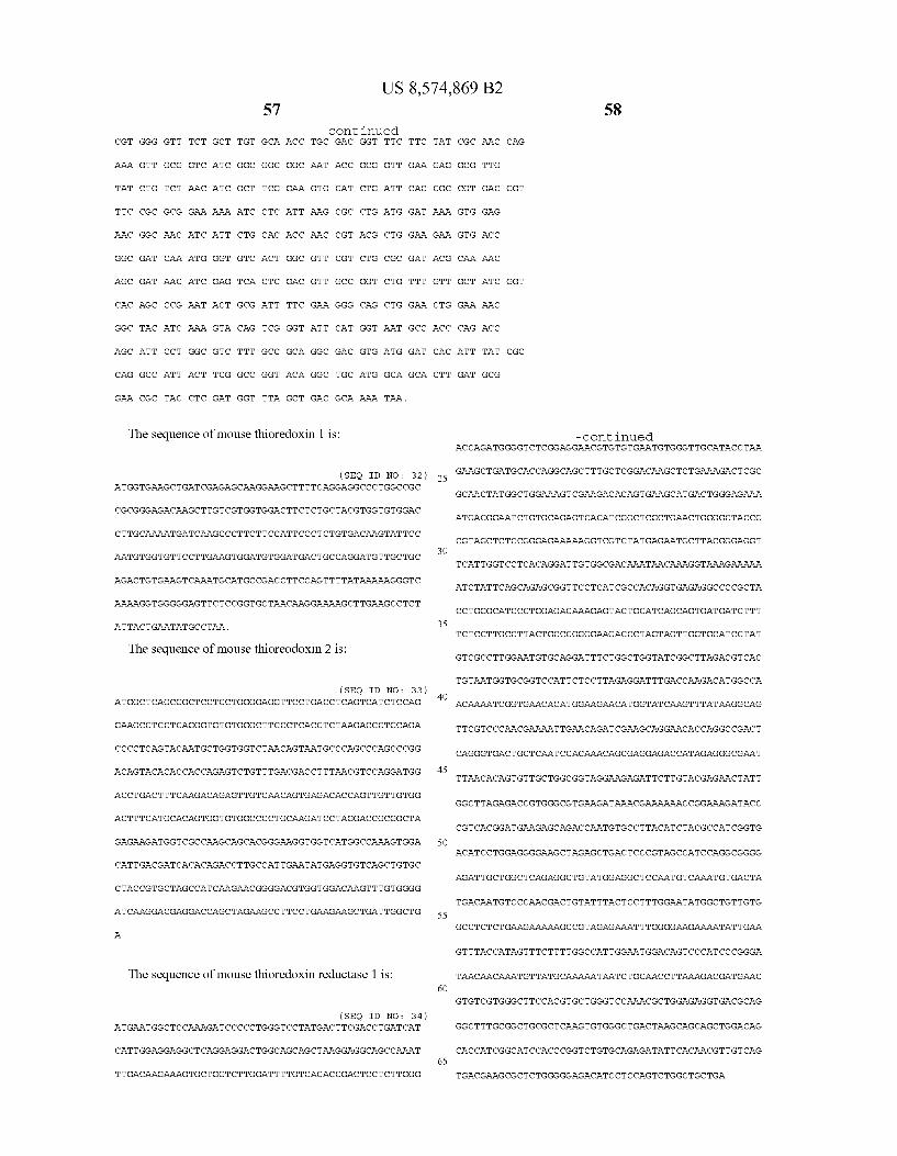

US 8,574,869 B2 59
The sequence of mouse thioredoxin reductase 2 is:
(SEO ID NO : 35) ATGGCGGCGATGGTGGCGGCGATGGTGGCGGCGCTGCGTGGACCCAGCAG
GCGCTTCCGGCCGCGGACACGGGCTCTGACACGCGGGACAAGGGGCGCGG
CGAGTGCAGCGGGAGGGCAGCAGAGCTTTGATCTCTTGGTGATCGGTGGG
GGATCCGGTGGCCTAGCTTGTGCCAAGGAAGCTGCTCAGCTGGGAAAGAA
GGTGGCTGTGGCTGACTATGTGGAACCTTCTCCCCGAGGCACCAAGTGGG
GCCTTGGTGGCACCTGTGTCAACGTGGGTTGCATACCCAAGAAGCTGATG
CATCAGGCTGCACTGCTGGGGGGCATGATCAGAGATGCTCACCACTATGG
CTGGGAGGTGGCCCAGCCTGTCCAACACAACTGGAAGACAATGGCAGAAG
CCGTGCAAAACCATGTGAAATCCTTGAACTGGGGTCATCGCGTCCAACTG
CAGGACAGGAAAGTCAAGTACTTTAACATCAAAGCCAGCTTTGTGGATGA
GCACACAGTTCGCGGTGTGGACAAAGGCGGGAAGGCGACTCTGCTTTCAG
CTGAGCACATTGTCATTGCTACAGGAGGACGGCCAAGGTACCCCACACAA
GTCAAAGGAGCCCTGGAATATGGAATCACAAGTGACGACATCTTCTGGCT
GAAGGAGTCCCCTGGGAAAACGTTGGTGGTTGGAGCCAGCTATGTGGCCC
TAGAGTGTGCTGGCTTCCTCACTGGAATTGGACTGGATACCACTGTCATG
ATGCGCAGCATCCCTCTCCGAGGCTTTGACCAGCAAATGTCATCTTTGGT
CACAGAGCACATGGAGTCTCATGGCACCCAGTTCCTGAAAGGCTGTGTCC
CCTCCCACATCAAAAAACTCCCAACTAACCAGCTGCAGGTCACTTGGGAG
GATCATGCTTCTGGCAAGGAAGACACAGGCACCTTTGACACTGTCCTGTG
GGCCATAGGGCGAGTTCCAGAAACCAGGACTTTGAATCTGGAGAAGGCTG
GCATCAGTACCAACCCTAAGAATCAGAAGATTATTGTGGATGCCCAGGAG
GCTACCTCTGTTCCCCACATCTATGCCATTGGAGATGTTGCTGAGGGGCG
GCCTGAGCTGACGCCCACAGCTATCAAGGCAGGAAAGCTTCTGGCTCAGC
GGCTCTTTGGGAAATCCTCAACCTTAATGGATTACAGCAATGTTCCCACA
ACTGTCTTTACACCACTGGAGTATGGCTGTGTGGGGCTGTCTGAGGAGGA
GGCTGTGGCTCTCCATGGCCAGGAGCATGTAGAGGTTTACCATGCATATT
ATAAGCCCCTAGAGTTCACGGTGGCGGATAGGGATGCATCACAGTGCTAC
ATAAAGATGGTATGCATGAGGGAGCCCCCACAACTGGTGCTGGGCCTGCA
CTTCCTTGGCCCCAACGCTGGAGAAGTCACCCAAGGATTTGCTCTTGGGA
TCAAGTGTGGGGCTTCATATGCACAGGTGATGCAGACAGTAGGGATCCAT
CCCACCTGCTCTGAGGAGGTGGTCAAGCTGCACATCTCCAAGCGCTCCGG
CCTGGAGCCTACTGTGACTGGTTGCTGA.
Example 10
In Vitro Trx/Trx Reductase Studies
Materials and Methods
A commercial TrxR (rat liver) solution (4 uM) was diluted with water to yield a 2.86 uM solution. Lyophilized Trx (human) was reconstituted with PBS (10 mM, pH 7.2) yield ing a 500 uM solution. A solution of 20 mM NADPH and 10 mM ATG and ATM solutions were prepared in water.
5
10
15
25
30
35
40
45
50
55
60
65
60 In a black polypropylene 1.5 mL micro centrifuge tube,
437 uL reaction buffer (10 mM histidine, 10 mM Na2SO4, 137 mM NaCl, 2.5 mM KC1, pH 7.0), 25uL NADPH, 16 uL formulated ocrelizumab solution (30.2 mg/mL) and 5 LL Trx were gently mixed. The reaction was initiated by the addition of 17.5 uL TrXR. The reaction was incubated at room tem perature for 24 hours. Aliquots of 20 uL were taken at each sampling time-point and stored at -70° C. until analyzed by the Bioanalyzer assay.
Inhibition of the Trx system was demonstrated using the same reaction conditions described above with the addition of various inhibitors.
1. In Vitro Activity of Thioredoxin System FIG. 24 shows a digital gel-like image from Bioanalyzer
analysis (each lane representing a time point) showing that incubation of intact ocrelizumab (“2H7, a humanized anti CD20 antibody, referred to as “Variant A’ above) (1 mg/mL) with 0.1 uM TrxR (rat liver), 5 uM Trx (human) and 1 mM NADPH in 10 mM histidine Sulfate buffer results in the reduction of ocrelizumab in less than one hour.
2. In Vitro Activity of Thioredoxin System Inhibited by Aurothioglucose
Aurothioglucose (ATG) was added to the ocrelizumab mixture described above, at the following concentrations: 1 mM: 0.6 uM (6:1 ATG:TrxR): 0.4 uM (4:1 ATG:TrxR); and 0.2 uM (2:1 ATG:TrxR). As attested by the digital gel-like images from Bioanalyzer
analysis shown in FIGS. 25-27, aurothioglucose added at concentrations 1 mM, 0.6 uM, and 0.4LM effectively inhibits the reduction of ocrelizumab by the thioredoxin system. However, as shown in FIG. 28, under these experimental conditions aurothioglucose added at a concentration of 0.2 uM cannot inhibit ocrelizumab reduction after 24 hours.
3. In Vitro Activity of Thioredoxin System Inhibited by Aurothiomalate
Aurothiomalate (ATM) was added to the ocrelizumab mix ture described above, at concentrations of 0.1 mM and 0.01 mM. As attested by the digital gel-like images from Bioana lyzer analysis shown in FIGS. 29 and 30, ATM effectively inhibits the reduction of ocrelizumab by the thioredoxin sys tem at both concentrations tested.
4. In Vitro Activity of Thioredoxin System Inhibited by CuSO4. CuSO was added to the ocrelizumab mixture described
above, at concentrations of 20 uM (4:1 Cu":Trx); 10um (2:1 Cu":Trx); and 5 uM (1:1 Cu":Trx). As shown in FIGS. 31-33, CuSO effectively inhibits thioredoxin-induced reduction of ocrelizumab at concentrations of 20 uM and 10 uM (FIGS. 31 and 32), but the 5 uM concentration is insuf ficient to result in a complete inhibition of reduction (FIG. 33).
5. In Vitro Activity of Thioredoxin System Inhibited by Cystamine
Cystamine was added to the ocrelizumab mixture describe above at the following concentrations: 532 uM (20:1 cysta mine:2H7 (Variant A) disulfide); 266 uM (10:1 cystamine: 2H7 (Variant A) disulfide); 133 uM (5:1 cystamine:2H7 dis ulfide); and 26.6 uM (1:1 cystamine:2H7 (Variant A) disulfide). As shown in FIGS. 34-37, cystamine effectively inhibits thioredoxin-induced reduction of ocrelizumab at concentrations of 532 uM (20:1 cystamine:2H7 (Variant A) disulfide) and 266 uM (10:1 cystamine:2H7 (Variant A)) (FIGS. 34 and 35) but the 133 uM (5:1 cystamine:2H7 (Vari ant A) disulfide) and 26.6 uM (1:1 cystamine:2H7 (VariantA) disulfide) concentrations are insufficient to inhibit the reduc tion of ocrelizumab after 24 hours (FIGS. 36 and 37).

US 8,574,869 B2 61
6. In Vitro Activity of Thioredoxin System Inhibited by Cystine
Cystine was added to the ocrelizumab mixture described above at a concentration of 2.6 mM. As shown in FIG. 38, at this concentration cystine effectively inhibits reduction of ocrelizumab by the thioredoxin system. It is noted that the minimum effective concentration of cystine (just as the effec tive minimum concentration of other inhibitors) depends on the actual circumstances, and might be different for different proteins, such as antibodies, and might vary depending on the timing of addition. Thus, for example, if cystine is added pre-lysis, the minimum effective concentration for antibody 2H7 (Variant A) is about 1.3 mM, for Apomab about 1 mM and for antibody Variant C about 4.5 mM. When cystine is added in the cell culture medium, the minimum effective concentration typically is somewhat higher, and is about 5.2 mM for 2H7 (Variant A), 6 mM for Apomab and 9 mM for antibody Variant C. Usually, for cystine, cystamine and oxi dized glutathione (see below) the minimum effective inhibi tory concentration is about 40x of the antibody concentration (in uM).
7. In Vitro Activity of Thioredoxin System Inhibited by Oxidized Glutathione (GSSG) GSSG was added to the ocrelizumab mixture described
above at a concentration of 2.6 mM. As shown in FIG. 39, at this concentration GSSG effectively inhibits reduction of ocrelizumab by the thioredoxin system. It is noted, however, that the minimum effective concentration of oxidize glu tathione (just as that of the other inhibitors) depends on the actual circumstances, such as, for example, on the nature of the protein (e.g. antibody) produced and the timing of addi tion. For example, for antibody 2H7 (VariantA) the minimum effective concentration is about 1.3 mM for addition prior to lysis.
8. In Vitro Activity of Enzymatic Reduction System FIG. 40 shows a digital gel-like image from Bioanalyzer
analysis (each lane representing a time point) showing that incubation of intact ocrelizumab (2H7, a humanized anti CD20 antibody, Variant A) (1 mg/mL) with 10 g/mL hex okinase, 50 ug/mL glucose-6-phosphate dehydrogenase, 5 uMthioredoxin, 0.1 uM thoredoxin reductase, 2 mM glucose, 0.6 mM ATP, 2 mM Mg, and 2 mM NADP in 50 mM histidine sulfate buffered at pH 7.38 results in the reduction of ocrelizumab in about one hour. Addition of 0.1 mM HDEA, a known glucose-6-phosphate dehydrogenase inhibitor does not inhibit the reduction.
9. In Vitro Activity of Enzymatic Reduction System Requires NADPH As shown in the digital gel-like image from Bioanalyzer
analysis of FIG. 41, incubation of intact ocrelizumab (1 mg/mL) with 5 uM thioredoxin, 0.1 uM thioredoxin reduc tase, and 2 mM NADP in 50 mM histidine sulfate buffer at pH 7.38 does not result in the reduction of the ocrelizumab anti body. Reduction of ocrelizumab could not occur without hex okinase and glucose-6-phosphate dehydrogenase and their substrates to generate NADPH. The invention illustratively described herein can suitably
be practiced in the absence of any element or elements, limi tation or limitations that is not specifically disclosed herein.
5
10
15
25
30
35
40
45
50
55
62 Thus, for example, the terms "comprising.” “including.” “containing.” etc. shall be read expansively and without limi tation. Additionally, the terms and expressions employed herein have been used as terms of description and not of limitation, and there is no intention in the use of such terms and expressions of excluding any equivalent of the invention shown or portion thereof, but it is recognized that various modifications are possible within the scope of the invention claimed. Thus, it should be understood that although the present invention has been specifically disclosed by preferred embodiments and optional features, modifications and varia tions of the inventions embodied herein disclosed can be readily made by those skilled in the art, and that such modi fications and variations are considered to be within the scope of the inventions disclosed herein. The inventions have been described broadly and generically herein. Each of the nar rower species and subgeneric groupings falling within the generic disclosure also form the part of these inventions. This includes within the generic description of each of the inven tions a proviso or negative limitation that will allow removing any subject matter from the genus, regardless or whether or not the material to be removed was specifically recited. In addition, where features or aspects of an invention are described in terms of the Markush group, those schooled in the art will recognize that the invention is also thereby described in terms of any individual member or subgroup of members of the Markush group. Further, when a reference to an aspect of the invention lists a range of individual members. as for example, SEQID NO:1 to SEQID NO: 100, inclusive. it is intended to be equivalent to listing every member of the list individually, and additionally it should be understood that every individual member may be excluded or included in the claim individually. The steps depicted and/or used in methods herein may be
performed in a different order than as depicted and/or stated. The steps are merely exemplary of the order these steps may occur. The steps may occur in any order that is desired Such that it still performs the goals of the claimed invention. From the description of the invention herein, it is manifest
that various equivalents can be used to implement the con cepts of the present invention without departing from its scope. Moreover, while the invention has been described with specific reference to certain embodiments, a person of ordi nary skill in the art would recognize that changes can be made in form and detail without departing from the spirit and the scope of the invention. The described embodiments are con sidered in all respects as illustrative and not restrictive. It should also be understood that the invention is not limited to the particular embodiments described herein, but is capable ofmany equivalents, rearrangements, modifications, and Sub stitutions without departing from the scope of the invention. Thus, additional embodiments are within the scope of the invention and within the following claims.
All U.S. patents and applications; foreign patents and applications; scientific articles: books; and publications men tioned herein are hereby incorporated by reference in their entirety as if each individual patent or publication was spe cifically and individually indicated to be incorporated by reference, including any drawings, figures and tables, as though set forth in full.
SEQUENCE LISTING
<16 O& NUMBER OF SEQ ID NOS: 35
<210s. SEQ ID NO 1 211 LENGTH: 107 212. TYPE: PRT

US 8,574,869 B2 63
- Continued
<213> ORGANISM: Artificial 22 Os. FEATURE:
<223> OTHER INFORMATION: light chain variable
<4 OOs, SEQUENCE: 1
Asp Ile Gln Met Thr Glin Ser Pro Ser Ser Leu Ser Ala Ser Val Gly 1. 5 1O 15
Asp Arg Val Thir Ile Thr Cys Arg Ala Ser Ser Ser Val Ser Tyr Met 2O 25 3O
His Trp Tyr Glin Gln Lys Pro Gly Lys Ala Pro Llys Pro Leu. Ile Tyr 35 4 O 45
Ala Pro Ser Asn Lieu Ala Ser Gly Val Pro Ser Arg Phe Ser Gly Ser SO 55 6 O
Gly Ser Gly Thr Asp Phe Thr Lieu. Thir Ile Ser Ser Lieu. Glin Pro Glu 65 70 7s 8O
Asp Phe Ala Thr Tyr Tyr Cys Glin Gln Trp Ser Phe Asn Pro Pro Thr 85 90 95
Phe Gly Glin Gly Thr Llys Val Glu Ile Lys Arg 1OO 105
<210s, SEQ ID NO 2 &211s LENGTH: 122 212. TYPE: PRT
<213> ORGANISM: Artificial 22 Os. FEATURE:
<223> OTHER INFORMATION: heavy chain variable
<4 OOs, SEQUENCE: 2
Glu Val Glin Lieu Val Glu Ser Gly Gly Gly Lieu Val Glin Pro Gly Gly 1. 5 1O 15
Ser Lieu. Arg Lieu Ser Cys Ala Ala Ser Gly Tyr Thr Phe Thr Ser Tyr 2O 25 3O
Asn Met His Trp Val Arg Glin Ala Pro Gly Lys Gly Lieu. Glu Trp Val 35 4 O 45
Gly Ala Ile Tyr Pro Gly Asn Gly Asp Thr Ser Tyr Asn Glin Llys Phe SO 55 6 O
Lys Gly Arg Phe Thir Ile Ser Val Asp Llys Ser Lys Asn. Thir Lieu. Tyr 65 70 7s 8O
Lieu. Glin Met Asn. Ser Lieu. Arg Ala Glu Asp Thr Ala Val Tyr Tyr Cys 85 90 95
Ala Arg Val Val Tyr Tyr Ser Asn Ser Tyr Trp Tyr Phe Asp Val Trp 1OO 105 11 O
Gly Glin Gly Thr Lieu Val Thr Val Ser Ser 115 12 O
<210s, SEQ ID NO 3 &211s LENGTH: 107 212. TYPE: PRT
<213> ORGANISM: Artificial 22 Os. FEATURE:
<223> OTHER INFORMATION: light chain variable
<4 OOs, SEQUENCE: 3
Asp Ile Gln Met Thr Glin Ser Pro Ser Ser Leu Ser Ala Ser Val Gly 1. 5 1O 15
Asp Arg Val Thir Ile Thr Cys Arg Ala Ser Ser Ser Val Ser Tyr Lieu. 2O 25 3O
His Trp Tyr Glin Gln Lys Pro Gly Lys Ala Pro Llys Pro Leu. Ile Tyr 35 4 O 45

US 8,574,869 B2 65
- Continued
Ala Pro Ser Asn Lieu Ala Ser Gly Val Pro Ser Arg Phe Ser Gly Ser SO 55 6 O
Gly Ser Gly Thr Asp Phe Thr Lieu. Thir Ile Ser Ser Lieu. Glin Pro Glu 65 70 7s 8O
Asp Phe Ala Thr Tyr Tyr Cys Glin Gln Trp Ala Phe Asn Pro Pro Thr 85 90 95
Phe Gly Glin Gly Thr Llys Val Glu Ile Lys Arg 1OO 105
<210s, SEQ ID NO 4 &211s LENGTH: 122 212. TYPE: PRT
<213> ORGANISM: Artificial 22 Os. FEATURE:
<223> OTHER INFORMATION: heavy chain variable
<4 OOs, SEQUENCE: 4
Glu Val Glin Lieu Val Glu Ser Gly Gly Gly Lieu Val Glin Pro Gly Gly 1. 5 1O 15
Ser Lieu. Arg Lieu Ser Cys Ala Ala Ser Gly Tyr Thr Phe Thr Ser Tyr 2O 25 3O
Asn Met His Trp Val Arg Glin Ala Pro Gly Lys Gly Lieu. Glu Trp Val 35 4 O 45
Gly Ala Ile Tyr Pro Gly Asn Gly Ala Thr Ser Tyr Asn Glin Llys Phe SO 55 6 O
Lys Gly Arg Phe Thir Ile Ser Val Asp Llys Ser Lys Asn. Thir Lieu. Tyr 65 70 7s 8O
Lieu. Glin Met Asn. Ser Lieu. Arg Ala Glu Asp Thr Ala Val Tyr Tyr Cys 85 90 95
Ala Arg Val Val Tyr Tyr Ser Ala Ser Tyr Trp Tyr Phe Asp Val Trp 1OO 105 11 O
Gly Glin Gly Thr Lieu Val Thr Val Ser Ser 115 12 O
<210s, SEQ ID NO 5 &211s LENGTH: 122 212. TYPE: PRT
<213> ORGANISM: Artificial 22 Os. FEATURE:
<223> OTHER INFORMATION: heavy chain variable
<4 OOs, SEQUENCE: 5
Glu Val Glin Lieu Val Glu Ser Gly Gly Gly Lieu Val Glin Pro Gly Gly 1. 5 1O 15
Ser Lieu. Arg Lieu Ser Cys Ala Ala Ser Gly Tyr Thr Phe Thr Ser Tyr 2O 25 3O
Asn Met His Trp Val Arg Glin Ala Pro Gly Lys Gly Lieu. Glu Trp Val 35 4 O 45
Gly Ala Ile Tyr Pro Gly Asn Gly Ala Thr Ser Tyr Asn Glin Llys Phe SO 55 6 O
Lys Gly Arg Phe Thir Ile Ser Val Asp Llys Ser Lys Asn. Thir Lieu. Tyr 65 70 7s 8O
Lieu. Glin Met Asn. Ser Lieu. Arg Ala Glu Asp Thr Ala Val Tyr Tyr Cys 85 90 95
Ala Arg Val Val Tyr Tyr Ser Tyr Arg Tyr Trp Tyr Phe Asp Val Trp 1OO 105 11 O
Gly Glin Gly Thr Lieu Val Thr Val Ser Ser 115 12 O

US 8,574,869 B2 67
- Continued
<210s, SEQ ID NO 6 &211s LENGTH: 213 212. TYPE: PRT
<213> ORGANISM: Artificial 22 Os. FEATURE:
<223> OTHER INFORMATION: full length light chain
<4 OOs, SEQUENCE: 6
Asp Ile Gln Met Thr Glin Ser Pro Ser Ser Leu Ser Ala Ser Val Gly 1. 5 1O 15
Asp Arg Val Thir Ile Thr Cys Arg Ala Ser Ser Ser Val Ser Tyr Met 2O 25 3O
His Trp Tyr Glin Gln Lys Pro Gly Lys Ala Pro Llys Pro Leu. Ile Tyr 35 4 O 45
Ala Pro Ser Asn Lieu Ala Ser Gly Val Pro Ser Arg Phe Ser Gly Ser SO 55 6 O
Gly Ser Gly Thr Asp Phe Thr Lieu. Thir Ile Ser Ser Lieu. Glin Pro Glu 65 70 7s 8O
Asp Phe Ala Thr Tyr Tyr Cys Glin Gln Trp Ser Phe Asn Pro Pro Thr 85 90 95
Phe Gly Glin Gly Thr Lys Val Glu Ile Lys Arg Thr Val Ala Ala Pro 1OO 105 11 O
Ser Val Phe Ile Phe Pro Pro Ser Asp Glu Gln Leu Lys Ser Gly Thr 115 12 O 125
Ala Ser Val Val Cys Lieu. Lieu. Asn. Asn. Phe Tyr Pro Arg Glu Ala Lys 13 O 135 14 O
Val Glin Trp Llys Val Asp Asn Ala Lieu. Glin Ser Gly Asn. Ser Glin Glu 145 150 155 160
Ser Val Thr Glu Glin Asp Ser Lys Asp Ser Thr Tyr Ser Leu Ser Ser 1.65 17O 17s
Thir Lieu. Thir Lieu. Ser Lys Ala Asp Tyr Glu Lys His Llys Val Tyr Ala 18O 185 19 O
Cys Glu Val Thr His Glin Gly Lieu Ser Ser Pro Val Thr Lys Ser Phe 195 2OO 2O5
Asn Arg Gly Glu. Cys 21 O
<210s, SEQ ID NO 7 &211s LENGTH: 452 212. TYPE: PRT
<213> ORGANISM: Artificial 22 Os. FEATURE:
<223> OTHER INFORMATION: full length heavy chain sequence
<4 OO > SEQUENCE: 7
Glu Val Glin Lieu Val Glu Ser Gly Gly Gly Lieu Val Glin Pro Gly Gly 1. 5 1O 15
Ser Lieu. Arg Lieu Ser Cys Ala Ala Ser Gly Tyr Thr Phe Thr Ser Tyr 2O 25 3O
Asn Met His Trp Val Arg Glin Ala Pro Gly Lys Gly Lieu. Glu Trp Val 35 4 O 45
Gly Ala Ile Tyr Pro Gly Asn Gly Asp Thr Ser Tyr Asn Glin Llys Phe SO 55 6 O
Lys Gly Arg Phe Thir Ile Ser Val Asp Llys Ser Lys Asn. Thir Lieu. Tyr 65 70 7s 8O
Lieu. Glin Met Asn. Ser Lieu. Arg Ala Glu Asp Thr Ala Val Tyr Tyr Cys 85 90 95

US 8,574,869 B2 69
- Continued
Ala Arg Val Val Tyr Tyr Ser Asn Ser Tyr Trp Tyr Phe Asp Val Trp 1OO 105 11 O
Gly Glin Gly Thr Lieu Val Thr Val Ser Ser Ala Ser Thr Lys Gly Pro 115 12 O 125
Ser Val Phe Pro Leu Ala Pro Ser Ser Lys Ser Thr Ser Gly Gly Thr 13 O 135 14 O
Ala Ala Lieu. Gly Cys Lieu Val Lys Asp Tyr Phe Pro Glu Pro Val Thr 145 150 155 160
Val Ser Trp Asn Ser Gly Ala Lieu. Thir Ser Gly Val His Thr Phe Pro 1.65 17O 17s
Ala Val Lieu. Glin Ser Ser Gly Lieu. Tyr Ser Leu Ser Ser Val Val Thr 18O 185 19 O
Val Pro Ser Ser Ser Lieu. Gly Thr Glin Thr Tyr Ile Cys Asn Val Asn 195 2OO 2O5
His Llys Pro Ser Asn. Thir Lys Val Asp Llys Llys Val Glu Pro Llys Ser 21 O 215 22O
Cys Asp Llys Thr His Thr Cys Pro Pro Cys Pro Ala Pro Glu Lieu. Leu 225 23 O 235 24 O
Gly Gly Pro Ser Val Phe Leu Phe Pro Pro Llys Pro Lys Asp Thr Lieu. 245 250 255
Met Ile Ser Arg Thr Pro Glu Val Thr Cys Val Val Val Asp Val Ser 26 O 265 27 O
His Glu Asp Pro Glu Val Llys Phe Asn Trp Tyr Val Asp Gly Val Glu 27s 28O 285
Val His Asn Ala Lys Thr Llys Pro Arg Glu Glu Gln Tyr ASn Ser Thr 29 O 295 3 OO
Tyr Arg Val Val Ser Val Lieu. Thr Val Lieu. His Glin Asp Trp Lieu. Asn 3. OS 310 315 32O
Gly Lys Glu Tyr Lys Cys Llys Val Ser Asn Lys Ala Lieu Pro Ala Pro 3.25 330 335
Ile Glu Lys Thir Ile Ser Lys Ala Lys Gly Glin Pro Arg Glu Pro Glin 34 O 345 35. O
Val Tyr Thr Lieu Pro Pro Ser Arg Glu Glu Met Thr Lys Asn Glin Val 355 360 365
Ser Lieu. Thr Cys Lieu Val Lys Gly Phe Tyr Pro Ser Asp Ile Ala Val 37 O 375 38O
Glu Trp Glu Ser Asn Gly Glin Pro Glu Asn Asn Tyr Lys Thr Thr Pro 385 390 395 4 OO
Pro Val Lieu. Asp Ser Asp Gly Ser Phe Phe Leu Tyr Ser Lys Lieu. Thr 4 OS 41O 415
Val Asp Llys Ser Arg Trp Glin Glin Gly Asn Val Phe Ser Cys Ser Val 42O 425 43 O
Met His Glu Ala Lieu. His Asn His Tyr Thr Gln Lys Ser Leu Ser Lieu. 435 44 O 445
Ser Pro Gly Lys 450
<210s, SEQ ID NO 8 &211s LENGTH: 452 212. TYPE: PRT
<213> ORGANISM: Artificial 22 Os. FEATURE:
<223> OTHER INFORMATION: full length heavy chain
<4 OOs, SEQUENCE: 8
Glu Val Glin Lieu Val Glu Ser Gly Gly Gly Lieu Val Glin Pro Gly Gly

Ser
Asn
Gly
Lys 65
Lell
Ala
Gly
Ser
Ala 145
Wall
Ala
Wall
His
Cys 225
Gly
Met
His
Wall
Tyr 3. OS
Gly
Ile
Wall
Ser
Glu 385
Pro
Wall
Luell
Met
Ala SO
Gly
Glin
Arg
Glin
Wall 13 O
Ala
Ser
Wall
Pro
Lys 21 O
Asp
Gly
Ile
Glu
His 29 O
Arg
Ala
Luell 37 O
Trp
Wall
Asp
Arg
His 35
Ile
Arg
Met
Wall
Gly 115
Phe
Luell
Trp
Luell
Ser 195
Pro
Pro
Ser
Asp
Asn
Wall
Glu
Ala
Thir 355
Thir
Glu
Luell
Luell 2O
Trp
Phe
Asn
Wall
Thir
Pro
Gly
Asn
Glin 18O
Ser
Ser
Thir
Ser
Arg 26 O
Pro
Ala
Wall
Tyr
Thir 34 O
Luell
Ser
Asp
Ser
42O
Ser
Wall
Pro
Thir
Ser 85
Tyr
Lell
Lell
Ser 1.65
Ser
Ser
Asn
His
Wall 245
Thir
Glu
Ser
Lys 3.25
Ile
Pro
Lell
Asn
Ser 4 OS
Arg
Arg
Gly
Ile 70
Lell
Wall
Ala
Lell 150
Gly
Ser
Lell
Thir
Thir 23 O
Phe
Pro
Wall
Thir
Wall 310
Ser
Pro
Wall
Gly 390
Asp
Trp
71
Ala
Glin
Asn 55
Ser
Arg
Ser
Thir
Pro 135
Wall
Ala
Gly
Gly
Lys 215
Lell
Glu
Lys 295
Lell
Ser
Lys 375
Glin
Gly
Glin
Ala
Ala 4 O
Gly
Wall
Ala
Asn
Wall 12 O
Ser
Luell
Luell
Thir
Wall
Pro
Phe
Wall
Phe 28O
Pro
Thir
Wall
Ala
Arg 360
Gly
Pro
Ser
Glin
Ser 25
Pro
Asp
Asp
Glu
Ser 105
Ser
Ser
Asp
Thir
Tyr 185
Glin
Asp
Pro
Pro
Thir 265
Asn
Arg
Wall
Ser
Lys 345
Glu
Phe
Glu
Phe
Gly 425
1O
Gly
Gly
Thir
Ser
Ser 17O
Ser
Thir
Pro 250
Trp
Glu
Luell
Asn 330
Gly
Glu
Asn
Phe 41O
Asn
Tyr
Lys
Ser
Ser
Thir
Trp
Ala
Ser
Phe 155
Gly
Luell
Pro 235
Wall
Glu
His 315
Lys
Glin
Met
Pro
ASn 395
Luell
Wall
US 8,574,869 B2
- Continued
Thir
Gly
Tyr 6 O
Ala
Ser
Thir 14 O
Pro
Wall
Ser
Ile
Wall 22O
Ala
Pro
Wall
Wall
Glin 3 OO
Glin
Ala
Pro
Thir
Ser
Tyr
Tyr
Phe
Phe
Lell 45
Asn
Asn
Wall
Phe
Thir 125
Ser
Glu
His
Ser
Cys
Glu
Pro
Wall
Asp 285
Tyr
Asp
Lell
Arg
Lys 365
Asp
Ser
Ser
Thir 3O
Glu
Glin
Thir
Asp 11 O
Gly
Pro
Thir
Wall 19 O
Asn
Pro
Glu
Asp
Asp 27 O
Gly
Asn
Trp
Pro
Glu 35. O
Asn
Ile
Thir
Cys 43 O
15
Ser
Trp
Luell
Tyr 95
Wall
Gly
Gly
Wall
Phe 17s
Wall
Wall
Luell
Thir 255
Wall
Wall
Ala
Luell
Ala 335
Pro
Glin
Ala
Thir
Luell 415
Ser
Tyr
Wall
Phe
Tyr
Cys
Trp
Pro
Thir
Thir 160
Pro
Thir
Asn
Ser
Luell 24 O
Luell
Ser
Glu
Thir
Asn
Pro
Glin
Wall
Wall
Pro 4 OO
Thir
Wall
72

US 8,574,869 B2 73
- Continued
Met His Glu Ala Lieu. His Asn His Tyr Thr Gln Lys Ser Leu Ser Lieu.
Ser
435 44 O 445
Pro Gly Lys 450
<210s, SEQ ID NO 9 &211s LENGTH: 213 212. TYPE: PRT
<213> ORGANISM: Artificial 22 Os. FEATURE:
<223> OTHER INFORMATION: full length light chain
<4 OOs, SEQUENCE: 9
Asp 1.
Asp
His
Ala
Gly 65
Asp
Phe
Ser
Ala
Wall 145
Ser
Thir
Asn
Ile Gln Met Thr Glin Ser Pro Ser Ser Lieu. Ser Ala 5 1O
Arg Val Thir Ile Thr Cys Arg Ala Ser Ser Ser Val 2O 25
Trp Tyr Glin Gln Lys Pro Gly Lys Ala Pro Llys Pro 35 4 O 45
Pro Ser Asn Lieu Ala Ser Gly Val Pro Ser Arg Phe SO 55 6 O
Ser Gly Thr Asp Phe Thr Lieu. Thir Ile Ser Ser Lieu. 70 7s
Phe Ala Thr Tyr Tyr Cys Glin Gln Trp Ala Phe Asn 85 90
Gly Glin Gly Thr Llys Val Glu Ile Lys Arg Thr Val 1OO 105
Val Phe Ile Phe Pro Pro Ser Asp Glu Gln Leu Lys 115 12 O 125
Ser Val Val Cys Lieu. Lieu. Asn. Asn. Phe Tyr Pro Arg 13 O 135 14 O
Gln Trp Llys Val Asp Asn Ala Lieu. Glin Ser Gly Asn 150 155
Val Thr Glu Gln Asp Ser Lys Asp Ser Thr Tyr Ser 1.65 17O
Lieu. Thir Lieu. Ser Lys Ala Asp Tyr Glu Lys His Llys 18O 185
Glu Val Thr His Glin Gly Lieu Ser Ser Pro Val Thr 195 2OO 2O5
Arg Gly Glu. Cys 21 O
<210s, SEQ ID NO 10 &211s LENGTH: 452 212. TYPE: PRT
<213> ORGANISM: Artificial 22 Os. FEATURE:
<223> OTHER INFORMATION: full length heavy chain
<4 OOs, SEQUENCE: 10
Glu 1.
Ser
Asn
Gly
Val Glin Lieu Val Glu Ser Gly Gly Gly Lieu Val Glin 5 1O
Lieu. Arg Lieu. Ser Cys Ala Ala Ser Gly Tyr Thr Phe 2O 25
Met His Trp Val Arg Glin Ala Pro Gly Lys Gly Lieu. 35 4 O 45
Ala Ile Tyr Pro Gly Asn Gly Ala Thr Ser Tyr Asn SO 55 6 O
Ser
Ser
Luell
Ser
Glin
Pro
Ala 11 O
Ser
Glu
Ser
Luell
Wall 19 O
Pro
Thir
Glu
Glin
Wall 15
Ile
Gly
Pro
Pro 95
Ala
Gly
Ala
Glin
Ser 17s
Ser
Gly 15
Ser
Trp
Gly
Luell
Tyr
Ser
Glu
Thir
Pro
Thir
Lys
Glu 160
Ser
Ala
Phe
Gly
Tyr
Wall
Phe
74

Lys 65
Lell
Ala
Gly
Ser
Ala 145
Wall
Ala
Wall
His
Cys 225
Gly
Met
His
Wall
Tyr 3. OS
Gly
Ile
Wall
Ser
Glu 385
Pro
Wall
Met
Ser
Gly
Glin
Arg
Glin
Wall 13 O
Ala
Ser
Wall
Pro
Lys 21 O
Asp
Gly
Ile
Glu
His 29 O
Arg
Ala
Luell 37 O
Trp
Wall
Asp
His
Pro 450
Arg
Met
Wall
Gly 115
Phe
Luell
Trp
Luell
Ser 195
Pro
Pro
Ser
Asp
Asn
Wall
Glu
Ala
Thir 355
Thir
Glu
Luell
Glu 435
Gly
Phe
Asn
Wall
Thir
Pro
Gly
Asn
Glin 18O
Ser
Ser
Thir
Ser
Arg 26 O
Pro
Ala
Wall
Thir 34 O
Luell
Ser
Asp
Ser
Ala
Thir
Ser 85
Tyr
Lell
Lell
Cys
Ser 1.65
Ser
Ser
Asn
His
Wall 245
Thir
Glu
Lys
Ser
Lys 3.25
Ile
Pro
Lell
Asn
Ser 4 OS
Arg
Lell
<210s, SEQ ID NO 11 &211s LENGTH:
212. TYPE : <213> ORGANISM: Artificial
PRT
452
Ile 70
Lell
Wall
Ala
Lell 150
Gly
Ser
Lell
Thir
Thir 23 O
Phe
Pro
Wall
Thir
Wall 310
Ser
Pro
Wall
Gly 390
Asp
Trp
His
75
Ser
Arg
Ser
Thir
Pro 135
Wall
Ala
Gly
Gly
Lys 215
Lell
Glu
Lys 295
Lell
Ser
Lys 375
Glin
Gly
Glin
Asn
Wall
Ala
Ala
Wall 12 O
Ser
Luell
Luell
Thir
Wall
Pro
Phe
Wall
Phe 28O
Pro
Thir
Wall
Ala
Arg 360
Gly
Pro
Ser
Glin
His 44 O
Asp
Glu
Ser 105
Ser
Ser
Asp
Thir
Tyr 185
Glin
Asp
Pro
Pro
Thir 265
Asn
Arg
Wall
Ser
Lys 345
Glu
Phe
Glu
Phe
Gly 425
Ser
Ser 17O
Ser
Thir
Pro 250
Trp
Glu
Luell
Asn 330
Gly
Glu
Asn
Phe 41O
Asn
Thir
Ser
Thir
Trp
Ala
Ser
Phe 155
Gly
Luell
Pro 235
Wall
Glu
His 315
Glin
Met
Pro
ASn 395
Luell
Wall
Glin
US 8,574,869 B2
- Continued
Lys
Ala
Ser
Thir 14 O
Pro
Wall
Ser
Ile
Wall 22O
Ala
Pro
Wall
Wall
Glin 3 OO
Glin
Ala
Pro
Thir
Ser 38O
Phe
Asn
Wall
Phe
Thir 125
Ser
Glu
His
Ser
Cys
Glu
Pro
Wall
Asp 285
Asp
Lell
Arg
Lys 365
Asp
Ser
Ser
Ser 445
Thir
Tyr
Asp 11 O
Gly
Pro
Thir
Wall 19 O
Asn
Pro
Glu
Asp
Asp 27 O
Gly
Asn
Trp
Pro
Glu 35. O
Asn
Ile
Thir
Cys 43 O
Luell
Luell
Tyr 95
Wall
Gly
Gly
Wall
Phe 17s
Wall
Wall
Luell
Thir 255
Wall
Wall
Ala
Luell
Ala 335
Pro
Glin
Ala
Thir
Luell 415
Ser
Ser
Tyr
Cys
Trp
Pro
Thir
Thir 160
Pro
Thir
Asn
Ser
Luell 24 O
Luell
Ser
Glu
Thir
Asn
Pro
Glin
Wall
Wall
Pro 4 OO
Thir
Wall
Luell
76

US 8,574,869 B2 77
- Continued
22 Os. FEATURE:
<223> OTHER INFORMATION: full length heavy chain sequence
<4 OOs, SEQUENCE: 11
Glu Val Glin Lieu Val Glu Ser Gly Gly Gly Lieu Val Glin Pro Gly Gly 1. 5 1O 15
Ser Lieu. Arg Lieu Ser Cys Ala Ala Ser Gly Tyr Thr Phe Thr Ser Tyr 2O 25 3O
Asn Met His Trp Val Arg Glin Ala Pro Gly Lys Gly Lieu. Glu Trp Val 35 4 O 45
Gly Ala Ile Tyr Pro Gly Asn Gly Ala Thr Ser Tyr Asn Glin Llys Phe SO 55 6 O
Lys Gly Arg Phe Thir Ile Ser Val Asp Llys Ser Lys Asn. Thir Lieu. Tyr 65 70 7s 8O
Lieu. Glin Met Asn. Ser Lieu. Arg Ala Glu Asp Thr Ala Val Tyr Tyr Cys 85 90 95
Ala Arg Val Val Tyr Tyr Ser Ala Ser Tyr Trp Tyr Phe Asp Val Trp 1OO 105 11 O
Gly Glin Gly Thr Lieu Val Thr Val Ser Ser Ala Ser Thr Lys Gly Pro 115 12 O 125
Ser Val Phe Pro Leu Ala Pro Ser Ser Lys Ser Thr Ser Gly Gly Thr 13 O 135 14 O
Ala Ala Lieu. Gly Cys Lieu Val Lys Asp Tyr Phe Pro Glu Pro Val Thr 145 150 155 160
Val Ser Trp Asn Ser Gly Ala Lieu. Thir Ser Gly Val His Thr Phe Pro 1.65 17O 17s
Ala Val Lieu. Glin Ser Ser Gly Lieu. Tyr Ser Leu Ser Ser Val Val Thr 18O 185 19 O
Val Pro Ser Ser Ser Lieu. Gly Thr Glin Thr Tyr Ile Cys Asn Val Asn 195 2OO 2O5
His Llys Pro Ser Asn. Thir Lys Val Asp Llys Llys Val Glu Pro Llys Ser 21 O 215 22O
Cys Asp Llys Thr His Thr Cys Pro Pro Cys Pro Ala Pro Glu Lieu. Leu 225 23 O 235 24 O
Gly Gly Pro Ser Val Phe Leu Phe Pro Pro Llys Pro Lys Asp Thr Lieu. 245 250 255
Met Ile Ser Arg Thr Pro Glu Val Thr Cys Val Val Val Asp Val Ser 26 O 265 27 O
His Glu Asp Pro Glu Val Llys Phe Asn Trp Tyr Val Asp Gly Val Glu 27s 28O 285
Val His Asn Ala Lys Thr Llys Pro Arg Glu Glu Glin Tyr Asn Ala Thr 29 O 295 3 OO
Tyr Arg Val Val Ser Val Lieu. Thr Val Lieu. His Glin Asp Trp Lieu. Asn 3. OS 310 315 32O
Gly Lys Glu Tyr Lys Cys Ala Val Ser Asn Lys Ala Lieu Pro Ala Pro 3.25 330 335
Ile Glu Ala Thir Ile Ser Lys Ala Lys Gly Glin Pro Arg Glu Pro Glin 34 O 345 35. O
Val Tyr Thr Lieu Pro Pro Ser Arg Glu Glu Met Thr Lys Asn Glin Val 355 360 365
Ser Lieu. Thr Cys Lieu Val Lys Gly Phe Tyr Pro Ser Asp Ile Ala Val 37 O 375 38O
Glu Trp Glu Ser Asn Gly Glin Pro Glu Asn Asn Tyr Lys Thr Thr Pro 385 390 395 4 OO

US 8,574,869 B2 79
- Continued
Pro Val Lieu. Asp Ser Asp Gly Ser Phe Phe Leu Tyr Ser Lys Lieu. Thr 4 OS 41O 415
Val Asp Llys Ser Arg Trp Glin Glin Gly Asn Val Phe Ser Cys Ser Val 42O 425 43 O
Met His Glu Ala Lieu. His Asn His Tyr Thr Gln Lys Ser Leu Ser Lieu. 435 44 O 445
Ser Pro Gly Lys 450
<210s, SEQ ID NO 12 &211s LENGTH: 452 212. TYPE: PRT
<213> ORGANISM: Artificial 22 Os. FEATURE:
<223> OTHER INFORMATION: full length heavy chain sequence
<4 OOs, SEQUENCE: 12
Glu Val Glin Lieu Val Glu Ser Gly Gly Gly Lieu Val Glin Pro Gly Gly 1. 5 1O 15
Ser Lieu. Arg Lieu Ser Cys Ala Ala Ser Gly Tyr Thr Phe Thr Ser Tyr 2O 25 3O
Asn Met His Trp Val Arg Glin Ala Pro Gly Lys Gly Lieu. Glu Trp Val 35 4 O 45
Gly Ala Ile Tyr Pro Gly Asn Gly Ala Thr Ser Tyr Asn Glin Llys Phe SO 55 6 O
Lys Gly Arg Phe Thir Ile Ser Val Asp Llys Ser Lys Asn. Thir Lieu. Tyr 65 70 7s 8O
Lieu. Glin Met Asn. Ser Lieu. Arg Ala Glu Asp Thr Ala Val Tyr Tyr Cys 85 90 95
Ala Arg Val Val Tyr Tyr Ser Ala Ser Tyr Trp Tyr Phe Asp Val Trp 1OO 105 11 O
Gly Glin Gly Thr Lieu Val Thr Val Ser Ser Ala Ser Thr Lys Gly Pro 115 12 O 125
Ser Val Phe Pro Leu Ala Pro Ser Ser Lys Ser Thr Ser Gly Gly Thr 13 O 135 14 O
Ala Ala Lieu. Gly Cys Lieu Val Lys Asp Tyr Phe Pro Glu Pro Val Thr 145 150 155 160
Val Ser Trp Asn Ser Gly Ala Lieu. Thir Ser Gly Val His Thr Phe Pro 1.65 17O 17s
Ala Val Lieu. Glin Ser Ser Gly Lieu. Tyr Ser Leu Ser Ser Val Val Thr 18O 185 19 O
Val Pro Ser Ser Ser Lieu. Gly Thr Glin Thr Tyr Ile Cys Asn Val Asn 195 2OO 2O5
His Llys Pro Ser Asn. Thir Lys Val Asp Llys Llys Val Glu Pro Llys Ser 21 O 215 22O
Cys Asp Llys Thr His Thr Cys Pro Pro Cys Pro Ala Pro Glu Lieu. Leu 225 23 O 235 24 O
Gly Gly Pro Ser Val Phe Leu Phe Pro Pro Llys Pro Lys Asp Thr Lieu. 245 250 255
Met Ile Ser Arg Thr Pro Glu Val Thr Cys Val Val Val Asp Val Ser 26 O 265 27 O
His Glu Asp Pro Glu Val Llys Phe Asn Trp Tyr Val Asp Gly Val Glu 27s 28O 285
Val His Asn Ala Lys Thr Llys Pro Arg Glu Glu Glin Tyr Asn Ala Thr 29 O 295 3 OO
Tyr Arg Val Val Ser Val Lieu. Thr Val Lieu. His Glin Asp Trp Lieu. Asn

US 8,574,869 B2 81
- Continued
3. OS 310 315 32O
Gly Lys Glu Tyr Lys Cys Llys Val Ser Asn Ala Ala Lieu Pro Ala Pro 3.25 330 335
Ile Ala Ala Thir Ile Ser Lys Ala Lys Gly Glin Pro Arg Glu Pro Glin 34 O 345 35. O
Val Tyr Thr Lieu Pro Pro Ser Arg Glu Glu Met Thr Lys Asn Glin Val 355 360 365
Ser Lieu. Thr Cys Lieu Val Lys Gly Phe Tyr Pro Ser Asp Ile Ala Val 37 O 375 38O
Glu Trp Glu Ser Asn Gly Glin Pro Glu Asn Asn Tyr Lys Thr Thr Pro 385 390 395 4 OO
Pro Val Lieu. Asp Ser Asp Gly Ser Phe Phe Leu Tyr Ser Lys Lieu. Thr 4 OS 41O 415
Val Asp Llys Ser Arg Trp Glin Glin Gly Asn Val Phe Ser Cys Ser Val 42O 425 43 O
Met His Glu Ala Lieu. His Asn His Tyr Thr Gln Lys Ser Leu Ser Lieu. 435 44 O 445
Ser Pro Gly Lys 450
<210s, SEQ ID NO 13 &211s LENGTH: 452 212. TYPE: PRT
<213> ORGANISM: Artificial 22 Os. FEATURE:
<223> OTHER INFORMATION: full length heavy chain sequence
<4 OOs, SEQUENCE: 13
Glu Val Glin Lieu Val Glu Ser Gly Gly Gly Lieu Val Glin Pro Gly Gly 1. 5 1O 15
Ser Lieu. Arg Lieu Ser Cys Ala Ala Ser Gly Tyr Thr Phe Thr Ser Tyr 2O 25 3O
Asn Met His Trp Val Arg Glin Ala Pro Gly Lys Gly Lieu. Glu Trp Val 35 4 O 45
Gly Ala Ile Tyr Pro Gly Asn Gly Ala Thr Ser Tyr Asn Glin Llys Phe SO 55 6 O
Lys Gly Arg Phe Thir Ile Ser Val Asp Llys Ser Lys Asn. Thir Lieu. Tyr 65 70 7s 8O
Lieu. Glin Met Asn. Ser Lieu. Arg Ala Glu Asp Thr Ala Val Tyr Tyr Cys 85 90 95
Ala Arg Val Val Tyr Tyr Ser Ala Ser Tyr Trp Tyr Phe Asp Val Trp 1OO 105 11 O
Gly Glin Gly Thr Lieu Val Thr Val Ser Ser Ala Ser Thr Lys Gly Pro 115 12 O 125
Ser Val Phe Pro Leu Ala Pro Ser Ser Lys Ser Thr Ser Gly Gly Thr 13 O 135 14 O
Ala Ala Lieu. Gly Cys Lieu Val Lys Asp Tyr Phe Pro Glu Pro Val Thr 145 150 155 160
Val Ser Trp Asn Ser Gly Ala Lieu. Thir Ser Gly Val His Thr Phe Pro 1.65 17O 17s
Ala Val Lieu. Glin Ser Ser Gly Lieu. Tyr Ser Leu Ser Ser Val Val Thr 18O 185 19 O
Val Pro Ser Ser Ser Lieu. Gly Thr Glin Thr Tyr Ile Cys Asn Val Asn 195 2OO 2O5
His Llys Pro Ser Asn. Thir Lys Val Asp Llys Llys Val Glu Pro Llys Ser 21 O 215 22O

Cys 225
Gly
Met
His
Wall
Tyr 3. OS
Gly
Ile
Wall
Ser
Glu 385
Pro
Wall
Met
Ser
US 8,574,869 B2 83
- Continued
Asp Llys Thr His Thr Cys Pro Pro Cys Pro Ala Pro 23 O 235
Gly Pro Ser Val Phe Leu Phe Pro Pro Llys Pro Llys 245 250
Ile Ser Arg Thr Pro Glu Val Thr Cys Val Val Val 26 O 265
Glu Asp Pro Glu Val Llys Phe Asn Trp Tyr Val Asp 27s 28O 285
His Asn Ala Lys Thr Llys Pro Arg Glu Glu Glin Tyr 29 O 295 3 OO
Arg Val Val Ser Val Lieu. Thr Val Lieu. His Glin Asp 310 315
Lys Glu Tyr Lys Cys Llys Val Ser Asn Ala Ala Lieu. 3.25 330
Ala Ala Thir Ile Ser Lys Ala Lys Gly Glin Pro Arg 34 O 345
Tyr Thr Lieu Pro Pro Ser Arg Glu Glu Met Thr Lys 355 360 365
Lieu. Thir Cys Lieu Val Lys Gly Phe Tyr Pro Ser Asp 37 O 375 38O
Trp Glu Ser Asn Gly Glin Pro Glu Asn. Asn Tyr Lys 390 395
Val Lieu. Asp Ser Asp Gly Ser Phe Phe Leu Tyr Ser 4 OS 41O
Asp Llys Ser Arg Trp Glin Glin Gly Asn Val Phe Ser 42O 425
His Glu Ala Lieu. His Trp His Tyr Thr Glin Lys Ser 435 44 O 445
Pro Gly Lys 450
<210s, SEQ ID NO 14 &211s LENGTH: 452 212. TYPE: PRT
<213> ORGANISM: Artificial 22 Os. FEATURE:
<223> OTHER INFORMATION: full length heavy chain
<4 OOs, SEQUENCE: 14
Glu 1.
Ser
Asn
Gly
Lys 65
Lell
Ala
Gly
Val Glin Lieu Val Glu Ser Gly Gly Gly Lieu Val Glin 5 1O
Lieu. Arg Lieu. Ser Cys Ala Ala Ser Gly Tyr Thr Phe 2O 25
Met His Trp Val Arg Glin Ala Pro Gly Lys Gly Lieu. 35 4 O 45
Ala Ile Tyr Pro Gly Asn Gly Ala Thr Ser Tyr Asn SO 55 6 O
Gly Arg Phe Thir Ile Ser Val Asp Llys Ser Lys Asn 70 7s
Gln Met Asn. Ser Lieu. Arg Ala Glu Asp Thr Ala Val 85 90
Arg Val Val Tyr Tyr Ser Tyr Arg Tyr Trp Tyr Phe 1OO 105
Gln Gly Thr Lieu Val Thr Val Ser Ser Ala Ser Thr 115 12 O 125
Glu
Asp
Asp 27 O
Gly
Asn
Trp
Pro
Glu 35. O
Asn
Ile
Thir
Cys 43 O
Luell
Pro
Thir
Glu
Glin
Thir
Asp 11 O
Luell
Thir 255
Wall
Wall
Ala
Luell
Ala 335
Pro
Glin
Ala
Thir
Luell 415
Ser
Ser
Gly 15
Ser
Trp
Luell
Tyr 95
Wall
Gly
Luell 24 O
Luell
Ser
Glu
Thir
Asn
Pro
Glin
Wall
Wall
Pro 4 OO
Thir
Wall
Luell
Gly
Tyr
Wall
Phe
Tyr
Cys
Trp
Pro
84
US 8,574,869 B2 85
- Continued
Ser Val Phe Pro Leu Ala Pro Ser Ser Lys Ser Thr Ser Gly Gly Thr 13 O 135 14 O
Ala Ala Lieu. Gly Cys Lieu Val Lys Asp Tyr Phe Pro Glu Pro Val Thr 145 150 155 160
Val Ser Trp Asn Ser Gly Ala Lieu. Thir Ser Gly Val His Thr Phe Pro 1.65 17O 17s
Ala Val Lieu. Glin Ser Ser Gly Lieu. Tyr Ser Leu Ser Ser Val Val Thr 18O 185 19 O
Val Pro Ser Ser Ser Lieu. Gly Thr Glin Thr Tyr Ile Cys Asn Val Asn 195 2OO 2O5
His Llys Pro Ser Asn. Thir Lys Val Asp Llys Llys Val Glu Pro Llys Ser 21 O 215 22O
Cys Asp Llys Thr His Thr Cys Pro Pro Cys Pro Ala Pro Glu Lieu. Leu 225 23 O 235 24 O
Gly Gly Pro Ser Val Phe Leu Phe Pro Pro Llys Pro Lys Asp Thr Lieu. 245 250 255
Met Ile Ser Arg Thr Pro Glu Val Thr Cys Val Val Val Asp Val Ser 26 O 265 27 O
His Glu Asp Pro Glu Val Llys Phe Asn Trp Tyr Val Asp Gly Val Glu 27s 28O 285
Val His Asn Ala Lys Thr Llys Pro Arg Glu Glu Glin Tyr Asn Ala Thr 29 O 295 3 OO
Tyr Arg Val Val Ser Val Lieu. Thr Val Lieu. His Glin Asp Trp Lieu. Asn 3. OS 310 315 32O
Gly Lys Glu Tyr Lys Cys Llys Val Ser Asn Ala Ala Lieu. Pro Ala Pro 3.25 330 335
Ile Ala Ala Thir Ile Ser Lys Ala Lys Gly Glin Pro Arg Glu Pro Glin 34 O 345 35. O
Val Tyr Thr Lieu Pro Pro Ser Arg Glu Glu Met Thr Lys Asn Glin Val 355 360 365
Ser Lieu. Thr Cys Lieu Val Lys Gly Phe Tyr Pro Ser Asp Ile Ala Val 37 O 375 38O
Glu Trp Glu Ser Asn Gly Glin Pro Glu Asn Asn Tyr Lys Thr Thr Pro 385 390 395 4 OO
Pro Val Lieu. Asp Ser Asp Gly Ser Phe Phe Leu Tyr Ser Lys Lieu. Thr 4 OS 41O 415
Val Asp Llys Ser Arg Trp Glin Glin Gly Asn Val Phe Ser Cys Ser Val 42O 425 43 O
Met His Glu Ala Lieu. His Asn His Tyr Thr Gln Lys Ser Leu Ser Lieu. 435 44 O 445
Ser Pro Gly Lys 450
<210s, SEQ ID NO 15 &211s LENGTH: 452 212. TYPE: PRT
<213> ORGANISM: Artificial 22 Os. FEATURE:
<223> OTHER INFORMATION: full length heavy chain
<4 OOs, SEQUENCE: 15
Glu Val Glin Lieu Val Glu Ser Gly Gly Gly Lieu Val Glin Pro Gly Gly 1. 5 1O 15
Ser Lieu. Arg Lieu Ser Cys Ala Ala Ser Gly Tyr Thr Phe Thr Ser Tyr 2O 25 3O
Asn Met His Trp Val Arg Glin Ala Pro Gly Lys Gly Lieu. Glu Trp Val

Gly
Lys 65
Lell
Ala
Gly
Ser
Ala 145
Wall
Ala
Wall
His
Cys 225
Gly
Met
His
Wall
Tyr 3. OS
Gly
Ile
Wall
Ser
Glu 385
Pro
Wall
Met
Ser
Ala SO
Gly
Glin
Arg
Glin
Wall 13 O
Ala
Ser
Wall
Pro
Lys 21 O
Asp
Gly
Ile
Glu
His 29 O
Arg
Ala
Luell 37 O
Trp
Wall
Asp
His
Pro
450
35
Ile
Arg
Met
Wall
Gly 115
Phe
Luell
Trp
Luell
Ser 195
Pro
Pro
Ser
Asp
Asn
Wall
Glu
Ala
Thir 355
Thir
Glu
Luell
Glu 435
Gly
Phe
Asn
Wall
Thir
Pro
Gly
Asn
Glin 18O
Ser
Ser
Thir
Ser
Arg 26 O
Pro
Ala
Wall
Tyr
Thir 34 O
Luell
Ser
Asp
Ser
Ala
Pro
Thir
Ser 85
Tyr
Lell
Lell
Ser 1.65
Ser
Ser
Asn
His
Wall 245
Thir
Glu
Ser
Lys 3.25
Ile
Pro
Lell
Asn
Ser 4 OS
Arg
Lell
Gly
Ile 70
Lell
Wall
Ala
Lell 150
Gly
Ser
Lell
Thir
Thir 23 O
Phe
Pro
Wall
Thir
Wall 310
Ser
Pro
Wall
Gly 390
Asp
Trp
His
87
Asn 55
Ser
Arg
Ser
Thir
Pro 135
Wall
Ala
Gly
Gly
Lys 215
Lell
Glu
Lys 295
Lell
Ser
Lys 375
Glin
Gly
Glin
Asn
4 O
Gly
Wall
Ala
Asn
Wall 12 O
Ser
Luell
Luell
Thir
Wall
Pro
Phe
Wall
Phe 28O
Pro
Thir
Wall
Ala
Arg 360
Gly
Pro
Ser
Glin
His 44 O
Asp
Asp
Glu
Ser 105
Ser
Ser
Asp
Thir
Tyr 185
Glin
Asp
Pro
Pro
Thir 265
Asn
Arg
Wall
Ser
Lys 345
Glu
Phe
Glu
Phe
Gly 425
Thir
Ser
Ser 17O
Ser
Thir
Pro 250
Trp
Glu
Luell
Asn 330
Gly
Glu
Tyr
Asn
Phe
Asn
Thir
Ser
Ser
Thir
Trp
Ala
Ser
Phe 155
Gly
Luell
Pro 235
Wall
Glu
His 315
Ala
Glin
Met
Pro
ASn 395
Luell
Wall
Glin
US 8,574,869 B2
- Continued
Tyr 6 O
Ala
Ser
Thir 14 O
Pro
Wall
Ser
Ile
Wall 22O
Ala
Pro
Wall
Wall
Glin 3 OO
Glin
Ala
Pro
Thir
Ser 38O
Phe
45
Asn
Asn
Wall
Phe
Thir 125
Ser
Glu
His
Ser
Cys
Glu
Pro
Wall
Asp 285
Tyr
Asp
Lell
Arg
Lys 365
Asp
Ser
Ser
Ser 445
Glin
Thir
Asp 11 O
Gly
Pro
Thir
Wall 19 O
Asn
Pro
Glu
Asp
Asp 27 O
Gly
Asn
Trp
Pro
Glu 35. O
Asn
Ile
Thir
Cys 43 O
Luell
Luell
Tyr 95
Wall
Gly
Gly
Wall
Phe 17s
Wall
Wall
Luell
Thir 255
Wall
Wall
Ala
Luell
Ala 335
Pro
Glin
Ala
Thir
Luell 415
Ser
Ser
Phe
Tyr
Cys
Trp
Pro
Thir
Thir 160
Pro
Thir
Asn
Ser
Luell 24 O
Luell
Ser
Glu
Thir
Asn
Pro
Glin
Wall
Wall
Pro 4 OO
Thir
Wall
Luell
88

US 8,574,869 B2 89
- Continued
<210s, SEQ ID NO 16 &211s LENGTH: 107 212. TYPE: PRT
<213> ORGANISM: Artificial 22 Os. FEATURE:
<223> OTHER INFORMATION: antibody
<4 OOs, SEQUENCE: 16
Asp Ile Gln Met Thr Glin Ser Pro Ser Ser Leu Ser Ala Ser Val Gly 1. 5 1O 15
Asp Arg Val Thir Ile Thr Cys Lys Ala Ser Glin Asp Wal Ser Ile Gly 2O 25 3O
Val Ala Trp Tyr Glin Gln Llys Pro Gly Lys Ala Pro Llys Lieu. Lieu. Ile 35 4 O 45
Tyr Ser Ala Ser Tyr Arg Tyr Thr Gly Val Pro Ser Arg Phe Ser Gly SO 55 6 O
Ser Gly Ser Gly Thr Asp Phe Thr Lieu. Thir Ile Ser Ser Leu Gln Pro 65 70 7s 8O
Glu Asp Phe Ala Thr Tyr Tyr Cys Glin Glin Tyr Tyr Ile Tyr Pro Tyr 85 90 95
Thr Phe Gly Glin Gly Thr Lys Val Glu Ile Llys 1OO 105
<210s, SEQ ID NO 17 &211s LENGTH: 119 212. TYPE: PRT
<213> ORGANISM: Artificial 22 Os. FEATURE:
<223> OTHER INFORMATION: antibody
<4 OOs, SEQUENCE: 17
Glu Val Glin Lieu Val Glu Ser Gly Gly Gly Lieu Val Glin Pro Gly Gly 1. 5 1O 15
Ser Lieu. Arg Lieu Ser Cys Ala Ala Ser Gly Phe Thr Phe Thr Asp Tyr 2O 25 3O
Thir Met Asp Trp Val Arg Glin Ala Pro Gly Lys Gly Lieu. Glu Trp Val 35 4 O 45
Ala Asp Val Asn Pro Asn. Ser Gly Gly Ser Ile Tyr Asn. Glin Arg Phe SO 55 6 O
Lys Gly Arg Phe Thr Lieu. Ser Val Asp Arg Ser Lys Asn. Thir Lieu. Tyr 65 70 7s 8O
Lieu. Glin Met Asn. Ser Lieu. Arg Ala Glu Asp Thr Ala Val Tyr Tyr Cys 85 90 95
Ala Arg Asn Lieu. Gly Pro Ser Phe Tyr Phe Asp Tyr Trp Gly Glin Gly 1OO 105 11 O
Thir Lieu Val Thir Wal Ser Ser 115
<210s, SEQ ID NO 18 &211s LENGTH: 107 212. TYPE: PRT
<213> ORGANISM: Artificial 22 Os. FEATURE:
<223> OTHER INFORMATION: light chain
<4 OOs, SEQUENCE: 18
Asp Ile Gln Met Thr Glin Ser Pro Ser Ser Leu Ser Ala Ser Val Gly 1. 5 1O 15
Asp Arg Val Thir Ile Thr Cys Arg Ala Ser Glin Asp Wall Asn. Thir Ala 2O 25 3O

Wall
Ser 65
Glu
Thir
Ala
Ser SO
Arg
Asp
Phe
Trp 35
Ala
Ser
Phe
Gly
Tyr
Ser
Gly
Ala
Glin
PRT
<4 OOs, SEQUENCE:
Glu 1.
Ser
Ala
Lys 65
Lell
Ser
Gly
Wall Glin Lieu.
Luell
Ile
Arg SO
Gly
Glin
Arg
Thir
Arg
His 35
Ile
Arg
Met
Trp
Luell 115
Luell 2O
Trp
Phe
Asn
Gly
Wall
PRT
FEATURE: OTHER INFORMATION:
SEQUENCE:
Asp Ile Gln Met 1.
Asp
Lell
Ser 65
Glu
Thir
Arg
Asn
Phe SO
Gly
Asp
Phe
Wall
Trp 35
Thir
Ser
Ile
Gly
Ile
Ser
Gly
Ala
Gly 1OO
Glin
Phe
Thir
Thir 85
Gly
SEQ ID NO 19 LENGTH: TYPE : ORGANISM: Artificial FEATURE:
OTHER INFORMATION: heavy chain
12O
19
Wall 5
Ser
Wall
Pro
Thir
Ser 85
Gly
Thir
SEQ ID NO 2 O LENGTH: TYPE : ORGANISM:
108
Glin
Lell
Asp 70
Thir
Glu
Arg
Thir
Ile 70
Lell
Asp
Wall
91
Lys
Tyr 55
Phe
Ser
Ala
Glin
Asn 55
Ser
Arg
Gly
Ser
artificial
Thir 5
Ile
Glin
Ser
Thir
Thir 85
Gly
Glin
Ser
Glin
Lell
Asp 70
Thir
Pro 4 O
Ser
Thir
Wall
Phe
Ser 12 O
Gly
Gly
Luell
Glin
Glu 105
Gly
Ser 25
Pro
Asp
Glu
Tyr 105
antibody
Thir
Lys
His 55
Thir
Ser
Pro 4 O
Ser
Ser
Cys
Luell
Ser
Ala 25
Asp
Gly
Luell
Glin
Glu 105
Wall
Thir
Glin 90
Ile
Gly
Gly
Gly
Thir
Thir
Asp 90
Ala
Ser 1O
Ser
Gly
Wall
Thir
Glin 90
Ile
Ala
Pro
Ile
His
Luell
Phe
Lys
Arg
Ser
Thir
Met
Luell
Glin
Thir
Pro
Ile
Tyr
Lys
US 8,574,869 B2
- Continued
Pro Llys Lieu. Lieu. Ile 45
Ser Arg Phe Ser Gly 6 O
Ser Ser Lieu. Glin Pro 8O
Tyr Thr Thr Pro Pro 95
Val Glin Pro Gly Gly 15
Asn. Ile Lys Asp Thr 3O
Gly Lieu. Glu Trp Val 45
Tyr Ala Asp Ser Val 6 O
Lys Asn. Thir Ala Tyr 8O
Ala Val Tyr Tyr Cys 95
Asp Tyr Trp Gly Glin 11 O
Ser Ala Ser Lieu. Gly 15
Asp Ile Ser Asn Tyr 3O
Val Llys Val Lieu. Ile 45
Ser Arg Phe Ser Gly 6 O
Ser Asn Lieu. Glu Pro 8O
Ser Thr Val Pro Trp 95
Arg
92

US 8,574,869 B2 93
- Continued
<210s, SEQ ID NO 21 &211s LENGTH: 123 212. TYPE: PRT
<213> ORGANISM; artificial 22 Os. FEATURE:
<223> OTHER INFORMATION: antibody
<4 OOs, SEQUENCE: 21
Glu Ile Glin Lieu Val Glin Ser Gly Pro Glu Lieu Lys Glin Pro Gly Glu 1. 5 1O 15
Thr Val Arg Ile Ser Cys Lys Ala Ser Gly Tyr Thr Phe Thr Asn Tyr 2O 25 3O
Gly Met Asn Trp Val Lys Glin Ala Pro Gly Lys Gly Lieu Lys Trp Met 35 4 O 45
Gly Trp Ile Asn Thr Tyr Thr Gly Glu Pro Thr Tyr Ala Ala Asp Phe SO 55 6 O
Lys Arg Arg Phe Thr Phe Ser Lieu. Glu Thir Ser Ala Ser Thr Ala Tyr 65 70 7s 8O
Lieu. Glin Ile Ser Asn Lieu Lys Asn Asp Asp Thr Ala Thr Tyr Phe Cys 85 90 95
Ala Lys Tyr Pro His Tyr Tyr Gly Ser Ser His Trp Tyr Phe Asp Val 1OO 105 11 O
Trp Gly Ala Gly Thr Thr Val Thr Val Ser Ser 115 12 O
<210s, SEQ ID NO 22 &211s LENGTH: 108 212. TYPE: PRT
<213> ORGANISM: Artificial 22 Os. FEATURE:
<223> OTHER INFORMATION: antibody
<4 OOs, SEQUENCE: 22
Asp Ile Gln Met Thr Glin Ser Pro Ser Ser Leu Ser Ala Ser Val Gly 1. 5 1O 15
Asp Arg Val Thir Ile Thr Cys Ser Ala Ser Glin Asp Ile Ser Asn Tyr 2O 25 3O
Lieu. Asn Trp Tyr Glin Glin Llys Pro Gly Lys Ala Pro Llys Val Lieu. Ile 35 4 O 45
Tyr Phe Thr Ser Ser Lieu. His Ser Gly Val Pro Ser Arg Phe Ser Gly SO 55 6 O
Ser Gly Ser Gly Thr Asp Phe Thr Lieu. Thir Ile Ser Ser Leu Gln Pro 65 70 7s 8O
Glu Asp Phe Ala Thr Tyr Tyr Cys Glin Glin Tyr Ser Thr Val Pro Trp 85 90 95
Thr Phe Gly Glin Gly Thr Lys Val Glu Ile Lys Arg 1OO 105
<210s, SEQ ID NO 23 &211s LENGTH: 123 212. TYPE: PRT
<213> ORGANISM: Artificial 22 Os. FEATURE:
<223> OTHER INFORMATION: antibody
<4 OOs, SEQUENCE: 23
Glu Val Glin Lieu Val Glu Ser Gly Gly Gly Lieu Val Glin Pro Gly Gly 1. 5 1O 15
Ser Lieu. Arg Lieu Ser Cys Ala Ala Ser Gly Tyr Thr Phe Thr Asn Tyr

Gly
Gly
Lys 65
Lell
Ala
Trp
Met
Trp SO
Arg
Glin
Gly
Asn 35
Ile
Arg
Met
Glin 115
Trp
Asn
Phe
Asn
Pro
Gly
PRT
<4 OOs, SEQUENCE:
Asp Ile Glin Lieu. 1.
Asp
Lell
Ser 65
Glu
Thir
Arg
Asn
Phe SO
Gly
Asp
Phe
Wall
Trp 35
Thir
Ser
Phe
Gly
Thir
Ser
Gly
Ala
Glin
PRT
<4 OOs, SEQUENCE:
Glu 1.
Ser
Gly
Gly
Lys 65
Lell
Ala
Wall Glin Lieu.
Luell
Met
Trp SO
Arg
Glin
Arg
Asn 35
Ile
Arg
Met
Luell
Trp
Asn
Phe
Asn
Pro
Wall
Thir
Thir
Ser 85
His
Thir
SEQ ID NO 24 LENGTH: TYPE : ORGANISM: Artificial FEATURE: OTHER INFORMATION:
108
24
Thir 5
Ile
Glin
Ser
Thir
Thir 85
Gly
SEO ID NO 25 LENGTH: TYPE : ORGANISM: Artificial FEATURE: OTHER INFORMATION:
123
25
Wall 5
Ser
Wall
Thir
Thir
Ser 85
Tyr
Arg
Phe 70
Lell
Lell
Glin
Thir
Glin
Lell
Asp 70
Thir
Glu
Phe 70
Lell
95
Glin
Thir 55
Ser
Arg
Wall
Ala 4 O
Gly
Luell
Ala
Gly
Thir 12 O
25
Pro
Glu
Asp
Glu
Ser 105
Wall
antibody
Ser
His 55
Phe
Pro
Ser
Pro 4 O
Ser
Thir
Wall
Ser
Ala 25
Gly
Gly
Luell
Glin
Glu 105
antibody
Ser
Ala
Glin
Thir 55
Ser
Arg
Gly
Ala
Ala 4 O
Gly
Luell
Ala
Gly
Gly
Ser 25
Pro
Glu
Asp
Glu
Thir
Gly
Pro
Thir
Asp 90
Ser
Ser
Ser 1O
Ser
Wall
Thir
Glin 90
Ile
Gly
Gly
Gly
Pro
Thir
Asp 90
Ser
Thir
Ser
Thir
His
Ser
Luell
Glin
Ala
Pro
Ile 7s
Luell
Thir
Ser
Thir
His
US 8,574,869 B2
- Continued
Gly
Tyr 6 O
Ala
Trp
Ser
Asp
Pro
Ser 6 O
Ser
Ser
Arg
Wall
Asp
Gly
Tyr 6 O
Ala
Trp
Lell 45
Ala
Ser
Wall
Ala
Ile
Lys 45
Arg
Ser
Thir
Glin
Phe
Lell 45
Ala
Ser
Wall
Glu
Ala
Thir
Phe 11 O
Ser
Ser
Wall
Phe
Luell
Wall
Pro
Thir
Glu
Ala
Thir
Phe
Trp
Asp
Ala
Tyr 95
Asp
Wall 15
Asn
Luell
Ser
Glin
Pro 95
Gly 15
His
Trp
Asp
Ala
Tyr 95
Asp
Wall
Phe
Tyr
Wall
Gly
Ile
Gly
Pro
Trp
Gly
Wall
Phe
Tyr
Wall
96

US 8,574,869 B2 97
- Continued
1OO 105 11 O
Trp Gly Glin Gly Thr Lieu Val Thr Val Ser Ser 115 12 O
<210s, SEQ ID NO 26 &211s LENGTH: 108 212. TYPE: PRT
<213> ORGANISM: Artificial 22 Os. FEATURE:
<223> OTHER INFORMATION: antibody
<4 OOs, SEQUENCE: 26
Asp Ile Gln Met Thr Glin Ser Pro Ser Ser Leu Ser Ala Ser Val Gly 1. 5 1O 15
Asp Arg Val Thir Ile Thr Cys Arg Ala Ser Lys Thir Ile Ser Lys Tyr 2O 25 3O
Lieu Ala Trp Tyr Glin Glin Llys Pro Gly Lys Ala Pro Llys Lieu. Lieu. Ile 35 4 O 45
Tyr Ser Gly Ser Thr Lieu. Glin Ser Gly Val Pro Ser Arg Phe Ser Gly SO 55 6 O
Ser Gly Ser Gly Thr Asp Phe Thr Lieu. Thir Ile Ser Ser Leu Gln Pro 65 70 7s 8O
Glu Asp Phe Ala Thr Tyr Tyr Cys Glin Glin His Asn Glu Tyr Pro Leu 85 90 95
Thr Phe Gly Glin Gly Thr Lys Val Glu Ile Lys Arg 1OO 105
<210s, SEQ ID NO 27 &211s LENGTH: 121 212. TYPE: PRT
<213> ORGANISM: Artificial 22 Os. FEATURE:
<223> OTHER INFORMATION: antibody
<4 OOs, SEQUENCE: 27
Glu Val Glin Lieu Val Glu Ser Gly Gly Gly Lieu Val Glin Pro Gly Gly 1. 5 1O 15
Ser Lieu. Arg Lieu Ser Cys Ala Ala Ser Gly Tyr Ser Phe Thr Gly His 2O 25 3O
Trp Met Asn Trp Val Arg Glin Ala Pro Gly Lys Gly Lieu. Glu Trp Val 35 4 O 45
Gly Met Ile His Pro Ser Asp Ser Glu Thr Arg Tyr Asn Glin Llys Phe SO 55 6 O
Lys Asp Arg Phe Thir Ile Ser Val Asp Llys Ser Lys Asn. Thir Lieu. Tyr 65 70 7s 8O
Lieu. Glin Met Asn. Ser Lieu. Arg Ala Glu Asp Thr Ala Val Tyr Tyr Cys 85 90 95
Ala Arg Gly Ile Tyr Phe Tyr Gly Thr Thr Tyr Phe Asp Tyr Trp Gly 1OO 105 11 O
Gln Gly Thr Lieu Val Thr Val Ser Ser 115 12 O
<210s, SEQ ID NO 28 &211s LENGTH: 214 212. TYPE: PRT
<213> ORGANISM: Artificial 22 Os. FEATURE:
<223> OTHER INFORMATION: antibody
<4 OOs, SEQUENCE: 28

Asp
Asp
Lell
Ser 65
Glu
Thir
Pro
Thir
Lys 145
Glu
Ser
Ala
Phe
99
Ile Gln Met Thr Glin Ser Pro Ser
Arg Val Thir Ile Thr Cys Arg Ala 2O 25
Ala Trp Tyr Glin Gln Lys Pro Gly 35 4 O
Ser Gly Ser Thr Lieu. Glin Ser Gly SO 55
Gly Ser Gly Thr Asp Phe Thr Lieu. 70
Asp Phe Ala Thr Tyr Tyr Cys Glin 85
Phe Gly Glin Gly Thr Lys Val Glu 1OO 105
Ser Wall Phe Ile Phe Pro Pro Ser 115 12 O
Ala Ser Val Val Cys Lieu. Lieu. Asn 13 O 135
Val Glin Trp Llys Val Asp Asn Ala 150
Ser Val Thr Glu Glin Asp Ser Lys 1.65
Thir Lieu. Thir Lieu. Ser Lys Ala Asp 18O 185
Cys Glu Val Thr His Glin Gly Lieu. 195 2OO
Asn Arg Gly Glu. Cys 21 O
<210s, SEQ ID NO 29 &211s LENGTH: 451 212. TYPE: PRT
<213> ORGANISM: Artificial 22 Os. FEATURE:
<223> OTHER INFORMATION: antibody
<4 OOs, SEQUENCE: 29
Glu 1.
Ser
Trp
Gly
Lys 65
Lell
Ala
Glin
Wall
Ala
Val Glin Lieu Val Glu Ser Gly Gly 5
Lieu. Arg Lieu. Ser Cys Ala Ala Ser 2O 25
Met Asn Trp Val Arg Glin Ala Pro 35 4 O
Met Ile His Pro Ser Asp Ser Glu SO 55
Asp Arg Phe Thir Ile Ser Val Asp 70
Gln Met Asn. Ser Lieu. Arg Ala Glu 85
Arg Gly Ile Tyr Phe Tyr Gly Thr 1OO 105
Gly Thr Lieu Val Thr Val Ser Ser 115 12 O
Phe Pro Leu Ala Pro Ser Ser Lys 13 O 135
Lieu. Gly Cys Lieu Val Lys Asp Tyr
Ser 1O
Ser
Wall
Thir
Glin 90
Ile
Asp
Asn
Luell
Asp 17O
Ser
Gly 1O
Gly
Gly
Thir
Asp 90
Thir
Ala
Ser
Phe
Luell
Lys
Ala
Pro
Ile
His
Glu
Phe
Glin 155
Ser
Glu
Ser
Luell
Tyr
Lys
Arg
Ser
Thir
Ser
Thir
Pro
US 8,574,869 B2
- Continued
Ser
Thir
Pro
Ser 6 O
Ser
Asn
Arg
Glin
Tyr 14 O
Ser
Thir
Pro
Wall
Ser
Gly
Tyr 6 O
Ala
Phe
Thir
Ser 14 O
Glu
Ala
Ile
Lys 45
Arg
Ser
Glu
Thir
Lell 125
Pro
Gly
His
Wall 2O5
Glin
Phe
Lell 45
Asn
Asn
Wall
Asp
Lys 125
Gly
Pro
Ser
Ser
Luell
Phe
Luell
Tyr
Wall 11 O
Arg
Asn
Ser
Lys 19 O
Thir
Pro
Thir
Glu
Glin
Thir
Tyr 11 O
Gly
Gly
Wall
Wall 15
Lys
Luell
Ser
Glin
Pro 95
Ala
Ser
Glu
Ser
Luell 17s
Wall
Gly 15
Gly
Trp
Luell
Tyr 95
Trp
Pro
Thir
Thir
Gly
Tyr
Ile
Gly
Pro
Luell
Ala
Gly
Ala
Glin 160
Ser
Tyr
Ser
Gly
His
Wall
Phe
Tyr
Cys
Gly
Ser
Ala
Wall
100

145
Ser
Wall
Pro
Asp 225
Gly
Ile
Glu
His
Arg 3. OS
Glu
Lell
Trp 385
Wall
Asp
His
Pro
Trp
Luell
Ser
Pro 21 O
Pro
Ser
Asp
Asn 29 O
Wall
Glu
Thir
Thir 37 O
Glu
Luell
Glu
Gly 450
Asn
Glin
Ser 195
Ser
Thir
Ser
Arg
Pro 27s
Ala
Wall
Thir
Luell 355
Ser
Asp
Ser
Ala 435
101
150
Ser Gly Ala Lieu. Thr 1.65
Ser Ser Gly Lieu. Tyr 18O
Ser Lieu. Gly Thr Glin
Asn Thr Llys Val Asp 215
His Thr Cys Pro Pro 23 O
Wall Phe Leu Phe Pro 245
Thir Pro Gu Wall. Thir 26 O
Glu Val Llys Phe Asn
Llys Thr Llys Pro Arg 295
Ser Wall Lieu. Thir Wall 310
Lys Cys Llys Val Ser 3.25
Ile Ser Lys Ala Lys 34 O
Pro Pro Ser Arg Glu 360
Lieu Val Lys Gly Phe 375
Asin Gly Glin Pro Glu 390
Ser Asp Gly Ser Phe 4 OS
Arg Trp Glin Glin Gly
Lieu. His Asn His Tyr
<210s, SEQ ID NO 3 O &211s LENGTH: 384 &212s. TYPE: DNA &213s ORGANISM: E.
<4 OOs, SEQUENCE: 30
atgttacacic aacaacgaaa
gataaaatta
gcgatcct cq
Ctggatgaaa
caaaac cctd
aaaaacggtg
titcc togacg
ttcacctgac
tcqatttctg
tcgctgacga
aagttggcggc
cta acctggc
44 O
c caacacgcc
tgacgacagt
ggcagagtgg
at atcagggc
gaaatatggc
alaccaaagtg
gtaa
155
Ser Gly Val 17O
Ser Lieu. Ser 185
Thr Tyr Ile
Llys Llys Val
Cys Pro Ala 235
Pro Llys Pro 250
Cys Val Val 265
Trp Tyr Val
Glu Glu Glin
Lieu. His Glin 315
Asn Lys Ala 330
Gly Glin Pro 345
Glu Met Thr
Tyr Pro Ser
Asn Asn Tyr 395
Phe Leu Tyr 41O
Asn. Wall Phe 425
Thr Gln Lys
coli. Thioredoxin TrixA
aggct tatt c
tittgacacgg
aaact gaccg
atc.cgtggta
ggtgcactgt
US 8,574,869 B2
- Continued
His
Ser
Glu 22O
Pro
Wall
Asp
Tyr 3 OO
Asp
Lell
Arg
Asp
Ser
Ser
Ser
Thir
Wall
Asn
Pro
Glu
Asp
Asp
Gly 285
Asn
Trp
Pro
Glu
Asn 365
Ile
Thir
Lell 445
Phe
Wall 19 O
Wall
Luell
Thir
Wall 27 O
Wall
Ser
Luell
Ala
Pro 35. O
Glin
Ala
Thir
Luell
Ser 43 O
Ser
Ctgtggagtt
atgtact caa
gcaaaatgat
ttgcaaaact
tcc.cgactict
Ctalaagg to a
160
Pro Ala 17s
Thir Wall
Asn His
Ser Cys
Lieu. Gly 24 O
Luell Met 255
Ser His
Glu Wall
Thr Tyr
Asin Gly 32O
Pro Ile 335
Glin Wall
Wall Ser
Wall Glu
Pro Pro 4 OO
Thir Wall 415
Wal Met
Luell Ser
atatatgagc
agcggacggg
cgcc.ccgatt
gaa catcgat
gctgctgttc
gttgaaagag
6 O
12 O
18O
24 O
3OO
360
384
102


US 8,574,869 B2 107 108
- Continued
gctgct cagc tigggaaagaa ggtggctgtg gctgactatgttgaacct t c toccc.gaggc 24 O
accalagtggg gccttggtgg cacctgtgtc. alacgtgggitt gcatacccala gaagctgatg 3OO
catcaggctg. cactgctggg ggg catgat C agagatgctic accactatgg Ctgggaggtg 360
gcc.ca.gc.ctg. tccaacacala Ctggalagaca atggcagaag cc.gtgcaaaa c catgtgaaa 42O
t ccttgaact ggggtcatcg cgt.ccaactg. Cagga cagga aagttcaagta Ctttalacat c 48O
aaagcc agct ttgttggatga gcacacagtt cqcggtgtgg acaaaggcgg gaaggcgact 54 O
Ctgctitt cag Ctgagcacat tt cattgct acaggaggac ggc.calaggta CCC cacacala 6OO
gtcaaaggag ccctggaata tigaat caca agtgacgaca tottctggct gaaggagtcC 660
Cctgggaaaa C9ttggtggit tagccagc tatgtggc.cc tagagtgtgc tiggct tcct c 72 O
actggaattig gactggatac Cactgtcatg atgcgcagda t cc ct ct cog aggctttgac 78O
Cagcaaatgt Catctttggt cacagagcac atggagt cto atggc accca gttcCtgaaa 84 O
ggctgtgtcc cct cocacat caaaaaactic ccaactalacc agctgcaggit cacttgggag 9 OO
gat catgctt Ctggcaagga agacacaggc acctttgaca Ctgtc.ctgtg ggc cataggg 96.O
cgagttcCag aalaccaggac tittgaatctg gagaaggctg gcatcagtac Caaccctaag O2O
aatcagaaga ttattgttgga tigcc.caggag got acct ctog titc.cccacat citatgcc att O8O
ggagatgttg Ctgagggg.cg gcctgagctg acgcc cacag Ctatcaaggc aggaaagctt 14 O
ctggct cago ggct ctittgg gaaatcctica accittaatgg attacagcaa tott.cccaca 2OO
actgtctitta Caccactgga gtatggctgt gtggggotgt Ctgaggagga ggctgtggct 26 O
CtcCatggcc aggagcatgt agaggitttac catgcatatt ataagcc cct agagttcacg 32O
gtggcggata gggatgcatc acagtgctac ataaagatgg tatgcatgag ggagc.ccc.ca 38O
Caactggtgc tigggcctgca Ctt cottggc cccaacgctg gagaagt cac cca aggattt 44 O
gct Cttggga t caagtgtgg ggctt catat gcacaggtga tigcagacagt agggatcCat SOO
Cccacctgct Ctgaggaggt ggt caagctg. Cacat ct coa agcgctc.cgg cctggagcct 560
actgttgactg gttgctga sf8
What is claimed is: 1. A method for the prevention of the reduction of a disul
fide bond in an antibody expressed in a recombinant host cell, comprising, following fermentation, sparging the pre-harvest or harvested culture fluid of said recombinant host cell with air, wherein the amount of dissolved oxygen (dO) in the pre-harvest or harvested culture fluid is at least 10%.
2. The method of claim 1 wherein said air sparging is continued until the pre-harvest or harvested culture fluid is at least 30% saturated with air.
3. The method of claim 1 wherein said air sparging is continued until the pre-harvest or harvested culture fluid is between about 100% saturated to about 30% saturated with a1.
4. The method of claim 1 wherein the amount of dissolved oxygen (dO) in the pre-harvest or harvested culture fluid is at least 30%.
45
50
55
5. The method of claim 1 wherein the antibody is a thera peutic antibody.
6. The method of claim 1 wherein the antibody is a bio logically functional fragment of an antibody.
7. The method of claim 1 wherein the host cell is eukaryotic host cell.
8. The method of claim 7 wherein the eukaryotic host cell is a mammalian host cell.
9. The method of claim 1 wherein the host cell is prokary otic host cell.
10. The method of claim9 wherein the prokaryotic host cell is a bacterial cell.