Embed Size (px)
Citation preview
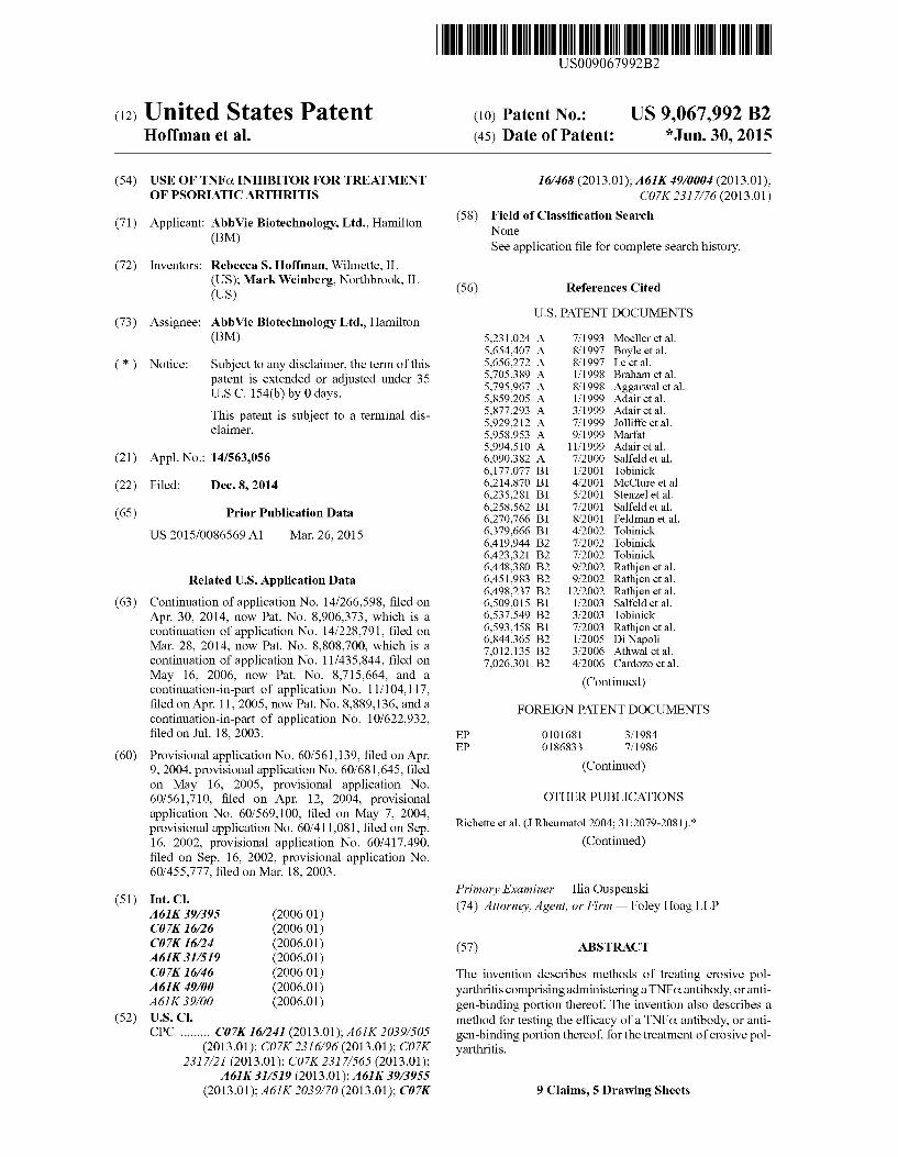
(12) United States Patent Hoffman et al.
US009067992B2
(10) Patent No.: US 9,067,992 B2 (45) Date of Patent: *Jun. 30, 2015
(54) USE OF TNFa. INHIBITOR FORTREATMENT OF PSORATICARTHRITIS
(71) Applicant: AbbVie Biotechnology, Ltd., Hamilton (BM)
(72) Inventors: Rebecca S. Hoffman, Wilmette, IL (US); Mark Weinberg, Northbrook, IL (US)
(73) Assignee: AbbVie Biotechnology Ltd., Hamilton (BM)
(*) Notice: Subject to any disclaimer, the term of this patent is extended or adjusted under 35 U.S.C. 154(b) by 0 days. This patent is Subject to a terminal dis claimer.
(21) Appl. No.: 14/563,056
(22) Filed: Dec. 8, 2014
(65) Prior Publication Data
US 2015/OO86569 A1 Mar. 26, 2015
Related U.S. Application Data (63) Continuation of application No. 14/266,598, filed on
Apr. 30, 2014, now Pat. No. 8,906,373, which is a continuation of application No. 14/228,791, filed on Mar. 28, 2014, now Pat. No. 8,808,700, which is a continuation of application No. 1 1/435,844, filed on May 16, 2006, now Pat. No. 8,715,664, and a continuation-in-part of application No. 1 1/104,117, filed on Apr. 11, 2005, now Pat. No. 8,889,136, and a continuation-in-part of application No. 10/622.932, filed on Jul.18, 2003.
(60) Provisional application No. 60/561,139, filed on Apr. 9, 2004, provisional application No. 60/681,645, filed on May 16, 2005, provisional application No. 60/561,710, filed on Apr. 12, 2004, provisional application No. 60/569,100, filed on May 7, 2004, provisional application No. 60/411,081, filed on Sep. 16, 2002, provisional application No. 60/417,490, filed on Sep. 16, 2002, provisional application No. 60/455,777, filed on Mar. 18, 2003.
(51) Int. Cl. A 6LX39/395 (2006.01) C07K 6/26 (2006.01) C07K 6/24 (2006.01) A 6LX3/59 (2006.01) C07K I6/46 (2006.01) A6 IK 49/00 (2006.01) A61 K39/00 (2006.01)
(52) U.S. Cl. CPC ......... C07K 16/241 (2013.01); A6 IK 2039/505
(2013.01); C07K 231.6/96 (2013.01); C07K 2317/21 (2013.01); C07K 2317/565 (2013.01);
A61K3I/519 (2013.01); A61K 39/3955 (2013.01); A61K 2039/70 (2013.01); C07K
I6/468 (2013.01); A61K 49/0004 (2013.01); C07K 2317/76 (2013.01)
(58) Field of Classification Search None See application file for complete search history.
(56) References Cited
U.S. PATENT DOCUMENTS
5,231,024 A 7/1993 Moeller et al. 5,654.407 A 8/1997 Boyle et al. 5,656,272 A 8, 1997 Le et al. 5,705,389 A 1/1998 Braham et al. 5,795,967 A 8/1998 Aggarwal et al. 5,859,205 A 1/1999 Adair et al. 5,877,293 A 3, 1999 Adair et al. 5,929,212 A 7, 1999 Jolliffe et al. 5,958,953 A 9, 1999 Marfat 5,994,510 A 11/1999 Adair et al. 6,090,382 A 7/2000 Salfeld et al. 6,177,077 B1 1/2001 Tobinick 6,214,870 B1 4/2001 McClure et al. 6,235,281 B1 5, 2001 Stenzel et al. 6,258,562 B1 7/2001 Salfeld et al. 6,270,766 B1 8, 2001 Feldman et al. 6,379,666 B1 4/2002 Tobinick 6,419,944 B2 7/2002 Tobinick 6,423.321 B2 7/2002 Tobinick 6,448,380 B2 9/2002 Rathjen et al. 6.451,983 B2 9/2002 Rathjen et al. 6,498,237 B2 12/2002 Rathjen et al. 6,509,015 B1 1/2003 Salfeld et al. 6,537,549 B2 3/2003 Tobinick 6,593.458 B1 7/2003 Rathjen et al. 6,844,365 B2 1/2005 Di Napoli 7,012,135 B2 3/2006 Athwal et al. 7,026,301 B2 4/2006 Cardozo et al.
(Continued)
FOREIGN PATENT DOCUMENTS
EP 01.01681 3, 1984 EP O186833 T 1986
(Continued)
OTHER PUBLICATIONS
Richette et al. (J Rheumatol 2004; 31:2079-2081).* (Continued)
Primary Examiner — Ilia Ouspenski (74) Attorney, Agent, or Firm — Foley Hoag LLP
(57) ABSTRACT
The invention describes methods of treating erosive pol yarthritis comprising administering a TNFC. antibody, oranti gen-binding portion thereof. The invention also describes a method for testing the efficacy of a TNFC. antibody, or anti gen-binding portion thereof, for the treatment of erosive pol yarthritis.
9 Claims, 5 Drawing Sheets
US 9,067,992 B2 Page 2
(56) References Cited 2009, 0226530 A1 9, 2009 Lassner et al. 2009,0258018 A1 10, 2009 Medich et al.
U.S. PATENT DOCUMENTS 2009/0271164 A1 10/2009 Peng et al. 2009,0280065 A1 11/2009 Willian et al.
7,070,775 B2 7/2006 Le et al. 20090304682 A1 12/2009 Hoffman et al. 7,192,584 B2 3/2007 Le et al. 2009/0317399 A1 12/2009 Pollack et al. 7,223,394 B2 5, 2007 Salfeld et al. 2010.0003243 A1 1/2010 Okun et al. 7,250,165 B2 7/2007 Heavner et al. 2010/0021451 A1 1/2010 Wong 7,276,239 B2 10/2007 Le et al. 2010.004O630 A1 2/2010 Elden et al. 7,438,907 B2 10/2008 Schuurman et al. 2010/0278822 A1 11/2010 Fraunhofer et al. 7,521,206 B2 4/2009 Heavner et al. 2011/0171227 A1 7/2011 Okun et al. 7,541,031 B2 6, 2009 Salfeld et al. 2012fOO 14956 A1 1, 2012 Kupper et al. 7,588,761 B2 9, 2009 Salfeld et al. 2013/O122011 A1 5, 2013 Hoffman et al. 7,674,769 B2 3/2010 Creasey 2013,0243786 A1 9/2013 Banerjee et al. 7,691.378 B2 4/2010 Heavner et al. 7,754.206 B2 7, 2010 Clarke et al. FOREIGN PATENT DOCUMENTS 7,807,389 B2 10/2010 Ritchlin et al. 7,833,525 B2 11/2010 Shenoy et al. EP O212489 3, 1987 7,842,709 B2 11/2010 Tartaglia et al. EP O351789 1, 1990 7,863,426 B2 1/2011 Wan et al. EP O366043 5, 1990 7.919,264 B2 4/2011 Maksymowych et al. EP O492448 7, 1992 8,034,906 B2 10/2011 Borhani et al. EP 26O 610 9, 1993 8,092,998 B2 1/2012 Stuhlmuller et al. EP O614984 9, 1994 8,093,045 B2 1/2012 Pla et al. JP 11127882 5, 1999 8, 187,836 B2 5/2012 Hsieh JP 2001-302542 10, 2001 8, 197,813 B2 6, 2012 Salfeld et al. WO WO-91/02078 2, 1991 8,206,714 B2 6, 2012 Salfeld et al. WO WO-91 03553 A1 3, 1991 8,216,583 B2 7/2012 Kruase et al. WO WO-91/O9967 A1 7, 1991 8,231,876 B2 7, 2012 Wan et al. WO WO-92, 11383 7, 1992 8,372.400 B2 2/2013 Salfeld et al. WO WO-92.16553 10, 1992 8.420,081 B2 4/2013 Fraunhofer et al. WO WO-93,06213 4f1993 8,636,704 B2 1/2014 Shang et al. WO WO-93, 11793 6, 1993 8,679,061 B2 3/2014 Julian et al. WO WO-94,29347 12/1994 8,715,664 B2 5/2014 Hoffman et al. ........... 424,142.1 WO WO-95/23813 9, 1995 8,753,839 B2 6/2014 Fraunhofer et al. WO WO-97,29131 8, 1997 8,808,700 B1 8/2014 Hoffman et al. ........... 424,141.1 WO WO-98.05357 2, 1998 8,846,046 B2 9/2014 Kaymakcalan et al. WO WO-98.22460 5, 1998 8,889,135 B2 11/2014 Fischkoff et al. WO WO-98.51344 A1 11F1998 8,889,136 B2 11/2014 Hoffman et al. WO WO-01/00229 1, 2001 8,906,373 B2 * 12/2014 Banerjee et al. ........... 424,145.1 WO WO-01/37874 5, 2001
2001, 0021380 A1 9, 2001 Pluenneke WO WO-O 1/43773 6, 2001 2002/0136723 A1 9, 2002 Feldmann et al. WO WO-01/62272 8, 2001 2003, OO12786 A1 1/2003 Teoh et al. WO WO-O2,125O2 2, 2002 2003.01.13318 A1 6, 2003 Tobinick WO WO-02/096461 12/2002 2003.0161828 A1 8/2003 Abdelghany et al. WO WO-O2/10O330 12/2002 2003/0204066 A1 10, 2003 Le et al. WO WO-2004/OO9776 A2 1, 2004 2003/0206898 A1 11, 2003 Fischkoffetal. WO WO-2004/O37205 A2 5, 2004 2003/0219438 A1 11, 2003 Salfeld et al. WO WO-2004/092448 10, 2004 2004.0009172 A1 1/2004 Fischkoffet al. WO WO-2006/041970 4/2006 2004/0033228 A1 2, 2004 Krause et al. 2004/0120952 A1 6/2004 Knight et al. OTHER PUBLICATIONS 2004/O126372 A1 7/2004 Banerjee et al. U.S. Appl. No. 1 1/104,117, filed Jul. 24, 2014, AbbVie, Inc., File 2004/O126373 A1 7/2004 Banerjee et al. History. 38. 8 A. Z39. E. al U.S. Appl. No. 10/622.932, filed Jul. 28, 2014, AbbVie Biotechnol
anerjee et al. ogy, Ltd., File History. 2004/O136990 A1 7/2004 Banerjee et al. -- 2004/O136991 A1 7/2004 Banerjee et al. U.S. RPN 9. filed Aug. 30, 2007, AbbVie Biotechnol 2004/0151722 A1 8/2004 Banerjee et al. ogy, Ltd. file History. 2004O1661 11 A1 8/2004 Kaymakcalan et al. ... 424/145.1 online Statement O a Nonproprietary Name Adopted by the USAN 2004/020931.6 A1* 10, 2004 Ritchlin et al. ................ 435/72 Council: Adalimumab, retrieved on May 19, 2011 Retrieved from: 2004/0219142 A1 1 1/2004 Banerjee et al. www.amaassn.org/resources/doc/usan/adalimumab.doc, p. 1. 2005/0249735 A1 11, 2005 Le et al. Aboulafia... “Etanercept for the treatment of human 2006, OO18907 A1 1/2006 Le et al. immunodeficiency virus-associated D psoriatic arthritis.” Mayo 2006, 0083741 A1 4/2006 Hoffman et al. Clinic Proceedings, 75(10): 10931098 (2002). 2006/0246073 Al 1 1/2006 Knight et al. Abraham et al., “Efficacy and Safety of Monoclonal Antibody to 2007/0041905 A1 2/2007 Hoffman et al. Human Tumor Necrosis Factora in Patients with Sepsis Syndrome.” 2007/0081996 A1 4/2007 Hoffman et al. JAMA, 273(12): 934-941 (1995). 2007/0196373 A1 8, 2007 Le et al. Alexander et al., “Elevated Levels of Proinflammatory Cytokines in 2007/0202104 A1 8/2007 Banerjee et al. th 2007,0298040 A1 12, 2007 Le et al. e Seman of Patients. With Chronic Prostatitis? Chronic Pelvic Pain 2008.0025976 A1 1/2008 Le et al. Syndrome. Urology, 52,744 (1998). 2008.0118496 A1 5, 2008 Medich et al. Antoni et al., “Open-label study of infliximab treatment for psoriatic 2008. O1313.74 A1 6, 2008 Medich et al. arthritis: clinical and C2 magnetic resonance imaging measurements 2008. O166348 A1 7/2008 Kupper et al. of reduction of inflammation.” Arthritis & Rheumatism, 47(5): 506 2008/0193466 A1 8/2008 Banerjee et al. 512 (2002). 2008/031 1043 A1 12/2008 Hoffman et al. Asadullah et al., “A high prevalence of cytomegalovirus 2009/0028794 A1 1/2009 Medich et al. antigenaemia in patients with moderate to severe chronic placque 2009/0110679 A1 4/2009 Li et al. psoriasis: an association with systemic tumor necrosis factor O. 2009/O123378 A1 5/2009 Wong et al. overexpression.” Br. J. Dermatol, 141(1):94-102 (1999).
US 9,067,992 B2 Page 3
(56) References Cited
OTHER PUBLICATIONS
Asakawa et al., “Effects of Cernitin Pollen-Extract (Cernilton) on Inflammatory Cytokines in Sex-Hormone Induced Nonbacterial Prostatitis Rats.” Hinyokika Kiyo, 47:459-465 (2001). Asli et al., “Inhibition of tumor necrosis factor alpha and ankylosing spondylitis.” N Engl J Med, 348(4):359-61 (2003). Awni et al., “Steady-State Pharmacokinetics (PK) of Adalimumab (HUMIRA1M, Abbott) Following 40 mg Subcutaneous (sc) Injec tion Every Other Week (eow) in Rheumatoid Arthritis (RA) Patients with and without Methotrexate (MTX) Background Therapy.” Arthritis Rheum, 48(9):S140 (2003). Baeten et al., “Immunomodulatory effects of anti-tumor necrosis factor alpha therapy on Synovium in spondylarthropathy: histologic findings in eight patients from an open-label pilot study.” Arthritis & Rheumatism, 44(1): 186-195 (2001). Bansback et al., “The Cost Effectiveness of Adalimumab (HUMIRATM, Abbott) in the Treatment of Patients with Moderate to Severe Rheumatoid Arthritis (RA).” Arthritis Rheum, 48(9):S611 (2003). Barbuto et al., “Production of Neutralizing Antibodies to Tumor Necrosis Factor by Human Tumor-Infiltrating B Lymphocytes.” Proc. Am. Assoc. Cancer Res, 34(487) Abstr. 2904 (1993). Barrera et al., “Drug Survival, efficacy and toxicity of monotherapy with a fully human antitumournecrosis with a fully human anti tumour necrosis factor-a antibody compared with methotrexate in long-standing rheumatoid arthritis.” Rheumatology, 41:430-439 (2002). Barrera et al., “Effect of a Fully Human Anti-TNFa. Monoclonal Antibody on the Local and Systemic Expression of TN Fa and IL-113.” Arthritis Rheum, 42(9): S75 (1999). Baugh et al., “Mechanisms for modulating TNFa in immune and inflammatory disease.” Current Opinion in Drug Discovery & Devel opment, 4(5):635-650 (2001). Beers et al., "Juvenile rheumatoid arthritis.” The Merck Manual of Diagnosis and Therapy, 17(270): 2402-2403 (1999). Bendtzen et al., “Auto-antibodies to IL-1a and TNFa in Normal Individuals and in Infectious and Immunoinflammatory Disorders.” The Physiological and Pathological Effects of Cytokines, 447-452 (1990). Billiau et al., “Infliximab for systemic onset juvenile idiopathic arthritis: experience in 3 children.” Journal of Rheumatology, 29(5): 1111-1114 (2002). Bodmer et al., “Preclinical review of anti-tumor necrosis factor monoclonal antibodies.” Critical Care Medicine, 21 (10):S441-S446 (1993). Boeger et al., “Treatment of ankylosing spondylitis with infliximab.” Ann Rheum Dis. 60(12): 1159-1160 (2001). Boekstegers et al., “Repeated administration of a F(ab')2 fragment of an anti-tumor necrosis factor alpha monoclonal antibody in patients with severe sepsis: effects on the cardiovascular system and cytokine levels.” Shock, 1(4):237-245 (1994). Bombardier et al., “Pattern of DMARD use in a North American Cohort of Patients with Early Rheumatoid Arthritis (RA) (SONORA).” Arthritis Rheum, 46(9):S344 (2002). Boyle et al., “A Novel Monoclonal Human IgM Autoantibody which Binds Recombinant Human and Mouse Tumor Necrosis Factor-a.” Cell. Immunol. 152:556-68 (1993). Boyle et al., “The B5Monoclonal Human Autoantibody Binds to Cell Surface TN Fa on Human Lymphoid Cells and Cell Lines and Appears to Recognize a Novel Epitope.” Cell. Immunol. 152:569-81 (1993). Brandt et al., “Successful short term treatment of severe undifferen tiated spondyloarthropathy with the anti-tumor necrosis factor-alpha monoclonal antibody infliximab,” J Rheumatol, 29(1):118-122 (2002). Brandt et at... “Successful treatment of active ankylosing spondylitis with the anti-tumor necrosis factor alpha monoclonal antibody infliximab.” Arthritis Rheum, 43(6): 1346-1352 (2000).
Braun et al., “Anti-TNFalpha: a new dimension in the pharmacotherapy of the spondyloarthropathies?” Ann Rheum Dis, 59(6):404-7 (2000). Braun et al., “Anti-tumour necrosis factor alpha therapy for ankylos ing spondylitis: international experience.” Ann Rheum Dis, 61(3):iii.51-iii.60 (2002). Braun et al., "Biologic therapies in the spondyloarthritis: new oppor tunities, new challenges.” Curr Opin Rheumatol, 15(4):394-407 (2003). Braun et al., “International ASAS consensus statement for the use of anti-tumour necrosis factor agents in patients with ankylosing spondylitis.” Ann Rheum Dis, 62(9):817-24 (2003). Braun et al., “New treatment options in spondyloarthropathies: increasing evidence for significant efficacy of anti-tumor necrosis factor therapy.” Curr Opin Rheumatol. 13(4):245-9 (2001). Braun et al., “Novel approaches in the treatment of ankylosing spondylitis and other spondyloarthritides.” Expert Opin Investig Drugs, 12(7): 1097-109 (2003). Braun et al., “Therapy of ankylosing spondylitis and other spondyloarthritides: established medical; treatment anti-TNF-a therapy and other novel approaches.” Arthritis Research, 4:307-321 (2002). Braun et al., “Treatment of active ankylosing spondylitis with infliximab: a randomized controlled multicentre trial.” Lancet, 659(93.13): 1187-93 (2002). Breban et at... “Efficacy of infliximab in refractory ankylosing spondylitis: results of a six-month open-label study.” Rheumatology, 41(11): 1280-5 (2002). Breedveld et al., “Sustained Efficacy Over 4Years with Adalimumab in Patients with Active Rheumatoid Arthritis.” Ann. Rheum. Dis, 62(1): 169 (2003). Breedveld et al., “Sustained Efficacy over 5 Years with Adalimumab (HUMIRA1M) in Patients with Active Rheumatoid Arthritis.” Arthritis Rheum, 48(9):S118 (2003). Breedveld et al., “The Fully Human Anti-TNF Antibody Adalimumab (D2E7) in Combination with Methotrexate (MTX) in the Treatment of Active Rheumatoid Arthritis: Results of a 2-Year Study.” EULAR, Prague, Czech Republic (2001). Breedveldet al., “The Long-term Efficacy and Safety of Adalimumab (D2E7), the Fully Human Anti-TNF Monoclonal Antibody, in Com bination with Methotrexate in the Treatment of Rheumatoid Arthritis: Results of a 2-Year Study,” JCR, 8(3):S46 (2002). Brekke et al., “Therapeutic Antibodies for Human Diseases at the Dawn of the Twenty-first Century.” Nat Rev Drug Discov. 2(3):240 (2003). Brisby et al., “Proinflammatory cytokines in cerebrospinal fluid and serum in patients with disc herniation and Sciatica, Eur Spine J., 11:62-66 (2002). Burmester et al., “Effect of Dose Interruptions on the Efficacy and Safety of Adalimumab in Patients with R.A.” Ann. Rheum. Dis. 62(1): 192 (2003). Burmester et al., “Long-Term Efficacy and Safety of Adalimumab (D2E7) Monotherapy in Patients With DMARD-Refractory Rheu matoid Arthritis—Results From a 2-Year Study.” Arthritis Rheum, 46(9):S537 (2002). Burmester et al., “Sustained Efficacy of Adalimumab Monotherapy for More than Four Years in DMARD-Refractory R.A.” Ann. Rheum. Dis., 62(1):192-3 (2003). Carlin et al., “A50% reduction in the psoriasis area and severity index (PASI.50) is a clinically significant endpoint in the assessment of psoriasis.” Journal of the American Academy of Dermatology, 50(6): 859-866 (2003). Case, "Old and New Drugs Used in Rheumatoid Arthritis: A Histori cal Perspective.” American Journal of Therapeutics, 8:163-179 (2001). Casset et al., “A peptide mimetic of an anti-CD4 monoclonal anti body by rational design.” Biochemical and Biophysical Research Communications, 307: 198-205 (2003). Cavagna et al., “Infliximab in the treatment of adult Still's disease refractory to conventional therapy.” Clin Exp Rheumatol, 19(3):329 332 (2001). Chartash et al., “Adalimumab Improves Fatigue in Patients with Active Rheumatoid Arthritis.” Ann. Rheum. Dis., 62(1):349 (2003).
US 9,067,992 B2 Page 4
(56) References Cited
OTHER PUBLICATIONS
Chaudhariet al., “Efficacy and safety of infliximab monotherapy for plaque-type psoriasis: a randomized trial.” Lancet, 357(927 1): 1842-7 (2001). Chikanza, 'Juvenile rheumatoid arthritis: therapeutic perspectives.” Pediatric Drugs 4(5):335-348 (2002). Chow et al., “Effect of monoclonal antibody on human tumor necro sis factor (TNF MAb) on NFa. IL-113. and IL-6 levels in patients with sepsis syndrome.” Clinical Research, 42(2): 299A (1994). Cohen et al., “InterSept: An international, multicenter, placebo-con trolled trial of monoclonal antibody to human tumor necrosis factor-a in patients with sepsis.” Grit Care Med., 24(9): 1431-1440 (1996). Colman... “Effects of amino acid sequence changes on antibody antigen interactions.” Research in Immunology, 145(1):33-36 (1994). Corluy, L. “Clinical Response Compared to DAS28 and ACR-Re sponse Criteria in Rheumatoid Arthritis Patients on Infliximab.” EULAR, abstract (2002). Cox et al. A directory of human germ-line V segments reveals a strong bias in their usage.” Eur, J. Immunol. 24(2):827-36 (1994). Davison et al., “Etanercept for severe psoriasis and psoriatic arthritis: observations on combination therapy.” British Journal of Dermatol ogy, 147(4):831-2 (2002). Dayer and Krane. "Anti-TNF-alpha therapy for ankylosing spondylitis-a specific or nonspecific treatment?” N Engl J Med, 346(18): 1399-400 (2002). den Broeder et al., “A Single Dose, Placebo Controlled Study of the Fully Human Anti-Tumor Necrosis Factor-a Antibody Adalimumab (D2E7) in Patients with Rheumatoid Arthritis.” The Journal of Rheumatology, 29(11): 2288-2298 (2002). den Broeder et al., “Long term anti-tumour necrosis factor a monotherapy in rheumatoid arthritis: effect on radiological course and prognostic value of markers of cartilage turnover and endothelial activation.” Ann. Rheum. Dis. 61:311-318 (2002). den Broeder et al., “The Effect of D2E7, a new human anti-TNFa. monoclonal antibody, on the oxidative burst of PMN in patients with RA.” Arthritis and Rheumatism, 41(9):S57 (1998). Department of Surgery, University of Toronto, Annual Report (1998 1999) found online at http://www.surQ.med.utoronto.ca/AnnRep? AR9899/index.html. Dernis et al., “Infliximab in spondylarthropathy-Influence on bone density.” Clin Exp Rheumatol, 2006 Suppl 28):S185-6 (2002). Egan et al., “A randomized, single-blind, pharmacokinetic and dose response study of subcutaneous methotrexate, 15 and 25 MG/week, for refractory ulcerative colitis and Crohn's Disease.” Gastroenterol ogy, 114(4):G3978 (1998). Eisermann et al., “Tumor necrosis factor in peritoneal fluid of women undergoing laparoscopic Surgery.” Fertility and Sterility, 50:573 (1988). Elewski. “Infliximab for the treatment of severe pustular psoriasis.” J. Am. Acad. Dermatol, 47(5):796-7 (2002). Elkayam et al., “From wheels to feet: a dramatic response of severe chronic psoriatic arthritis to etanercept.” Ann. Rheumatic Diseases, 59.839 (2000). Elliott et al., “Suppression of fever and the acute-phase response in a patient with juvenile chronic arthritis treated with monoclonal anti body to tumour necrosis factor-alpha (cA2).” British Journal of Rheumatology, 36(5): 589-593 (1997). Elliott et al., “Treatment of rheumatoid arthritis with chimeric monoclonal antibodies to tumor necrosis factora.” Arthritis & Rheu matism, 36(12): 1681-90 (1993). Emery et al., “Changes in PRO-MMP-1 in Relation to Standard Measures of Disease Activity Over a 6 Month Treatment Period with Adalimumab (D2E7) in Rheumatoid Arthritis.” Arthritis & Rheuma tism, 44(9):S215 (2001). Emery et al., “Improvement in HAQ Disability in Rheumatoid Arthri tis (RA) with Adalimumab (HUMIRATM) Based on Duration of Disease.” Arthritis Rheum, 48(9):S313 (2003). Enbrel (etanercept) Label, 2007.
Ettehadi et al., “Elevated tumor necrosis factor-alpha (TNF-a) bio logical activity in psoriatic skin lesions.” Clin. Exp. Immunol, 96:146-151 (1994). FDA approval of Humira (adalimumab): Prescribing information for Humira (adalimumab), Abbott Laboratories, North Chicago, IL, USA, Dec. 20, 2002, pp. 1-16. Feldmann et al., “Anti-TNFa. Therapy of Rheumatoid Arthritis: What Have We Learned.” Annu. Rev. Immunol. 19:163-196 (2001). Figiniet al., “In Vitro Assembly of Repertoires of Antibody Chains on the Surface of Phage by Renaturation.” J. Mol. Biol., 239:68-78 (1994). Flendrie et al., “Survival during treatment with tumor necrosis factor blocking gents in rheumatoid arthritis.” Ann. Rheum. Dis., 62(2): ii.30-ii.33 (2003). Fomsgaard et al., “Auto-antibodies to Tumour Necrosis Factor a in Healthy Humans and Patients with Inflammatory Diseases and Gram-Negative Bacterial Infections.” Scand. J. Immunol. 30:219-23 (1989). Foote et al., “Antibody Framework Residues Affecting the Confor mation of the Hypervariable Loops.” J. Mol. Biol., 224:487-499 (1992). Foster et al., “Secondary glaucoma in patients with juvenile rheuma toid arthritis-associated iridocyclitis.” Acta Opthalamol. Scand. 78(5):576-579 (2000). Fox et al., "Sjogren's Syndrome.” Arthritis and Rheumatism, 29:577 85 (1986). Furst et al., “Adalimumab, a Fully Human Anti-Tumor Necrosis Factor-a Monoclonal Antibody, and Concomitant Standard Antirheumatic Therapy for the Treatment of Rheumatoid Arthritis: Results of STAR (Safety Trial of Adalimumab in Rheumatoid Arthri tis).” The Journal of Rheumatology, 30(12):2563-2571 (2003). Furst et al., “Improvement of the Individual ACR Components in ACR20 Responders in an Adalimumab (HUMIRATM) RA Clinical Trial.” Arthritis Rheum, 48(9):5106 (2003). Furst et al., “Safety and Efficacy of Adalimumab (D2E7), a Fully Human Anti-TNF-a Monoclonal Antibody, Given in Combination with Standard Antirheumatic Therapy: Safety Trial of Adalimumab in Rheumatoid Arthritis.” Arthritis Rheum., 46(9): S572 (2002). Furst et al., “TNF Blockade by the Fully Human Monoclonal Anti body Adalimumab (D2E7), in the Armada Trial Results in Decreases in Serum Matrix Metalloproteinase (MMP) Levels Along with Impressive Clinical Improvement in Refractory RAPatients.” Arthri tis Rheum., 44(9):S215 (2001). Genetic Engineering & Biotechnology News, “Top 20 Best-Selling Drugs of 2012.” Mar. 5, 2013. Genovese et al., “Adalimumab efficacy in patients with psoriatic arthritis who failed prior DMARD therapy.” Ann Rheum Dis. 64(3):313 (2005). Gerloni et al., “Infliximab in the treatment of persistently active refractory juvenile idiopathic (chronic) arthritis: a short-term pilot study.” Arthritis & Rheumatism 43(9): S256, abstract #1139 (2000). Giannini et al., “Preliminary definition of improvement in juvenile arthritis.” Arthritis & Rheumatism, 40: 1202 (1997). Gordon et al., “Clinical Response to Adalimumab Treatment in Patients with Moderate to Severe psoriasis: Double-Blind, Random ized Controlled Trial and Open-Label Extension Study,” J. Am. Acad. Derm., 55(1):598-606 (2006). Gorman et al., “Treatment of ankylosing spondylitis by inhibition of tumor necrosis factor alpha,” N Engl J Med., 346(18): 1349-56 (2002). Goto et al., “Adalimumab.” Medline ACNLM12510366 (2002). Goto et al., “Adalimumab.” Nippon Rinsho (Japanese Journal of Clinical Medicine), 60(12): 2384-2389 (2002). Granneman et al., “Pharmacokinetic/Pharmacodynamic (PKIPD) Relationships of Adalimumab (HUMIRATM, Abbott) in Rheuma toid Arthritis (RA) Patients during Phase II/III Clinical Trials.” Arthritis. Rheum., 48(9):S140 (2003). Griffiths et al. “Human anti-self antibodies with high specificity from phage display libraries.” The EMBO.J., 12(2):725-34 (1993). Grom et al., “Patterns of Expression of Tumor Necrosis Factor a, Tumor Necrosis Factora, and Their Receptors in Synovia of Patients
US 9,067,992 B2 Page 5
(56) References Cited
OTHER PUBLICATIONS
with Juvenile Rheumatoid Arthritis and Juvenile Spondylarthropathy.” Arthritis & Rheumatism, 39(10): 1703-1710 (1996). Halme. “Release of tumor necrosis factor-a by a human peritoneal macrophages in vivo and in vitro.” Am J ObstetGynecol. 161: 1718 (1989). Harris et al., “Expression of proinflammatory Genes During Estro gen-Induced Inflammation of the Rat Prostate.” Prostate, 44:19-25 (2000). Hawkins et al., “Selection of Phage Antibodies by Binding Affinity Mimicking Affinity Maturation.” J. Mol. Biol., 226:889-896 (1992). Highlights of Humira Prescribing Information, Abbott Laboratories, North Chicago, IL, USA, pp. 1-70, updated Mar. 2011. Highlights of Humira Prescribing Information, Abbott Laboratories, North Chicago, IL, USA, pp. 1-53, updated Mar. 2009. Holler et al., “Modulation of Acute Graft-Versus-Host Disease. After Allogeneic Bone Marrow Transplantation by Tumor Necrosis Factor a (TN Fa) Release in the Course of Pretransplant Conditioning: Role of Conditioning Regimens and Prophylactic Application of a Monoclonal Antibody Neutralizing Human TNFa. (MAK 195F).” Blood, 86(3):890-899 (1995). Holliger et al., “Engineered antibody fragments and the rise of single domains.” Nature Biotechnology, 23(9): 1126-1136 (2005). Honkanen et al., “Infliximab Treatment in the refractory chronic uveitis of juvenile idiopathic arthritis (JRA).” Arthritis & Rheuma tism, 44:277-390, (2001) abstract #1438. Hoogenboom et al., "Converting rodent into human antibodies by guided selection.” Antibody Engineering. 8:169-185 (1996). Horneffet al., “TNF-alpha antagonists for the treatment of juvenile onset spondyloarthritides.” Clin Exp Rheumatol, 20(6 Supp 28):S137-42 (2002). HUMIRA (adalimumab). Data Sheet online). Abbott Laboratories, Dec. 20, 2002 retrieved on Jun. 7, 2013. Retrieved from the Internet: URL: www.fda.gov/downloads/Drugs/Development Ap provalProcess HowDrugsareDevelopedandApproved Approval Ap plications. TherapeuticBiologic Applications/ucm092762.pdf. HUMIRA. Highlights of Prescribing Information, Abbott Laborato ries, North Chicago, IL, USA, pp. 1-56, Nov. 2009. Humira FDA approval letter for PSA, Oct. 3, 2005. HUMIRA Prescribing Information, Abbott Laboratories, North Chi cago, IL, USA, pp. 1-13, Jan. 2003. HUMIRA Prescribing Information, Abbott Laboratories, North Chi cago, IL, USA, pp. 1-33, Sep. 27, 2005. HUMIRA Prescribing Information, Abbott Laboratories, North Chi cago, IL, USA, pp. 1-24, Jul. 30, 2004. Huse et al., “Generation of a large combinatorial library of the immunoglobulin repertoire in phage lambda” Science, 246:1275-81 (1989). International Preliminary Examination Report for PCT/US2003/ 022566 same as WO 04/009776. International Preliminary Examination Report for WO 04/009776. Iyer et al., “Etanercept for severe psoriasis and psoriatic arthritis: observations on combination therapy.” Br. J. Dermatol, 146(1):118 21 (2002). Janeway C. “The protein products of MHC class I and class II genes are highly polymorphic.” Immunobiology (3rd Edition) 4:24-4:30 (1997). Janeway C. “The structure of a typical antibody molecule.” Immunobiology, 5 (2001). Jespers et al., “Guiding the Selection of Human Antibodies from Phage Display Repertoires to a SinGie Epitope of an Antigen.” Bio/ Technology, 12:899-903 (1994). Kaiser et al., “Efficacy of infliximab (Remicade) in the treatment of spondyloarthropathies two case reports.' Joint Bone Spine, 68(6):525-7 (2001). Kalden et al., “Emerging role of anti-tumor necrosis factor therapy in rheumatic diseases.” Arthritis Research, 4(2): S34-40 (2002).
Kamarashev et al., “Generalised pustular psoriasis induced by cyclosporin a withdrawal responding to the tumour necrosis factor alpha inhibitor etanercept.” Dermatology, 205(2):213-6 (2002). Kanakoudi-Tsakalidou et al., “Influenza vaccination in children with chronic rheumatic diseases and long-term immunosuppressive therapy,” Clinical and Experimental Rheumatology, 19:589-594 (2001). Katsanos, K.H. et al., “Axillary hidradenitis Suppurativa. Successfully treated with infliximab in a Grahn's disease patient.” AJG 97:2155 2156 (2002). Kavanaugh et al., “Adalimumab treatment with and without methotrexate in patients with moderate to severe psoriatic arthritis: results from ADEPT.” Ann Rheum Dis. 64(3):325 FRIO227 (2005). Kavanaugh et al., “Immune Response is Not Affected by Adalimumab. Therapy.” Ann. Rheum. Dis., 62(1): 169 (2003). Kavanaugh et al., “The Armada Trial: 12-Month Efficacy and Safety of Combination Therapy with Adalimumab (D2E7), the First Fully Human Anti-TNF Monoclonal Antibody, and Methotrexate (MTX) in Patients with Active Rheumatoid Arthritis.” Ann. Rheum. Dis. 61(1):S168 (2002). Kavanaugh et al., “Treatment with Adalimumab (D2E7) does not Affect Normal Immune Responsiveness.” Arthritis Rheum. 46(9):S132 (2002). Kaymakcalan et al., “Comparison of Adalimumab (D2E7), Infliximab, and Etanercept in the Prevention of Polyarthritis in the Transgene Murine Model of Rheumatoid Arthritis.” Arthritis, Rheum., 46(9):S304 (2002). Kaymakcalan et al., “Murine Model for Assessing Adalimumab, Inflixmab, and Etanercept to Prevent Polyarthritis.” Ann. Rheum. Dis., 62(1): 136-7 (2003). Keffer et al., “Transgenic Mice 1 Expressing Human Tumour Necro sis Factor: A Predictive Genetic Model of Arthritis.” EMBO (Euro pean Molecular Biology Organization) Journal, 10(13):4025-4031 (1991). Kempeni, “Preliminary Results of early clinical trials with the fully human anti-TNFO. monoclonal antibody D2E7.” Ann. Rheum. Dis. 58(1): 170-172 (1999). Kempeni, Joachim, "Update on D2E7: a fully human anti-tumour necrosis factor a monoclonal antibody.” Ann. Rheum. Dis. 59(1): 144-145 (2000). Keystone et al., “Adalimumab Inhibits the Progression of Structural Joint Damage in Patients with Active R.A.” Ann. Rheum. Dis. 62(1):64-5 (2003). Keystone et al., “Efficacy and Safety of Adalimumab (D2E7), the Fully Human Anti-TNF Monoclonal Antibody, in MTX Partial Responders: Results of the 24-week ARMADA Trial.” JCR: Journal of Clinical Rheumatology, 8(3): S69 (2002). Keystone et al., “Subgroup Analysis of Radiographic Progression in RA Patients with Moderate Disease Treated with Adalimumab (HUMIRACR).” Ann. Rheum. Dis., 62(1): 169 (2003). Keystone et al., “Sustained Radiographic Inhibition with Adalimumab (HUMIRAr) over 2 years in Patients with Long Stand ing Rheumatoid Arthritis (RA).” Arthritis Rheum., 48(9): S315 (2003). Keystone et al., “The Armada Trial: A Double-Blind Placebo Con trolled Trial of the Fully Human Anti-TNF Monoclonal Antibody, Adalimumab (D2E7), in Patients with Active RA on Methotrexate (MTX).” Arthritis & Rheumatism, 44(9): S213 (2001). Keystone et al., “The Fully Human Anti-TNF Monoclonal Antibody, Adalimumab (D2E7), Dose Ranging Study: The 24-Week Clinical Results in Patients with Active RA on Methotrexate Therapy (The ARMADA Trial).” (EULAR), Prague, Czech Republic, (2001). Keystone, E. et al., “Response to Adalimumab in Patients with Early Versus Late Rheumatoid Arthritis (RA).” Ann. Rheum. Dis. 62(1): 170 (2003). Kietz et al., "Clinical response to etanercept in polyarticular course juvenile rheumatoid arthritis.” J. Rheumatology, 28(2):360-362 (2001). Kirby et al., “Successful treatment of severe recalcitrant psoriasis with combination infliximab and methotrexate.” Clin. Exp. Dermatol, 26(1):27-9 (2001). Klippel et al., “A. Epidemiology, Pathology, and Pathogenesis.” Primer on Rheumatic Diseases, 11:155 (1997).
US 9,067,992 B2 Page 6
(56) References Cited
OTHER PUBLICATIONS
Klippel et al., “A. Juvenile Rheumatoid Arthritis and Juvenile Spondyloarthropathies.” Primer on Rheumatic Diseases, 11:393 (1997). Klippel, J.H. et al., “Juvenile Idiopathic Arthritis C. Treatment and Assessment.” Primer on Rheumatic Diseases, 13:154-162 (2008). Koski et al., “Tumor necrosis factor-alpha and receptors for it in labial Salivary glands in Sjogren's syndrome.” Clin Exp Rheumatol., 19:131 (2001). Kraetsch et al., “Successful treatment of a small cohort of patients with adult onset of Still's disease with infliximab: first experiences.” Annals of the Rheumatic Diseases, 60(3):iii.55-iii.57 (2001). Kremer, J., “Rational Use of New and Existing Disease-Modifying Agents in Rheumatoid Arthritis.” Ann. Intern. Med., 134:695-706 (2001). Kurschat et al., “Treatment of psoriatic arthritis with etanercept.” JAM Acad Dermatology, 44(6): 1052 (2001). Lahdenne et al., “Infliximab or etantercept in the treatment of chil dren with refractoryjuvenile idiopathic arthritis: an open label study.” Ann. Rhem. Dis., 62(3):245-247 (2003). Lahdenne, P. and Honkanen, V. “Infliximab vs Etanercept in the treatment of severe juvenile chronic arthritis.” Arthritis & Rheuma tism, 43(1): S381 (2001) abstract #1888. Lerner et al., “Antibodies without immunization. 258:13.13-14 (1992). Leusch et al., “Failure to demonstrate TNFa-specific autoantibodies in human sera by ELISA and Western blot.” J. Immunol Methods, 139: 145-47 (1991). Lewis et al., “Use of alanine Scanning mutagenesis to improve the affinity of an antigp120 (HIV) antibody.” J. Cell. Biochem., 18D:215 (1994). Lipsy R. “Etanercept and its implications for managed care.” Am J of Managed Care, 8(6):5194-5200 (2002). Lorenz et al., “Perspectives for TNF-alpha-targeting therapies.” Arthritis Research, 4(3):S17-24 (2002). Lorenz et al., “Technology evaluation: Adalimumab, Abbott Labo ratories.” Current Opinions in Molecular Therapeutics, 4(2): 185-190 (2002). Low et al., “Mimicking Somatic Hypermutation: Affinity Maturation of Antibodies Displayed on Bacteriophage Using a Bacterial Mutator Strain.” J. Mol. Biol. 260:359-368 (1996). Low, Nigel M., thesis extract, Cambridge University (1996). MacCallum et al., “Antibody-antigen Interactions: Contact Analysis and Binding Site Topography,” J. Mol. Biol. 262:732-745 (1996). MacDonald et al., “Tumor necrosis factor-alpha and interferon gamma production measured at the single cell level in normal and inflamed human intestine.” Clin. Exp. Immunol. 81:301-305 (1990). Machold et al., “Adalimumab—a new TNF-a antibody for treatment of inflammatory joint disease.” Expert Opin. Biol. Ther., 3(2):351 360 (2003). Mackiewicz et al., “Dual effects caspase-1, interleukin-1beta, tumour necrosis factor-alpha and nerve growth factor receptor in inflamma tory myopathies.” Clin. Exp. Rheumatol, 21(1):41-8 (2003). Mader et al., “Does injectable gold retard radiologic evidence of joint damage in psoriatic arthritis?” Clin. Invest. Med., 18(2): 139-143 (1995) abstract. Maini et al., “How does infliximab work in rheumatoid arthritis?' Arthrit. Res, 4(2):S22-S28 (2002). Maksymowych et al., "Canadian Rheumatology Association Con sensus on the use of anti-tumor necrosis factor-alpha directed thera pies in the treatment of spondyloarthritis.” J Rheumatol, 30(6): 1356 63 (2003). Mang et al., “Response of severe psoriasis to infliximab.” Dermatol ogy, 204(2):156-7 (2002). Mangge et al., "Serum cytokines in jevenile rheumatoid arthritis.” Arthritis Rheum., 8:211 (1995). Mangge et al., “Therapeutic experience with infliximab in a patient with polyarticular juvenile idiopathic arthritis and uveitis.” Rheumatol Int., 5:258-261 (2003).
Science,
Marks et al., “By-Passing Immunization: Building High Affinity Human Antibodies by Chain Shuffling.” Nature Biotechnology, 10:779-783 (1992). Marks et al., “By-passing immunization: Human antibodies from V-gene libraries displayed on phage.” J.Mol. Biol., 222:581-97 (1991). Martinez et al., “Hidradenitis Suppurativa and Grahn's disease: Response to treatment with infliximab.” Inflammatory Bowel Dis eases, 7(4):323-326 (2001). Marzi et al., “Effect of anti-tumor necrosis factor a on leukocyte adhesion in the liver after hemorrhagic shock: An intravital micro scopic study in the rat.” Shock, 3(1): 27-33 (1995). Marzo-Ortega H. et al., “Inhibition of tumor necrosis factor alpha and ankylosing spondylitis.” N Engl J Med., 348(4):359-61 (2003). Massarotti et al., “Treatment Patterns in Early-onset Rheumatoid Arthritis (RA): Results from the Sonora Study.” Ann. Rheum. Dis. 61(1):S93 (2002). Mease et al., “Etanercept in the treatment of psoriatic arthritis and psoriasis: a randomised trial.” Lancet, 356(9227):385-90 (2000). Mease, P.J. et al., “Adalimumab for the treatment of patients with moderately to severely active psoriatic arthritis: results of a double blind, randomized, placebo-controlled trial.” Arthritis and Rheuma tism, 52(10):3279-3289 (2005). Mease, P.J. et al., “Psoriatic Arthritis Treatment: Biological Response Modifiers.” Annals of the Rheumatic Diseases, 64(2):ii78-ii82 (2005). Mease, P.J., “Adalimumab: an anti-TNF agent for the treatment of psoriatic arthritis.” Expert Opin. Biol. Ther. 5(11): 1491-1504 (2005). Mease, P.J., “Cytokine blockers in psoriatic arthritis.” Ann Rheum Dis, 60:iii.37-iii.40 (2001). Mease, P.J., “Etanercept: A new era in the treatment of psoriatic arthritis.” Am J of Managed Care, 8(6):S181-S193 (2002). Mease, P.J., “Tumour necrosis factor (TNF) in psoriatic arthritis: pathophysiology and treatment with TNF inhibitors.” Ann Rheum Dis. 61:298-304 (2002). Medynski, Dan. “Phage Display: All Dressed Up and Ready to Role.” Bio/Technology, 12: 1134-1136 (1994). Mishra, N. et al., “Histone deacetylase inhibitors modulate renal disease in the MRL-lprlpr mouse.” J. Clin Invest. 111(4):539-552 (2003). Moller, A. et al., “Monoclonal antibodies to human tumor necrosis factor C.: in vitro and vivo application.” Cytokine, 2(3):162-69 (1990). Moretti et al., “New insights in the pathogenesis of vitiligo: Imbal ance of epidermal cytokines at sites of lesions.” Pigment Cell Res., 15(2):81-92 (2002). Moretti et al., “Vitiligo and Epidermal Microenvironment: Possible Involvement of Keratinocyte-Derived Cytokines.” Arch. Dermatol, 138(2):273-4 (2002). Morietal., “Peritoneal fluid interleukin-1/band tumor necrosis factor in patients with benign gynecologic disease.” Am J ReprodImmunol, 26:62 (1991). Mullan, R.H. and Bresnihan, B., “Disease-modifying anti-rheumatic drug therapy and structural damage in early rheumatoid arthritis.” Clinical and Experimental Rheumatology, 21 (31):S158-164 (2003). Murota et al., “Disruption of tumor necrosis receptor P55 impairs collagen turnover in experimentally induced Sclerodermic skin fibro blasts.” Arthritis Rheum, 48(4): 1117-25 (2003). Mussiet al., "Serum TNF-alpha levels correlate with disease severity and are reduced by effective therapy in plaque-type psoriasis.' J Bil Reul Homeost Agents, 11(3): 115-8 (1997). Nadler et al., “11-1 Band TNF-a in prostatic secretions are indicators in the evaluation of men with chronic prostatitis.” Journal Urology, 164:214 (2000). Neuner et al., “Cytokine release by peripheral blood mononuclear cells is affected by 8-methoxypsoralen plus UV-A.” Photochem Photobiol. 59(2): 182-188 (1994). Newland et al., “Rapid response to infliximab in severe pustular psoriasis, von Zumbusch type.” Int. J. Derma tol., 41(7):449-52 (2002).
US 9,067,992 B2 Page 7
(56) References Cited
OTHER PUBLICATIONS
Nickoloff et al., “Cellular Localization of Interleukin-8 and Its Inducer, Tumor Necrosis Factor-alpha in Psoriasis.” Am. J. Pathology 138(1): 129-140 (1991). Nilsson, Bjorn, "Antibody engineering.” Current Opinion in Struc tural Biology, 5:450-456 (1995). Ogilvie et al., “Treatment of psoriatic arthritis with antitumor necro sis factor-a antibody clears skin lesions of psoriasis resistant to treat ment with methotrexate.” British Journal of Dermatology, 144(3):587-589, (2001). Oh et al., “The potential angiogenic role of macrophages in the formation of choroidal neovascular membranes.” Invest Ophthalmol Visual Sci., 40: 1891 (1999). Oh et al., “Treatment with anti-tumor necrosis factor alpha (TNF alpha) monoclonal antibody dramatically decreases the clinical activity of psoriasis lesions.” Journal of the American Academy of Dermatology, 42(5 Pt 1):829-30 (2000). O'Quinnet al. “The effectiveness of tumor necrosis factora antibody (infliximab) in treating recalcitrant psoriasis: a report of 2 cases.” Arch. Dermatol., 138(5):644-8 (2002). Orhan et al., “Seminal plasma cytokine levels in the diagnosis of chronic pelvic pain syndrome.” IntJ Urol. 8:495 (2001). Osbourn et al., “From rodent reagents to human therapeutics using antibody guided selection.” Methods, 36:61-68 (2005). Overton et al., “Peritoneal fluid cytokines and the relationship with endometrosis and pain.” Hum Reprod, 11:380 (1996). Ozaktay et al., “Dorsal root sensitivity to interleukin-1 beta, interleukin-6 and tumor necrosis factor in rats.” Eur Spine Journal, 11:467 (2002). Partschet al., "Highly increased levels of tumor necrosis factor-alpha and other proinflammatory cytokines in psoriatic arthritis synovial fluid.” J. Rheumatol., 24(3):518-23 (1997). Partsch et al., “T cell derived cytokines derived in psoriatic arthritis synovial fluids.” Annals Rheumatoid Disease, 57:691 (1998). Paul, William. “Immunogenicity and Antigen Structure.” Funda mental Immunology, 3(242): 292-295 (1993). Pham et al., “Initiation of biological agents in patients with ankylos ing spondylitis: results of a Delphi study by the ASAS Group.” Ann Rheum Dis., 62(9):812-6 (2003). Pincus, Theodore et al., "Combination Therapy with Multiple Dis ease-Modifying Antirheumatic Drugs in Rheumatoid Arthritis: A Preventive Strategy.” Ann. Intern. Med., 131:768-774 (1999). Product Monograph for Humiraadalimumab (#00148) CAS Registry No. 331731-18-1, Abbott Laboratories, Jul. 10, 2012: 1-93. Queen et al., “A humanized antibody that binds to the interleukin 2 receptor.” Proc. Natl. Acad. Sci. USA, 86:1.0029-10033 (1989). Rau et al., “2.5-Year Treatment Results with Adalimumab (D2E7), the First Fully Human Anti-TNF Monoclonal Antibody, in Combi nation with Methotrexate in Active Rheumatoid Arthritis.” Ann. Rheum. Dis. 61(1):S55 (2002). Rau et al., “Adalimumab Inhibits Radiographic Disease Progression in Long-Standing, Rapidly Progressive Rheumatoid Arthritis.” Ann. Rheum. Dis., 62(1):191 (2003). Rau et al., “Combination therapy with the human anti-TNF antibody D2E7 and methotrexate in active chronic polyarthritis.” Z. Rheumatol., 58(1): 1/35, F20 (1999). Rau et al., “Effect and compatibility of repeated intravenous doses of the human anti-TNF antibody D2E7 in patients with chronic polyarthritis.” Z. Rheumatol., 58(1): 1/41, p. 12 (1999). Rau et al., “Erfahrungen mit D2E7.” Akt. Rheumatol., 25:83-88 (2000). Rau et al., “Long-term efficacy and tolerability of multiple I.V. doses of the fully human Anti-TNF-Antibody D2E7 in patients with Rheu matoid Arthritis.” Arthritis & Rheumatism, 41(137): S55 (1998). Rau et al., “Long-term Treatment with the Fully Human Anti-TNF Antibody D2E7 Slows Radiographic Disease Progression in Rheu matoid Arthritis.” Arthritis and Rheumatism, 42(9):S400 (1999). Rau et al., “Low dose prednilsolone therapy (LDPT) retards radio graphically detectable destruction in early rheumatoid arthritis—
Preliminary results of a multicenter, randomized, parallel, double blind study.” Z. Rheumatol. 59(2):II/90-II/96 (2000). Rau, R. et al., “Treatment with Adalimumab (D2E7), the Fully Human Anti-TNF Monoclonal Antibody, Slows Radiographic Dis ease Progression in Rheumatoid Arthritis: Results of a 12-Month Study,” J. Clin. Rheum., 8:S78 (2002). Rau, R., Adalimumab (a fully human anti-tumour necrosis factor a monoclonal antibody) in the treatment of active rheumatoid arthritis: the initial results of five trials.” Ann. Rheum. Dis... 61(2):ii70-ii73 (2002). Rau, R., “Experiments with D2E7.” Z. Rheumatol., 58(1):1-21, S51 (1999). Reilly and Gilkeson. “Use of genetic knockouts to modulate disease expression in a murine model of lupus, MRUipr mice.” Immunologic Research, 25(2): 143-153 (2002). Reimold, “New indications for treatment of chronic inflammation by TNF-alpha blockade.” Am J Med Sci., 325(2):75-92 (2003). Reinhart et al., “Assessment of the safety and efficacy of the monoclonal anti-tumor necrosis factor antibody-fragment, MAK 195F, in patients with sepsis and septic shock: A multicenter, ran domized, placebo-controlled, dose-ranging study.” Crit. Care. Med., 24(5): 733-742 (1996). Remicade (Infliximab) Drug Information: Uses, Side Effects, Drug Interactions and Warnings http://www.rxlist.com/remicadedrug.htm. Remicade (infliximab) Product Label (Jun. 2002). Reuss-Borst et al., “Sweet's syndrome associated with myelodysplasia: possible role of cytokines in the pathogenesis of the disease.” Br. J. Haematol., 84(2):356-8 (1993). Revicki et al., “Treatment with Adalimumab (D2E7), a Fully Human Anti-TNF Monoclonal Antibody, Improves Physical Function, Vital ity, and Mental Health While Reducing Bodily Pain in Patients with Active Rheumatoid Arthritis (RA).” Arthritis Rheum., 46(9): S537 (2002). Riechmann et al., “Phage Display and Selection of a Site-Directed Randomized Single-Chain Antibody FV Fragment for Its Affinity Improvement.” Biochemistry, 32:8848-8855 (1993). Ritchlin et al., “Patterns of cytokine productions in psoriatic synovium.” J. Rheumatol, 25:1544 (1998). Rudikoff et al., “Single amino acid Substitution altering antigen binding specificity.” Proc. Natl. Acad. Sci. USA, 79(6):1979-1983 (1982). Ruperto, N., “48-Week Data From the Study of Adalimumab in Children With Juvenile Rheumatoid Arthritis (JRA).” Ann. Rheum. Dis. 65(2):56 (2006) Note that the abstract Ruperto N., et al. “48 Week Data from the Study of Adalimumab in Children with Juvenile Rheumatoid Arthritis (JRA): Presented at EULAR Scientific Meet ing, Jul. 21-24, 2006; Amsterdam, Netherlands Abstract OP0007” is duplicative. Russell-Jones et al., “High-dose interferon and the U.K. guidelines for cutaneous melanoma.” Br. J. Dermatol., 147(4):832-4 (2002). Salfeld et al., “Generation of Fully Human Anti-TNF Antibody D2E7.” Arthritis Rheum., 41(9):S57 (1998). Sandborn et al., “CDP571, a humanised monoclonal antibody to tumour necrosis factor O, for moderate to severe Crohn's disease: a randomized, double blind, placebo controlled trial.” Gut, 53:1485 1491 (2004). Sandbornet al., “Etanercept for Active Crohn's Disease: A Random ized, Double-blind, Placebo-Controlled Trial.” Gastroenterol, 121:1088-1094 (2001). Sandbornet al., “An engineered human antibody to TNF (CDP571) for active Crohn's disease: a randomized double-blind placebo-con trolled trial.” Gastroenterology, 120:1330-1338 (2001). Santora et al., "Characterization of Noncovalent Complexes of Recombinant Human Monoclonal Antibody and Antigen Using Car bon Exchange, Size Exclusion Chromatography,and BIAcore.” Ana lytical Biochemistry, 299(2): 119-129 (2001). Santora et al., “Characterization of Recombinant Human Monoclonal Tissue Necrosis Factor-a Antibody. Using Cation-Ex change HPLC and Capillary Isoelectric Focusing.” Analytical Bio chemistry, 275:98-108 (1999). Schattenkirchner et al. “Efficacy and Tolerability of Weekly Subcu taneous Injections of the Fully Human Anti-TNF-Antibody D2E7 in
US 9,067,992 B2 Page 8
(56) References Cited
OTHER PUBLICATIONS
Patients with Rheumatoid Arthritis—Results of a Phase I Study.” Arthritis and Rheumatism, 41(9): S57 (1998). Schattenkirchner et al., “Long-term Use of the Fully Human Anti TNF Antibody Adalimumab (D2E7) in DMARD-refractory Rheu matoid Arthritis.” EULAR, Prague, Czech Republic, Jun. 2001. Schattenkirchner et al., “Long-term Use of the Fully Human Anti TNF Antibody D2E7 in Combination with Methotrexate in Active Rheumatoid Arthritis.” EULAR 43(9) (suppl.) S228 (2000). Schattenkirchner et al., “Phase 1 study on the effectiveness and compatibility of weekly Subcutaneous injections of the human anti TNF antibody D2E7 in chronic polyarthritis.” Z. I Rheumatol. 58(1):1-42, P14 (1999). Schiff et al., “A Randomized, Controlled, Safety Trial of Adalimumab (D2E7), a Fully Human Anti-TNF Monoclonal Anti body, Given to RA Patients in Combination with Standard Rheumatologic Care: The STAR (Safety Trial of Adalimumab in Rheumatoid Arthritis) Trial.” Ann. Rheum Dis. 61(1):S169 (2002). Schiff et al., “Efficacy of Adalimumab Measured by the Disease Activity Score 28 (DAS28) and EULAR Response Criteria.” Ann. Rheum. Dis., 62(1): 170 (2003). Schiff et al., “Malignancies in Rheumatoid Arthritis (RA) Clinical Trials with Adalimumab (HUMIRA).” Arthritis Rheum., 48(9):S700 (2003). Schiffet al., “Rates of Infection in Adalimumab Rheumatoid Arthritis Clinical Trials.” Ann. Rheum. Dis., 62(1): 184 (2003). Schiff et al., “Sustained Efficacy of Adalimumab (HUMIRATM) Plus Methotrexate in Rheumatoid Arthritis (RA) Patients.” Arthritis Rheum., 48(9):S314 (Poster 740) (2003). Schnarr et al., “Anti-tumour necrosis factor (TNF)-alpha therapy in undifferentiated spondyloarthropathy.” Clin Exp Rheumatol, 2006 Supp. 28):S126-9 (2002). Schopfet al., “Treatment of psoriasis with the chimeric monoclonal antibody against tumor necrosis factor alpha, infliximab. J. Am. Acad. Dermatol, 46(6):886-91 (2002). Shealy et al., “Characterization of golimumab, a human monoclonal antibody specific for human tumor necrosis factor alpha, mAbs, 2(4): 1-12 (2010). Shikiaret al., “The validity and responsiveness of three quality of life measures in the assessment of psoriasis patients: results of a phase II study.” Health and Quality of Life Outcomes 4:71 (2006). Shvidel et al., “Cytokin release by activated T-cells in large granular lymphocytic leukemia associated with autoimmune disorders.” Hematol J., 3:32 (2002). Sibilia, J., “Combinaison de traitements de fond dans la polyarthrite rhumatoide.” Ann. Med. Interne. 153(1):41-52 (2002). Siegel et al., “Evidence of Effects of a TNF Blocking Agent in ACR20 Non-Responders.” Arthritis Rheum., 48(9):S127 (2003). Sieper et al., “New treatment options in ankylosing spondylitis: a role for anti-TNFalpha therapy.” Ann Rheum Dis. 60(3):iii.58-61 (2001). Simon et al., “Studies on efficacy in psoriasis and psoriatic arthritis initiated.” Dermatol. Psychosom., 4:100-102 (2003). Slatko, J., “Contender to the crown.” MedAdNews, 29(7): 1-3 (2010). Smith, "Ibuprofen in psoriatic arthritis.” Arthritis Rheum. 23(8):961-962 (1980). Smolen et al., “A Comparison of the SDAI and DAS28 as Measures of Response in Adalimumab (HUMIRAT) Clinical Trials in Rheu matoid Arthritis (RA).” Arthritis Rheum., 48(9):S107 (2003). Smolen et al., “Objectives and Strategies for Rheumatoid Arthritis Therapy: Yesterday vs. Today.” Drugs of Today, 39(B):3-8 (2003). Spencer-Green, "Etanercept (Enbrel): update on therapeutic use.” Ann Rheum Dis. 59(1): i46-i49 (2000). Stokes et al., “Potential of tumor necrosis factor neutralization strat egies in Rheumatologle disorders other than rheumatoid arthritis.” Semin Arthritis Rheum., 33(1):1-18 (2003). Stone et al., "Clinical and imaging correlates of response to treatment with infliximab in patients with ankylosing spondylitis.” J Rheumatol., 28(7):1605-14 (2001). Strand et al., “Treatment with Adalimumab (D2E7), a Fully Human Anti-TNF Monoclonal Antibody, Improves Physical Function and
Health Related Quality of Life (HRQOL) in Patients with Active Rheumatoid Arthritis (RA).” Ann. Rheum. Dis... 61(1):S175 (2002). Strand, V. et al., “Adalimumab Improves Health-related Quality of Life in Rheumatoid Arthritis Patients.” Ann. Rheum. Dis., 62(1):356 (2003). Strand, Vibekeet al., “Improvement in Health-related Quality of Life, Health Utility, and Fatigue in Patients with Active Rheumatoid Arthritis (RA) on Adalimumab (Humiratm. Abbott) Therapy.” Arthri tis Rheum., 48(9):S402 (2003). Studnicka-Benke, A. et al., “Tumor necrosis factor alpha and its Soluble receptors parallel clinical disease and autoimmune activity in systemic lupus erythematosus.” Br J Rheumatol., 35: 1067 (1996). Sun et al., “Bowel necrosis induced by tumor necrosis factor in rats is mediated by platelet-activating factor.” J. Clin. Linvest., 81: 1328 1331 (1988). Sun et al., “Individually Distinct Ig Homology Domains in PECAM-1 Regulate Homophilic Binding and Modulate Receptor Affinity.” J. Biol. Chem., 271:11090-1 1098 (1996). Takematsu, H., "Absence of tumor necrosis factor-alpha in Suction blister fluids and stratum corneum from patients with psoriasis.” Arch Dermatol Res., 281 (6):398-400 (1989). Taketani et al., “Comparison of cytokine levels and embryo toxicity in peritoneal fluid in infertile women with untreated or treated endometrosis.” Am J Obstet. Gynecol., 167:265 (1992). Thomas, Clayton L., Taber's Cyclopedic Medical Dictionary, 13:118-119 (1977). Thompson, Julia et al., "Affinity Maturation of a High-affinity Human Monoclonal Antibody Against the Third Hypervariable Loop of Human Immunodeficiency Virus: Use of Phage Display to Improve Affinity and Broaden Strain Reactivity.” J. Mol. Biol. 256:77-88 (1996). Thomson, "Abbott seeks U.S. and E.U. approval for D2E7 in rheu matoid arthritis.” Reuters Drug News, Apr. 10, 2002, Retrieved from https ://integrity.thomsonpharma.coml/integrity/xmlx.sl/pk ref list.xml show ficha ref?p refid=662437. Tomlinson et al., “The Repertoire of Human Germline VHSequences Reveals about Fifty Groups of VH Segments with Different Hypervariable Loops.” J. Mol. Biol., 227:776-798 (1992). Tomlinson et al., “The structural repertoire of the human VK domain.” The EMBO Journal, 14(18):4628-4638 (1995). Tracey et al., “Tumor Necrosis Factor: A Pleiotropic Cytokine and Therapeutic Target.” Annu. Rev. Med. 45:491-503 (1994). Tracy et al., "Shock and tissue injury induced by recombinant human cachectin.” Science, 234:470-474 (1986). Tsutsumimoto et al., “TNF-a and IL-1 B Suppress N-Cadherin Expression in MC3T3-El Cells,” J Bone Miner Res., 14:1751 (1999). Tugwell, P. et al., “Adalimumab Improves Utility and Quality-ad justed Life Days in Rheumatoid Arthritis.” Ann. Rheum. Dis. 62(1): 107-8 (2003). Tugwell, P. et al., “Relationship Between ACR Response and HRQL in Adalimumab Clinical Trials.” Ann. Rheum. Dis. 62(1):536 (2003). Tutuncu et al., “Anti-TNF therapy for other inflammatory condi tions.” Clin. Exp. Rheumatol., 20(6Suppl28):S146-51 (2002). Tyring et al., “Efficacy and Safety of HUMIRA Every-Other-Week Dosing: Pooled Clinical Trial Experience.” Abstract, Presented at the 21st World Congress of Dermatology, Buenos Aires, Argentina, Sep. 30-Oct. 5, 2007. Ueda et al., “Two Mouse Monoclonal Antibodies Detecting Two Different Epitopes of an Activated Lymphocyte Antigen on Adult T-Cell Leukemia Cells.” Cancer Res. 45:1314-1319 (1985). Vajdos et al., "Comprehensive Functional Maps of the Antigen-bind ing Site of an Anti-ErbB2 Antibody Obtained with Shotgun Scanning Mutagenesis,” J. Mol. Biol. 320:415-428 (2002). Van de Putte et al., “A placebo-controlled phase 1 study of the human anti-TNP-antibody D2E7 in patients with active chronic polyarthritis.” Z. Rheumatol., 58(1):1-34, F19 (1999). Van de Putte et al., “A Single Dose Placebo Controlled Phase I Study of the Fully Human Anti-TNF Antibody D2E7 in Patients with Rheu matoid Arthritis.” Arthritis Rheum., 41:S57 (1998). Van De Putte et al., “Adalimumab (D2E7), the Fully Human Anti TNF Monoclonal Antibody, in the Treatment of Patients with Rheu
US 9,067,992 B2 Page 9
(56) References Cited
OTHER PUBLICATIONS
matoid Arthritis Who Failed Previous DMARD Therapy: Efficacy and Safety Results from a 6-Month Phase III Study.” JCR, 8(3):S89 (2002). Van de Putte et al., “Adaliumuab.” TNFa-Inhibition in the Treatment of Rheumatoid Arthritis, 71-93 (2003). Van de Putte et al., “Efficacy and Safety of Adalimumab (D2E7), the First Fully Human Anti-TNF Monoclonal Antibody, in Patients with Rheumatoid Arthritis Who Failed Previous DMARD Therapy: 6-Month Results from a Phase III Study.” Ann. Rheum. Dis. 61(1):S168 (2002). Van de Putte et al., “Efficacy and safety of the fully human anti tumour necrosis factor a monoclonal antibody adalimumbo (D2E7) in DMARD refractory patients with rheumatoid arthritis: a 12 week. phase II study.” Ann. Rheum. Dis., 62:1 168-1177 (2003). Van de Putte et al., “Efficacy of the Fully Human anti-TNF Antibody D2E7 in Rheumatoid Arthritis.” Arthritis & Rheumatism, 42(1977):S400 (1999). Van de Putte et al., “One Year Efficacy Results of the Fully Human Anti-TNF Antibody D2E7 in Rheumatoid Arthritis.” Arthritis Rheum., 43(9):S269 (2000). Van de Putte et al., “Sustained 5-Year Efficacy of Adalimumab (HUMIRA) Monotherapy in DMARD-Refractory rheumatoid arthritis (RA).” Arthritis Rheum., 48(9):S314 (2003). Van de Putte, Atkins Malaise et al., “Adalimumab (D2E7) Monotherapy in the Treatment of Patients with Severely Active Rheumatoid Arthritis (RA).” Arthritis Rheum., 46(9):S171 (2002). Van den Bosch F, et al. Crohn's disease associated with spondyloarthropathy: effect of TNF-alpha blockade with infliximab on articular symptoms. Lancet. 356(9244): 1821-2 (2000). vander Pollet al., “Effect of postponed treatment with an anti-tumour necrosis factor (TNF) F(ab')2 fragment on endotoxin-induced cytokine and neutrophil responses in chimpanzees.” Clin. Exp. Immunol., 100:21-25 (1995). van Riel "A Comparison of CRP and ESR to Measure the DAS28 in Adalimumab Clinical Trials.” Ann Rheum Dis., 62: 169-70 (Poster THUO 199). (2003). van Riel et al., “Long-Term Treatment with Adalimumab (D2E7) Using Background Methotrexate in Active Rheumatoid Arthritis: Results of a 3 Year Study.” Arthritis Rheum., 46(9):S534 (2002). Vasilli, Pierre, “The pathophysiology of tumor necrosis factors'. Annu. Rev. Immunol., 10:411-452 (1992). Vaughan et al., “Human antibodies by design.” Nature Biotechnol ogy, 16:535-539 (1998). Velagapudi et al., “Pharmacokinetics of Adalimumab (D2E7), a Fully Human Anti-TNF-a Monoclonal Antibody, Following a Single Intra venous Injection in Rheumatoid Arthritis Patient Treated with Methotrexate.” Arthritis Rheum., 46(9): S133 (2002). Velagapudi, Raja B. et al., “Effect of Methotrexate (MTX) Coadministration on the Pharmacokinetics (PK) of Adalimumab (HUMIRATM, Abbott) Following a Single Intravenous (iv) Injec tion.” Arthritis Rheum., 48(9):S141 (2003). Venn, G. et al., “Elevated synovial fluid levels of interleukin-6 and tumor necrosis factor associated with early experimental canine osteoarthritis.” Arthritis Rheum., 36:819-826 (1993). Victor and Gottlieb, "TNF-alpha and apoptosis: implications for the pathogenesis and treatment of psoriasis.' J Drugs Dermatol, 1(3):264-75 (2002). Vitali et al., “Preliminary criteria for the classification of sjogren's syndrome.” Arthritis Rheum, 36:3407 (1993). Wakefield and Lloyd, “The role of cytokines in the Pathogenesis of inflammatory eye disease.” Cytokine, 4:1 (1992). Ward, E. Sally et al., “Binding activities of a repertoire of single immunoglobulin variable domains secreted from Escherichia coli.” Nature, 341:544-546 (1989). Weinblatt et al., “Adalimumab, a Fully Human Anti-Tumor Necrosis Factor a Monoclonal Antibody, for the Treatment of Rheumatoid Arthritis in Patients Taking Concomitant Methotrexate.” Arthritis & Rheumatism, 48(1):35-45 (2003).
Weinblatt et al., “The ARMADA Trial: Efficacy and Safety of Adalimumab in Patients with Active RA at 24 Months.” Ann. Rheum. Dis., 62(1):98 (2003). Weinblatt et al., “The Armada Trial: Sustained Improvement and Tolerability in Long-Term Follow-Up of Patients Treated with Adalimumab (HUMIRATM).” Arthritis Rheum., 48(9):S314 (2003). Weisman et al., “Efficacy, Pharmacokinetic, and Safety Assessment of Adalimumab, a Fully Human Anti-Tumor Necrosis Factor-Alpha Monoclonal Antibody, in Adults with Rheumatoid Arthritis Receiv ing Concomitant Methotrexate: A Pilot Study.” Clinical Therapeu tics, 25(6): 1700-1721 (2003). Weisman et al., “The Importance of Pain and the Impact of Adalimumab on Pain in RA Patients.” Ann. Rheum. Dis., 62(1):351 (2003). Weisman, Michael et al., “A Dose Escalation Study Designed to Demonstrate the Safety, Tolerability and Efficacyy of the Fully Human Anti-TNF Antibody, D2E7. Given in Combination with Methotraxate.” Arthritis Rheum., 43(9):S391 (2000). Wellbome et al., “Adalimumab (D2E7), a Fully Human Anti-TNF-a Monoclonal Antibody, Improved Health-Related Quality of Life in Patients with Active Rheumatoid Arthritis Despite Concomitant Methotrexate Therapy.” Arthritis Rheum., 46(9):S518 (2002). Wells et al., “Incidence of Injection-Site Reactions Associated with Adalimumab (D2E7) Give Subcutaneously for at Least 6 Months: A Retrospective Analysis of 4 Phase II/III Clinical Trials.” Arthritis Rheum., 46(9):S171 (2002). Wells et al., “Injection-site Reactions in Adalimumab Rheumatoid Arthritis (RA) Pivotal Clinical Trials.” Ann. Rheum. Dis., 62(1):411 (2003). Westacott et al., “Tumor necrosis factor-a receptor expression on chondrocytes isolated from human articular cartilage. J. Rheumatol ogy, 21:1710 (1994). Williams et al., "Anti-tumor necrosis factor ameliorates joint disease in murine collagen-induced arthritis.” Proc Natl Acad Sci USA, 89:9784 (1992). Winter et al., “Humanized antibodies.” Immunology Today, 14(6):243-246 (1993). Winter et al., “Making Antibodies by Phage Display Technology.” Annu. Rev. Immunol. 12:433-455 (1994). Wollina U. et al., “Treatment of recalcitrant psoriatic arthritis with anti-tumor necrosis factor-alpha antibody.” J. Eur, Acad. Dermatol ogy and Venereology, 16(2): 127-129 (2002). Woon, M. et al., “Kinetics of cytokine production in experimental autoimmune anterior uveitis (EAAU).” Current Eye Research, 17:955 (1998). Yamauchi et al., “Adalimubab in the Management of Hidradenitis Suppurativa.” JAm Acad. Deam. AB41: P504 (2007). Yazici et al., “A preliminary study of etanercept in the treatment of severe, resistant psoriatic arthritis.” Clinical and Experimental Rheumatology, 18:732-734 (2000). Yazici, Y., et al., “Etanercept in the treatment of severe, resistant psoriatic arthritis: Continued efficacy and changing patterns of use after two years.”J Am Acad Dermatology, vol. 44(6):1052 (2001). Zou JX, et at. Immunological basis for the use of TNF-alpha-block ing agents in ankylosing spondylitis and immunolollical changes during treatment. Clin Exp Rheumatol. Nov.-Dec. 2002.20(6 Suppl 25):S34-7. Bhole et al., “Differences in body mass index among individuals with PSA, psoriasis, RA and the general population.” Rhem. 51:552-556 (2012). Calabrese, “Human immunodeficiency virus (HIV) infection and arthritis.” Rheum Dis Clin North Am., 19(2):477-88 (1993). Ching et al., “Induction of Intratumoral Tumor Necrosis Factor (TNF) Synthesis and Hemorrhagic Necrosis by 5,6- Dimethylxanthenone-4-Acetic Acid (DMXAA) in TNF Knockout Mice.” Cancer Research, 59:3304-3307 (1999). Clinical Trial NCT00 195507, “Study Evaluating Etanercept in the Treatment of Subjects With Psoriasis” Sep. 13, 2005 Wyeth Clinicai Trials.gov Identifier: NCT00 195507. Clinical Trial NCT00 195689, “Safety and Efficacy of Adalimumab in Patients with Moderate to Severely Active Psoriatic Arthritis, Primary Outcome Measures: ACR20/50/70, HAQ”.
US 9,067,992 B2 Page 10
(56) References Cited
OTHER PUBLICATIONS
Clinical Trial NCT00235885, “Safety and Efficacy Study of Adalimumab in Patients with Active Psoriatic Arthritis, Primary Out come Measures: PsARC, ACR20. Clinical Trial NCT0065.9412, “A Placebo-Controlled Study With an Extension Examining the Safety and Efficacy of Alefacept in Psoriatic Arthritis.” Apr. 14, 2008 Astellas Pharma Inc. Clinical Tri als.gov Identifier: NCT0065.9412. Decision Resources website, “The Highest Proportions of Surveyed Rheumatologists and Surveyed MCO Pharmacy Directors Selected Humira as the Most Efficacious Therapy for Moderate to Severe Psoriatic Arthritis, When Compared to Other Available Therapies.” Web. Mar. 28, 2011. Emerald BioSystems Wizard I & II Instructions online), Jan. 22. 2001 retrieved Jan. 6, 2015. Retrieved from Internet Archive wayback machine: <https://web.archive.org/web/2001.0122011 100/ http://www.emeraldbiostructures.com/wiZ instructions.htm>, 4 pageS. Emerald BioSystems Wizard II Formulations online). Dec. 17, 2000 retrieved Jan. 6, 2015. Retrieved from Internet Archive wayback machine: <https://web.archive.org/web/2000 1217030900/http:// www.emeraldbiostructures.com/wiz2 formulations.htm>, 3 pages. Farhi et al., “Global Assessment of Psoriasis Severity and Change from Photographs: A Valid and Consistent Method.” Journal ofInves tigative Dermatology, 128: 2198-2203 (2008). Feldman et al., “Psoriasis assessment tools in clinical trials.” Ann Rheum Dis, 64(2):ii65-i68 (2005). Goodman. “Novel EGFR Inhibitor Added to Radiotherapy Fails to Improve Outcomes in Head and Neck Cancer.” ASCO Post 4(19):1-2 (2013). Helfrich et al., “Topical becocalcidiol for the treatment of psoriasis vulgaris: a randomized, placebo-controlled, double-blind, multicentre study.” British Journal of Dermatology, 157: 369-374 (2007). Ho et al., “Genetic epidemiology of psoriatic arthritis.” Modern Rheumatology 14(2): 91-100, (2004) Abstract only. http://www.marketwatch.com/story/biogen-slumps-cdp-571 studyresults-miss-endpoint (Jul. 30, 2002). Humira product mongraph, Jul. 10, 2012. Jackson. “Immunomodulating drugs in the management of psoriatic arthritis.” Am J Clin Dermatol. 2(6):367-75 (2001). Kirson, et al., “Matching-adjusted indirect comparison of adalimumab vs. etanercept and infliximab for the treatment of psoriatic arthritis.” J Med. Econ. 16(4):479-89 (2013). Kvien et al., “Prediction of diagnosis in acute and Subacute oligoarthritis of unknown origin.” British Journal of Rheumatology, 35(4):359-63 (1996). Langley et al., “Evaluating psoriasis with Psoriasis Area and Severity Index, Psoriasis Global Assessment, and Lattice System Physician's Global Assessment.” J. Am. Acad. Dermatol., 51(4):563-9 (2004).
Markenson., “Psoriatic Arthritis.” In Manual of Rheumatology and Outpatient Orthopedic Disorders: Diagnosis and Therapy, 4(35):279 283 (2000). Mease et al., “Adalimumabtherapy in patients with psoriatic arthritis: 24-week results of a phase III study.” Arthritis & Rheumatism, 50(12):4097 (2004). Mease, P.J., “Psoriatic arthritis therapy advances.” Current Opinion in Rheumatology, 17(4):426-432 (2005). Mease, P.J., “Targeting therapy in psoriatic arthritis. Drug Discovery Today: Therapeutics Strategies, 1(3):389-396 (2004). MedicineNet.com (http://www.medterms.com/scripUmain/art. asp?articlekey=17659#), accessed Nov. 1, 2010. Newsire, “Abbott's HUMIRACR) (adalimumab) Honored With Pres tigious Galen Prize for Innovation in Patient Care.” PRNewswire, 1 (2007). Office Action cited during prosecution of U.S. Appl. No. 10/163,657. dated Jun. 18, 2007. Office Action cited during prosecution of U.S. Appl. No. 10/163,657. dated Sep. 21, 2006. Office Action cited during prosecution of U.S. Appl. No. 10/422,287. dated Jan. 16, 2009. Office Action cited during prosecution of U.S. Appl. No. 10/422,287. dated Jul. 18, 2008. Office Action cited during prosecution of U.S. Appl. No. 1 1/435,844. dated Aug. 8, 2008. Office Action cited during prosecution of U.S. Appl. No. 1 1/435,844. dated Feb. 9, 2009. Office Action cited during prosecution of U.S. Appl. No. 1 1/435,844. dated Jan. 16, 2008. Papp et al., “Approaches to discontinuing efalizumab: an open-label study of therapies for managing inflammatory recurrence.” BMC Dermatology, 6:9 (2006). Pitarchet al., “Treatment of psoriasis with adalimumab.” Clinical and Experimental Dermatology, 32(1):18-22 (2007). Prous et al., “Annual update 2004/2005 Treatment of musculoskeletal disorders.” Drugs of the Future, Prous Science, 30(2): 181-232 (2005). Richette et al., “Sensory neuropathy revealing necrotizing vasculitis during infliximab therapy for rheumatoid arthritis.” J. Rheumatol. 31:2079-2081 (2004). Tobin et al., “TNF alpha inhibitors in the treatment of psoriasis and psoriatic arthritis.” BIODRUGS: Clinical Immunotherapeutics, Biopharmaceuticals and Gene Therapy, 19(1):47-57 (2005). van der Kerkhof, “The psoriasis area and severity index and alterna tive approaches for the assessment of severity: persisting areas of confusion.” Br J Dermatol, 137:661-662 (1997). van Deventer et al., “Transmembrane TNF-alpha, induction of apoptosis, and the efficacy of TNF-targeting therapies in Crohn's disease.” Gastroenterol. 121:1242-1246 (2001).
* cited by examiner
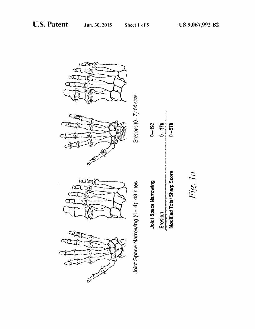
U.S. Patent Jun. 30, 2015 Sheet 1 of 5 US 9,067,992 B2
3 wer
i cy
R st to es: its
i.

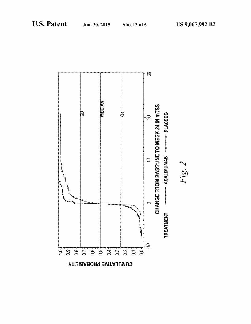
U.S. Patent Jun. 30, 2015 Sheet 2 of 5 US 9,067,992 B2
US 9,067,992 B2 U.S. Patent
|
*{{}{} if fif
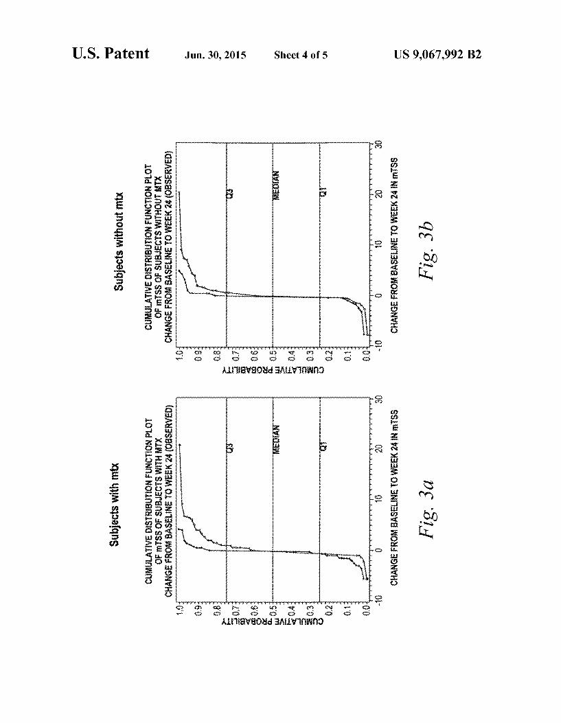
US 9,067,992 B2 U.S. Patent
US 9,067,992 B2 Sheet 5 of 5 Jun. 30, 2015 U.S. Patent
sy#33ff),
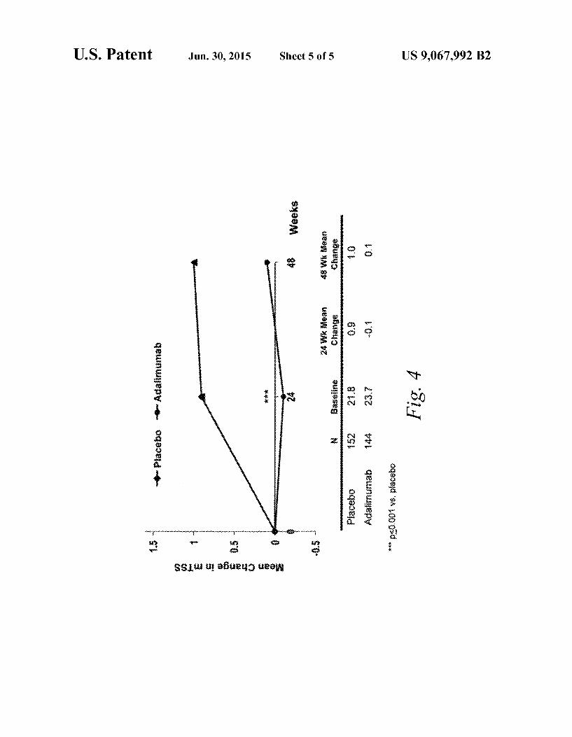
ssiti is sitetic tisee
US 9,067,992 B2 1.
USE OF TNFO INHIBITOR FORTREATMENT OF PSORATCARTHRTS
RELATED APPLICATIONS
This application claims priority to U.S. Provisional Applin. No. 60/681,645, which was filed on May 16, 2005.
This application is related to U.S. Pat. Nos. 6,090,382, 6.258,562, and 6,509,015. This application is also related to U.S. patent application Ser. No. 09/801,185, filed Mar. 7, 2001; U.S. patent application Ser. No. 10/163,657, filed Jun. 5, 2002; and U.S. patent application Ser. No. 10/422.287. filed Apr. 26, 2002; U.S. patent application Ser. No. 10/525, 292, filed Aug. 16, 2002; U.S. patent application Ser. No. 10/693.233, filed Oct. 24, 2003: U.S. patent application Ser. No. 10/622.932, filed Jul.18, 2003, published as U.S. Patent Application Publication No. 2004/0126372; U.S. patent application Ser. No. 10/623,039, filed Jul. 18, 2003: U.S. patent application Ser. No. 10/623,076, filed Jul. 18, 2003: U.S. patent application Ser. No. 10/623,065, filed Jul. 18, 2003: U.S. patent application Ser. No. 10/622,928, filed Jul. 18, 2003: U.S. patent application Ser. No. 10/623,075, filed Jul. 18, 2003: U.S. patent application Ser. No. 10/623,035, filed Jul. 18, 2003: U.S. patent application Ser. No. 10/622, 683, filed Jul. 18, 2003: U.S. patent application Ser. No. 10/622.205, filed Jul. 18, 2003: U.S. patent application Ser. No. 10/622.210, filed Jul.18, 2003; and U.S. patent applica tion Ser. No. 10/623.318, filed Jul.18, 2003. This application is also related to PCT/US05/12007, filed Apr. 11, 2005. The entire contents of each of these patents and patent applica tions, including the above-mentioned U.S. Patent Application Publication No. 2004/0126372, are hereby incorporated herein by reference. The International Patent Application No. PCT/US05/12007 corresponds to U.S. Patent Application Publication No. 2006/0009385, and the entire content of U.S. Patent Application Publication No. 2006/0009385 (excluding the claims) is incorporated herein by reference.
BACKGROUND OF THE INVENTION
Polyarthritis may be erosive or non-erosive. In the erosive form, the underlying disease process erodes the cartilage; in the non-erosive form, the cartilage is not affected. Erosive polyarthritis is an inflammatory disease of joints that results in tissue destruction and erosion within the affected joint. Erosive polyarthritis occurs in many patients having inflam matory disorders, including psoriatic arthritis, spondyloar thropathies, such as ankylosing spondylitis, and juvenile rheumatoid arthritis. Many of the current treatments of dis orders in which erosive polyarthritis is a manifestation fail to focus on decreasing radiographic progression of joint dis CaSC.
SUMMARY OF THE INVENTION
There is a need to treat erosive polyarthritis in a safe and effective manner. While traditional treatments of erosive pol yarthritis, such as administration of DMARDs, may delay disease progression, traditional treatments may be slow to become effective, may lose efficacy with time, and may be associated with potentially serious toxic effects. The present invention provides a safe and effective means for treating erosive polyarthritis and slowing the progression of joint disease. The present invention includes methods of treating erosive
polyarthritis comprising administering TNF inhibitors. The invention also provides a method for treating a human Subject
10
15
25
30
35
40
45
50
55
60
65
2 Suffering from erosive polyarthritis, comprising administer ing to the subject an anti-TNFC. antibody, such that erosive polyarthritis is treated. Kits and articles of manufacture com prising a TNFC. inhibitor are also included in the invention.
In one embodiment, the TNFC. inhibitor is selected from the group consisting of an anti-TNFC. antibody, oran antigen binding portion thereof, a TNF fusion protein, or a recombi nant TNF binding protein. In one embodiment, the TNF fusion protein is etanercept. In another embodiment, the anti TNFC. antibody, or antigen-binding portion thereof, is an antibody selected from the group consisting of a humanized antibody, a chimeric antibody, and a multivalent antibody. In one embodiment, the anti-TNFC. antibody, or antigen-bind ing portion thereof, is infliximab, golimumab, or adali mumab. In still another embodiment, the anti-TNFC. anti body, or antigen-binding portion thereof, is a human antibody. The invention provides a method for treating a human
Subject Suffering from erosive polyarthritis, comprising administering to the Subject a TNFC. antibody, or antigen binding portion thereof, such that erosive polyarthritis is treated.
In one embodiment, the TNFC. antibody, or antigen-bind ing portion thereof, is an antibody selected from the group consisting of a humanized antibody, a chimericantibody, and a multivalent antibody. In another embodiment, the TNFC. antibody, or antigen-binding portion thereof, is infliximab or golimumab.
In one embodiment, the TNFC. antibody, or antigen-bind ing portion thereof, is a human antibody. In one embodiment, the human antibody, or an antigen-binding portion thereof, dissociates from human TNFC. with a K of 1x10 Morless and a K-rate constant of 1 x10s' or less, both determined by Surface plasmon resonance, and neutralizes human TNFC. cytotoxicity in a standard in vitro L929 assay with an ICs of 1x10⁷M or less. In another embodiment, the human anti body, oran antigen-binding portion thereof, has the following characteristics:
a) dissociates from human TNFC. with a K-rate constant of 1x10s or less, as determined by surface plasmon reso nance,
b) has a light chain CDR3 domain comprising the amino acid sequence of SEQ ID NO:3, or modified from SEQ ID NO:3 by a single alanine substitution at position 1, 4, 5, 7 or 8 or by one to five conservative amino acid substitutions at positions 1, 3, 4, 6, 7, 8 and/or 9:
c) has a heavy chain CDR3 domain comprising the amino acid sequence of SEQ ID NO: 4, or modified from SEQ ID NO. 4 by a single alanine substitution at position 2, 3, 4, 5, 6, 8, 9, 10 or 11 or by one to five conservative amino acid substitutions at positions 2, 3, 4, 5, 6, 8, 9, 10, 11 and/or 12. In still another embodiment, the human antibody, or an antigen binding portion thereof, comprises a light chain variable region (LCVR) having a CDR3 domain comprising the amino acid sequence of SEQ ID NO:3, or modified from SEQ ID NO:3 by a single alanine substitution at position 1, 4, 5, 7 or 8, and comprises a heavy chain variable region (HCVR) having a CDR3 domain comprising the amino acid sequence of SEQID NO: 4, or modified from SEQID NO. 4 by a single alanine substitution at position 2, 3, 4, 5, 6, 8, 9, 10 or 11. In yet another embodiment, the human antibody, or an antigen binding portion thereof, comprises a light chain variable region (LCVR) comprising the amino acid sequence of SEQ ID NO: 1 and a heavy chain variable region (HCVR) com prising the amino acid sequence of SEQ ID NO: 2. In one embodiment, the human antibody, or an antigen-binding por tion thereof, is adalimumab.
US 9,067,992 B2 3
In one embodiment, the TNFC. antibody, or antigen-bind ing portion thereof, is administered to the Subject on a biweekly dosing regimen.
In one embodiment, the subject has a disorder in which TNFC. activity is detrimental. In one embodiment, the disor der in which TNFC. activity is detrimental is selected from the group consisting of psoriatic arthritis, ankylosing spondylitis, and juvenile rheumatoid arthritis. In another embodiment, the disorder in which TNFO activity is detrimental is psoriatic arthritis. In still another embodiment, the disorder in which TNFC. activity is detrimental is rheumatoid arthritis.
In one embodiment, the invention includes further com prising administering an additional therapeutic agent to the Subject. In one embodiment, the additional therapeutic agent is methotrexate. In another embodiment, the additional thera peutic agent is a Disease Modifying Anti-Rheumatic Drug (DMARD) or a Nonsteroidal Antiinflammatory Drug (NSAID) or a steroid, or any combination thereof. The invention includes a method for testing the efficacy of
a TNFC. antibody, or antigen-binding portion thereof, for decreasing radiographic progression of joint disease associ ated with erosive polyarthritis. In one embodiment, the method for testing the efficacy of a TNFC. antibody, or anti gen-binding portion thereof, comprises determining the effi cacy of the TNFC. antibody, or antigen-binding portion thereof, using a modified Total Sharp Score (mTSS) of a patient population having joint disease associated with ero sive polyarthritis and a mTSS of the patient population fol lowing administration of the TNFC. antibody, or antigen binding portion thereof, wherein no change or a decrease in the mTSS indicates that the TNFC. antibody, or antigen-bind ing portion thereof, is efficacious for decreasing radiographic progression of joint disease associated with erosive pol yarthritis. In one embodiment, the decrease in the mTSS is about -0.2.
In one embodiment, the patient population also has a dis order in which TNFC. is detrimental. In one embodiment, the disorder in which TNFC. activity is detrimental is selected from the group consisting of psoriatic arthritis, ankylosing spondylitis, and juvenile rheumatoid arthritis.
In one embodiment, the TNFC. antibody, or antigen-bind ing portion thereof, is an antibody selected from the group consisting of a humanized antibody, a chimericantibody, and a multivalent antibody. In one embodiment, the TNFC. anti body, or antigen-binding portion thereof, is infliximab or golimumab. In another embodiment, the TNFC. antibody, or antigen-binding portion thereof, is a human antibody. In one embodiment, the human antibody, or an antigen-binding por tion thereof, dissociates from human TNFC. with a K of 1x10 Morless and a Karate constant of 1 x10s or less, both determined by Surface plasmon resonance, and neutral izes human TNFC. cytotoxicity in a standard in vitro L929 assay with an ICso of 1x10⁷ Morless.
In another embodiment, the human antibody, oran antigen binding portion thereof, has the following characteristics:
a) dissociates from human TNFC. with a K-rate constant of 1x10s' or less, as determined by surface plasmon reso nance,
b) has a light chain CDR3 domain comprising the amino acid sequence of SEQID NO:3, or modified from SEQ ID NO:3 by a single alanine substitution at position 1, 4, 5, 7 or 8 or by one to five conservative amino acid substitutions at positions 1, 3, 4, 6, 7, 8 and/or 9:
c) has a heavy chain CDR3 domain comprising the amino acid sequence of SEQID NO: 4, or modified from SEQ ID NO. 4 by a single alanine substitution at position 2, 3, 4, 5, 6, 8, 9, 10 or 11 or by one to five conservative amino acid
5
10
15
25
30
35
40
45
50
55
60
65
4 substitutions at positions 2, 3, 4, 5, 6, 8, 9, 10, 11 and/or 12. In still another embodiment, the human antibody, or an antigen binding portion thereof, comprises a light chain variable region (LCVR) having a CDR3 domain comprising the amino acid sequence of SEQ ID NO:3, or modified from SEQ ID NO:3 by a single alanine substitution at position 1, 4, 5, 7 or 8, and comprises a heavy chain variable region (HCVR) having a CDR3 domain comprising the amino acid sequence of SEQID NO: 4, or modified from SEQID NO. 4 by a single alanine substitution at position 2, 3, 4, 5, 6, 8, 9, 10 or 11.
In another embodiment, the human antibody, oran antigen binding portion thereof, comprises a light chain variable region (LCVR) comprising the amino acid sequence of SEQ ID NO: 1 and a heavy chain variable region (HCVR) com prising the amino acid sequence of SEQ ID NO: 2. In still another embodiment of the invention, the human antibody, or an antigen-binding portion thereof, is adalimumab.
In one embodiment of the invention, the TNFC. antibody, or antigen-binding portion thereof, is administered to the Sub ject on a biweekly dosing regimen. In one embodiment, anti body, or antigen-binding portion thereof, is administered in combination with an additional therapeutic agent, including, for example methotrexate The invention describes a method for monitoring the effec
tiveness of a TNFC. antibody, or antigen-binding portion thereof, for the treatment of erosive polyarthritis in a human Subject comprising determining the effectiveness of the TNFC. antibody, or antigen-binding portion thereof, using a baseline modified Total Sharp Score (mTSS) of a patient population having erosive polyarthritis and a mTSS score of a patient population following administration of the TNFC. antibody, or antigen-binding portion thereof, wherein a result selected from the group consisting of a decrease in the mTSS in about 9-27% of the patient population; no change in the mTSS in about 65-73% of the patient population; and an increase in the mTSS in about 9-28% of the patient popula tion, indicates that the TNFC. antibody, or antigen-binding portion thereof, is effective at treating erosive polyarthritis.
In one embodiment of the invention, the TNFC. antibody, or antigen-binding portion thereof, is an antibody selected from the group consisting of a humanized antibody, a chimeric antibody, and a multivalent antibody. In one embodiment, the TNFC. antibody, or antigen-binding portion thereof, is inflix imab or golimumab. In another embodiment, the TNFC. anti body, or antigen-binding portion thereof, is a human anti body.
In another embodiment, the human antibody, oran antigen binding portion thereof, dissociates from human TNFC. with a K of 1x10 Morless and a K-rate constant of 1x10s or less, both determined by Surface plasmon resonance, and neutralizes human TNFC. cytotoxicity in a standard in vitro L929 assay with an ICso of 1x10⁷M or less. In still another embodiment, the human antibody, or an antigen-binding por tion thereof, is adalimumab. The invention also includes a method for testing the effi
cacy of a TNFC. antibody, or antigen-binding portion thereof, to treat erosive polyarthritis associated with psoriatic arthri tis, comprising determining the efficacy of the TNFC. anti body, or antigen-binding portion thereof, using a baseline modified Total Sharp Score (mTSS) and either a baseline Psoriasis Area and Severity Index (PAST) score or a baseline ACR score of a patient population having erosive polyarthri tis in comparison with the mTSS and either the PASI or the ACR score of the patient population following administration of the NFC. antibody, or antigen-binding portion thereof, wherein no change or a decrease in the mTSS and either an ACR20 response achieved in at least about 57% or a PASI 50
US 9,067,992 B2 5
response achieved in at least about 75% of the patient popu lation, indicates that the TNFC. antibody, or antigen-binding portion thereof, is efficacious for the treatment of erosive polyarthritis associated with psoriatic arthritis. In one embodiment, an ACR50 response is achieved in at least about 39% of the patient population. In another embodiment, an ACR70 response is achieved in at least about 23% of the patient population. In still another embodiment, a PASI75 response is achieved in at least about 59% of the patient population. In yet another embodiment, a PASI90 response is achieved in at least about 42% of the patient population. In one embodiment, the TNFC. antibody, orantigen-binding por tion thereof, is adalimumab.
The invention describes a method for treating erosive pol yarthritis comprising administering to a subject having ero sive polyarthritis, adalimumab on a biweekly dosing regimen. In one embodiment, the dose of adalimumab is about 40 mg. The invention also includes a kit comprising a pharmaceu
tical composition comprising a TNFC. antibody, or an anti gen-binding portion thereof, and a pharmaceutically accept able carrier, and instructions for administration of the pharmaceutical composition for the treatment of erosive pol yarthritis. In one embodiment, the pharmaceutical composi tion comprises the TNFC. antibody, or antigen-binding por tion thereof, adalimumab. In one embodiment, pharmaceutical composition comprises about 40 mg of adali mumab. In another embodiment, the kit further comprises an additional therapeutic agent. In one embodiment, the addi tional therapeutic agent is methotrexate.
The invention describes an article of manufacture compris ing a packaging material; a TNFC. antibody, or antigen-bind ing portion thereof, and a label or package insert contained within the packaging material indicating that the TNFC. anti body, or antigen-binding portion thereof, can be used for the treatment of erosive polyarthritis.
The invention also includes an article of manufacture com prising a packaging material; a TNFC. antibody, or antigen binding portion thereof, and a label or package insert con tained within the packaging material indicating that the TNFC. antibody, or antigen-binding portion thereof, can be used for to inhibit radiographic progression of joint disease.
In one embodiment, the article of manufacture comprises an antibody selected from the group consisting of a human ized antibody, a chimeric antibody, and a multivalent anti body.
In one embodiment, the TNFC. antibody, or antigen-bind ing portion thereof, is infliximab or golimumab. In another embodiment, the TNFC. antibody, or antigen-binding portion thereof, is a human antibody. In one embodiment, the human antibody, or an antigen-binding portion thereof, dissociates from human TNFC. with a K of 1x10M or less and a Kar rate constant of 1x10s' or less, both determined by surface plasmon resonance, and neutralizes human TNFC. cytotoxic ity in a standard in vitro L929 assay with an ICso of 1x10⁷M or less. In another embodiment, the human antibody, or an antigen-binding portion thereof, is adalimumab.
FIGURES
FIGS. 1a and 1b show a diagram of the modified total sharp score (mTSS) (FIG. 1a) and radiographic findings associated with PSA (FIG. 1b).
FIG. 2 shows a cumulative distribution function plot of modified Total Sharps Score (mTSS). The graph shows the change in baseline to Week 24 for subjects with both baseline and Week 24 radiographic films.
10
15
25
30
35
40
45
50
55
60
65
6 FIGS. 3a and 3b show cumulative distribution function
plots of mTSS of subjects with (FIG. 3a) and without (FIG. 3b) methotrexate (mtx).
FIG. 4 shows a graph of the mean change in mTSS at Week 48.
DETAILED DESCRIPTION OF THE INVENTION
I. Definitions
In order that the present invention may be more readily understood, certain terms are first defined. The term “human TNFo” (abbreviated herein as hTNFO,
or simply hTNF), as used herein, is intended to refer to a human cytokine that exists as a 17 kD secreted form and a 26 kD membrane associated form, the biologically active form of which is composed of a trimer of noncovalently bound 17 kD molecules. The structure of hTNFC. is described further in, for example, Pennica, D., et al. (1984) Nature 312:724 729; Davis, J. M., et al. (1987) Biochemistry 26:1322-1326; and Jones, E.Y., et al. (1989) Nature 338:225-228. The term human TNFC. is intended to include recombinant human TNFC. (rhTNFC), which can be prepared by standard recom binant expression methods or purchased commercially (R& D Systems, Catalog No. 210-TA, Minneapolis, Minn.). TNFO is also referred to as TNF. The term “TNFC. inhibitor refers to an agent which inter
feres with TNFC. activity. The term also includes each of the anti-TNFC. human antibodies and antibody portions described herein as well as those described in U.S. Pat. Nos. 6,090,382; 6.258,562: 6,509,015, and in U.S. patent applica tion Ser. Nos. 09/801,185 and 10/302,356. In one embodi ment, the TNFC. inhibitor used in the invention is an anti TNFC. antibody, or a fragment thereof, including infliximab (Remicade(R), Johnson and Johnson; described in U.S. Pat. No. 5,656.272, incorporated by reference herein), CDP571 (a humanized monoclonal anti-TNF-alpha IgG4 antibody), CDP870 (a humanized monoclonal anti-TNF-alpha antibody fragment), an anti-TNF dAb (Peptech), CNTO 148 (goli mumab: Medarex and Centocor, see WO 02/12502), and adalimumab (Humira R. Abbott Laboratories, a human anti TNF mAb, described in U.S. Pat. No. 6,090,382 as D2E7). Additional TNF antibodies which can be used in the invention are described in U.S. Pat. Nos. 6,593,458; 6,498,237; 6,451, 983; and 6,448,380, each of which is incorporated by refer ence herein. In another embodiment, the TNFC. inhibitor is a TNF fusion protein, e.g., etanercept (Enbrel(R), Amgen; described in WO91/03553 and WO09/406,476, incorporated by reference herein). In another embodiment, the TNFC. inhibitor is a recombinant TNF binding protein (r-TBP-I) (Serono). The term “antibody', as used herein, is intended to refer to
immunoglobulin molecules comprised of four polypeptide chains, two heavy (H) chains and two light (L) chains inter connected by disulfide bonds. Each heavy chain is comprised of a heavy chain variable region (abbreviated hereinas HCVR or VH) and a heavy chain constant region. The heavy chain constant region is comprised of three domains, CH1, CH2 and CH3. Each light chain is comprised of a light chain variable region (abbreviated herein as LCVR or VL) and a light chain constant region. The light chain constant region is comprised of one domain, CL. The VH and VL regions can be further subdivided into regions of hypervariability, termed comple mentarity determining regions (CDR), interspersed with regions that are more conserved, termed framework regions (FR). Each VHand VL is composed of three CDRs and four FRS, arranged from amino-terminus to carboxy-terminus in
US 9,067,992 B2 7
the following order: FR1, CDR1, FR2, CDR2, FR3, CDR3, FR4. The antibodies of the invention are described in further detail in U.S. Pat. Nos. 6,090,382; 6,258,562; and 6,509,015, each of which is incorporated herein by reference in its entirety. The term “antigen-binding portion of an antibody (or
simply “antibody portion'), as used herein, refers to one or more fragments of an antibody that retain the ability to spe cifically bind to an antigen (e.g., hTNFC). It has been shown that the antigen-binding function of an antibody can be per formed by fragments of a full-length antibody. Examples of binding fragments encompassed within the term “antigen binding portion of an antibody include (i) a Fab fragment, a monovalent fragment consisting of the VL, VH, CL and CH1 domains; (ii) a F(ab')2 fragment, a bivalent fragment compris ing two Fab fragments linked by a disulfide bridge at the hinge region; (iii) a Fd fragment consisting of the VH and CH1 domains; (iv) a Fv fragment consisting of the VL and VH domains of a single arm of an antibody, (v) a dAb fragment (Ward et al. (1989) Nature 341:544-546), which consists of a VH domain; and (vi) an isolated complementarity determin ing region (CDR). Furthermore, although the two domains of the Fv fragment, VL and VH, are coded for by separate genes, they can be joined, using recombinant methods, by a synthetic linker that enables them to be made as a single protein chain in which the VL and VH regions pair to form monovalent molecules (known as single chain FV (ScFV); see e.g., Bird et al. (1988) Science 242:423-426; and Huston et al. (1988) Proc. Natl. Acad. Sci. USA 85:5879-5883). Such single chain antibodies are also intended to be encompassed within the term “antigen-binding portion of an antibody. Other forms of single chain antibodies, such as diabodies are also encom passed. Diabodies are bivalent, bispecific antibodies in which VH and VL domains are expressed on a single polypeptide chain, but using a linker that is too short to allow for pairing between the two domains on the same chain, thereby forcing the domains to pair with complementary domains of another chain and creating two antigen binding sites (see e.g., Hol liger et al. (1993) Proc. Natl. Acad. Sci. USA 90:6444-6448: Poljak et al. (1994) Structure 2:1121-1123). The antibody portions of the invention are described in further detail in U.S. Pat. Nos. 6,090,382, 6,258,562, 6,509,015, each of which is incorporated herein by reference in its entirety.
Binding fragments are produced by recombinant DNA techniques, or by enzymatic or chemical cleavage of intact immunoglobulins. Binding fragments include Fab., Fab'. F(ab'), Fabc. Fv, single chains, and single-chain antibodies. Other than “bispecific' or “bifunctional immunoglobulins or antibodies, an immunoglobulin or antibody is understood to have each of its binding sites identical. A “bispecific' or “bifunctional antibody' is an artificial hybrid antibody hav ing two different heavy/light chain pairs and two different binding sites. Bispecific antibodies can be produced by a variety of methods including fusion of hybridomas or linking of Fab' fragments. See, e.g., Songsivilai & Lachmann, Clin. Exp. Immunol. 79:315-321 (1990); Kostelny et al., J. Immu mol. 148, 1547-1553 (1992). A “conservative amino acid Substitution', as used herein, is
one in which one amino acid residue is replaced with another amino acid residue having a similar side chain. Families of amino acid residues having similar side chains have been defined in the art, including basic side chains (e.g., lysine, arginine, histidine), acidic side chains (e.g., aspartic acid, glutamic acid), uncharged polar side chains (e.g., glycine, asparagine, glutamine, serine, threonine, tyrosine, cysteine), nonpolar side chains (e.g., alanine, Valine, leucine, isoleu cine, proline, phenylalanine, methionine, tryptophan), beta
10
15
25
30
35
40
45
50
55
60
65
8 branched side chains (e.g., threonine, Valine, isoleucine) and aromatic side chains (e.g., tyrosine, phenylalanine, tryp tophan, histidine). The term “human antibody', as used herein, is intended to
include antibodies having variable and constant regions derived from human germline immunoglobulin sequences. The human antibodies of the invention may include amino acid residues not encoded by human germline immunoglo bulin sequences (e.g., mutations introduced by random or site-specific mutagenesis in vitro or by Somatic mutation in vivo), for example in the CDRs and in particular CDR3. However, the term “human antibody', as used herein, is not intended to include antibodies in which CDR sequences derived from the germline of another mammalian species, Such as a mouse, have been grafted onto human framework Sequences. The term “recombinant human antibody', as used herein,
is intended to include all human antibodies that are prepared, expressed, created or isolated by recombinant means, such as antibodies expressed using a recombinant expression vector transfected into a host cell (described further below), antibod ies isolated from a recombinant, combinatorial human anti body library (described further below), antibodies isolated from an animal (e.g., a mouse) that is transgenic for human immunoglobulin genes (see e.g., Taylor et al. (1992) Nucl. Acids Res. 20:6287) or antibodies prepared, expressed, cre ated or isolated by any other means that involves splicing of human immunoglobulin gene sequences to other DNA sequences. Such recombinant human antibodies have vari able and constant regions derived from human germline immunoglobulin sequences. In certain embodiments, how ever, such recombinant human antibodies are subjected to in vitro mutagenesis (or, when an animal transgenic for human Ig sequences is used, in vivo Somatic mutagenesis) and thus the amino acid sequences of the VH and VL regions of the recombinant antibodies are sequences that, while derived from and related to human germline VH and VL sequences, may not naturally exist within the human antibody germline repertoire in vivo. An "isolated antibody', as used herein, is intended to refer
to an antibody that is substantially free of other antibodies having different antigenic specificities (e.g., an isolated anti body that specifically binds hTNFC. is substantially free of antibodies that specifically bind antigens other than hTNFC). An isolated antibody that specifically binds hTNFC. may, however, have cross-reactivity to other antigens, such as TNFC. molecules from other species (discussed in further detail below). Moreover, an isolated antibody may be sub stantially free of other cellular material and/or chemicals. A "neutralizing antibody', as used herein (or an “antibody
that neutralized hTNFO activity'), is intended to refer to an antibody whose binding to hTNFC. results in inhibition of the biological activity of hTNFC. This inhibition of the biological activity of hTNFC. can be assessed by measuring one or more indicators of hTNFC. biological activity, such as hTNFC.- induced cytotoxicity (either in vitro or in vivo), hTNFC.- induced cellular activation and hTNFC. binding to hTNFC. receptors. These indicators of hTNFC. biological activity can be assessed by one or more of several standard in vitro or in vivo assays known in the art (see U.S. Pat. No. 6,090,382). Preferably, the ability of an antibody to neutralize hTNFC. activity is assessed by inhibition of hTNFO-induced cytotox icity of L929 cells. As an additional or alternative parameter of hTNFO activity, the ability of an antibody to inhibit hTNFC.-induced expression of ELAM-1 on HUVEC, as a measure of hTNFC.-induced cellular activation, can be assessed.
US 9,067,992 B2 9
The term "surface plasmon resonance', as used herein, refers to an optical phenomenon that allows for the analysis of real-time biospecific interactions by detection of alterations in protein concentrations within a biosensor matrix, for example using the BIAcore system (Pharmacia Biosensor AB, Uppsala, Sweden and Piscataway, N.J.). For further descriptions, see Example 1 of U.S. Pat. No. 6.258,562 and Jonsson et al. (1993) Ann. Biol. Clin. 51:19; Jonsson et al., (1991) Biotechniques 11:620-627; Johnsson et al. (1995).J. Mol. Recognit. 8:125; and Johnnson et al. (1991) Anal. Bio chem. 198:268.
The term “K, as used herein, is intended to refer to the off rate constant for dissociation of an antibody from the antibody/antigen complex. The term “K, as used herein, is intended to refer to the
dissociation constant of a particular antibody-antigen inter action. The term “ICso as used herein, is intended to refer to the
concentration of the inhibitor required to inhibit the biologi cal endpoint of interest, e.g., neutralize cytotoxicity activity. The term “nucleic acid molecule', as used herein, is
intended to include DNA molecules and RNA molecules. A nucleic acid molecule may be single-stranded or double stranded, but preferably is double-stranded DNA. The term "isolated nucleic acid molecule', as used herein
in reference to nucleic acids encoding antibodies or antibody portions (e.g., VH, VL, CDR3) that bind hTNFC., is intended to refer to a nucleic acid molecule in which the nucleotide sequences encoding the antibody orantibody portion are free of other nucleotide sequences encoding antibodies or anti body portions that bind antigens other than hTNFC, which other sequences may naturally flank the nucleic acid in human genomic DNA. Thus, for example, an isolated nucleic acid of the invention encoding a VH region of an anti-hTNFC. antibody contains no other sequences encoding other VH regions that bind antigens other than hTNFC. The term “vector', as used herein, is intended to refer to a
nucleic acid molecule capable of transporting another nucleic acid to which it has been linked. One type of vector is a “plasmid', which refers to a circular double stranded DNA loop into which additional DNA segments may be ligated. Another type of vector is a viral vector, wherein additional DNA segments may be ligated into the viral genome. Certain vectors are capable of autonomous replication in a host cell into which they are introduced (e.g., bacterial vectors having a bacterial origin of replication and episomal mammalian vectors). Other vectors (e.g., non-episomal mammalian vec tors) can be integrated into the genome of a host cell upon introduction into the host cell, and thereby are replicated along with the host genome. Moreover, certain vectors are capable of directing the expression of genes to which they are operatively linked. Such vectors are referred to herein as “recombinant expression vectors' (or simply, “expression vectors'). In general, expression vectors of utility in recom binant DNA techniques are often in the form of plasmids. In the present specification, "plasmid' and “vector” may be used interchangeably as the plasmid is the most commonly used form of vector. However, the invention is intended to include Such other forms of expression vectors, such as viral vectors (e.g., replication defective retroviruses, adenoviruses and adeno-associated viruses), which serve equivalent functions. The term “recombinant host cell’ (or simply “host cell’).
as used herein, is intended to refer to a cell into which a recombinant expression vector has been introduced. It should be understood that such terms are intended to refer not only to the particular subject cell but to the progeny of Such a cell. Because certain modifications may occur in Succeeding gen
10
15
25
30
35
40
45
50
55
60
65
10 erations due to either mutation or environmental influences, Such progeny may not, in fact, be identical to the parent cell, but are still included within the scope of the term “host cell as used herein. The term "dose, as used herein, refers to an amount of
TNFC. inhibitor which is administered to a subject. The term “multiple-variable dose' includes different doses
of a TNFC. inhibitor which are administered to a subject for therapeutic treatment. “Multiple-variable dose regimen” or “multiple-variable dose therapy” describe a treatment sched ule which is based on administering different amounts of TNFC. inhibitor at various time points throughout the course of treatment. In one embodiment, the invention describes a multiple-variable dose method of treatment of erosive pol yarthritis comprising an induction phase and a treatment phase, wherein a TNFC. inhibitor is administered at a higher dose during the induction phase than the treatment phase. Multiple-variable dose regimens are described in PCT appli cation no. PCT/US05/12007. The term “dosing, as used herein, refers to the adminis
tration of a Substance (e.g., an anti-TNFC. antibody) to achieve a therapeutic objective (e.g., the treatment of erosive polyarthritis). The terms “biweekly dosing regimen”, “biweekly dosing,
and “biweekly administration’, as used herein, refer to the time course of administering a Substance (e.g., an anti-TNFC. antibody) to a subject to achieve a therapeutic objective (e.g., the treatment of erosive polyarthritis). The biweekly dosing regimen is not intended to include a weekly dosing regimen. Preferably, the substance is administered every 9-19 days, more preferably, every 11-17 days, even more preferably, every 13-15 days, and most preferably, every 14 days. The term “combination” as in the phrase “a first agent in
combination with a secondagent' includes co-administration of a first agent and a second agent, which for example may be dissolved or intermixed in the same pharmaceutically accept able carrier, or administration of a first agent, followed by the second agent, or administration of the second agent, followed by the first agent. The present invention, therefore, includes methods of combination therapeutic treatment and combina tion pharmaceutical compositions. In one embodiment, the invention provides a combination therapy for treating erosive polyarthritis comprising administering an anti-TNF antibody. The term “concomitant as in the phrase “concomitant
therapeutic treatment includes administering an agent in the presence of a second agent. A concomitant therapeutic treat ment method includes methods in which the first, second, third, or additional agents are co-administered. A concomi tant therapeutic treatment method also includes methods in which the first or additional agents are administered in the presence of a second or additional agents, wherein the second or additional agents, for example, may have been previously administered. A concomitant therapeutic treatment method may be executed step-wise by different actors. For example, one actor may administer to a subject a first agent and a second actor may to administer to the Subject a second agent, and the administering steps may be executed at the same time, or nearly the same time, or at distant times, so long as the first agent (and additional agents) are after administration in the presence of the second agent (and additional agents). The actor and the Subject may be the same entity (e.g., human). The term “combination therapy”, as used herein, refers to
the administration of two or more therapeutic Substances, e.g., an anti-TNFC. antibody and another drug. The other drug(s) may be administered concomitant with, prior to, or following the administration of an anti-TNFC. antibody.
US 9,067,992 B2 11
The term "kit' as used herein refers to a packaged product or article of manufacture comprising components. The kit preferably comprises a box or container that holds the com ponents of the kit. The box or container is affixed with a label or a Food and Drug Administration approved protocol. The box or container holds components of the invention which are preferably contained within plastic, polyethylene, polypro pylene, ethylene, or propylene vessels. The vessels can be capped-tubes or bottles. The kit can also include instructions for administering the TNFC. antibody, orantigen-binding por tion thereof. In one embodiment the kit of the invention includes the formulation comprising the human antibody D2E7, as described in PCT/IB03/04502 and U.S. application Ser. No. 10/222,140.
Various aspects of the invention are described in further detail herein.
II. TNFO Inhibitors
This invention provides a method of treating erosive pol yarthritis in which the administration of a TNFC. inhibitor e.g., a TNFC. antibody, or antigen-binding portion thereof, is beneficial. In one embodiment, these methods include admin istration of isolated human antibodies, or antigen-binding portions thereof, that bind to human TNFC. with high affinity and a low off rate, and have a high neutralizing capacity. Preferably, the human antibodies of the invention are recom binant, neutralizing human anti-hTNFC. antibodies.
In one embodiment, the TNFC. inhibitor used in the inven tion is an anti-TNFC. antibody, or a fragment thereof, includ ing infliximab (Remicade(R), Johnson and Johnson; described in U.S. Pat. No. 5,656.272, incorporated by reference herein), CDP571 (a humanized monoclonal anti-TNF-alpha IgG4 antibody), CDP870 (a humanized monoclonal anti-TNF alpha antibody fragment), an anti-TNF dAb (Peptech), CNTO 148 (golimumab: Medarex and Centocor, see WO 02/12502), and adalimumab (Humira R. Abbott Laboratories, a human anti-TNF mAb, described in U.S. Pat. No. 6,090,382 as D2E7). Additional TNF antibodies which can be used in the invention are described in U.S. Pat. Nos. 6,593,458: 6,498,237; 6,451,983; and 6,448,380, each of which is incor porated by reference herein. The most preferred recombinant, neutralizing antibody
used in the invention is referred to herein as D2E7, also referred to as HUMIRAR and adalimumab (the amino acid sequence of the D2E7VL region is shown in SEQID NO: 1; the amino acid sequence of the D2E7VH region is shown in SEQ ID NO: 2). The properties of D2E7 (HUMIRAR) have been described in Salfeld et al., U.S. Pat. Nos. 6,090,382, 6.258,562, and 6,509,015, which are each incorporated by reference herein. Other examples of TNFC. inhibitors include chimeric and humanized murine anti-hTNFC. antibodies which have undergone clinical testing for treatment of rheu matoid arthritis (see e.g., Elliott et al. (1994) Lancet 344: 1125-1127; Elliot et al. (1994) Lancet 344:1105-1110: Rankin et al., (1995) Br. J. Rheumatol. 34:334-342). In another embodiment, the anti-TNFC. antibody is multivalent.
In one embodiment, the method of treating erosive pol yarthritis of the invention includes the administration of D2E7 antibodies and antibody portions, D2E7-related anti bodies and antibody portions, and other human antibodies and antibody portions with equivalent properties to D2E7. such as high affinity binding to hTNFC. with low dissociation kinetics and high neutralizing capacity. In one embodiment, the invention provides a method for treating erosive pol yarthritis with an isolated human antibody, or an antigen binding portion thereof, that dissociates from human TNFC.
10
15
25
30
35
40
45
50
55
60
65
12 with a K of 1x10 M or less and a K rate constant of 1x10s' or less, both determined by surface plasmon reso nance, and neutralizes human TNFC. cytotoxicity in a stan dard in vitro L929 assay with an ICso of 1x10⁷M or less. More preferably, the isolated human antibody, or antigen binding portion thereof, dissociates from human TNFC. with a K of 5x10's or less, or even more preferably, with a Kar of 1x10's or less. More preferably, the isolated human antibody, or antigen-binding portion thereof, neutralizes human TNFC. cytotoxicity in a standard in vitro L929 assay with an ICso of 1x10 Morless, even more preferably with an ICso of 1x10M or less and still more preferably with an ICso of 1x10" M or less. In a preferred embodiment, the antibody is an isolated human recombinant antibody, or an antigen-binding portion thereof.
It is well known in the art that antibody heavy and light chain CDR3 domains play an important role in the binding specificity/affinity of an antibody for an antigen. Accord ingly, in another aspect, the invention pertains to methods of treating erosive polyarthritis by administering human anti bodies that have slow dissociation kinetics for association with hTNFC. and that have light and heavy chain CDR3 domains that structurally are identical to or related to those of D2E7. Position 9 of the D2E7 VLCDR3 can be occupied by Ala or Thr without substantially affecting the K. Accord ingly, a consensus motif for the D2E7 VLCDR3 comprises the amino acid sequence: Q-R-Y-N-R-A-P-Y-(T/A) (SEQID NO:3). Additionally, position 12 of the D2E7 VHCDR3 can be occupied by Tyror ASn, without substantially affecting the K. Accordingly, a consensus motif for the D2E7 VHCDR3 comprises the amino acid sequence: V-S-Y-L-S-T-A-S-S-L- D-(Y/N) (SEQ ID NO: 4). Moreover, as demonstrated in Example 2 of U.S. Pat. No. 6,090,382, the CDR3 domain of the D2E7 heavy and light chains is amenable to substitution with a single alanine residue (at position 1, 4, 5, 7 or 8 within the VLCDR3 or at position 2,3,4, 5, 6, 8, 9, 10 or 11 within the VHCDR3) without substantially affecting the K Still further, the skilled artisan will appreciate that, given the ame nability of the D2E7 VL and VHCDR3 domains to substitu tions by alanine, substitution of other amino acids within the CDR3 domains may be possible while still retaining the low off rate constant of the antibody, in particular substitutions with conservative amino acids. Preferably, no more than one to five conservative amino acid Substitutions are made within the D2E7VL and/or VHCDR3 domains. More preferably, no more than one to three conservative amino acid substitutions are made within the D2E7 VL and/or VH CDR3 domains. Additionally, conservative amino acid Substitutions should not be made at amino acid positions critical for binding to hTNFC. Positions 2 and 5 of the D2E7 VLCDR3 and posi tions 1 and 7 of the D2E7 VHCDR3 appear to be critical for interaction with hTNFC. and thus, conservative amino acid Substitutions preferably are not made at these positions (al though an alanine substitution at position 5 of the D2E7 VL CDR3 is acceptable, as described above) (see U.S. Pat. No. 6,090,382).
Accordingly, in another embodiment, the invention pro vides methods of treating erosive polyarthritis by the admin istration of an isolated human antibody, or antigen-binding portion thereof. The antibody or antigen-binding portion thereof preferably contains the following characteristics:
a) dissociates from human TNFC. with a K-rate constant of 1x10s or less, as determined by surface plasmon reso nance,
b) has a light chain CDR3 domain comprising the amino acid sequence of SEQ ID NO:3, or modified from SEQ ID NO:3 by a single alanine substitution at position 1, 4, 5, 7 or
US 9,067,992 B2 13
8 or by one to five conservative amino acid substitutions at positions 1, 3, 4, 6, 7, 8 and/or 9:
c) has a heavy chain CDR3 domain comprising the amino acid sequence of SEQID NO: 4, or modified from SEQ ID NO. 4 by a single alanine substitution at position 2, 3, 4, 5, 6, 8, 9, 10 or 11 or by one to five conservative amino acid substitutions at positions 2, 3, 4, 5, 6, 8, 9, 10, 11 and/or 12. More preferably, the antibody, or antigen-binding portion
thereof, dissociates from human TNFC. with a K of 5x10's or less. Even more preferably, the antibody, or antigen-binding portion thereof, dissociates from human TNFa. with a K of 1x10's or less.
In yet another embodiment, the invention provides a method of treating erosive polyarthritis by administration of an isolated human antibody, or antigen-binding portion thereof. The antibody orantigen-binding portion thereofpref erably contains a light chain variable region (LCVR) having a CDR3 domain comprising the amino acid sequence of SEQ IDNO:3, or modified from SEQID NO:3 by a single alanine substitution at position 1, 4, 5, 7 or 8, and with a heavy chain variable region (HCVR) having a CDR3 domain comprising the amino acid sequence of SEQID NO: 4, or modified from SEQID NO. 4 by a single alanine substitution at position 2, 3, 4, 5, 6, 8, 9, 10 or 11. Preferably, the LCVR further has a CDR2 domain comprising the amino acid sequence of SEQ ID NO: 5 (i.e., the D2E7 VLCDR2) and the HCVR further has a CDR2 domain comprising the amino acid sequence of SEQ ID NO: 6 (i.e., the D2E7 VHCDR2). Even more pref. erably, the LCVR further has CDR1 domain comprising the amino acid sequence of SEQ ID NO: 7 (i.e., the D2E7 VL CDR1) and the HCVR has a CDR1 domain comprising the amino acid sequence of SEQID NO: 8 (i.e., the D2E7 VH CDR1). The framework regions for VL preferably are from the VI human germline family, more preferably from the A20 human germline Vk gene and most preferably from the D2E7VL framework sequences shown in FIGS. 1A and 1B of U.S. Pat. No. 6,090,382.The framework regions for VH pref erably are from the V3 human germline family, more pref erably from the DP-31 human germline VH gene and most preferably from the D2E7VH framework sequences shown in FIGS. 2A and 2B of U.S. Pat. No. 6,090,382.
Accordingly, in another embodiment, the invention pro vides a method of treating erosive polyarthritis by the admin istration of an isolated human antibody, or antigen-binding portion thereof. The antibody or antigen-binding portion thereof preferably contains a light chain variable region (LCVR) comprising the amino acid sequence of SEQID NO: 1 (i.e., the D2E7 VL) and a heavy chain variable region (HCVR) comprising the amino acid sequence of SEQID NO: 2 (i.e., the D2E7 VH). In certain embodiments, the antibody comprises a heavy chain constant region, such as an IgG1. IgG2, IgG3, IgG4, IgA, IgE, IgM or Ig|D constant region. Preferably, the heavy chain constant region is an IgG1 heavy chain constant region oran IgG4 heavy chain constant region. Furthermore, the antibody can comprise a light chain constant region, either a kappa light chain constant region or a lambda light chain constant region. Preferably, the antibody com prises a kappa light chain constant region. Alternatively, the antibody portion can be, for example, a Fab fragment or a single chain Fv fragment.
In still other embodiments, the invention describes a method of treating erosive polyarthritis in which the admin istration of an anti-TNFC. antibody wherein the antibody is an isolated human antibody, or an antigen-binding portion thereof. The antibody orantigen-binding portion thereofpref erably contains D2E7-related VL and VHCDR3 domains, for example, antibodies, or antigen-binding portions thereof,
10
15
25
30
35
40
45
50
55
60
65
14 with a light chain variable region (LCVR) having a CDR3 domain comprising an amino acid sequence selected from the group consisting of SEQID NO:3, SEQID NO: 11, SEQID NO: 12, SEQID NO: 13, SEQID NO: 14, SEQID NO: 15, SEQID NO: 16, SEQ ID NO: 17, SEQID NO: 18, SEQ ID NO: 19, SEQID NO: 20, SEQID NO: 21, SEQID NO:22, SEQID NO. 23, SEQID NO: 24, SEQID NO: 25 and SEQ ID NO: 26 or with a heavy chain variable region (HCVR) having a CDR3 domain comprising an amino acid sequence selected from the group consisting of SEQID NO: 4, SEQID NO: 27, SEQID NO: 28, SEQID NO: 29, SEQID NO:30, SEQID NO:31, SEQ ID NO:32, SEQID NO:33, SEQ ID NO:34 and SEQID NO:35. The TNFC. antibody used in the invention can also be
modified. In some embodiments, the TNFC. antibody or anti gen binding fragments thereof, is chemically modified to provide a desired effect. For example, pegylation of antibod ies and antibody fragments of the invention may be carried out by any of the pegylation reactions known in the art, as described, for example, in the following references: Focus on Growth Factors 3:4-10 (1992); EP 0 154316; and EP 0401 384 (each of which is incorporated by reference herein in its entirety). Preferably, the pegylation is carried out via an acy lation reaction or an alkylation reaction with a reactive poly ethylene glycol molecule (or an analogous reactive water soluble polymer). A preferred water-soluble polymer for pegylation of the antibodies and antibody fragments of the invention is polyethylene glycol (PEG). As used herein, "polyethylene glycol is meant to encompass any of the forms of PEG that have been used to derivatize other proteins, such as mono (C1-CIO) alkoxy- or aryloxy-polyethylene glycol. Methods for preparing pegylated antibodies and antibody
fragments of the invention will generally comprise the steps of (a) reacting the antibody or antibody fragment with poly ethylene glycol, Such as a reactive ester or aldehyde derivative of PEG, under conditions whereby the antibody or antibody fragment becomes attached to one or more PEG groups, and (b) obtaining the reaction products. It will be apparent to one of ordinary skill in the art to select the optimal reaction conditions or the acylation reactions based on known param eters and the desired result.
Pegylated antibodies and antibody fragments may gener ally be used to treat erosive polyarthritis and TNFC.-related disorders of the invention by administration of the TNFC. antibodies and antibody fragments described herein. Gener ally the pegylated antibodies and antibody fragments have increased half-life, as compared to the nonpegylated antibod ies and antibody fragments. The pegylated antibodies and antibody fragments may be employed alone, together, or in combination with other pharmaceutical compositions.
In yet another embodiment of the invention, TNFC. anti bodies or fragments thereof can be altered wherein the con stant region of the antibody is modified to reduce at least one constant region-mediated biological effector function relative to an unmodified antibody. To modify an antibody of the invention such that it exhibits reduced binding to the Fc receptor, the immunoglobulin constant region segment of the antibody can be mutated at particular regions necessary for Fc receptor (FcR) interactions (see e.g., Canfield and Morrison (1991).J. Exp. Med. 173: 1483-1491; and Lundet al. (1991).J. of Immunol. 147:2657-2662). Reduction in FcRbinding abil ity of the antibody may also reduce other effector functions which rely on FcR interactions, such as opSonization and phagocytosis and antigen-dependent cellular cytotoxicity. An antibody or antibody portion of the invention can be
derivatized or linked to another functional molecule (e.g., another peptide or protein). Accordingly, the antibodies and
US 9,067,992 B2 15
antibody portions of the invention are intended to include derivatized and otherwise modified forms of the human anti hTNFC. antibodies described herein, including immunoadhe sion molecules. For example, an antibody orantibody portion of the invention can be functionally linked (by chemical cou pling, genetic fusion, noncovalent association or otherwise) to one or more other molecular entities, such as another antibody (e.g., a bispecific antibody or a diabody), a detect able agent, a cytotoxic agent, a pharmaceutical agent, and/or a protein or peptide that can mediate associate of the antibody or antibody portion with another molecule (such as a strepta vidin core region or a polyhistidine tag). One type of derivatized antibody is produced by crosslink
ing two or more antibodies (of the same type or of different types, e.g., to create bispecific antibodies). Suitable crosslinkers include those that are heterobifunctional, having two distinctly reactive groups separated by an appropriate spacer (e.g., m-maleimidobenzoyl-N-hydroxySuccinimide ester) or homobifunctional (e.g., disuccinimidyl Suberate). Such linkers are available from Pierce Chemical Company, Rockford, Ill.
Useful detectable agents with which an antibody or anti body portion of the invention may be derivatized include fluorescent compounds. Exemplary fluorescent detectable agents include fluorescein, fluorescein isothiocyanate, rhodamine, 5-dimethylamine-1-napthalenesulfonyl chloride, phycoerythrin and the like. An antibody may also be deriva tized with detectable enzymes, such as alkaline phosphatase, horseradish peroxidase, glucose oxidase and the like. When an antibody is derivatized with a detectable enzyme, it is detected by adding additional reagents that the enzyme uses to produce a detectable reaction product. For example, when the detectable agent horseradish peroxidase is present, the addition of hydrogen peroxide and diaminobenzidine leads to a colored reaction product, which is detectable. An antibody may also be derivatized with biotin, and detected through indirect measurement of avidin or streptavidin binding. An antibody, or antibody portion, of the invention can be
prepared by recombinant expression of immunoglobulin light and heavy chain genes in a host cell. To express an antibody recombinantly, a host cell is transfected with one or more recombinant expression vectors carrying DNA fragments encoding the immunoglobulin light and heavy chains of the antibody Such that the light and heavy chains are expressed in the host cell and, preferably, secreted into the medium in which the host cells are cultured, from which medium the antibodies can be recovered. Standard recombinant DNA methodologies are used to obtain antibody heavy and light chain genes, incorporate these genes into recombinant expression vectors and introduce the vectors into host cells, such as those described in Sambrook, Fritsch and Maniatis (eds), Molecular Cloning, A Laboratory Manual, Second Edition, Cold Spring Harbor, N.Y., (1989), Ausubel et al. (eds.) Current Protocols in Molecular Biology, Greene Pub lishing Associates, (1989) and in U.S. Pat. No. 4,816,397 by Boss et al. To express D2E7 or a D2E7-related antibody, DNA frag
ments encoding the light and heavy chain variable regions are first obtained. These DNAs can be obtained by amplification and modification of germline light and heavy chain variable sequences using the polymerase chain reaction (PCR). Ger mline DNA sequences for human heavy and light chain vari able region genes are known in the art (see e.g., the “Vbase' human germline sequence database; see also Kabat et al. (1991) Sequences of Proteins of Immunological Interest, Fifth Edition, U.S. Department of Health and Human Services, NIH Publication No. 91-3242: Tomlinson et al. (1992) “The
10
15
25
30
35
40
45
50
55
60
65
16 Repertoire of Human Germline V, Sequences Reveals about Fifty Groups of V, Segments with Different Hypervariable Loops' J. Mol. Biol. 227:776-798; and Cox et al. (1994) “A Directory of Human Germ-line V7s Segments Reveals a Strong Bias in their Usage' Eur: J. Immunol. 24:827-836; the contents of each of which are expressly incorporated herein by reference). To obtain a DNA fragment encoding the heavy chain variable region of D2E7, or a D2E7-related antibody, a member of the V3 family of human germline VH genes is amplified by standard PCR. Most preferably, the DP-31 VH germline sequence is amplified. To obtain a DNA fragment encoding the light chain variable region of D2E7, or a D2E7 related antibody, a member of the VI family of human ger mline VL genes is amplified by standard PCR. Most prefer ably, the A20 VL germline sequence is amplified. PCR primers suitable for use in amplifying the DP-31 germline VH and A20 germline VL sequences can be designed based on the nucleotide sequences disclosed in the references cited Supra, using standard methods. Once the germline VH and VL fragments are obtained,
these sequences can be mutated to encode the D2E7 or D2E7 related amino acid sequences disclosed herein. The amino acid sequences encoded by the germline VH and VL DNA sequences are first compared to the D2E7 or D2E7-related VH and VL amino acid sequences to identify amino acid residues in the D2E7 or D2E7-related sequence that differ from germline. Then, the appropriate nucleotides of the ger mline DNA sequences are mutated such that the mutated germline sequence encodes the D2E7 or D2E7-related amino acid sequence, using the genetic code to determine which nucleotide changes should be made. Mutagenesis of the ger mline sequences is carried out by Standard methods, such as PCR-mediated mutagenesis (in which the mutated nucle otides are incorporated into the PCR primers such that the PCR product contains the mutations) or site-directed mutagenesis. Once DNA fragments encoding D2E7 or D2E7-related VH
and VL segments are obtained (by amplification and mutagenesis of germline VH and VL genes, as described above), these DNA fragments can be further manipulated by standard recombinant DNA techniques, for example to con Vert the variable region genes to full-length antibody chain genes, to Fab fragment genes or to a scFV gene. In these manipulations, a VL- or VH-encoding DNA fragment is operatively linked to another DNA fragment encoding another protein, such as an antibody constant region or a flexible linker. The term “operatively linked’, as used in this context, is intended to mean that the two DNA fragments are joined Such that the amino acid sequences encoded by the two DNA fragments remain in-frame. The isolated DNA encoding the VH region can be con
Verted to a full-length heavy chain gene by operatively linking the VH-encoding DNA to another DNA molecule encoding heavy chain constant regions (CH1, CH2 and CH3). The sequences of human heavy chain constant region genes are known in the art (see e.g., Kabat et al. (1991) Sequences of Proteins of Immunological Interest, Fifth Edition, U.S. Department of Health and Human Services, NIH Publication No. 91-3242) and DNA fragments encompassing these regions can be obtained by standard PCR amplification. The heavy chain constant region can be an IgG1, IgG2, IgG3. IgG4, IgA, IgE, IgM or Ig|D constant region, but most pref erably is an IgG1 or IgG4 constant region. For a Fab fragment heavy chain gene, the VH-encoding DNA can be operatively linked to another DNA molecule encoding only the heavy chain CH1 constant region.
US 9,067,992 B2 17
The isolated DNA encoding the VL region can be con Verted to a full-length light chain gene (as well as a Fab light chain gene) by operatively linking the VL-encoding DNA to another DNA molecule encoding the light chain constant region, CL. The sequences of human light chain constant region genes are known in the art (see e.g., Kabat et al. (1991) Sequences of Proteins of Immunological Interest, Fifth Edi tion, U.S. Department of Health and Human Services, NIH Publication No. 91-3242) and DNA fragments encompassing these regions can be obtained by standard PCR amplification. The light chain constant region can be a kappa or lambda constant region, but most preferably is a kappa constant region.
To create a schv gene, the VH- and VL-encoding DNA fragments are operatively linked to another fragment encod ing a flexible linker, e.g., encoding the amino acid sequence (Gly-Ser), such that the VH and VL sequences can be expressed as a contiguous single-chain protein, with the VL and VH regions joined by the flexible linker (see e.g., Bird et al. (1988) Science 242:423-426; Huston et al. (1988) Proc. Natl. Acad. Sci. USA 85:5879-5883; McCafferty et al., Nature (1990) 348:552-554). To express the antibodies, or antibody portions of the
invention, DNAS encoding partial or full-length light and heavy chains, obtained as described above, are inserted into expression vectors such that the genes are operatively linked to transcriptional and translational control sequences. In this context, the term “operatively linked' is intended to mean that an antibody gene is ligated into a vector Such that transcrip tional and translational control sequences within the vector serve their intended function of regulating the transcription and translation of the antibody gene. The expression vector and expression control sequences are chosen to be compatible with the expression host cell used. The antibody light chain gene and the antibody heavy chain gene can be inserted into separate vector or, more typically, both genes are inserted into the same expression vector. The antibody genes are inserted into the expression vector by standard methods (e.g., ligation of complementary restriction sites on the antibody gene frag ment and vector, or bluntend ligation if no restriction sites are present). Prior to insertion of the D2E7 or D2E7-related light or heavy chain sequences, the expression vector may already carry antibody constant region sequences. For example, one approach to converting the D2E7 or D2E7-related VHandVL sequences to full-length antibody genes is to insert them into expression vectors already encoding heavy chain constant and light chain constant regions, respectively, such that the VH segment is operatively linked to the CH segment(s) within the vector and the VL segment is operatively linked to the CL segment within the vector. Additionally or alterna tively, the recombinant expression vector can encode a signal peptide that facilitates secretion of the antibody chain from a host cell. The antibody chain gene can be cloned into the vector Such that the signal peptide is linked in-frame to the amino terminus of the antibody chain gene. The signal pep tide can be an immunoglobulin signal peptide or a heterolo gous signal peptide (i.e., a signal peptide from a non-immu noglobulin protein).
In addition to the antibody chain genes, the recombinant expression vectors of the invention carry regulatory sequences that control the expression of the antibody chain genes in a host cell. The term “regulatory sequence' is intended to include promoters, enhancers and other expres sion control elements (e.g., polyadenylation signals) that con trol the transcription or translation of the antibody chain genes. Such regulatory sequences are described, for example, in Goeddel; Gene Expression Technology. Methods in Enzy
10
15
25
30
35
40
45
50
55
60
65
18 mology 185, Academic Press, San Diego, Calif. (1990). It will be appreciated by those skilled in the art that the design of the expression vector, including the selection of regulatory sequences may depend on Such factors as the choice of the host cell to be transformed, the level of expression of protein desired, etc. Preferred regulatory sequences for mammalian host cell expression include viral elements that direct high levels of protein expression in mammalian cells, such as promoters and/or enhancers derived from cytomegalovirus (CMV) (such as the CMV promoter/enhancer), Simian Virus 40 (SV40) (such as the SV40 promoter/enhancer), adenovi rus, (e.g., the adenovirus major late promoter (AdMLP)) and polyoma. For further description of viral regulatory elements, and sequences thereof, see e.g., U.S. Pat. No. 5,168,062 by Stinski, U.S. Pat. No. 4,510,245 by Bell et al. and U.S. Pat. No. 4,968,615 by Schaffner et al.
In addition to the antibody chain genes and regulatory sequences, the recombinant expression vectors of the inven tion may carry additional sequences, such as sequences that regulate replication of the vector in host cells (e.g., origins of replication) and selectable marker genes. The selectable marker gene facilitates selection of host cells into which the vector has been introduced (see e.g., U.S. Pat. Nos. 4.399, 216, 4,634,665 and 5,179,017, all by Axel et al.). For example, typically the selectable marker gene confers resis tance to drugs. Such as G418, hygromycin or methotrexate, on a host cell into which the vector has been introduced. Pre ferred selectable marker genes include the dihydrofolate reductase (DHFR) gene (for use in dhfr host cells with methotrexate selection/amplification) and the neogene (for G418 selection).
For expression of the light and heavy chains, the expression vector(s) encoding the heavy and light chains is transfected into a host cell by standard techniques. The various forms of the term “transfection” are intended to encompass a wide variety of techniques commonly used for the introduction of exogenous DNA into a prokaryotic or eukaryotic host cell, e.g., electroporation, calcium-phosphate precipitation, DEAE-dextran transfection and the like. Although it is theo retically possible to express the antibodies of the invention in either prokaryotic or eukaryotic host cells, expression of anti bodies in eukaryotic cells, and most preferably mammalian host cells, is the most preferred because Such eukaryotic cells, and in particular mammalian cells, are more likely than prokaryotic cells to assemble and secrete a properly folded and immunologically active antibody. Prokaryotic expression of antibody genes has been reported to be ineffective for production of high yields of active antibody (Boss and Wood (1985) Immunology Today 6:12-13).
Preferred mammalian host cells for expressing the recom binant antibodies of the invention include Chinese Hamster Ovary (CHO cells) (including dhfr-CHO cells, described in Urlaub and Chasin, (1980) Proc. Natl. Acad. Sci. USA 77:4216-4220, used with a DHFR selectable marker, e.g., as described in Kaufman and Sharp (1982) Mol. Biol. 159:601 621), NS0 myeloma cells, COS cells and SP2 cells. When recombinant expression vectors encoding antibody genes are introduced into mammalian host cells, the antibodies are produced by culturing the host cells for a period of time sufficient to allow for expression of the antibody in the host cells or, more preferably, secretion of the antibody into the culture medium in which the host cells are grown. Antibodies can be recovered from the culture medium using standard protein purification methods.
Host cells can also be used to produce portions of intact antibodies, such as Fab fragments or ScFv molecules. It is understood that variations on the above procedure are within
US 9,067,992 B2 19
the scope of the present invention. For example, it may be desirable to transfect a host cell with DNA encoding either the light chain or the heavy chain (but not both) of an antibody of this invention. Recombinant DNA technology may also be used to remove some or all of the DNA encoding either or both of the light and heavy chains that is not necessary for binding to hTNFC. The molecules expressed from such trun cated DNA molecules are also encompassed by the antibodies of the invention. In addition, bifunctional antibodies may be produced in which one heavy and one light chain are an antibody of the invention and the other heavy and light chain are specific for an antigen other than hTNFC. by crosslinking an antibody of the invention to a second antibody by standard chemical crosslinking methods.
In a preferred system for recombinant expression of an antibody, orantigen-binding portion thereof, of the invention, a recombinant expression vector encoding both the antibody heavy chain and the antibody light chain is introduced into dhfr-CHO cells by calcium phosphate-mediated transfection. Within the recombinant expression vector, the antibody heavy and light chain genes are each operatively linked to CMV enhancer/AdMLP promoter regulatory elements to drive high levels of transcription of the genes. The recombi nant expression vector also carries a DHFR gene, which allows for selection of CHO cells that have been transfected with the vector using methotrexate selection/amplification. The selected transformant host cells are culture to allow for expression of the antibody heavy and light chains and intact antibody is recovered from the culture medium. Standard molecular biology techniques are used to prepare the recom binant expression vector, transfect the host cells, select for transformants, culture the host cells and recover the antibody from the culture medium.
Recombinant human antibodies of the invention in addi tion to D2E7 or an antigenbinding portion thereof, or D2E7 related antibodies disclosed herein can be isolated by screen ing of a recombinant combinatorial antibody library, preferably a Sclv phage display library, prepared using human VL and VH cDNAs prepared from mRNA derived from human lymphocytes. Methodologies for preparing and screening Such libraries are known in the art. In addition to commercially available kits for generating phage display libraries (e.g., the Pharmacia Recombinant Phage Antibody System, catalog no. 27-94.00-01; and the Stratagene Sur f/APTM phage display kit, catalog no. 240612), examples of methods and reagents particularly amenable for use in gen erating and screening antibody display libraries can be found in, for example, Ladner et al. U.S. Pat. No. 5,223,409: Kang et al. PCT Publication No. WO92/18619: Dower et al. PCT Publication No. WO 91/17271: Winter et al. PCT Publication No. WO92/20791; Markland et al. PCT Publication No. WO 92/15679; Breitling et al. PCT Publication No. WO 93/01288; McCafferty et al. PCT Publication No. WO 92/01047: Garrard et al. PCT Publication No. WO92/09690; Fuchs et al. (1991) Bio/Technology 9:1370-1372; Hay et al. (1992) Hum Antibod Hybridomas 3:81-85; Huseet al. (1989) Science 246:1275-1281; McCafferty et al., Nature (1990) 348:552-554; Griffiths et al. (1993) EMBO.J. 12:725-734; Hawkins et al. (1992).J Mol Biol 226:889-896; Clackson etal. (1991) Nature 352:624-628; Gram et al. (1992) PNAS 89:3576-3580; Garrard et al. (1991) Bio/Technology 9:1373 1377; Hoogenboom et al. (1991) Nuc Acid Res 19:4133 4137; and Barbas et al. (1991) PNAS 88:7978-7982.
In a preferred embodiment, to isolate human antibodies with high affinity and a low off rate constant for hTNFC., a murine anti-hTNFC. antibody having high affinity and a low off rate constant for hTNFC. (e.g., MAK 195, the hybridoma
10
15
25
30
35
40
45
50
55
60
65
20 for which has deposit number ECACC 87 050801) is first used to select human heavy and light chain sequences having similar binding activity toward hTNFC., using the epitope imprinting methods described in Hoogenboom et al., PCT Publication No. WO93/06213. The antibody libraries used in this method are preferably schv libraries prepared and screened as described in McCafferty et al., PCT Publication No. WO92/01047, McCafferty et al., Nature (1990)348:552 554; and Griffiths et al., (1993) EMBO.J. 12:725-734. The schv antibody libraries preferably are screened using recom binant human TNFC. as the antigen. Once initial human VL and VH segments are selected,
“mix and match' experiments, in which different pairs of the initially selected VL and VH segments are screened for hTNFC. binding, are performed to select preferred VL/VH pair combinations. Additionally, to further improve the affin ity and/or lower the off rate constant for hTNFC. binding, the VL and VH segments of the preferred VL/VH pair(s) can be randomly mutated, preferably within the CDR3 region of VH and/or VL, in a process analogous to the in vivo Somatic mutation process responsible for affinity maturation of anti bodies during a natural immune response. This in vitro affin ity maturation can be accomplished by amplifying VH and VL regions using PCR primers complimentary to the VH CDR3 or VL CDR3, respectively, which primers have been "spiked with a random mixture of the four nucleotide bases at certain positions such that the resultant PCR products encode VH and VL segments into which random mutations have been introduced into the VH and/or VLCDR3 regions. These randomly mutated VH and VL segments can be rescreened for binding to hTNFC. and sequences that exhibit high affinity and a low off rate for hTNFC. binding can be selected.
Following screening and isolation of an anti-hTNFC. anti body of the invention from a recombinant immunoglobulin display library, nucleic acid encoding the selected antibody can be recovered from the display package (e.g., from the phage genome) and Subcloned into other expression vectors by standard recombinant DNA techniques. If desired, the nucleic acid can be further manipulated to create other anti body forms of the invention (e.g., linked to nucleic acid encoding additional immunoglobulin domains, such as addi tional constant regions). To express a recombinant human antibody isolated by screening of a combinatorial library, the DNA encoding the antibody is cloned into a recombinant expression vector and introduced into a mammalian host cells, as described in further detail in above. Methods of isolating human antibodies with high affinity
and a low off rate constant for hTNFC. are also described in U.S. Pat. Nos. 6,090,382, 6,258,562, and 6,509,015, each of which is incorporated by reference herein. The TNFC. inhibitor may also be a TNF fusion protein, e.g.,
etanercept (Enbrel R, Amgen; described in WO91/03553 and WO 09/406,476, incorporated by reference herein). In another embodiment, the TNFO inhibitor is a recombinant TNF binding protein (r-TBP-I) (Serono).
III. Treatment of Erosive Polyarthritis
The invention provides methods of treating erosive pol yarthritis comprising administering a TNFC. inhibitor, includ ing, for an example, a TNFC. antibody, to a subject having erosive polyarthritis. The invention also describes methods for determining the efficacy of a TNFC. inhibitor for the treat ment of erosive polyarthritis. Preferably, the TNFC. is human TNFC. and the subject is a human subject. In one embodiment, the TNFC. inhibitor is adalimumab, also referred to as
US 9,067,992 B2 21
HUMIRAR) or D2E7. The use of TNFC. inhibitors, including antibodies and antibody portions, in the treatment of erosive polyarthritis, as well as methods for determining the efficacy of a TNFC. inhibitor for the treatment of erosive polyarthritis, is discussed further below: The term “polyarthritis' generally refers to inflammation,
i.e., Swelling, tenderness, or warmth, at two or more joints of a subject. As used herein, the term “erosive polyarthritis' refers to a
Subject who has polyarthritis which is damaging to the joint. The invention provides a method for treating erosive pol
yarthritis comprising administration of a TNFC. inhibitor, including, for an example, a TNFC. antibody. The invention also provides a method for inhibiting radiographic progres sion of joint disease associated with erosive polyarthritis. Methods for administering a TNFC. antibody, or an antigen binding portion thereof, for the treatment of erosive pol yarthritis are described in more detail below.
The invention provides a method for determining the effi cacy of an anti-TNFC. treatment for erosive polyarthritis. Measures for determining such efficacy include tests which determine whether joint destruction or erosion is improved following treatment. For example, the Total Modified Sharp Score (mTSS) of a subject may be used to determine improve ments in erosive polyarthritis in the subject. The mTSS may also be used as an assay to determine the efficacy of a treat ment for erosive polyarthritis. A Sharp score is an X-ray measurement in changes in total
joint damage as assessed by bone erosions and joint space narrowing (Sharp et al. (1971) Arthritis & Rheumatism 14:706: Sharp et al. (1985) Arthritis & Rheumatism 28:16). The mTSS is a measure of the extent and severity of joint damage based on evaluations of X-rays of patients hands and feet. Joints are scored for both joint erosions and joint space narrowing. The mTSS is the sum of the erosion score (ES) and the joint space narrowing (JSN) score and has, for example, a range of about 0 to about 398, where 0-no damage. The ES is the Sum of joint scores collected for 46 joints and has a range, for example, of about 0 to about 230. The JSN is the sum of joint scores collected for 42 joints and has a range, for example, of about 0 to about 168. A score of 0 would indicate no change. In one embodiment of the invention, the mTSS is determined by combining the joint space narrowing score having a range of about 0-192 and an erosion score having a range of about 0-378. An improved or constant mTSS demonstrates that the
TNFC. inhibitor is effective for treating erosive polyarthritis. In one embodiment, efficacy of a TNFC. inhibitor for the treatment of erosive polyarthritis is evidenced by a lack of progression of joint disease, e.g., no change in Sharp score, in mTSS over time in a subject having erosive polyarthritis. In another embodiment, efficacy of a TNFC. inhibitor for the treatment of erosive polyarthritis is evidenced by a decrease in the radiographic progression of joint disease, e.g., decrease in Sharp score, in mTSS over time in a subject having erosive polyarthritis.
In one embodiment, the overall change in the mTSS between baseline and a time period following treatment with a TNFC. inhibitor is between about 0.9 and about -0.2. In another embodiment, the overall change in the mTSS between baseline and a time period following treatment with a TNFC. inhibitor is between about 0.5 and about -0.2. In still another embodiment, the overall change in the mTSS between baseline and a time period following treatment with a TNFC. inhibitor is between about 0.2 and about -0.2.
It should be noted that ranges intermediate to the above recited scores, e.g., about -0.1 to about 0.3, are also intended
10
15
25
30
35
40
45
50
55
60
65
22 to be part of this invention. For example, ranges of values using a combination of any of the above recited values as upper and/or lower limits are intended to be included.
While treatment of an inflammatory disease with an anti inflammatory agent may result in clinical improvements fol lowing treatment, there may still be progressive joint damage resulting from erosive polyarthritis (Gladman et al. (1990).J. Rheumatol 17:809; Hanly et al. (1988) Ann Rheum Dis 47:386). Thus, it is a feature of this invention to provide a method for treating erosive polyarthritis which may be asso ciated with another disorder. In a preferred embodiment, the invention includes treatment of erosive polyarthritis associ ated with a disorder in which TNFC. activity is detrimental, including, but not limited to, rheumatoid arthritis (including juvenile rheumatoid arthritis), Crohn's disease, psoriasis, psoriatic arthritis, and ankylosing spondylitis. Erosive pol yarthritis may also be associated with multicentric reticulo histiocytosis (MRH) (Santillietal. (2002) Ann Rehum Dis 61: 485). As used herein, the term “a disorder in which TNFO activ
ity is detrimental' is intended to include diseases and other disorders in which the presence of TNFC. in a subject suffer ing from the disorder has been shown to be or is suspected of being either responsible for the pathophysiology of the dis order or a factor that contributes to a worsening of the disor der. Accordingly, a disorder in which TNFC. activity is detri mental is a disorder in which inhibition of TNFO activity is expected to alleviate the symptoms and/or progression of the disorder. Such disorders may be evidenced, for example, by an increase in the concentration of TNFC. in a biological fluid of a Subject Suffering from the disorder (e.g., an increase in the concentration of TNFC. in serum, plasma, synovial fluid, etc. of the Subject), which can be detected, for example, using an anti-TNFC. antibody as described above. There are numer ous examples of disorders in which TNFC. activity is detri mental. The use of TNFC. inhibitors for the treatment of erosive polyarthritis associated with specific disorders is dis cussed further below: A. Autoimmune Diseases
In one embodiment, the invention includes treatment of erosive polyarthritis associated with an autoimmune disease. Erosive polyarthritis may be found in patients suffering from autoimmune diseases, including forms of arthritis Such as rheumatoid arthritis and juvenile rheumatoid arthritis (Ver loes (1998) Med Genet. 35:943). TNFC. antibodies, such as adalimumab, may be used to treat autoimmune diseases, in particular those associated with erosive polyarthritis. Examples of Such autoimmune conditions include rheuma toid arthritis, rheumatoid spondylitis, osteoarthritis and gouty arthritis, allergy, multiple Sclerosis, autoimmune diabetes, autoimmune uveitis and nephrotic syndrome. Other examples of autoimmune conditions include multisystem autoimmune diseases and autoimmune hearing loss. Other examples of autoimmune disease are described in U.S. application Ser. No. 10/622.932, incorporated by reference herein. Juvenile Rheumatoid Arthritis Tumor necrosis factor has been implicated in the patho
physiology of juvenile arthritis, including juvenile rheuma toid arthritis (Grom et al. (1996) Arthritis Rheum. 39:1703; Mangge et al. (1995) Arthritis Rheum. 8:211). In one embodi ment, the TNFC. antibody of the invention is used to treat juvenile rheumatoid arthritis. The term “juvenile rheumatoid arthritis' or “JRA as used
herein refers to a chronic, inflammatory disease which occurs before age 16 that may cause joint or connective tissue dam age. JRA is also referred to as juvenile chronic polyarthritis and Still's disease.
US 9,067,992 B2 23
JRA causes joint inflammation and stiffness for more than 6 weeks in a child of 16 years of age or less. Inflammation causes redness, Swelling, warmth, and Soreness in the joints. Any joint can be affected and inflammation may limit the mobility of affected joints. One type of JRA can also affect the internal organs. JRA is often classified into three types by the number of
joints involved, the symptoms, and the presence or absence of certain antibodies found by a blood test. These classifications help the physician determine how the disease will progress and whether the internal organs or skin is affected. The clas sifications of JRA include the following:
a. Pauciarticular JRA, wherein the patient has four or fewer joints are affected. Pauciarticular is the most common form of JRA, and typically affects large joints, such as the knees.
b. Polyarticular HRA, wherein five or more joints are affected. The Small joints, such as those in the hands and feet, are most commonly involved, but the disease may also affect large joints.
c. Systemic JRA is characterized by joint swelling, fever, a light skin rash, and may also affect internal organs such as the heart, liver, spleen, and lymph nodes. Systemic JRA is also referred to as it Still's disease. A small percentage of these children develop arthritis in many joints and can have severe arthritis that continues into adulthood. B. Spondyloarthropathies
In one embodiment, the invention includes treatment of erosive polyarthritis associated with a spondylarthopathy. Erosive polyarthritis may be found in patients suffering from inflammatory diseases, such as spondyloarthopathies, asso ciated with detrimental TNFO activity (see e.g., Moeller et al. (1990) Cytokine 2:162; U.S. Pat. No. 5.231,024; European Patent Publication No. 260 610). As used herein, the term “spondyloarthropathy” or
“spondyloarthropathies” is used to refer to any one of several diseases affecting the joints of the spine, wherein such dis eases share common clinical, radiological, and histological features. A number of spondyloarthropathies share genetic characteristics, i.e. they are associated with the HLA-B27 allele. In one embodiment, the term spondyloarthropathy is used to refer to any one of several diseases affecting the joints of the spine, excluding ankylosing spondylitis, wherein Such diseases share common clinical, radiological, and histologi cal features. Examples of spondyloarthropathies include ankylosing spondylitis, psoriatic arthritis/spondylitis, entero pathic arthritis, reactive arthritis or Reiter's syndrome, and undifferentiated spondyloarthropathies. Examples of animal models used to study spondyloarthropathies include ank/ank transgenic mice, HLA-B27 transgenic rats (see Taurog et al. (1998) The Spondylarthritides. Oxford: Oxford University Press).
Examples of subjects who are at risk of having spondyloar thropathies include humans Suffering from arthritis. Spondy loarthropathies can be associated with forms of arthritis, including rheumatoid arthritis. In one embodiment of the invention, a TNFC. inhibitor is used to treat a subject who Suffers from a spondyloarthropathy associated with erosive polyarthritis. Examples of spondyloarthropathies which can be treated with a TNFO inhibitor are described below: Ankylosing Spondylitis (AS)
In one embodiment, the invention includes treatment of erosive polyarthritis associated with ankylosing spondylitis using a TNFC. antibody, or antigen-binding portion thereof. Tumor necrosis factor has been implicated in the pathophysi ology of ankylosing spondylitis (see Verjans et al. (1991) Arthritis Rheum. 34:486; Verjans et al. (1994) Clin Exp Immunol. 97:45; Kaijtzel et al. (1999) Hum Immunol.
5
10
15
25
30
35
40
45
50
55
60
65
24 60:140). Ankylosing spondylitis (AS) is an inflammatory dis order involving inflammation of one or more vertebrae. AS is a chronic inflammatory disease that affects the axial skeleton and/or peripheral joints, including joints between the verte brae of the spine and sacroiliac joints and the joints between the spine and the pelvis. AS can eventually cause the affected Vertebrae to fuse or grow together. Spondyarthropathies, including AS, can be associated with psoriatic arthritis (PSA) and/or inflammatory bowel disease (IBD), including ulcer ative colitis and Crohn's disease.
Early manifestations of AS can be determined by radio graphic tests, including CT scans and MRI scans. Early mani festations of AS often include scroiliitis and changes in the sacroliac joints as evidenced by the blurring of the cortical margins of the subchrondral bone, followed by erosions and Sclerosis. Fatigue has also been noted as a common symptom of AS (Duffy et al. (2002) ACR 66th Annual Scientific Meet ing Abstract). Psoriatic Arthritis
In one embodiment, the invention includes treatment of erosive polyarthritis associated with psoriatic arthritis using a TNFC. antibody, or antigen-binding portion thereof. Tumor necrosis factor has been implicated in the pathophysiology of psoriatic arthritis (PSA) (Partschet al. (1998) Ann Rheum Dis. 57:691; Ritchlin et al. (1998) J Rheumatol. 25:1544). As referred to herein, psoriatic TNFC. has been implicated in activating tissue inflammation and causing joint destruction in rheumatoid arthritis (see e.g., Moeller, A., et al. (1990) Cytokine 2:162-169; U.S. Pat. No. 5,231,024 to Moelleret al.: European Patent Publication No. 260 610 B1 by Moeller, A.; Tracey and Cerami, supra: Arend, W. P. and Dayer, J-M. (1995) Arth. Rheum. 38:151-160; Fava, R. A., et al. (1993) Clin. Exp. Immunol. 94:261-266). TNFC. also has been impli cated in promoting the death of islet cells and in mediating insulin resistance in diabetes (see e.g., Tracey and Cerami, supra: PCT Publication No. WO94/08609). TNFC. also has been implicated in mediating cytotoxicity to oligodendro cytes and induction of inflammatory plaques in multiple scle rosis (see e.g., Tracey and Cerami, Supra). Chimeric and humanized murine anti-hTNFC. antibodies have undergone clinical testing for treatment of rheumatoid arthritis (see e.g., Elliott, M.J., et al. (1994) Lancet 344:1125-1127; Elliot, M. J., et al. (1994) Lancet 344:1105-1110; Rankin, E. C., et al. (1995) Br. J. Rheumatol. 34:334-342).
Psoriatic arthritis refers to chronic inflammatory arthritis which is associated with psoriasis, a common chronic skin condition that causes red patches on the body. About 1 in 20 individuals with psoriasis will develop arthritis along with the skin condition, and in about 75% of cases, psoriasis precedes the arthritis. PSA exhibits itself in a variety of ways, ranging from mild to severe arthritis, wherein the arthritis usually affects the fingers and the spine. When the spine is affected, the symptoms are similar to those of ankylosing spondylitis, as described above. A TNFC. antibody, or antigen-binding fragment thereof, can be used for treatment of erosive pol yarthritis associated with PSA. PSA is sometimes associated with arthritis mutilans.
Arthritis mutilans refers to a disorder which is characterized by excessive bone erosion resulting in a gross, erosive defor mity which mutilates the joint.
Characteristic radiographic features of PSA include joint erosions, joint space narrowing, bony proliferation including periarticular and shaft periostitis, osteolysis including "pencil in cup' deformity and acro-osteolysis, ankylosis, spurforma tion, and spondylitis (Wassenberg et al. (2001) Z Rheumatol 60:156). Unlike rheumatoid arthritis (RA), joint involvement in PSA is often asymmetrical and may be oligoarticular;
US 9,067,992 B2 25
osteoporosis is atypical. Although erosive changes in early PSA are marginal as in RA, they become irregular and ill defined with disease progression because of periosteal bone formation adjacent to the erosions. In severe cases, erosive changes may progress to development of pencil in cup defor mity or gross osteolysis (Gold et al. (1988) Radiol Clin North Am 26:1195: Resnick et al. (1977)) J Can Assoc Radiol 28:187). Asymmetrical erosions may be visible radiographi cally in the carpus and in the metacarpophalangeal (MCP), proximal interphalangeal (PIP), and distal interphalangeal (DIP) joints of the hands, but the DIP joints are often the first to be affected. Abnormalities are seen in the phalangeal tufts and at the sites of attachments oftendons and ligaments to the bone. The presence of DIP erosive changes may provide both sensitive and specific radiographic findings to Support the diagnosis of PSA. Also, the hands tend to be involved much more frequently than the feet with a ratio of nearly 2:1.
Other examples of spondyloarthropathies are described in U.S. application Ser. No. 10/622.932 (U.S. Pat. Publ. 2004/ 0126372), incorporated by reference herein. C. Skin and Nail Disorders
In one embodiment, the invention includes treatment of erosive polyarthritis associated with skin and nail disorders. As used herein, the term "skin and nail disorder in which TNFC. activity is detrimental is intended to include skin and/or nail disorders and other disorders in which the pres ence of TNFC. in a subject suffering from the disorder has been shown to be or is suspected of being either responsible for the pathophysiology of the disorder or a factor that con tributes to a worsening of the disorder, e.g., psoriasis. Accord ingly, skin and nail disorders in which TNFC. activity is det rimental are disorders in which inhibition of TNFO activity is expected to alleviate the symptoms and/or progression of the disorder. The use of the antibodies, antibody portions, and other TNFC. inhibitors for the treatment of specific skin and nail disorders is discussed further below. In certain embodi ments, the antibody, antibody portion, or other TNFC. inhibi tor of the invention is administered to the subject in combi nation with another therapeutic agent, as described below. In one embodiment, a TNFC. antibody is administered to the Subject in combination with another therapeutic agent for the treatment of erosive polyarthritis associated with psoriasis and the treatment of psoriasis associated with arthritis. Psoriasis Tumor necrosis factor has been implicated in the patho
physiology of psoriasis (Takematsu et al. (1989) Arch Der matol Res. 281:398: Victor and Gottlieb (2002).J Drugs Der matol. 1(3):264). Psoriasis is described as a skin inflammation (irritation and redness) characterized by fre quent episodes of redness, itching, and thick, dry, silvery scales on the skin. In particular, lesions are formed which involve primary and secondary alterations in epidermal pro liferation, inflammatory responses of the skin, and an expres sion of regulatory molecules Such as lymphokines and inflammatory factors. Psoriatic skin is morphologically char acterized by an increased turnover of epidermal cells, thick ened epidermis, abnormal keratinization, inflammatory cell infiltrates into the epidermis and polymorphonuclear leuko cyte and lymphocyte infiltration into the epidermis layer resulting in an increase in the basal cell cycle. Psoriasis often involves the nails, which frequently exhibit pitting, separa tion of the nail, thickening, and discoloration. Psoriasis is often associated with other inflammatory disorders, for example arthritis, including rheumatoid arthritis, inflamma tory bowel disease (IBD), and Crohn's disease.
Evidence of psoriasis is most commonly seen on the trunk, elbows, knees, Scalp, skin folds, or fingernails, but it may
10
15
25
30
35
40
45
50
55
60
65
26 affect any or all parts of the skin. Normally, it takes about a month for new skin cells to move up from the lower layers to the Surface. In psoriasis, this process takes only a few days, resulting in a build-up of dead skin cells and formation of thick Scales. Symptoms of psoriasis include: skin patches, that are dry or red, covered with silvery scales, raised patches of skin, accompanied by red borders, that may crack and become painful, and that are usually lovated on the elbows, knees, trunk, Scalp, and hands; skin lesions, including pus tules, cracking of the skin, and skin redness; joint pain or aching which may be associated with of arthritis, e.g., psori atic arthritis.
Treatment for psoriasis often includes a topical corticos teroids, vitamin D analogs, and topical or oral retinoids, or combinations thereof. In one embodiment, the TNFC. inhibi tor of the invention is administered in combination with or the presence of one of these common treatments. Additional therapeutic agents which can also be combined with the TNFC. inhibitor for treatment of psoriasis are described in more detail below. The diagnosis of psoriasis is usually based on the appear
ance of the skin. Additionally a skin biopsy, or scraping and culture of skin patches may be needed to rule out other skin disorders. An X-ray may be used to check for psoriatic arthri tis if joint pain is present and persistent.
In one embodiment of the invention, a TNFC. inhibitor is used to treat psoriasis, including chronic plaque psoriasis, guttate psoriasis, inverse psoriasis, pustular psoriasis, pem phigus Vulgaris, erythrodermic psoriasis, psoriasis associated with inflammatory bowel disease (IBD), and psoriasis asso ciated with rheumatoid arthritis (RA). Specific types of pso riasis included in the treatment methods of the invention include chronic plaque psoriasis, guttate psoriasis, inverse psoriasis, and pustular psoriasis. Other examples of psoriasis and other types of skin and nail disorders are described in U.S. application Ser. No. 10/622.932, incorporated by reference herein.
Methods for determining the efficacy of a TNFC. inhibitor for the treatment of erosive polyarthritis in association a dis order in which TNFC. activity is detrimental include any assay which measures the degree of joint destruction, including joint space narrowing and/or joint erosion. In one embodi ment, joint destruction is measured using radiography. Such assays may be used to examine the efficacy of the TNFC. inhibitor by determining whetheran improvement occurs in a subject or patient population treated with the TNFC. inhibitor. Generally, improvements are determined by comparing a baseline score determined prior to treatment, and a score determined at a time following treatment with the TNFC. inhibitor.
Additional improvements in arthritic conditions. Such as Ra, PSA, and JRA, may be determined by measuring the ACR response. ACR thresholds, e.g., ACR20, ACR50, ACR70, may be used for defining improvement in RA and PSA, and indicate the percentage improvement in seven disease activity measures. Criteria include percentage improvement intender and swollen joint count and improvement of at least 3 of the following criteria: patient pain assessment, patient global assessment, physician global assessment, patient self-as sessed disability, or laboratory measures of disease activity (i.e., erythrocyte sedimentation rate or C-reactive protein level) (Felson et al. (1993) Arthritis Rheum. 36(6):729).
Other assays used to determine improvement for a given therapy for the treatment of RA, JRA, and PSA, include the EULAR response, DAS score, FACIT-F, HAO score, and SJC and/or TJC counts.
US 9,067,992 B2 27
The EULAR criteria uses a DAS for defining response. Response is defined as both: (a) change in disease activity from baseline and (b) the level of disease activity reached during follow-up. Criteria used to define DAS include: Ritchie articular index, Swollen joint count (44-joint count), erythrocyte sedimentation rate, and Health Assessment Ques tionnaire. A modified version of the DAS criteria, DAS28, uses a 28-joint count for Swollen and tenderjoints. Response is defined as a combination of a significant change from baseline and the level of disease activity attained. Good response is defined as a significant decrease in DAS (>1.2) and a low level of disease activity (< or -2.4). Non-response is defined as a decrease < or =0.6, or a decrease >0.6 and < or =1.2 with an attained DAS>3.7. Any other scores are regarded as moderate responses. The DAS is a score is based on the Ritchie articular index,
a 44 Swollen joint count, ESR, and a general health assess ment on a VAS. Range varies from 1 to 9. Serial measure ments of the DAS and DAS28 are strong predictors of physi cal disability and radiological progression, and both indices are sensitive discriminators between patients with high and low disease activity and between active and placebo treated patient groups.
FACIT-F (Functional Assessment of Chronic Illness Therapy-Fatigue) is a validated questionnaire designed to measure patients assessment of fatigue-related factors in chronic illness (see Cella and Webster (1997) Oncology (Huntingt). 11:232 and Laietal. (2003) Oual Life Res. 12(5): 485). The Health Assessment Questionnaire (HAQ) is a vali
dated questionnaire designed to assess patients’ ability to perform activities of daily living, particularly in adult arthrit ics. Instrument consists of the HAQ Disability Index (20 items), Pain Scale (1 item), and Global Health Status (1 item) that measure disability/physical functioning and quality of life (Fries et al. (1982) J Rheumatol. 9(5):789).
Swollen and tender joints (SJC and TJC) are the most characteristic features of RA, and disease severity is directly related to the number of swollen and tender joints. Counting Swollen and tender joints is a key component of the clinical assessment of RA.
Improvements in PSA and psoriasis may also be deter mined using the PASI response, DLOI, and the BSA score. DLOI (Dermatology Life Quality Index) is a health-related quality of life measure widely used for a variety of dermato logical diseases, including PSA and psoriasis. The body Sur face Area (BSA) score provides a measurement of surface area based on height and weight and expressed in m. The PASI (Psoriasis Area and Severity Index) is a composite mea Sure of the erythema, induration, descquamation and body Surface area that is affected by psoriasis for a particular patient. Patients are evaluated for head, trunk, upper and lower limb involvement. Scores range from 0 (clear) to 72 (maximum severity).
Improvements in treatment for PSA may also be measured using the PSARC (Psoriatic Arthritis Response Criteria), which provides a clinical measure of the change intender and Swollenjoint scores, along with a series of global assessments of disease activity.
Improvements in AS may be measured by using any num ber of instruments to evaluate various AS symptoms. Some of the commonly used scales include the Assessment in Anky losing Spondylitis (ASAS), the Bath Ankylosing Spondylitis Disease Activity Index (BASDAI) (Garrett et al. (1994) J Rheumatol 21:2286), the Bath Ankylosing Spondylitis Metrology Index (BASMI) (Jenkinson et al. (1994) J Rheu matol 21:1694), and the Bath Ankylosing Spondylitis Func
10
15
25
30
35
40
45
50
55
60
65
28 tional Index (BASFI) (Calin et al. (1994) J Rheumatol 21:2281). These indices can be used to monitor a patient over time and to determine improvement. Additional description measurements for assessing improvements in AS are described in U.S. application Ser. No. 10/622.932, incorpo rated by reference herein.
IV. Pharmaceutical Compositions and Pharmaceutical Administration
A. Compositions and Administration Antibodies, antibody-portions, and other TNFC. inhibitors
for use in the methods of the invention, may be incorporated into pharmaceutical compositions Suitable for administration to a Subject having erosive polyarthritis. Typically, the phar maceutical composition comprises an antibody, antibody portion, or other TNFC. inhibitor of the invention and a phar maceutically acceptable carrier. As used herein, “pharmaceu tically acceptable carrier includes any and all solvents, dis persion media, coatings, antibacterial and antifungal agents, isotonic and absorption delaying agents, and the like that are physiologically compatible. Examples of pharmaceutically acceptable carriers include one or more of water, saline, phos phate buffered saline, dextrose, glycerol, ethanol and the like, as well as combinations thereof. In many cases, it is prefer able to include isotonic agents, for example, Sugars, polyal cohols such as mannitol, Sorbitol, or Sodium chloride in the composition. Pharmaceutically acceptable carriers may fur ther comprise minor amounts of auxiliary Substances such as wetting or emulsifying agents, preservatives or buffers, which enhance the shelf life or effectiveness of the antibody, anti body portion, or other TNFC. inhibitor. The compositions for use in the methods of the invention
may be in a variety of forms. These include, for example, liquid, semi-solid and Solid dosage forms, such as liquid Solutions (e.g., injectable and infusible solutions), disper sions or Suspensions, tablets, pills, powders, liposomes and suppositories. The preferred form depends on the intended mode of administration and therapeutic application. Typical preferred compositions are in the form of injectable or infus ible solutions, such as compositions similar to those used for passive immunization of humans with other antibodies or other TNFC. inhibitors. The preferred mode of administration is parenteral (e.g., intravenous, Subcutaneous, intraperito neal, intramuscular). In one embodiment, the antibody or other TNFC. inhibitor is administered by intravenous infusion or injection. In another embodiment, the antibody or other TNFC. inhibitor is administered by intramuscular or subcuta neous injection.
Therapeutic compositions typically must be sterile and stable under the conditions of manufacture and storage. The composition can be formulated as a solution, microemulsion, dispersion, liposome, or other ordered structure Suitable to high drug concentration. Sterile injectable solutions can be prepared by incorporating the active compound (i.e., anti body, antibody portion, or other TNFC. inhibitor) in the required amount in an appropriate solvent with one or a combination of ingredients enumerated above, as required, followed by filtered sterilization. Generally, dispersions are prepared by incorporating the active compound into a sterile vehicle that contains a basic dispersion medium and the required other ingredients from those enumerated above. In the case of sterile powders for the preparation of sterile inject able solutions, the preferred methods of preparation are vacuum drying and freeze-drying that yields a powder of the active ingredient plus any additional desired ingredient from a previously sterile-filtered solution thereof. The proper flu
US 9,067,992 B2 29
idity of a solution can be maintained, for example, by the use of a coating Such as lecithin, by the maintenance of the required particle size in the case of dispersion and by the use of Surfactants. Prolonged absorption of injectable composi tions can be brought about by including in the composition an agent that delays absorption, for example, monostearate salts and gelatin.
Supplementary active compounds can also be incorporated into the compositions. In certain embodiments, an antibody or antibody portion for use in the methods of the invention is coformulated with and/or coadministered with one or more additional therapeutic agents, including an erosive polyarthri tis inhibitor or antagonist. For example, an anti-hTNFC. anti body or antibody portion of the invention may be coformu lated and/or coadministered with one or more additional antibodies that bind other targets associated with erosive pol yarthritis (e.g., antibodies that bind other cytokines or that bind cell Surface molecules), one or more cytokines, soluble TNFC receptor (see e.g., PCT Publication No. WO94/06476) and/or one or more chemical agents that inhibit hTNFC. pro duction or activity (such as cyclohexane-ylidene derivatives as described in PCT Publication No. WO 93/19751) or any combination thereof. Furthermore, one or more antibodies of the invention may be used in combination with two or more of the foregoing therapeutic agents. Such combination therapies may advantageously utilize lower dosages of the adminis tered therapeutic agents, thus avoiding possible side effects, complications or low level of response by the patient associ ated with the various monotherapies.
In one embodiment, the invention includes pharmaceutical compositions comprising an effective amount of a TNFC. inhibitor and a pharmaceutically acceptable carrier, wherein the effective amount of the TNFC. inhibitor may be effective to treaterosive polyarthritis. In one embodiment, the antibody or antibody portion for use in the methods of the invention is incorporated into a pharmaceutical formulation as described in PCT/IB03/04502 and U.S. application Ser. No. 10/222, 140, incorporated by reference herein. This formulation includes a concentration 50 mg/ml of the antibody D2E7. wherein one pre-filled Syringe contains 40 mg of antibody for Subcutaneous injection. In another embodiment, the formu lation of the invention includes D2E7. The antibodies, antibody-portions, and other TNFC. inhibi
tors of the present invention can be administered by a variety of methods known in the art, although for many therapeutic applications, the preferred route/mode of administration is Subcutaneous injection. In another embodiment, administra tion is via intravenous injection or infusion. As will be appre ciated by the skilled artisan, the route and/or mode of admin istration will vary depending upon the desired results. In certain embodiments, the active compound may be prepared with a carrier that will protect the compound against rapid release, Such as a controlled release formulation, including implants, transdermal patches, and microencapsulated deliv ery systems. Biodegradable, biocompatible polymers can be used. Such as ethylene vinyl acetate, polyanhydrides, polyg lycolic acid, collagen, polyorthoesters, and polylactic acid. Many methods for the preparation of such formulations are patented or generally known to those skilled in the art. See, e.g., Sustained and Controlled Release Drug Delivery Sys tems, Robinson, ed., Dekker, Inc., New York, 1978. TNFC. antibodies may also be administered in the form of
protein crystal formulations which include a combination of protein crystals encapsulated within a polymeric carrier to form coated particles. The coated particles of the protein crystal formulation may have a spherical morphology and be microspheres of up to 500 micro meters in diameter or they
5
10
15
25
30
35
40
45
50
55
60
65
30 may have some other morphology and be microparticulates. The enhanced concentration of protein crystals allows the antibody of the invention to be delivered subcutaneously. In one embodiment, the TNFC. antibodies of the invention are delivered via a protein delivery system, wherein one or more of a protein crystal formulation or composition, is adminis tered to a subject with a TNFC.-related disorder. Composi tions and methods of preparing stabilized formulations of whole antibody crystals or antibody fragment crystals are also described in WO 02/072636, which is incorporated by reference herein. In one embodiment, a formulation compris ing the crystallized antibody fragments described in PCT/ IB03/04502 and U.S. application Ser. No. 10/222,140, incor porated by reference herein, are used to treat erosive polyarthritis using treatment methods of the invention.
In certain embodiments, an antibody, antibody portion, or other TNFC. inhibitor of the invention may be orally admin istered, for example, with an inert diluent or an assimilable edible carrier. The compound (and other ingredients, if desired) may also be enclosed in a hard or soft shell gelatin capsule, compressed into tablets, or incorporated directly into the subjects diet. For oral therapeutic administration, the compounds may be incorporated with excipients and used in the form of ingestible tablets, buccal tablets, troches, cap Sules, elixirs, Suspensions, syrups, wafers, and the like. To administer a compound of the invention by other than parenteral administration, it may be necessary to coat the compound with, or co-administer the compound with, a mate rial to prevent its inactivation. The pharmaceutical compositions of the invention may
include a “therapeutically effective amount’ or a “prophylac tically effective amount of an antibody or antibody portion of the invention. A “therapeutically effective amount” refers to an amount effective, at dosages and for periods of time necessary, to achieve the desired therapeutic result. A thera peutically effective amount of the antibody, antibody portion, or otherTNFC. inhibitor may vary according to factors such as the disease state, age, sex, and weight of the individual, and the ability of the antibody, antibody portion, other TNFC. inhibitor to elicit a desired response in the individual. A therapeutically effective amount is also one in which any toxic or detrimental effects of the antibody, antibody portion, or other TNFC. inhibitor are outweighed by the therapeuti cally beneficial effects. A “prophylactically effective amount refers to an amount effective, at dosages and for periods of time necessary, to achieve the desired prophylactic result. Typically, since a prophylactic dose is used in Subjects prior to or at an earlier stage of disease, the prophylactically effective amount will be less than the therapeutically effective amount.
Dosage regimens may be adjusted to provide the optimum desired response (e.g., a therapeutic or prophylactic response). For example, a single bolus may be administered, several divided doses may be administered over time or the dose may be proportionally reduced or increased as indicated by the exigencies of the therapeutic situation. It is especially advantageous to formulate parenteral compositions in dosage unit form for ease of administration and uniformity of dosage. Dosage unit form as used herein refers to physically discrete units Suited as unitary dosages for the mammalian Subjects to be treated; each unit containing a predetermined quantity of active compound calculated to produce the desired therapeu tic effect in association with the required pharmaceutical carrier. The specification for the dosage unit forms of the invention are dictated by and directly dependent on (a) the unique characteristics of the active compound and the par ticular therapeutic or prophylactic effect to be achieved, and
US 9,067,992 B2 31
(b) the limitations inherent in the art of compounding Such an active compound for the treatment of sensitivity in individu als.
In one embodiment, the invention provides a single dose method for treating erosive polyarthritis, comprising admin istering to a subject in need thereof a single dose of a TNFC. inhibitor, Such as a human antibody. In one embodiment, the TNFC. inhibitor is the anti-TNFC. antibody adalimumab. The single dose of TNFC. inhibitor can be any therapeutically or prophylactically effective amount. In one embodiment, a Sub ject is administered either a 20 mg, a 40 mg, or an 80 mg single dose of adalimumab (also referred to as D2E7). The single dose may be administered through any route, includ ing, for example, Subcutaneous administration. Biweekly dosing regimens can be used to treat erosive polyarthritis and are further described in U.S. application Ser. No. 10/163,657. Multiple variable dose methods of treatment or prevention can also be used to treat erosive polyarthritis, and are further described in PCT applin. no. PCT/US05/012007.
It is to be noted that dosage values may vary with the type and severity of the condition to be alleviated. It is to be further understood that for any particular Subject, specific dosage regimens should be adjusted over time according to the indi vidual need and the professional judgment of the person administering or Supervising the administration of the com positions, and that dosage ranges set forth herein are exem plary only and are not intended to limit the scope or practice of the claimed composition. It should also be noted that the invention pertains to methods of treatment of erosive pol yarthritis including acute management and chronic manage ment of the disease. The invention also pertains to packaged pharmaceutical
compositions or kits for administering the anti-TNF antibod ies of the invention for the treatment of erosive polyarthritis. In one embodiment of the invention, the kit comprises a TNFC. inhibitor, such as an antibody, a second pharmaceutical composition comprising an additional therapeutic agent, and instructions for administration for treatment of erosive pol yarthritis. The instructions may describe how, e.g., Subcuta neously, and when, e.g., at week 0 and week 2, the different doses of TNFC. inhibitor and/or the additional therapeutic agent shall be administered to a subject for treatment.
Another aspect of the invention pertains to kits containing a pharmaceutical composition comprising an anti-TNFC. antibody and a pharmaceutically acceptable carrier and one or more pharmaceutical compositions each comprising a drug useful for treating erosive polyarthritis and a pharmaceuti cally acceptable carrier. Alternatively, the kit comprises a single pharmaceutical composition comprising an anti-TNFC. antibody, one or more drugs useful for treating erosive pol yarthritis and a pharmaceutically acceptable carrier. The kits contain instructions for dosing of the pharmaceutical compo sitions for the treatment of erosive polyarthritis. The package or kit alternatively can contain the TNFC.
inhibitor and it can be promoted for use, either within the package or through accompanying information, for the uses or treatment of the disorders described herein. The packaged pharmaceuticals or kits further can include a second agent (as described herein) packaged with or copromoted with instruc tions for using the second agent with a first agent (as described herein). B. Additional Therapeutic Agents The invention also describes methods of treating erosive
polyarthritis comprising administering a TNFC. inhibitor in combination with an additional therapeutic agent. The inven tion also pertains to pharmaceutical compositions and meth ods of use thereof for the treatment of erosive polyarthritis in
10
15
25
30
35
40
45
50
55
60
65
32 combination with an additional therapeutic agent. The phar maceutical compositions comprise a first agent that treats erosive polyarthritis. The pharmaceutical composition also may comprise a second agent that is an active pharmaceutical ingredient; that is, the second agent is therapeutic and its function is beyond that of an inactive ingredient, such as a pharmaceutical carrier, preservative, diluent, or buffer. In one embodiment, the second agent may be useful in treating or preventing erosive polyarthritis. In another embodiment, the second agent may diminish or treat at least one symptom(s) associated with the disorder which is associated with erosive polyarthritis, e.g., psoriatic arthritis. In yet another embodi ment, the additional agent is useful for the treatment of both erosive polyarthritis and the additional disorder. The first and second agents may exert their biological effects by similar or unrelated mechanisms of action; or either one or both of the first and second agents may exert their biological effects by a multiplicity of mechanisms of action. A pharmaceutical com position may also comprise a third compound, or even more yet, wherein the third (and fourth, etc.) compound has the same characteristics of a second agent.
It should be understood that the pharmaceutical composi tions described herein may have the first and second, third, or additional agents in the same pharmaceutically acceptable carrier or in a different pharmaceutically acceptable carrier for each described embodiment. It further should be under stood that the first, second, third and additional agent may be administered simultaneously or sequentially within described embodiments. Alternatively, a first and second agent may be administered simultaneously, and a third or additional agent may be administered before or after the first two agents. The combination of agents used within the methods and
pharmaceutical compositions described herein may have a therapeutic additive or synergistic effect on the condition(s) or disease(s) targeted for treatment. The combination of agents used within the methods or pharmaceutical composi tions described herein also may reduce a detrimental effect associated with at least one of the agents when administered alone or without the other agent(s) of the particular pharma ceutical composition. For example, the toxicity of side effects of one agent may be attenuated by another agent of the com position, thus allowing a higher dosage, improving patient compliance, and improving therapeutic outcome. The addi tive or synergistic effects, benefits, and advantages of the compositions apply to classes of therapeutic agents, either structural or functional classes, or to individual compounds themselves.
Supplementary active compounds can also be incorporated into the compositions. In certain embodiments, an antibody or antibody portion of the invention is coformulated with and/or coadministered with one or more additional therapeu tic agents that are useful for treating erosive polyarthritis. For example, an anti-hTNFC. antibody, antibody portion, or other TNFC. inhibitor of the invention may be coformulated and/or coadministered with one or more additional antibodies that bind other targets (e.g., antibodies that bind other cytokines or that bind cell Surface molecules), one or more cytokines, soluble TNFC. receptor (see e.g., PCT Publication No. WO 94/06476) and/or one or more chemical agents that inhibit hTNFC. production or activity (such as cyclohexane-ylidene derivatives as described in PCT Publication No. WO 93/19751). Furthermore, one or more antibodies or other TNFC. inhibitors of the invention may be used in combination with two or more of the foregoing therapeutic agents. Such combination therapies may advantageously utilize lower dos
US 9,067,992 B2 33
ages of the administered therapeutic agents, thus avoiding possible toxicities or complications associated with the Vari ous monotherapies.
TNFC. inhibitors, e.g., a TNFC. antibody, or antigen-bind ing portion thereof, described herein may be used in combi nation with additional therapeutic agents for the treatment erosive polyarthritis. Preferably the other drug is a Disease Modifying Anti-Rheumatic Drug (DMARD) or a Nonsteroi dal Antiinflammatory Drug (NSAID) or a steroid or any com bination thereof. Preferred examples of a DMARD are hydroxychloroquine, leflunomide, methotrexate, parenteral gold, oral gold and Sulfasalazine.
Nonlimiting additional agents which can also be used in combination with a TNFC. inhibitor, e.g., a TNFC. antibody, or antigen-binding portion thereof, to treat erosive polyarthritis include, but are not limited to, the following: non-steroidal anti-inflammatory drug(s) (NSAIDs); cytokine Suppressive anti-inflammatory drug(s) (CSAIDs); CDP-571/BAY-10 3356 (humanized anti-TNFC. antibody: Celltech/Bayer): cA2/infliximab (chimeric anti-TNFC. antibody: Centocor); 75kdTNFR-IgG/etanercept (75 kDTNF receptor-IgG fusion protein; Immunex; see e.g., Arthritis & Rheumatism (1994) Vol. 37, 5295, J. Invest. Med. (1996) Vol. 44, 235A); 55 kdTNF-IgG (55 kD TNF receptor-IgG fusion protein; Hoff mann-LaRoche); IDEC-CE9.1/SB 210396 (non-depleting primatized anti-CD4 antibody; IDEC/SmithKline; see e.g., Arthritis & Rheumatism (1995) Vol. 38, S185); DAB 486 IL-2 and/or DAB 389-IL-2 (IL-2 fusion proteins; Seragen; see e.g., Arthritis & Rheumatism (1993) Vol. 36, 1223); Anti Tac (humanized anti-IL-2RC.: Protein Design Labs/Roche): IL-4 (anti-inflammatory cytokine; DNAX/Schering); IL-10 (SCH 52000; recombinant IL-10, anti-inflammatory cytok ine; DNAX/Schering); IL-4, IL-10 and/or IL-4 agonists (e.g., agonist antibodies); IL-1RA (IL-1 receptor antagonist; Syn ergen/Amgen); anakinra (KineretR/Amgen): TNF-bp/s-TNF (soluble TNF binding protein; see e.g., Arthritis & Rheuma tism (1996) Vol. 39, No. 9 (supplement), 5284, Amer: J. Physiol.—Heart and Circulatory Physiology (1995) Vol. 268, pp. 37-42); R973401 (phosphodiesterase Type IV inhibitor; see e.g., Arthritis & Rheumatism (1996) Vol. 39, No. 9 (supplement), S282); MK-966 (COX-2 Inhibitor; see e.g., Arthritis & Rheumatism (1996) Vol.39, No. 9 (supplement), S81); Iloprost (see e.g., Arthritis & Rheumatism (1996) Vol. 39, No. 9 (supplement), S82); methotrexate; thalidomide (see e.g., Arthritis & Rheumatism (1996) Vol.39, No. 9 (supple ment), 5282) and thalidomide-related drugs (e.g., Celgen); leflunomide (anti-inflammatory and cytokine inhibitor; see e.g., Arthritis & Rheumatism (1996) Vol.39, No. 9 (supple ment), 5131. Inflammation Research (1996) Vol. 45, pp. 103 107); tranexamic acid (inhibitor of plasminogen activation; see e.g., Arthritis & Rheumatism (1996) Vol. 39, No. 9 (supplement), S284); T-614 (cytokine inhibitor; see e.g., Arthritis & Rheumatism (1996) Vol.39, No. 9 (supplement), S282); prostaglandin E1 (see e.g., Arthritis & Rheumatism (1996) Vol. 39, No. 9 (supplement), S282); Tenidap (non steroidal anti-inflammatory drug; see e.g., Arthritis & Rheu matism (1996) Vol.39,No.9 (supplement), S280); Naproxen (non-steroidal anti-inflammatory drug; see e.g., Neuro Report (1996) Vol. 7, pp. 1209-1213); Meloxicam (non-steroidal anti-inflammatory drug), Ibuprofen (non-steroidal anti-in flammatory drug); Piroxicam (non-steroidal anti-inflamma tory drug); Diclofenac (non-steroidal anti-inflammatory drug); Indomethacin (non-steroidal anti-inflammatory drug); Sulfasalazine (see e.g., Arthritis & Rheumatism (1996) Vol. 39, No. 9 (supplement), S281); Azathioprine (see e.g., Arthri tis & Rheumatism (1996) Vol.39,No.9 (supplement), S281); ICE inhibitor (inhibitor of the enzyme interleukin-10 convert
10
15
25
30
35
40
45
50
55
60
65
34 ing enzyme); Zap-70 and/or lck inhibitor (inhibitor of the tyrosine kinase zap-70 or lck); VEGF inhibitor and/or VEGF-R inhibitor (inhibitors of vascular endothelial cell growth factor or vascular endothelial cell growth factor recep tor; inhibitors of angiogenesis); corticosteroid anti-inflam matory drugs (e.g., SB203580); TNF-convertase inhibitors: anti-IL-12 antibodies; anti-IL-18 antibodies; interleukin-11 (see e.g., Arthritis & Rheumatism (1996) Vol. 39, No. 9 (supplement), S296); interleukin-13 (see e.g., Arthritis & Rheumatism (1996) Vol.39,No.9 (supplement), S308); inter leukin-17 inhibitors (see e.g., Arthritis & Rheumatism (1996) Vol. 39, No. 9 (supplement), S120); gold; penicillamine; chloroquine; chlorambucil; hydroxychloroquine; cyclospo rine, cyclophosphamide; total lymphoid irradiation; anti-thy mocyte globulin; anti-CD4 antibodies; CD5-toxins; orally administered peptides and collagen; lobenzarit disodium; Cytokine Regulating Agents (CRAs) HP228 and HP466 (Houghten Pharmaceuticals, Inc.); ICAM-1 antisense phos phorothioate oligo-deoxynucleotides (ISIS 2302; Isis Phar maceuticals, Inc.); soluble complement receptor 1 (TP10; T Cell Sciences, Inc.); prednisone; orgotein; glycosaminogly can polysulphate; minocycline; anti-IL2R antibodies; marine and botanical lipids (fish and plant seed fatty acids; see e.g., DeLuca et al. (1995) Rheum. Dis. Clin. North Am. 21:759 777): auranofin; phenylbutazone; meclofenamic acid; flufe namic acid; intravenous immune globulin, Zileuton; azarib ine: mycophenolic acid (RS-61443); tacrolimus (FK-506); Sirolimus (rapamycin); amiprilose (therafectin); cladribine (2-chlorodeoxyadenosine); methotrexate; antivirals; and immune modulating agents. Any of the above-mentioned agents can be administered in combination with the TNFC. inhibitor, including a TNFC. antibody, to treat erosive pol yarthritis or to inhibit radiographic progression of joint dis CaSC.
In one embodiment, the TNFC. inhibitor, e.g., a TNFC. antibody, or antigen-binding portion thereof, is administered in combination with one of the following agents for the treat ment of rheumatoid arthritis: small molecule inhibitor of KDR (ABT-123), small molecule inhibitor of Tie-2; methotr exate; prednisone; celecoxib; folic acid; hydroxychloroquine sulfate; rofecoxib; etanercept; infliximab; leflunomide: naproxen; Valdecoxib; SulfaSalazine; methylprednisolone; ibuprofen; meloxicam; methylprednisolone acetate; gold Sodium thiomalate; aspirin; azathioprine; triamcinolone acetonide; propxyphene napsylate/apap; folate: nabumetone; diclofenac; piroxicam, etodolac, diclofenac sodium; oxaprozin, oxycodone hcl: hydrocodone bitartratefapap; diclofenac sodium/misoprostol; fentanyl; anakinra, human recombinant; tramadol hel; Salsalate; Sulindac; cyanocobal amin/fa/pyridoxine; acetaminophen; alendronate Sodium; prednisolone; morphine Sulfate; lidocaine hydrochloride; indomethacin, glucosamine Sulfatefchondroitin; cyclospo rine; amitriptyline hcl; Sulfadiazine; oxycodone hcl/acetami nophen, olopatadine hcl; misoprostol; naproxen Sodium; omeprazole; mycophenolate mofetil, cyclophosphamide; rit uximab, IL-1 TRAP; MRA; CTLA4-IG: IL-18 BP; ABT-874; ABT-325 (anti-IL 18); anti-IL 15; BIRB-796; SC10-469; VX-702; AMG-548; VX-740; Roflumilast; IC-485; CDC 801; and mesopram. In another embodiment, the TNFC. anti body of the invention is administered for the treatment of a TNFC. related disorder in combination with one of the above mentioned agents for the treatment of rheumatoid arthritis.
In one embodiment, the TNFC. inhibitor, e.g., a TNFC. antibody, or antigen-binding portion thereof, is used in com bination with a drug used to treat Crohn's disease or a Crohn's-related disorder. Examples of therapeutic agents which can be used to treat Crohn's include mesalamine, pred
US 9,067,992 B2 35
nisone, azathioprine, mercaptopurine, infliximab, budes onide, SulfaSalazine, methylprednisolone sod succ, diphe noxylate/atrop Sulf, loperamide hydrochloride, methotrexate, omeprazole, folate, ciprofloxacin/dextrose-water, hydroc odone bitartratefapap, tetracycline hydrochloride, fluocinon ide, metronidazole, thimerosal/boric acid, cholestyramine? Sucrose, ciprofloxacin hydrochloride, hyoscyamine Sulfate, meperidine hydrochloride, midazolam hydrochloride, oxyc odone hcl/acetaminophen, promethazine hydrochloride, Sodium phosphate, Sulfamethoxazole/trimethoprim, cele coxib, polycarbophil, propoxyphene napsylate, hydrocorti Sone, multivitamins, balsalazide disodium, codeine phos phatefapap, colesevelam hel, cyanocobalamin, folic acid, levofloxacin, methylprednisolone, natalizumab, and inter feron-gamma.
In one embodiment, the TNFC. inhibitor, e.g., a TNFC. antibody, or antigen-binding portion thereof, is administered in combination with an agent which is commonly used to treat spondyloarthropathies, such as AS. Examples of Such agents include nonsteroidal, anti-inflammatory drugs (NSAIDs), COX 2 inhibitors, including Celebrex R, Vioxx(R), and Bex tra R, and etoricoxib. Physiotherapy is also commonly used to treat spondyloarthropathies, usually in conjunction with non steroidal inflammatory drugs.
In one embodiment, the TNFC. inhibitor, e.g., a TNFC. antibody, or antigen-binding portion thereof, is administered in combination with an additional therapeutic agent to treat ankylosing spondylitis. Examples of agents which can be used to reduce or inhibit the symptoms of ankylosing spondylitis include ibuprofen, diclofenac and misoprostol, naproxen, meloxicam, indomethacin, diclofenac, celecoxib, rofecoxib, sulfasalazine, prednisone, methotrexate, azathio prine, minocyclin, prednisone, etanercept, and infliximab.
In one embodiment, the TNFC. inhibitor, e.g., a TNFC. antibody, or antigen-binding portion thereof, is administered in combination with an additional therapeutic agent to treat psoriatic arthritis. Examples of agents which can be used to reduce or inhibit the symptoms of psoriatic arthritis include methotrexate; etanercept; rofecoxib; celecoxib; folic acid; Sulfasalazine; naproxen; leflunomide; methylprednisolone acetate; indomethacin; hydroxychloroquine Sulfate; Sulin dac; prednisone; betamethasone diprop augmented; inflix imab, methotrexate; folate; triamcinolone acetonide; diclofenac; dimethylsulfoxide; piroXicam, diclofenac Sodium, ketoprofen; meloxicam, prednisone; methylpred nisolone; nabumetone; tolmetin Sodium; calcipotriene; cyclosporine; diclofenac, Sodium/misoprostol; fluocinonide; glucosamine Sulfate; gold sodium thiomalate; hydrocodone; bitartratefapap; ibuprofen, risedronate Sodium; Sulfadiazine; thioguanine; Valdecoxib; alefacept; and efalizumab.
In one embodiment, the TNFC. inhibitor, e.g., a TNFC. antibody, or antigen-binding portion thereof, is administered in combination with topical corticosteroids, vitamin Dana logs, and topical or oral retinoids, or combinations thereof, for the treatment of psoriasis. In addition, the TNFC. antibody of the invention is administered in combination with one of the following agents for the treatment of psoriasis: Small molecule inhibitor of KDR (ABT-123), small molecule inhibitor of Tie-2, calcipotriene, clobetasol propionate, triam cinolone acetonide, halobetasol propionate, tazarotene, methotrexate, fluocinonide, betamethasone diprop aug mented, fluocinolone, acetonide, acitretin, tar shampoo, betamethasone Valerate, mometasone furoate, ketoconazole, pramoxine/fluocinolone, hydrocortisone Valerate, flurandre nolide, urea, betamethasone, clobetasol propionate/emoll. fluticasone propionate, azithromycin, hydrocortisone, mois turizing formula, folic acid, desonide, coal tar, diflorasone
10
15
25
30
35
40
45
50
55
60
65
36 diacetate, etanercept, folate, lactic acid, methoXSalen, he/bis muth Subgal/ZnOX/resor, methylprednisolone acetate, pred nisone, Sunscreen, Salicylic acid, halcinonide, anthralin, clo cortolone pivalate, coal extract, coal tarisalicylic acid, coal tarisalicylic acid/sulfur, desoximetaSone, diazepam, emol lient, pimecrolimus emollient, fluocinonide/emollient, min eral oil/castor oil/na lact, mineral oil/peanut oil, petroleum/ isopropyl myristate, psoralen, salicylic acid, soap/ tribromsalan, thimerosal/boric acid, celecoxib, infliximab, alefacept, efalizumab, tacrolimus, pimecrolimus, PUVA, UVB, and sulfasalazine. An antibody, antibody portion, or other TNFC. inhibitor,
e.g., a TNFC. antibody, or antigen-binding portion thereof, may be used in combination with other agents to treat skin conditions. For example, an antibody, antibody portion, or other TNFC. inhibitor of the invention is combined with PUVA therapy. PUVA is a combination of psoralen (P) and long-wave ultraviolet radiation (UVA) that is used to treat many different skin conditions. The antibodies, antibody por tions, or other TNFC. inhibitors of the invention can also be combined with pimecrolimus. In another embodiment, the antibodies of the invention are used to treat psoriasis, wherein the antibodies are administered in combination with tacroli mus. In a further embodiment, tacrolimus and TNFC. inhibi tors are administered in combination with methotrexate and/ or cyclosporine. In still another embodiment, the TNFC. inhibitor of the invention is administered with excimer laser treatment for treating psoriasis.
Nonlimiting examples of other therapeutic agents with which a TNFC. inhibitor, e.g., a TNFC. antibody, or antigen binding portion thereof, can be combined to treat a skin or nail disorder include UVA and UVB phototherapy. Other nonlim iting examples which can be used in combination with a TNFC. inhibitor include anti-IL-12 and anti-IL-18 therapeutic agents, including antibodies. Any one of the above-mentioned therapeutic agents, alone
or in combination therewith, can be administered to a subject suffering from erosive polyarthritis, in combination with the TNFC, a inhibitor, including a TNFC. antibody, or antigen binding portion thereof. A TNFC. antibody, or antigen-bind ing portion thereof, may be used in combination with addi tional therapeutic agents known to be effective at acute management of subjects with erosive polyarthritis. A TNFC. antibody, or antigen-binding portion thereof, may also be used in combination with additional therapeutic agents known to be effective at management of subjects with erosive polyarthritis.
This invention is further illustrated by the following example which should not be construed as limiting. The con tents of all references, patents and published patent applica tions cited throughout this application are incorporated herein by reference.
EXAMPLE
D2E7 in Human Subjects with Psoriatic Arthritis
Patients with moderate to severe psoriatic arthritis of any Subtype (arthritis of the distal interphalangeal joints, arthritis mutilans, symmetric polyarthritis, asymmetric oligoarthritis and/or spoyloarthropathy) are selected for the study. Patients have either failed or exhibited intolerance to non-steroidal antiinflamatory drugs (NSAIDs) or disease modifying anti rheumatic drugs (DMARDs). Therapy is given alone and/or in combination with NSAIDs and DMARDS.
Dosage ranges being evaluated include 40 mg every other week, which is the D2E7 dose which has been found to be
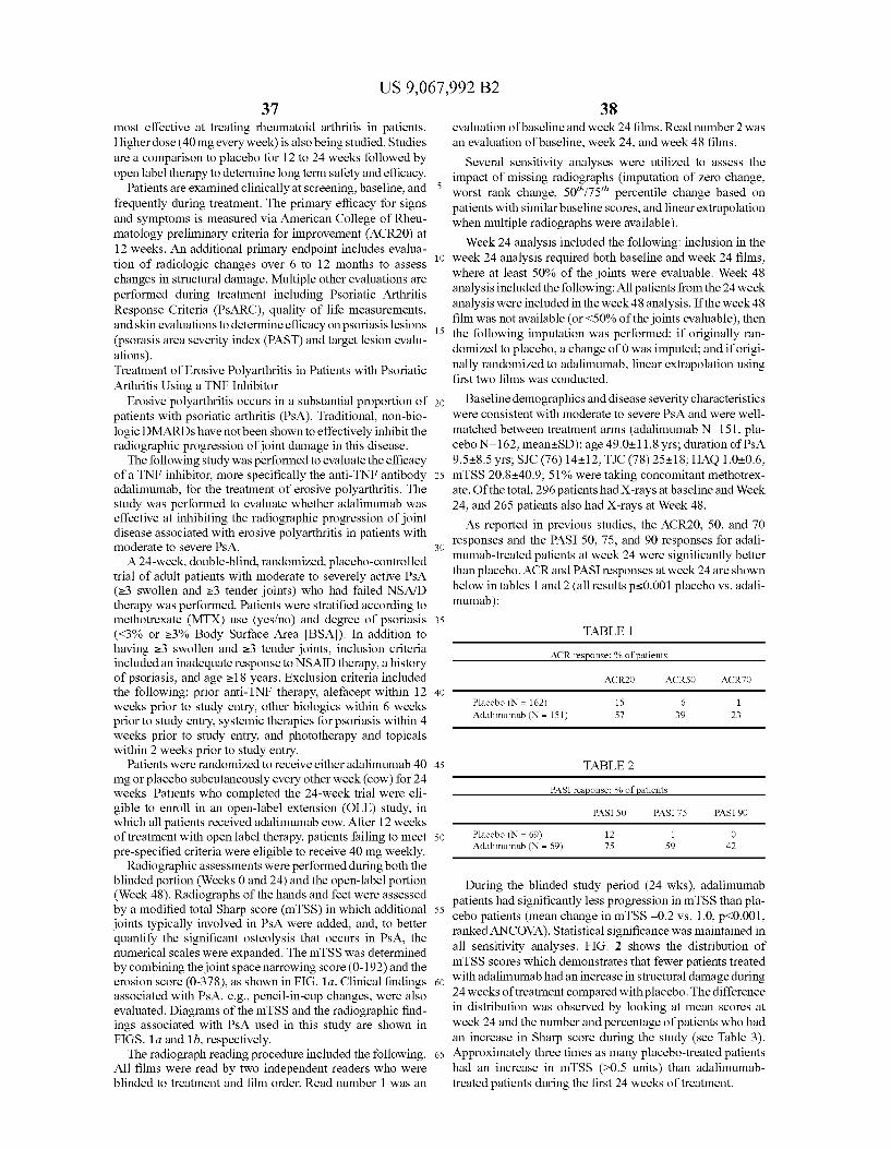
US 9,067,992 B2 37
most effective at treating rheumatoid arthritis in patients. Higher dose (40 mg every week) is also being studied. Studies are a comparison to placebo for 12 to 24 weeks followed by open label therapy to determine long term safety and efficacy.
Patients are examined clinically at Screening, baseline, and frequently during treatment. The primary efficacy for signs and symptoms is measured via American College of Rheu matology preliminary criteria for improvement (ACR20) at 12 weeks. An additional primary endpoint includes evalua tion of radiologic changes over 6 to 12 months to assess changes in structural damage. Multiple other evaluations are performed during treatment including Psoriatic Arthritis Response Criteria (PSARC), quality of life measurements, and skin evaluations to determine efficacy on psoriasis lesions (psorasis area severity index (PAST) and target lesion evalu ations). Treatment of Erosive Polyarthritis in Patients with Psoriatic Arthritis. Using a TNF Inhibitor
Erosive polyarthritis occurs in a Substantial proportion of patients with psoriatic arthritis (PSA). Traditional, non-bio logic DMARDs have not been shown to effectively inhibit the radiographic progression of joint damage in this disease. The following study was performed to evaluate the efficacy
of a TNF inhibitor, more specifically the anti-TNF antibody adalimumab, for the treatment of erosive polyarthritis. The study was performed to evaluate whether adalimumab was effective at inhibiting the radiographic progression of joint disease associated with erosive polyarthritis in patients with moderate to severe PSA. A 24-week, double-blind, randomized, placebo-controlled
trial of adult patients with moderate to severely active PSA (>3 swollen and >3 tender joints) who had failed NSA/D therapy was performed. Patients were stratified according to methotrexate (MTX) use (yes/no) and degree of psoriasis (<3% or >3% Body Surface Area (BSA). In addition to having 23 Swollen and 23 tender joints, inclusion criteria included an inadequate response to NSAID therapy, a history of psoriasis, and age 218 years. Exclusion criteria included the following: prior anti-TNF therapy, alefacept within 12 weeks prior to study entry, other biologics within 6 weeks prior to study entry, systemic therapies for psoriasis within 4 weeks prior to study entry, and phototherapy and topicals within 2 weeks prior to study entry.
Patients were randomized to receive eitheradalimumab 40 mg or placebo Subcutaneously every other week (eow) for 24 weeks. Patients who completed the 24-week trial were eli gible to enroll in an open-label extension (OLE) study, in which all patients received adalimumab eow. After 12 weeks of treatment with open label therapy, patients failing to meet pre-specified criteria were eligible to receive 40 mg weekly.
Radiographic assessments were performed during both the blinded portion (Weeks 0 and 24) and the open-label portion (Week 48). Radiographs of the hands and feet were assessed by a modified total Sharp score (mTSS) in which additional joints typically involved in PSA were added, and, to better quantify the significant osteolysis that occurs in PSA, the numerical scales were expanded. The mTSS was determined by combining the joint space narrowing score (0-192) and the erosion score (0-378), as shown in FIG. 1a. Clinical findings associated with PSA, e.g., pencil-in-cup changes, were also evaluated. Diagrams of the mTSS and the radiographic find ings associated with PSA used in this study are shown in FIGS. 1a and 1b, respectively. The radiograph reading procedure included the following.
All films were read by two independent readers who were blinded to treatment and film order. Read number 1 was an
5
10
15
25
30
35
40
45
50
55
60
65
38 evaluation of baseline and week 24 films. Read number 2 was an evaluation of baseline, week 24, and week 48 films.
Several sensitivity analyses were utilized to assess the impact of missing radiographs (imputation of Zero change, worst rank change, 50/75" percentile change based on patients with similar baseline scores, and linear extrapolation when multiple radiographs were available). Week 24 analysis included the following: inclusion in the
week 24 analysis required both baseline and week 24 films, where at least 50% of the joints were evaluable. Week 48 analysis included the following: All patients from the 24 week analysis were included in the week 48 analysis. If the week 48 film was not available (or <50% of the joints evaluable), then the following imputation was performed: if originally ran domized to placebo, a change of 0 was imputed; and if origi nally randomized to adalimumab, linear extrapolation using first two films was conducted.
Baseline demographics and disease severity characteristics were consistent with moderate to severe PSA and were well matched between treatment arms (adalimumab N=151, pla cebo N=162; meant-SD): age 49.0+11.8 yrs; duration of PSA 9.5+8.5 yrs: SJC (76) 14:12; TJC (78) 25+18; HAQ 1.0+0.6; mTSS 20.8+40.9; 51% were taking concomitant methotrex ate. Of the total, 296 patients had X-rays at baseline and Week 24, and 265 patients also had X-rays at Week 48. As reported in previous studies, the ACR20, 50, and 70
responses and the PASI 50, 75, and 90 responses for adali mumab-treated patients at week 24 were significantly better than placebo. ACR and PASI responses at week 24 are shown below in tables 1 and 2 (all results ps0.001 placebo vs. adali mumab):
TABLE 1
ACR response: % of patients
ACR2O ACRSO ACR70
Placebo (N = 162) 15 6 1 Adalimumab (N = 151) 57 39 23
TABLE 2
PASI response: % of patients
PASISO PASI 75 PASI90
Placebo (N = 69) 12 1 O Adalimumab (N = 69) 75 59 42
During the blinded study period (24 wks), adalimumab patients had significantly less progression in mTSS than pla cebo patients (mean change in mTSS -0.2 vs. 1.0, p<0.001, ranked ANCOVA). Statistical significance was maintained in all sensitivity analyses. FIG. 2 shows the distribution of mTSS scores which demonstrates that fewer patients treated with adalimumab had an increase in structural damage during 24 weeks of treatment compared with placebo. The difference in distribution was observed by looking at mean scores at week 24 and the number and percentage of patients who had an increase in Sharp score during the study (see Table 3). Approximately three times as many placebo-treated patients had an increase in mTSS (>0.5 units) than adalimumab treated patients during the first 24 weeks of treatment.
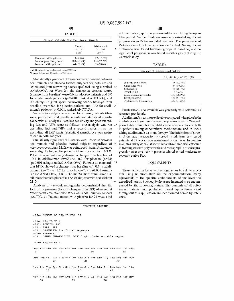
US 9,067,992 B2 39
TABLE 3
Change' in Modified Total Sharp Score at Week 24
Placebo Adalimumab N = 152 N = 144
n (%) n (%)
Decrease in Sharp Score 8 (5.3%) 27 (18.8%) No change in Sharp Score 100 (65.8%) 104 (72.2%) Increase in Sharp Score 44 (28.9%) 13 (9.0%)
ps 0.001 placebo vs. adalimumab using CMH test *Change defined as >0.5 units in mTSS Score
Statistically significant differences were observed between adalimumab and placebo treated subjects for both erosion scores and joint narrowing scores (ps0.001 using a ranked ANACOVA). At Week 24, the change in erosion scores (change from baseline) were 0.6 for placebo patients and 0.0 for adalimumab patients (p<0.001, ranked ANCOVA), and the change in joint space narrowing scores (change from baseline) were 0.4 for placebo patients and -0.2 for adali mumab patients (p<0.001, ranked ANCOVA).
Sensitivity analyses to account for missing patients films were performed and results maintained statistical signifi cance with all analyses. Post-hoc sensitivity analyses exclud ing feet and DIPs were as follows: one analysis was run excluding feet and DIPS and a second analysis was run excluding all DIP joints. Statistical significance was main tained in both analyses.
Statistically significant differences were observed between adalimumab and placebo treated Subjects regardless of whether concomitant MTX was being used. Mean differences were slightly higher for patients taking concomitant MTX. Patients on monotherapy showed a change from baseline of -0.1 in adalimumab (n=68) vs. 0.9 for placebo (n=74) (ps0.001 using a ranked ANACOVA). Patients on concomi tant MTX showed a change from baseline of -0.3 in adali mumab (n=76) vs. 1.2 for placebo (n=78) (ps0.001 using a ranked ANACOVA). FIGS. 3a and 3b show cumulative dis tribution function plots of mTSS of subjects with and without MTX.
Analysis of 48-week radiographs demonstrated that the lack of progression (lack of changes in mTSS) observed at Week 24 was maintained to Week 48 in adalimumab patients (see FIG. 4). Patients treated with placebo for 24 weeks did
SEQUENCE LISTING
10
15
25
30
35
40
40 not have radiographic progression of disease during the open label period. Neither treatment arm demonstrated significant progression in PSA-associated features. The prevalence of PSA-associated findings are shown in Table 4. No significant difference was found between groups at baseline, and no significant progression was found in either group during the 24-week study.
TABLE 4
Prevalence of PSA-associated findings
All patients (N = 313) n (%)
Joint space widening Gross osteolysis Subluxation Pencil-in-cup Juxta-articular periostitis Shaft periostitis Phalangeal tuft resorption
38 (12.1%) 60 (19.2%) 49 (15.7%) 9 (2.9%)
247 (78.9%) 140 (44.7%) 224 (71.6%)
Furthermore, adalimumab was generally well-tolerated as reported previously. Adalimumab was more effective compared with placebo in
inhibiting radiographic disease progression over a 24-week period. Adalimumab showed differences versus placebo both in patients taking concomitant methotrexate and in those taking adalimumab as monotherapy. The inhibition of struc tural damage progression observed in adalimumab-treated patients at 24 weeks was maintained at one year. In conclu sion, this study demonstrated that adalimumab was effective in treating erosive polyarthritis and radiographic disease pro gression over one year in patients who also had moderate to severely active PSA.
EQUIVALENTS
Those skilled in the art will recognize, or be able to ascer tain using no more than routine experimentation, many equivalents to the specific embodiments of the invention described herein. Such equivalents are intended to be encom passed by the following claims. The contents of all refer ences, patents and published patent applications cited throughout this application are incorporated herein by refer CCC.
<16 Os NUMBER OF SEO ID NOS: 37
<21 Os SEQ ID NO 1 &211s LENGTH: 107 212s. TYPE: PRT
<213> ORGANISM: Artificial Sequence 22 Os. FEATURE:
<223> OTHER INFORMATION: D2E7 light chain variable region
<4 OOs SEQUENCE: 1
Asp Ile Gln Met Thr Glin Ser Pro Ser Ser Leu Ser Ala Ser Val Gly 1. 5 1O 15
Asp Arg Val Thir Ile Thr Cys Arg Ala Ser Glin Gly Ile Arg Asn Tyr 2O 25 3 O
Lieu Ala Trp Tyr Glin Glin Llys Pro Gly Lys Ala Pro Llys Lieu. Lieu. Ile 35 4 O 45
Tyr Ala Ala Ser Thr Lieu. Glin Ser Gly Val Pro Ser Arg Phe Ser Gly SO 55 60
Ser 65
Glu
Thir
US 9,067,992 B2 41
- Continued
Gly Ser Gly Thr Asp Phe Thr Lieu. Thir Ile Ser Ser Leu Gln Pro 70 7s 8O
Asp Val Ala Thr Tyr Tyr Cys Glin Arg Tyr Asn Arg Ala Pro Tyr 85 90 95
Phe Gly Glin Gly Thr Lys Val Glu Ile Llys 1OO 105
<210s, SEQ ID NO 2 &211s LENGTH: 121 212. TYPE: PRT
<213> ORGANISM: Artificial Sequence 22 Os. FEATURE:
<223> OTHER INFORMATION: D2E7 heavy chain variable region
<4 OOs, SEQUENCE: 2
Glu 1.
Ser
Ala
Ser
Glu 65
Lell
Ala
Glin
Val Glin Lieu Val Glu Ser Gly Gly Gly Lieu Val Glin Pro Gly Arg 5 1O 15
Lieu. Arg Lieu. Ser Cys Ala Ala Ser Gly Phe Thr Phe Asp Asp Tyr 2O 25 3O
Met His Trp Val Arg Glin Ala Pro Gly Lys Gly Lieu. Glu Trp Val 35 4 O 45
Ala Ile Thir Trp Asn. Ser Gly His Ile Asp Tyr Ala Asp Ser Val SO 55 6 O
Gly Arg Phe Thir Ile Ser Arg Asp Asn Ala Lys Asn. Ser Lieu. Tyr 70 7s 8O
Gln Met Asn. Ser Lieu. Arg Ala Glu Asp Thr Ala Val Tyr Tyr Cys 85 90 95
Llys Val Ser Tyr Lieu. Ser Thr Ala Ser Ser Lieu. Asp Tyr Trp Gly 1OO 105 11 O
Gly Thr Lieu Val Thr Val Ser Ser 115 12 O
<210s, SEQ ID NO 3 &211s LENGTH: 9 212. TYPE: PRT
<213> ORGANISM: Artificial Sequence 22 Os. FEATURE:
<223> OTHER INFORMATION: D2E7 light chain variable region CDR3 22 Os. FEATURE:
<221s NAME/KEY: VARIANT <222s. LOCATION: ) 9 <223> OTHER INFORMATION: Xaa = Any Amino Acid
<4 OOs, SEQUENCE: 3
Glin 1.
Arg Tyr Asn Arg Ala Pro Tyr Xaa 5
<210s, SEQ ID NO 4 &211s LENGTH: 12 212. TYPE: PRT
<213> ORGANISM: Artificial Sequence 22 Os. FEATURE:
<223> OTHER INFORMATION: D2E7 heavy chain variable region CDR3 22 Os. FEATURE:
<221s NAME/KEY: VARIANT &222s. LOCATION: 12
<223> OTHER INFORMATION: Xaa = Any Amino Acid
<4 OOs, SEQUENCE: 4
Wall 1.
Ser Tyr Lieu. Ser Thr Ala Ser Ser Lieu. Asp Xaa 5 1O
42
US 9,067,992 B2 43
- Continued
<210s, SEQ ID NO 5 &211s LENGTH: 7 212. TYPE: PRT
<213> ORGANISM: Artificial Sequence 22 Os. FEATURE:
<223> OTHER INFORMATION: D2E7 light chain variable region CDR2
<4 OOs, SEQUENCE: 5
Ala Ala Ser Thr Lieu. Glin Ser 1. 5
<210s, SEQ ID NO 6 &211s LENGTH: 17 212. TYPE: PRT
<213> ORGANISM: Artificial Sequence 22 Os. FEATURE:
<223> OTHER INFORMATION: D2E7 heavy chain variable region CDR2
<4 OOs, SEQUENCE: 6
Ala Ile Thir Trp Asn. Ser Gly His Ile Asp Tyr Ala Asp Ser Val Glu 1. 5 1O 15
Gly
<210s, SEQ ID NO 7 &211s LENGTH: 11 212. TYPE: PRT
<213> ORGANISM: Artificial Sequence 22 Os. FEATURE:
<223> OTHER INFORMATION: D2E7 light chain variable region CDR1
<4 OO > SEQUENCE: 7
Arg Ala Ser Glin Gly Ile Arg Asn Tyr Lieu Ala 1. 5 1O
<210s, SEQ ID NO 8 &211s LENGTH: 5 212. TYPE: PRT
<213> ORGANISM: Artificial Sequence 22 Os. FEATURE:
<223> OTHER INFORMATION: D2E7 heavy chain variable region CDR1
<4 OOs, SEQUENCE: 8
Asp Tyr Ala Met His 1. 5
<210s, SEQ ID NO 9 &211s LENGTH: 107 212. TYPE: PRT
<213> ORGANISM: Artificial Sequence 22 Os. FEATURE:
<223> OTHER INFORMATION: 2SD4 light chain variable region
<4 OOs, SEQUENCE: 9
Asp Ile Gln Met Thr Glin Ser Pro Ser Ser Leu Ser Ala Ser Ile Gly 1. 5 1O 15
Asp Arg Val Thir Ile Thr Cys Arg Ala Ser Glin Gly Ile Arg Asn Tyr 2O 25 3O
Lieu Ala Trp Tyr Glin Glin Llys Pro Gly Lys Ala Pro Llys Lieu. Lieu. Ile 35 4 O 45
Tyr Ala Ala Ser Thr Lieu. Glin Ser Gly Val Pro Ser Arg Phe Ser Gly SO 55 6 O
Ser Gly Ser Gly Thr Asp Phe Thr Lieu. Thir Ile Ser Ser Leu Gln Pro 65 70 7s 8O
Glu Asp Val Ala Thr Tyr Tyr Cys Gln Lys Tyr Asn Ser Ala Pro Tyr
44
US 9,067,992 B2 45
- Continued
85 90 95
Ala Phe Gly Glin Gly. Thir Lys Val Glu Ile Llys 1OO 105
SEQ ID NO 10 LENGTH: 121 TYPE PRT
ORGANISM: Artificial Sequence FEATURE:
OTHER INFORMATION: 2SD4 heavy chain variable region
SEQUENCE: 1.O
Glin Val Glin Lieu Val Glu Ser Gly Gly Gly Lieu Val Glin Pro Gly Arg 1. 5 1O 15
Ser Lieu. Arg Lieu. Ser Cys Ala Ala Ser Gly Phe Thr Phe Asp Asp Tyr
Ala Met His Trp Val Arg Glin Ala Pro Gly Lys Gly Lieu. Asp Trp Val 35 4 O 45
Ser Ala Ile Thir Trp Asn. Ser Gly His Ile Asp Tyr Ala Asp Ser Val SO 55 6 O
Glu Gly Arg Phe Ala Val Ser Arg Asp Asn Ala Lys Asn Ala Lieu. Tyr 65
Lieu. Glin Met Asn. Ser Lieu. Arg Pro Glu Asp Thr Ala Val Tyr Tyr Cys 85 90 95
Thr Lys Ala Ser Tyr Lieu Ser Thr Ser Ser Ser Lieu. Asp Asn Trp Gly 1OO 105 11 O
Gln Gly Thr Lieu Val Thr Val Ser Ser 115 12 O
SEQ ID NO 11 LENGTH: 9 TYPE PRT
ORGANISM: Artificial Sequence FEATURE:
OTHER INFORMATION: 2SD4 light chain variable region CDR3
SEQUENCE: 11
Glin Llys Tyr Asn. Ser Ala Pro Tyr Ala 1. 5
SEQ ID NO 12 LENGTH: 9 TYPE PRT
ORGANISM: Artificial Sequence FEATURE:
OTHER INFORMATION: EP B12 light chain variable region CDR3
SEQUENCE: 12
Glin Llys Tyr Asn Arg Ala Pro Tyr Ala 1. 5
SEQ ID NO 13 LENGTH: 9
TYPE PRT
ORGANISM: Artificial Sequence FEATURE:
OTHER INFORMATION: VL1OE4 light chain variable region CDR3
SEQUENCE: 13
Gln Lys Tyr Glin Arg Ala Pro Tyr Thr 1. 5
46
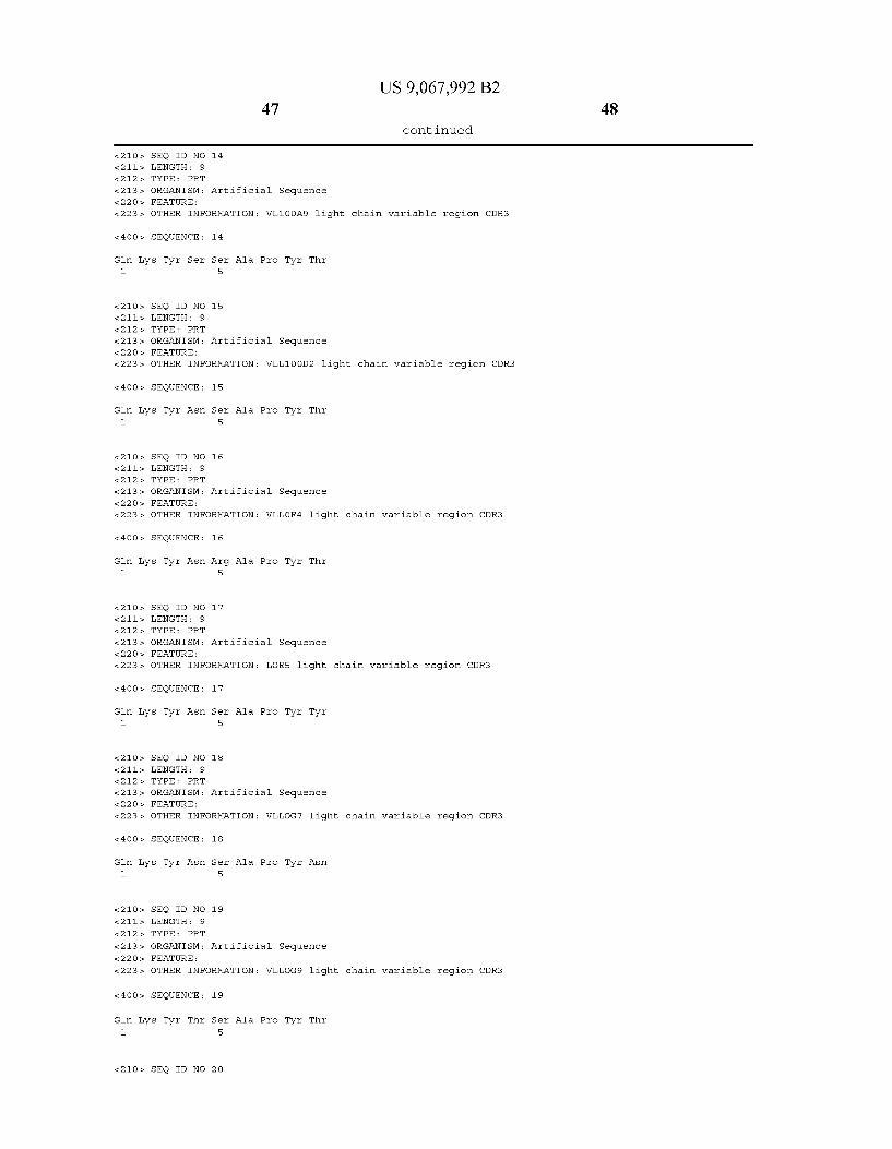
US 9,067,992 B2 47
- Continued
<210s, SEQ ID NO 14 &211s LENGTH: 9 212. TYPE: PRT
<213> ORGANISM: Artificial Sequence 22 Os. FEATURE:
<223> OTHER INFORMATION: VL100A9 light chain variable region CDR3
<4 OOs, SEQUENCE: 14
Gln Lys Tyr Ser Ser Ala Pro Tyr Thr 1. 5
<210s, SEQ ID NO 15 &211s LENGTH: 9 212. TYPE: PRT
<213> ORGANISM: Artificial Sequence 22 Os. FEATURE:
<223> OTHER INFORMATION: WLL1OOD2 light chain variable region CDR3
<4 OOs, SEQUENCE: 15
Gln Lys Tyr Asn Ser Ala Pro Tyr Thr 1. 5
<210s, SEQ ID NO 16 &211s LENGTH: 9 212. TYPE: PRT
<213> ORGANISM: Artificial Sequence 22 Os. FEATURE:
<223> OTHER INFORMATION: WLL OF4 light chain variable region CDR3
<4 OOs, SEQUENCE: 16
Gln Lys Tyr Asn Arg Ala Pro Tyr Thr 1. 5
<210s, SEQ ID NO 17 &211s LENGTH: 9 212. TYPE: PRT
<213> ORGANISM: Artificial Sequence 22 Os. FEATURE:
<223> OTHER INFORMATION: LOE5 light chain variable region CDR3
<4 OOs, SEQUENCE: 17
Gln Lys Tyr Asn Ser Ala Pro Tyr Tyr 1. 5
<210s, SEQ ID NO 18 &211s LENGTH: 9 212. TYPE: PRT
<213> ORGANISM: Artificial Sequence 22 Os. FEATURE:
<223> OTHER INFORMATION: WLLOG7 light chain variable region CDR3
<4 OOs, SEQUENCE: 18
Gln Lys Tyr Asn Ser Ala Pro Tyr Asn 1. 5
<210s, SEQ ID NO 19 &211s LENGTH: 9 212. TYPE: PRT
<213> ORGANISM: Artificial Sequence 22 Os. FEATURE:
<223> OTHER INFORMATION: WLLOG9 light chain variable region CDR3
<4 OOs, SEQUENCE: 19
Gln Lys Tyr Thr Ser Ala Pro Tyr Thr 1. 5
<210s, SEQ ID NO 2 O
48
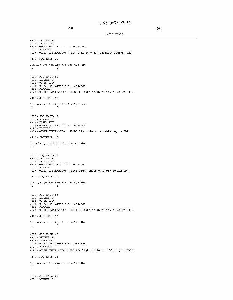
US 9,067,992 B2 49
- Continued
&211s LENGTH: 9 212. TYPE: PRT
<213> ORGANISM: Artificial Sequence 22 Os. FEATURE:
<223> OTHER INFORMATION: WLLOH1 light chain variable region CDR3
<4 OOs, SEQUENCE: 2O
Glin Llys Tyr Asn Arg Ala Pro Tyr Asn 1. 5
<210s, SEQ ID NO 21 &211s LENGTH: 9 212. TYPE: PRT
<213> ORGANISM: Artificial Sequence 22 Os. FEATURE:
<223> OTHER INFORMATION: WLLOH10 light chain variable region CDR3
<4 OOs, SEQUENCE: 21
Glin Llys Tyr Asn. Ser Ala Ala Tyr Ser 1. 5
<210s, SEQ ID NO 22 &211s LENGTH: 9 212. TYPE: PRT
<213> ORGANISM: Artificial Sequence 22 Os. FEATURE:
<223> OTHER INFORMATION: VL1B7 light chain variable region CDR3
<4 OOs, SEQUENCE: 22
Gln Glin Tyr Asn Ser Ala Pro Asp Thr 1. 5
<210s, SEQ ID NO 23 &211s LENGTH: 9 212. TYPE: PRT
<213> ORGANISM: Artificial Sequence 22 Os. FEATURE:
<223> OTHER INFORMATION: VL1C1 light chain variable region CDR3
<4 OOs, SEQUENCE: 23
Gln Lys Tyr Asn Ser Asp Pro Tyr Thr 1. 5
<210s, SEQ ID NO 24 &211s LENGTH: 9 212. TYPE: PRT
<213> ORGANISM: Artificial Sequence 22 Os. FEATURE:
<223> OTHER INFORMATION: VLO.1F4 light chain variable region CDR3
<4 OOs, SEQUENCE: 24
Gln Lys Tyr Ile Ser Ala Pro Tyr Thr 1. 5
<210s, SEQ ID NO 25 &211s LENGTH: 9 212. TYPE: PRT
<213> ORGANISM: Artificial Sequence 22 Os. FEATURE:
<223> OTHER INFORMATION: VLO. 1H8 light chain variable region CDR3
<4 OOs, SEQUENCE: 25
Gln Lys Tyr Asn Arg Pro Pro Tyr Thr 1. 5
<210s, SEQ ID NO 26 &211s LENGTH: 9
50
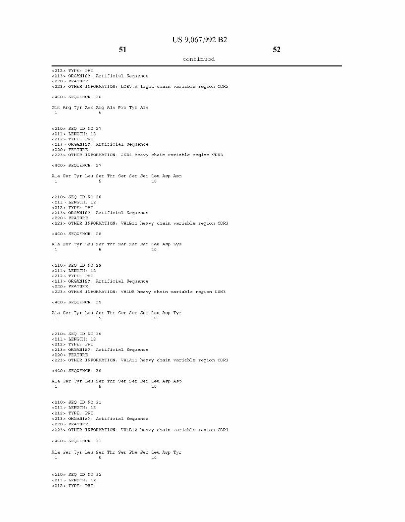
US 9,067,992 B2 51
- Continued
212. TYPE: PRT
<213> ORGANISM: Artificial Sequence 22 Os. FEATURE:
<223> OTHER INFORMATION: LOE7. A light chain variable region CDR3
<4 OOs, SEQUENCE: 26
Glin Arg Tyr Asn Arg Ala Pro Tyr Ala 1. 5
<210s, SEQ ID NO 27 &211s LENGTH: 12 212. TYPE: PRT
<213> ORGANISM: Artificial Sequence 22 Os. FEATURE:
<223> OTHER INFORMATION: 2SD4 heavy chain variable region CDR3
<4 OOs, SEQUENCE: 27
Ala Ser Tyr Lieu. Ser Thr Ser Ser Ser Lieu. Asp Asn 1. 5 1O
<210s, SEQ ID NO 28 &211s LENGTH: 12 212. TYPE: PRT
<213> ORGANISM: Artificial Sequence 22 Os. FEATURE:
<223> OTHER INFORMATION: VH1B11 heavy chain variable region CDR3
<4 OOs, SEQUENCE: 28
Ala Ser Tyr Lieu. Ser Thr Ser Ser Ser Lieu. Asp Llys 1. 5 1O
<210s, SEQ ID NO 29 &211s LENGTH: 12 212. TYPE: PRT
<213> ORGANISM: Artificial Sequence 22 Os. FEATURE:
<223> OTHER INFORMATION: VH1D8 heavy chain variable region CDR3
<4 OOs, SEQUENCE: 29
Ala Ser Tyr Lieu Ser Thr Ser Ser Ser Lieu. Asp Tyr 1. 5 1O
<210s, SEQ ID NO 3 O &211s LENGTH: 12 212. TYPE: PRT
<213> ORGANISM: Artificial Sequence 22 Os. FEATURE:
<223> OTHER INFORMATION: VH1A11 heavy chain variable region CDR3
<4 OOs, SEQUENCE: 30
Ala Ser Tyr Lieu. Ser Thr Ser Ser Ser Lieu. Asp Asp 1. 5 1O
<210s, SEQ ID NO 31 &211s LENGTH: 12
212. TYPE: PRT
<213> ORGANISM: Artificial Sequence 22 Os. FEATURE:
<223> OTHER INFORMATION: VH1B12 heavy chain variable region CDR3
<4 OOs, SEQUENCE: 31
Ala Ser Tyr Lieu Ser Thr Ser Phe Ser Lieu. Asp Tyr 1. 5 1O
<210s, SEQ ID NO 32 &211s LENGTH: 12 212. TYPE: PRT
52
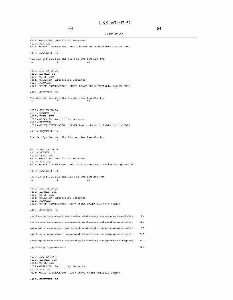
US 9,067,992 B2 53
- Continued
<213> ORGANISM: Artificial Sequence 22 Os. FEATURE:
<223> OTHER INFORMATION: VH1E4 heavy chain variable region CDR3
<4 OOs, SEQUENCE: 32
Ala Ser Tyr Lieu Ser Thr Ser Ser Ser Lieu. His Tyr 1. 5 1O
<210s, SEQ ID NO 33 &211s LENGTH: 12 212. TYPE: PRT
<213> ORGANISM: Artificial Sequence 22 Os. FEATURE:
<223> OTHER INFORMATION: VH1F6 heavy chain variable region CDR3
<4 OOs, SEQUENCE: 33
Ala Ser Phe Leu Ser Thr Ser Ser Ser Lieu. Glu Tyr 1. 5 1O
<210s, SEQ ID NO 34 &211s LENGTH: 12 212. TYPE: PRT
<213> ORGANISM: Artificial Sequence 22 Os. FEATURE:
<223> OTHER INFORMATION: 3C-H2 heavy chain variable region CDR3
<4 OOs, SEQUENCE: 34
Ala Ser Tyr Lieu Ser Thr Ala Ser Ser Lieu. Glu Tyr 1. 5 1O
<210 SEQ ID NO 35 &211s LENGTH: 12 212. TYPE: PRT
<213> ORGANISM: Artificial Sequence 22 Os. FEATURE:
<223> OTHER INFORMATION: VH1-D2.N heavy chain variable region CDR3
<4 OOs, SEQUENCE: 35
Val Ser Tyr Lieu. Ser Thr Ala Ser Ser Lieu. Asp Asn 1. 5 1O
<210s, SEQ ID NO 36 &211s LENGTH: 321 &212s. TYPE: DNA
<213> ORGANISM: Artificial Sequence 22 Os. FEATURE:
<223> OTHER INFORMATION: D2E7 light chain variable region
<4 OOs, SEQUENCE: 36
gacatccaga tigacccagtic ticcatcct co citgtctgcat citg tagggga cagagt cacc
at Cacttgtc. g.ggcaagt ca gggcatcaga aattact tag cctggitatica gcaaaaacca
gggaaag.ccc ctaagctic ct gat citatgct gcatccactt togcaatcagg ggit cocatct
cggttcagtg gcagtggat.c tdggacagat ttcactict ca C catcagcag cct acagcct
gaagatgttg caactt atta citgtcaaagg tataaccotg caccgtatac ttittggc.ca.g
gggaccalagg taaatcaa a
<210s, SEQ ID NO 37 &211s LENGTH: 363 &212s. TYPE: DNA
<213> ORGANISM: Artificial Sequence 22 Os. FEATURE:
<223> OTHER INFORMATION: D2E7 heavy chain variable region
<4 OO > SEQUENCE: 37
6 O
12 O
18O
24 O
3OO
321
54
US 9,067,992 B2 55 56
- Continued
gaggtgcagc tiggtggagtic tiggggaggc titggtacago CC9gcaggt C cct gagactic 6 O
t cct gtgcgg cct Ctggatt Cacct ttgat gattatgcca to actgggit cc.ggcaa.gct 12 O
cCagggaagg gcctggaatgggit ct cagct atc acttgga at agtggtca catagacitat 18O
gcggactctg tdgagggc.cg attcaccatc. tccagagaca acgc.caagaa ctic cctgtat 24 O
Ctgcaaatga acagtctgag agctgaggat acggc.cgitat attactgtgc gaaagtict cq 3OO
taccttagca cc.gcgt.cct c ccttgact at tdgggccaag gtaccctggit caccgtc.tc.g 360
agt 363
What is claimed: 1. A method of treatment of moderate to severe active
psoriatic arthritis in adult patients, wherein each said patient has a 3 Swollen and 23 tenderjoints prior to the treatment and has failed NSAID therapy, comprising subcutaneously administering to each said patient 40 mg of adalimumab every other week, wherein 23% of said patients achieve 70% reduction in American College of Rheumatology (ACR) score at week 24 of the treatment.
2. A method for reducing or inhibiting symptoms in a patient with psoriatic arthritis, comprising Subcutaneously administering to said patient 40 mg of adalimumab every other week.
3. The method of claim 2, wherein said 40 mgadalimumab is comprised in a pre-filled Syringe for Subcutaneous injec tion.
25
30
4. The method of claim3, wherein said 40 mgadalimumab is formulated at a concentration of 50 mg/ml.
5. The method of claim 2, wherein the patient achieves at least a 50% reduction in ACR score at week 24 of the treat ment.
6. The method of claim 5, wherein the patient achieves at least a 70% reduction in ACR score at week 24 of the treat ment.
7. The method of claim 2, wherein said symptoms are progression of structural damage assessed by radiograph.
8. The method of claim 7, wherein said 40 mgadalimumab is comprised in a pre-filled Syringe for Subcutaneous injec tion.
9. The method of claim8, wherein said 40 mgadalimumab is formulated at a concentration of 50 mg/ml.
: : : : :
UNITED STATES PATENT AND TRADEMARK OFFICE
CERTIFICATE OF CORRECTION
PATENT NO. : 9,067,992 B2 APPLICATIONNO. : 14/563056 DATED : June 30, 2015 INVENTOR(S) : Rebecca S. Hoffman et al.
Page 1 of 1
It is certified that error appears in the above-identified patent and that said Letters Patent is hereby corrected as shown below:
ON THE TITLE PAGE
Item 72 the Inventors should read as follows:
Rebecca S. Hoffman, Wilmette, IL (US) Mark Weinberg, Northbrook, IL (US) Subhashis Banerjee, Princeton, NJ (US) Lori K. Taylor, Boston, MA (US) Clive E. Spiegler, Skillman, NJ (US) Daniel E. Tracey, Harvard, MA (US) Elliot K. Chartash, Marietta, GA (US) William T. Barchuk, San Diego, CA (US) Philip Yan, Vernon Hills, IL (US) Anwar Murtaza, Westborough, MA (US) Jochen G. Salfeld, North Grafton, MA (US) Steven A. Fischkoff, Short Hills, NJ (US) George R. Granneman, Marco Island, FL (US)
Signed and Sealed this Eighth Day of March, 2016
74.4.4.2% 4 Michelle K. Lee
Director of the United States Patent and Trademark Office