Embed Size (px)
Citation preview

3,350+OPEN ACCESS BOOKS
108,000+INTERNATIONAL
AUTHORS AND EDITORS115+ MILLION
DOWNLOADS
BOOKSDELIVERED TO
151 COUNTRIES
AUTHORS AMONG
TOP 1%MOST CITED SCIENTIST
12.2%AUTHORS AND EDITORS
FROM TOP 500 UNIVERSITIES
Selection of our books indexed in theBook Citation Index in Web of Science™
Core Collection (BKCI)
Chapter from the book Advances in OphthalmologyDownloaded from: http://www.intechopen.com/books/advances-in-ophthalmology
PUBLISHED BY
World's largest Science,Technology & Medicine
Open Access book publisher
Interested in publishing with IntechOpen?Contact us at [email protected]

10
Etiology and Clinical Presentation of Astigmatism
Sanja Masnec Olujić Ghethaldus Ophthalmology Policlinics, Zagreb
Croatia
1. Introduction
A perfect point image of an object point is called a stigmatic image. The term „stigmatic“is
derived from the Greek word stigma, which refers to a sharply pointed stylus (Liesegang et
al., 2002). Thus, a stigmatic optical system is one able to focus all the light rays from a point
source onto a single point. However, in most cases, images are not stigmatic.
In paraxial optics, the focus is stigmatic, and all paraxial rays (those extremely close to the
optical axis) focus onto a point. With nonparaxial rays, the focus is generally not stigmatic.
Deviations from stigmatic imaging are called aberrations, and, one of a number of ways to
classify such aberrations is clinically: into spherical aberrations, regular astigmatism and
irregular astigmatism.
Astigmatism is the unequal refraction of the same eye in two different meridians. Unlike the
basic types of refraction- emetropia, myopia and hyperopia- where all the light rays enter
one focus (on the retina, behind it, or in front of it), in astigmatism there is no a single focus.
In basic types of refraction, the cornea is spherical and it refracts equally in all the meridians.
In astigmatism variations in the curvature of the cornea or lens along different meridians
prevent the light rays from focusing onto a single point. Corneal refraction depends on the
corneal curvature. If the cornea is more curved, the power of refraction is higher and vice
versa. Corneal and lenticular astigmatism can complement or cancel each other. Their
summation represents the so called “total astigmatism”.
Due to the lack of a single focus, an astigmatic eye is not able to see clearly without
correction. Astigmatic patients complain of visual disturbances in both far (in myopia) and
near vision (in hyperopia), which causes astenopic problems (headache, dizziness, fatigue
etc). Additionally, regular, symmetric objects might seem to them irregular, and /or
elongated.
2. Astigmatism types
Astigmatism can be regular or irregular. It can also be divided into myopic, hyperopic and mixed/compound astigmatism (where one of the main meridians is hyperopic and the other one myopic).
www.intechopen.com

Advances in Ophthalmology
168
2.1 Regular astigmatism
In regular astigmatism each meridian refracts regularly and equally, but differently from the other meridians. One of the meridians refracts the most and another one the least. Those two meridians are called the main meridians, and they are perpendicular to each other.
In most cases, one of the main meridians is located vertically and the other one horizontally, but there can also be oblique, still maintaining the 90° angle to each other.
In the remaining meridians, located between the both two main meridians, refraction changes gradually –increases or decreases (Fig 1).
Fig. 1. With-the-rule astigmatism of 5.4 D shown on the topographical map of the anterior corneal surface , with quantitative information on the corneal curvature, astigmatism and its meridians.
Vertical meridian, in most of cases, refracts more then the horizontal, most likely due to the pressure of eyelids onto the cornea. This type of astigmatism is called with- the- rule astigmatism and is more common in children (Mohindra et al., 1978).
If the horizontal meridian refracts more, this is called against- the- rule astigmatism and is more common in older adults.
The difference in refraction of the two main meridians represents the amount of astigmatism and is represented in diopters (D).
When that difference is no more than 1/2 -3/4 diopters it is a so-called physiological astigmatism and a correction is usually not needed, as it does not lead to visual deterioration or subjective symptoms. Mostly, it is neutralized by the lenticular astigmatism (Parunovic et al., 1995).
www.intechopen.com

Etiology and Clinical Presentation of Astigmatism
169
Astigmatism over ¾ D can lead to visual disturbances and other subjective symptoms, because the lenticular astigmatism, which rarely exceeds 1-1.5 D, is not able to compensate for the corneal astigmatism.
Regular type astigmatism is rarely greater than 6-7 D.
For a better understanding of astigmatism and its correction, the most important optical concept is the conoid of Sturm.
The refractive power, as mentioned above, changes from one meridian to the next, and an
astigmatic surface cannot bring a pencil of light rays to a point focus. Two focal lines instead
of a single focus are formed. This complex geometrical envelope of a pencil of light rays
refracted by a circular spherocylindrical lens is called the conoid of Sturm.
The conoid of Sturm has two focal lines, each parallel to one of the main meridians of the
spherocylindrical lens. All the light rays in the pencil pass through each of the focal lines.
The cross sections of the conoid of Sturm at various points along its length are mostly
elliptical, including the portion of the conoid external to the two focal lines. At the dioptric
mean of the two focal lines there is a circular cross section of the conoid of Sturm. This
circular patch of light rays is called the circle of least confusion, and represents the best overall
focus for the spherocylindrical lens. The circle of least confusion is the position where all the
rays would be brought to focus if the lens had a spherical power equal to the average
spherical power of all the meridians of the spherocylindrical lens (Michaels, 1980).
This average spherical power of a spherocylindrical lens represents the spherical equivalent
of the lens, and can be calculated by the following equation:
Spherical equivalent = sphere + cylinder/2.
About 50% of infants in their first years of life show astigmatism of over 1D (Bennet et al.,
1989; Mohindra et al., 1978).
Some authors have suggested that this high astigmatism helps the infant to bracket the
position of best focus while it learns to accommodate (Howland et al., 1978).
In adults, this high incidence of astigmatism is much smaller. Different studies show that
about 15 % of adults have astigmatism greater than 1D, and only 2 % of more then 3D
(Yanoff & Duker, 2004).
In the leater group, it is most likely that much of the high astigmatism is due to some form
of intraocular surgery, such as cataract surgery (particularly when extracapsular cataract
extraction is performed), corneal transplants, corneal lacerations repair, …etc.
Regular astigmatism can be congenital and acquired. Congenital astigmatism is usually
inherited. Acquired astigmatism is mostly against-the rule and as mentioned above, can
result from various intraocular surgeries.
Most oftenly, astigmatism is static, but it can also change during the life time. With –the –
rule astigmatism tends to lessen over the years, and the position of the main meridians can
also slightly change during the lifetime.
Regular astigmatism is correctable by cylindrical spectacle lenses and contact lenses.
www.intechopen.com

Advances in Ophthalmology
170
In with-the rule astigmatism, a correcting plus cylinder lens should be used at or near the 90° axis. In against-the rule astigmatism, a correcting plus cylinder lens should be used at or near the 180° axis.
2.2 Irregular astigmatism
Irregular astigmatism appears when the refraction of light is unequal and irregular in the same meridian of the eye.
That is usually a consequence of pathological changes especially to the cornea (maculae centrales corneae, ulcus, pannus, keratoconus etc) or lens (cataract, posterior capsular opacification, lens subluxation etc).
The visual acuity of such an eye is deteriorated and sometimes monocular diplopia or poliopia occurs. All eyes have at least a small amount of irregular astigmatism, but the term is used clinically only for the stronger irregularities, such as those mentioned above, or occurring with keratoconus.
In the past, irregular astigmatism was not a field of strong interest by clinicians, as it was not very common, and was not treatable. It could not be corrected with spectacles, and rigid contact lenses could alleviate the problem to some extent only if the irregular astigmatism was corneal in origin.
With the development of keratorefractive surgical procedures, it became increasingly of interest, as keratorefractive surgeries may produce visually significant irregular astigmatism, or be used to treat it.
2.2.1 Keratoconus
Keratoconus (conical cornea) is a condition characterized by a progressive corneal steepening, usually inferior to the center of the cornea (Fig. 2.). As a result of a noninflammatory thinning of the corneal stroma eventually myopia is induced, together with irregular astigmatism, leading to an impairment in the quality of vision.
Keratoconus is a bilateral, usually asymmetric, noninflammatory corneal ectasia, which belongs to a group of corneal shape disorders, together with pellucid marginal corneal degeneration and keratoglobus.
By keratometry keratoconus is classified as:
1. mild (< 48 D) 2. moderate (48-54 D) 3. severe (> 54 D).
Epidemiology and pathogenesis
Keratoconus occurs in all ethnic groups with no male or female preponderance.
Rarely, it can be congenital (Smolin, 1987).
In majority of the cases, its onset is at puberty and then progresses until the third to fourth decade of life, with an incidence of approximately 1 per 2000 in the general population, and a prevalence of 54.5 per 100,000 (Hofstetter, 1959; Kennedy et al., 1986).
www.intechopen.com

Etiology and Clinical Presentation of Astigmatism
171
Fig. 2. Keratoconus (conical cornea)
In some cases it may also start later in life and progress at any age.
The etiology of the disease can be divided into three groups:
1. inherited 2. sporadic 3. acquired (secondary or in association to /with other diseases).
The inheritance of keratoconus is still not defined completely. Before the presence of videokeratography it was believed that more than 90 % of cases were sporadic, but recent studies revealed evidence that suggests an autosomal dominant pattern of inheritance (Gonzales & McDonnell, 1992).
Corneal thinning appears to result from a loss of structural components in the cornea, but why this happens is still not clear.
Recent biochemical studies of cornea with keratoconus suggest that enzyme abnormalities in the corneal epithelium, such as increased expression of proteases and other catabolic enzymes (Sawagamuchi et al., 1989) and decreased levels of inhibitors of proteolytic enzymes, may play a role in corneal stromal digestion and degradation (Fukuchi et al., 1994).
Investigations of corneal α1 proteinase inhibitor and α2 macroglobulin (also proteinase inhibitor) support the hypothesis that the degradation process may be aberrant in keratoconus (Sawagamuchi et al., 1994)
Evidence has been provided regarding abnormalities in gene promoters involved in these enzyme activities (Maruyama et al., 2001).
www.intechopen.com
Advances in Ophthalmology
172
Other studies have reported abnormalities in corneal collagen and its cross-linking, as a potential cause of keratoconus (Bron, 1988).
Some authors have proposed a role for an IL-1 system in the cornea in the pathogenesis of keratoconus. It is suggested that the increased expression of the IL-1 receptor sensitizes the keratocytes to IL-1 released from the epithelium or endothelium, causing a loss of keratocytes through apoptosis and a decrease in stromal mass over time (Wilson et al., 1996).
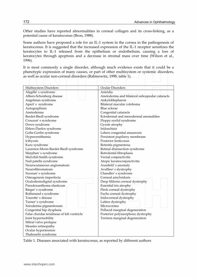
It is most commonly a single disorder, although much evidence exists that it could be a phenotypic expression of many causes, or part of other multisystem or systemic disorders, as well as ocular non-corneal disorders (Rabinowitz, 1998, table 1).
Multisystem Disorders Ocular Disorders
Alagille' s syndrome Albers-Schonberg disease Angelman syndrome Apert' s syndrome Autographism Anetoderma Bardet-Biedl syndrome Crouzon' s syndrome Down syndrome Ehlers-Danlos syndrome Goltz-Gorlin syndrome Hyperornithemia Icthyosis Kurz syndrome Laurence-Moon-Bardet-Biedl syndrome Marphan' s syndrome Mulvihil-Smith syndrome Nail patella syndrome Neurocutaneous angiomatosis Neurofibromatosis Noonan' s syndrome Osteogenesis imperfecta Oculodentodigital syndrome Pseudoxanthoma elasticum Rieger' s syndrome Rothmund s syndrome Tourette' s disease Turner' s syndrome Xeroderma pigmentosum Congenital hip dysplasia False chordae tendineae of left ventricle Joint hypermobility Mitral valve prolapse Measles retinopathy Ocular hypertension Thalesselis syndrome
Aniridia Anetoderma and bilateral subcapsular cataracts Ankyloblepharon Bilateral macular coloboma Blue sclerae Congenital cataracts Ectodermal and mesodermal anomalities Floppy eyelid syndrome Gyrate atrophy Iridoschisis Lebers congenital amaurosis Persistent pupilarry membrane Posterior lenticonus Retinitis pigmentosa Retinal disinsection syndrome Retrolental fibroplasia Vernal conjunctivitis Atopic keratoconjunctivitis Axenfeld' s anomaly Avellino' s dystrophy Chandler' s syndrome Corneal amyloidosis Deep filiform corneal dystrophy Essential iris atrophy Fleck corneal dystrophy Fuchs corneal dystrophy Iridocorneal dystrophy Lattice dystrophy Microcornea Pellucid marginal degeneration Posterior polymorphous dystrophy Terriens marginal degeneration
Table 1. Diseases associated with keratoconus, as reported by different authors
www.intechopen.com
Etiology and Clinical Presentation of Astigmatism
173
The most common association is with Down syndrome, connective tissue disorders, and
Leber's congenital amaurosis (Cullen & Butler, 1963; Iwaszkiewicz, 1989).
Acquired etiologies can be divided into those which are attributed to inflammatory
conditions, such as vernal conjunctivitis and those secondary to eye rubbing which can also
release inflammatory mediators, such as Leber's congenital amaurosis and Down syndrome.
Many authors report a high incidence of mitral valve prolapse (58%) in patients with
advanced keratoconus, while others report of eye rubbing in systemic atopy and cytokine
interleukin-1, as a mediator of stromal degradation. Other factors, such as contact lens wear,
may play a role in the development of the cone (Krachmer et al.,1984; Sharif et al., 1992).
Also, some studies report 6-8% of cases with positive family history or evidence of familiar
transmission (Hallerman & Wilson, 1977; Krachmer et al., 1984).
Ocular manifestations
Early stages of the disorder may not present any symptoms. Cornea may appear normal on
slit-lamp examination and there may be no symptoms of the disease. It maya be suspected
by an ophthalmologist because the patient cannot be refracted to a 20/20 corrected vision.
There might only be a mild steepening of keratometry mires, inferiorly or centrally. In such
cases, anterior topography of the central and paracentral cornea will confirm the suspected
diagnosis (Krachmer et al, 1984). In advanced cases, there can be a significant visual
disturbance followed by a significant visual loss, but, fortunately, patients with keratoconus
never become totally blind. Symptoms, as well as clinical signs, are variable and depend on
the stage of the progression and on the severity of the disease.
The corneal manifestations include steepening (centrally or paracentrally, most commonly
inferiorly or inferotemporally), thinning of the corneal apex, conical protrusion, a ring of
iron deposits partially or completely accumulating in the epithelium surrounding the cone
(Fleischer's ring), anterior scars at the level of Bowman's membrane, enlarged corneal nerves,
increased intensity of the corneal endothelial reflex, and deep vertical lines in stroma and
Descemet' s membrane, that disappear momentally under digital pressure during slit-
lamp examination (Vogt' s striae).
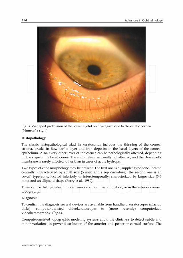
The steeping of the cornea leads to clinical signs, which include the V-shaped protrusion of
the lower eyelid on downgaze due to the ectatic cornea (Munson’s sign, Fig. 3.), sharply
focused light beam at the nasal limbus, produced by lateral illumination (Rizzuti's sign), and
a dark reflex using the retroillumination techniques in the area of the cone, when observing
the cornea with the pupil dilatation (Charleaux' s sign), and an irregular “scissor” reflex on
retinoscopy.
Some patients with advanced keratoconus may occasionally experience sudden visual loss
and pain due to the acute rupture of Descemet's membrane (acute hydrops). In such cases
the conjunctive may be injected, and diffuse stromal edema appears. These breaks in
Descemet’s membrane result in acute overhydration and a stromal imbibing of aqueous. The
overlying corneal epithelium may become edematous. The edema may last for months and,
after the resolution of redness and relief of pain over time, can be replaced by scarring (and
the corneal steepness may be reduced).
www.intechopen.com
Advances in Ophthalmology
174
Fig. 3. V-shaped protrusion of the lower eyelid on downgaze due to the ectatic cornea (Munson' s sign )
Histopathology
The classic histopathological triad in keratoconus includes the thinning of the corneal stroma, breaks in Bowman' s layer and iron deposits in the basal layers of the corneal epithelium. Also, every other layer of the cornea can be pathologically affected, depending on the stage of the keratoconus. The endothelium is usually not affected, and the Descemet’s membrane is rarely affected, other than in cases of acute hydrops.
Two types of cone morphology may be present. The first one is a „nipple“ type cone, located centrally, characterized by small size (5 mm) and steep curvature; the second one is an „oval“ type cone, located inferiorly or inferotemporally, characterized by larger size (5-6 mm), and an ellipsoid shape (Perry et al., 1980).
These can be distinguished in most cases on slit-lamp examination, or in the anterior corneal topography.
Diagnosis
To confirm the diagnosis several devices are available from handheld keratoscopes (placido disks), computer-assisted videokeratoscopes to (more recently) computerized videokeratography (Fig.4).
Computer-assisted topographic modeling systems allow the clinicians to detect subtle and minor variations in power distribution of the anterior and posterior corneal surface. The
www.intechopen.com
Etiology and Clinical Presentation of Astigmatism
175
forme fruste, or subclinical keratoconus, recognized by Placido disk-based topography, requires caution and is considered a contraindication to refractive surgery. With regard to the identification of forme fruste keratoconus, classification programs on corneal topographers may assist in differentiating between keratoconus suspects, corneal distortion, and even patients having undergone refractive surgery, and normal variations of corneal topography.
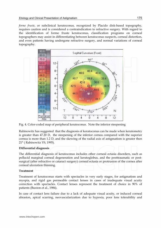
Fig. 4. Color-coded map of peripheral keratoconus. Note the inferior steepening
Rabinowitz has suggested that the diagnosis of keratoconus can be made when keratometry is greater than 47.20 D, the steepening of the inferior cornea compared with the superior cornea is more than 1.2 D, and the skewing of the radial axis of astigmatism is greater then 21° ( Rabinowitz YS, 1995).
Differential diagnosis
The differential diagnosis of keratoconus includes other corneal ectasia disorders, such as pellucid marginal corneal degeneration and keratoglobus, and the posttraumatic or post-surgical (after refractive or cataract surgery) corneal ectasia or protrusion of the cornea after corneal ulceration thinning.
Treatment
Treatment of keratoconus starts with spectacles in very early stages, for astigmatism and myopia, and rigid gas permeable contact lenses in cases of inadequate visual acuity correction with spectacles. Contact lenses represent the treatment of choice in 90% of patients (Buxton et al., 1984).
In case of contact lens failure due to a lack of adequate visual acuity, or induced corneal abrasion, apical scarring, neovascularization due to hypoxia, poor lens tolerability and
www.intechopen.com

Advances in Ophthalmology
176
discomfort and lens displacement, surgical procedure is indicated. The most frequent surgical procedure is penetrating keratoplasty, which refers to the full-thickness replacement of diseased corneal tissue with a healthy donor. The main difficulty during surgery is suturing to the thin corneal bed.
Lamellar keratoplasty is also effective, but is not as frequently used as the penetrating
keratoplasty, due to technical challenges and it being more time consuming. It is a
procedure in which a partial-thickness graft of donor tissue (donor stroma or sclera) is used
to provide tectonic stability and /or optical improvement.
There are generally two types of lamellar keratoplasty: anterior and posterior. In the
anterior lamellar keratoplasty, the transplanted tissue does not include corneal endothelium
and this procedure avoids endothelial rejection. The aim in deep lamellar and posterior
lamellar keratoplasty is to replace diseased corneal endothelium while keeping the anterior
corneal surface intact, as well as reducing refractive error and irregular astigmatism. In a
short amount of time, deep lamellar endothelial keratoplasty (DLEK), Descemet-stripping
endothelial keratoplasty (DSEK), Descemet-stripping automated endothelial keratoplasty
(DSAEK) and Descemet’ s membrane endothelial keratoplasty (DMEK) have become the
techniques of choice for partial corneal transplant surgeries.
Descemet’s membrane endothelial keratoplasty (DMEK) and Descemet-stripping automated
endothelial keratoplasty (DSAEK) are new types of partial-thickness corneal graft
operations, in which only the innermost corneal layers are replaced. Visual recovery is
quicker, there is less physical restriction on activities and no related suture problems.
The results of lamellar techniques are far better than epikeratoplasty which has been
abandoned due to the suboptimal visual outcomes. A keratoconic patient has a 10-20%
chance, over her/his lifetime, of needing a corneal transplant (Smiddy et al., 1988; Tuft et
al.,1994).
Intracorneal ring segments Intacts can be also used in cases without corneal scarring to
reduce myopia and astigmatism. The purpose of Intacts segment implantation is to defer the
need for corneal transplantation and restore contact lens tolerance. The placement of Intacts
generates the response that interrupts the biomechanical disease progression, and a
biomechanical response that allows visual improvement over six months. The improvement
in visual acuity and refraction is accomplished by shortening the path length of the portion
of the collagen lamellae that are central to the segments. The redistribution of corneal
curvature leads to a redistribution of corneal stress, interrupting the biomechanical cycle of
the keratoconus progression.
A relatively new method of treating progressive corneal ectasia is corneal collagen
crosslinking (CXL). Its clinical use has been rapidly increasing since it was originally
introduced in 1997 as the first treatment that could improve the biomechanical stability of
the weakened cornea. This method is based on combined action of the photo-sensitizer
riboflavin (vitamin B2) and ultraviolet A light, which induce the formation of new covalent
bonds between the collagen fibers.
This method has been widely accepted since the first introduction by Spoerl and his co-workers at Dresden University in Germany, in 1997 (Spörl et al., 1997).
www.intechopen.com
Etiology and Clinical Presentation of Astigmatism
177
The recent advances in diagnostic devices resulted in the detection of more subtle corneal
changes, suggesting that the sublinical forms are more common than fully developed
keratoconus. The iatrogenic post-LASIK ectasia is the second most common corneal ectasia.
It usually appears as a result of refractive surgery performed in predisposed individuals or
in patients with an undetected early form of keratoconus. Before the introduction of corneal
collagen crosslinking, the only treatment that could halt the progression of keratoconus in
most cases was the penetrating keratoplasty.
Studies which have been conducted so far demonstrated the beneficial effect in halting the
progression of the diseases, with a very low complication rate (Koller et al., 2009; Tomkins &
Garzozi, 2008).
The term „crosslinking“refers to the formation of bonds between natural polymer
molecules.
The original crosslinking technique described by Wollensak and co-workers is still being
used today, with the minor modifications. (Wollensak et al., 2003).
The photopolymerizing effect is induced by the combined action of riboflavin as a photo-
sensitizer, and long wavelength ultraviolet (UVA) light of 370 nm. This particular
wavelength was chosen so that riboflavin can achieve a maximal absorption while still
remaining bellow harmful radiation levels. During the exposure, riboflavin is excited into a
triplet state, generating the so-called reactive oxygen species which, in turn, induce the
formation of new covalent bonds (disulphide /s-s) between the amino acids of neighboring
collagen fibers.
After the application of a local anesthetic, a lid speculum is inserted and central corneal
abrasion up to 9.0 mm in diameter is made. According to the protocol of the Institute for
Refractive and Ophthalmic Surgery in Zürich, Switzerland, 0.1% riboflavin solution
containing 10 mg of riboflavin-5-phosphate diluted in 10 ml of a 20% dextran solution is
instilled every 3 minutes, for 30 minutes. After that, central corneal thickness (CCT) is
measured. For safety reasons, additional riboflavin 0.1% hypoosmotic drops without
dextran should be applied in corneas whose thickness is less than 400 µm. Otherwise, UVA
light should not be applied. The cornea is then irradiated for 30 minutes with an UVA
illumination device. The device must provide a homogenous UV radiation with irradiance
of 3 mW/ cm2 at the working distance of 5 cm. During irradiation, the cornea is moistened
every 3 minutes with the riboflavin 0.1% drops. The total dosage delivered to the cornea in
that way is 5.4 J/cm2. At the end, antibiotic ointment is applied and bandage lens inserted to
facilitate the epithelial healing.
Beside the standard CXL, as described above (with removal of the epithelium), the
treatment can also be the transepithelial CXL (without removal of the epithelium). The later
has less complications, such as death of keratocytes. Not being invasive, it is very well
tolerated.
Indications for cross-linking today are clinical and instrumental progression (refractive,
topographic, pachimetric, aberrometric) of corneal ectasia disorders such as keratoconus
and pellucid marginal degeneration in the last 6-12 months, iatrogenic keratectasia after
refractive lamellar surgery and corneal melting not responding to conventional therapy.
www.intechopen.com
Advances in Ophthalmology
178
Contraindications include corneal thickness of less than 400 µm, central corneal opacity,
epithelial healing disorders, such as map dot dystrophy and rheumatic disorders, refractive
keratotomy, previous herpes simplex virus keratitis (UV-A may induce herpes reactivation),
corneal melting disorders, concurrent infection, severe ocular surface disease, and
pregnancy.
In recent years, corneal collagen crosslinking has become a standard treatment for the
progressive corneal ectasia in numerous centers throughout the world. Additional basic and
clinical research is necessary in order to establish more precise indications and to
demonstrate the permanence of the treatment. Despite the evidence in support of the safety
of new procedure, future studies are necessary to define its limitations and its long-term
efficacy. It has the potential to reduce corneal curvature by approximately 1.0-1.5 D.
Despite the fact that no sight-threatening complications were recorded in the large
prospective studies, there are some sporadic reports of stromal haze resistant to topical
steroid treatment (Mazzota et al., 2007), diffuse lamellar keratitis in post-LASIK ectasia,
herpetic keratitis and iritis (Kymionis et al., 2007), Acanthamoeba keratitis with corneal
perforation (Rama et al., 2009), and microbial keratitis (Pollhammer & Cursiefen, 2009;
Zamora & Males, 2009). Other potential complications include delay in reepithelization,
sterile infiltrates, potential for induction of herpes simples virus and dendritic ulcer, and
ocular surface disorders and tear dysfunction (Corkin, 2009).
The reliable data on the turnover rate of the collagen fibers are still insufficient due to the
almost complete absence of corneal remodeling (Wollensak & Iomdina., 2009).
However, data such as the increased resistance to the enzymatic degradation in the
crosslinked corneas support the theory of the long –lasting effect of the treatment (Spoerl et
al., 2004).
2.3 Other corneal ectasias
Pellucid marginal corneal degeneration
Pellucid marginal corneal degeneration is a variant of keratoconus, characterized by a
peripheral band of thinning of the inferior cornea and protrusion from the 4 to 8 o'clock
position but a 2 mm uninvolved surface of the cornea between the thinning and the limbus.
It has also been described in scleroderma (Sii et al., 2004) and in Sjögren syndrome
(Fernández-Barboza et al., 2009).
The thickness of the central cornea is usually normal but with marked against-the-rule
astigmatism. It can nicely be recognized by videokeratography due to the typical
„butterfly“appearance that represents a large degree of against-the-rule astigmatism
(Maguire et al., 1987).
Treatment includes spectacles or contact lenses. Due to the extensive against-the-rule
astigmatism, the success rate for fitting those patients with the hard contact lens is lesser
than in patients with keratoconus. In case of inadequately vision corrected patients or in
case of lens intolerability, large, eccentric penetrating keratoplasty may be considered.
www.intechopen.com

Etiology and Clinical Presentation of Astigmatism
179
Keratoglobus
In keratoglobus, in contrast to the localized thinning centrally or paracentrally in
keratoconus, the entire cornea is thinned out, especially near the limbus, and has a globular
protrusion (Krachmer et al., 1984).
It comes in two forms: a congenital or juvenile form and an acquired, adult form. The
congenital form can be part of Ehlers-Danlos syndrome type Vl, or part of the corneal
syndrome associated with blue sclera and red hair (Royce et al., 1990).
The adult form can be associated with blepharitis, vernal keratoconjunctivitis, and orbital
diseases than cause proptosis (Cameron, 1993).
It has a recessive pattern of inheritance and is often associated with blue sclerae and other
systemic features, such as Ehlers-Danlos syndrome and other systemic connective tissue
abnormalities. These kinds of corneas can be so thinned that they are prone to corneal
rupture from a minimal trauma.
It was also described in thyroid orbitopathy (Jacobs et al., 1974), and was acquired in
pellucid marginal degeneration after extracapsular cataract extraction (Rumelt & Rehany,
1998).
Treatment includes protection from trauma, and protective spectacles are strongly
recommended. Hard contact lenses are contraindicated. Lamellar epikeratoplasty should be
considered to reinforce thin corneas, and in case of acquired keratoglobus, central
penetrating keratoplasty may be successful.
Terrien's marginal corneal degeneration
Terrien's marginal degeneration is a bilateral, slowly- progressive condition with marginal corneal ectasia associated with corneal neovascularisation, lipid deposition along the central edge, thinning and opacification. The cause is unknown, but it is likely to be different from the causes of most degenerations that occurs with age. It can occur at any age, but most frequently in men, 20-40 years old. Two types have been described. First one is slowly- progressive and mostly asymptomatic, and occurs in older population. The second one occurs in younger patients, it is more inflammatory in type and may be associated with episcleritis and scleritis (Iwamoto et al., 1972).
Terrien's marginal degeneration initially starts superiorly with peripheral corneal haze,
gradually vascularizes, and is followed by corneal thinning that starts between the limbus
and line of lipid deposition. Characteristically, a steeper sloping of the cornea occurs at the
advancing edge, without the overlying edge like as in Mooren's ulcer (see below).
The thinning progresses circumferentially, but the overlying epithelium is intact.
Unlike the Mooren's ulcer, usually there is no pain or inflammation, but occasionally, it may
present with recurrent painful episodes of inflammation. Perforation may occur, but is rare.
A pseudopterygium may occur in an oblique axis in some patients.
Irregular astigmatism is characteristic in the progressive flattening of vertical meridian, and
the high degree of against-the rule astigmatism.
www.intechopen.com
Advances in Ophthalmology
180
Treatment includes the use of rigid gas-permeable contact lenses. More severe thinning may require crescentic, full-thickness or lamellar keratoplasty (Hahn & Kim, 1993).
Mooren's ulcer
Mooren’s ulcer is a progressive, crescentic, peripheral corneal ulceration. Two clinical types have been documented. One type occurs primarily in the older population, it is typically unilateral and more responsive to local therapy. The other is bilateral, painful, progressive corneal destruction, mostly in younger individuals, more resistant to systemic immunosuppression. The pathogenesis of Mooren’s ulcer is unknown but appears to involve an autoimmune reaction against a specific target molecule in the stroma, which may occur in genetically susceptible individuals (Gottsch et al., 1999).
It has a characteristic extensive, “overhanging” edge which is absent in Terrien’s disease, and progresses with a stromal, yellowish infiltrate at the advancing margin. Over time, overlying epithelial defect develops, followed by stromal melting. In the second type, the inflammation may affect all the layers of cornea and perilimbal tissue, and perforation can occur.
Patients complain of severe pain, photophobia and tearing.
In chronic cases, topographical maps show severe irregular astigmatism and peripheral steepening as a result of peripheral corneal thinning and scarring. Unlike the other forms of peripheral ulcerative keratitis, no clear zone between the ulcer and limbus can be seen.
Treatment includes local, systemic, and surgical therapy. Local therapy includes topical corticosteroids, followed by conjunctival resection if inflammation is not controlled, as well as topical cyclosporine drops. Systemic immunosuppressive treatment of the more aggressive bilateral disease has included corticosteroids, cyclosporine and methotrexate (Brown & Mondino, 1984; Foster, 1985).
Other surgical procedures include epikeratoplasty, lamellar keratoplasty, delimiting keratotomy, conjunctival flap and patch grafts of periostium.
2.4 Residual astigmatism (non-corneal astigmatism)
Corneal astigmatism of more than 2D is present in 5% of general population, while at least 93% of the population has at least 0.5D of corneal astigmatism. The percentage of residual astigmatism is between 10% and 80% (Parunovic et al., 1995).
Spherical rigid contact lens can correct total astigmatism if the correction of cylinder with the spectacles is the same value as the value of corneal astigmatism. In some cases there is a difference in value between astigmatism corrected with the spectacles and corneal astigmatism. Astigmatism that rises from this difference is called residual astigmatism, and does not correlate with the anterior surface of the cornea. Residual astigmatism is in most cases lenticular, but can also be derived from a badly fitted contact lens (a decentrated or deformed contact lens).
Lenticular astigmatism may also be caused by lens subluxation, like in Marfan's syndrome (Konradsen et al., 2010; Yeung & Weissman, 1997), changes in lens contour, like in anterior and posterior lenticonus in Alport' s syndrome (Al-Mahmood et al., 2010; Blaise et al., 2003; Hentati et al., 2008; Kim et al., 2010), lenticular trauma etc.
www.intechopen.com

Etiology and Clinical Presentation of Astigmatism
181
Residual, lenticular astigmatism is most always against-the-rule astigmatism, rarely more than 1D, and usually is masked by correction of total refractive astigmatism. Residual astigmatism (RA) is calculated by subtracting the value of corneal astigmatism (CA) from the value of astigmatism corrected by the spectacles (SC), according the formula: RA = SC - CA.
Example: Keratometry: 43.00 / 43.50 x 90 Spectacle correction: -2.00 /-1.50 x 180 Residual astigmatism = (- 1.00 x 180) - (- 0.50 x 180) = - 0.50 x 180
2.5 Surgically induced astigmatism
One of the possible complications of cataract surgery (especially extracapsular and intracapsular cataract extraction), as well as penetrating keratoplasty, is induced astigmatism, which is a major cause of functional disturbance and insufficient uncorrected visual acuity.
The aim of cataract surgery today is rapid visual rehabilitation, the best possible uncorrected visual acuity, and minimal postoperative astigmatism.
The phacoemulsification procedure results in less surgically induced astigmatism than extracapsular cataract extraction, in which the incision is much larger.
Clear corneal incision (CCI) is the most used type of incision in phacoemulsification surgery, because it is less time-consuming and doesn't require cauterization or wound suturing. The location of the CCI affects the degree of postoperative astigmatism.
CCI is made deliberately in the steepest meridian if astigmatism is addressed. It can be made at superior, oblique or temporal locations.
Temporal CCI induces regular astigmatism 90 degrees away from the incision (with-the-rule astigmatism) thus minimizing the postoperative astigmatism (Cilino et al., 1997; Cravy, 1991; Hayashi et al., 1994). It is known to induce the least postoperative astigmatism. Also, the smaller the CCI, the lesser the induced astigmatism.
Oblique scleral tunnel incision predictably reduces astigmatism by simultaneously producing corneal flattening and steepening (Simsek et al., 1998).
Some studies have shown that a small superior CCI induces greater postoperative astigmatism than a small supero-oblique CCI, and a small supero-oblique CCI induces higher postoperative astigmatism than a small temporal CCI (Mendivil, 1996; Reiner et al., 1999; Wirbelauer et al.,1997).
Some authors reported that, although temporal CCI is reported to result in the least induced astigmatism, locating the incision superotemporally or superonasally may ease surgical manipulations during the phacoemulsification cataract surgery for a right-handed surgeon who works from the 12 o' clock position relative to the patient (Ermis et al., 2004).
Performing the procedure from the patient’s temporal side may not be possible with the most operating tables, and locating the CCI temporally in left eye may be difficult for a right-handed surgeon who sits at the 12 o' clock position.
www.intechopen.com

Advances in Ophthalmology
182
Several groups of authors analyzed refractive astigmatism in patients who have had
phacoemulsification cataract surgery performed by the oblique clear corneal incision.
They provided evidence that the supero-oblique clear corneal incision does not induce the
clinically significant amount of oblique astigmatism (Jacobs et al., 1999; Brian et al., 2001;
Masnec et al., 2007).
Also, evidence is provided that the superotemporal or superonasal CCI has minimal effect
on corneal astigmatism (Masnec et al., 2007).
Further studies on more patients should provide definitive conclusions about the influence
of the superotemporal or superonasal clear corneal incision on postoperative astigmatism.
Many studies investigated the influence of different factors, such as the type of a surgery,
length of incision and its type (curved, straight, frown), location and width of incision
(central vs. peripheral-limbal or scleral), presence or absence of a suture and the suturing
method, on postoperative astigmatism (Azar et al., 1997; Roman et al., 1998; Simsek et
al.,1998; Wirbelauer et al.,1997)..
Any incisions that are made in the cornea have the potential to change the curvature and
therefore the dioptric power of the cornea in that meridian.
The location as well as the width of the incision affects the degree of postoperative
astigmatism.
Typically, corneal incisions cause flattening at the axis where they are made. The basic
concepts are:
The larger the incisions, the greater the flattening
The larger the arc length of the corneal incisions, the more effect it has in flattening the
cornea at that meridian. Due to the coupling effect, arc lengths of more than 90° are
ineffective.
The more central the incisions, the greater the flattening
Most surgeons prefer performing limbal relaxing incisions (LRIs) at the periphery of the
clear cornea. Due to this location, they tend to be more forgiving, heal better, and are less
likely to cause irregular astigmatism. But, due to the distance from the central cornea and
increased thickness of the cornea at the periphery, these incisions have less effect than
astigmatic keratotomy (AK) incisions, which are more centrally placed.
For penetrating incisions, the shorter the tunnel length, the greater the flattening
Creating an “astigmatically neutral” clear corneal incision during cataract surgery requires
the incision to have a sufficient tunnel length. This reduces the compromise of the corneal
structure at the incision site and induces little change in the corneal astigmatism. For
increasing the astigmatic effect of the corneal incisions, the surgeon can make the tunnel
length shorter; however, this results in an incision that may be more prone to leaking during
the postoperative period. Surgeon can also vary the position of the corneal incision so that it
is placed on the steep axis and, therefore, any induced flattening will help the patient by
reducing astigmatism.
www.intechopen.com
Etiology and Clinical Presentation of Astigmatism
183
For nonpenetrating incisions, the deeper the incision, the greater the flattening
Most nomograms for LRIs call for nonpenetrating incisions that are placed perpendicularly to corneal tissue. With incisions that are made at 80% or 90% of the corneal thickness, as measured by pachymetry, there is a significant flattening of the corneal astigmatism. As the incisions become more shallow, their effect is lesser, and incisions at less than half corneal depth have little effect on the corneal curvature and power (Budak et al., 1998; Gills et al., 2003; Nichamin, 2006; Thornton, 1994; Wang et al., 2003).
3. Conclusion
Beside the congenital astigmatism which is usually inherited, acquired form of astigmatism
can be a result of various intraocular surgeries.
Regular astigmatism is correctable by cylindrical spectacle lenses and contact lenses. In case
of irregular astigmatism, cylindrical lenses usually do not help and rigid contact lenses may
be useful.
That is usually a consequence of pathological changes of cornea, such as keratoconus, or the
lens.
In the past, irregular astigmatism was not very interesting for clinicians because it was not
very common and not treatable. However, with the development of keratorefractive surgical
procedures, it became increasingly of interest as, in many cases, keratorefractive surgeries
produce visually significant irregular astigmatism and, at the same time, may also be able to
treat it.
Special focus still remains on keratoconus, a disorder characterized by progressive corneal
steepening due to the loss of structural components in the cornea, but its cause is still
unclear. It is difficult to treat, and a keratoconic patient has a 10-20% chance, over their
lifetime, of needing a corneal transplant. Still, in 90 % of patients contact lenses represent
the treatment of choice.
In recent years, corneal collagen crosslinking has become a standard treatment for the
progressive corneal ectasia in numerous centers throughout the world, because much
evidence has been provided that it can improve biomechanical stability of the weakened
cornea.
Additional basic and clinical research is necessary in order to establish more precise
indications and to demonstrate the permanence of the treatment. Despite the evidence in
support of the safety of this new procedure, future studies are necessary to define its
limitations and its long-term efficacy.
4. References
Al-Mahmood, AM., Al-Swailem, SA., Al-Khalaf A., Al-Binali GY. (2010). Progressive
Posterior Lenticonus In a Patient with Alport Syndrome. Middle East Afr J
Ophthalmol, Vol. 17, No. 4 (2010), pp. 379-381
www.intechopen.com
Advances in Ophthalmology
184
Azar, DT., Stark, WJ., Dodick, J., Khoury, JM., Vitale, S., Enger, C., & Reed, C.
(1997). Prospective, randomized vector analysis of astigmatism after three-, one-,
and no-suture phacoemulsification. J Cataract Refract Surg, Vol. 23, (1997), pp.
1164-73
Bennet, AG., & Rabbits, RB. (1989) Clinical Visual Optics, (2nd ed.) , Butterworths, London
Blaise, P., Delanaye, P., Martalo, O., Pierard, GE., Rorive, G., Galand, A. (2003). Anterior
lenticonus: diagnostic aid in Alport syndrome. J Fr Ophthalmol, Vol. 26, No.10.
(Dec 2003), pp. 1075-82
Brian, J., Jacobs, BS., Bruce, I., Gaynes, OD., Phar, MD., Thomas, A., & Deutch, MD.
J Cataract Refractive Surg, Vol. 27, (2001), pp. 1176-9
Bron, AJ. (1988). Keratoconus. Cornea, Vol. 7, ( 1988), pp. 163-9
Brown, SI., Mondino BJ. (1984). Therapy of Mooren s ulcer. Am J Ophthalmol. Vol. 98. (1984),
pp. 1-6
Budak, K., Friedman, NK., Koch, DD. (1998). Limbal relaxing incisions with cataract
surgery. . J Cataract Refract Surg, Vol. 24, (1998), pp. 503-508
Buxton, JN. (1978) .Contact lenses in keratoconus. Contact Intraocular Lans Med J, Vol. 4.
(1978), pp. 74
Buxton, JN., Keates, RH., & Hoefle FB. (1984). The contact lens correction of keratoconus. The
CLAO Guide to Basic Science and Clinical Practice : Contact lenses, (ed), Grune and
Stratton, Orlando
Cameron, JA., Cotter, JB., Risco, JM., & Alvarez, H. (1991). Epikeratoplasty for keratoglobus
associated with blue sclera. Ophthalmology, Vol. 98, (1991), pp. 446-52
Cameron, JA. (1993). Keratoglobus. Cornea, Vol. 12, (1993), pp. 124-30
Cameron, JA. (1993). Corneal abnormalities in Ehlers-Danlos syndrome type VI. Cornea, Vol.
12, ( 1993), pp. 54-9
Caporossi, A., Mazzotta, C., Baiocchi, S., & Caporossi, T. (2010). Long-term results of
riboflavin ultraviolet a corneal collagen cross-linking for keratoconus in Italy: The
Siena eye cross study. Am J Ophthalmol, (Feb 2010)
Chan, CC., Sharma, M & Wachler, BS. (2007). Effect of inferior-segment Intacts with and
without C3-R on keratoconus. J Cataract Refract Surg, Vol. 33, No. 1. (Januar 2007),
pp. 75-80
Chang, DF.(2008). Mastering refractive IOLs-the art and science, Slack, ISBN 978-1-55642-
859-3
Cillino, S., Morreale, D., Mauceri, A., Ajovalasit, C., & Ponte, F. (1997). Temporal versus
superior approach phacoemulsification: short-term postoperative astigmatism. J
Cataract Refract Surg, Vol. 23, (1997), pp. 267-71
Collin, J., Cochenee, B., & Savary, G. (2001). Intacts inserts for treating keratoconus: one -
year results. Ophthalmology, Vol. 108, ( 2001), pp. 1409-14
Corkin, R. (2009). CXL Indications and Patient Selection. Cataract & Refract Surg Today,
(April 2009), pp.33-35
Cravy, TV. (1991). Routine use of a lateral approach to cataract extraction to achive rapid
and sustained stabilisation of postoperative astigmatism. J Cataract Refract Surg,
Vol. 17, ( 1991), pp. 415-23
www.intechopen.com

Etiology and Clinical Presentation of Astigmatism
185
Cullen, JF., & Butler, HG. (1963). Mongolism (Down s syndrome) and keratoconus. Br J
Ophthalmol, Vol .47, (1963), pp. 321-30
Cupak, K. (2004). Oftalmologija (2nd ed), Naknadni zavod globus, ISBN 953-167-163-x,
Zagreb
Dana , MR., Putz, JS., & Viana, MAG. (1992). Contact lens failure in keratoconus
menagement. Ophthalmology, Vol. 99, (1992), pp. 1187-92
Ermis, SS., Inan, UU., & Ozturk, F. (2004). J Cataract Refract Surg, Vol. 30, (2004), pp. 1316-
19
Ernest, PH., Lavery, KT., & Kiessling, LA. (1994). Relative strenght of scleral corneal and
clear corneal incisions constructed in cadaver eyes. J Cataract Refract Surg, Vol. 20,
(1994), pp. 626-9
Ernest, PH., Fenzl, R., Lavery, KT., & Sensoli, A. (1995) Relative stability of clear corneal
incisions in a cadaver eye model. J Cataract Refract Surg, Vol. 21, (1995), pp. 39-42
Fernández-Barboza, F., Verdiguel-Sotelo K., Hernández-Löpez, A. (2009). Pellucid marginal
degeneration and corneal ulceration, associated with Sjögren syndrome. Rev Med
Inst Mex Seguro Soc, Vol. 47.(2009), pp. 77-82
Filip, O., Golu, T., & Filip I. (1994). Keratoconus in Albers-Schonberd diases. Ophthalmologia,
Vol 38. (1994), pp. 247-251
Foote, CS. (1968). Mechanisms of photosensitized oxidation. There are several different
types of photosensitized oxidation which may be important in biological systems.
Science, Vol. 162, No. 857 (November, 1968), pp. 963-70
Foster, CS. (1985). Systemic immunosuppressive therapy for progressive bilateral Mooren’ s
ulcer. Ophthalmology, Vol. 92, (1985), pp. 1436-9
Fukuchi , T., Yue, B., Sugar, J.,S.( 1994). Lysosomal enzyme activities in conjunctival tissues
of patients with keratoconus. Arch Ophthalmol, Vol. 112, (1994), pp: 1368-1374
Gills, JP., Rowsey, JJ. (2003). Managing coupling in secondary astigmatism keratotomy. In:
Fine, IH., Packer, M., Hoffman, RS. Eds. A Complete Surgical Guide for Correcting
Astigmatism. (2003). Thorofare, Slack Incorporated, Nj pp. 131-140
Gills, JP., Wallace, RB., Miller, K. (2003). Reducing pre-existing astigmatism with limbal
relaxing incisions. In: Gills, J, ed. A Complete Surgical Guide for Correcting
Astigmatism. (2003). Thorofare, Slack Incorporated, Nj pp. 99-119
Gonzales, V., & McDonell PJ. (1992). Computer-assisted corneal topography in parents of
patients with keratoconus. Arch Ophthalmol, Vol. 110, (1992), pp. 1412-4
Gottsch, JD., Li, Q., Ashraf, F. (1999) Cytokine-induced calgranulin C expression in
keratocytes. Clin immunol, Vol. 91 (1999), pp: 34-40
Grewal, DS., Brar, GS., Jain, R., Sood, V., Singla, M., & Grewal, SP. (2009). Corneal collagen
crosslinking using riboflavin and ultraviolet-A light for keratoconus: one year
analysis using Scheimpflug imaging. J Cataract Refract Surg, Vol. 35, No. 3 (March
2009), pp. 425-32
Hafezi, F., Kanellopoulos, J., Wiltfang, R., & Seiler, T. (2007). Corneal collagen crosslinking
with riboflavin and ultraviolet A to treat induced keratectasia after laser in situ
keratomileusis. J Cataract Refract Surg, Vol. 33, No. 12 (December 2007), pp. 2035-40
Hahn, TW., Kim, JH. (1993). Two step annular tectonic lamellar keratoplasty in severe
Terrien s marginal degeneration. Ophthalmic Surg. Vol. 24, (1993), pp. 831-4
www.intechopen.com

Advances in Ophthalmology
186
Hallerman , W . & Wilson, EJ. (1977). Genetische Betractungen uber den Keratoconus. Klin
Monatsb Augenheilk, Vol. 170, (1977), pp. 906-908
Hayashi, K., Nakao, F., & Hayashi, F. (1994). Corneal topographic analysis of superolateral
incision cataract surgery. Cataract Refract Surg, Vol. 20, ( 1994), pp. 392-9
Hentati, N., Sellami, D., Makni, K., Kharrat, M., Hachicha, J., Hammadi, A., Feki, J. (2008).
Ocular findings in Alport syndrome: 32 case studies. J Fr Ophthalmol, Vol. 31, No. 6.
(Jun 2008), pp. 597-604
Hofstetter, H. (1959). A keratoscopic survey of 13,395 eyes. Am J Optom Acad Optom, Vol. 36.
(1959), pp. 3-11
Howland, HC., Atkinson, J., Braddick, O., & French, J. (1978). Astigmatism measured by
Photorefraction. Science, Vol. 202, ( 1978), pp. 331-3
Iwamoto, T., DeVoe AG., Farris RL. (1972). Electron microscopy in cases of marginal
degenerations of cornea. Invest Ophthalmol Vis Sci., Vol 11, (1972), pp. 241-57
Iwaszkiewicz, E. (1989). Keratoconus II. Coexisting diseases and theories on its etiology and
pathogenesis. Klin Oczna, Vol. 91, (1989), pp. 210-211
Jacobs, BJ., Gaynes, BI., & Deutch, TA. (1999). Refractive astigmatism after oblique clear
corneal phacoemulsification cataract incision. J Cataract Refract Surg, Vol. 25.
(1999), pp. 949-52
Jacobs, DS., Green WR., Maumenee, AE. (1974). Acquired keratoglobus. AJO, Vol.77, (1974),
pp.393-9
Karseras, AG. , & Ruben, M. (1976). Aetiology of keratoconus. Br J Ophthalmol, Vol. 60, (
1976), pp. 522-5
Kennedy, RH., Bourne, WM., & Dyer JA. (1986). A 48-year clinical and epidemiology study
of keratoconus. Am J Ophthalmol, Vol. 101, (1986), pp. 267-73
Kim, KS., Kim, MS., Kim JM., Choi, CY. (2010). Evaluation of anterior lenticonus in alport
syndrome using tracy wavefront aberrometry and transmission electron
microscopy. Ophthalmic Surg Lasers Imaging, Vol. 41, No.3 (May-Jun 2010), pp.330-6
Koller, T., Mrochen, M., & Seiler, T. (2009). Complications and failure rates after corneal
crosslinking. J Cataract Refract Surg, Vol. 35, No. 8 (August 2009), pp. 1358-62.
Konradsen, TR., Koivula, A., Kugelberg, M., Zetterström, C. (2010). Corneal curvature,
pachymetry, and endothelial cell density in Marfan syndrome. Acta Ophthalmol
(September 2010)
Krachmer, JH., Feder, RS., & Belin, MW. (1984). Keratoconus and related noninflammatory
corneal thinning disorders. Surv Ophthalmol, Vol. 28, (1984), pp. 293-322
Kymionis, GD., Bouzoukis, DI., Diakonis, VF. (2007). Diffuse lamellar keratitis after corneal
crosslinking in patient with post-laser in situ keratomileusis corneal ectasia. J
Cataract Refract Surg, Vol. 33, (2007), pp. 2135-7
Kymionis, GD., Portaliou, DM., Bouzoukis, DI.(2007). Herpetic keratitis with iritis after
corneal crosslinking with riboflavin and ultraviolet A for keratoconus. J Cataract
Refract Surg, Vol. 33, (2007), pp. 1982-4
Liesegang, TJ., Deutch, TA., & Gilbert Grand, M. (2002-2003). Optics, Refraction, and Contact
lenses, Basic and Clinical Science Course, American Academy of Ophthalmology, United
states of America, ISSN
www.intechopen.com

Etiology and Clinical Presentation of Astigmatism
187
Maguire, LJ., Klyce, SD., McDonald ME., & Kaufmann HE. (1987). Corneal topography of
pellucid marginal degeneration . Ophthalmology, Vol. 94, (1987), pp. 519-524
Mandic, Z., Petric ,I., Bencic, G., Vatavuk, Z., & Bojic, L. ( 2005). Postoperative outcomes
after implantation of intraocular lenses in eyes with cataract and uveitis. Coll
Antropol. Vol .29, No. 1, (March 2005), pp. 9-12, ISSN 03506134
Marcsai, MS., Varley, GA:, & Krachmer, JH. (1990). Development of keratoconus after
contact lens wear: patient characteristics. Arch Ophthalmol, Vol. 108, (1990), pp. 534-
8
Maruyama, Y., Wang, X., & Li, Y. (2001). Involment of sp 1 elements in the promoter activity
of genes affected in keratoconus. Invest Ophthalmol Vis Sci, Vol. 42, (2001), pp.
1980-5
Masket, S. (1991). Horizontal anchor suture closure method for small incision cataract
surgery. J Cataract Refract Surg, Vol. 17, (1991), pp. 689-95
Masnec-Paškvalin, S., Čima, I., Iveković, R., Matejčić, A., Novak-Lauš, K., & Mandić, Z.
(2007). Comparison of Preoperative and Postoperative Astigmatism after
Superotemporal or Superonasal Clear Corneal Incision in Phacoemulsiphication.
Coll Antropol , Vol. 31, No. ( 2007), pp. 199-202
Mazzota, C., Balestrazzi, A., Baiocchi, S. (2007). Stromal haze after combined riboflavin-UVa
corneal cross-linking in keratoconus: in vivo confocal microscopic evaluation. Clin
Exper Ophthalmol, Vol. 35, (2007), pp. 580-2
Mendivil, A. (1996). Comparative study of astigmatism through superior and lateral small
incisions. Eur J Ophthalmol, Vol. 6, (1996), pp. 389-92
Michaels, DD. (1980). A clinical aprprouch. Visual optics and refraction, (2nd ed.), Mosby, St
Louis
Mohindra, I., Held, R., Gwiazda, J., & Brill, S. (1978) . Astigmatism in Infants. Science, Vol.
202, (1978), pp. 329-31
Morlet, N., Minassian, D., & Dart, J. (2001). Astigmatism and the analysis of its surgical
correction. Br J Ophthalmol, Vol. 85, (2001), pp. 1127-38
Nesburn, AB., Bahri, S., & Salz, J. (1995). Keratoconus detected by videokeratography in
candidates for photorefractive keratectomy. J Refractive Surg, Vol. 11, (1995), pp.
194-201
Nichamin, LD. (2006). Astigmatism control. Ophthalmol Clin North Am, Vol. 19, (2006), pp.
485-493
Oshika, T., Tsuboi, S., & Yaguchi, S. (1994). Comparative study of intraocular lens
implantation through 3.2 and 5.5 mm incisions. Ophthalmology, Vol. 101, (1994), pp.
1183-90
Oshima, Y., Tsujikawa, K., Oh, A., & Harino, S. (1997). Comparative study of intraocular
lens implantation through 3.0 mm temporal clear corneal and superior scleral
tunnel self-sealing incisions. J Cataract Refract Surg, Vol. 23, (1997), pp. 347-53
Parunovic, A. (1995). Korekcija refrakcionih anomalija oka, Bjeletic, ISSN 86-17-04525-6, Beograd
Perry, HD., Buxton, JN., & Fine BS. (1980). Round and oval cones in keratoconus.
Ophthalmology, Vol. 87, (1980), pp. 905-909
Pollhammer, M., Cursiefen, C. (2009). Bacterial keratitis early after corneal crosslinking with
riboflavin and ultraviolet-A. . J Cataract Refract Surg, Vol. 3, No.35, (2009), pp.588-9
www.intechopen.com

Advances in Ophthalmology
188
Pouliquen, Y., Dhermy, P., & Espinasse, MA: (1985). Keratoglobus. J Fr Ophthalmol, Vol. 8,
(1985), pp. 43-54
Pramanik, S., Musch, DC., Sutphin, JE., & Farjo, AA. (2006). Extended long term outcomes
of penetrating keratoplasty for keratoconus. Ophthalmology, Vol. 9, No. 113
(September 2006), pp. 1633-8
Rabinowitz, YS. (1995). Videokeratographic indices to aid in screening for keratoconus. J
Refrac Surg , Vol. 11, (1995), pp. 371-9
Rabinowitz, YS. (1998). Keratoconus. Surv Ophthalmol, Vol. 42,No. 4 (Januar 1998), pp. 297-
319
Rahi, A., Davies, P., & Ruben, M. (1977). Kratoconus and coexisting atopic disease. Br J
Ophthalmol, Vol. 61,(1977), pp. 761-4
Rainer, G., Menapace, R., & Vass, C. (1999). Corneal shape changes after temporal and
superolateral 3.0 mm clear corneal incisions. J Cataract Refract Surg, Vol. 25, (1999),
pp. 1121-26
Rama, P., Di Matteo, F., Matuska, S., Paganoni, G., Spinelli, A. (2009). Acanthamoeba
keratitis with perforation after corneal crosslinking and bandage contact lens use. .
J Cataract Refract Surg, Vol. 35, No. 4, (2009), pp. 788-91
Ricchi, B., Lepore, D., & Iossa, M. (1991). Ocular anomalies in Alagille s syndrome, J Fr
Ophthalmol , Vol 14 (1991), pp. 481-485
Roman, SJ., Auclin, FX., Chong-Sit, DA., & Ullern, MM. (1998). Surgically induced
astigmatism with superior and temporal incisions in cases of with-the-rule
preoperative astigmatism. J Cataract Refract Surg, Vol. 24, (1998), pp. 1636-41
Royce ,PM., Steinmann, B., & Vogel, A. ( 1990). Brittle cornea syndroma: an haritable
connective tissue disorder distinct from Ehlers-Danlos syndrome type VI and
fragilitas oculi, with spontaneous perforation of the eye, blue sclerae, red hair, and
normal collagen lysyl hydroxylation. Eur J Pediatr, Vol. 149, (1990), pp. 465-9
Rumelt, S., & Rehany, U. (1998). Surgically induced keratoglobus in pellucid marginal
degeneration. Eye, Vol.12, (1998), pp.156-158
Sawagamuchi, S., Yue ,BYT., Sugar J, Giljoy, JE. (1989). Lysosomal abnormalities in
keratoconus. Arch Ophthalmol, Vol 108, (1989), pp. 1507-10
Sawagamuchi, S., Twinning, SS., & Yue, BYT. (1994). Alpha 2 macroglobulin levels in
normal human and keratoconus corneas. Invest Ophthalmol Vis Sci, Vol. 35, (1994),
pp. 4008-4014
Sharif, KW., Casey, TA., & Colart, J. (1992). Prevalence of mitral valve prolapse in
keratoconus patients. J R Soc Med, Vol. 85, (1992), pp. 446-448
Seiler, T., Huhle, S., Spoerl, E., & Kunath, H. (2000). Manifest diabetes and keratoconus : a
retrospective case control study. Graefes Arch Clin Exp Ophthalmol, Vol. 10, No. 238
(Oct ober 2000), pp. 822-5
Seiler, T., & Hafezi, F. (2006). Corneal cross- linking-induced stromal demarcation line.
Cornea, Vol. 25, (2006), pp. 1057-9
Sii, F., Lee, GA., Sanfilippo, P., Stephensen, DC. (2004). Pellucid marginal degeneration and
scleroderma. Clin Exp Optom, Vol 87, (2004), pp. 180-4
www.intechopen.com

Etiology and Clinical Presentation of Astigmatism
189
Simsek, S., Yasar, T., & Demirok, A. (1998). Effect of superior and temporal clear corneal
incisions on astigmatism after sutureless phacoemulsification. J Cataract Refract
Surg, Vol. 24, ( 1998), pp. 515-18
Smiddy, WE., Hamburg, TR., Kracher, GP & Stark WJ. (1988). Keratoconus. Contact lens or
keratoplasty?. Ophthalmology, Vol. 95, (1988), pp. 487-92
Smolin, G. (1987). Dystrophies and degenerations, The Cornea, (2nd ed), Little, Brown,
Boston
Spörl, E., Huhle, M., Kasper, M., & Seiler, T. (1997). Erhöhung der Festigkeit der Hornhaut
durch Vernetzung. Ophthalmologe, Vol. 94, No. 12 (December 1997), pp. 902-6
Spörl, E., Huhle, M., & Seiler, T. (1998). Induction of cross-links in corneal tissue. Exp Eye
Res. Vol. 66, No. 1 (Januar 1998), pp. 97-103
Spoerl, E., Wollensak, G., & Seiler, T. (2004). Increased resistance of crosslinked cornea
against enzymatic digestion. Curr Eye Res, Vol. 29 (December 2004), pp. 35-40
Spoerl, E., Mrochen, M., & Sliney, D. (2007). Safety of UVA-riboflavin cross-linking of the
cornea. Cornea, Vol. 26, (2007), pp. 385-9
Thornton, SP. (1994). Radial and Astigmatic Keratotomy: The American System of Precise,
Predictable Refractive Surgery, Thorofare, Slack Incorporated, NJ
Tomkins, O., & Garzozi, HJ. (2008). Collagen cross-linking: Strengthening the unstable
cornea. Clin Ophthalmol, Vol. 4,No. 2 (December 2008), pp. 863-7
Tuft, SJ., Moodaley, LC., & Gregory, WM. (1994). Prognostic factors of progression to
keratoconus. Ophthalmology, Vol. 101, (1994), pp. 439-447
Waller, SG., Steinert, RE., & Wagoner, MD. (1995). Long term results of epikeratoplasty for
keratoconus. Cornea, Vol. 14, (1995), pp. 84-8
Wang, L., Misra, M., Koch, DD. (2003). Peripheral corneal relaxing incisions combined with
cataract surgery. J Cataract Refract Surg, Vol. 29, (2003), pp. 712-722
Wilson, SE., Guang, HE., & Weng, J. (1996). Epithelial injury induces keratocyte apoptosis:
hypothesized reole of the interleukin-1 system in the modulation of corneal tissue
organisation and wound healing. Exp Eye Res, Vol. 62, (1996), pp. 325-7
Wirbelauer, C., Anders, N., Pham, DT., & Wollensak, J. (1997). Effect of incision location on
preoperative oblique astigmatism after scleral tunnel incision. Cataract Refract Surg,
Vol. 23, (1997), pp. 365-71
Wollensak, G., Spoerl, E., & Seiler, T. (2003). Riboflavin /ultraviolet-a-induced collagen
crosslinking for the treatment of keratoconus. Am J Ophthalmol, Vol. 135 (December
2003), pp. 620-7
Wollensak, G., & Iomdina, E. (2008). Long- term biomechanical properties after collagen
crosslinking of sclera using glyceraldehyde. Acta Ophthalmol, Vol.86 ,No. 8,
(December 2008), pp. 887-93
Wollensak, G., & Iomdina, E. (2009). Long term biomechanical properties of rabbit cornea
after photodynamic collagen crosslinking. Acta Ophthalmol, Vol. 87, No.1, (Februar
2009), pp. 48-51
Yanoff, M., Duker, JS. (2004). Ophthalmology, (2nd ed.), Mosby, ISSN 0-323-01634-0,
Philadelphia
Yeung, KK., Weissman, BA. (1997). Contact lens correction of patients with Marfan
syndrome. J Am Optom Assoc, Vol. 68, No. 6, (Jun 1997), pp.367-72
www.intechopen.com

Advances in Ophthalmology
190
Zamora, KV., Males, JJ. (2009). Polymicrobial keratitis after a collagen cross-linking
procedure with postoperative use of a contact lens: a case report. Cornea, Vol. 28,
No.4, (May 2009), pp. 474-6
www.intechopen.com
Advances in OphthalmologyEdited by Dr Shimon Rumelt
ISBN 978-953-51-0248-9Hard cover, 568 pagesPublisher InTechPublished online 07, March, 2012Published in print edition March, 2012
InTech EuropeUniversity Campus STeP Ri Slavka Krautzeka 83/A 51000 Rijeka, Croatia Phone: +385 (51) 770 447 Fax: +385 (51) 686 166www.intechopen.com
InTech ChinaUnit 405, Office Block, Hotel Equatorial Shanghai No.65, Yan An Road (West), Shanghai, 200040, China
Phone: +86-21-62489820 Fax: +86-21-62489821
This book focuses on the different aspects of ophthalmology - the medical science of diagnosis and treatmentof eye disorders. Ophthalmology is divided into various clinical subspecialties, such as cornea, cataract,glaucoma, uveitis, retina, neuro-ophthalmology, pediatric ophthalmology, oncology, pathology, andoculoplastics. This book incorporates new developments as well as future perspectives in ophthalmology andis a balanced product between covering a wide range of diseases and expedited publication. It is intended tobe the appetizer for other books to follow. Ophthalmologists, researchers, specialists, trainees, and generalpractitioners with an interest in ophthalmology will find this book interesting and useful.
How to referenceIn order to correctly reference this scholarly work, feel free to copy and paste the following:
Sanja Masnec Olujić (2012). Etiology and Clinical Presentation of Astigmatism, Advances in Ophthalmology, DrShimon Rumelt (Ed.), ISBN: 978-953-51-0248-9, InTech, Available from:http://www.intechopen.com/books/advances-in-ophthalmology/astigmatism