Embed Size (px)
Citation preview

3,350+OPEN ACCESS BOOKS
108,000+INTERNATIONAL
AUTHORS AND EDITORS114+ MILLION
DOWNLOADS
BOOKSDELIVERED TO
151 COUNTRIES
AUTHORS AMONG
TOP 1%MOST CITED SCIENTIST
12.2%AUTHORS AND EDITORS
FROM TOP 500 UNIVERSITIES
Selection of our books indexed in theBook Citation Index in Web of Science™
Core Collection (BKCI)
Chapter from the book Underlying Mechanisms of EpilepsyDownloaded from: http://www.intechopen.com/books/underlying-mechanisms-of-epilepsy
PUBLISHED BY
World's largest Science,Technology & Medicine
Open Access book publisher
Interested in publishing with IntechOpen?Contact us at [email protected]
17
Monoamines and Sleep: Effects of Oxcarbazepine
Alfonso Alfaro-Rodríguez1, Emilio Arch-Tirado2 and Rigoberto González-Piña3
1Laboratorio de Neuroquímica 2Laboratorio de Bioacústica
3Laboratorio de Neuroplasticidad Departamento de Neurofisiología, Instituto Nacional de Rehabilitación, SSA
México
1. Introduction
Oxcarbazepine (OXC) is a second generation antiepileptic drug (AED) whose chemical structure resembles carbamazepine but has a different metabolism. OXC is rapidly reduced to 10, 11-dihiydro-10-hydroxy-carbamazepine (monohydroxy derivative, MHD), the clinically relevant metabolite of OXC (Kalis & Huff, 2001). OXC is used for the treatment of partial seizures as a monotherapy or as an adjunctive therapy in adults and children aged 4 to 16 years. OXC Is also sometimes used to treat acute mania in adults, as well as bipolar disorder, a disease that causes episodes of depression, episodes of frenzied, abnormal excitement, and neuropathic pain (Landmark, 2007, Tidwell & Swims; 2003). The mechanism of action for OXC is not completely understood. Electrophysiological studies suggest that the presence of the active MHD metabolite results in the blockage of voltage-sensitive sodium channels, possibly through the modulation of high-voltage calcium channels and an increase in K+ channel conductance (Mclean et al., 1994). The ability of OXC and MHD to limit repetitive high-frequency firing of sodium-dependent action potentials on neurons can contribute to inhibition of the spread of seizure activity from a focal point (Kalis & Huff; 2001, Landmark; 2007). On the other hand, Clinckers (2005) showed that hippocampal DA and 5-HT levels are critically involved in the anticonvulsant activity of OXC. These anticonvulsant effects were restricted to a well-defined anticonvulsant concentration range and were proven to be mediated by D2 and 5-HT1A receptor stimulation (Clinckers et al., 2004). The lack of effect after systemic administration might originate from pharmacodynamic interactions with other brain areas suppressing increases in hippocampal monoamines. The neurobiological relationships between epilepsy and sleep are receiving increased experimental attention. A key role for limbic monoamines in epilepsy has been established, and recently some studies showed the importance of hippocampal monoamines in limbic seizure control (Wójtowicz, et al. 2009). Epileptic phenomena may provoke sleep modifications, such as those that have been observed in idiopathic generalized epilepsies, in partial epilepsy with or without seizures, and in secondarily generalized seizures, as well as in animal models of temporal lobe
www.intechopen.com
Underlying Mechanisms of Epilepsy
304
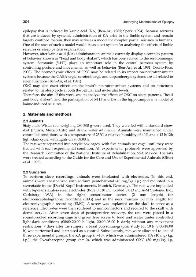
epilepsy that is induced by kainic acid (KA) (Ben-Ari, 1985; Sperk, 1994). Because seizures that are induced by systemic administration of KA arise in the limbic system and remain largely confined therein, they may serve as a model for complex partial seizures in humans. One of the uses of such a model would be as a test system for analyzing the effects of limbic seizures on sleep pattern organization. However, after kainic acid (KA) administration, animals currently display a complex pattern of behavior known as “head and body shakes”, which has been related to the serotoninergic system. Serotonin (5-HT) plays an important role in the central nervous system by controlling posture and movements, as well as behavior (Ben-Ari, et al. 1981; Osorio-Rico, 2003). The normothymic effects of OXC may be related to its impact on neurotransmitter systems because the GABA-ergic, serotoninergic and dopaminergic systems are all related to sleep functions (Ben-Ari, et al. 1981). OXC may also exert effects on the brain’s neurotransmitter systems and on structures related to the sleep cycle at both the cellular and molecular levels. Therefore, the aim of this work was to analyze the effects of OXC on sleep patterns, “head and body shakes”, and the participation of 5-HT and DA in the hippocampus in a model of kainic-induced seizures.
2. Materials and methods
2.1 Animals
Sixty male Wistar rats weighing 280-300 g were used. They were fed with a standard chow diet (Purina, México City) and drank water ad libitum. Animals were maintained under controlled conditions, with a temperature of 25°C, a relative humidity of 40% and a 12 h:12h light-dark cycle, with lights on at 08:00 h. The rats were separated into acrylic box cages, with five animals per cage, until they were treated with each experimental condition. All experimental protocols were approved by the Research Committee of the National Institute of Rehabilitation, SSA México. The rats were treated according to the Guide for the Care and Use of Experimental Animals (Olfert et al. 1993).
2.2 Surgeries
To perform sleep recordings, animals were implanted with electrodes. To this end, animals were anesthetized with sodium pentobarbital (40 mg/kg i.p.) and mounted in a stereotaxic frame (David Kopff Instruments, Munich, Germany). The rats were implanted with bipolar stainless steel electrodes (Bore 0.010 in., Coated 0.013 in., A-M Systems, Inc., Carlsborg, WA) in the right sensorimotor cortex (2 mm length) for electroencephalographic recording (EEG) and in the neck muscles (50 mm length) for electromyographic recording (EMG). A screw was implanted on the skull to serve as a reference. Electrodes were then soldered to miniconnectors and secured to the skull with dental acrylic. After seven days of postoperative recovery, the rats were placed in a soundproofed recording cage and given free access to food and water under controlled light-dark conditions (08:00-20:00 h light, 20:00-08:00 h dark) without any movement restrictions. 7 days after the surgery, a basal polysomnographic study for 10 h (8:00-18:00 h) was performed and later used as a control. Subsequently, rats were allocated to one of three experimental groups: the KA group (n=10), which was administered KA (10 mg/kg, i.p.); the Oxcarbazepine group (n=10), which was administered OXC (50 mg/kg, i.p,
www.intechopen.com
Monoamines and Sleep: Effects of Oxcarbazepine
305
dissolved in ethanol (5%) and carboxymethylcellulose ); and the OXC+KA group (n=10), which was administered KA 30 minutes after OXC-treatment. A new session of polygraphic recordings were performed for 10 h over 3 days. The rats were then returned to the animal facility, where the veterinarians would care for them.
2.3 Behavioral assessment
Three additional groups of rats that were administered either saline solution (n=7), KA (n=11) or OXC+KA (n=12) were used to assess the frequency of head and body shakes in each condition. There was no need for an OXC group since it is widely known that this substance does not elicit seizures. Head and body shakes were visually recorded for a total period of two hours (8:30-12:30 h) by an observer who was unaware of the drug treatment, according to the protocol of Altagracia et al. (1994).
2.4 Analysis of serotonin (5-HT), 5-hydroxy-indol-acetic acid (5-HIAA), dopamine (DA), noradrenaline (NA) and Homovanilic acid (HVA)
The levels of serotonin and its metabolite were studied, because KA induces seizures that are promoted by elevated levels of serotonin. It was found that pretreatment with OXC increased the levels of serotonin and its metabolite in the brain, which was consistent with the finding that OXC reduced the frequency of head and body shakes. After 4 hours of behavioral assessment, rats from all three groups were sacrificed by decapitation. The hippocampus was dissected out according to the techniques of Glowinski and Iversen (Glowinski & Iversen 1996). The tissues were immediately placed into Teflon tubes that were kept on ice, sonicated in 0.4 N of percloric acid with 0.1% (w/v) of sodium metabisulfite followed by 10 min of centrifugation at 15,000 rpm at 4°C. The supernatants were kept frozen at 70°C until chromatographic analysis. The contents of 5-HT, its metabolite 5-HIAA, and catecholamines (DA, NA and HVA), were analyzed by high-performance liquid chromatography (HPLC) with an electrochemical detector according to the protocol of Alfaro-Rodríguez et al. (2006). A Perkin-Elmer LC-250 liquid chromatograph with a metrohm electrochemical detector was used. Calibration curves for monoamines were constructed using known concentrations of standards prepared in percloric metabisulfite solution that were injected into the 20 µl loop of chromatograph. Peaks were integrated with the Perkin Elmer LC 1020 program. The concentration of indolamine in the samples was obtained by interpolation of their respective standard curves. We used an Alltech adsorbosphere catecholamine 3U analytical column (particle size= 3 µm). The mobile phase consisted of an aqueous phosphate buffer solution (0.1 M, pH 3.2) that contained 0.2 mM sodium octyl sulfate, 0.1mM of EDTA and 14% v/v methanol. The flow rate was 1.2 ml/min, and the potential was fixed at 0.80 V against an Ag/AgCl reference electrode.
2.5 Analysis of sleep and statistical analysis of results
Each of the printed polygraphic recordings was analyzed visually, according to Alfaro-Rodríguez and Gonzalez-Pina (2005). Briefly, they were classified as follows: Wakefulness (W), which was characterized by the desynchronization of the EEG and the presence of muscle tone (EMG) that was accentuated during movements; Slow Wave Sleep (SWS), which was characterized by the presence of sleep spindles, slow waves with voltage higher than 75 µV, and a decrease in EMG voltage; and Paradoxical Sleep (PS), which was
www.intechopen.com
Underlying Mechanisms of Epilepsy
306
characterized by the desynchronization of the EEG and an absence of EMG voltage. Mean duration values (mean ± S.E.M.) of each EEG state were statistically compared by a one-way analysis of variance (ANOVA), and subsequent comparisons within groups were performed using a Tukey test, with p≤ 0.01. Results from the counting of the head and body shakes were analyzed by ANOVA followed
by Dunnett’s test. Values of P<0.01 were considered to be significant.
Monoamine concentration and metabolite/neurotransmitter rate values were analyzed statistically by ANOVA, and subsequent comparison within groups were carried out by a Tukey test.
3. Results
3.1 Effects of kainic acid on sleep
Table 1 shows the results that were obtained from the sleep recordings. A single dose of KA (10 mg/kg) affected the organization of the sleep patterns. Kainic acid induced animals to stay awake for the whole initial 10 hours of electroencephalographic recording. During the follow-up on the next day, the W increased its total duration, and therefore, the SWS and PS decreased their respective total durations. The mean duration of the SWS and PS did not show any significant changes. On the second day, all parameters returned to control levels, suggesting that the rats did recovered from the effects of KA on the sleep-wake cycle 48hrs after treatment.
3.2 Effect of oxcarbazepine on sleep
OXC had a strong and immediate effect on the sleep-wake cycle. On the treatment day, the animals in the OXC group increased their sleep behavior throughout the recording period, remaining in sleep posture most of the time, only moving to eat or drink. OXC induced a significant decrease in W (21.5%), with a concomitant increase in total sleep time (SWS: 12.56% and PS: 28.7%). The sleep-wake cycle returned to their control values by day 1, and remained unchanged on day 2 (table 1).
3.3 Effect of oxcarbazepine + kainic acid on sleep
The animals treated with OXC 30 min before KA injection showed significant changes in
almost all the sleep-wake parameters that were measured. The latency of SWS increased
to 289.98 min (p<0.001) and that of PS to 304.45 min (p<0.01). There was also a significant
increase in total time of W (p<0.05). However, these changes were much less dramatic
than those observed under treatment with kainic acid alone. The present results suggested
that there was an anticonvulsive effect of OXC on the KA-induced changes in the sleep
patterns and a protective activity on seizures. During the following days of recordings,
the amount of wakefulness progressively decreased, returning to control values by day 1
(Table 1).
3.4 Effects of KA and OXC+KA on the head and body shakes
KA administration produced a significant increase in the frequencies of the head and body
shakes during the first two hours following drug administration (Table 2). Administration
of OXC 30 min prior to kainic acid treatment reduced the frequency of head and body
shakes to 42% of those of the KA group. The increase in head and body shakes observed
www.intechopen.com
Monoamines and Sleep: Effects of Oxcarbazepine
307
seemed to be related to sleep behavior. The frequency of head and body shakes in animals
began to decrease after the second hour (Table 2).
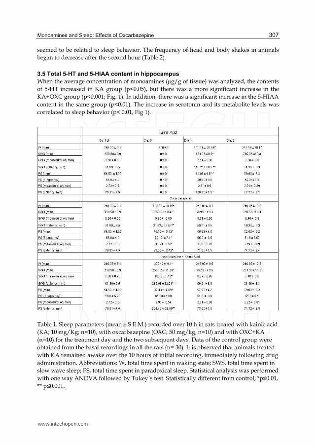
3.5 Total 5-HT and 5-HIAA content in hippocampus
When the average concentration of monoamines (µg/g of tissue) was analyzed, the contents
of 5-HT increased in KA group (p<0.05), but there was a more significant increase in the
KA+OXC group (p<0.001; Fig. 1). In addition, there was a significant increase in the 5-HIAA
content in the same group (p<0.01). The increase in serotonin and its metabolite levels was
correlated to sleep behavior (p< 0.01, Fig 1).
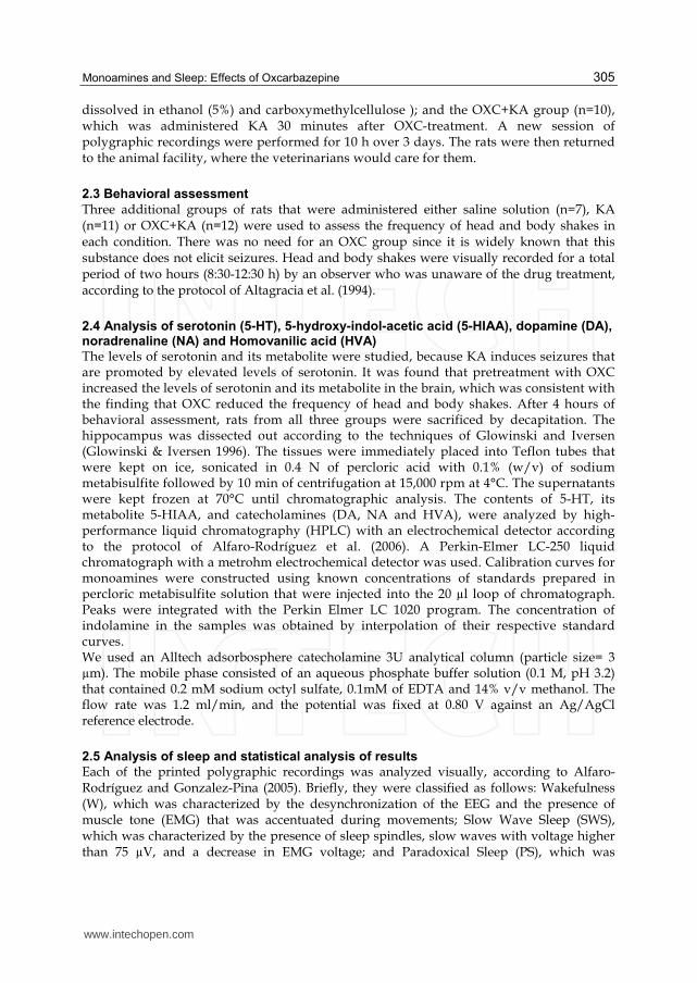
Table 1. Sleep parameters (mean ± S.E.M.) recorded over 10 h in rats treated with kainic acid
(KA; 10 mg/Kg; n=10), with oxcarbazepine (OXC; 50 mg/kg, n=10) and with OXC+KA
(n=10) for the treatment day and the two subsequent days. Data of the control group were
obtained from the basal recordings in all the rats (n= 30). It is observed that animals treated
with KA remained awake over the 10 hours of initial recording, immediately following drug
administration. Abbreviations: W, total time spent in waking state; SWS, total time spent in
slow wave sleep; PS, total time spent in paradoxical sleep. Statistical analysis was performed
with one way ANOVA followed by Tukey´s test. Statistically different from control; *p≤0.01,
** p≤0.001.
www.intechopen.com
Underlying Mechanisms of Epilepsy
308
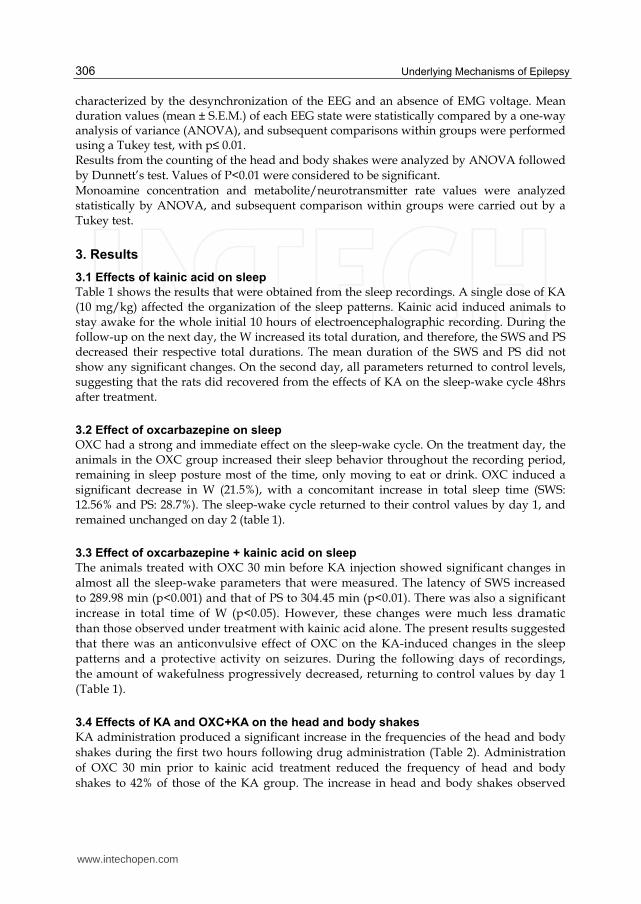
Table 2. Effects of oxcarbazepine + kainic acid on the frequency of head and body shakes induced by kainic acid. Behavioral results are expressed as mean ± S.E.M. of n=7-12 independent experiments. *P<0.05, **p<0.01, statistically different from the kainic acid group. ANOVA followed by Dunnet´s test.
Fig. 1. Total content of serotonin (5-HT) and its metabolite 5-hydroxy-indol-acetic acid (5-HIAA) in the hippocampus. There was a significant increase in the concentrations of both 5-HT and 5-HIAA in the kainic acid (KA) and oxcarbazepine /KA-treated (OXC/CA) rats, in comparison with control (C). One-way ANOVA and pos hoc Tukey test *p≤0.05 **p≤0.01.
The 5-HIAA/5-HT ratio revealed that the metabolite 5-HIAA increased more than its precursor in both, the KA alone and the OXC+KA conditions (Fig 2).
www.intechopen.com

Monoamines and Sleep: Effects of Oxcarbazepine
309
Fig. 2. 5-HIAA/5-HT ratio estimated in the hippocampus. The 5-HIAA/5-HT ratio was significantly increased in the kainic acid (KA) and oxcarbazepine /KA-treated (OXC/CA) rats compared to control (C). One-way ANOVA and pos hoc Tukey test *p≤0.05.
3.6 Total DA, NA and HVA content in hippocampus
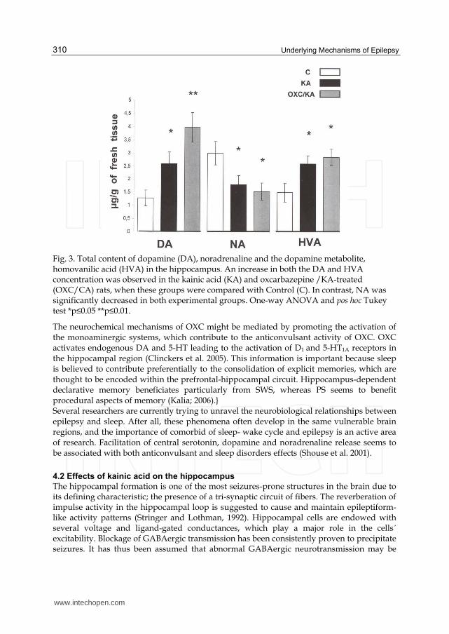
When the average concentration of monoamines (µg/g of tissue) was examined, a significant increase in DA and HVA was observed for both the KA (DA, HVA: p<0.05) and the KA+OXC groups (DA: p<0.01, HVA: p<0.05). Moreover, this increase in catecholamine levels is related to sleep behavior. By contrast, the metabolite NA decreased significantly in both the KA and KA+ OXC groups (p<0.05) (Fig. 3).
4. Discussion
4.1 Effects on sleep
As described previously in the rat model of temporal lobe epilepsy, we observed a significant KA-induced disorganization of the sleep-wakefulness cycle that involved both the SWS and PS phases (Alfaro et al. 2009). This suggests that such sleep inhibition and longtime insomnia is not only due to a physical effect of the immediate pharmacological action of KA, but also to an action that is exercised on the neurophysiological mechanisms that regulate the sleep-wakefulness cycle. It is also interesting, from a pathophysiological point of view, that the reduction of sleep that is induced by epileptic seizures did not produce a compensatory increase, as is normally observed with sleep inhibition that is produced by other means (Frank et al. 1997). Whereas our findings showed that OXC induces an increase in both sleep phases, CBZ has an inhibitory effect on PS (Alfaro-Rodríguez et al. 2002; Alfaro et al. 2009, Ayala-Guerrero et al. 2002). In contrast to CBZ, which had no effects on the sleep-wake cycle (Alfaro et al. 2009), animals treated with OXC adopted a sleep behavior through the observation period. Time sleep initiation (latency) decreased and mean duration and frequency are increased significantly in OXC treated animals. On the other hand, in OXC-pretreated animals, the frequency and duration of behavioral and electrophysiological manifestations of KA-evoked seizures decreased.
www.intechopen.com
Underlying Mechanisms of Epilepsy
310
Fig. 3. Total content of dopamine (DA), noradrenaline and the dopamine metabolite, homovanilic acid (HVA) in the hippocampus. An increase in both the DA and HVA concentration was observed in the kainic acid (KA) and oxcarbazepine /KA-treated (OXC/CA) rats, when these groups were compared with Control (C). In contrast, NA was significantly decreased in both experimental groups. One-way ANOVA and pos hoc Tukey test *p≤0.05 **p≤0.01.
The neurochemical mechanisms of OXC might be mediated by promoting the activation of the monoaminergic systems, which contribute to the anticonvulsant activity of OXC. OXC activates endogenous DA and 5-HT leading to the activation of D1 and 5-HT1A receptors in the hippocampal region (Clinckers et al. 2005). This information is important because sleep is believed to contribute preferentially to the consolidation of explicit memories, which are thought to be encoded within the prefrontal-hippocampal circuit. Hippocampus-dependent declarative memory beneficiates particularly from SWS, whereas PS seems to benefit procedural aspects of memory (Kalia; 2006).} Several researchers are currently trying to unravel the neurobiological relationships between epilepsy and sleep. After all, these phenomena often develop in the same vulnerable brain regions, and the importance of comorbid of sleep- wake cycle and epilepsy is an active area of research. Facilitation of central serotonin, dopamine and noradrenaline release seems to be associated with both anticonvulsant and sleep disorders effects (Shouse et al. 2001).
4.2 Effects of kainic acid on the hippocampus
The hippocampal formation is one of the most seizures-prone structures in the brain due to its defining characteristic; the presence of a tri-synaptic circuit of fibers. The reverberation of impulse activity in the hippocampal loop is suggested to cause and maintain epileptiform-like activity patterns (Stringer and Lothman, 1992). Hippocampal cells are endowed with several voltage and ligand-gated conductances, which play a major role in the cells´ excitability. Blockage of GABAergic transmission has been consistently proven to precipitate seizures. It has thus been assumed that abnormal GABAergic neurotransmission may be
www.intechopen.com

Monoamines and Sleep: Effects of Oxcarbazepine
311
related to epilepsy (Prince, 1978). However, in vivo epilepsy can also be induced with exposure to an agonist of glutamate receptors, in a manner that may not be directly related to GABAergic mechanisms. One such agent is kainic acid, whose induced seizures are a well-established model of temporal lobe epilepsy, and KA-induced epileptic seizures reliably occur at the systemic dose of 10mg/kg used in this study. Khazipov & Holmes (2003) implicate GABA inhibitory mechanisms in the KA- induced emergence of synchronized epileptiform-like activity patterns in the hippocampus.
4.3 Relationship between monoamines and OXC and their effects on sleep and seizures
Clinckers (2005) provided evidence that OXC and MHD led to an increase in the release of monoamines to the extracellular space. Thus, their anticonvulsant effects seem to depend upon the enhancement of endogenous DA and 5-HT transmission and the subsequent activation of D2 and 5HT1A receptors. In recent studies, OXC and its active metabolite (MHD) have been shown to exert important dopamine- and serotonin-promoting effects in the limbic area (Kovacs, 2008). Therefore, in our study, we assumed that the OXC-induced decrease in seizures and increase in sleep was mediated by serotonergic and dopaminergic system. In a previous study (Clinckers, et al. 2005), it was shown that hippocampal DA and 5-HT levels are critically involved in the anticonvulsant activity of OXC. Furthermore, this study suggested that the lack of effect after systemic administration might originate from the pharmacodynamic interactions with other brain areas which result in the suppression of hippocampal monoamine increases. In addition, it was also shown that the selective block of either the D2 receptor or the 5-HT1 receptor was able to completely abolish the anticonvulsant effects of OXC and MHD. These results indicate that activation of both receptor types is necessary for the anticonvulsant effects of OXC and MHD. We also found that 5-HT, 5-HIAA and DA were also increased, an effect possibly exerted by the alteration of the excitation-inhibition balance. In our results the increase of 5-HIAA suggests the active participation of 5-HT metabolism in the pattern of changes by KA and KA+OXC. Excitatory and inhibitory actions of DA have been reported in hippocampus (Barone, et al. 1991 Starr, 1996). High DA concentrations enhance glutamate release via D1/D5 receptor stimulation, while low concentrations reduce excitatory responses via D2
receptors. Both excitatory and inhibitory hippocampal transmission can be reduced via receptor 5-HT3 activation (Dorostkar & Boehm, 2007 ). Additionally, hippocampal 5-HT reduces glutamate release by acting on presynaptic 5-HT1A receptors (Mauler et al. 2001). During selective 5-HT1A blockade, 5-HT produces fast excitation probably mediated by 5-HT2C receptors (Beck 1992). Moreover, 5-HT inhibits GABA-ergic hippocampal interneurons via presynaptic 5-HT1A receptors (Schmitz et al.1995a). The inhibitory effect of 5-HT on glutamatergic transmission may therefore be partially counterbalanced by a 5-HT-mediated disinhibition of the principal hippocampal output cells (Schmitz et al.1998). However, in our results we found that the levels of catecholamines such as NA were decreased from the control values in both KA and KA+OXC groups. In contrast, HVA was a significantly increased in both groups. These results suggested an effect of OXC on the metabolism of catecholamines.
4.4 Effects of KA and KA/OXC on limbic seizures
It’s known that KA produces neurochemical changes in monoamines (Bourne et al. 2001).
KA also produces alterations in the complex behavioral pattern known as “limbic seizures”.
www.intechopen.com
Underlying Mechanisms of Epilepsy
312
One of the components of this pattern, the head and body shakes, is frequently associated
with the intensity of seizures (Sperk 1994), increased levels of amine metabolites for both 5-
HT and DA and the NA content after KA administration (Ben-Ari, 1981). Other studies have
demonstrated that 5HT is involved in the development of the wet dog-shakes, which is a
response in rats and some other models that involves central 5HT activity (Osorio-Rico,
2003). In these cases, the participation of 5HT that is induced by KA may cause toxic effects.
Another report has shown the participation of D2 dopamine receptors in the susceptibility of
mice to kainic acid-evoked hippocampal cell death (Bozzi et al. 2000).
In our results we found that neurochemical changes produced as a consequence of KA
administration involved increases in the levels of 5-HT, 5-HIAA and DA, HVA, and a
decrease in the levels of NA. With the addition of OXC (KA/OXC group), there was an ever
larger increase in all these systems, except for NA, which remained decreased. The
increment in 5-HT and its metabolite together with the DA increment in the KA/OXC
treatment mediate disinhibition of the principal hippocampal output cells probably induced
by MHD, the active metabolite of OXC. The anti-convulsive effects of OXC are achieved by
5HT and other neurotransmitters, such as NA and DA, which generate SWS and the first
minutes of PS. OXC significantly reduced motor seizures. The 50 mg/kg dose of OXC was
also able to diminish the kainic acid-evoked body and head shakes (47%), as has been
previously shown in studies (Landmark, 2007; Mclean et al. 1994).
These results explain why, KA-induced seizures disappear within the third hour of OXC
treatment, while in previous study using CBZ (Alfaro, et al. 2009) the seizures disappeared
six hours after administration of treatment.
Several anti-epileptic drugs such as OXC increase extracellular levels of DA and/or 5-HT in
brain areas involved in epileptogenesis (Biggs et al. 1995; Southam et al.1998; Murakami et
al. 2001). It is not clear whether these increases in monoamine levels have a direct
anticonvulsant effect, contribute to the total anticonvulsant effect, or are just a drug side-
effect.
4.5 Mechanisms of action of OXC on ionic channels
Like CBZ, OXC and MHD are considered to exert their pharmacological effects by
stabilization of Na+ channels in a voltage-, frequency-, and time-dependent manner (Mclean
et al. 1994; Malow et al. 1998). They also block high-threshold Ca2+ current (Akaike et al.
2001) and increase K+ channel conductance (Malow et al. 1998). The mechanism of action of
OXC has been proposed to differ that of CBZ by the modulation of the Ca2+ channels
(Calabresi, 1995; Wellinton & Goa, 2001), although the study by Sitges, et al. (2007) does not
show remarkable differences between the inhibition exerted by the older and newer
anticonvulsants on channel-mediated release of glutamate evoked by high K+.
The effect of OXC appears to be related to the dose and to the serum concentrations of MHD.
In general, daily fluctuations in MHD concentration are relatively slight, smaller than would
be expected from the elimination half-life of the compound. Therapeutic monitoring may help
to decide whether adverse effects are dependent on MHD concentrations (May, et al., 2003).
5. Conclusions
We, therefore, believe that the effects of OXC and MHD on hippocampal monoaminergic
transmission are contributors to the anticonvulsive effects of these compounds.
www.intechopen.com
Monoamines and Sleep: Effects of Oxcarbazepine
313
These results are clinically relevant as hippocampus plays a significant role in seizures in many diseases. These results also offer a better understanding of the mechanisms by which anticonvulsants affect the seizures along with the origin of seizures.
6. References
Akaike, K., Tanaka, H., Tojo, S., Fukumoto, S., Imamura,S. & Takigawa, M. (2001) Kainic acid-induced dorsal and ventral hippocampal seizures in rats. Brain Res. Vol. 900, No. 1, (May 2001), pp. 65-71, ISSN 0006-8993
Alfaro-Rodríguez, A., González-Piña, R., Arch-Tirado, E., Carrasco-Portugal, M., Pérez-Guillé, B., Soriano-Rosales, R. E., Padilla-Martin, K., Uribe-Escamilla, R. & Labra-Ruiz, N. (2009) Neuro-protective effects of carbamazepine on sleep patterns and head and body shakes in kainic acid-treated rats. Chem. Biol. Interact. Vol. 180, No. 3, (August, 2009), pp. 376-82, ISSN 0009-2797
Alfaro-Rodríguez, A., González-Piña, R., González-Maciel, A. & Arch-Tirado, E. (2006) Serotonin and 5-hydroxy-indole-acetic acid contents in dorsal raphe and suprachiasmatic nuclei in normal, malnourished and rehabilitated rats under 24 h of sleep deprivation. Brain Res. Vol. 1110, No. 1, (September 2006), pp. 95-101, ISSN 0006-8993
Alfaro-Rodríguez, A. & González-Piña, R. (2005) Ozone-induced paradoxical sleep decrease is related to diminished acetylcholine levels in the medial preoptic area in rats. Chem. Biol. Interact. Vol. 151, No. 3, (February 2005), pp. 151-158, ISSN 0009-2797
Alfaro-Rodríguez, A., Labra-Ruiz, N., Carrasco-Portugal, M., Gonzalez-Maciel, A., Perez-Guille, B &, Soriano-Rosales, R. (2002) Effect of Carbamazepine on sleep patterns disturbed by epilepsy. Proc. West. Pharmacol. Soc. Vol. 45, (December 2002), pp. 62-64, ISSN 0083-8969
Altagracia, M., Kravzov, J., Santamaría, A., Ríos, C., Ordaz, H. & Gonzalez, L. (1994) Dapsone administration prevents quinolinate-induced neurotoxicity in rats. Proc. West. Pharmacol. Soc. Vol.37, (December 1994), pp. 63, ISSN 0083-8969
Ayala-Guerrero, F., Alfaro-Rodríguez, A., Martínez, C., Campos-Sepúlveda, E., Vargas, L. & Mexicano G. (2002) Effect of kainic acid-induced seizures on sleep patterns. Proc. West. Pharmacol. Soc. Vol 45, (December 2002) pp. 178-180, ISSN 0083-8969
Barone, P., Palma, V., DeBartolomeis, A., Tedeschi, E., Muscettola, G. & Campanella, G. (1991) Dopamine D1 and D2 receptors mediate opposite functions in seizures induced by lithium-pilocarpine. Eur. J. Pharmacol. Vol. 195, No. 1, (March 1991), pp. 157-62, ISSN 0014-2999
Bazil, C. W., Castro, L. H. & Walczakt, T. S. (2000) Reduction of rapid eye movement sleep by diurnal and nocturnal seizures in temporal lobe epilepsy. Arch Neurol. Vol. 57, No. 3, (March 2000), pp. 363-368, ISSN 0003-9942
Bazil, C. W. & Walczak, T. S. (1997) Effects of sleep and sleep stage on epileptic and nonepileptic seizures. Epilepsia. Vol. 38, No. 1, (January 1997), pp. 56-62, ISSN 1528-1167
Beck, S. G. (1992) 5-Hydroxytryptamine increases excitability of CA1 hippocampal pyramidal cells. Synapse, Vol. 10, No. 4, (April 1992), pp. 334-340, ISSN 1098-2396
Bedard, P. & Pycock, C. J. (1977) “Wet-dog” shakes behaviour in the rat: a possible quantitative model of central 5-hydroxytryptamine activity. Neuropharmacology, Vol. 16, No. 10, (October 1977), pp. 663-670, ISSN 0028-3908
www.intechopen.com

Underlying Mechanisms of Epilepsy
314
Ben-Ari, Y. (1985) Limbic seizure and brain damage produced by kainic acid: mechanisms and relevance to human temporal lobe epilepsy. Neuroscience, Vol. 14, No. 2, (February 1985), pp. 375-403, ISSN 0306-4522
Ben-Ari, Y., Tremblay, E., Riche, D., Ghilini, G. & Naquel, R. (1981) Electroencephalographic, clinical and pathological alterations following systemic administration of kainic acid, bicuculine or pentetrazole: metabolic mapping using the deoxyglucose method with special reference to the pathology of epilepsy. Neuroscience, Vol. 6, No. 7, pp. 1361-1391, ISSN 0306-4522
Clinckers, R., Smolders, I., Meurs, A., Ebinger, G. & Michotte, Y. (2005). Hippocampal dopamine and serotonin elevations as pharmacodynamic markers for the anticonvulsant efficacy of oxcarbazepine and 10,11-dihydro-10-hydroxycarbamazepine. Neurosci. Lett., Vol. 390, No. 1, (December 2005 ), pp. 48-53, ISSN 0304-3940
Clinckers, R., Smolders, I., Meurs, A., Ebinger, G. & Michotte, Y. (2004) Anticonvulsant action of hippocampal dopamine and serotonin is independently mediated by D and 5-HT receptors. J. Neurochem., Vol. 89, No. 4, (May 2004), pp. 834-43, ISSN 1471-4159.
Crespel, A., Coubes P. & Baldy-Molulinier, M. (2000) Sleep influence on seizures and epilepsy effects on sleep in parcial frontal and temporal lobe epilepsias. Clin. Neurophysiol., Vol. 111, No. Suppl 2, (September 2000), pp. S54-59, ISSN 0736-0258
Degen, R. & Rodin, E. A. (1991) Epilepsy, sleep and sleep deprivation (Eds.) Amsterdam, Elsevier 1991.
Dorostkar, M. M. & Boehm, S. (2007) Opposite effects of presynaptic 5-HT3 receptor activation on spontaneous and action potential-evoked GABA release at hippocampal synapses. J. Neurochem., Vol 100, No. 2, (January 2007), pp. 395-405, ISSN 1471-4159.
Frank, M. G., Page, J. & Helle, H. C. (1997) The effects of REM sleep-inhibiting drugs in neonatal rats: evidence for a distinction between neonatal active sleep and REM sleep. Brain Res., Vol 778, No. 1, (December 1997), pp. 64-72, ISSN 0006-8993
Gigli, G. L., Placidini, F., Diomedi, M., Maschio, M., Silvestre, G., Scalise, A. & Marciani, M. G. (1997) Nocturnal sleep and daytime somnolence in untreated patiens with temporal lobe epilepsy: Changes after treatment with controlled-release carbamazepine. Epilepsia, Vol. 38, No. 6, (June 1997), pp. 696-701, ISSN 1528-1167
Glowinski, J. & Iversen, L. L. (1966) Regional studies of catecholamines in the rat brain. I. The disposition of (3H)norepinephrine, (3H)dopamine and (3H)dopa in various regions of the brain. J. Neurochem., Vol. 13, No. 8, (August 1966), pp. 655-669, ISSN 1470-4159
Grabenstatter, H. L., Clark, S. & Dudet, F. E. (2007) Anticonvulsivant effects of carbamazepine on spontaneous seizures in rats with kainite-induced epilepsy: comparison of intraperitoneal injections with drug-in-food protocols. Epilepsia, Vol. 48, No. 12, (December 2007), pp. 2287-2295, ISSN 1528-1167
Halász, P. (1984) Sleep arousal and electroclinical manifestations of generalized epilepsy with spike wave pattern. In: Degen, R., Niedermeyer, E., (Eds.) Epilepsy, sleep and sleep deprivation. Amsterdam, Elsevier, pp. 97-107.
Landmark, C.J. (2007) Targets for antiepileptic drugs in the synapse. Med. Sci. Monit., Vol 13, No. 1, (January 2007), pp. RA1-7, ISSN 1234-1010
www.intechopen.com
Monoamines and Sleep: Effects of Oxcarbazepine
315
Kalia, M. (2006) Neurolobiology of sleep. Metabolism, Vol. 55, No. 10Suppl 2, (October 2006), pp. S2-6, ISSN 0026-0495
Kalis, M. M. & Huff, N.A. (2001) Oxcarbazepine, an antiepileptic agent. Clinical therapeutics, Vol. 23, No. 5, (May 2001), pp. 680-700, ISSN 0149-2918
Khazipov, R. & Holmes, G. L. (2003) Synchronization of kainate-induced epileptic activity via GABAergic inhibition in the superfused rat hippocampus in vivo. J. Neurosci., Vol. 23, No. 12, (June 2003), pp. 5337-41, ISSN 0270-6474
Lerma, J., Paternain, A. V., Rodríguez-Moreno, A. & Lopez Garcia, J. C. (2001) Molecular physiology of Kainate receptors. Physiol. Rev., Vol. 81, (July 2001), pp. 971-978, ISSN 0031-9333
Lothman, E. W. & Collins, R. C. (1981) Kainic acid induced limbic seizures: metabolic, behavioral, electroencephalographic and neuropathological correlates. Brain Res., Vol. 218, No. 1-2, (August, 1981), pp. 299-318, ISSN 0006-8993
Malow, B. A., Lin, X., Kushwaha, R. & Aldrich, M. S. (1998) Interictal spiking increases with sleep depth in temporal lobe epilepsy. Epilepsia, Vol. 39, No. 12, (December 1998), pp. 1309-1316, ISSN 1528-1167
Mauler, F., Fahrig, T., Horvath, E. & Jork, R. (2001) Inhibition of evoked glutamate release by the neuroprotective 5-HT (1 A) receptor agonist BAY x 3702 in vitro and in vivo. Brain Res., Vol. 888, No. 1, (January 2001), pp. 150-157, ISSN 0006-8993
May, T. W., Korn-Merker, E. & Rambeck, B. (2003) Clinical pharmacokinetics of oxcarbazepine. Clin. Pharmakinet., Vol. 42, No. 12, (December 2003), pp. 1023-1042, ISSN 0312-5963
McLean, M. J., Schmutz, M. & Wamil, A. W. (1994) Oxcarbazepine: Mechanism of action. Epilepsia, Vol. 35, No. Suppl. 3, (June 1994), pp. S5-9, ISSN 1528-1167
Shin, C. & McNamara, J. O. 1994. Mechanism of epilepsy. Annu. Rev. Med., Vol. 45, (February 1994), pp. 379-389.
Min, M. Y., Meyland, Z. & Kullmann, D. M. (1999) Synaptically released glutamate reduces gamma-aminobutyric acid (GABA)ergic inhibition in the hippocampus via kainate receptors. Proc. Natl. Acad. Sci. U. S. A., Vol. 96, No. 17, (August 1999), pp. 9932-9937, ISSN 1091-6490
Olfert, E. D., Cross, B. M. & McWilliam, A. A. (1993) Guide for the care and use of experimental animals. Can. Council Anim. Care 1-211.
Osorio-Rico, L., Mancera-Flores, M. & Ríos, C. (2003) Changes in brain serotonin turnover, body and head shakes in kainic acid-treated rats. Pharmacol and Toxicol., Vol. 92, No. 3, (March 2003), pp. 143-147, ISSN 0901-9928
Placidi, F., Marciani, M. G., Diomedi, M., Sauri, F., Giacomini, P. & Gigli, G. L. (2000) Effects of lamotrogine on nocturnal sleep, daytime somnolence and cognitive functions in focal epilepsy. Acta Neurol. Scand., Vol. 102, No. 2, (August 2000), pp. 81-86, ISSN 1600-0404
Prince, D. A. (1978) Neurophysiology of epilepsy. Annu. Rev. Neurosci., Vol. 1, (March 1978), pp. 395-415, ISSN 0325-0395
Roberts, R. (1998) Differential diagnosis of sleep disorders, non-epileptic attacks and epileptic seizures. Curr. Opin. Neurol., Vol. 11, No. 2, (april 1998), pp. 135-139, ISSN 1350-7540
Sammaritano, M. & Sherwin, A. (2000) Effect of anticonvulsants on sleep. Neurology., Vol. 54, No 5 Suppl 1 ,(March 14, 2000), S16-24, ISSN 0028-3878
www.intechopen.com
Underlying Mechanisms of Epilepsy
316
Schliebs, R., Zilvin, M., Steinbach, J. & Rothe, T. (1989) Changes in cholinergic but not in GABAergic markers in amygdale, piriform cortex and nucleus basalis of the rat brain following systemic administration of kainic acid. J. Neurochem., Vol. 53, No. 1, (July 1989), pp. 212-218, ISSN 1471-4159
Shouse, M. N., Staba, R. J., Saquib, S. F &, Farber, P. R. (2001) Long-lasting effects of feline amygdala kindling on monoamines, seizures and sleep. Brain Res., Vol. 892, No. 1, (February 2001), pp. 147-65, ISSN 0006-8993
Schmitz, D., Empeson, R. M. & Heinemann, U. (1995) Serotonin reduces inhibition via 5-HT1A receptors in area CA1 of rat hippocampal slices in vitro. J. Neurosci., Vol. 15, No. 11, (November 1995), pp. 7217-7225, ISSN 0270-6474
Schmitz, D., Gloveli, T., Empeson, R. M. & Heinemann, U. (1998) Comparison of the effects of serotonin in the hippocampus and the entorhinal cortex. Mol. Neurobiol., Vol. 17, No. 1-3, (December 1998), pp. 59-72, ISSN 0893-7648
Sitges, M., Guarneros, A. & Nekrassov V. (2007) Effects of carbamazepine, phenytoin, valproic acid, oxcarbazepine, lamotrogine, topiramate and vinpocetine on the presynaptic Ca2+ channel-mediated release of (3H) glutamate: Comparison with the Na+ channel-mediated release. Neuropharmacology, Vol. 53, No. 7, (December 2007), pp. 854-862, ISSN 0028-3908
Sloviter, R. S. & Damiano, B. P. (1981) Sustained electrical stimulation of the perforant path duplicates kainite induced electrophysiologic effects and hippocampal damage in rats. Neurosci. Lett., Vol. 24, No. 3), (July 1981), pp. 229-284, ISSN 0304-3940
Sperk, G. (1994) Kainic acid seizures in the rat. Prog. Neurobiol., Vol. 42, No. 1, (August 1994), pp. 1-3,. ISSN 0301-0082
Stewart, R. M., Growdon, J. H., Cancian, D. & Baldessarini, R. J. (1976) 5-hydroxitryptophan-induced myoclonus: increased sensitivity to serotonin after intracranial 5,7-dihydroxytryptamine in the adult rat. Neuropharmacol., Vol. 15, No. 8, (August 1976), pp. 449-455, ISSN 0028-3908
Stringer, J. L. & Lothman, E. W. (1992) Reverberatory seizure discharges in hippocampal-parahippocampal circuits. Exp. Neurol., Vol. 116, No. 2, (May 1992), pp. 198-203, ISSN 0014-4886
Tidwell, A. & Swims, M. (2003) Review of the newer Antiepileptic drugs. Am. J. Manag. Care, Vol. 9, No. 3, (March 2003), pp. 253-276, ISSN 1936-2692
Wang, L., Zuo, C. H., Zhao, D. Y. & Wu, X. R. (2000) Brain distribution and efficacy of carbamazepine in kainic acid induced seizure in rats. Brain Dev., Vol. 22, No. 3, (May 2000), pp. 154-157, ISSN 0387-7604
Wójtowicz, A. M., van den Boom, L., Chakrabarty, A., Maggio, N., Haq, R. U., Behrens, C. J. & Heinemann, U. (2009) Monoamines block kainate- and carbachol-induced gamma-oscillations but augment stimulus-induced gamma-oscillations in rat hippocampus in vitro. Hippocampus, Vol. 19, No. 3, (March 2009), pp. 273-288, ISSN 1098-1063
www.intechopen.com
Underlying Mechanisms of EpilepsyEdited by Prof. Fatima Shad Kaneez
ISBN 978-953-307-765-9Hard cover, 354 pagesPublisher InTechPublished online 26, September, 2011Published in print edition September, 2011
InTech EuropeUniversity Campus STeP Ri Slavka Krautzeka 83/A 51000 Rijeka, Croatia Phone: +385 (51) 770 447 Fax: +385 (51) 686 166www.intechopen.com
InTech ChinaUnit 405, Office Block, Hotel Equatorial Shanghai No.65, Yan An Road (West), Shanghai, 200040, China
Phone: +86-21-62489820 Fax: +86-21-62489821
This book is a very provocative and interesting addition to the literature on Epilepsy. It offers a lot of appealingand stimulating work to offer food of thought to the readers from different disciplines. Around 5% of the totalworld population have seizures but only 0.9% is diagnosed with epilepsy, so it is very important to understandthe differences between seizures and epilepsy, and also to identify the factors responsible for its etiology so asto have more effective therapeutic regime. In this book we have twenty chapters ranging from causes andunderlying mechanisms to the treatment and side effects of epilepsy. This book contains a variety of chapterswhich will stimulate the readers to think about the complex interplay of epigenetics and epilepsy.
How to referenceIn order to correctly reference this scholarly work, feel free to copy and paste the following:
Alfonso Alfaro-Rodriguez, Emilio Arch-Tirado and Rigoberto Gonza lez-Pin a (2011). Monoamines and Sleep:Effects of Oxcarbazepine, Underlying Mechanisms of Epilepsy, Prof. Fatima Shad Kaneez (Ed.), ISBN: 978-953-307-765-9, InTech, Available from: http://www.intechopen.com/books/underlying-mechanisms-of-epilepsy/monoamines-and-sleep-effects-of-oxcarbazepine


![Fire Protection Plan/Fuel Management Plan [TPM 20747] … · 2014. 5. 10. · modeling data, on-site vegetation, access, surrounding area fuel conditions, slope, and worst-case weather](https://img.pdfslide.us/doc/110x75/60e8e1025facf035c43ab6f2/fire-protection-planfuel-management-plan-tpm-20747-2014-5-10-modeling-data.jpg)


![imageserv11.team-logic.com...ONETOUCH TEST STRIPS; ULTRA, VERIO ONEXTON OPSUMIT ORACEA ORFADIN ORTHOVISC [INJ] oseltamivir OTEZLA OTOVEL OTREXUP [INJ] OVIDREL [INJ] oxcarbazepine oxybutynin](https://img.pdfslide.us/doc/110x75/604de3e06a6bae6edd6eaa2d/-onetouch-test-strips-ultra-verio-onexton-opsumit-oracea-orfadin-orthovisc.jpg)