Embed Size (px)
Citation preview
1086 30 April 1966 Congenital Clubfoot-Shaw IB:MRLH
It is surprising that, except in Brockman's (1930) mono-graph, none of the communications on the results of primarymanagement of clubfoot present a statistical analysis. Thosesurgeons who use the Denis Browne splint or a modificationclaim to have very gratifying results (Browne, 1937, 1956 ; Belland Grice, 1944; Thomson, 1949). However, in a recent sur-vey by McCauley, Lusskin, and Bromley (1964), these workersstated that: " Because of the development of flat-foot followingtreatment in some of the patients we question the use of theDenis Browne splint as a corrective device."
In the light of the present findings it is suggested that thenumerous modifications which have been devised for the DenisBrowne splint (Thomson, 1942, 1949; Bell and Grice, 1944)disclose the discontent with the results obtained by its use.There is a paucity of statistical evidence in the literature onthe efficiency of the splint. Its virtues are much vaunted, itsweaknesses concealed.The statistics of the present survey on the treatment by
stretching and Robert Jones's strapping compare favourablywith those of Brockman (1930), who used a similar method:in 73 patients 50.6% required only manipulation and strappingto cure the deformity; 20.5% also had division of the tendocalcaneus with plaster splintage; 24.6% were not cured byeither of these methods and required some form of openoperation; 4.1 % still retained this deformity after all thesemethods and an operation on bone was performed. His laterdespondency (Brockman, 1937) was hardly warranted by thequality of his results.More recently Thomas (1964) claims that 90% of patients
can be cured by simple manipulation followed by splinting, ifthe baby is treated daily as soon after birth as possible.The evidence presented here leaves no doubt that the success-
ful treatment of talipes equinovarus must begin at birth.Though it is not invariably successful, repeated gentle
stretching and the application of adhesive strapping to main-tain correction is a more satisfactory technique than either theDenis Browne splint or serial plaster-of-Paris casts. We have
been too easily persuaded to relinquish Robert Jones's methodof treatment, which, apart from its high rate of success, hasthe merit of simplicity and the additional appeal of beingold-fashioned.
SummaryThree different methods for the primary treatment of con-
genital clubfoot have been compared. These methods were theDenis Browne splint, stretching and adhesive strapping, andserial plaster-of-Paris casts. There were 140 patients in theseries and 221 feet were affected. Only 20 (19%) of 105 feetwere successfully treated by the Denis Browne method alone,whereas 68 feet out of 96 (71%) were treated successfully bystretching and strapping. None of the 20 feet treated bymanipulation and serial plaster-of-Paris casts could be regardedas corrected. These results show that splintage alone is of littlevalue for the correction of clubfoot deformity and that correc-tion followed by fixation with adhesive strapping is far moreeffective.
I owe my gratitude to Mr. G. C. Lloyd-Roberts and Mr. H. H.Nixon for their encouragement and help during this survey of theirpatients. My thanks are also due to Mr. R. A. Denham, of theRoyal Hospital, Portsmouth, for his generosity in allowing me toreview the patients under his care.
REFERENCES
Bell, J. F., and Grice, D. S. (1944). 7. Bone 7t. Surg., 26, 799.Brockman, E. P. (1930). Congenital Club-Foot. Wright, Bristol.
(1937). Brit. med. 7., 2, 572.Browne, D. (1937). Ibid., 2, 570.
(1956). Clinfcal Orthopaedics-No. 8. Lippincott, Philadelphia.Elnslie, R. C. (1920). 7. orthop. Surg., 28, 668.Jones, R. (1894). Trans. Brit. orthop. Soc., 1, 20.
and Lovett, R. W. (1929). Orthopaedic Surgery, 2nd ed. OxfordUniversity Press, London.
McCauley, J., Lusskin, R., and Bromley, J. (1964). 7. Bone 7t. Surg.,46A, 525.
Thomas, G. E. (1964). Ibid., 46B, 792.Thomson, S. A. (1942). Ibid., 24, 291.
(1949). Ibid., 31A, 431.
Hepatic Dysfunction in the Congenital Rubella Syndrome
GILLES R. G. MONIF,* M.D.; RICHARD ASOFSKY,* M.D.; JOHN L. SEVER,* M.D., PH.D.
Brit. med. j., 1966, 1, 1086-1088
The congenital rubella syndrome once defined a morphologicallystatic condition: specific malformations which were the sequelaeof maternal infection with rubella virus early in gestation.It is now apparent that this term is conceptually limited, andmust be expanded to include an " acute " component due tocontinued virus action in the neonatal period and beyond(Alford et al., 1964; Monif et al., 1965). Thrombocytopenia,encephalitis, interstitial pneumonia, myocardial injury, andhepatitis have all recently been described in infants with therubella syndrome (Rudolph et al., 1965 ; Korones et al., 1965;Hardy et al., 1966). It is the purpose of this paper tocharacterize the hepatitis more completely.
Materials and Methods
Six cases of congenital rubella syndrome, confirmed by virusisolation from either throat or urine specimens, were identifiedin which, in addition to the classical stigmata of the rubella
syndrome, there was evidence of hepatic dysfunction. Two ofthe six infants with biochemical evidence of hepatic dysfunctioncame to necropsy. In addition to the review of the clinicaldata and histological analysis, the livers from these two infantsand a percutaneous liver biopsy specimen from a third infantwere studied for the recovery of virus.The two livers were homogenized in Hanks's balanced salt
solution containing penicillin, 400 units/ml., and streptomycin,0.4 mg./ml.; and then the homogenate was diluted to a 20%suspension. Each specimen was then centrifuged at 600 g. for15 minutes, and 0.2-ml. aliquots of the supernatant wereinoculated into four primary African green monkey kidneycultures in roller tubes. The cultures were maintained with1.5 ml. of medium consisting of Eagle's basal medium con-
* From the Section on Infectious Diseases, Perinatal Research Branch,National Institute of Neurological Diseases and Blindness, and theLaboratory of Infectious Diseases, National Institutes of Allergy andInfectious Diseases, National Institutes of Health, Bethesda,Maryland.

3109 A 196 '
antibiotics and 02% SWS rabbit antisra accoringto the procedures previously described (Sever -et a., 1962). Thecult~res- were incubited at 370 C. in a stationary positionfor eight days, at which time the medsiumwas removed andfresh medium aded. Two tissvpe paltures f ach set Of fourtubes -were inoculated 24 hours latr withI 100 TOlD50 ofCoxsackie A-9. Th remaining two cultures were allowe4 toincubate for an additional four days, after which 0.2 ml./tube ofthe fluid and cells were Inoculated into four additional primaryAfrican green monkey, kidney - cultures. The challenge con-sisted of 100 TCID50 of Coxsackie A-9, injected eight days laterinto each of these two tubes. All specimens were carried in thismanner through three passages. /
Interferencewith the cytopatbic effect sen with CoxsackieA-9 was interpreted as suggesting the presen of rula vus.
Interfering agents were identified-by neutralization with specificrubella antisehim in primary African green monkey iAidneycultuies, utilizing the method previously reported (Schiff, et a.,
1963). The frants of iver obtained by percutaneous punchbiopsy were tensed out with glass needles and then placeddirectly on to a sheet of primary African green mbokey kidney
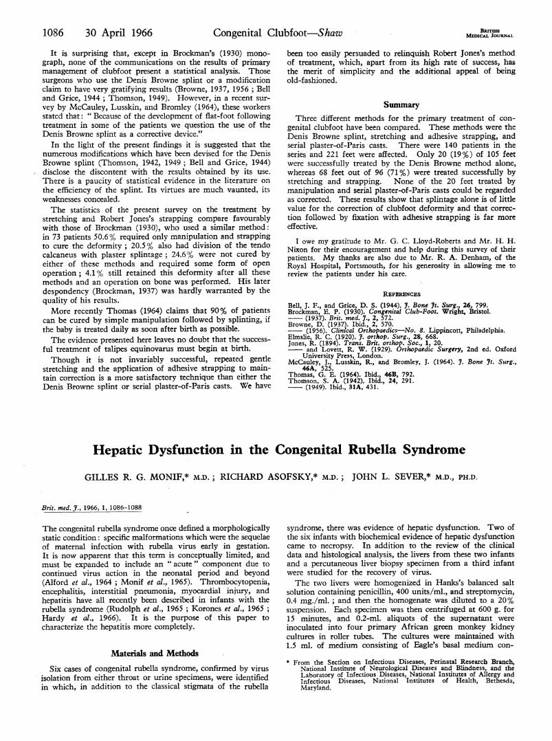
FIG. 1.-Swollen Kupifer cells engorged with bile pigment. (H. andE. X160.)
iels. After'sevendays 0.2 ni /tub of ih fluid andl: ell .i1 oculated or green
culturroe tubes. The subu ing of thesecwas--as described above.
Re sults
Hepati Function.-The clinical and biochemical data bctthe six cases are sumnrized the Table. Jaundice was nodwithin the first 24 hours in th'ree cases, and in all cases iti
48 hours. The livers were palpable 3-5 cm. and the sph
1-5-cm. -below flte costal margin. Hepatosplenomegaly pesduring the period of overt hepatic dysfunction. Clay-colo
stools were observed for Cases 1 and 6. After prmin
studies had excluded Rh.-ABO ilcompatibilities, nenatalcongenital syphilis, toxoplasmosis, systemic herpetic infection,and, in Cases 1, 3, and 4i infection with salivary-glandvrthe, clinical diagnosis was congenital biliary atresia or hepatitof unknown aetiology.-Histologye-The livers" in Cases 4 and 5 were similar. Th
most striking feature was swelling and engorgement of
in both cases careful dissection of the extrahepatic biliary ducts ha4,!-ccordig to the, available protocols, failed to demonatrate-;'
evidence of obstruction. -
FiG. 2.-Ductular bile sta"s. (H. 'and
Biochemical Indices of Hepatic Funcion '
.*h -°< X4it-S -r t?B x 63t) -24
e;. f t
.\.,9.
< Z
o-
casel- Case2 Case3 Case4 Case5 Cu. ee6Days 1-4 .. Totalbilirbin 24 (2), Bilirubi, 76/I27 (1), S.G.O.T. 82 (4); t(l); total bilirubin Total bilirubin 10*01),
19 (3) 17 3/7-5 (3) S.G.P.T. 57 (4) 8&0 (2), 10-0 (4) 7-8/5-0 (3);* alk.-
S.G.O.T. 130(YDays 5-7 .. .. Bilirubin 102/5-8 (7) Bilirubin 68/4 0 (5), Bilitubin 14 5/5 1 (6)Bilirubin 11822 (6), Total bilirubin 9-6 (5) Total bilirubin 144(O10-8/3-0 (7)
Second week .. - - Bilirubin 8-9/4-6 Bilirubiii 72/296; Bilirubin 4-6/46 Bilirubin 8'315*8;S.G.0.T. 179 S.G.O.T. 155,Third , . .. _ Bilirubin 2-9/0-9; Total bilirubin 5 3* Bilirubin 5 3/3-1 .Billrubin 4.0/5-3, 4 3/; Bilirubin 45/2 2 ;C.F.S.G.O.T. 56; .C.F. S.G.O.T. 141; 4 6; S.G.O.T. 760, +2;- S.G.O.T. 3
neg.; prothrombin S.G.P.T. 67 chol- 640;,S.G.P.T. 400,.time normal steol ; .FS.F. 270- T.S.P.T5-9. ~~~~~~~~~~~6-7WAI 3-3/3 4) (A/6; 3 8i2 i)
Fourth Bilirubin 6-1/5-9 ; C.F. neg. _ Bilirubin 4-5/1-5 _ Bilirubin 4012,.S.G*0.T. 89, 170; S.G.0.T. 92C.F. +2; -T.T.ner. T b 04Pginaldcier n - Bilirubin 5175 T05Tot bilubin04(69 (Non-icteric at time 6f (Died on 31st day of (Died on 22nd day of Bilirubin 1-8/0-9*days); prot bi days); S.G.O.T. 49 discharge-39 days) life.) Rubella virus ife.) ubella virus S.G.O.T. 70; Ctime normal; rblla recovered fromliver reovered from liver +2 (28 day)virus recoveredfom
liver biopsy (65 days)
* Exchange tansfio both mother and baby 0 Rh +, Coomba positive or z4egative.t Exchange ansfusion. both mother and baby 0 Rh+, Coomba positive or negative..
1088 30 April 1966 Congenital, Rubella Syndrome-Monif et al. BDCUmKupffer cells with green pigment (Fig. 1). This was mostprominent in areas of apparent drop-out of parenchymal cells.The pigment ranged from finely to very coarsely granular.It gave a positive Stein test and a negative Gmelin reaction;it stained with methylene blue but not with iron stains, anddid not fluoresce. There were, in addition, foci of canalicularand ductular bile stasis (Fig. 2). Bile ductules were apparentlyreduced in number. Zones of haematopoeisis were present inboth livers; in Case 5 there were many blast-like cells in theseareas. There was a slight increase in periportal fibrous tissuein both cases.
Virology.-Three livers were studied for the presence ofrubella virus. Recovery' and specific identification of the viruswas achieved in all three of the cases studied.
Comment
While the biochemical changes were primarily related toelevations of indirect bilirubin, abnormally high values for thetransaminases were recorded in Cases 5 and 6. Late in thecourse of the disease there was a' reversal of the albumin-globulin ratio in Case 3.Two clinical patterns were present. Either there was gradual
biochemical and clinical amelioration, to normal values andstatus, or a chronic indolent course was pursued associatedwith persistent jaundice and biochemical derangement. Thelivers at necropsy in Cases 4 and 5 revealed the histologicalcorollary of this latter clinical observation when observed at22 and 34 days of age. The presence of increased periportaifibrous tissue which extends into the lobules at 34 days suggeststhat this process may progress and indeed be one of the aetio-logies for juvenile cirrhosis. Almost equally important are thepercutaneous liver biopsy specimens obtained in Cases 2 and 6,which revealed normal hepatic architecture coinciding with thereturn to normal of biochemical indices measuring liverfunction (F. Fuste, personal communication, 1965). Withrespect to the unusual histological picture, it is noteworthythat Beard (1956) described a very similar finding in whatappears to'be a' case of transplacental hepatitis except that Beardreported considerably more necrosis than we have observed.The infants cited (Cases 1-6) were the products of full-term
pregnancies with a range of 38-41 weeks' gestation. Cases 1,2, and 4 weighed less than 2,500 g. The presence of markedbilirubin elevation on the first or second day of life indicatedan abnormal event superimposed on prematurity. The serumglutamine oxaloacetic transaminase (S.G.O.T.) in Cases 5 and 6was abnormally elevated. However, the differential diagnosiswas complicated owing to the haemolytic process associatedwith the rubella syndrome (Zinkham and Medearis, 1966).Nucleated red cells and reticulocytosis were present in Cases 2and 4. Serial haematocrit readings for both groups wereinvariably at the lower limits of normal values for a given age.Additional evidence of haemolysis was furnished by the con-tinued erythropoiesis in lung, liver, and spleen well beyond
that time when it normally disappears. These findings arepresent in most cases of the congenital rubella syndrome(Korones et al., 1965; Hardy et al., 1966); and unpublisheddata). The critical anatomical and biochemical differentiationwas between haemolytic disease of the newborn associated withprolonged " obstructive jaundice" and a cholangitic type ofhepatitis of viral origin. While Zuelzer and Brown (1961)have observed normally elevated S.G.O.T. values in the formercondition, the early elevation of these values in Case 6 suggeststhat they were the result of the aetiological process rather thanthe consequence of a secondary effect.
Rubella virus was isolated from the liver when there wasbiochemical and/or histological evidence of dysfunction. Thiswas in contrast to our past experience with three rubella syn-drome infants who died in the newborn period (Monif et al.,1965). In these cases virus was recovered from only one of fivelivers studied, and there. was no evidence of biochemical orhistological hepatic dysfunction. The incidence of recovery ofrubella virus from livers with apparent derangement whencoupled with a characteristic histological picture favours thehypothesis that this is a hepatitis of viral origin.
Summary
Six cases are described in which hepatic dysfunction wasassociated with the classical finding of the congenital rubellasyndrome. Necropsy liver specimens and a liver biopsy fromthree of these cases when studied by histological and virusisolation techniques revealed a correlation between hepaticderangement and recovery of the virus. One of the cases atnecropsy had histological changes suggestive of early cirrhosis.
We are indebted to Drs. Marvin Kuschner and Leon Sokolofffor their review of the histological material and for their constructivecriticism. A debt of gratitude must be expressed to Drs. SheldonB. Korones, Gordon B. Avery, and Janet Hardy for making thisstudy possible; to Drs. F. Fuste, D. Kamala, and B. Ruebner forsupplying tissue blocks for histological sections; and to Betty J.Sanders and Barbara G. Mance for technical assistance.
REFERENCES
Alford, C. A., Neva, F. A., and Weller, T. H. (1964). New Engl. 7.Med., 271, 1275.
Monif, G. R. G., Avery, G. B., Korones, S. B., and Sever, J. L. (1965).Lancet, 1, 723.
- Hardy, J. B., and Sever, J. L. (1966). Bull. 7ohns Hopk. Hosp.,118, 85.
Rudolph, A. J., Yow, M. D., Phillips, C. A., Desmond, M. M., Blattner,R. J., and Melnick, J. L. (1965). 7. Amer. med. Ass., 191, 843.
Korones, S. B., Ainger, L. E., Monif, G. R. G., Roane, J., Sever, J. L.,and Fuste, F. (1965). 7. Pediat., 67, 166.
Schiff, G. M., Sever, J. L., and 1{uebner, R. J. (1963). Science, 142, 58.Sever, J. L., Schiff, G. M., and Traub, R. G. (1962). 7. Amer. med. Ass.,
182, 663.Beard, A. G. (1956). 7. Pediat., 49, 454.Zinkham, W. H., and Medearis, D. N. (1966). 7. Pediat. In press.Zuelzer, W. W., and Brown, A. K. (1961). Amer. 7. Dis. Child., 101, 87.
![Treatment Of Congenital Clubfoot Using The Ponseti · PDF fileIris Lohan Treatment Of Congenital Clubfoot Using The Ponseti Method Workshop Manual [2nd Edition]](https://img.pdfslide.us/doc/110x75/5a9d8bd67f8b9a21688c2ac8/treatment-of-congenital-clubfoot-using-the-ponseti-lohan-treatment-of-congenital.jpg)


![Clubfoot: Ponseti Management [Italian]](https://img.pdfslide.us/doc/110x75/613d460c736caf36b75b61e2/clubfoot-ponseti-management-italian.jpg)