Embed Size (px)
Citation preview

10.4 Special SensesSmell – olfactory organsTaste – taste budsHearing/Equilibriuim – EarsSight – eyesSpidey Sense

10.5 Sense of Smell
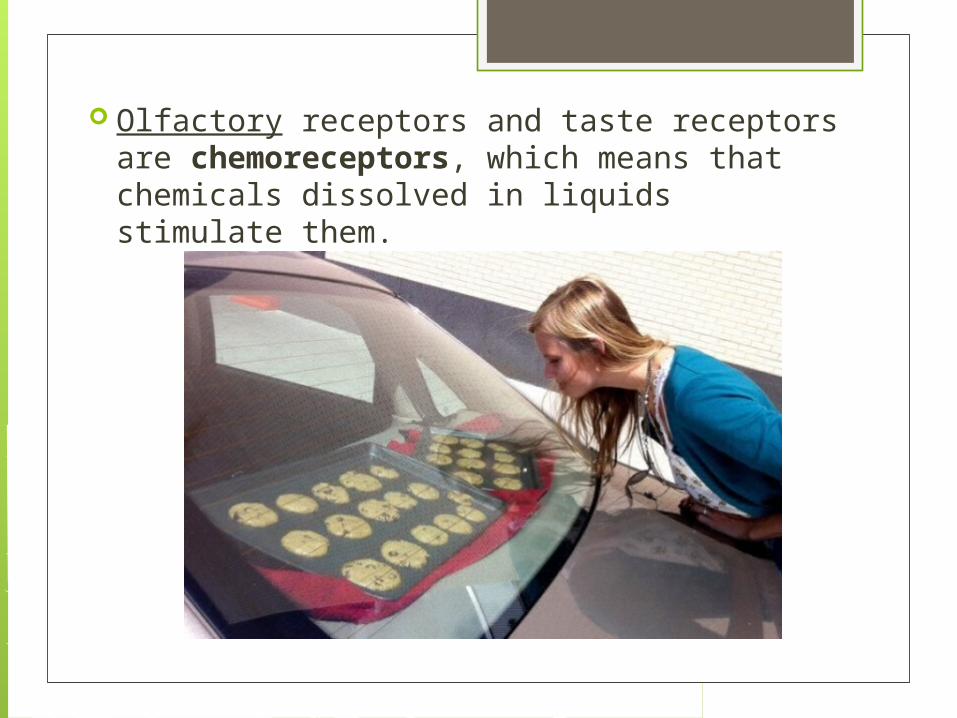
Olfactory receptors and taste receptors are chemoreceptors, which means that chemicals dissolved in liquids stimulate them.
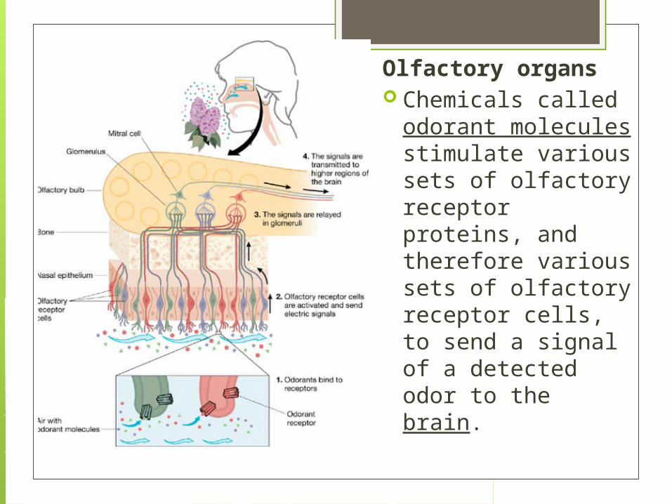
Olfactory organs Chemicals called
odorant molecules stimulate various sets of olfactory receptor proteins, and therefore various sets of olfactory receptor cells, to send a signal of a detected odor to the brain.
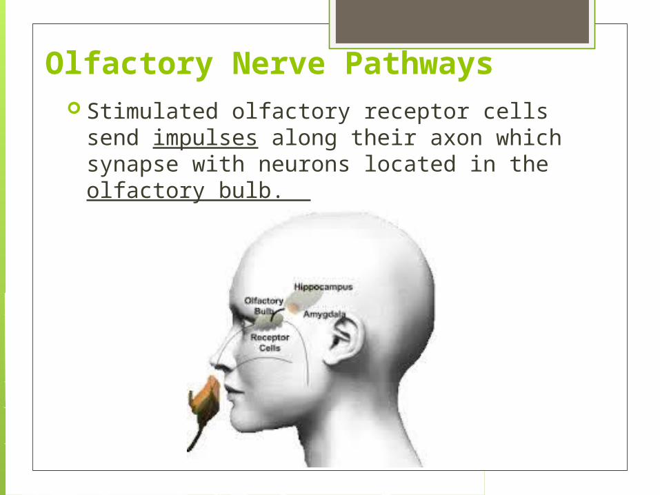
Olfactory Nerve Pathways Stimulated olfactory receptor cells send
impulses along their axon which synapse with neurons located in the olfactory bulb.
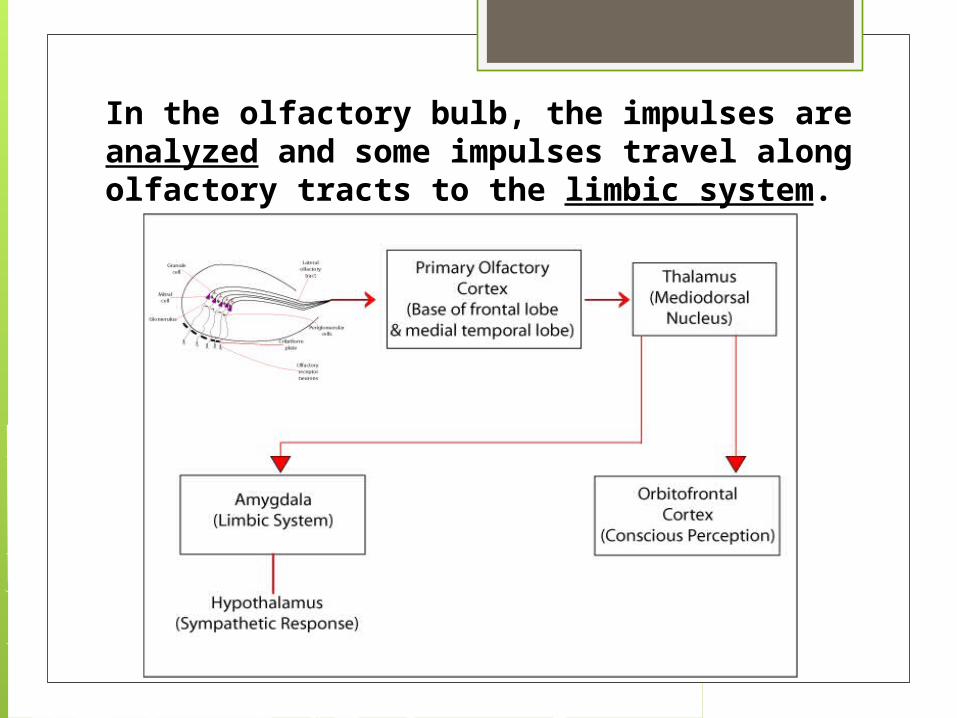
In the olfactory bulb, the impulses are analyzed and some impulses travel along olfactory tracts to the limbic system.
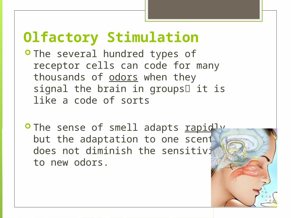
Olfactory Stimulation The several hundred types of receptor
cells can code for many thousands of odors when they signal the brain in groups it is like a code of sorts
The sense of smell adapts rapidly, but the adaptation to one scent does not diminish the sensitivity to new odors.

TAS
TE
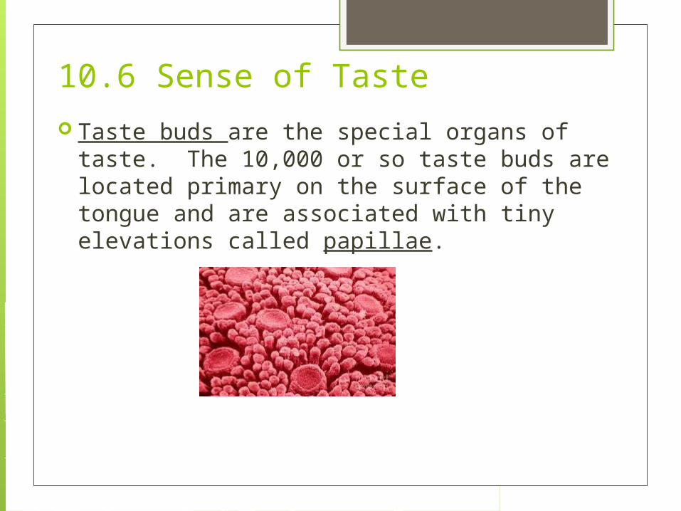
10.6 Sense of Taste Taste buds are the special organs of taste. The
10,000 or so taste buds are located primary on the surface of the tongue and are associated with tiny elevations called papillae.
10.6 Sense of Taste
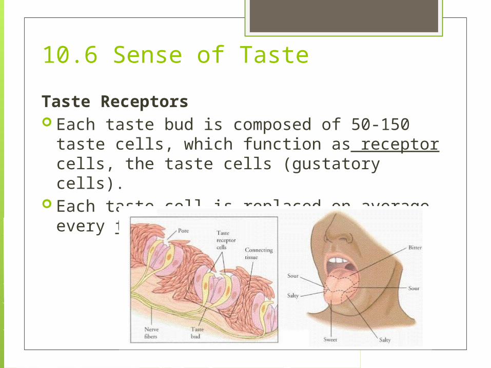
Taste Receptors Each taste bud is composed of 50-150 taste
cells, which function as receptor cells, the taste cells (gustatory cells).
Each taste cell is replaced on average every ten days.
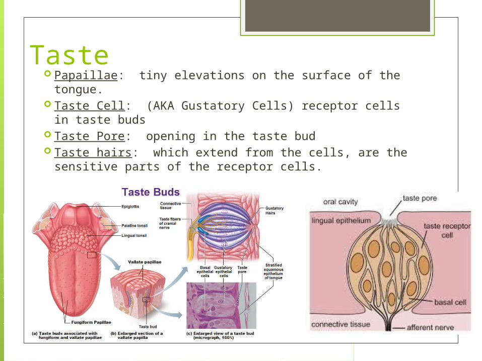
Taste Papaillae: tiny elevations on the surface of the tongue. Taste Cell: (AKA Gustatory Cells) receptor cells in taste
buds Taste Pore: opening in the taste bud Taste hairs: which extend from the cells, are the
sensitive parts of the receptor cells.
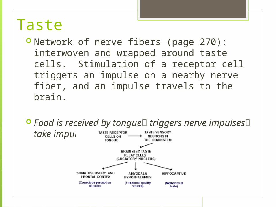
Taste Network of nerve fibers (page 270):
interwoven and wrapped around taste cells. Stimulation of a receptor cell triggers an impulse on a nearby nerve fiber, and an impulse travels to the brain.
Food is received by tongue triggers nerve impulses take impulse to the brain
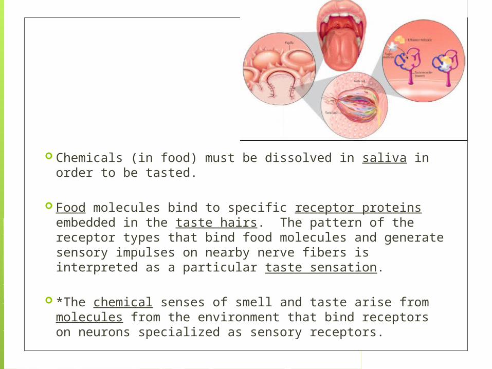
Chemicals (in food) must be dissolved in saliva in order to be tasted.
Food molecules bind to specific receptor proteins embedded in the taste hairs. The pattern of the receptor types that bind food molecules and generate sensory impulses on nearby nerve fibers is interpreted as a particular taste sensation.
*The chemical senses of smell and taste arise from molecules from the environment that bind receptors on neurons specialized as sensory receptors.
Taste Sensations
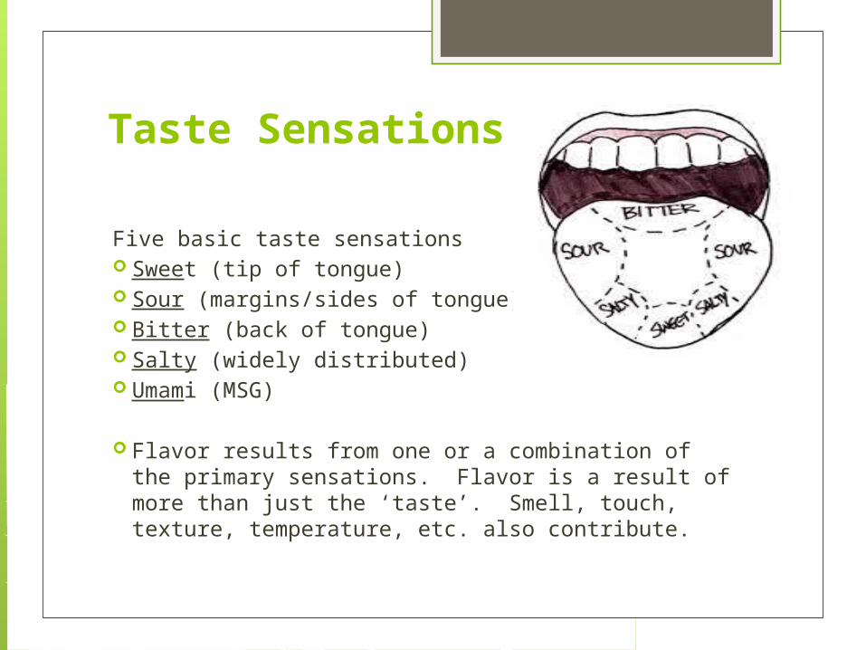
Five basic taste sensations Sweet (tip of tongue) Sour (margins/sides of tongue) Bitter (back of tongue) Salty (widely distributed) Umami (MSG)
Flavor results from one or a combination of the primary sensations. Flavor is a result of more than just the ‘taste’. Smell, touch, texture, temperature, etc. also contribute.

Hearing
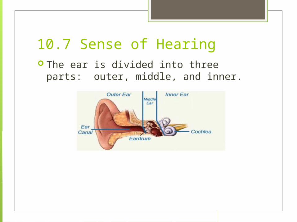
10.7 Sense of Hearing The ear is divided into three parts:
outer, middle, and inner.
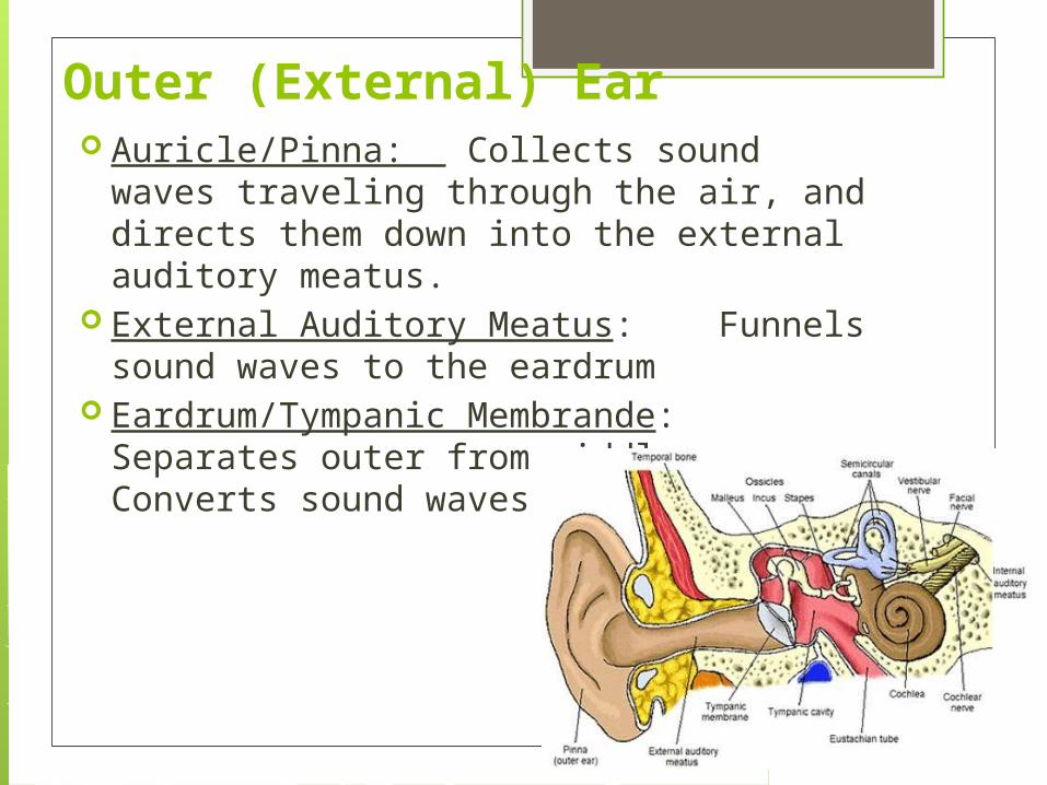
Outer (External) Ear Auricle/Pinna: Collects sound waves
traveling through the air, and directs them down into the external auditory meatus.
External Auditory Meatus: Funnels sound waves to the eardrum
Eardrum/Tympanic Membrande: Separates outer from middle ear. Converts sound waves into vibrations
Middle Ear Parts:
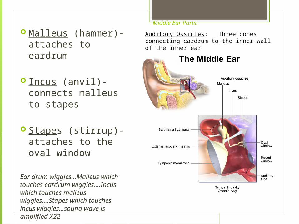
Malleus (hammer)- attaches to eardrum
Incus (anvil)- connects malleus to stapes
Stapes (stirrup)- attaches to the oval window
Ear drum wiggles…Malleus which touches eardrum wiggles….Incus which touches malleus wiggles….Stapes which touches incus wiggles…sound wave is amplified X22
Auditory Ossicles: Three bones connecting eardrum to the inner wall of the inner ear
Middle Ear Parts:
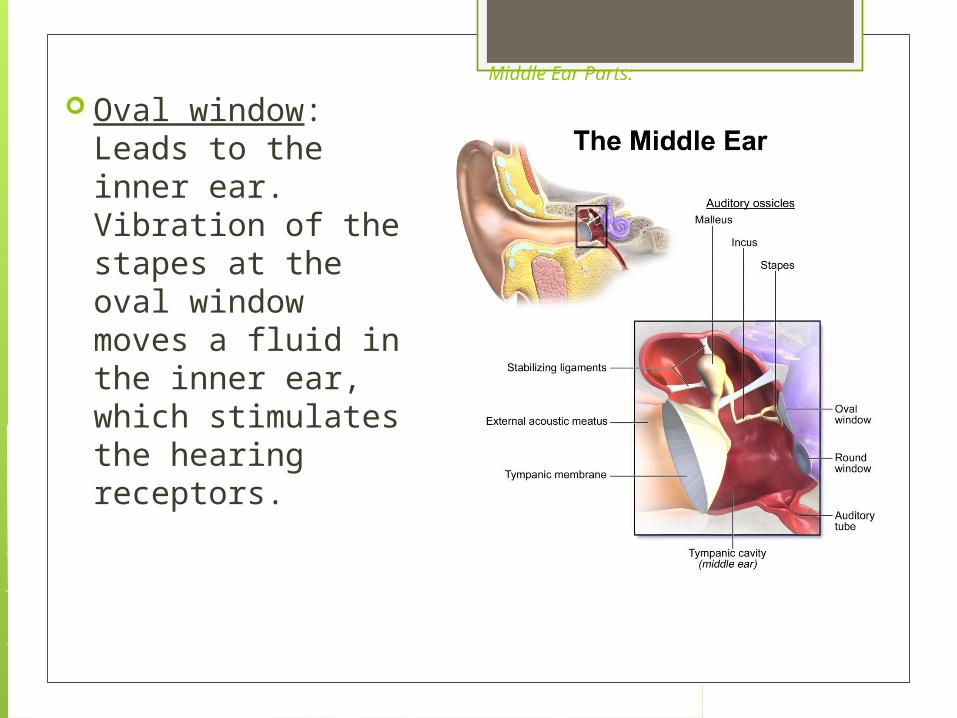
Oval window: Leads to the inner ear. Vibration of the stapes at the oval window moves a fluid in the inner ear, which stimulates the hearing receptors.
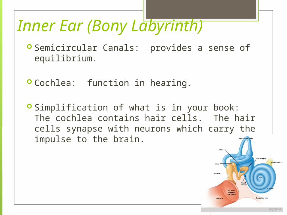
Inner Ear (Bony Labyrinth) Semicircular Canals: provides a sense of
equilibrium.
Cochlea: function in hearing.
Simplification of what is in your book: The cochlea contains hair cells. The hair cells synapse with neurons which carry the impulse to the brain.
Middle Ear Stapes which touches oval window touches the inner
ear fludi filled structures. The sound waves are then transferred to mechanical fluid waves. Oval window touches the
The pitch is deteremined by how far wave travelsing to coclia.
There are hair cells in the coclia. The hair cells synapes (ie send messges) to sensory neurons.
So Far: Sound wave-eardrum-malleas-incus-stapes
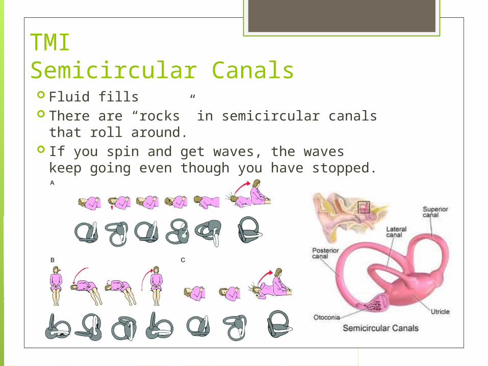
TMISemicircular Canals Fluid fills There are “rocks” in semicircular canals that roll
around. If you spin and get waves, the waves keep
going even though you have stopped.
TMIHearing Nerve Pathways
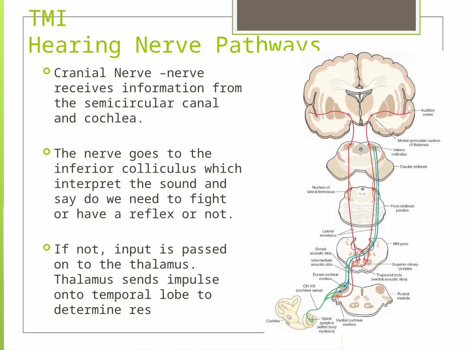
Cranial Nerve –nerve receives information from the semicircular canal and cochlea.
The nerve goes to the inferior colliculus which interpret the sound and say do we need to fight or have a reflex or not.
If not, input is passed on to the thalamus. Thalamus sends impulse onto temporal lobe to determine res
Steps of hearing The external ear picks up sound waves and focuses
them onto the eardrum. The eardrum vibrates and converts the sound impulses
to mechanical waves. The three ossicles move in response to the eardrum
vibration, amplifying the sound. The stapes pushes on the oval window. Fluid waves causes membranes within the cochlea to
move, bending hair cells The hair cells bending causes a nerve impulse. The nerve impulse is carried to the temporal lobes by
nerve fibers where they are interpreted.
Sight

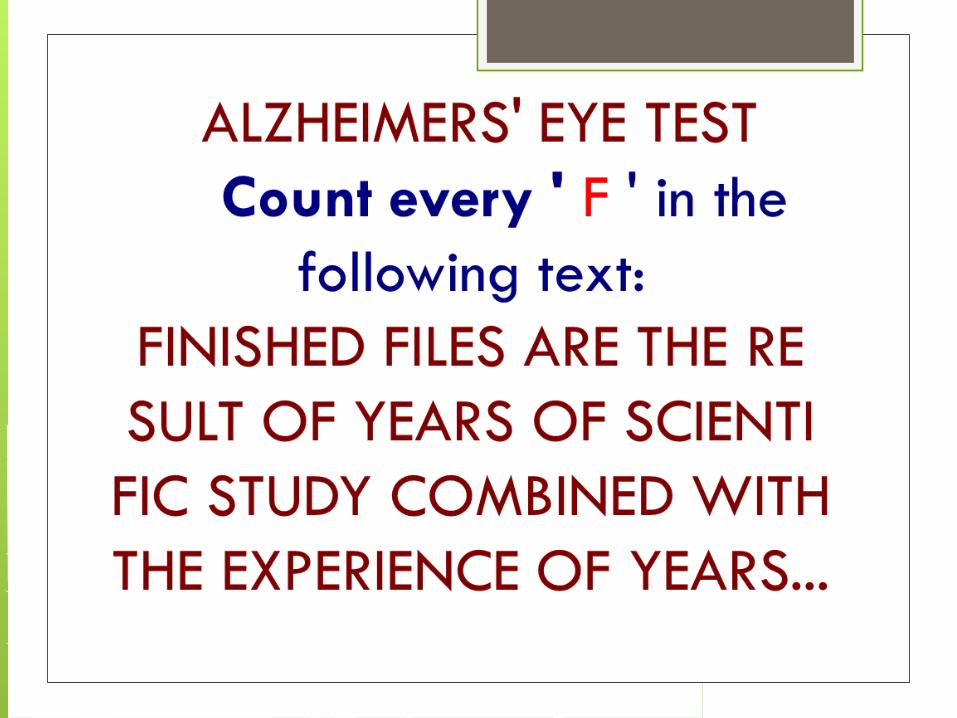
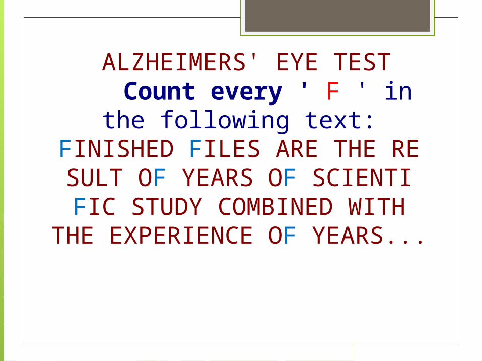
ALZHEIMERS' EYE TEST Count every ' F ' in the
following text:FINISHED FILES ARE THE RESULT OF YEARS OF SCIENTIFIC STUDY COMBINED WITH
THE EXPERIENCE OF YEARS...
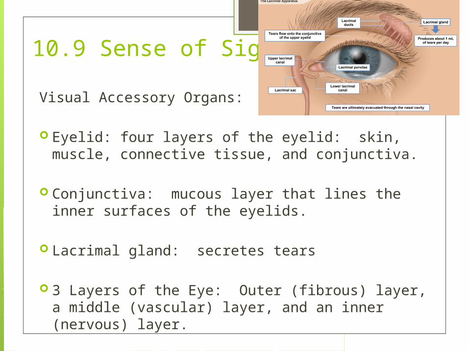
10.9 Sense of Sight
Visual Accessory Organs:
Eyelid: four layers of the eyelid: skin, muscle, connective tissue, and conjunctiva.
Conjunctiva: mucous layer that lines the inner surfaces of the eyelids.
Lacrimal gland: secretes tears
3 Layers of the Eye: Outer (fibrous) layer, a middle (vascular) layer, and an inner (nervous) layer.
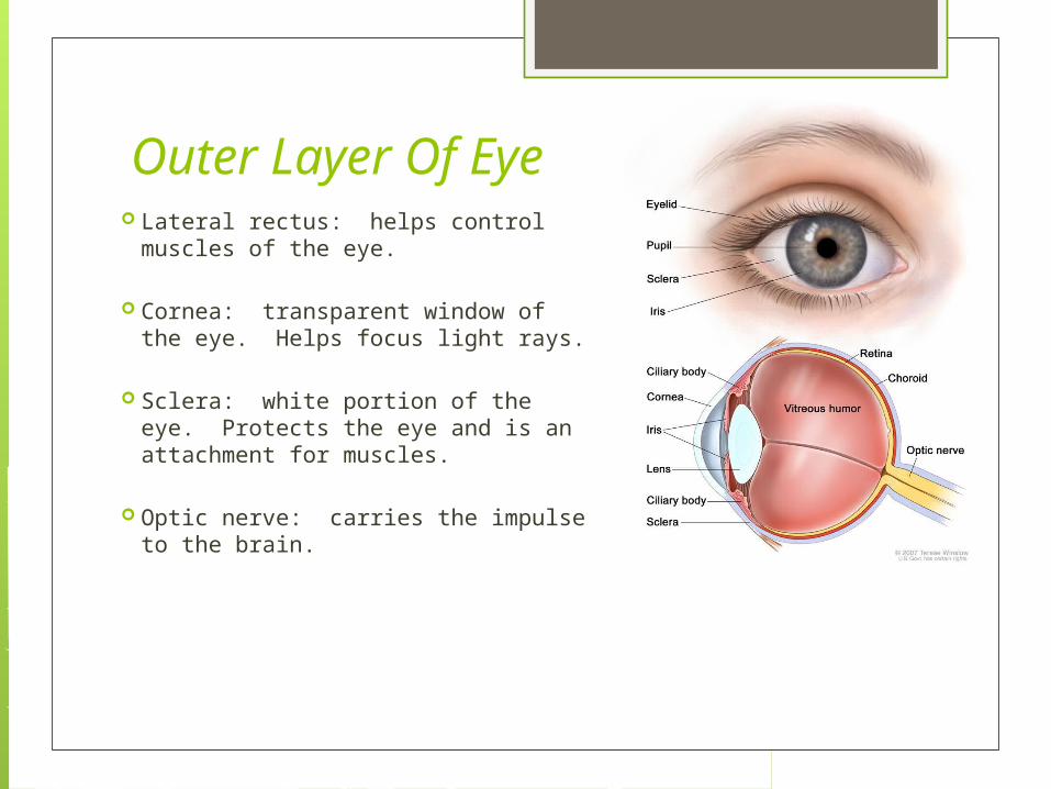
Outer Layer Of Eye Lateral rectus: helps control
muscles of the eye.
Cornea: transparent window of the eye. Helps focus light rays.
Sclera: white portion of the eye. Protects the eye and is an attachment for muscles.
Optic nerve: carries the impulse to the brain.
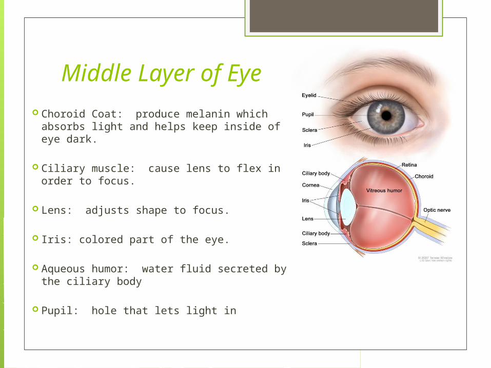
Middle Layer of Eye Choroid Coat: produce melanin which
absorbs light and helps keep inside of eye dark.
Ciliary muscle: cause lens to flex in order to focus.
Lens: adjusts shape to focus.
Iris: colored part of the eye.
Aqueous humor: water fluid secreted by the ciliary body
Pupil: hole that lets light in
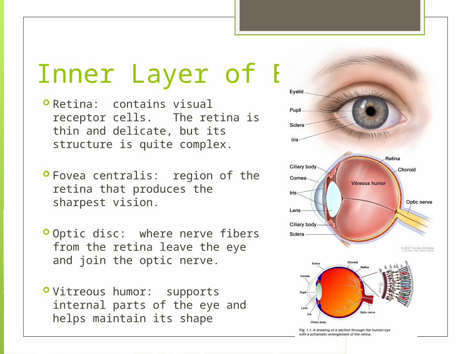
Inner Layer of Eye Retina: contains visual receptor
cells. The retina is thin and delicate, but its structure is quite complex.
Fovea centralis: region of the
retina that produces the sharpest vision.
Optic disc: where nerve fibers from the retina leave the eye and join the optic nerve.
Vitreous humor: supports internal parts of the eye and helps maintain its shape
GLAUCOMA
Visual Defects/Diseases Glaucoma
Aqueous humor forms faster than it is removed Fluid in eye increases and the fovea is compressed, leading to restricted vision
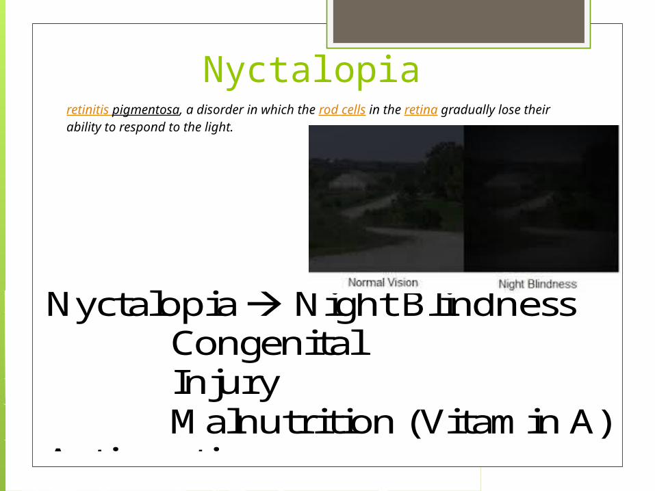
Nyctalopia Night Blindness Congenital Injury Malnutrition (Vitamin A)
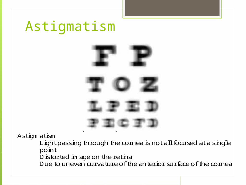
Astigmatism Light passing through the cornea is not all focused at a single point Distorted image on the retina Due to uneven curvature of the anterior surface of the cornea
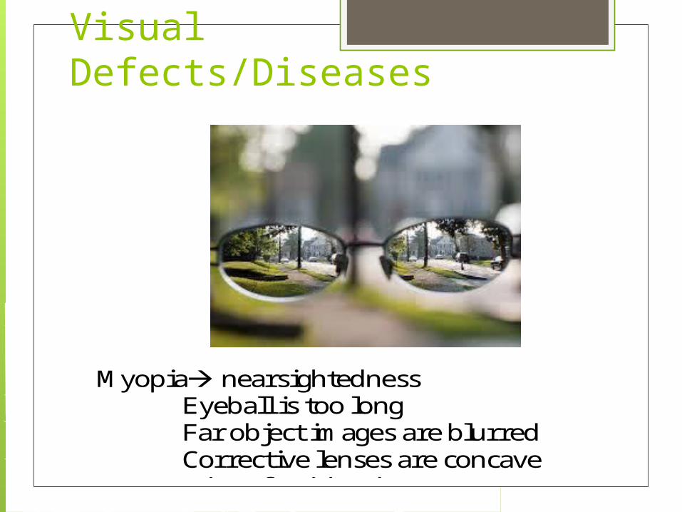
Myopia nearsightedness Eyeball is too long Far object images are blurred Corrective lenses are concave
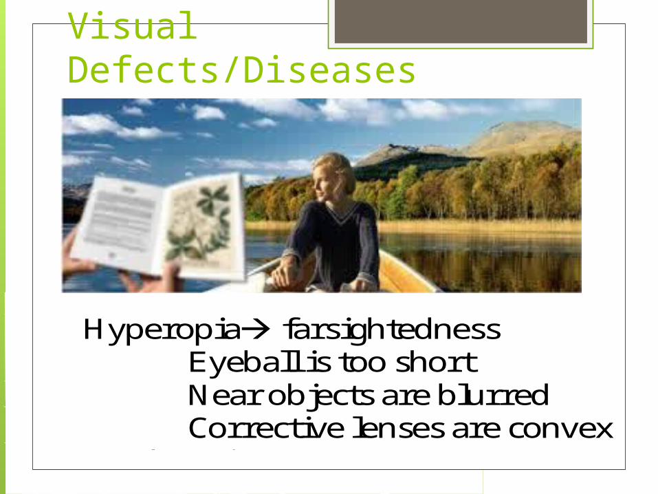
Hyperopia farsightedness Eyeball is too short Near objects are blurred Corrective lenses are convex
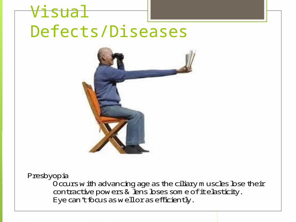
Presbyopia Occurs with advancing age as the ciliary muscles lose their contractive powers & lens loses some of it elasticity. Eye can’t focus as well or as efficiently.
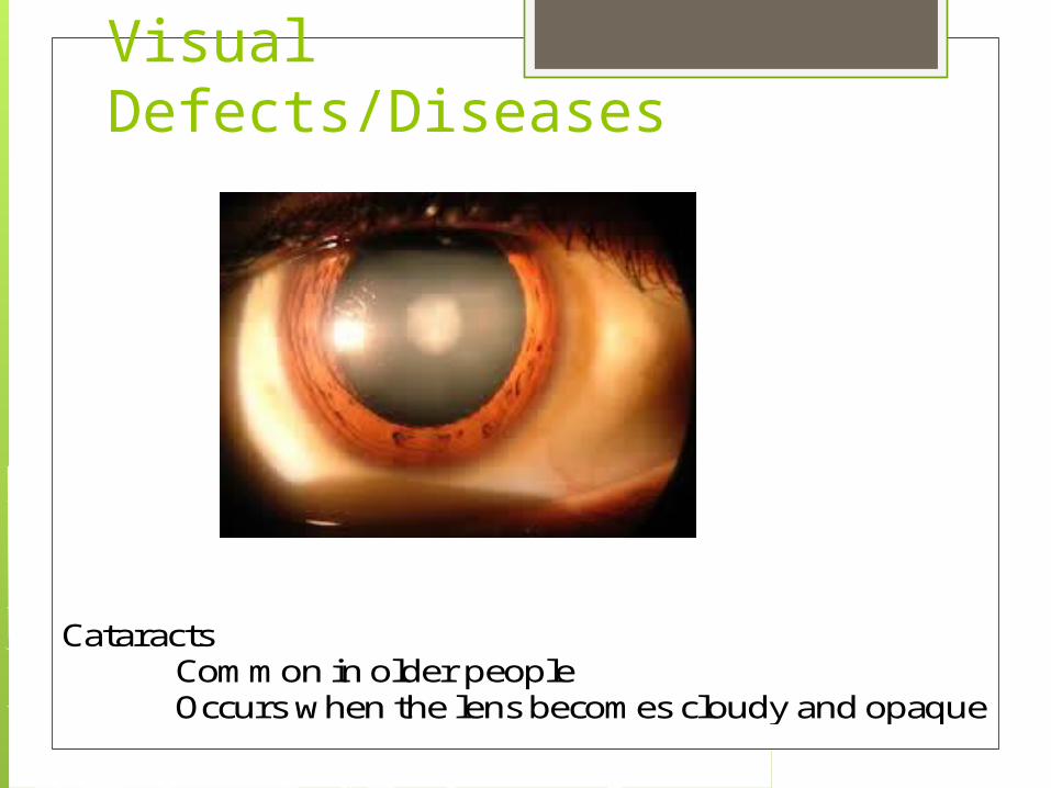
Cataracts Common in older people Occurs when the lens becomes cloudy and opaque
NyctalopiaVisual Defects/Diseases
Glaucoma Aqueous humor forms faster than it is removed Fluid in eye increases and the fovea is compressed, leading to restricted vision
Nyctalopia Night Blindness Congenital Injury Malnutrition (Vitamin A)
Astigmatism Light passing through the cornea is not all focused at a single point Distorted image on the retina Due to uneven curvature of the anterior surface of the cornea
Myopia nearsightedness Eyeball is too long Far object images are blurred Corrective lenses are concave
Hyperopia farsightedness Eyeball is too short Near objects are blurred Corrective lenses are convex
Presbyopia Occurs with advancing age as the ciliary muscles lose their contractive powers & lens loses some of it elasticity. Eye can’t focus as well or as efficiently.
Cataracts Common in older people Occurs when the lens becomes cloudy and opaque
retinitis pigmentosa, a disorder in which the rod cells in the retina gradually lose their ability to respond to the light.
Astigmatism
Visual Defects/Diseases Glaucoma
Aqueous humor forms faster than it is removed Fluid in eye increases and the fovea is compressed, leading to restricted vision
Nyctalopia Night Blindness Congenital Injury Malnutrition (Vitamin A)
Astigmatism Light passing through the cornea is not all focused at a single point Distorted image on the retina Due to uneven curvature of the anterior surface of the cornea
Myopia nearsightedness Eyeball is too long Far object images are blurred Corrective lenses are concave
Hyperopia farsightedness Eyeball is too short Near objects are blurred Corrective lenses are convex
Presbyopia Occurs with advancing age as the ciliary muscles lose their contractive powers & lens loses some of it elasticity. Eye can’t focus as well or as efficiently.
Cataracts Common in older people Occurs when the lens becomes cloudy and opaque
Visual Defects/Diseases
Visual Defects/Diseases Glaucoma
Aqueous humor forms faster than it is removed Fluid in eye increases and the fovea is compressed, leading to restricted vision
Nyctalopia Night Blindness Congenital Injury Malnutrition (Vitamin A)
Astigmatism Light passing through the cornea is not all focused at a single point Distorted image on the retina Due to uneven curvature of the anterior surface of the cornea
Myopia nearsightedness Eyeball is too long Far object images are blurred Corrective lenses are concave
Hyperopia farsightedness Eyeball is too short Near objects are blurred Corrective lenses are convex
Presbyopia Occurs with advancing age as the ciliary muscles lose their contractive powers & lens loses some of it elasticity. Eye can’t focus as well or as efficiently.
Cataracts Common in older people Occurs when the lens becomes cloudy and opaque
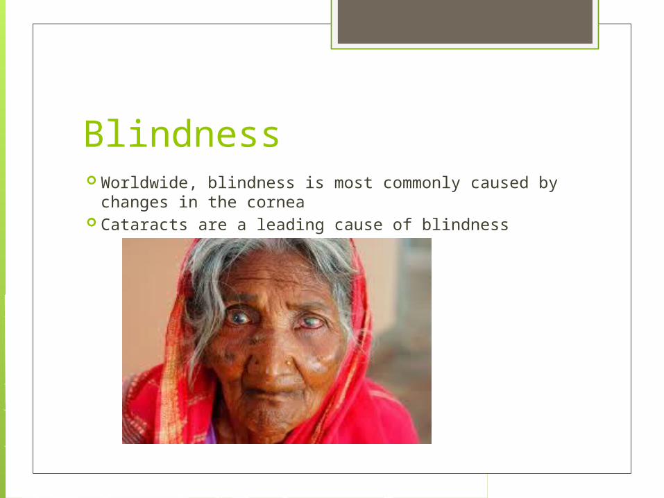
Blindness Worldwide, blindness is most commonly caused by
changes in the cornea Cataracts are a leading cause of blindness
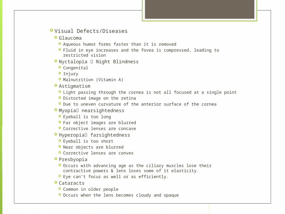
Visual Defects/Diseases Glaucoma
Aqueous humor forms faster than it is removed Fluid in eye increases and the fovea is compressed, leading to restricted
vision Nyctalopia Night Blindness
Congenital Injury Malnutrition (Vitamin A)
Astigmatism Light passing through the cornea is not all focused at a single point Distorted image on the retina Due to uneven curvature of the anterior surface of the cornea
Myopia nearsightedness Eyeball is too long Far object images are blurred Corrective lenses are concave
Hyperopia farsightedness Eyeball is too short Near objects are blurred Corrective lenses are convex
Presbyopia Occurs with advancing age as the ciliary muscles lose their contractive
powers & lens loses some of it elasticity. Eye can’t focus as well or as efficiently.
Cataracts Common in older people Occurs when the lens becomes cloudy and opaque