Embed Size (px)
Citation preview
Chapter 2 Review of Literature
School of Pharmaceutical Sciences 21
2. REVIEW OF LITERATURE
2.1. Based on colon specific drug delivery system
2.1.1. Mahale et al., 2013 have studied the colon specific delivery systems. The colonic
environment in the lower GIT has attained important value in the design of colon targeted drug
delivery systems. Targeted drug delivery into colon is highly recommended for local treatment of
a variety of bowel diseases like ulcerative colitis, Crohn’s disease, amebiosis, colonic cancer etc.
Mechanisms of drug targeting to the colon are pH-dependent delivery, time-dependent delivery,
pressure-dependent delivery and bacteria dependent delivery etc.
2.1.2. Srivastava et al., 2012 have developed calcium-pectinate matrix tablet for colon-targeted
delivery of meloxicam (MLX) microsponges. Modified Quassi-emulsion solvent diffusion
method was used to formulate microsponges (MS). The effects of volume of dichloromethane
and Eudragit RS100 content (independent variables) were determined on the particle size,
entrapment efficiency and %cumulative drug release of MS1–MS9. The optimized formulation
was developed into colon-targeted matrix tablet using calcium pectinate as the matrix. The
optimized colon-targeted tablet shielded MLX loaded microsponges in gastrointestinal region
and selectively delivered them to colon, as visualized by vivo fluoroscopy in rabbits. The
pharmacokinetic evaluation in rabbits, revealed drug in plasma after a lag time of 7 h, a tmax of 30
h with Fr = 61.047%, thus presenting a formulation suitable for targeted colonic delivery.
Calcium pectinate matrix tablet loaded with MLX microsponges were developed as a promising
system for the colon-specific delivery.
2.1.3. Pandey et al., 2012 have worked on tinidazole microbeads for colon targeting. The
objective of study was to develop and evaluate multiparticulate system exploiting pH-sensitive
property and specific biodegradability of calcium alginate microbeads, for colon- targeted
delivery of tinidazole for the treatment of amoebic colitis. The calcium alginate beads containing
tinidazole were prepared by ionotropic gelation technique followed by coating with Eudragit
S100 using solvent evaporation method to obtain pH sensitive microbeads. Various formulation
parameters were optimized which included concentration of sodium alginate (2% w/v), curing
time (20 min) and concentration of pectin (1% w/v). All the formulations were evaluated for
surface morphology, particle size analysis, entrapment efficiency and in-vitro drug release in
Chapter 2 Review of Literature
School of Pharmaceutical Sciences 22
conditions simulating colonic fluid in the presence of rat caecal (2% w/v) content. The result
showed that average size of beads of optimized formulation (FT4) was found to be 998.73 ±5.12
µm with entrapment efficiency of 87.28±2.19 %. The in-vitro release of Eudragit S100 coated
beads in presence of rat caecal content was found to be 70.73%± 1.91% in 24 hours. Data of in-
vitro release was fitted into Higuchi kinetics and Korsmeyer Peppas equation to explain release
profile. The optimized formulation (FT 4) showed zero order release.
2.1.4. Mura et al., 2012 have worked on 5- aminosalicyclic acid N- succinyl- chitosan
microparticles for colon specific delivery. The objective of study was to prepare NS-chitosan
microparticles for the delivery of 5-aminosalicylic acid (5-ASA) to the colon. N-Succinyl-
chitosan was chosen as carrier system because it has excellent pharmaceutical properties in colon
drug targeting such as poor solubility in acid environment, biocompatibility, mucoadhesive
properties, and low toxicity. 5-ASA loaded NS-chitosan microparticles were prepared using
spray-drying method. As a control, a matrix obtained by freeze-drying technique was also
prepared and evaluated. The result showed that mean size of the microparticles was around 5µm.
SEM images showed an acceptable spherical non porous structure of microparticles. In-vitro
swelling and drug release studies were in accordance with the polymer properties, showing the
highest swelling ratio and drug release at pH = 7.4 (colonic pH) where microparticles were able
to deliver more than 90% of 5-ASA during 24 h.
2.1.5. Juan et al., 2012 have prepared azo- reductase activated budesodine prodrugs for colon
targeting. The objective of present study was to produce local topical effect, improving safety
and increasing anti-inflammatory efficacy. The budesodine prodrug were synthesized and tested
using an in-vitro azo reductase assay simulating human colonic microflora. The kinetics of
amino steroid ester cyclization and its pH dependence was also evaluated. The result showed that
prodrug of budesodine was potential in management of ulcerative colitis.
2.1.6. Sirisha et al., 2012 have developed colon targeting mesalamine matrix tablet. The
objective of present study was to deliver tablet directly into the colon by using both hydrophilic
and hydrophobic polymers. Matrix tablets were prepared by direct compression method using
different concentration of HPME and EC. Evaluation like hardness, friability, weight variation,
thickness and drug content of prepared formulations were done. The results showed that
combination of both polymers exhibit best release profile and able to sustain the drug release for
prolong period of time. The test batch comparison analysis was conformed that combination of
Chapter 2 Review of Literature
School of Pharmaceutical Sciences 23
both hydrophilic and hydrophobic polymer successfully employed for formulating the sustain
release colon targeted matrix tablet of mesalamine.
2.1.7. Mishra et al., 2012 have synthesized and characterized amino acid conjugate of naproxen.
The objective of work was to enhance the solubility without affecting permeability and deliver
naproxen to colon without significant reversion of prodrug in gastrointestinal conditions. The
naproxen- glycine conjugate was prepared by conventional coupling method and prodrug was
characterized by FTIR, FTNMR, FABMS, and element analysis. The results showed that
prodrug possessed therapeutically efficacious drug delivery system with less pharmaceutical
limitations.
2.1.8. Nangude et al., 2012 have studied colon targeted oral matrix tablets of naproxen and
esomeprazole. The objective of this study was to develop colon targeted drug delivery system of
naproxen and esomeprazole using different polymers for treatment of IBD. In this, matrix tablet
was prepared and various evaluation tests were carried out. Maximum drug release occurred in
phosphate buffer (pH 6.8). The results showed that optimized formulation showed no change
either in physical appearance, drug content or dissolution pattern after storage at 40 ̊C/ 75% RH
for three months.
2.1.9. Sarkar et al., 2011 had investigated prednisolone tablets for colon targeting delivery
system. The prednisolone tablets were prepared using wet granulation method with various
additive and coating. Different concentration of avicel and PVP were used; acted as canalizing
and binding agent. The results showed that lactose as diluents provided reasonable release for
prednisolone among other diluents. The 1% Eudragit RS showed 100% release of drug in
comparison with other concentration. 10% PVP gave the best results. The prednisolone modified
release tablet was successfully formulated using wet granulation method as a potential colon
delivery system.
2.1.10. Potu et al., 2011 have studied on fenoprofen calcium compressed coated tablets for colon
specific drug delivery. The main objective of study was to release the drug maximum in targeted
area i.e. physiological environment of colon. Various formulations were prepared using different
ratio of guar gum and HPMC. HPMC was included in this study to control the solubility of guar
gum and to prevent premature drug release in stomach and small intestine. Dissolution studies in
pH 6.8 phosphate buffer were carried out. The results of in-vitro study indicated the formulation
containing 60% guar gum was able to release less than 1% of drug in environment of stomach

Chapter 2 Review of Literature
School of Pharmaceutical Sciences 24
and small intestine while 98% release of drug in targeted area. In vivo X-ray study showed that
design dosage form reaches the targeted site.
2.1.11. Yadav et al., 2011 have prepared polymeric prodrug of 4- aminosalicyclic acid for
inflammatory bowel disease. The objective of work was to reduce the frequency of
administration and avoid the gastrointestinal adverse effects associated with 4-ASA. The
synthesized prodrug was characterized by melting point, Rf value, FT-IR and 1H NMR. In-vitro
drug release was conducted at pH 1.2, 7.4 and in presence of rat faecal matter (pH 7.4). The
results showed that maximum 92.8% of drug release from prodrug and time taken for 50% drug
release was found to be nearly 3.5 hours. The amount of 4- aminosalicyclic acid released in
colon was found to be very high as compared to stomach and intestine.
2.1.12. Trombino et al., 2011 have synthesized lysine based prodrug of 5- Aminosalicyclic acid
and 6- Mercaptopurine for colon specific release. The aim of work was to design and
characterization of prodrug for the colon targeting. In this, prodrugs were synthesized and
characterized by FT-IR, 1H NMR and GC/ MS spectroscopy. The results suggested that the
prodrug could have high potential in tumors treatment, targeting 6-MP to the colon and
outweighing the disadvantage occur with the conventional treatment system.
2.1.13. Dube et al., 2011 have developed colon targeted lornoxicam matrix tablet. The objective
of study was to target drug directly to the colon and reducing systemic side effect. Matrix tablets
were prepared by direct compression method using different concentration of HPMC and EC.
Evaluation like hardness, friability, thickness, % drug content, weight variation and in-vitro
study of prepared formulation was done. The results showed that combination of both polymers
exhibit best release profile and able to sustain the drug release for prolong period of time.
2.1.14. Jose et al., 2011 have developed colon-specific chitosan microspheres for chronotherapy
of chronic angina. Chitosan microspheres were formulated by emulsion cross-linking method
and tested for chronotherapy of chronic stable angina. Diltiazem hydrochloride was encapsulated
in the chitosan microspheres following coating with Eudragit S-100 by solvent evaporation
technique, exploiting the advantages of microbiological properties of chitosan and pH dependent
solubility of Eudragit S-100. Different microsphere formulations were prepared by varying the
ratio diltiazem hydrochloride: chitosan. The effects of these variables on the particle size and
encapsulation parameters were evaluated to develop an optimized formulation. In-vitro release
study of non-coated chitosan microspheres in simulated gastrointestinal (GI) fluid exhibited a

Chapter 2 Review of Literature
School of Pharmaceutical Sciences 25
burst release while Eudragit coated microspheres showed release at colonic pH. Chitosan
biodegradability was proved by the enhanced release rate of diltiazem hydrochloride in presence
of rat caecal contents.
2.1.15. Kothawade et al., 2011 have studied conventional and novel approaches for colon
specific drug delivery. Colon specific drug delivery is not only useful for targeting the drugs
required in the treatment of diseases associated with colon, but also as a potential site for the
local and systemic delivery of peptide and proteins and other therapeutic drugs. Precise colon
drug delivery requires the triggering mechanism in the delivery system that can respond only to
the physiological conditions specific to the colon. The primary approaches used to obtain colon-
specific delivery were based on prodrugs, pH and time dependent systems or microflora
activated systems. Recently continuous efforts have been taken on designing colon-specific
delivery systems with improved site specificity and versatile drug release kinetics to accomplish
different therapeutic needs. Different studies provided detailed insight into the conventional as
well as recent approaches used to target the therapeutic agents specifically to the colon.
2.1.16. Challa et al., 2011 have studied novel approaches on colon specific drug delivery. Colon
specific drug delivery has gained increased importance not just for delivery of the drugs in the
treatment associated with the colon, but also as a potential site for the systemic delivery of
therapeutic peptides and proteins. To achieve successful colon targeted drug delivery, a drug
need to be protected from degradation, release and absorption in the upper portion of the GI tract
and then to be ensured abrupt or controlled release in the proximal colon.
2.1.17. Philip et al., 2010 have studied primary and novel approaches for colon targeted drug
delivery system. The colon is a site where both local and systemic delivery of drugs can take
place. Local delivery allows topical treatment of inflammatory bowel disease. However,
treatment can be made effective if the drugs can be targeted directly into the colon, thereby
reducing the systemic side effects.
2.1.18. Patel et al., 2010 have studied enteric coated tablets of prednisolone for colon targeted
drug delivery. The main objective of this study was to reduce the frequency of dose
administration, prevent ulcerative colitis and also reduce the side effect of anti-inflammatory
drug in GI tract by developing delay release (DR) tablet of prednisolone using Eudragit S 100 as
enteric coating. The matrix tablet of prednisolone tablet was formulated by wet granulation
method. In-vitro drug release was performed by using simulating colonic fluid of pH 7.4 as

Chapter 2 Review of Literature
School of Pharmaceutical Sciences 26
dissolution media. The results showed that Eudragit S100 can successfully be used to coat the
tablets for colon targeted delivery of drug.
2.1.19. Udo et al., 2010 have worked on 5- Fluorouracil acetic acid/ β- cyclodextrin conjugates:
Drug release behavior in enzymatic and rat caecal media. In this, 5- fluorouracil-1- acetic acid
was prepared and covalently conjugated to β- cyclodextrin through ester o amide linkage. The
drug release behavior of the conjugates in enzymatic solution and rat caecal contents were
investigated. The 5-FUA/β-CyD ester conjugate was slowly hydrolyzed to 5-FUA in aqueous
solutions, whereas the amide conjugate was hardly hydrolyzed at these physiological conditions,
but hydrolyzed only in strong alkaline solutions (>0.1MNaOH) at 60 ̊C, both ester and amide
conjugates were degraded in solutions of a sugar-degrading enzyme, α-amylase to 5-
FUA/maltose and triose conjugates, but the release of 5-FUA was only slight in α-amylase
solutions. In solutions of an ester-hydrolyzing enzyme carboxylic esterase, the ester conjugate
was hydrolyzed to 5-FUA at the same rate as that in the absence of the enzyme, whereas the
amide conjugate was not hydrolyzed by the enzyme. The results indicated that the ester
conjugate was hydrolyzed to 5- FUA. The in-vitro release behavior of the ester conjugate was
clearly reflected in the hydrolysis in rat caecal contents and in the in vivo release after oral
administration to rats.
2.1.20. Philip et al., 2009 have worked on colon targeting drug delivery system. Prodrug was
synthesizing by coupling ketoprofen with glycine. Reversion of KET- GLY to ketoprofen was
carried out at different pH and at pH 6.8 containing rat faecal contents. In-vitro reversion study
showed that KET- GLY remained intact in stomach but released the free drug at pH 6.8
containing fresh faecal material. In vivo study showed that KET- GLY was less toxic in stomach
with enhanced anti-inflammatory potential in the colonic region. KET- GLY was better in action
as compared with the parent drug.
2.1.21. Varshosaz et al., 2009 have worked on colon specific delivery system of budesonide to
increase the efficacy in the treatment of ulcerative colitis. The main objective of study was to
prepare Dextran- budesonide conjugates with different molecular weight of dextran (10,000,
70,000 and 500,000) in the presence of dimethylaminopyridine using succinate spacer.
Conjugate prepared by dextran 70,000 showed the most desirable solubility, stability and release
properties. In vivo evaluation was carried out to analyze potential clinical use in the treatment of
ulcerative colitis.

Chapter 2 Review of Literature
School of Pharmaceutical Sciences 27
2.1.22. Makhlof et al., 2009 have design pH- sensitive nanospheres for colon - specific drug
delivery in experimentally induced colitis rat model. Nanospheres were prepared using
polymeric mixture of poly (lactic-co-glycolic) acid and pH sensitive methacrylate copolymer.
Budesonide, active corticosteroid, was entrapped as a model drug. The therapeutic efficacy of the
prepared nanospheres was assessed using the TNBS (Trinitrobenzene sulfonate) colitis in rat, in
comparison with conventional enteric microparticles. Colon targeting properties were evaluated
using coumarin-6 loaded nanospheres. The results indicated that prepared nanospheres showed
strongly pH dependent drug release properties in acidic and neutral pH values followed by
sustain release phase at pH 7.4. In–vivo studies using coumarin-6 loaded nanospheres displayed
higher colon levels and lower systemic availability of fluorescent marker when compared with
simple enteric coating. The nanosphere system combined the properties of pH- sensitivity,
control release and particulate targeting that was useful for colon specific delivery in
inflammatory bowel disease.
2.1.23. Wei et al., 2008 have studied colon specific pectin/ ethylcellulose film coated 5-
fluorouracil pellets in rats. The objective of study was to assess the bio-distribution and
pharmacokinetics of pectin/ ethylcellulose film-coated and uncoated pellets containing 5-
fluorouracil. Both coated and uncoated pellets were orally administered to the rats at a dosage
equivalent to 15 mg/kg. 5-FU concentrations in different parts of the gastrointestinal (GI) tract
and plasma were quantitatively analyzed using HPLC. The results suggested that 5-FU released
from uncoated pellets mainly distributed in the upper GI tract, however, 5-FU released from
coated pellets mainly distributed in the cecum and colon. In plasma, the observed mean Cmax
from the coated pellets group (3.65±2.3µg/mL) was lower than that of the uncoated pellets group
(23.54±2.9µg/mL). The AUC (area under curve) values obtained from the uncoated pellets and
the coated pellets were 49.08±3.1 and 9.06±1.2µgh/mL. The relatively high local drug
concentration with prolonged exposure time provided a potential to enhance anti-tumor efficacy
with low systemic toxicity for the treatment of colon cancer.
2.1.24. Pertuit et al., 2007 have worked on 5- amino salicyclic acid bound nanoparticles for the
therapy of inflammatory bowel disease. The objective of study was to design nanoparticles of 5-
ASA and reduce the side effect of 5-ASA in upper GIT. 5-ASA was covalently bound to
polycaprolactone. The nanoparticles were prepared by oil/water emulsification or
nanoprecipitation methods. Particle diameters were 200 nm and 350 nm for emulsification and
Chapter 2 Review of Literature
School of Pharmaceutical Sciences 28
nanoprecipitation. Toxicity of the different formulations was evaluated on Caco-2 and HEK cell
culture. In-vitro drug release demonstrated significant drug retention inside the NP formulation.
Toxicity was slightly increased for 5-ASA grafted NP in comparison to blank NP. In-vivo,
clinical activity score and myeloperoxidase activity decreased after administration of all 5-ASA
containing formulations (untreated control: 28.0±5.6 U/mg; 5-ASA–NP (0.5 mg/kg): 15.2±5.6
U/mg; 5-ASA solution (30 mg/kg): 16.2±3.6 U/mg). Nanoparticle formulations allowed
lowering significantly the dose of 5-ASA. These oral nanoparticle formulations demonstrated
their therapeutic potential and appear to be a promising approach for the therapy of inflammatory
bowel disease.
2.1.25. Paharia et al., 2007 have developed Eudragit-coated pectin microspheres of 5-
fluorouracil for colon targeting. Pectin microspheres were prepared by emulsion dehydration
method. The yield of preparation and the encapsulation efficiencies were high for all pectin
microspheres. Eudragit-coating of pectin microspheres was performed by oil-in-oil solvent
evaporation method and evaluated for surface morphology, particle size and size distribution,
swellability, percentage drug entrapment, and in-vitro drug release in simulated gastrointestinal
fluids. The in-vitro drug release study of optimized formulation was also performed in simulated
colonic fluid in the presence of 2% rat caecal content. The release profile of fluorouracil from
Eudragit-coated pectin microspheres was pH dependent. In acidic medium, the release rate was
much slower and the drug was released quickly at pH 7.4. Eudragit-coated pectin microspheres
presented promising controlled release carriers for colon-targeted delivery of Fluorouracil.
2.1.26. Cai et al., 2003 have synthesized 5- aminosalicyclic acid and 5- acetyl aminosalicyclic
acid of polyanhydride- P (CBFAS). The main objective of work was to attain high local
concentration of 5- ASA in the colon site via oral administration. A novel polyanhydride,
poly[(5- carboxybutyl formamide)-2-acetyl salicylic anhydride] (P(CBFAS)), with 5-
aminosalicylic acid (5-ASA) incorporated into the polymer backbone was synthesized and
characterized by infrared, 1H-nuclear magnetic resonance, differential scanning calorimetry and
vapor pressure osmometry. The factors influencing the release profile of formulation was
examined. The results showed that the release rate of 5-ASA and 5-acetyl ASA increased with
increasing pH value and with decreasing molecular weights. In PBS (pH 8.0, 37 ̊C) total ASA
released was 8.0% for P (CBFAS) (Mn 10770) in 13 h, but only 1.1 and 2.6% at pH 2.0 and 6.5,
respectively. Enzymes including pepsin and trypsin, as well as rat gastric and jejunum contents

Chapter 2 Review of Literature
School of Pharmaceutical Sciences 29
had little effect on the release rate of 5-ASA and 5-acetyl ASA at pH 2.0 and 6.5 (less than 4% in
13 h). However, the release rate of 5-ASA and 5-acetyl ASA was much fast in PBS (pH 8.0)
containing 5% of caecal contents, the total ASA released was 13.6% for the polymer in 13 h.
Study suggested that P (CBFAS) may be potentially useful in the colon specific delivery of 5-
ASA.
2.1.27. Wiwattanapatapee et al., 2003 have prepared dendrimeric conjugates for colonic
delivery of 5- aminosalicyclic acid. The objective of study was to design water soluble PAMAM
dendrimers conjugates for colonic delivery of 5-aminosalicyclic acid. The drug was bound to the
dendrimer using two different spacers containing azo-bond, p-aminobenzoic acid (PABA) and p-
aminohippuric acid (PAH). The results showed that PAMAM dendrimer conjugates containing
PABA and PAH spacers gradually released 5-ASA with time and the amount of drug released
was 45.6 and 57.0% of the dose in 24 h. The release of the drug from the commercial prodrug,
sulfasalazine was significantly faster than both conjugates. No 5-ASA was detected from the
incubation of dendrimer conjugates with the homogenate of the stomach or phosphate buffer at
pH 1.2 and 6.8. Only a small amount of 5-ASA was found after incubation of both conjugates
with the homogenate of the small intestine for 12 h. This indicated that the PAMAM dendrimer
can be used as a carrier for colon specific drug delivery.
2.1.28. Li et al., 2002 have evaluated in-vitro dissolution behavior for colon-specific drug
delivery system (CODES™) in multi-pH media using United States Pharmacopeia Apparatus II
and III. United States Pharmacopeia dissolution apparatus II (paddle) and III (reciprocating
cylinder) coupled with automatic sampling devices and software were used to develop a testing
procedure for acquiring release profiles of colon-specific drug delivery system (CODES™) drug
formulations in multi-pH media using acetaminophen as a model drug. Re-lease profiles in
artificial gastric fluid (pH 1.2), intestinal fluid (pH 6.8), and pH 5.0 buffer were determined. The
percent release of acetaminophen from coated core tablets was highly pH dependent. A release
profile exhibiting a negligible release in pH 1.2 and 6.8 buffers followed by a rapid release in pH
5.0 buffer was established. The release rate was reduced significantly with the increase in acid-
soluble Eudragit E coating levels, but lactulose loading showed only a negligible effect.
Apparatus III was demonstrated to be more convenient and efficient than apparatus II by
providing various programmable options in sampling times, agitation rates, and medium

Chapter 2 Review of Literature
School of Pharmaceutical Sciences 30
changes, which suggested that the apparatus III approach has better potential for in-vitro
evaluation of colon-specific drug delivery systems.
2.1.29. Yano et al., 2002 have developed colon specific delivery of prednisolone appended α-
cyclodextrin conjugate. The aim of present work was to reduce the absorption of drug into upper
GIT track and also reduce systemic side- effect. In this, anti-inflammatory effect and systemic
side-effect of PD succinate/ α- cyclodextrin ester conjugation after oral administration was
studied using IBD model in rats. Anti-inflammatory effect of PD suc/ α-CyD conjugation was
compared with PD alone. The results indicated that PD suc/ α- CyD conjugate was useful as
delayed release type prodrug for colon specific delivery.
2.1.30. Krishnaiah et al., 2002 have investigated in-vitro drug release studies on guar gum-
based colon targeted oral drug delivery systems of 5- fluorouracil. The objective of study was to
develop novel tablet formulation for site- specific delivery of 5- fluorouracil to the colon without
release the drug in stomach and small intestine using guar gum as carrier. Fast disintegrating 5-
fluorouracil core table were coated with different ratio of guar gum. In-vitro drug release study
was carried out and amount of drug release was estimated by HPLC method. The result showed
that compression coated tablets containing 80% (FHV- 80) of guar gum provided maximum drug
release in the colon since they release only 2.38% of drug in the stomach and small intestine.
2.1.31. Tozaki et al., 2002 have designed chitosan capsules for colon specific drug delivery and
enhanced localization of 5- aminosalicyclic acid in the large intestine. The objective of this study
was to achieve the colon-specific delivery of an anti-ulcerative colitis drug using chitosan
capsules and to accelerate healing of 2,4,6-trinitrobenzene sulfonic acid sodium salt (TNBS)-
induced colitis in rats. In this, 5- Aminosalicylic acid (5-ASA) was used as a model of an anti-
inflammatory drug. The gastrointestinal transit of chitosan capsules was determined by counting
the number of capsules in the gastrointestinal lumen by celiotomy at certain times after their oral
administration to rats. The chitosan capsules reached the large intestine 3.5 h after oral
administration in rats. The release study of 5-ASA from chitosan capsule was carried out by
rotating basket method. The result showed that release of 5-ASA from the chitosan capsule
increased in the presence of rat caecal contents. After oral administration of chitosan capsules
containing 5-ASA, the concentrations of 5-ASA in the large intestinal mucosa were higher than
those in the CMC suspension. When 5-ASA was orally administered using chitosan capsules in
TNBS-induced colitis rats, showed better therapeutic effects with 5-ASA than with a 5-ASA

Chapter 2 Review of Literature
School of Pharmaceutical Sciences 31
CMC suspension, as evaluated by the MPO activities, C/B ratio and the damage score. Therefore
chitosan capsules were useful carriers for the colon-specific delivery of anti-inflammatory drugs
including 5- ASA and the healing of TNBS-induced colitis in rats.
2.1.32. Krishnaiah et al., 2001 have prepared colon targeted drug delivery system for
mebendazole. The main objective of work was to develop colon targeted drug delivery system
for mebendazole using guar gum as carrier. The matrix tablets were prepared by wet granulation
method using different proportion of guar gum. The tablets were evaluated for content
uniformity and in-vitro drug release study of mebendazole from matrix tablets at different time
interval, which was estimated by HPLC method. The mebendazole matrix tablets containing
either 20% or 30% of guar gum showed no change either in physical appearance, drug content or
dissolution after storage at 45 ̊C/ 75% RH for three months.
2.2. Based on Curcumin delivery system
2.2.1. Ahmed et al., 2012 have studied the emulsion-based delivery systems for curcumin.
Curcumin has been reported to have many biological activities, but its application as a functional
ingredient is currently limited because of its poor water-solubility and bioaccessibility. This
study investigated the impact of different lipid-based formulations on curcumin encapsulation
and bioaccessibility. Oil-in-water nanoemulsion, were prepared with different lipids: long,
medium, and short chain triacylglycerols. An in-vitro model simulating small intestine digestion
conditions, characterized the rate and extent of lipid phase digestion. The bioaccessibility of
curcumin appeared to be slightly higher in conventional emulsions than in nanoemulsion.
2.2.2. Zhang Lin et al., 2012 have developed a novel folate-modified self-micro emulsifying
drug delivery system of curcumin for colon targeting. The objective of this study was to prepare,
characterize and evaluate a folate-modified self-micro emulsifying drug delivery system
(FSMEDDS) with the aim to improve the solubility of curcumin and its delivery to the colon,
facilitating endocytosis of FSMEDDS mediated by folate receptors on colon cancer cells. The in-
vitro release results indicated that the obtained formulation of curcumin could reach the colon
efficiently and release the drug immediately. Cellular uptake studies analyzed with fluorescence
microscopy and flow cytometry indicated that the FSMEDDS formulation could efficiently bind
with the folate receptors on the surface of positive folate receptors cell lines. In addition,

Chapter 2 Review of Literature
School of Pharmaceutical Sciences 32
FSMEDDS showed greater cytotoxicity than SMEDDS in the above two cells. FSMEDDS-filled
colon-targeted capsules may be a potential carrier for colon delivery of curcumin.
2.2.3. Kumar et al., 2012 have prepared mucoadhesive gels for treatment of oral sub mucous
fibrosis, which provide effect for extended period of time. Stress was given for improvised local
action of the drug with the addition of mucoadhesive polymer in the formulation. Curcumin was
taken as a model drug as it exhibits profound antitumor activity. The semisolid preparations
comprised of stabilizer like sodium meta-bisulphite, mucoretention / mucoadhesive polymers
like HEC, NaCMC and equal mixture of HEC & NaCMC, and were subjected for various
physicochemical parameters like pH, spreadability, drug content uniformity, extrudability, and
viscosity and I.R. studies. In-vitro drug release studies were carried out in phosphate buffer (pH
6.4). In vivo oral sub mucous fibrosis was induced in mice using marketed Gutkha preparation
and formulating into a mucoadhesive gel form and applying to mice oral mucosa for 6 months.
Histopathological observations reported that the study of mucoadhesive semi-solid drug design
for the treatment of oral sub mucous fibrosis can be useful for patients suffering from oral sub
mucous fibrosis.
2.2.4. Gandhy et al., 2012 determined the effects of curcumin and synthetic analogs on colon
cancer cell proliferation and apoptosis using standardized assays. The changes in Sp proteins and
Sp-regulated gene products were analyzed by western blots, and real time PCR was used to
determine microRNA-27a (miR-27a), miR-20a, miR-17-5p and ZBTB10 and ZBTB4 mRNA
expression.
2.2.5. Basnet et al., 2011 have performed clinical studies which suggested that cancer could be
prevented or reduced by treatment with anti-oxidant and anti-inflammatory drugs, therefore,
curcumin, a principal component of turmeric (a curry spice) showing strong anti-oxidant and
anti-inflammatory activities can prevent and treat cancer and other chronic diseases. However,
curcumin, a highly pleiotropic molecule with an excellent safety profile targeting multiple
diseases with strong evidence on the molecular level, could not achieve its optimum therapeutic
outcome in past clinical trials, largely due to its low solubility and poor bioavailability.
Curcumin can be developed as a therapeutic drug by improving delivery systems, enabling its
enhanced absorption and cellular uptake.
2.2.6. Vajpayee et al., 2011 have formulated and evaluated microspheres using natural polymers
for colon targeting. Curcumin is used to treat colon cancer, but it has very poor absorption in

Chapter 2 Review of Literature
School of Pharmaceutical Sciences 33
upper GIT. As a part of drug delivery, colon offers near neutral pH, reduced digestive enzymatic
activity, a long transit time and increased responsiveness to absorption enhancers. Aim of study
was to identify suitable polymer based microspheres promising in-vitro mouth-to-colon release
profile. Curcumin loaded microspheres were prepared by ionic cross linking technique using
calcium chloride. Three formulations with each polymer using different concentrations were
formulated by ionic cross linking technique. In-vitro drug release study was performed using
simulated gastric fluid and simulated intestinal fluid for 8 hrs. Natural polysaccharides degraded
by the human colonic flora, have thus been investigated as colonic drug delivery carriers.
2.2.7. Upmanyu et al., 2011 have formulated, characterized and evaluated floating microspheres
of curcumin to achieve an extended retention in upper GIT, which resulted in enhanced
absorption and there by improved bioavailability. The microspheres were prepared by solvent
evaporation method using polymers such as hydroxyl propyl methyl cellulose (HPMC K 15 M),
ethyl cellulose in different ratios and curcumin in each formulation. In-vitro drug release was
performed by USP apparatus type I. The yield, particle size, Buoyancy percentage, drug
entrapment efficiency, and in-vitro drug release were studied.
2.2.8. Goindi et al., 2011 have developed Gastro-retentive floating beads of curcumin β-
cyclodextrin complex to treat stomach tumors by targeted and sustained release characteristics.
Aqueous solubility of curcumin was enhanced by complex formation with β-cyclodextrin. This
complex with enhanced solubility profile was further used to prepare multiple unit floating
beads. Floating beads of curcumin β-cyclodextrin complex were prepared by dripping a mixture
of sodium alginate and hydroxypropyl methylcellulose solution into calcium chloride solution
acidified with acetic acid. FBCC were evaluated for percent drug entrapment, diameter, surface
topography, buoyancy, in-vitro release and pharmacodynamic activity against forestomach
papillomas in albino female mice. The investigation revealed that floating beads possessed
optimum formulation characteristics. Results of in-vitro studies and anti-tumor studies in animals
suggested that FBCC can be safely and effectively used to treat neoplasia of stomach.
2.2.9. Wilken et al., 2011 have studied anticancer properties and therapeutic activity in head and
neck squamous cell carcinoma. Curcumin (bisferuloylmethane) is a polyphenol derived from the
Curcuma longa plant, commonly known as turmeric. More recently curcumin has been found to
possess anti-cancer activities via its effect on a variety of biological pathways involved in
mutagenesis, oncogene expression, cell cycle regulation, apoptosis, tumorigenesis and

Chapter 2 Review of Literature
School of Pharmaceutical Sciences 34
metastasis. This study presents an overview of the current in-vitro and in vivo data supporting its
therapeutic activity in head and neck cancer as well as some of the challenges concerning its
development as an adjuvant chemotherapeutic agent.
2.2.10. Wichitnithad et al., 2011 had synthesized prodrug of curcuminoids for colon cancer
treatment. In this, succinyl derivatives of three curcuminoids were synthesized and anti-colon
cancer activity of the compound was evaluated using Caco-2 cells. Hydrolysis of prodrug in
phosphate buffer pH 7.4 and in human plasma followed pseudo 1st order kinetics. The result
suggested that succinate prodrugs of curcuminoid were stable in phosphate buffer and released
the parent curcumin derivatives readily in human plasma and showed anti-colon cancer activity.
2.2.11. Das et al., 2010 have encapsulated curcumin in alginate-chitosan-pluronic composite
nanoparticles for delivery to cancer cells. The composite nanoparticles were prepared by using
three biocompatible polymers alginate, chitosan, and pluronic by ionotropic pre-gelation
followed by polycationic cross-linking. Pluronic F127 was used to enhance the solubility of
curcumin in the Alginate-Chitosan nanoparticles. The in-vitro drug release profile along with
release kinetics and mechanism from the composite nanoparticles were studied under simulated
physiological conditions for different incubation periods. Cellular Internalization of curcumin-
loaded composite nanoparticles was confirmed from green fluorescence inside the HeLa cells.
The half-maximal inhibitory concentrations for free curcumin and encapsulated curcumin were
found to be 13.28 and 14.34 µm respectively.
2.2.12. Patel et al., 2009 have developed and characterized curcumin loaded transfersome for
transdermal delivery. Curcumin is widely used in potent anti-inflammatory herbal drug. Its
activity is similar to the NSAIDs in inflammatory pain management but main problem with
curcumin when given orally is its poor bioavailability due to less GI absorption. The
transfersomes were formulated by modified hand shaking method using surfactant such as
Tween 80 and Span 80 in various concentrations. Transfersome entrapped curcumin gel showed
better permeation as compared to plain drug gel.
2.2.13. Johnson et al., 2007 have focused on describing the pre-clinical and clinical evidence of
curcumin as a chemoprotective compound in colorectal cancer. The most practical approach to
reduce the morbidity and mortality of cancer is to delay the process of carcinogenesis through
the use of chemopreventive agents. This necessitates that safer compounds, especially those
derived from natural sources must be critically examined for chemoprevention. A spice common

Chapter 2 Review of Literature
School of Pharmaceutical Sciences 35
to India and the surrounding regions, is turmeric, derived from the rhizome of Curcuma longa.
Pre-clinical studies in a variety of cancer cell lines including breast, cervical, colon, gastric,
hepatic, leukemia, oral epithelial, ovarian, pancreatic, and prostate have consistently shown that
curcumin possessed anti-cancer activity in-vitro and in pre-clinical animal models. The robust
activity of curcumin in colorectal cancer has led to five phase I clinical trials being completed
showing the safety and tolerability of curcumin in colorectal cancer patients. In-vitro evidence
and clinical trials suggested that curcumin proved to be useful for the chemoprevention of colon
cancer in humans.
2.2.14. Cole et al., 2007 have studied neuro-protective actions of curcumin. Curcumin has an
outstanding safety profile and a number of pleiotropic actions with potential for neuroprotective
efficacy, including anti-inflammatory, antioxidant, and antiprotein-aggregate activities. Despite
concerns about poor oral bioavailability, curcumin has at least 10 known neuroprotective actions
and many of these might be realized in vivo. Indeed, accumulating cell culture and animal model
data showed that dietary curcumin is a strong candidate for use in the prevention or treatment of
major disabling age-related neurodegenerative diseases like Alzheimer’s, Parkinson’s, and
stroke.
2.3. Based on Flurbiprofen delivery system
2.3.1. Kawadkar et al., 2012 prepared genipin cross-linked chitosan microspheres of
flurbiprofen for intra-articular (i.a.) delivery. Emulsion-cross-linking method was used to prepare
the microspheres using different concentrations of genipin and drug-to-polymer ratios. The mean
particle size was found to be in the range of 5.18–9.74 µm with good % drug entrapment up to
80.97%. SEM indicated the spherical shape with smooth surface of drug-loaded cross-linked
microspheres. FTIR also indicated cross-linking of genipin with chitosan and the absence of
chemical interactions between drug, polymer, and cross-linker, which was further confirmed by
TGA. DSC and XRD revealed the molecular dispersion of drug within microspheres. The
optimized microspheres were able to release the drug for more than 10 h. The biocompatibility of
the microspheres in the rat (Sprague-Dawley) knee joints was confirmed by histopathology.
2.3.2. Mishra et al., 2011 designed a new formulation and evaluated of mucoadhesive buccal
film of flurbiprofen, which is designed for anti – inflammatory and analgesic therapy in the oral
Chapter 2 Review of Literature
School of Pharmaceutical Sciences 36
cavity method. The film containing PVP and NaCMC was selected best promising film for the
delivery of the anti – inflammatory drug.
2.3.3. Deglon et al., 2011 developed an automated system for the on-line bioanalysis of dried
blood spots (on-line DBS). In this way, a prototype was designed for integration into a
conventional LC/MS/MS, allowing the successive extraction of 30 DBS toward the analytical
system without any sample pretreatment. The developed method was assessed for the DBS
analysis of flurbiprofen (FLB) and its metabolite 4- hydroxyflurbiprofen (OH-FLB) in human
whole blood (i.e. 5µL). The on-line DBS automated system was then successfully applied to a
pharmacokinetic study performed on healthy male volunteers after oral administration of a single
50-mg dose of FLB. Additionally, a comparison between finger capillary DBS and classic
venous plasma concentrations was investigated.
2.3.4. Veerappan et al., 2010 has formulated controlled release lipospheres of flurbiprofen by
using microencapsulation technology. By formulating sustained release lipospheres gastro
intestinal side effect were minimized. The formulation variables were studied with different
levels of butyl alcohol (co-surfactant), water and drug. In-vitro drug release profile study
revealed the formulations D1-D4 lipospheres showed sustained release.
2.3.5. Han et al., 2008 developed a method based on cloud-point extraction (CPE) for the
determination of flurbiprofen (FP) in rat plasma after oral and transdermal administration by
high-performance liquid chromatography coupled with UV detection (HPLC–UV). The non-
ionic surfactant Genapol X-080 was chosen as the extract solvent. Variables parameter affecting
the CPE efficiency were evaluated and optimized. Chromatography separation was performed on
a Diamond C18 column by isocratic elution with UV detection at 254 nm. The assay was linear
over the range of 0.2–50 and 0.1–10µg/ml for oral and transdermal administration, respectively,
and the lower limit of quantification (LLOQ) was 0.1µg/ml. After strict validation, the method
indicated good performance in terms of reproducibility, specificity, linearity, precision and
accuracy, and it was successfully applied to the pharmacokinetic study of flurbiprofen in rats
after oral and transdermal administration.
2.3.6. Mokhtar et al., 2008 developed Proniosomal gels or solutions of flurbiprofen based on
span 20, span 40, span 60 and span 80 without and with cholesterol. Nonionic surfactant vesicles
(niosomes) formed immediately upon hydrating proniosomal formulae. The entrapment
efficiency (EE%) of flurbiprofen (a poorly soluble drug) was either determined by exhaustive

Chapter 2 Review of Literature
School of Pharmaceutical Sciences 37
dialysis of freshly prepared niosomes or centrifugation of freeze-thawed vesicles. The influence
of different processing and formulation variables such as surfactant chain length, cholesterol
content, drug concentration, total lipid concentration, negatively or positively charging lipids and
the pH of the dispersion medium on flurbiprofen EE% was demonstrated.
2.3.7. El-Kamel et al., 2008 have worked on oral colon targeted drug delivery systems for
treatment of inflammatory bowel disease. The aim of the study was to investigate prodrug of
NSAIDs as colon targeted delivery system for treatment bowel disease. For this purpose,
naproxen, sulindac and flurbiprofen were used. The carboxylic group of these drugs was
conjugated with amino group of L- aspartic acid or hydroxyl group of α- or β- cyclodextrin. In-
vitro and in-vivo study of prodrug was carried out. The results showed no significant hydrolysis
of prodrug in buffer having range pH 1.2- 7.2 over 72 h. Negligible % of drug release from Fbp-
α-CyD or Fbp-β-CyD prodrug was detected in rat stomach contents, intestine tissue and
intestinal contents homogenates. In rat colon homogenate, Fbp- α-CyD or Fbp-β-CyD released
60% of Fbp within 4 h. Oral administration of Fbp-β-CyD to rats after induction of colitis
significantly attenuated the severity of the colonic injury and reduced the score of the
macroscopic and microscopic damage. Additionally, there was a significant increase in the level
of GSH. The present study showed that Fbp-β-CyD prodrug was beneficial in treatment of
inflammatory bowel disease.
2.3.8. Xiu-Jin et al., 2008 developed stereoselective reversed-phase HPLC assay to determine
the enantiomers of flurbiprofen, ketoprofen and etodolac in human plasma. Chiral drug
enantiomers were extracted from human plasma with liquid–liquid extraction. Then flurbiprofen
and ketoprofen enantiomers reacted with the acylation reagent thionyl chloride and pre-column
chiral derivatization reagent (S)-(−)-α-(1-naphthyl)ethylamine (S-NEA), and etodolac
enantiomers reacted with S-NEA using 1-(3-dimethylaminopropyl)-3-ethylcarbodiimide (EDC)
and 1-hydroxybenzotriazole (HOBT) as coupling agents. The derivatized products were
separated on an Agilent Zorbax C18 (4.6mm×250 mm, 5µm) column with a mixture of
acetonitrile-0.01 mol·L−1
phosphate buffer (pH 4.5) (70:30, v/v) for flurbiprofen enantiomers,
acetonitrile-0.01 mol·L−1
phosphate buffer (pH 4.5) (60:40, v/v) for ketoprofen enantiomers and
methonal-0.01 mol·L−1
potassium dihydrogen phosphate buffer (pH 4.5) (88:12, v/v) for
etodolac enantiomers as mobile phase.

Chapter 2 Review of Literature
School of Pharmaceutical Sciences 38
2.3.9. Orlu et al., 2006 have designed and evaluated colon specific drug delivery system
containing flurbiprofen microsponges. Microsponges containing flurbiprofen and Eudragit
RS100 were prepared by Quassi-emulsion solvent diffusion method. Flurbiprofen was entrapped
into a commercial Microsponge 5640 system using entrapment method. The thermal behavior,
surface morphology, particle size and pore structure of microsponges were examined. The colon
specific formulations were prepared by compression coating and also pore plugging of
microsponges with pectin: hydroxypropylmethyl cellulose (HPMC) mixture followed by
tableting. In-vitro dissolution studies were done on all formulations and the results were
kinetically and statistically evaluated. The pore shapes of microsponges prepared by quasi-
emulsion solvent diffusion method and entrapment method were found as spherical and
cylindrical holes, respectively.

Chapter 2 Review of Literature
School of Pharmaceutical Sciences 39
2.4. DRUG REVIEW
2.4.1. CURCUMIN
Curcumin ‘yellow-colored’ Indian spice is used worldwide for health care, prevention and
treatment of various diseases as well as preservation of food and coloring agent. It is a naturally
occurring bioactive phytochemical which is considered most successful in modern medicine. It is
used worldwide as spice, food additive or dietary pigment. Chemically, it is a polyphenol and
potent curcuminoid which is responsible for imparting yellow color. It is mainly derived from
rhizomes of plant Curcuma longa, family Zingiberaceae (Jurenka et al., 2009, Huang et al.,
2011). Turmeric consists of three curcuminoids i.e., curcumin, desmethoxycurcumin and bis-
desmethoxycurcumin. Other chemical constituents present are zingiberene, curcumenol,
curcuma, eugenol, tetrahydrocurcumin, triethylcurcumin, turmerin, turmerones, and turmeronols.
These natural phenols are responsible for the yellow colour of turmeric (Anand et al., 2003,
Aggarwal et al., 2007). Curcumin was first isolated in 1815 by Vogel and its chemical structure
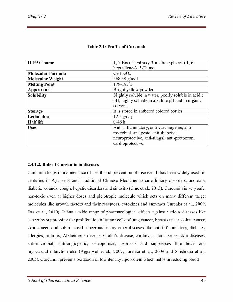
(Fig. 2.1) was determined by Roughley and Whiting in 1973 (Chattopadhyay et al., 2004). Table
2.1 shows the profile of Curcumin.
2.4.1.1. Structure of curcumin
OCH3
OH
O OH
H3CO
HO
Fig. 2.1: Molecular structure of curcumin
Chapter 2 Review of Literature
School of Pharmaceutical Sciences 40
Table 2.1: Profile of Curcumin
IUPAC name 1, 7-Bis (4-hydroxy-3-methoxyphenyl)-1, 6-
heptadiene-3, 5-Dione
Molecular Formula C21H20O6
Molecular Weight 368.38 g/mol
Melting Point 179-183ᵒC
Appearance Bright yellow powder
Solubility Slightly soluble in water, poorly soluble in acidic
pH, highly soluble in alkaline pH and in organic
solvents.
Storage It is stored in ambered colored bottles.
Lethal dose 12.5 g/day
Half life 0-48 h
Uses Anti-inflammatory, anti-carcinogenic, anti-
microbial, analgesic, anti-diabetic,
neuroprotective, anti-fungal, anti-protozoan,
cardioprotective.
2.4.1.2. Role of Curcumin in diseases
Curcumin helps in maintenance of health and prevention of diseases. It has been widely used for
centuries in Ayurveda and Traditional Chinese Medicine to cure biliary disorders, anorexia,
diabetic wounds, cough, hepatic disorders and sinusitis (Cine et al., 2013). Curcumin is very safe,
non-toxic even at higher doses and pleiotropic molecule which acts on many different target
molecules like growth factors and their receptors, cytokines and enzymes (Jurenka et al., 2009,
Das et al., 2010). It has a wide range of pharmacological effects against various diseases like
cancer by suppressing the proliferation of tumor cells of lung cancer, breast cancer, colon cancer,
skin cancer, oral sub-mucosal cancer and many other diseases like anti-inflammatory, diabetes,
allergies, arthritis, Alzheimer’s disease, Crohn’s disease, cardiovascular disease, skin diseases,
anti-microbial, anti-angiogenic, osteoporosis, psoriasis and suppresses thrombosis and
myocardial infarction also (Aggarwal et al., 2007, Jurenka et al., 2009 and Shishodia et al.,
2005). Curcumin prevents oxidation of low density lipoprotein which helps in reducing blood

Chapter 2 Review of Literature
School of Pharmaceutical Sciences 41
cholesterol level (Huang et al., 2011). Curcumin is non-toxic and have decreased side-effects
without any loss to its therapeutic efficacy. Along with the pharmacological actions of curcumin,
it also has the photo stabilizing property to protect photo-labile drugs present in solutions, topical
formulations and soft gelatin capsules (Zandi et al., 2010). Curcumin is used in various health
supplements too. Clinically, curcumin has already been used to reduce post-operative
inflammation. Safety evaluation studies indicate that both turmeric and curcumin are well
tolerated at a very high dose without any toxic effects.
2.4.1.3. Mechanism of action of Curcumin on Inflammatory Bowel Disease [IBD]
IBD- Crohn’s disease and Ulcerative colitis
Inflammatory bowel disease (IBD) is characterized by chronic inflammation in the mucosal
membrane of the small and/or large intestine. Although many treatments have been
recommended for IBD, they do not treat the cause but are effective only in reducing the
inflammation and accompanying symptoms in up to 80% of patients (Philip et al., 2009).
Curcumin is a highly pleiotropic molecule capable of interacting with numerous molecular
targets involved in inflammation. Curcumin modulates the inflammatory response by down-
regulating the activity of cyclooxygenase-2 (COX-2), lipoxygenase, and inducible nitric oxide
synthase (iNOS) enzymes; inhibits the production of the inflammatory cytokines tumor necrosis
factor-alpha (TNF-a), interleukins (Chattopadhyay et al., 2004). Curcumin acts via inhibiting
COX-2 and iNOS by suppressing necrotic factor kappa B (NF-κB) activation. NF-κB, a
ubiquitous eukaryotic transcription factor, is involved in regulation of inflammation, cellular
proliferation, transformation, and tumorigenesis. Curcumin suppresses NF-κB activation and
proinflammatory gene expression by blocking phosphorylation of inhibitory factor I-kappa B
kinase (IκB). Suppression of NF-κB activation subsequently down-regulates COX-2 and iNOS
expression, inhibiting the inflammatory process and tumorigenesis (Chattopadhyay et al., 2004,
Jobin et al., 1999 and Surh et al., 2001).

Chapter 2 Review of Literature
School of Pharmaceutical Sciences 42
2.4.2. FLURBIPROFEN
Flurbiprofen, a propionic acid derivative, is a nonsteroidal anti-inflammatory drug (NSAID) with
antipyretic and analgesic activity (Han et al., 2008). Oral formulations of flurbiprofen may be
used for the symptomatic treatment of rheumatoid arthritis, osteoarthritis and other inflammatory
conditions. Flurbiprofen may also be used topically prior to ocular surgery to prevent or reduce
intraoperative miosis. Flurbiprofen is structurally and pharmacologically related to fenoprofen,
ibuprofen, and ketoprofen (Fig. 2.2). Table 2.2 shows the profile of Flurbiprofen.
Category:
• Anti-inflammatory Agent
• Cyclooxygenase Inhibitors
• Analgesics
• Analgesics, Non-Narcotic
• Antipyretics
• Non-steroidal Anti-inflammatory Drugs (NSAIDs)
2.4.2.1. Structure of Flurbiprofen
OH
F
O
Fig. 2.2: Molecular structure of Flurbiprofen

Chapter 2 Review of Literature
School of Pharmaceutical Sciences 43
Table 2.2: Profile of Flurbiprofen
IUPAC name 2-(3-fluoro-4-phenylphenyl) propanoic acid
Molecular Formula C15H13FO2
Molecular Weight 244.2609g/mol
Melting Point 114-117ᵒC
Appearance White to slightly yellow crystalline powder
Solubility Slightly soluble in water, soluble in organic
solvents.
Storage Stored at room temperature
Half life 4.7-5.7 h
pka value 4.22
Log P 4.24
Uses Anti-inflammatory, anti-pyretic, analgesic.
2.4.2.2. Mechanism of Action: The anti-inflammatory effect of flurbiprofen occurs via
reversible inhibition of cyclooxygenase (COX), the enzyme responsible for the conversion of
arachidonic acid to prostaglandin G2 (PGG2) and PGG2 to prostaglandin H2 (PGH2) in the
prostaglandin synthesis pathway (Kawadkar et al., 2012). This effectively decreases the
concentration of prostaglandins involved in inflammation, pain, swelling and fever. Flurbiprofen
is a non-selective COX inhibitor and inhibits the activity of both COX-1 and COX -2.
2.4.2.3. Pharmacokinetics
Absorption: Flurbiprofen is rapidly and almost completely absorbed following oral
administration. Peak plasma concentrations are reached 0.5 - 4 hours after oral administration
(Yan-Mei et al., 2009).
Distribution: Distribution into human body tissues and fluids not fully characterized. Distribute
into milk very small amount.
Plasma Protein Binding: >99% (principally albumin).
Metabolism: Extensively metabolized. CYP2C9 plays an important role in the metabolism of
flurbiprofen to its major metabolite, 4′-hydroxyflurbiprofen, which has weak anti-inflammatory
activity.

Chapter 2 Review of Literature
School of Pharmaceutical Sciences 44
Excretion: Following oral dosing, approximately 70% of the flurbiprofen dose is eliminated in
urine as parent drug and metabolites, with <3% excreted as unchanged drug.
2.5. EXCIPIENTS REVIEW
2.5.1. Chitosan: Chitosan is a linear polysaccharide, composed of glucosamine and N-
acetylglucosamine, produced by partial deacetylation of chitin by alkaline or enzymatic
hydrolysis (Shaji et al., 2010). Chitin is the major component of the exoskeleton of crustaceans,
insects, cell wall of fungi and yeast. For the commercial production, chitin from shells of prawns,
crabs, or other crustacean is used. Chitosan is a polysaccharide (Fig. 2.3) obtained by N-
deacetylation from chitin and it has been widely investigated as a carrier for novel delivery
systems owing to its biodegradability, biocompatibility and safety. Chitosan (low mol. wt.,
viscosity 20-200 cP) is prone to degradation by the colonic microflora and therefore it can be
used for colon specific drug delivery incorporated in pH sensitive polymer (Rabiskova et al.,
2012). Table 2.3 shows the profile of chitosan.
2.5.1.1. Structure of Chitosan
O
O
O
O
O
CH2OH
OHOH
HN
OH
HN
CH2OH
OH
HN
OH
CH2OH
CH3 CH3 CH3
O O O
n
Fig. 2.3: Molecular structure of Chitosan

Chapter 2 Review of Literature
School of Pharmaceutical Sciences 45
Table 2.3: Profile of Chitosan
Synonyms 2-Amino-2-deoxy-(1,4)-β-D-glucopyranan; deacetylated chitin;
deacetylchitin; β-1,4-poly-D-glucosamine; poly-D-glucosamine;
poly-(1,4-β-D-glucopyranosamine).
Molecular weight 10 000–1 000 000 Daltons
Functional category Coating agent; disintegrant; film-forming agent; mucoadhesive;
tablet binder; viscosity-increasing agent.
Applications Chitosan is used in cosmetics and is under investigation for use in
a number of pharmaceutical formulations. The suitability and
performance of chitosan as a component of pharmaceutical
formulations for drug delivery applications has been investigated
in numerous studies. These include controlled drug delivery
applications, use as a component of mucoadhesive dosage forms,
rapid release dosage forms, improved peptide delivery, colonic
drug delivery systems and use for gene delivery. Chitosan has
been processed into several pharmaceutical forms including gels,
films, beads, microspheres, tablets and coatings for liposomes.
Solubility Sparingly soluble in water; practically insoluble in ethanol (95%),
other organic solvents, and neutral or alkali solutions at pH above
approximately 6.5.
Storage and Stability Chitosan powder is a stable material at room temperature,
although it is hygroscopic after drying. Chitosan should be stored
in a tightly closed container in a cool, dry place. The PhEur 2005
specifies that chitosan should be stored at a temperature of 2–8°C.
2.5.2. Eudragit S 100 [Polymethacrylates]: Eudragit is anionic copolymer based on
methacrylic acid and methacrylate (Fig. 2.4). It is a pH sensitive polymer mainly used for colon
targeting (Pandey et al., 2012). Polymethacrylates are primarily used in oral capsule and tablet
formulations as film-coating agents. Depending on the type of polymer used, films of different
solubility characteristics can be produced. Eudragit E is used as a plain or insulating film former;
it is soluble in gastric fluid below pH 5. Eudragit L, S and FS types are used as enteric coating
agents because they are resistant to gastric fluid. Different types are available that are soluble at
different pH values: e.g. Eudragit L is soluble at pH > 6; Eudragit S and FS are soluble at pH >
7. Table 2.4 shows the profile of Eudragit S 100.
Chapter 2 Review of Literature
School of Pharmaceutical Sciences 46
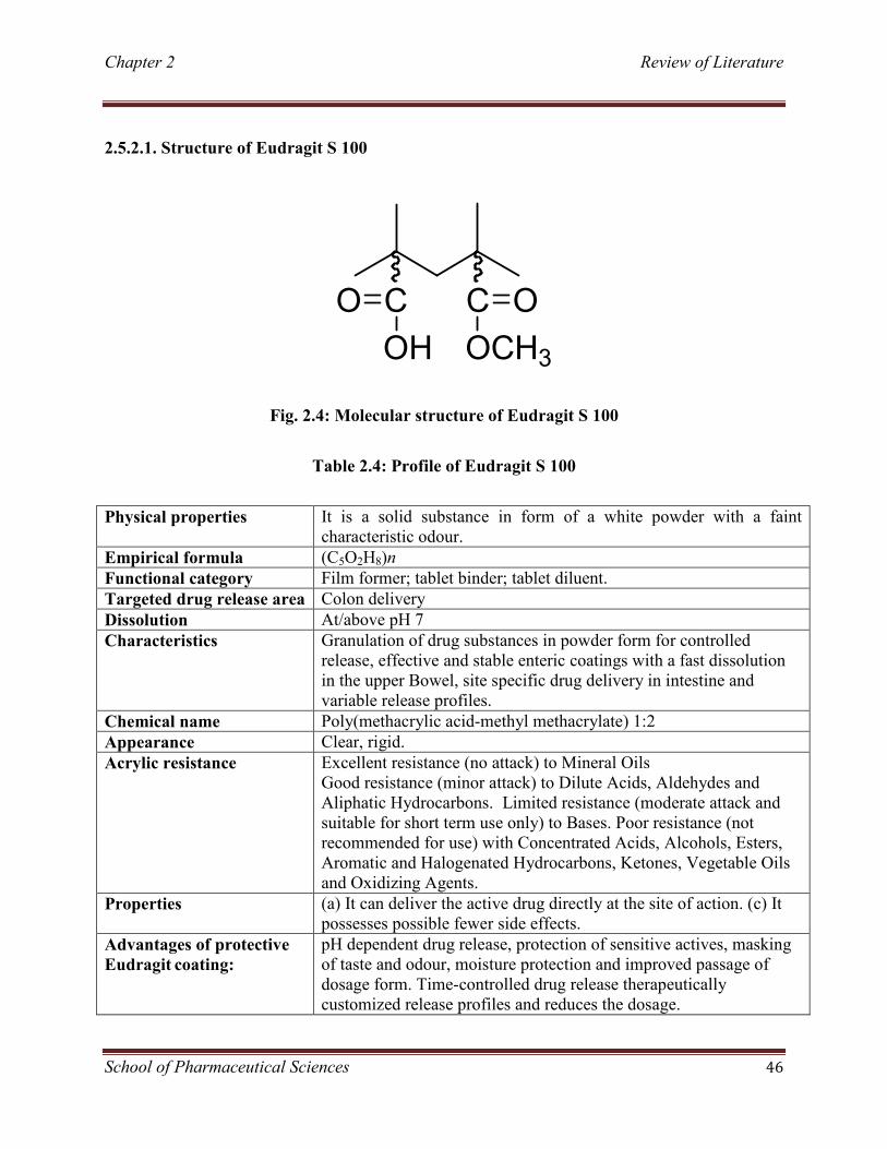
2.5.2.1. Structure of Eudragit S 100
C CO
OH
O
OCH3
Fig. 2.4: Molecular structure of Eudragit S 100
Table 2.4: Profile of Eudragit S 100
Physical properties It is a solid substance in form of a white powder with a faint
characteristic odour.
Empirical formula (C5O2H8)n
Functional category Film former; tablet binder; tablet diluent.
Targeted drug release area Colon delivery
Dissolution At/above pH 7
Characteristics Granulation of drug substances in powder form for controlled
release, effective and stable enteric coatings with a fast dissolution
in the upper Bowel, site specific drug delivery in intestine and
variable release profiles.
Chemical name Poly(methacrylic acid-methyl methacrylate) 1:2
Appearance Clear, rigid.
Acrylic resistance Excellent resistance (no attack) to Mineral Oils
Good resistance (minor attack) to Dilute Acids, Aldehydes and
Aliphatic Hydrocarbons. Limited resistance (moderate attack and
suitable for short term use only) to Bases. Poor resistance (not
recommended for use) with Concentrated Acids, Alcohols, Esters,
Aromatic and Halogenated Hydrocarbons, Ketones, Vegetable Oils
and Oxidizing Agents.
Properties (a) It can deliver the active drug directly at the site of action. (c) It
possesses possible fewer side effects.
Advantages of protective
Eudragit coating:
pH dependent drug release, protection of sensitive actives, masking
of taste and odour, moisture protection and improved passage of
dosage form. Time-controlled drug release therapeutically
customized release profiles and reduces the dosage.

Chapter 2 Review of Literature
School of Pharmaceutical Sciences 47
2.5.3. Ethanol: Used as an organic solvent for drug and polymers (Fig. 2.5) (Merck Index,
2006). Table 2.5 shows the profile of ethanol.
2.5.3.1. Structure of Ethanol
OH
Fig. 2.5: Molecular structure of Ethanol
Table 2.5: Profile of Ethanol
Synonyms Ethyl alcohol, ethyl hydroxide, grain alcohol, methyl carbinol.
Empirical formula C2H5OH
Molecular weight 46.07g/mol
Functional category Anti-microbial preservative [10% v/v], disinfectant [90% v/v],
skin penetrant, solvent [up to 85% v/v].
Applications Ethanol and aqueous ethanol solutions of various concentrations
are widely used in pharmaceutical formulations and cosmetics;.
Although ethanol is primarily used as a solvent, it is also
employed in solutions as an antimicrobial preservative. Topical
ethanol solutions are also used as penetration enhancers and as
disinfectants [Karabit et. al., 1989, Liu et. al., 1991 and Chiori et.
al., 1983].
Uses Solvent in film coating, solvent in injectable solutions, solvent in
oral liquids at variable concentrations (%v/v) and as solvent in
topical products 60-90% v/v.
Boiling Point 78.15◦C
Flammability Readily flammable, burns with blue and smokeless flame.
Solubility Miscible with chloroform, ether, glycerin, and water.
Storage and Stability Aqueous ethanol solutions may be sterilized by autoclaving or by
filtration and should be stored in airtight containers, in a cool
place.
2.5.4. SPAN 80: Sorbitan monoesters are a series of mixtures of partial esters of sorbitol and its
mono- and dianhydrides with fatty acids (Fig. 2.6). Sorbitan diesters are a series of mixtures of
partial esters of sorbitol and its monoanhydride with fatty acids. Sorbitan esters are widely used

Chapter 2 Review of Literature
School of Pharmaceutical Sciences 48
in cosmetics, food products, and pharmaceutical formulations as lipophilic nonionic surfactants.
They are mainly used in pharmaceutical formulations as emulsifying agents in the preparation of
creams, emulsions, and ointments for topical application. When used alone, sorbitan esters
produce stable water-in-oil emulsions and microemulsions but are frequently used in
combination with varying proportions of a polysorbate to produce water-in-oil or oil-in-water
emulsions or creams of varying consistencies (Merck Index, 2006). Table 2.6 shows the profile
of span 80.
2.5.4.1. Structure of SPAN 80
O
O
OH
OH
O
OH
Fig. 2.6: Molecular structure of SPAN 80
Table 2.6: Profile of SPAN 80
Synonyms Sorbitan monooleate, ionets 80, montan 80, glycomulo, sorgen 80
Empirical Formula C24H44O6
Molecular weight 428g/mol
Appearance Liquid, clear, viscous, yellow coloured
Stability Stable and combustible.
Functional Category Emulsifying agent, nonionic surfactant, solubilizing agent,
wetting and dispersing/suspending agent.
Solubility Generally soluble or dispersible in oils, they are also soluble in
most organic solvents. In water, although insoluble, they are
generally dispersible.
Applications Used in cosmetics, food products, and pharmaceutical
formulations as lipophilic nonionic surfactants.
Chapter 2 Review of Literature
School of Pharmaceutical Sciences 49
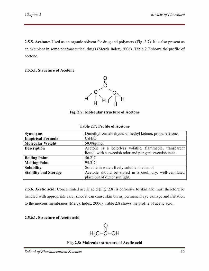
2.5.5. Acetone: Used as an organic solvent for drug and polymers (Fig. 2.7). It is also present as
an excipient in some pharmaceutical drugs (Merck Index, 2006). Table 2.7 shows the profile of
acetone.
2.5.5.1. Structure of Acetone
C
C C
HH
H H
HH
O
Fig. 2.7: Molecular structure of Acetone
Table 2.7: Profile of Acetone
Synonyms Dimethylformaldehyde; dimethyl ketone; propane 2-one.
Empirical Formula C3H6O
Molecular Weight 58.08g/mol
Description Acetone is a colorless volatile, flammable, transparent
liquid, with a sweetish odor and pungent sweetish taste.
Boiling Point 56.2ᵒ C
Melting Point 94.3ᵒ C
Solubility Soluble in water, freely soluble in ethanol
Stability and Storage Acetone should be stored in a cool, dry, well-ventilated
place out of direct sunlight.
2.5.6. Acetic acid: Concentrated acetic acid (Fig. 2.8) is corrosive to skin and must therefore be
handled with appropriate care, since it can cause skin burns, permanent eye damage and irritation
to the mucous membranes (Merck Index, 2006). Table 2.8 shows the profile of acetic acid.
2.5.6.1. Structure of Acetic acid
CH3C
O
OH
Fig. 2.8: Molecular structure of Acetic acid

Chapter 2 Review of Literature
School of Pharmaceutical Sciences 50
Table 2.8: Profile of Acetic acid
Nonproprietary Names BP: Glacial acetic acid, JP: Glacial acetic acid,
USP: Glacial acetic acid
Synonyms Ethanoic acid, Vinegar acid
Empirical formula and Molecular
weight
C2H4O2, 60.05
Functional category Acidifying agent
Description It occurs as a crystalline mass or a clear, colorless
volatile solution with a pungent odor.
Solubility Miscible with ethanol, ether, glycerin, water, and
other fixed and volatile oils.
Stability and Storage conditions Stored in an airtight container in a cool, dry place.
Boiling and Melting point 118°C (b. p.), 17°C (m. p.)
2.5.6. Carboxy methyl cellulose (CMC): Carboxymethylcellulose is widely used in oral and
topical pharmaceutical formulations, primarily for its viscosity-increasing properties (Fig. 2.9).
Viscous aqueous solutions are used to suspend powders intended for either topical application or
oral and parenteral administration. Carboxymethylcellulose sodium may also be used as a tablet
binder and disintegrant and to stabilize emulsions (Merck Index, 2006). Table 2.9 shows the
profile of carboxy methyl cellulose.
2.5.6.1. Structure of Carboxy methyl cellulose
O
OH
OH
CH2OCH2COOH
O
O
O
CH2OCH2COOH
OH
Fig. 2.9: Molecular structure of Carboxy methyl cellulose

Chapter 2 Review of Literature
School of Pharmaceutical Sciences 51
Table 2.9: Profile of Carboxy methyl cellulose
Nonproprietary Names BP: Carmellose Sodium, JP: Carmellose Sodium,
USP: Carboxy MethylCellulose
Synonyms cellulose gum; CMC
Empirical Formula and Mol. wt. [C6H7O2(OH)x(OCH2COOH)y]n 90 000–700 000
Functional Category Suspending agent, viscosity-increasing agent and
water-absorbing agent.
Description It occurs as a white to almost white, odorless, granular
powder.
Solubility Practically insoluble in acetone, ethanol (95%), ether
and toluene.
Stability and Storage Conditions
Aqueous solutions stored for prolonged periods should
contain an antimicrobial preservative.
The bulk material should be stored in a well-closed
container in a cool and dry place.
Melting point Browns at approximately 227°C, and chars at
approximately 252°C.
Uses Emulsifying agent, Gel forming agent.
2.5.7. Liquid Paraffin: It is used primarily as an excipient in topical pharmaceutical
formulations where its emollient properties are exploited in ointment bases (Fig. 2.10). It is also
used in ophthalmic formulations. Light mineral oil is additionally used in oil-in-water and
polyethylene glycol/glycerol emulsions, as a solvent and lubricant in capsules and tablets, as a
solvent and penetration enhancer in transdermal preparations and as the oily medium used in the
microencapsulation of many drugs (Merck Index, 2006). Table 2.10 shows the profile of liquid
paraffin.
2.5.7.1. Structure of Liquid Paraffin
CnH2n 2
Fig. 2.10: Molecular structure of Liquid Paraffin

Chapter 2 Review of Literature
School of Pharmaceutical Sciences 52
Table 2.10: Profile of Liquid Paraffin
Nonproprietary Names BP: Light liquid paraffin
JP: Light liquid paraffin
USPNF: Light mineral oil
Synonyms Light white mineral oil, light liquid petrolatum.
Empirical Formula and Molecular
Weight
Light mineral oil is a mixture of refined liquid saturated
hydrocarbons obtained from petroleum. It is less viscous
and has a lower specific gravity than mineral oil.
Functional Category Emollient, lubricant.
Description Light mineral oil is a transparent, colorless, viscous oily
liquid without fluorescence in daylight.
Solubility
Soluble in chloroform, ether, and hydrocarbons;
sparingly soluble in ethanol (95%); practically insoluble
in water.
Storage Conditions Light mineral oil should be stored in an airtight container
in a cool, dry place and protected from light.
Boiling point >360°C
Uses Ophthalmic ointments, Otic preparations, Topical
emulsions.
2.5.8. Zinc Chloride: ZnCl2 is hygroscopic and even deliquescent (Fig. 2.11). Samples should
therefore be protected from sources of moisture including the water vapor present in ambient air.
Zinc chloride finds wide application in textile processing, metallurgical fluxes and chemical
synthesis (Merck Index, 2006). Table 2.11 shows the profile of zinc chloride.
2.5.8.1. Structure of Zinc Chloride
Zn
ClCl
Fig. 2.11: Molecular structure of Zinc Chloride

Chapter 2 Review of Literature
School of Pharmaceutical Sciences 53
Table 2.11: Profile of Zinc Chloride
Synonyms Zinc dichloride fume.
Empirical Formula and Molecular Weight ZnCl2, 136.3
Description Zinc chloride is a white crystalline solid.
Solubility Acetone: slightly soluble and water solubility
435% at 70° F.
Stability and Storage Conditions Zinc chloride can be stored in properly closed
containers under cold to warm environment, in
a temperature range of 2 to 40 degree Celsius.
Boiling point and Melting point 1350°F (b.p.), 293°C (m.p.)
Uses It is used for preserving wood, in soldering
fluxes, as a catalyst in chemical metals.