Embed Size (px)
Citation preview
2/4/14
1
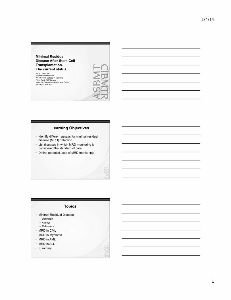
Minimal Residual Disease After Stem Cell Transplantation. The current status Sergio Giralt, MD Professor of Medicine Weill Cornell College of Medicine Chief, Adult BMT Service Memorial Sloan Kettering Cancer Center New York, New York
Learning Objectives
• Identify different assays for minimal residual disease (MRD) detection
• List diseases in which MRD monitoring is considered the standard of care
• Define potential uses of MRD monitoring
Topics
• Minimal Residual Disease – Definition – Assays – Relevance
• MRD in CML • MRD in Myeloma • MRD in AML • MRD in ALL • Summary
2/4/14
2
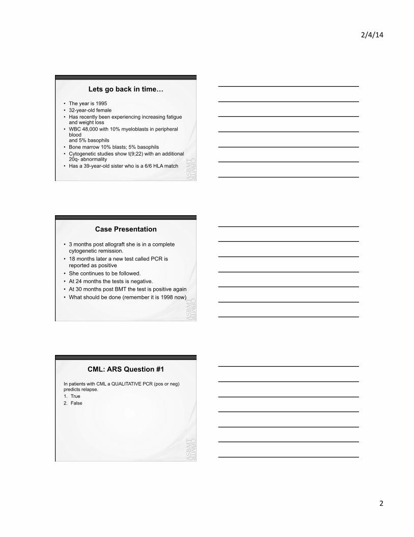
Lets go back in time…
• The year is 1995 • 32-year-old female • Has recently been experiencing increasing fatigue
and weight loss • WBC 48,000 with 10% myeloblasts in peripheral
blood and 5% basophils
• Bone marrow 10% blasts; 5% basophils • Cytogenetic studies show t(9;22) with an additional
20q- abnormality • Has a 39-year-old sister who is a 6/6 HLA match
Case Presentation
• 3 months post allograft she is in a complete cytogenetic remission.
• 18 months later a new test called PCR is reported as positive
• She continues to be followed. • At 24 months the tests is negative. • At 30 months post BMT the test is positive again • What should be done (remember it is 1998 now)
CML: ARS Question #1
In patients with CML a QUALITATIVE PCR (pos or neg) predicts relapse. 1. True 2. False
2/4/14
3
CML: ARS Question #2
In this patient what would the appropriate next step be with this positive test for minimal residual disease (MRD)? 1. Request donor lymphocyte infusion (DLI) as soon as possible 2. Start interferon 3. Wait until 1999 and get her on a protocol with the new STI571 4. Perform bone marrow aspiration and determine whether there was cytogenetic evidence of disease
ARS Question #3: What is MRD anyway?
Which is the correct definition for MRD? 1.Minimal residual disease refers to disease that is left over after treatment that only can be seen by an expert pathologist. 2. Minimal residual disease only relates to CML and represents presence of disease at a 1 in 100000 level. 3.Minimal residual disease is the name given to small numbers of leukemic or other tumor cells detected by very sensitive methods that remain in the patient during treatment, or after treatment when the patient is in remission. It is the major cause of relapse in cancer and leukemia.
MRD-Definition When in doubt ask WIKIPEDIA
• “Minimal residual disease is the name given to small numbers of leukaemic cells that remain in the patient during treatment, or after treatment when the patient is in remission. It is the major cause of relapse in cancer and leukaemia.”
2/4/14
4
Minimal Residual Disease
• Not totally true • MRD has usually referred to disease detected by
non-traditional methods (xray or pathology). The two most commonly used methods are flow cytometry and polymerase chain reaction.
• MRD by flow cytometry or PCR predicts for a higher risk of relapse after chemotherapy and also after transplantation in SOME but NOT ALL diseases.
MRD Detection
• Cytogenetic methods, including FISH – Generally not sensitive enough to be real minimal
residual disease measure • Flow cytometry
– Based on aberrant antigen expression (“Leukemia-associated immunophenotype”)
• PCR – Adaptable to different targets – Can measure clonal abnormality or abnormal
expression
Polymerase Chain Reaction
• Developed in 1983 by Kary Mullis, PCR is now a common and indispensable technique for DNA cloning and sequencing which has ubiquitous applications in diagnosis of hereditary diseases, forensic sciences, minimal residual disease detection and infectious diseases.
• In 1993, Mullis and Michael Smith were awarded the Nobel Prize for their work on PCR.
• The method relies on thermal cycling, consisting of cycles of repeated heating and cooling of the reaction for DNA melting and enzymatic replication of the DNA. Primers (short DNA fragments) containing sequences complementary to the target region along with a DNA Polymerase
• Almost all PCR applications employ a heat-stable DNA polymerase, such as Taq polymerase (an enzyme originally isolated from the bacterium Thermus aquaticus
2/4/14
5
PCR DNA-based tests
Detect tumor specific DNA sequences using the polymerase chain reaction (PCR), a highly sensitive technique. Useful for chromosomal translocation, microsatellites (chimerism), immunoglobulin and T cell receptor rearrangements.
RNA-based tests Detect tumor specific RNA sequence. Uses reverse transcription of the RNA followed by polymerase chain reaction. RNA-based tests are normally utilized when a DNA test is impractical. BCR-ABL most commonly used The markers used for RNA-based testing are almost exclusively chromosomal translocations such as t(9;22) BCR-ABL, t(15;17) PML-RARA and t(12;21) ETV-RUNX1 (TEL-AML1).
Types of PCR Methods
• Antigen receptor PCR – Most suited to lymphoid malignancies
• Fusion transcript PCR – Several tumor types but only limited subsets of most
tumors (CML excepted) • PCR for gene mutations
– AML subsets, e.g. FLT3 or NPM1 • mRNA PCR
– Suitable for upregulated genes, e.g. WT1
• Immunological tests • Flow cytometry is an immunological-based testing of
leukemias or other cancers utilizes proteins on the surface of the cells. Leukemic and other cancer cells often show quite unusual and unique combinations (leukemic phenotype) of these cell surface proteins. These proteins can be stained with fluorescent dye labeled antibodies and detected using flow cytometry. The limit of detection of immunological tests is generally about 1 in 10,000 cells and cannot be used on cancers that don’t have an identifiable and stable phenotype
2/4/14
6
Flow Cytometry-WIKIPEDIA
• Flow cytometry is a laser-based, biophysical technology employed in cell counting, sorting, biomarker detection and protein engineering.
• Principle: suspend cells in a stream of fluid and passing them by an electronic detection apparatus. It allows simultaneous multi-parametric analysis of the physical and chemical characteristics of up to thousands of particles per second.
• Flow cytometry is routinely used in the diagnosis of health disorders, especially blood cancers.
• History • Mack Fulwyler was the inventor of the forerunner to today's flow cytometers • Wolfgang Gohde developed in 1968 fluorescent based flow cytometry
• Patient-specific testing • Patient-specific MRD detection using
immunoglobulin (IG) or T cell receptors (TCR). • Measures MRD in tumors that do not contain a
chromosomal translocation or other specific marker. • These tests are very specific, and detect leukaemic
cells at levels down to one cell in a million, though the limit typically achieved is 1 in 10,000 to 1 in 100,000 cells. For comparison, the limit of what one can detect using traditional morphologic examinations using a microscope is about 1 cell in 100.
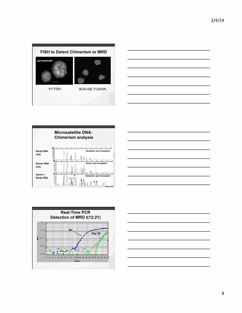
Methods For Detecting Chimerism
• XY FISH – Easy, but not very sensitive; only applicable to a subset of
patients • PCR methods
– Microsatellite markers(short tandem repeats (STRs) or variable number tandem repeats (VNTR))
• Informative in nearly all cases; sensitivity around 1-5% • Most widely used
– TaqMan qPCR against single nucleotide polymorphisms • Sensitivity 0.1% or better, and quantitation better but perhaps
not informative as often; more limited data – Y chromosome PCR even more sensitive (1/105)
• Lineage-specific chimerism more specific
2/4/14
7
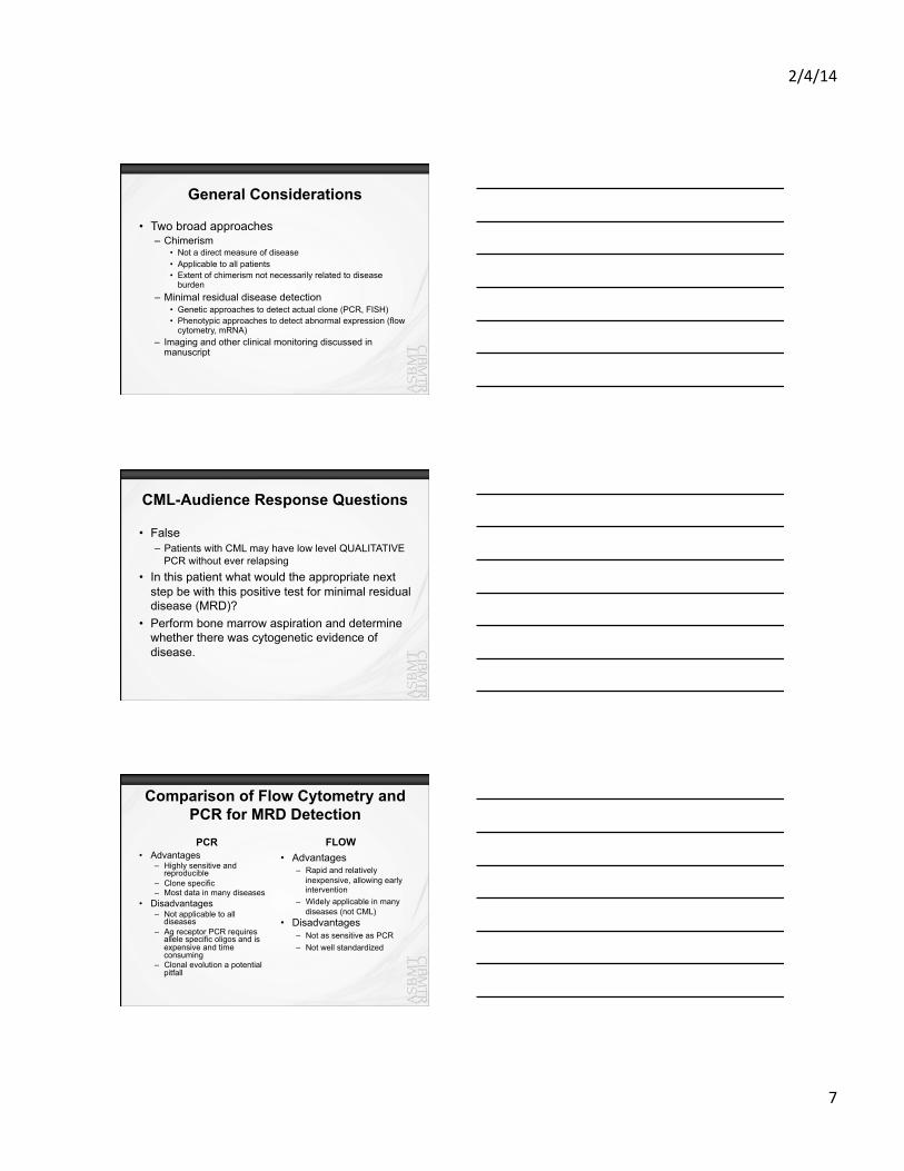
General Considerations
• Two broad approaches – Chimerism
• Not a direct measure of disease • Applicable to all patients • Extent of chimerism not necessarily related to disease
burden – Minimal residual disease detection
• Genetic approaches to detect actual clone (PCR, FISH) • Phenotypic approaches to detect abnormal expression (flow
cytometry, mRNA) – Imaging and other clinical monitoring discussed in
manuscript
CML-Audience Response Questions
• False – Patients with CML may have low level QUALITATIVE
PCR without ever relapsing • In this patient what would the appropriate next
step be with this positive test for minimal residual disease (MRD)?
• Perform bone marrow aspiration and determine whether there was cytogenetic evidence of disease.
Comparison of Flow Cytometry and PCR for MRD Detection
PCR • Advantages
– Highly sensitive and reproducible
– Clone specific – Most data in many diseases
• Disadvantages – Not applicable to all
diseases – Ag receptor PCR requires
allele specific oligos and is expensive and time consuming
– Clonal evolution a potential pitfall
FLOW • Advantages
– Rapid and relatively inexpensive, allowing early intervention
– Widely applicable in many diseases (not CML)
• Disadvantages – Not as sensitive as PCR – Not well standardized
2/4/14
8
FISH to Detect Chimerism or MRD
BCR-ABL FUSION XY FISH
Recipient: pre-‐transplant
Donor: pre-‐transplant
Recipient: post-‐transplant
Microsatellite DNA: Chimerism analysis
Recip DNA only
Donor DNA only
Donor + Recip DNA
K Berg, JHU
Dx Day 28
Real-Time PCR Detection of MRD t(12;21)
2/4/14
9
Quantitating Leukaemic Cell Load in CML
Num
ber o
f leu
kem
ic c
ells
1012
1
106
CCR
108
1010
102
104
3 log reduction
MCR
Limits of detection
4 log reduction
Cytogenetic response
Q-PCR result
CHR
Complete Molecular Remission
BC
R-A
BL/A
BL ratio (%
)
0.0001
0.001
0.01
0.1
1
10
100
<0.0001
Quantitative RT-PCR and relapse post BMT
0 12 24 36 48 60 72 84 96 108 120
Months post BMT
0
20
40
60
80
100
n = 113 p < 0.0001
Prob
abili
ty o
f rel
apse
Low or falling BCR-ABL levels (n =80) < 0.02% BCR-ABL/ABL
High or rising BCR-ABL levels (n =33) > 0.02% BCR-ABL/ABL
Complete molecular response to DLI
0 6 12 18 24 30 0
20
40
60
80
100
61%
47%
87%
Overall
Haematologic relapse
Molecular/Cytogenetic relapse
Months post DLI
Mol
ecul
ar re
mis
sion
(%)
p = 0.004
2/4/14
10
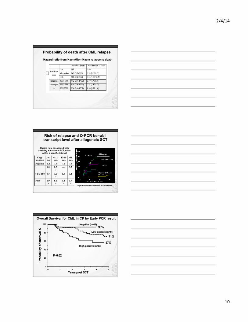
Probability of death after CML relapse
Hazard ratio from Haem/Non-Haem relapse to death
Copy number
3-6 mo.
6-12 mo.
12-18 mo.
>18 mo.
Negative 1.0 1.0 1.0 1.0 1 1.0 2.5
* ---- 3.2
>1 to 100 0.7
3.6
* 1.9
3.4
>100 1.9 *
5.1 *
3.2 *
3.9 *
Hazard ratio associated with attaining a maximum PCR value
within a specific interval
Days after max PCR achieved at 6-12 months
Risk of relapse and Q-PCR bcr-abl transcript level after allogeneic SCT
0 1 2 3 4 5 Y e a r s p o s t S C T
0
2 0
4 0
6 0
8 0
1 0 0
P r o b
a b i l i
t y o
f s u r
v i v a
l %
L o w p o s i t i v e ( n = 1 4 )
H i g h p o s i t i v e ( n = 6 3 )
7 1 %
5 7 %
9 3 % N e g a t i v e ( n = 6 1 )
P = 0 . 0 2
Overall Survival for CML in CP by Early PCR result
2/4/14
11
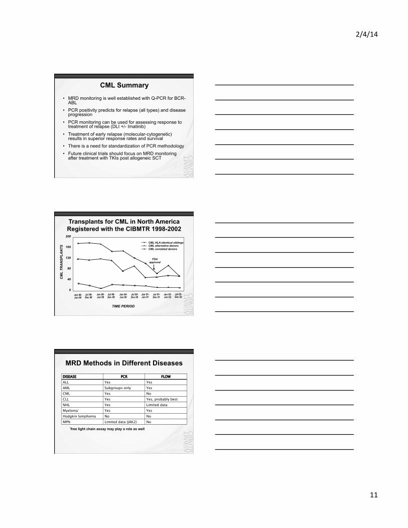
CML Summary
• MRD monitoring is well established with Q-PCR for BCR-ABL
• PCR positivity predicts for relapse (all types) and disease progression
• PCR monitoring can be used for assessing response to treatment of relapse (DLI +/- Imatinib)
• Treatment of early relapse (molecular-cytogenetic) results in superior response rates and survival
• There is a need for standardization of PCR methodology • Future clinical trials should focus on MRD monitoring
after treatment with TKIs post allogeneic SCT
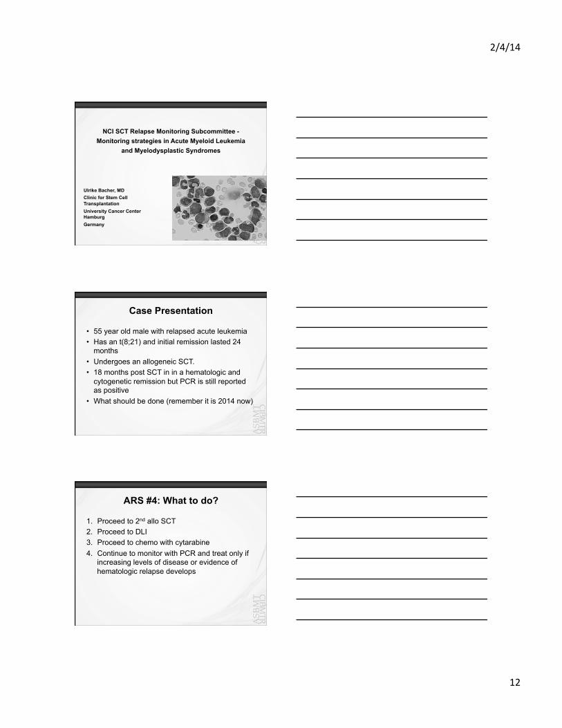
Transplants for CML in North America Registered with the CIBMTR 1998-2002
TIME PERIOD
0
40
80
120
160
200
CM
L TR
AN
SPLA
NTS
CML HLA-identical siblings CML alternative donors CML unrelated donors
Jan 98- Jun 98
Jul 98- Dec 98
Jan 99- Jun 99
Jul 99- Dec 99
Jul 00- Dec 00
Jan 01- Jun 01
Jul 01- Dec 01
Jan 00- Jun 00
Jan 02- Jun 02
Jul 02- Dec 02
FDA approval
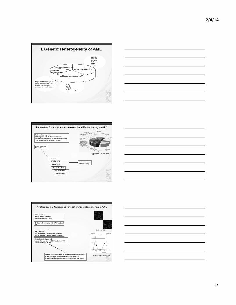
MRD Methods in Different Diseases
DISEASE PCR FLOW ALL Yes Yes AML Subgroups only Yes CML Yes No CLL Yes Yes, probably best NHL Yes Limited data Myeloma* Yes Yes Hodgkin lymphoma No No MPN Limited data (JAK2) No
*free light chain assay may play a role as well
2/4/14
12
NCI SCT Relapse Monitoring Subcommittee - Monitoring strategies in Acute Myeloid Leukemia
and Myelodysplastic Syndromes
Ulrike Bacher, MD Clinic for Stem Cell Transplantation University Cancer Center Hamburg Germany
Case Presentation
• 55 year old male with relapsed acute leukemia • Has an t(8;21) and initial remission lasted 24
months • Undergoes an allogeneic SCT. • 18 months post SCT in in a hematologic and
cytogenetic remission but PCR is still reported as positive
• What should be done (remember it is 2014 now)
ARS #4: What to do?
1. Proceed to 2nd allo SCT 2. Proceed to DLI 3. Proceed to chemo with cytarabine 4. Continue to monitor with PCR and treat only if
increasing levels of disease or evidence of hematologic relapse develops
2/4/14
13
Balanced
Normal karyotype - 45%
Balanced translocations - 25%
Unbalanced karyotypes - 15%
FLT3-ITD FLT3-TKD MLL-PTD RAS NPM1 TET2,..
Single monosomies (-7, -Y, -X,…) Single trisomies (+8, +21, +11,..) Structural alterations Unbalanced translocations
Complex aberrant - 15%
t(8;21) inv(16) t(15;17) 11q23 rearrangements
I. Genetic Heterogeneity of AML
Reciprocal rearrangements: MRD diagnostic with RQ-PCR well established “favorable“ rearrangements >> minor role for allo-SCT Other suitable markers for the SCT setting?
NPM1: 55%?
FLT-ITD: 35%?
NRAS: 10%
FLT3-TKD: 55%
MLL-PTD: 10%
Parameters for post-transplant molecular MRD monitoring in AML?
RUNX1: 10%
Normal karyotype: 45% of all cases
Post-transplant MRD monitoring?
Haferlach et al., Curr Opin Hemat, 2006
NPM1 mutation: - 55% in normal karyotype - association with FLT3-ITD
Nucleophosmin1 mutations for post-transplant monitoring in AML
Thiede et al., 2006
Morphological relapse: n=9 Preceded by increase of NPM1A mutation: 100% Interval: ø 24 days (12-38)
Post Transplant: NPM1A negative > indicator for remission NPM1A positive > always relapse post-SCT
Bacher et al., Exp Hematol, 2009 NPM1A-mutation> suitable for post-transplant MRD monitoring in AML (although underrepresented in SCT patients) Short interval between increase of mutation load and relapse!
13 stem cell recipients with NPM1 mutated AML
2/4/14
14
FLT3 mutations for post-transplant MRD monitoring in AML?
Scholl et al., 2005: 4 AML patients + FLT3-ITD/TKD-mutation >> RQ-PCR in the post-transplant period
Strong correlation of the mutation load with post-transplant outcomes
- Instability of FLT3 mutations at relapse? - FLT3-ITD: RQ-PCR requires design of specific primers
Frohling et al., Cancer Cell, 2007
FLT3 mut: ~40% in normal karyotype AML Adverse prognosis
Scholl et al., Clin Cancer Res, 2005
?
Flow cytometry for post-transplant monitoring in AML?
Leukemia associated immunophenotypes in AML: - “Cross-lineage“ expression: CD7 - Loss of antigens: HLA-DR - Aberrant levels of expression
Diez-Campelo et al., 2008 • Flow cytometry in 41 stem cell recipients with
AML/MDS • ≥10-3 leukemia cells at 3 months: 4-yr EFS < 20% • < 10-3 leukemia cells: >70%
Flow cytometry might contribute to MRD in the post-transplant period of AML, but very few studies have so far been performed.
Diez-Campelo et al., Am J H, 2008
II. Genetic heterogeneity of MDS
Duesseldorf Registry, 2000
Cytogenetic alterations in MDS AML1/RUNX1
15%
no marker detected51%
TET220%
NRAS7%FLT3-ITD
3%
MLL-PTD3%
KITD8161%
Molecular mutations in advanced MDS
2/4/14
15
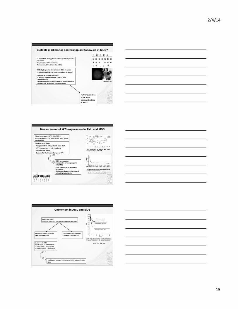
Suitable markers for post-transplant follow-up in MDS?
So far, no MRD strategy for the follow-up of MDS patients is available Only exception: WT1 monitoring (Tamura et al., 2006; Cilloni et al., 2003)
MDS: Cytogenetic alterations in 55% of cases >> Interphase FISH as post-transplant strategy?
Fuehrer et al., Int J Mol Med, 2005: 23 pediatric patients (of those, 8 AML, 2 MDS) > Interphase FISH > Stable remission: n=19 >> no aberrant interphase nuclei > relapse: n=4 >> aberrant interphase nuclei
Further evaluation in the post-transplant setting of MDS?
T. Haferlach, MLL
Measurement of WT1-expression in AML and MDS
Wilms tumor gene (WT1): RQ-PCR >> overexpression in AML/MDS and other malignancies
WT1 expression in patients with post-transplant relapse of the AML
WT1 expression in AML patients with stable post-transplant remission
Candoni et al., 2009: • Relapse in 6/38 AML patients post SCT • WT1 expression ↑ in all 6 patients • Progression: n=5/6 • Successful DLI/chemotherapy: n=1/6
WT1- expression: Irrespective of subgroups in AML/MDS Less specific than molecular mutations Background expression as well in healthy individuals Candoni et al., Eur J Haemat, 2009
Chimerism in AML and MDS
Bader et al., 2004: STR-PCR chimerism in 81 pediatric patients with AML
Bader et al., BMT, 2004
Complete DC/decreasing MC > Relapse: 13% (p<0.05)
Increasing mixed chimerism (MC): > Relapse: 47%
Zeiser et al., 2005: CD34+ chim. in 168 AML/MDS > mixed chim.: relapses 89% > full donor chim.: relapses 6%
The kinetics of mixed chimerism is highly relevant in AML/MDS
2/4/14
16
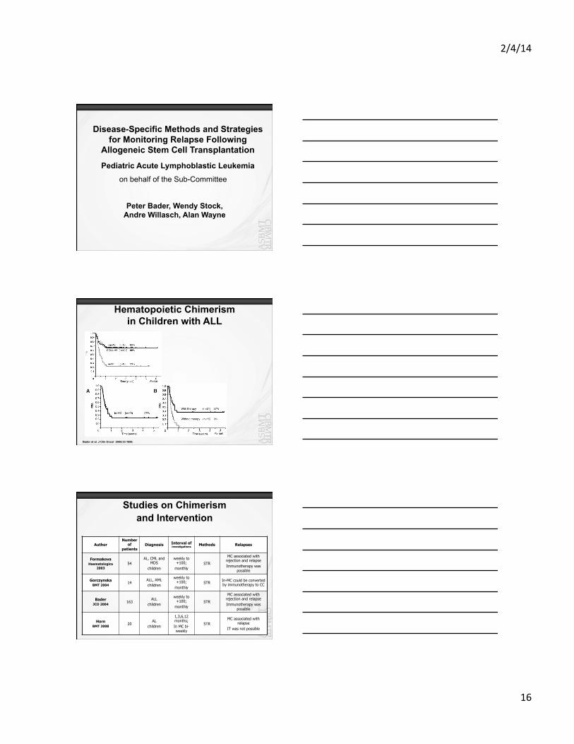
Disease-Specific Methods and Strategies for Monitoring Relapse Following
Allogeneic Stem Cell Transplantation
Pediatric Acute Lymphoblastic Leukemia on behalf of the Sub-Committee
Peter Bader, Wendy Stock, Andre Willasch, Alan Wayne
Hematopoietic Chimerism in Children with ALL
Bader et al. J Clin Oncol 2004;33:1696.
Studies on Chimerism and Intervention
Author Number
of patients
Diagnosis Interval of investigations Methods Relapses
Formakova Haematologica
2003 54
AL, CML and MDS
children
weekly to +100;
monthly STR
MC associated with rejection and relapse Immunotherapy was
possible
Gorczynska BMT 2004
14 ALL, AML children
weekly to +100;
monthly STR In-MC could be converted
by immunotherapy to CC
Bader JCO 2004
163 ALL
children
weekly to +100;
monthly STR
MC associated with rejection and relapse Immunotherapy was
possible
Horn BMT 2008
20 AL
children
1,3,6,12 months; In MC bi-weekly
STR MC associated with
relapse IT was not possible
2/4/14
17
Retrospective Studies - MRD prior to SCT Literature
Author Number of patients Diagnosis Time of
investigation Methods Survival
according to MRD status
Knechtli Blood 1998
64 ALL prior to conditioning
Ig / TCR PCR
high level pos. – 0% low level pos. – 36%
negative – 73%
Bader Leukemia 2002
41 ALL prior to conditioning
Ig / TCR PCR
high level pos. – 23% low level pos. – 48%
negative – 78%
Uzunel Blood 2001
30 ALL prior to conditioning
Ig / TCR PCR
high level pos. – 47% low level pos. – 50%
negative – 100%
Sramkova Ped Blood Cancer
2007 25 ALL prior to
conditioning Ig / TCR
PCR positive – 0%
negative – 94%
Prospective Study: MRD Prior SCT ALL REZ BFM Group: CR2
EFS CI
Years after SCT
MRD < 10-4
MRD ≥ 10-4
0 1 2 3 4 5 60.0
0.2
0.4
0.6
0.8
1.0
Eve
nt-f
ree
Sur
viva
lPro
babi
lity
Years after SCT
MRD < 10-4
MRD ≥ 10-4
0 1 2 3 4 5 60.0
0.2
0.4
0.6
0.8
1.0
Eve
nt-f
ree
Sur
viva
lPro
babi
lity
Years after SCT
MRD < 10-4
MRD ≥ 10-4
0 1 2 3 4 5 60.0
0.2
0.4
0.6
0.8
1.0
Cum
ulat
ive
Inci
denc
e
Years after SCT
MRD < 10-4
MRD ≥ 10-4
0 1 2 3 4 5 60.0
0.2
0.4
0.6
0.8
1.0
Cum
ulat
ive
Inci
denc
e
EFS CIR
MRD < 10-4: n = 46; cens.= 29; pEFS = .60 ± .08 CI (relapse) = .13 ± .06 ≥ 10-4: n = 45; cens.= 14; pEFS = .27 ± .07 CI (relapse) = .57 ± .08
p = .0004 p < .001
Bader et al. J Clin Oncol 2009;27:377-84.
Conclusions II
• MRD prior to stem cell transplantation has a profound impact on post transplant outcome!
• What adds MRD post transplant?
2/4/14
18
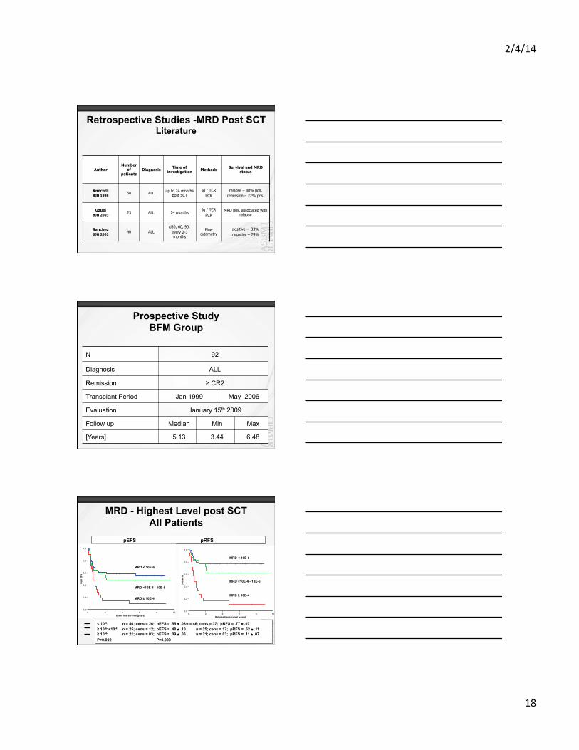
Retrospective Studies -MRD Post SCT Literature
Author Number
of patients
Diagnosis Time of investigation Methods Survival and MRD
status
Knechtli BJH 1998
68 ALL up to 24 months post SCT
Ig / TCR PCR
relapse – 88% pos. remission – 22% pos.
Uzuel BJH 2003
23 ALL 24 months Ig / TCR
PCR MRD pos. associated with
relapse
Sanchez BJH 2002
40 ALL d30, 60, 90, every 2-3 months
Flow cytometry
positive – 33% negative – 74%
Prospective Study BFM Group
N 92
Diagnosis ALL
Remission ≥ CR2
Transplant Period Jan 1999 May 2006
Evaluation January 15th 2009
Follow up Median Min Max
[Years] 5.13 3.44 6.48
MRD - Highest Level post SCT All Patients
pEFS pRFS
< 10-6: n = 46; cens.= 26; pEFS = .55 ± .08 n = 46; cens.= 37; pRFS = .77 ± .07 ≥ 10-6- <10-4 n = 25; cens.= 12; pEFS = .48 ± .10 n = 25; cens.= 17; pRFS = .62 ± .11 ≥ 10-4: n = 21; cens.= 03; pEFS = .09 ± .06 n = 21; cens.= 03; pRFS = .11 ± .07 P=0.002 P=0.000
Event free survival [years]1086420
Cum
EFS
1,0
0,8
0,6
0,4
0,2
0,0
Relapse free survival [years]1086420
Cum
RFS
1,0
0,8
0,6
0,4
0,2
0,0
MRD ≥ 10E-4 MRD ≥ 10E-4
MRD < 10E-6
MRD < 10E-6
MRD <10E-4 - 10E-6
MRD <10E-4 - 10E-6
2/4/14
19
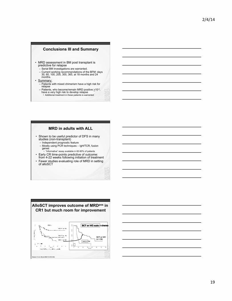
Conclusions III and Summary
• MRD assessment in BM post transplant is predictive for relapse – Serial BM investigations are warranted. – Current working recommendations of the BFM: days
30, 60, 100, 200, 300, 365, at 18 months and 24 months.
• Summary: – Patients with mixed chimerism have a high risk for
relapse – Patients, who become/remain MRD positive >10-4,
have a very high risk to develop relapse • Additional treatment in these patients is warranted
MRD in adults with ALL
• Shown to be useful predictor of DFS in many studies (non-transplant) – Independent prognostic feature – Mostly using PCR techniques – IgH/TCR, fusion
genes • “Informative” assay available in 60-90% of patients
• Early CR time-points predictive of outcome: from 4-22 weeks following initiation of treatment
• Fewer studies evaluating role of MRD in setting of alloSCT
AlloSCT improves outcome of MRDpos in CR1 but much room for improvement
0.00
0.25
0.50
0.75
1.00
Cum
ulat
ive
Sur
viva
l
0 12 24 36 48 60 72 84Months
Kaplan-Meier survival estimates, by allo_iper2
SCT or H/C (n = 36)
rest (n = 18)
SCT or H/C-auto v chemo
Bassan, R. et al. Blood 2009;113:4153-4162.
2/4/14
20
MRD following alloSCT in Adults with ALL
Author Number
of patients
Diagnosis Time of investigation Methods DFS and MRD status
Mortuza JCO 2002
19 ALL
(B-lineage)
From 1-20 mos.
Ig / TCR PCR
Semi-quant.
positive – 0% negative – 100% CCR
Spinelli Haematologica
2007 37 ALL Day +100
Ig/TCR or fusion gene
PCR Quantitative
positive >10-4: 20% negative: 93%
Bassan* Blood 2009
18
ALL *All were
PCR+ prior to
transplant
Not defined Ig / TCR
PCR positive >10-4: 0 negative: 50%
Dombret, H et al. Blood 2002;100:2357-66.
MRD status prior to transplant predicts DFS
Achievement of Molecular Remission Prior to AlloSCT is Important in Ph+ ALL
Wassmann, B. et al. Blood 2005;106:458-463.
Imatinib Treatment of Molecular Relapse with Following Allo-SCT for Ph+ ALL
2/4/14
21
Summary
• MRD detection both prior to and following alloSCT for adults with ALL is associated with poor DFS
• Clinical interventions based on MRD measurements suggest utility but data are very limited: – Allocation to alloSCT in CR1 – Post-transplant intervention to prevent relapse
• Targeted therapy (e.g. imatinib) following transplant
• Challenge: implementation of standardized MRD assays that can be done in “real-time” – IgH/TCR qPCR assays are laborious – Data on flow cytometric measurements of MRD in adults with
ALL are lacking
Nicolaus Kröger
Disease specific Monitoring of Relapse after Allogeneic Hematopoietic Cell Transplantation Multiple Myeloma NCI Workshop 1./2.-11.2009
Conventional techniques for monitoring
• Bone marrow aspiration: infiltration often underestimated
• Serum/24h urine electrophoresis (agarose gel or capillary zone): lowest detectable level of M-component: 0.2 - 0.6 g/L
• Immunofixation (serum/urine): lowest detectable level of M-component: 0.12 - 0.25 g/L
• Free light chain assay (κ/λ ratio) : useful in light chain disease and non-secretory, necessary to determine sCR, early response assessment due to short half time (6h)
2/4/14
22
Imaging monitoring • More than 80% of the pts develop osteolytic bone lesions • The hallmark of myeloma bone disease is an increased
osteoclastic bone resorption and an exhausted osteoblast function resulting in a reduced bone formation even in patients in complete remission
• Standard: conventional radiology as skeletal survey involving cervical, thoracic and lumbar spine, skull, chest, pelvis, humeri and femora
• Disadvantage: low sensitivity, no exact response assessment
• CT: high sensitivity, but higher radiation dose • MRI: high sensitivity, no radiation dose, detect
extramedullary disease • PET-CT: highest sensitivity for extramedullary disease
Flow-cytometry • Flow cytometry has become an easy applicable
method to detect residual myeloma cells The European Myeloma Network recommends a minimal panel including
• CD19, CD56, CD20, CD117, CD28 and CD27.
• Plasma cell gating should be based on CD38 vs. CD138 expression
• This method is less sensitive (10-4) than allele-specific oligonucleotides PCR (ASO-PCR)
Rawston A.C., et al. Haematologica 2008;93:431-438.
Allele-specific oligonucleotides PCR (ASO-PCR)
• Patient-specific primers (IgH rearrangement)
• High sensitivity of (10-5 - 10-6) and highly specific (100%)
• Time-consuming (for each patients), does not detect extramedullary disease
2/4/14
23
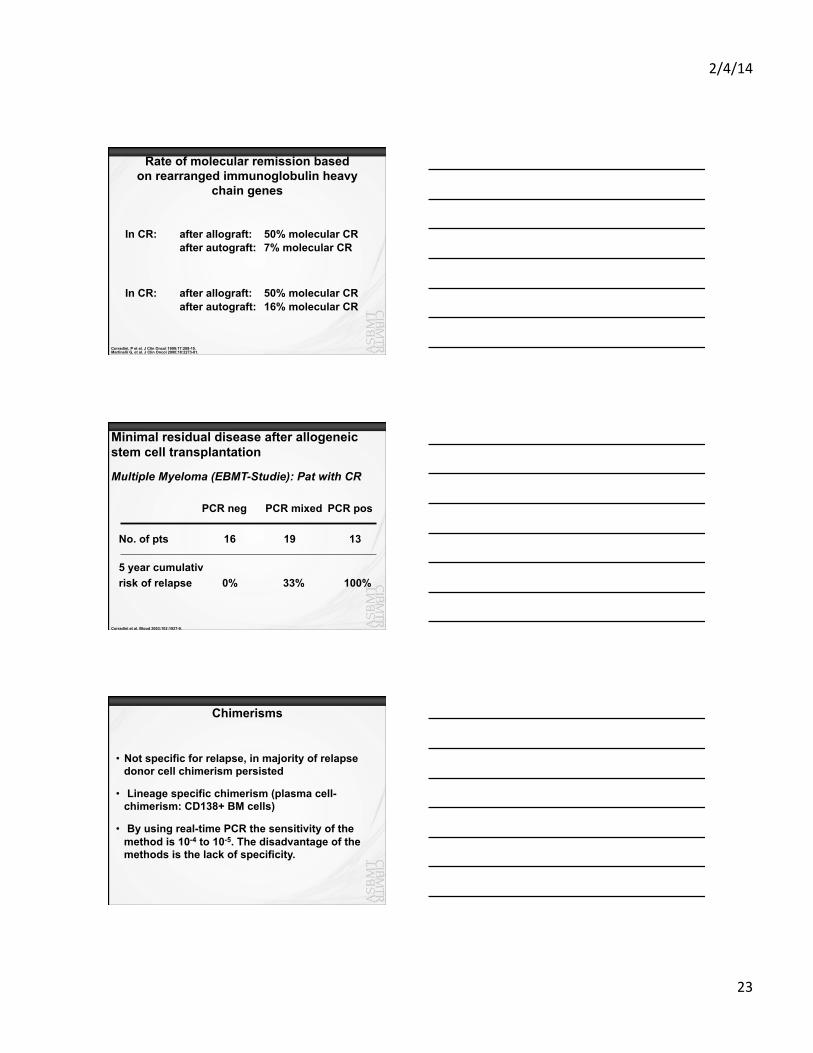
Rate of molecular remission based on rearranged immunoglobulin heavy
chain genes
In CR: after allograft: 50% molecular CR after autograft: 7% molecular CR
In CR: after allograft: 50% molecular CR
after autograft: 16% molecular CR
Martinelli G, et al. J Clin Oncol 2000;18:2273-81. Corradini, P et al. J Clin Oncol 1999;17:208-15.
No. of pts 16 19 13
5 year cumulativ risk of relapse 0% 33% 100%
Minimal residual disease after allogeneic stem cell transplantation
Multiple Myeloma (EBMT-Studie): Pat with CR
PCR neg PCR mixed PCR pos
Corradini et al. Blood 2003;102:1927-9.
Chimerisms
• Not specific for relapse, in majority of relapse donor cell chimerism persisted
• Lineage specific chimerism (plasma cell-chimerism: CD138+ BM cells)
• By using real-time PCR the sensitivity of the method is 10-4 to 10-5. The disadvantage of the methods is the lack of specificity.
2/4/14
24
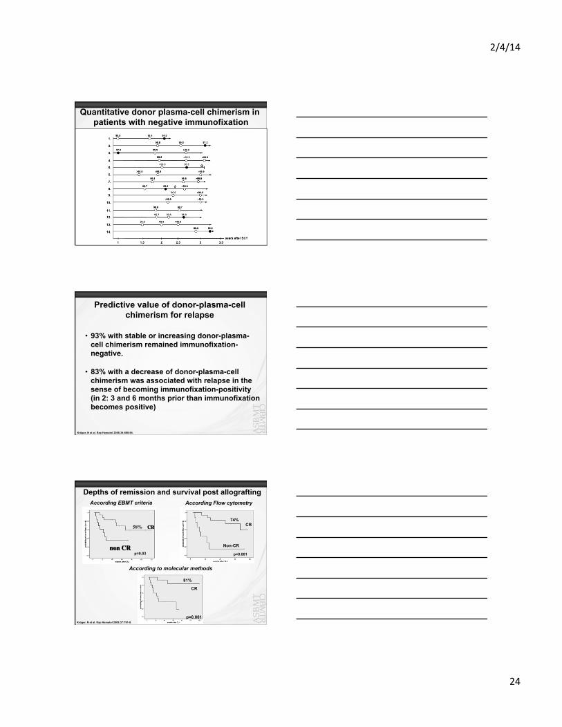
Quantitative donor plasma-cell chimerism in patients with negative immunofixation
Predictive value of donor-plasma-cell chimerism for relapse
• 93% with stable or increasing donor-plasma-cell chimerism remained immunofixation-negative.
• 83% with a decrease of donor-plasma-cell chimerism was associated with relapse in the sense of becoming immunofixation-positivity (in 2: 3 and 6 months prior than immunofixation becomes positive)
Kröger, N et al. Exp Hematol 2006;34:688-94.
CR
non CR
Depths of remission and survival post allografting
p=0.03
According EBMT criteria
58%
According Flow cytometry
74% CR
Non-CR
p=0.001
According to molecular methods
81%
CR
p=0.001 Kröger, N et al. Exp Hematol 2009;37:791-8.
2/4/14
25
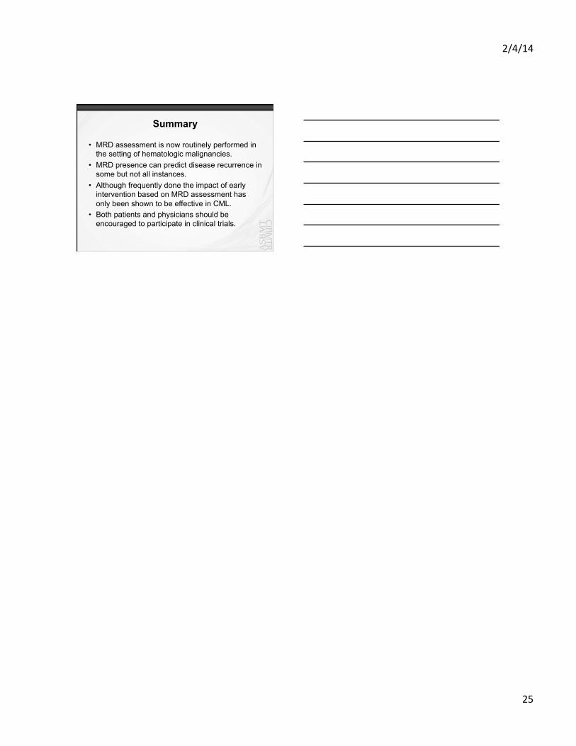
Summary
• MRD assessment is now routinely performed in the setting of hematologic malignancies.
• MRD presence can predict disease recurrence in some but not all instances.
• Although frequently done the impact of early intervention based on MRD assessment has only been shown to be effective in CML.
• Both patients and physicians should be encouraged to participate in clinical trials.