Embed Size (px)
Citation preview
1
Tissue Factor Activated Coagulation Cascade in The Tumor Microenvironment Is
Critical For Tumor Progression And an Effective Target For Therapy
Yuan Liu1, Pengfei Jiang1, Katerina Capkova2, Dong Xue1, Longwu Ye1, Subhash C
Sinha3, Nigel Mackman4, Kim D Janda2, and Cheng Liu1
1 Department of Immunology and Microbial Science, 2 Chemistry, 3Molecular Biology,
The Scripps Research Institute, 10550 North Torrey Pines Road, La Jolla, CA92037,
USA.
4 Division of Hematology/Oncology, University of North Carolina at Chapel Hill, 917
Mary Ellen Jones, Chapel Hill, NC, 27599, USA
Address for correspondence:
Cheng Liu, M.D, Ph.D.
The Scripps Research Institute
10550 North Torrey Pines Road
La Jolla, CA92037, USA
Tel: 858-785-7734
Fax: 858-785-7756
E-mail: [email protected]
Precis: Targeting the coagulation cascade activated in the tumor microenvironment may
be a highly effective strategy for therapeutic eradication of metastatic cancer.
Research. on November 27, 2018. © 2011 American Association for Cancercancerres.aacrjournals.org Downloaded from
Author manuscripts have been peer reviewed and accepted for publication but have not yet been edited. Author Manuscript Published OnlineFirst on August 31, 2011; DOI: 10.1158/0008-5472.CAN-11-1145

2
Keywords Cancer, Tissue Factor Factor VIIa, Thrombin, Prodrug,
Abstract
Tissue factor(TF), a rate limiting enzyme cofactor in activating coagulation, is
highly expressed in a wide spectrum of human tumor and tumor stromal cells. Using TF-
deficient cancer cells and a conditional TF-knockout mouse model, we show that TF
expressed by cancer cells, but not by the host stromal cells, plays a critical role in tumor
growth. In the tumor microenvironment, serum coagulation factors are readily
extravasated and therefore lead to continuous TF-mediated activation of coagulation
proteases. To target this highly specific cascade of serine proteases, we used both a
TF:VIIa inhibitor and doxorubicin-based prodrugs that are selectively activated by
TF:FVIIa, FXa, and thrombin. Treatment with the TF:FVIIa inhibitor led to growth
retardation in breast tumor models. In contrast, treatment with the prodrug eliminated
primary tumor cells and lung metastases without apparent toxicity. Our findings offer
preclinical proof of principle that targeting the coagulation cascade that is activated in
the tumor microenvironment can be a highly effective approach for cancer therapy.
Research. on November 27, 2018. © 2011 American Association for Cancercancerres.aacrjournals.org Downloaded from
Author manuscripts have been peer reviewed and accepted for publication but have not yet been edited. Author Manuscript Published OnlineFirst on August 31, 2011; DOI: 10.1158/0008-5472.CAN-11-1145

3
Introduction
Tissue Factor(TF) is the plasmalemma receptor and obligatory cofactor for cell
surface assembly and initiation of the coagulation protease cascade (1). It is widely
expressed and a key determinant of tumor progression (2-6). In addition to its function
as an initiator of hemostasis, TF can signal through TF:FVIIa, FXa, and thrombin
mediated activation of protease-activated receptors. Human TF is a glycoprotein of 263
residues, which shares structural homology with the cytokine receptor families (7-9). To
initiate the coagulation protease cascade, TF binds and facilitates activation of factor
VII(FVII) to the serine protease factor VIIa(FVIIa) and allosterically conforms the
catalytic site of the bound FVIIa and associates with the physiologic substrate zymogens
factor X(FX) and factor IX(10, 11), leading to thrombin generation, fibrin deposition, and
platelet activation. TF presence is prominent in the perivascular cells that form a
hemostatic barrier and serve to limit hemorrhage after vessel injury. Therefore, TF
mediated coagulation is otherwise inactive elsewhere in the body, unless there is an
injury to the vasculature.
To assess the role of TF in tumor progression, we generated TF knockout cancer
cells by crossing a PyVmT mouse with TF flox/flox mice. Multiple cancer lines were
established from the tumors developed in TF flox/flox.PyVmT(+) mice. In addition, TF
knockout was achieved by infecting the established TF flox/flox.PyVmT(+) tumor cell line
with adenovirus vector express Cre recombinase. The contribution of the host TF was
evaluated in TFflox/flox LysM Cre(+) mice with the deleted TF gene in mature
macrophages and granulocytes, and the TFflox/flox Tie2 Cre(+) mice specifically deleted
Research. on November 27, 2018. © 2011 American Association for Cancercancerres.aacrjournals.org Downloaded from
Author manuscripts have been peer reviewed and accepted for publication but have not yet been edited. Author Manuscript Published OnlineFirst on August 31, 2011; DOI: 10.1158/0008-5472.CAN-11-1145

4
TF gene in endothelial cells and hematopoietic cells. Through these genetic models, we
demonstrate that TF expressed by cancer cells is the key contributor to tumor growth,
but not the host TF expressed by myeloid cells and endothelial cells.
Due to enhanced tumor vasculature permeability, the coagulation factors and
albumin are extravasated and present in the tumor microenvironment where the
coagulation cascade is constantly activated by the high level of the tissue factor on the
tumor and stromal cell surface. Fibrin deposit in tumor stroma is commonly observed.
Therefore, the coagulation cascade is an attractive target for prodrug therapy. Peptide
conjugates of doxorubicin designed for activation by plasmin (12, 13) and cathepsins
(14, 15) have been described. Indeed, the TF:FVIIa and proteases in the coagulation
cascade have the highest substrate specificity among serine proteases and are very
well characterized (16, 17). We synthesized two types of doxorubicin prodrugs targeting
coagulation proteases. The first type of prodrug contained a short polyethylene glycol or
a succinyl substituent to enhance the prodrug solubility, while the second prodrug
includes an albumin binding �-maleimidocaproic acid (EMC) moiety to prolong half-life.
The prodrugs were prepared by incorporating a small peptide appended to the amino
functionality of doxorubicin resulting in an inactive compound, unless hydrolyzed to
leucine-doxorubicin by TF:FVIIa and proteases within the coagulation cascade. Both
types of prodrugs were found to be effective against primary tumors. However, the EMC
containing prodrug demonstrated superior efficacy as a single agent. In vivo
administration of the EMC prodrug achieves complete tumor growth arrest and size
reduction in the aggressive 4T1 murine mammary carcinoma. This strategy is similar to
Research. on November 27, 2018. © 2011 American Association for Cancercancerres.aacrjournals.org Downloaded from
Author manuscripts have been peer reviewed and accepted for publication but have not yet been edited. Author Manuscript Published OnlineFirst on August 31, 2011; DOI: 10.1158/0008-5472.CAN-11-1145

5
the cell-impermeable prodrug activation by cell surface associated legumain in tumors
(18, 19). The prodrugs significantly reduced the number of established lung metastasis
and resulted in tumor eradication in human breast cancer xenogragh. The coagulation
protease activated prodrugs were stable in plasma indicating that the activation of
coagulation is discreet and only occurs in the tumor microenvironment and during injury.
Taken together, these data support that coagulation cascade is activated in the tumor
microenvironment and may serve as an enzymatic target to achieve chemotherapeutic
eradication of cancer and metastasis.
Research. on November 27, 2018. © 2011 American Association for Cancercancerres.aacrjournals.org Downloaded from
Author manuscripts have been peer reviewed and accepted for publication but have not yet been edited. Author Manuscript Published OnlineFirst on August 31, 2011; DOI: 10.1158/0008-5472.CAN-11-1145

6
Material and methods
Reagents and cell lines. Biotinylated mouse TF antibody was purchased from
American Diagnostica. Rabbit FVII antibody and human albumin were from Abcam.
FVIIa and FXa were from HTI. Thrombin was from R&D systems. Doxorubicin and
biotinylated albumin were from Sigma. Tissue factor was kindly provided by Wolfram
Ruf. The 4T1 and MDA-MB231 cell lines were purchased from ATCC and were
characterized according to ATCC instructions. The cells were maintained in RPMI-1640
or DMEM medium supplemented with 10% fetal bovine serum in a humidified incubator
containing 5% CO2 at 37°C. All the cell lines were used within 20 passages.
Immunochemical analysis. 4T1 and PyVmT tumor sections were stained by using
biotinylated anti-TF and Goat anti-FVII antibody. Fluorescein streptavidin and Texas red
were used as the secondary reporting reagent. FVII was i.v. injected into mice to
increase FVII extravasation. Bio-Rad Radiance 2100 laser scanning confocal
microscope was used for analysis.
Albumin extravasation assay. Albumin or biotinylated albumin(30μmol/kg) was i.v.
injected into the tail vein for observation. Fluorescein-avidin was used for biotinylated
albumin staining. Nuclei were stained with DAPI.
Tissue distribution. Drug auto fluorescence was observed 12 hours after 10μmol/kg
i.v. injection. Nuclei were stained with DAPI. Tissues was homogenized and diluted with
50% ethanol containing 0.6mol/L HCL. After normalization of protein concentration, the
Research. on November 27, 2018. © 2011 American Association for Cancercancerres.aacrjournals.org Downloaded from
Author manuscripts have been peer reviewed and accepted for publication but have not yet been edited. Author Manuscript Published OnlineFirst on August 31, 2011; DOI: 10.1158/0008-5472.CAN-11-1145

7
relative fluorescent intensity was measured by using of a Perkin-Elmer LS-50-B
spectrofluorometer(excitation: 470nm; emission: 590nm).
Colony-forming cell assays. Bone marrow single cell suspension was (1x10⁶ per ml)
cultured in MethoCult medium containing SCF, IL-3 and IL-6(Stemcell Technologies) for
10 days at 37°C and 5% CO2 and the number of colony are counted.
Plasma pharmacokinetics. Blood from mice injected with 15μmol/kg prodrugs i.v. were
collected at 10min, 6hr, 12hr and 24hr post injection and 1:10 diluted. Samples were
measured by spectrofluorometer (excitation: 470nm; emission: 590nm).
Mouse tumor models. PyVmT murine breast carcinoma was produced by injection of
5 × 10⁵ TF normal and deficient PyVmT tumor cells into the right flank of wild or
conditional knock out C57BL/6J mice. 4T1 murine breast carcinoma model was
produced by injection 1 × 10⁶ 4T1 cells into the s.c. site on the back of BALB/c mice.
MDA-MB231 human breast carcinoma model was generated by injection of 1 × 10⁶
MDA-MB231 cells into the s.c. site on the back Hsd:Athymic nude mice. Treatment was
initiated when the tumors reached 4 mm in diameter through i.p. injections of the
indicated reagents. The lungs were removed on day 24 and fixed in the Bouin’s solution
to count spontaneous lung metastases. Number of lung metastases was counted under
an anatomy microscope. Experimental lung metastases were induced by i.v. injection of
2 × 10⁶ 4T1 in the tail vein of BALB/c mice. EMC-LTPRL-DOX treatment was started on
day 16 with i.p. injection of 15μmol/kg daily. Experimental lung metastases were count
Research. on November 27, 2018. © 2011 American Association for Cancercancerres.aacrjournals.org Downloaded from
Author manuscripts have been peer reviewed and accepted for publication but have not yet been edited. Author Manuscript Published OnlineFirst on August 31, 2011; DOI: 10.1158/0008-5472.CAN-11-1145
8
on day 25. Tumor growth (volume= length × width × width/2) and signs of physical
discomfort were monitored including for any gross evidence of tumor necrosis, local
tumor ulceration, as well as evidence of toxicity including the mobility of animals,
response to stimulus, piloerection, eating, and weight. These procedures have been
reviewed and approved by the Institutional Animal Care and Use Committee at The
Scripps Research Institute.
Statistical analysis. Statistical significance of data was determined by the two-tailed
Student's t test.
Research. on November 27, 2018. © 2011 American Association for Cancercancerres.aacrjournals.org Downloaded from
Author manuscripts have been peer reviewed and accepted for publication but have not yet been edited. Author Manuscript Published OnlineFirst on August 31, 2011; DOI: 10.1158/0008-5472.CAN-11-1145

9
Results
TF activates coagulation within the tumor microenvironment.
To determine cancer cell-surface expression of TF:FVIIa complex, we examined
4T1 murine syngeneic mammary carcinoma and polyoma virus middle T antigen
(PyVmT) murine spontaneous mammary carcinoma by immunohistochemical analysis
using the rabbit anti-mouse TF antibody. The staining shows that there is high level
expression of the TF on 4T1 tumor cell surface (Figure 1a). It also shows that FVII can
be extravasated readily from the vessel into the tumor microenvironment of primary
tumors and TF:FVIIa complex are detectable on the tumor cell surface (Figure 1b and
1c). The TF expression was furthermore found on the cell face of tumor associate
macrophage in PyVmT tumor (Figure 1d). Expression of TF is also confirmed by
western blot analysis (Figure 1. e). A schematic presentation of TF:FVIIa complex
formation in primary tumor microenvironment is proposed (Figure 1f). Based on the size
of the coagulation factors and the enhanced permeability of tumor vasculature
(Supplemental table 1), FVII and FX extravasate from leaky tumor vessels into the
tumor microenvironment of primary tumors and bind to the cell surface TF to active
coagulation cascade.
Albumin readily extravasates from the tumor vasculature into tumor
microenvironment.
To further evaluate the enhanced permeability of tumor vascular of serum
proteins, we examined the extravasation of albumin by injecting biotinylated albumin
into circulation of tumor bearing mice. Albumin and biotinylated albumin(30μmol/kg)
Research. on November 27, 2018. © 2011 American Association for Cancercancerres.aacrjournals.org Downloaded from
Author manuscripts have been peer reviewed and accepted for publication but have not yet been edited. Author Manuscript Published OnlineFirst on August 31, 2011; DOI: 10.1158/0008-5472.CAN-11-1145

10
were i.v. injected into the tail vein of tumor bearing mice. Tumor and normal organ
specimens were collected and frozen sections were probed with FITC labeled
strepavidin to visualize the biotinylated albumin. Albumin is readily extravasated in
tumor, but not in normal organs such as liver (Figure 1g). In tumor tissue, biotinylated
albumin is accumulated in the tumor microenvironment (Figure 1g, tumor), but absent in
normal liver (Figure 1g, liver) and other organs (data not shown).
TF overexpression is a critical mediator of the tumor growth.
A transgenic animal expressing the polyoma virus middle T antigen(PyVmT) is a
robust breast cancer model developing multiple palpable mammary carcinomas
spontaneously. To generate TF knockout cancer cells, we crossed TF+/+-PyVmT(+)
mice with TF flox/flox-PyVmT(-) mice to acquire TF flox/flox.PyVmT(+) mice (Supplemental
1a). The mouse tail PCR genotyping were performed to confirm TF flox/flox.PyVmT(+)
mice (Supplemental 1b). TF flox/flox.PyVmT(+) cancer cell lines were established from the
spontaneous tumors developed in TF flox/flox.PyVmT(+) mice. The TF-/- PyVmT(+) cancer
cell line was achieved by infecting the established TF flox/flox.PyVmT(+) tumor cell line
with adenovirus vector expressing Cre recombinase and GFP+ marker (Supplemental
1c). The Cre recombinase eliminates a segment of TF gene from the genome and
disrupts TF expression completely (Supplemental 1d).
To examine the role of TF expressed by cancer cell in tumor growth, TF normal
(TF+/+-PyVmT+) and TF deficient (TF-/- PyVmT+) tumor cells were inoculated into
C56BL/6 mice. The tumor induced by TF deficient cells grow much slower than that
Research. on November 27, 2018. © 2011 American Association for Cancercancerres.aacrjournals.org Downloaded from
Author manuscripts have been peer reviewed and accepted for publication but have not yet been edited. Author Manuscript Published OnlineFirst on August 31, 2011; DOI: 10.1158/0008-5472.CAN-11-1145

11
induced by TF normal cells (Figure 2a and 2b). TF deficient cells only developed and
grew small tumors in mice, indicating a critical role of Tissue Factor in tumor
progression. H&E staining of tumor specimens indicated the TF deficient tumors contain
scarce and small lobular of tumor cells, but abundance of stromal component between
tumor cells (Supplemental 1e).
The contribution of TF expressed by stromal cells was evaluated in mice with
conditional TF gene knock-out in different host cells. We have transplanted TF normal
cancer cells into the TFflox/flox·LysM Cre(+) mice in which TF gene is deleted in mature
macrophage and granulocyte, and the TFflox/flox Tie2 Cre (+) mice in which TF gene
deleted endothelial cells and hematopoitic cells. We found no difference in tumor growth
patterns in these models, and all the tumor volume are larger than 1200 mm3 (Figure 2c
and 2d). However, when we transplanted TF-/- cancer cells into the TFflox/flox·LysM Cre(+)
and Tie2 Cre (+) mice, tumor growth is retarded and all the tumor volume is less than
600 mm3 (Figure 2e and 2f). TF deletion in stromal cells does not appear to suppress
tumor growth. Taken together these results indicate that TF expressed by cancer cells
plays a significant role in tumor progression, in contrast to TF provided by stromal cells.
FVIIa inhibitor suppresses tumor growth.
To evaluate the role of TF:FVIIa on tumor growth, a reported high affinity inhibitor
of FVIIa (Supplemental 2a) was synthesized and applied in to 4T1 in vivo model with
0.2mg/kg dosage daily. At this bleeding time was prolonged three-fold (Supplemental
2b) in mouse tail-bleeding experiment. The FVIIa inhibitor had an inhibitory effect on
Research. on November 27, 2018. © 2011 American Association for Cancercancerres.aacrjournals.org Downloaded from
Author manuscripts have been peer reviewed and accepted for publication but have not yet been edited. Author Manuscript Published OnlineFirst on August 31, 2011; DOI: 10.1158/0008-5472.CAN-11-1145
12
tumor growth (Supplemental 2c) that is consistent with the role of TF:FVIIa activity in
tumor growth. However, the FVIIa inhibitor has a low solubility and short half-life in
blood. The lack of more pronounced effects is due to the poor pharmacokinetic
properties of this compound. These experiments underscore the difficulty in the
development of successful inhibitors for this class of serine proteases in cancer therapy,
especially due to the risk of bleeding.
Synthesis of doxorubicin based prodrugs.
To validate the strategy of tumor microenvironment activated prodrugs and to
take advantage of an enhanced vascular permeability of the blood vessels found in
malignant tissue for circulating macromolecules, we synthesized two doxorubicin
prodrugs targeting coagulation proteases containing: a) a short polyethylene glycol
substituent to enhance the prodrug solubility, b) An albumin binding �-maleimidocaproic
acid moiety introduced to react with cysteine-34 of the albumin circulating in the
bloodstream, increasing the prodrug half-life, and significantly reducing drug toxicity.
The plasma protein albumin preferentially accumulates in solid tumors (20, 21), and can
be used as an endogenous drug carrier to enhance the pharmacologic properties of
these drugs. The synthesis of both prodrugs commenced with the preparation of
peptides 2a-b using standard Fmoc solid phase synthesis, where 2-(2-
methoxyethoxyacetic acid) or �-maleimidocaproic acid was incorporated as the last
amino acid residue in the growing sequence. After cleavage from the resin, these
peptides were purified by RP HPLC. Subsequent coupling with doxorubicin in the
Research. on November 27, 2018. © 2011 American Association for Cancercancerres.aacrjournals.org Downloaded from
Author manuscripts have been peer reviewed and accepted for publication but have not yet been edited. Author Manuscript Published OnlineFirst on August 31, 2011; DOI: 10.1158/0008-5472.CAN-11-1145

13
presence of PyBOP and diisopropylethylamine afforded the target prodrugs a and b,
which were purified by RP HPLC and characterized by HR MS (Figure 3a).
The activation of Peg-LTPRL-DOX by FVIIa and TF:FVIIa was carried out in vitro
and analyzed by LC-MS (Figure 3b and 3c). In the control sample, there was only one
component, the Peg-LTPRL-DOX(WM 1240.5) (Figure 3c, peak 2). Hence, incubation
of Peg-LTPRL-DOX with thrombin, FVIIa, and FXa induced activation of the prodrug
producing Leu-DOX(WM 542.5) (Figure 3c, peak 1), which could be separated by liquid
chromatography and detected by mass spectrometry (Figure 3d). Inclusion of equal
molar soluble TF extracellular domain with FVIIa significantly enhanced the activity of
FVIIa as expected. However, this still underestimates the catalytic activity of TF:FVIIa
complexes on the cell surface that are of native TF proteins and in an appropriate cell-
surface lipid environment. Indeed, non-specific serine protease trypsin has a very low
activity toward this prodrug. While the catalytic efficiency of the serine proteases in the
coagulation cascade and trypsin toward prodrug activation were determined (Table 1)
and support the specificity of activation of the prodrug by serine proteases in the
coagulation cascade.
Prodrug activation and accumulation are enhanced in tumor.
The profile of prodrug distribution and activation in vivo were determined by
detecting prodrug and parent drug using a fluorescent microscope and a fluorometer.
Prodrug and active prodrug accumulation were visualized in tumor sections after i.v.
injection of 10μmol/kg DOX, Peg-LTPRL-DOX and EMC-LTPRL-DOX, respectively
Research. on November 27, 2018. © 2011 American Association for Cancercancerres.aacrjournals.org Downloaded from
Author manuscripts have been peer reviewed and accepted for publication but have not yet been edited. Author Manuscript Published OnlineFirst on August 31, 2011; DOI: 10.1158/0008-5472.CAN-11-1145

14
(Figure 4a, red color). The accumulation of the prodrug in tumors of Peg-LTPRL-DOX
treated mice and EMC-LTPRL-DOX treated mice was greatly enhanced compared to
that of doxorubicin treated mice. Doxorubicin is cell permeable and has a high
clearance rate in tumor tissue, because doxorubicin is a small molecular compound.
The EMC-LTPRL-DOX is cell-impermeable until activated and is not taken up by normal
tissue cells. The fluorescence signal in the liver and kidneys of mice treated with EMC-
LTPRL-DOX were primarily extracellular, suggesting that EMC-LTPRL-DOX is not
activated in the normal tissues (Figure 4a. liver and kidney). Nuclear positivity of active
EMC-LTPRL-DOX is detected in tumor indicating that the prodrugs are only activated in
the tumor microenvironment (Figure 4a, tumor). Tumor accumulation and activation of
the EMC-LTPRL-DOX are supported by quantitative assay in tumor bearing mice
(Figure 4b). Binding to albumin (half-life 9~12 days) with EMC group, EMC-LTPRL-
DOX shows a longer half–life in mouse circulating blood than that of doxorubicin, which
almost disappeared after six hours from injection (Figure 4c).
Coagulation cascade serine protease activated prodrug has low toxicity and is
stable in serum.
First, we assessed the stability of the prodrug compounds by mixing the Peg-
LTPRL-DOX with mouse serum and incubated at 37 oC for 24 hours. The compounds
were subjected to HPLC analysis at the end of incubation (Figure 5a) and the prodrugs
appeared very stable in serum and only minor degradation products were observed that
was distinct from activated drug. With 3 mg/kg injection every two days, doxorubicin
caused significant toxicity as demonstrated by weight loss (Figure 5b) whereas both
Research. on November 27, 2018. © 2011 American Association for Cancercancerres.aacrjournals.org Downloaded from
Author manuscripts have been peer reviewed and accepted for publication but have not yet been edited. Author Manuscript Published OnlineFirst on August 31, 2011; DOI: 10.1158/0008-5472.CAN-11-1145

15
Peg-LTPRL-DOX and EMC-LTPRL-DOX lacked apparent toxicity and weight loss. We
further evaluated the effect on white blood cells by EMC-LTPRL-DOX prodrug and
compared this to doxorubicin (Supplemental table 2). Single injection of doxorubicin at
MTD caused significant WBC reduction, whereas EMC-LTPRL-DOX given at a “save-
dosage” had no effect on the WBC count. Next, we examined the effect of EMC-LTPRL-
DOX on hematopoietic stem cells using a colony forming assay. Doxorubicin
demonstrated significant suppression on the formation of CFU-GM. In contrast EMC-
LTPRL-DOX at the same concentration had no effect on colony formation (Figure 5c)
indicating the coagulation cascade is not activated in the bone marrow
microenvironment and is consistent with the lack of TF expression by hematopoietic
cells and circulating blood cells. The EMC-LTPRL-DOX was significantly less toxic than
doxorubicin when evaluated in vivo and its accumulated MTD is greater than twenty-fold
of that of the doxorubicin (Supplemental table 3).
The coagulation activated prodrugs are affective against tumor.
The 4T1 murine mammary carcinoma model was used to evaluate the efficacy
and safety of Peg-LTPRL-DOX, EMC-LTPRL-DOX, and doxorubicin. On 21-day post-
treatment, tumor volumes of the prodrug treated groups (3 μmol/kg) were significantly
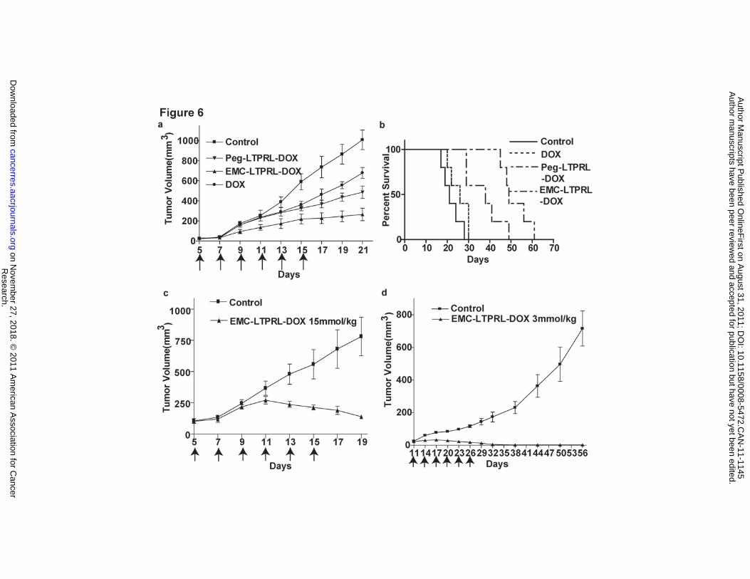
smaller than the control group (Figure 6a) (p<0.01 for EMC-LTPRL-DOX, n=6). The
MTD of Peg-LTPRL-DOX (i.p. 5X) was greater than 54 μmol/kg versus that of DOX at
2.8 μmol/kg. Treatment of 4T1 at MTD of DOX demonstrated growth retardation in this
model versus untreated control.
Research. on November 27, 2018. © 2011 American Association for Cancercancerres.aacrjournals.org Downloaded from
Author manuscripts have been peer reviewed and accepted for publication but have not yet been edited. Author Manuscript Published OnlineFirst on August 31, 2011; DOI: 10.1158/0008-5472.CAN-11-1145
16
Prodrug with the EMC tag has enhanced efficacy.
The efficacy of the albumin-binding prodrug EMC-LTPRL-DOX was further
enhanced, (Figure 6a), compared to Peg-LTPRL-DOX at 3 μmol/kg dosage and
resulted in near complete growth inhibition at 3 μmol/kg. Thus, prodrug treatment
significantly prolonged survival of tumor bearing mice compared to untreated controls,
and DOX treated groups (Figure 6b). Both prodrug treatments caused massive tumor
death upon histological analysis (Supplemental 3). In groups treated with EMC-LTPRL-
DOX at a higher dose, the tumor growth was completely halted and started to reduce
upon continued treatment (Figure 6c). Importantly, in a human MBA-MD-231 xenograph
model, EMC-LTPRL-DOX treatment at 3 μmol/kg led to complete tumor eradication and
non-recurrence with no weight loss and no other apparent signs of toxicity (Figure 6d).
Albumin bound prodrug eliminates established breast cancer metastasis in lung.
EMC-LTPRL-DOX treatment at 3 μmol/kg has significant inhibitory effects on
spontaneous metastasis of 4T1 murine mammary carcinoma (Figure 7a and 7b). H&E
staining of lung sections demonstrated metastasis and extensive lung architecture
destruction in untreated control mice, whereas in the treated mice, metastasis was
significantly inhibited and the lung architecture was nearly normal (Figure 7c).
Next, we evaluated the effect of high dosage treatment on established
metastasis. The lung metastasis was induced by i.v. injection of 4T1 murine mammary
carcinoma cells. On day 16, tumor metastasis was already established in the lung of
tumor bearing animals (Figure 7d, control-16days). The high dose EMC-LTPRL-DOX
Research. on November 27, 2018. © 2011 American Association for Cancercancerres.aacrjournals.org Downloaded from
Author manuscripts have been peer reviewed and accepted for publication but have not yet been edited. Author Manuscript Published OnlineFirst on August 31, 2011; DOI: 10.1158/0008-5472.CAN-11-1145

17
treatment was initiated on day 16 with 10 μmol/kg dose for 7 injections. The lungs were
dissected on day 25. Lung metastasis of the treated group on day 25 was compared to
that of the control on day 16 (Figure 7d). The lung metastasis in the EMC-LTPRL-DOX
treated group on day 25 was significantly less than that found in control mice on the 16
day indicating the high dose EMC-LTPRL-DOX treatment is capable of eliminating
established lung metastasis in these mice (Figure 7e). H&E staining of lung sections
revealed that treatment of metastasis greatly improves lung tissue morphology (Figure
7f). Elimination of established metastasis is a very challenging assay and has great
clinical relevance. Taken together, the effect of EMC-TPRL-DOX represents a
promising treatment for metastatic cancer a major cause of mortality of cancer patients.
Research. on November 27, 2018. © 2011 American Association for Cancercancerres.aacrjournals.org Downloaded from
Author manuscripts have been peer reviewed and accepted for publication but have not yet been edited. Author Manuscript Published OnlineFirst on August 31, 2011; DOI: 10.1158/0008-5472.CAN-11-1145

18
Discussion
TF is overexpressed in many types of human cancers, and clinical studies have
shown a correlation between the levels of TF expression and poor prognosis (22-25).
Not only does TF expression occur in a wide spectrum of cancers, but also the level of
expression in cancer cells is drastic upregulated by 1000-fold compared to the levels of
normal cells that these cancer cells are derived from (26, 27). This drastic upregulation
may, to some extent, be caused by the existence of common tumor microenvironmental
stimuli, such as those responsible for TF upregulation during inflammation and hypoxia.
Recently, it was demonstrated that TF expression is under the direct control of
oncogenic pathways activated by genetic mutations sustained by cancer cells (28). This
is demonstrated experimentally by the effect of several mutant oncogenes, including K-
ras, EGFR, EGFRvIII, HER-2 and PML-RARa on TF transcription, translation, half-life,
and encryption. The effect of these oncogenes was operative in colorectal, mammary,
cutaneous, and astrocytic cancer cells (28-31). In addition, similar changes are also
found in loss of function mutations of major tumor suppressor genes such as p53 and
PTEN (2, 30-32).
Several recent studies indicate that TF plays a role in tumor biology (33). The
formation of the TF:FVIIa and TF:FVIIa:FXa complexes leads to cleavage of protease-
activated receptors (PARs) at the cell surface (34). The TF:FVIIa–PAR-2 pathway
induces expression of the proangiogenic cytokine IL-8 in tumor cells (35) and also
contributes to retinal neoangiogenesis (36). The TF cytoplasmic domain can regulate
the p38 mitogenactivated kinase and extracellular signal-regulated kinase1/2 and the
Research. on November 27, 2018. © 2011 American Association for Cancercancerres.aacrjournals.org Downloaded from
Author manuscripts have been peer reviewed and accepted for publication but have not yet been edited. Author Manuscript Published OnlineFirst on August 31, 2011; DOI: 10.1158/0008-5472.CAN-11-1145

19
rac pathways (37) as well as suppress integrin-mediated migration of cells (36).
Increased intravascular TF expression is observed in cancers (33, 38). TF is also
expressed by tumor cells as well stromal cells such as monocytes, macrophages, and
endothelial cells (38). This may contribute to the prothrombotic state (Trousseau's
syndrome) associated with a high percentage of cancer patients (27). In addition, TF
containing procoagulant microparticles may be shed from different cells, including
platelets, leukocytes, and endothelial cells, or from cancer cells (39-44) and recently
these particles were shown to be important to tumor growth (4).
Breeding conditional TF knockout mice with PyVmT murine spontaneous
mammary carcinoma model, we generated for the first time conditional TF knockout
tumor lines that are tumorigenic and metastatic in C57Bl/6 mice. Multiple stable cancer
lines were established from this tumor model. They express a high level of TF and the
TF gene can be removed by Cre expressing adenovirus in vitro. The growth patterns of
the TF wild type and the TF deficient cancer cells provided clear evidence that TF
expressed by tumor cells is required for the accelerated growth of cancers in this model.
In contrast, the growth of tumor cells in mice lacking TF either in myeloid derived cells
and endothelial cells is barely affected, suggesting the contribution of TF expressed by
these two types of stromal cells is small.
Consistent with enhanced permeability of the tumor vasculature, we have
demonstrated that coagulation factors are readily extravasated in tumors. Furthermore,
TF initiates the activation of the coagulation cascade in the tumor microenvironment.
Research. on November 27, 2018. © 2011 American Association for Cancercancerres.aacrjournals.org Downloaded from
Author manuscripts have been peer reviewed and accepted for publication but have not yet been edited. Author Manuscript Published OnlineFirst on August 31, 2011; DOI: 10.1158/0008-5472.CAN-11-1145

20
Based on these findings, we designed and validated PEG-LTPRL-DOX and
EMC-LTPRL-DOX prodrugs that can be selectively catalytically converted to the
cytotoxic end product by the tissue factor activated coagulation cascade in the tumor
microenvironment. PEG-LTPRL-DOX and EMC-LTPRL-DOX activation is not found in
any significant amount in normal tissues since TF:FVIIa and thrombin are not activated
without injury. Importantly, PEG-LTPRL-DOX and EMC-LTPRL-DOX are very stable in
plasma. We found the little effect of PEG-LTPRL-DOX and EMC-LTPRL-DOX on cells
of myeloid lineage, as mice showed a negligible reduction in peripheral blood or marrow
myeloid cells at elevated therapeutic doses. Although this therapeutic approach showed
little toxicity in our mouse models, the potential toxic risks of off site activation require
careful evaluation in cancer patients especially when persistent thrombotic conditions
such as deep vein thrombosis exist. Selection criteria to exclude such higher-risk
patients may be necessary in initial clinical trial.
Due to the reduced toxicity, larger cumulative dosage of PEG-LTPRL-DOX and
EMC-LTPRL-DOX can be administered more frequently. Consequently, significant
eradicated effect of EMC-LTPRL-DOX on tumor has been observed in the 4T1 mouse
carcinoma model. In high dose treatment of EMC-LTPRL-DOX in the human MDA-
MB231 carcinoma model, the tumor was completely eradicated, and the host survives a
long term. In high dose treatment, EMC-LTPRL-DOX not only reduced 4T1 tumor
metastasis to lung, but also halted and eradicated established metastasis.
Research. on November 27, 2018. © 2011 American Association for Cancercancerres.aacrjournals.org Downloaded from
Author manuscripts have been peer reviewed and accepted for publication but have not yet been edited. Author Manuscript Published OnlineFirst on August 31, 2011; DOI: 10.1158/0008-5472.CAN-11-1145

21
In summary, Tissue Factor expression in malignant cancers is widely recognized,
yet its functions during tumor progression are unknown. Our findings implicate TF
expressed by cancer cells can activate a coagulation cascade in the tumor
microenvironment and is an essential contributor to tumor progression. For robust and
critical hydrolases, including proteases and glycosidases that are enriched and
activated in the tumor microenvironment, sufficient inhibition may prove difficult to
achieve pharmacologically without significant toxicity. Target the enzymatic activity for
prodrug activation is an attractive alternative and amenable to rationally design drug
candidates. Development of clinically viable inhibitors for these enzymes has proven to
be difficult and has taken up an overwhelming majority of our collective cancer drug
discovery efforts. Herein we demonstrated that eradication of primary and metastatic
cancers can be achieved by functionally targeted prodrugs in animal models; our
findings provide a logical path for future preclinical and clinical studies of this form of
targeted cancer therapy.
Acknowledgments
Authors thank to the US National Cancer Institute (CA127535) and the US
Department of Defense (W81XWH-07-1-0389, W81XWH-05-1-0091, W81XWH-05-1-
0318 W81XWH-09-1-0690) for the funding support.
References
Research. on November 27, 2018. © 2011 American Association for Cancercancerres.aacrjournals.org Downloaded from
Author manuscripts have been peer reviewed and accepted for publication but have not yet been edited. Author Manuscript Published OnlineFirst on August 31, 2011; DOI: 10.1158/0008-5472.CAN-11-1145

22
1. Davie, E. W., Fujikawa, K., and Kisiel, W. The coagulation cascade: initiation, maintenance, and regulation. Biochemistry, 30: 10363-10370, 1991.
2. Milsom, C., Yu, J., May, L., Magnus, N., and Rak, J. Diverse roles of tissue factor-expressing cell subsets in tumor progression. Semin Thromb Hemost, 34: 170-181, 2008.
3. Mueller, B. M. and Ruf, W. Requirement for binding of catalytically active factor VIIa in tissue factor-dependent experimental metastasis. J Clin Invest, 101: 1372-1378, 1998.
4. Palumbo, J. S., Talmage, K. E., Massari, J. V., La Jeunesse, C. M., Flick, M. J., Kombrinck, K. W., et al. Tumor cell-associated tissue factor and circulating hemostatic factors cooperate to increase metastatic potential through natural killer cell-dependent and-independent mechanisms. Blood, 110: 133-141, 2007.
5. Schaffner, F. and Ruf, W. Tissue factor and protease-activated receptor signaling in cancer. Semin Thromb Hemost, 34: 147-153, 2008.
6. Versteeg, H. H., Schaffner, F., Kerver, M., Petersen, H. H., Ahamed, J., Felding-Habermann, B., et al. Inhibition of tissue factor signaling suppresses tumor growth. Blood, 111: 190-199, 2008.
7. Bazan, J. F. WKS motifs and the cytokine receptor framework of tissue factor. Trends Biochem Sci, 16: 329, 1991.
8. Edgington, T. S., Mackman, N., Brand, K., and Ruf, W. The structural biology of expression and function of tissue factor. Thromb Haemost, 66: 67-79, 1991.
9. Morrissey, J. H., Fakhrai, H., and Edgington, T. S. Molecular cloning of the cDNA for tissue factor, the cellular receptor for the initiation of the coagulation protease cascade. Cell, 50: 129-135, 1987.
10. Bauer, K. A., Kass, B. L., ten Cate, H., Hawiger, J. J., and Rosenberg, R. D. Factor IX is activated in vivo by the tissue factor mechanism. Blood, 76: 731-736, 1990.
11. ten Cate, H., Bauer, K. A., Levi, M., Edgington, T. S., Sublett, R. D., Barzegar, S., et al. The activation of factor X and prothrombin by recombinant factor VIIa in vivo is mediated by tissue factor. J Clin Invest, 92: 1207-1212, 1993.
12. de Groot, F. M., de Bart, A. C., Verheijen, J. H., and Scheeren, H. W. Synthesis and biological evaluation of novel prodrugs of anthracyclines for selective activation by the tumor-associated protease plasmin. J Med Chem, 42: 5277-5283., 1999.
13. Chakravarty, P. K., Carl, P. L., Weber, M. J., and Katzenellenbogen, J. A. Plasmin-activated prodrugs for cancer chemotherapy. 2. Synthesis and biological activity of peptidyl derivatives of doxorubicin. J Med Chem, 26: 638-644., 1983.
14. Satchi, R., Connors, T. A., and Duncan, R. PDEPT: polymer-directed enzyme prodrug therapy. I. HPMA copolymer-cathepsin B and PK1 as a model combination. Br J Cancer, 85: 1070-1076., 2001.
15. Dubowchik, G. M. and Firestone, R. A. Cathepsin B-sensitive dipeptide prodrugs. 1. A model study of structural requirements for efficient release of doxorubicin. Bioorg Med Chem Lett, 8: 3341-3346., 1998.
16. Parker, K. A. and Tollefsen, D. M. The protease specificity of heparin cofactor II. Inhibition of thrombin generated during coagulation. J Biol Chem, 260: 3501-3505, 1985.
17. Butenas, S., DiLorenzo, M. E., and Mann, K. G. Ultrasensitive fluorogenic substrates for serine proteases. Thromb Haemost, 78: 1193-1201, 1997.
18. Liu, C., Sun, C., Huang, H., Janda, K., and Edgington, T. Overexpression of legumain in tumors is significant for invasion/metastasis and a candidate enzymatic target for prodrug therapy. Cancer Res, 63: 2957-2964, 2003.
Research. on November 27, 2018. © 2011 American Association for Cancercancerres.aacrjournals.org Downloaded from
Author manuscripts have been peer reviewed and accepted for publication but have not yet been edited. Author Manuscript Published OnlineFirst on August 31, 2011; DOI: 10.1158/0008-5472.CAN-11-1145

23
19. Wu, W., Luo, Y., Sun, C., Liu, Y., Kuo, P., Varga, J., et al. Targeting cell-impermeable prodrug activation to tumor microenvironment eradicates multiple drug-resistant neoplasms. Cancer Res, 66: 970-980, 2006.
20. Kratz, F., Warnecke, A., Scheuermann, K., Stockmar, C., Schwab, J., Lazar, P., et al. Probing the cysteine-34 position of endogenous serum albumin with thiol-binding doxorubicin derivatives. Improved efficacy of an acid-sensitive doxorubicin derivative with specific albumin-binding properties compared to that of the parent compound. J Med Chem, 45: 5523-5533, 2002.
21. kratz F.;Beyer, U. Serum proteins as drug carriers of anticancer agents, a review. Drug Delivery, 5: 281-299, 1998.
22. Akashi, T., Furuya, Y., Ohta, S., and Fuse, H. Tissue factor expression and prognosis in patients with metastatic prostate cancer. Urology, 62: 1078-1082, 2003.
23. Poon, R. T., Lau, C. P., Ho, J. W., Yu, W. C., Fan, S. T., and Wong, J. Tissue factor expression correlates with tumor angiogenesis and invasiveness in human hepatocellular carcinoma. Clin Cancer Res, 9: 5339-5345, 2003.
24. Shigemori, C., Wada, H., Matsumoto, K., Shiku, H., Nakamura, S., and Suzuki, H. Tissue factor expression and metastatic potential of colorectal cancer. Thromb Haemost, 80: 894-898, 1998.
25. Ueno, T., Toi, M., Koike, M., Nakamura, S., and Tominaga, T. Tissue factor expression in breast cancer tissues: its correlation with prognosis and plasma concentration. Br J Cancer, 83: 164-170, 2000.
26. Mueller, B. M., Reisfeld, R. A., Edgington, T. S., and Ruf, W. Expression of tissue factor by melanoma cells promotes efficient hematogenous metastasis. Proc Natl Acad Sci U S A, 89: 11832-11836, 1992.
27. Rickles, F. R. Mechanisms of cancer-induced thrombosis in cancer. Pathophysiol Haemost Thromb, 35: 103-110, 2006.
28. Rak, J., Milsom, C., May, L., Klement, P., and Yu, J. Tissue factor in cancer and angiogenesis: the molecular link between genetic tumor progression, tumor neovascularization, and cancer coagulopathy. Semin Thromb Hemost, 32: 54-70, 2006.
29. Tallman, M. S., Lefebvre, P., Baine, R. M., Shoji, M., Cohen, I., Green, D., et al. Effects of all-trans retinoic acid or chemotherapy on the molecular regulation of systemic blood coagulation and fibrinolysis in patients with acute promyelocytic leukemia. J Thromb Haemost, 2: 1341-1350, 2004.
30. Yu, J. L., May, L., Klement, P., Weitz, J. I., and Rak, J. Oncogenes as regulators of tissue factor expression in cancer: implications for tumor angiogenesis and anti-cancer therapy. Semin Thromb Hemost, 30: 21-30, 2004.
31. Yu, J. L., May, L., Lhotak, V., Shahrzad, S., Shirasawa, S., Weitz, J. I., et al. Oncogenic events regulate tissue factor expression in colorectal cancer cells: implications for tumor progression and angiogenesis. Blood, 105: 1734-1741, 2005.
32. Rong, Y., Post, D. E., Pieper, R. O., Durden, D. L., Van Meir, E. G., and Brat, D. J. PTEN and hypoxia regulate tissue factor expression and plasma coagulation by glioblastoma. Cancer Res, 65: 1406-1413, 2005.
33. Pawlinski, R. and Mackman, N. Use of mouse models to study the role of tissue factor in tumor biology. Semin Thromb Hemost, 34: 182-186, 2008.
34. Ruf, W., Dorfleutner, A., and Riewald, M. Specificity of coagulation factor signaling. J Thromb Haemost, 1: 1495-1503, 2003.
Research. on November 27, 2018. © 2011 American Association for Cancercancerres.aacrjournals.org Downloaded from
Author manuscripts have been peer reviewed and accepted for publication but have not yet been edited. Author Manuscript Published OnlineFirst on August 31, 2011; DOI: 10.1158/0008-5472.CAN-11-1145

24
35. Hjortoe, G. M., Petersen, L. C., Albrektsen, T., Sorensen, B. B., Norby, P. L., Mandal, S. K., et al. Tissue factor-factor VIIa-specific up-regulation of IL-8 expression in MDA-MB-231 cells is mediated by PAR-2 and results in increased cell migration. Blood, 103: 3029-3037, 2004.
36. Belting, M., Dorrell, M. I., Sandgren, S., Aguilar, E., Ahamed, J., Dorfleutner, A., et al. Regulation of angiogenesis by tissue factor cytoplasmic domain signaling. Nat Med, 10: 502-509, 2004.
37. Ahamed, J., Niessen, F., Kurokawa, T., Lee, Y. K., Bhattacharjee, G., Morrissey, J. H., et al. Regulation of macrophage procoagulant responses by the tissue factor cytoplasmic domain in endotoxemia. Blood, 109: 5251-5259, 2007.
38. Pawlinski, R., Pedersen, B., Schabbauer, G., Tencati, M., Holscher, T., Boisvert, W., et al. Role of tissue factor and protease-activated receptors in a mouse model of endotoxemia. Blood, 103: 1342-1347, 2004.
39. Combes, V., Simon, A. C., Grau, G. E., Arnoux, D., Camoin, L., Sabatier, F., et al. In vitro generation of endothelial microparticles and possible prothrombotic activity in patients with lupus anticoagulant. Journal of Clinical Investigation, 104: 93-102, 1999.
40. Dvorak, H. F., Quay, S. C., Orenstein, N. S., Dvorak, A. M., Hahn, P., Bitzer, A. M., et al. Tumor shedding and coagulation. Science, 212: 923-924, 1981.
41. Dvorak, H. F., Van DeWater, L., Bitzer, A. M., Dvorak, A. M., Anderson, D., Harvey, V. S., et al. Procoagulant activity associated with plasma membrane vesicles shed by cultured tumor cells. Cancer Research, 43: 4434-4442, 1983.
42. Martin, S. J., Reutelingsperger, C. P., McGahon, A. J., Rader, J. A., van Schie, R. C., LaFace, D. M., et al. Early redistribution of plasma membrane phosphatidylserine is a general feature of apoptosis regardless of the initiating stimulus: inhibition by overexpression of Bcl-2 and Abl. Journal of Experimental Medicine, 182: 1545-1556, 1995.
43. Mesri, M. and Altieri, D. C. Endothelial cell activation by leukocyte microparticles. Journal of Immunology, 161: 4382-4387, 1998.
44. Zwaal, R. F. and Schroit, A. J. Pathophysiologic implications of membrane phospholipid asymmetry in blood cells. Blood, 89: 1121-1132, 1997.
Table
Table 1
Steady-state kinetic constants for activation enzymes
Enzyme Km(nM) kcat (min-1) kcat / Km (min-1 nM-1)
Thrombin 989 169.38 0.142
VIIa 1887 8.94 0.004
VIIa+TF 1423 16.08 0.011
Xa 1558 4.32 0.003
Trypsin 2106 3.42 0.001
Research. on November 27, 2018. © 2011 American Association for Cancercancerres.aacrjournals.org Downloaded from
Author manuscripts have been peer reviewed and accepted for publication but have not yet been edited. Author Manuscript Published OnlineFirst on August 31, 2011; DOI: 10.1158/0008-5472.CAN-11-1145

25
Figure legends
Figure 1. TF:FVIIa complex is formed on cell surface in the tumor
microenvironment. TF (a, green) and FVII (b, red) is detected on 4T1 tumor cell
surface. TF and FVIIa are colocalized (c, yellow). TF (d, green) is also observed on
cellular surface of PyVmT tumor cells and tumor associated macrophages (red, CD68).
(e)Western blotting of TF expression (f)Schematic presentation of extravasation of
coagulation factors (g)Assessment of albumin extravasation with fluorescein-labeled
strepavidin (green).
Figure 2. TF deletion in cancer cell suppresses tumor growth. (a)Tumor growth and
tumor weight (b) of mice planted with TF deficient and TF +/+ PyVmT cells. Tumor
growth in wild and TF conditional knock-out mice that were planted with TF +/+(c, d)
and TF-/-(e, f) PyVmT cancer cells.
Figure 3. Prodrug synthesis and activation. (a)Synthesis scheme. (b)Scheme of
prodrug activation. (c)Analysis of prodrug activation. (d)Peak 1 and peak 2 are prodrug
and activated prodrug (+Na+) respectively.
Figure 4. In vivo distribution of prodrugs. (a)Auto-fluorescence of prodrugs (red) in
samples. EMC-LTPRL-DOX demonstrated nuclear positivity in tumor tissue.
(b)Biodistribution of EMC-LTPRL-DOX. (c)Plasma pharmacokinetics of prodrugs.
Research. on November 27, 2018. © 2011 American Association for Cancercancerres.aacrjournals.org Downloaded from
Author manuscripts have been peer reviewed and accepted for publication but have not yet been edited. Author Manuscript Published OnlineFirst on August 31, 2011; DOI: 10.1158/0008-5472.CAN-11-1145

26
Figure 5. EMC-LTPRL-DOX prodrug has favorable in vivo properties and low
toxicity. (a)Liquid Chromatography of Peg-LTPRL-DOX incubated with serum.
(b)Weight loss of mice treated prodrugs and doxorubicin. (c) Colony formation assay.
Figure 6. Tumoricidal effect of prodrugs in mammary carcinoma. (a)In vivo effect of
low dose treatment of prodrugs on 4T1 carcinoma. (6x i.p. 3μmol/kg) and Kaplan-Meier
survival curves(b). (c)In vivo effect of high dose treatment of EMC-LTPRL-DOX on 4T1
carcinoma. (6x i.p 15μmol/kg, p<0.001). (d)In vivo effect of EMC-LTPRL-DOX on MDA-
MB231 carcinoma. (6x i.p 3μmol/kg, (p<0.001)).
Figure 7. EMC-LTPRL-DOX eliminates established lung metastasis. Representative
lung specimens(a), number(p<0.001)(b), and H&E staining(c) of spontaneous lung
metastasis in 4T1 models. Representative lung specimens(d), number(p<0.001)(e), and
H&E staining(f) of experimental lung metastasis in 4T1 models. Metastatic sites are
already established at day 16 and are eliminated at day 25 by treatment with EMC-
LTPRL-DOX.
Research. on November 27, 2018. © 2011 American Association for Cancercancerres.aacrjournals.org Downloaded from
Author manuscripts have been peer reviewed and accepted for publication but have not yet been edited. Author Manuscript Published OnlineFirst on August 31, 2011; DOI: 10.1158/0008-5472.CAN-11-1145

Research.
on Novem
ber 27, 2018. © 2011 A
merican A
ssociation for Cancer
cancerres.aacrjournals.org D
ownloaded from
Author m
anuscripts have been peer reviewed and accepted for publication but have not yet been edited.
Author M
anuscript Published O
nlineFirst on A
ugust 31, 2011; DO
I: 10.1158/0008-5472.CA
N-11-1145

Research. on November 27, 2018. © 2011 American Association for Cancercancerres.aacrjournals.org Downloaded from
Author manuscripts have been peer reviewed and accepted for publication but have not yet been edited. Author Manuscript Published OnlineFirst on August 31, 2011; DOI: 10.1158/0008-5472.CAN-11-1145

Research.
on Novem
ber 27, 2018. © 2011 A
merican A
ssociation for Cancer
cancerres.aacrjournals.org D
ownloaded from
Author m
anuscripts have been peer reviewed and accepted for publication but have not yet been edited.
Author M
anuscript Published O
nlineFirst on A
ugust 31, 2011; DO
I: 10.1158/0008-5472.CA
N-11-1145
Published OnlineFirst August 31, 2011.Cancer Res Yuan liu, Pengfei Jiang, Katerina Capkova, et al. Effective Target For TherapyMicroenvironment Is Critical For Tumor Progression And an Tissue Factor Activated Coagulation Cascade in The Tumor
Updated version
10.1158/0008-5472.CAN-11-1145doi:
Access the most recent version of this article at:
Material
Supplementary
http://cancerres.aacrjournals.org/content/suppl/2011/08/31/0008-5472.CAN-11-1145.DC1
Access the most recent supplemental material at:
Manuscript
Authoredited. Author manuscripts have been peer reviewed and accepted for publication but have not yet been
E-mail alerts related to this article or journal.Sign up to receive free email-alerts
Subscriptions
Reprints and
To order reprints of this article or to subscribe to the journal, contact the AACR Publications
Permissions
Rightslink site. Click on "Request Permissions" which will take you to the Copyright Clearance Center's (CCC)
.http://cancerres.aacrjournals.org/content/early/2011/08/30/0008-5472.CAN-11-1145To request permission to re-use all or part of this article, use this link
Research. on November 27, 2018. © 2011 American Association for Cancercancerres.aacrjournals.org Downloaded from
Author manuscripts have been peer reviewed and accepted for publication but have not yet been edited. Author Manuscript Published OnlineFirst on August 31, 2011; DOI: 10.1158/0008-5472.CAN-11-1145