Embed Size (px)
Citation preview


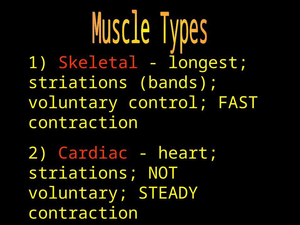
1) Skeletal - longest; striations (bands); voluntary control; FAST contraction
2) Cardiac - heart; striations; NOT voluntary; STEADY contraction
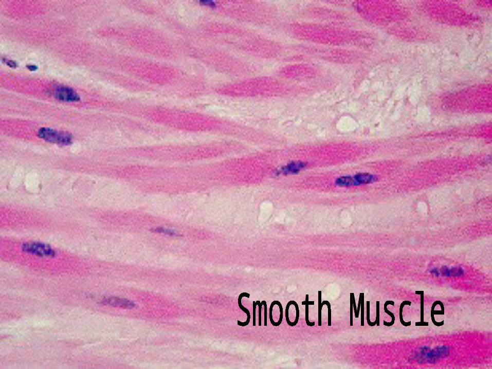
3) Smooth - visceral; NO striations; NOT voluntary; SLOW contraction
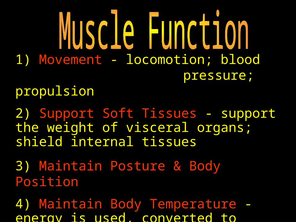
1) Movement - locomotion; blood pressure; propulsion
2) Support Soft Tissues - support the weight of visceral organs; shield internal tissues
3) Maintain Posture & Body Position
4) Maintain Body Temperature - energy is used, converted to heat, heat is released
5) Guard Entrances & Exits - digestive/urinary openings provide voluntary control
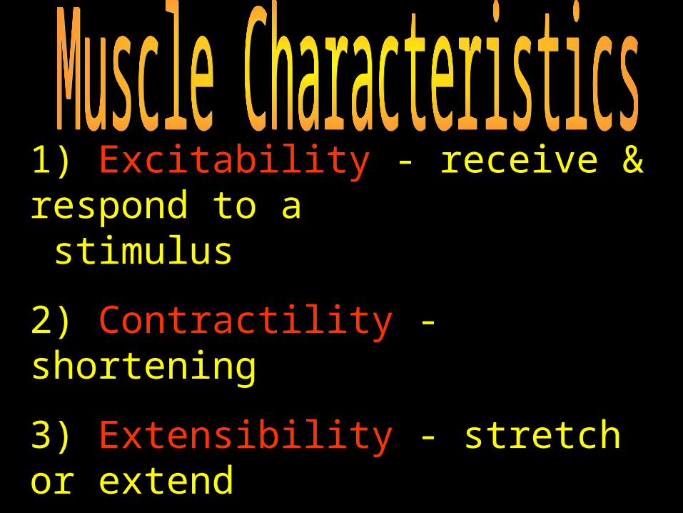
1) Excitability - receive & respond to a stimulus
2) Contractility - shortening
3) Extensibility - stretch or extend
4) Elasticity - resume resting length
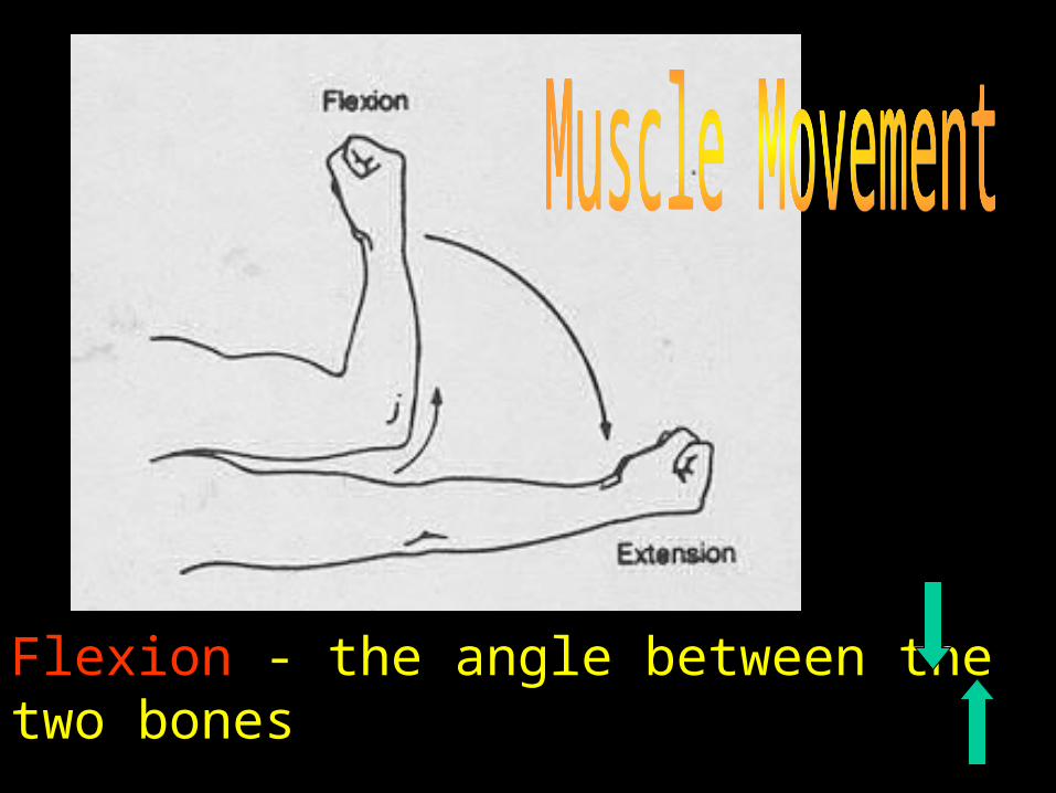
Flexion - the angle between the two bones
Extension - the angle between the two bones
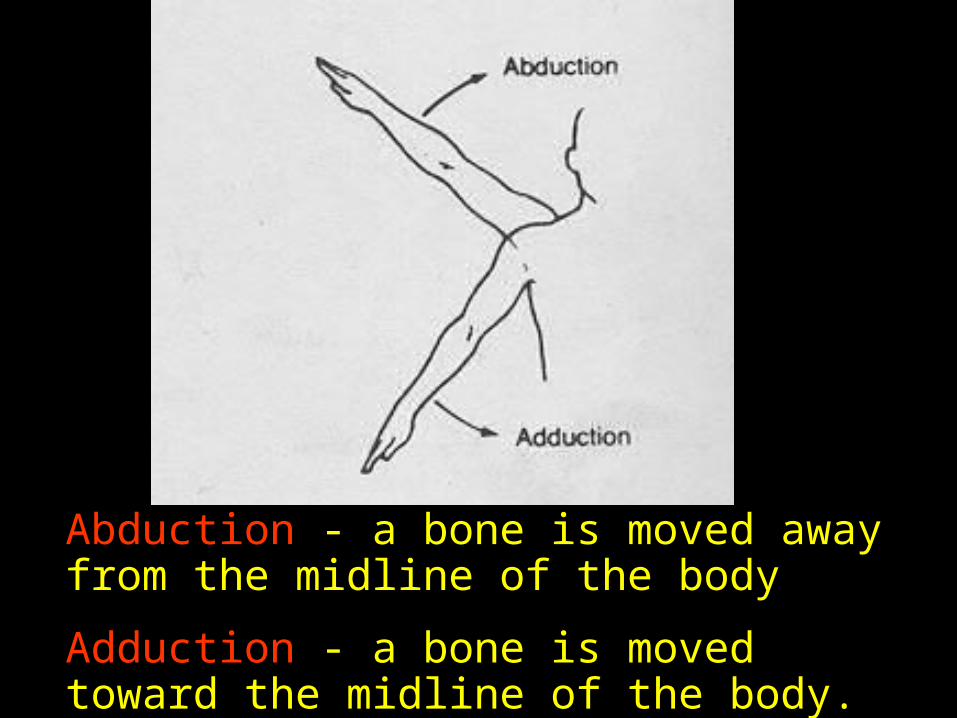
Abduction - a bone is moved away from the midline of the body
Adduction - a bone is moved toward the midline of the body.
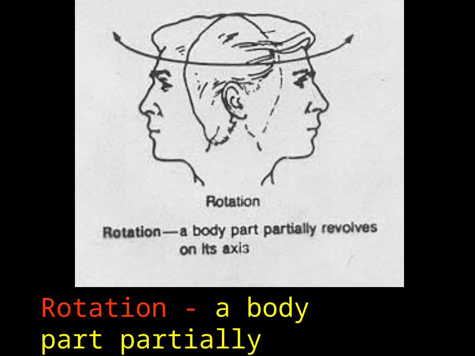
Rotation - a body part partially revolves on its axis
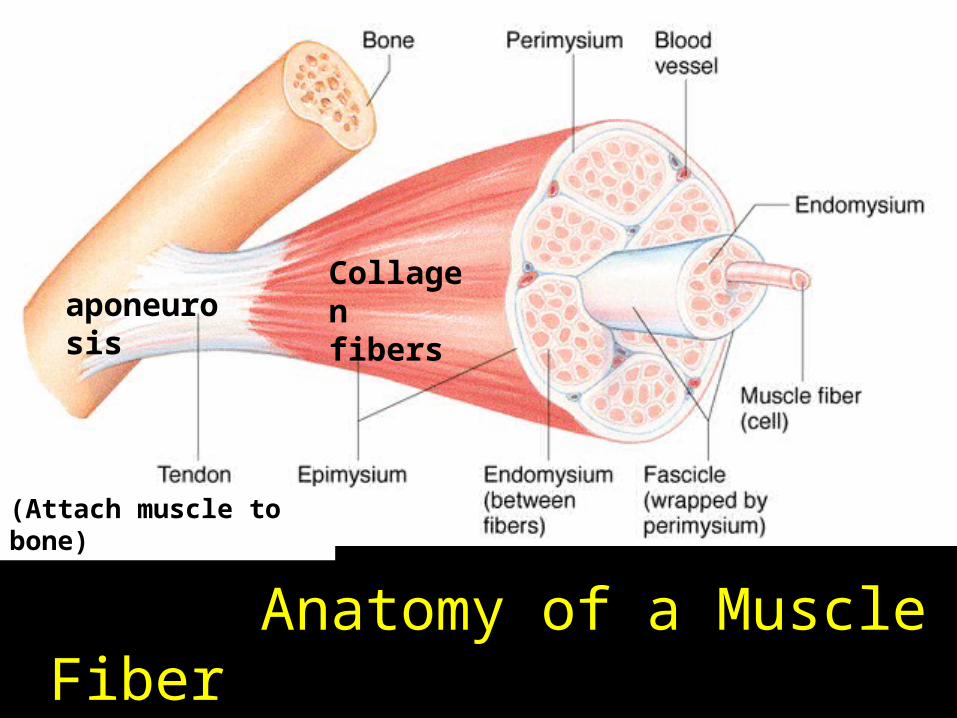
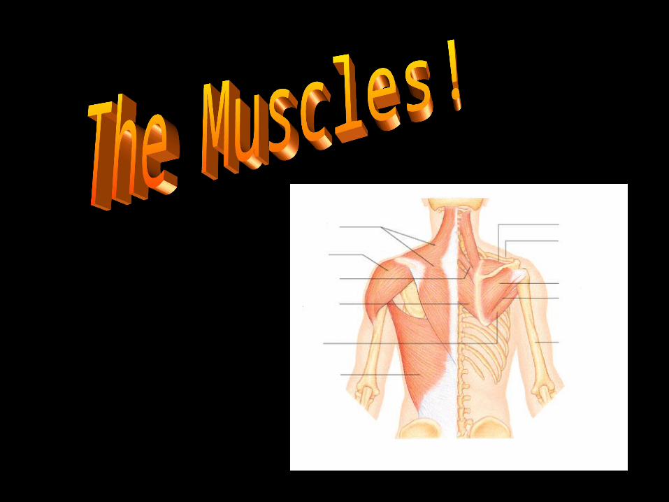
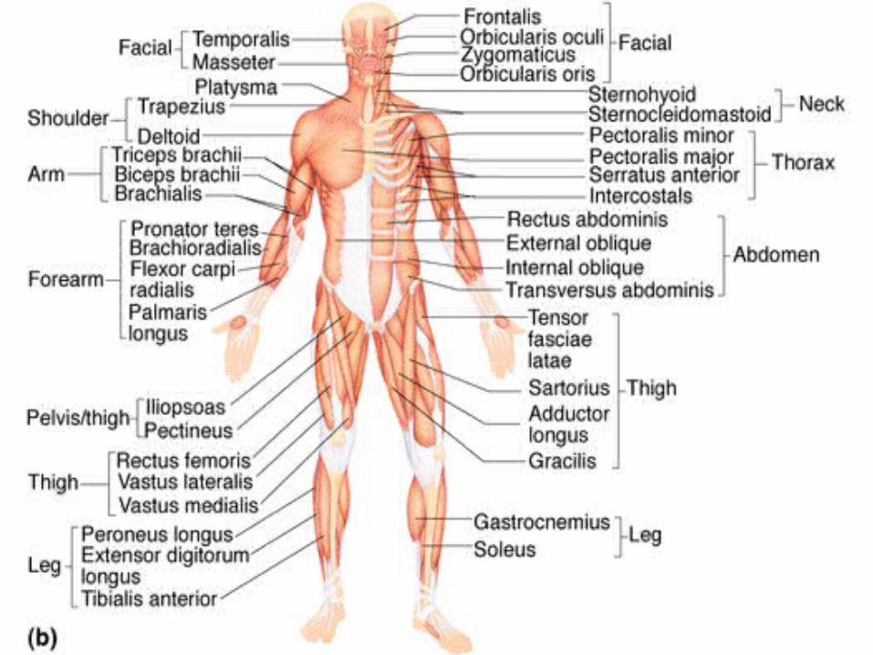
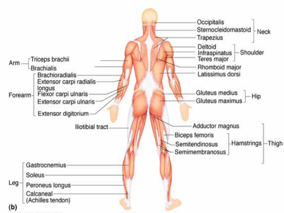
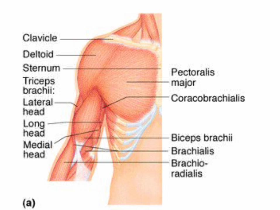
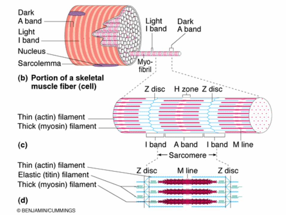
Anatomy of a Muscle Fiber
Collagen fibers
(Attach muscle to bone)
aponeurosis
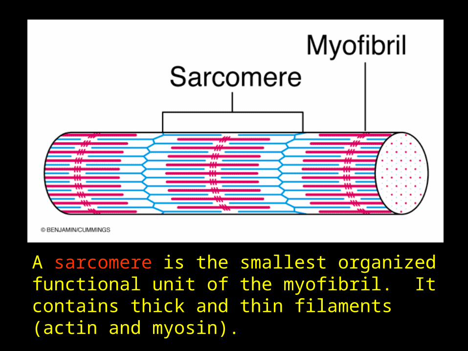
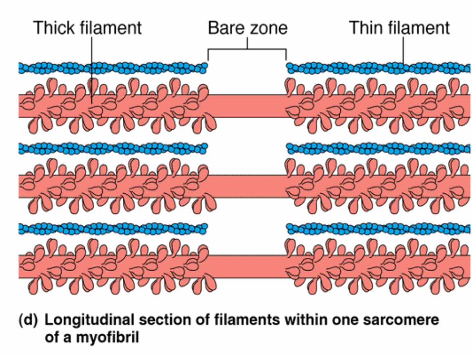
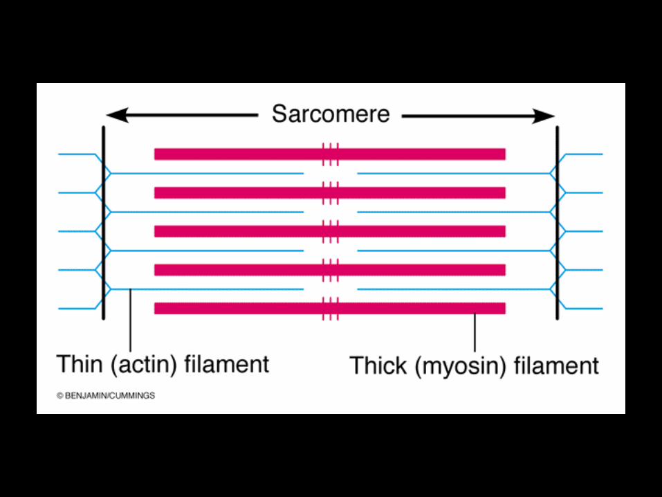
A sarcomere is the smallest organized functional unit of the myofibril. It contains thick and thin filaments (actin and myosin).
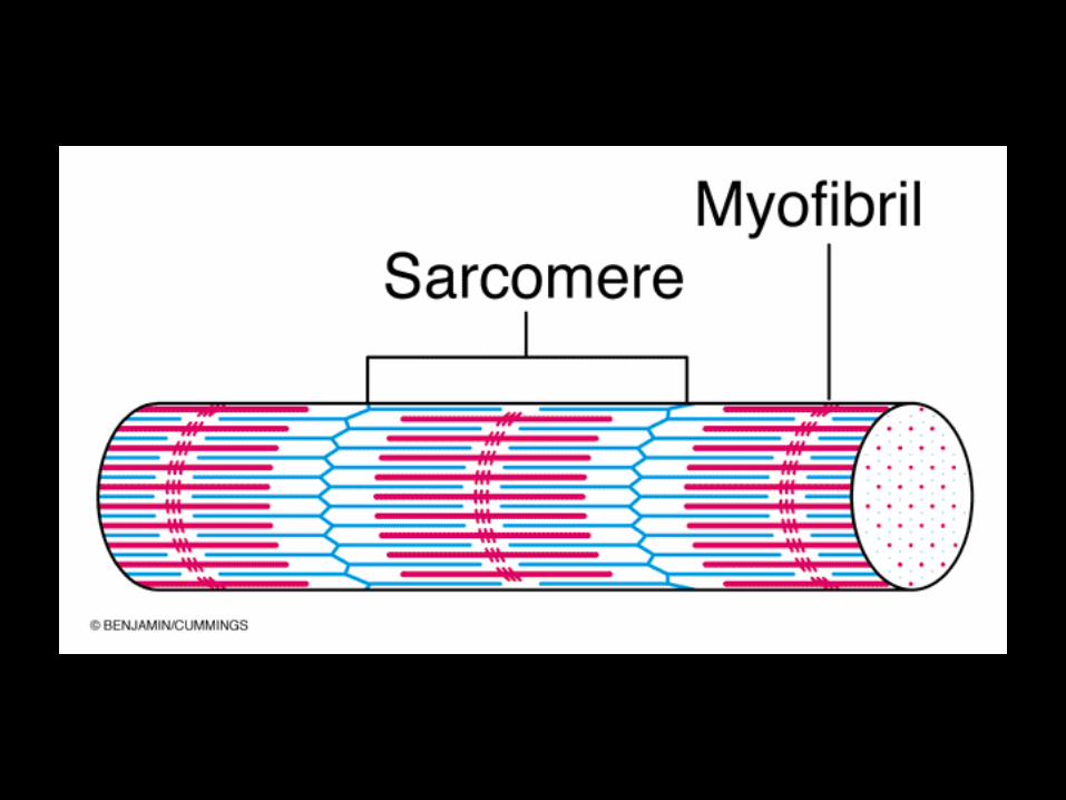
A myofibril consists of about 10,000 sarcomeres.
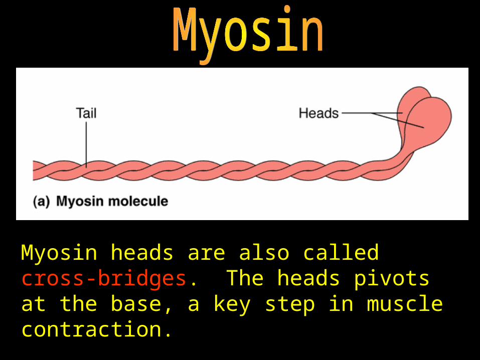
Myosin heads are also called cross-bridges. The heads pivots at the base, a key step in muscle contraction.
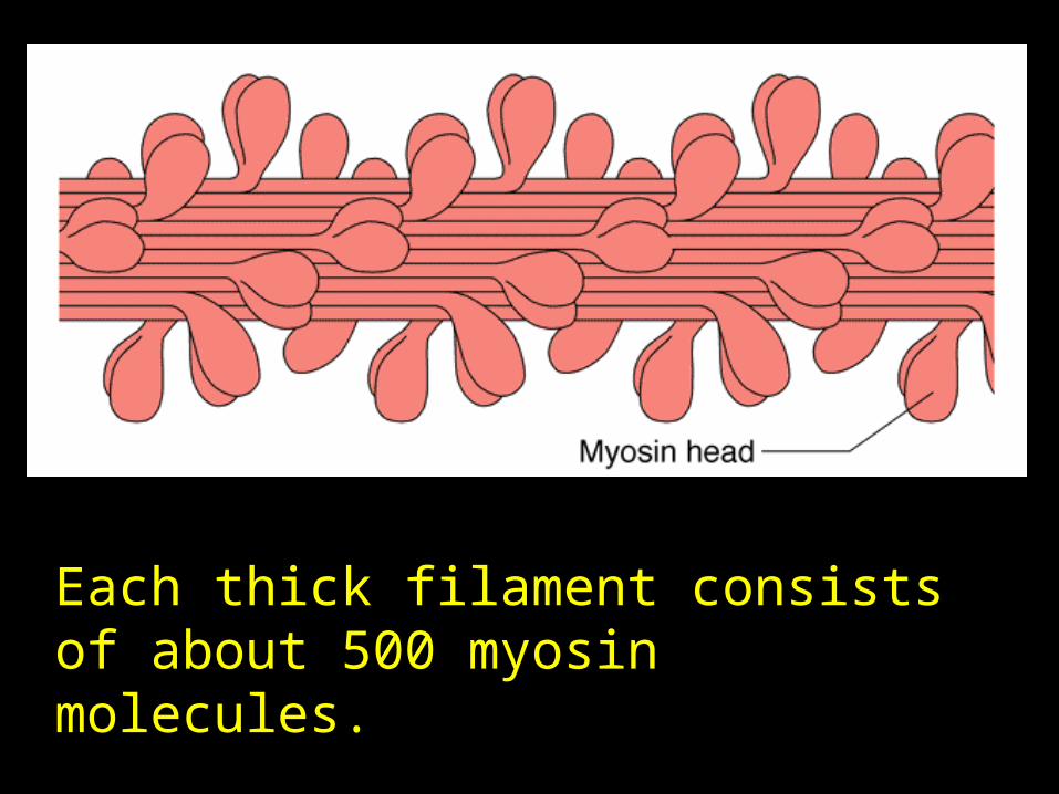
Each thick filament consists of about 500 myosin molecules.
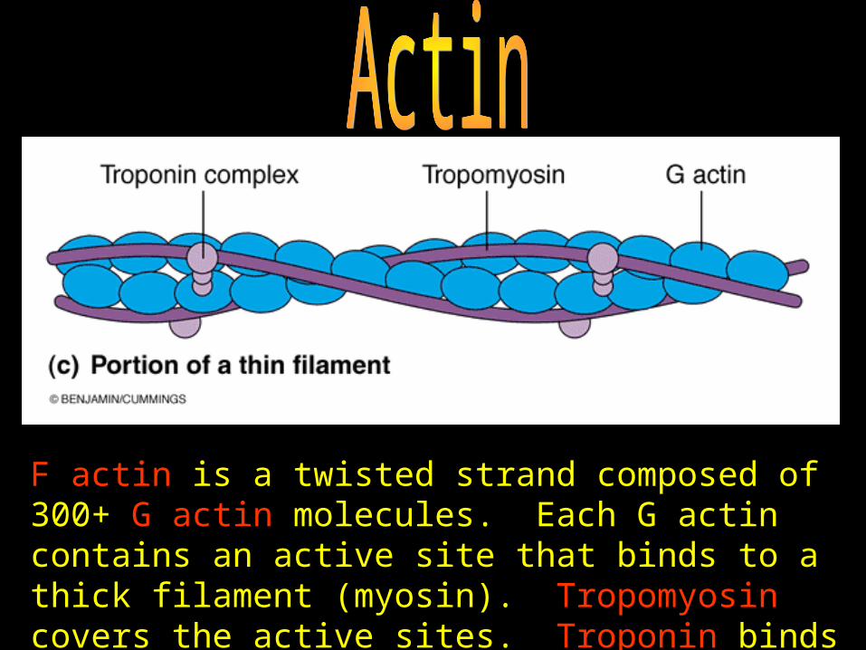
F actin is a twisted strand composed of 300+ G actin molecules. Each G actin contains an active site that binds to a thick filament (myosin). Tropomyosin covers the active sites. Troponin binds tropomyosin and G actin.
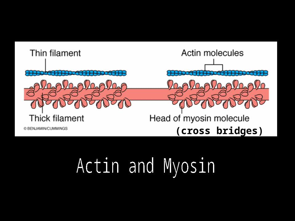
(cross bridges)

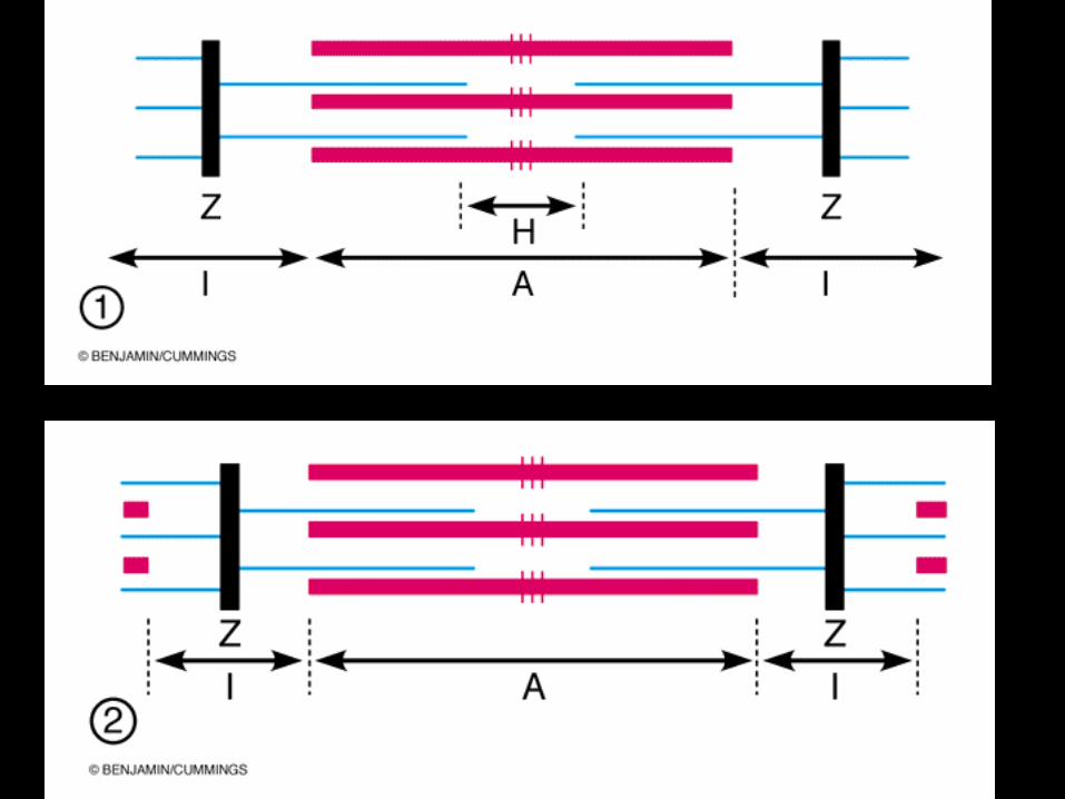
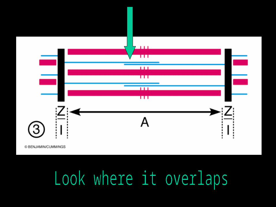
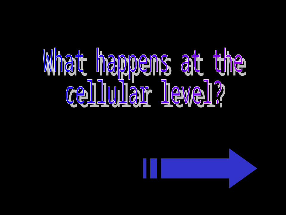
The Sliding Filament Theory
The thin actin filaments slide past the thick myosin filaments
The I bands shrinkThe H zone disappearsThe A bands move closer
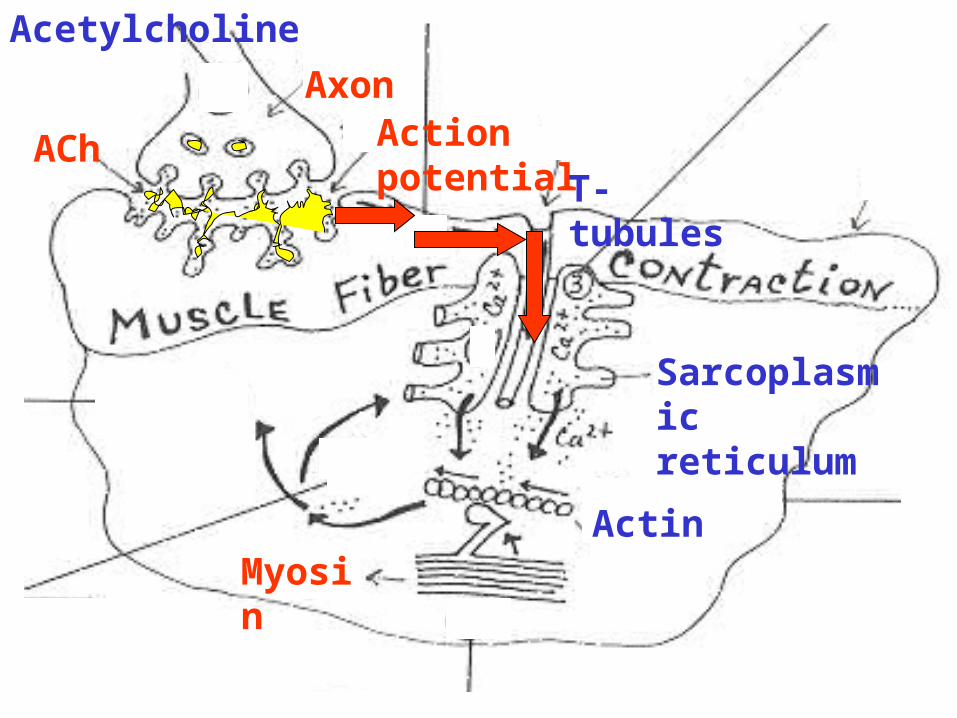
Axon
ACh
Acetylcholine
Sarcoplasmic reticulum
Action potential
T-tubules
ActinMyosin
7
1
2
3
46
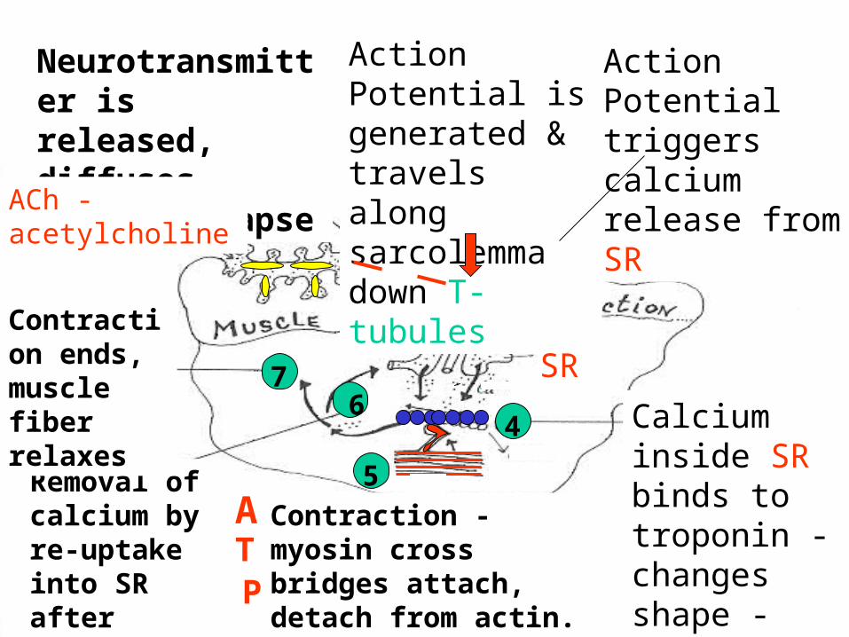
Neurotransmitter is released, diffuses across synapse
ACh - acetylcholine
Action Potential is generated & travels along sarcolemma down T-tubules
Action Potential triggers calcium release from SR
Calcium inside SR binds to troponin - changes shape - actin active sites exposed
Contraction - myosin cross bridges attach, detach from actin. Cross bridges split ATP & store energy.
ATP
Removal of calcium by re-uptake into SR after action potential ends
Contraction ends, muscle fiber relaxes
SR
5
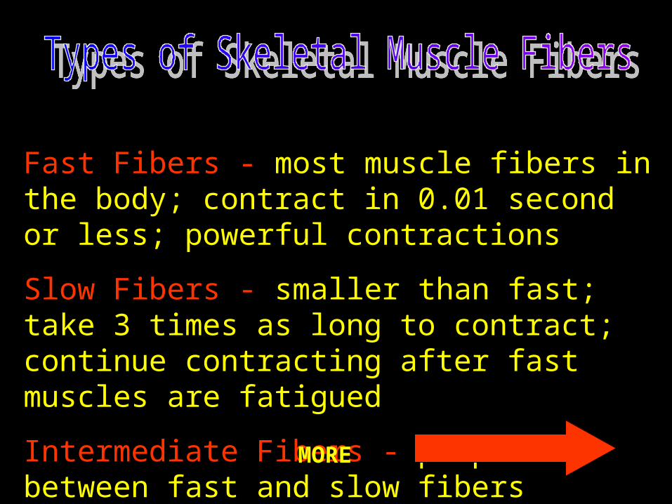
Fast Fibers - most muscle fibers in the body; contract in 0.01 second or less; powerful contractions
Slow Fibers - smaller than fast; take 3 times as long to contract; continue contracting after fast muscles are fatigued
Intermediate Fibers - properties between fast and slow fibers
MORE
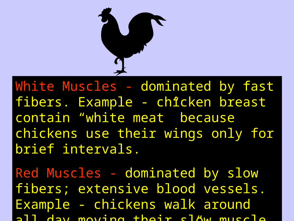
White Muscles - dominated by fast fibers. Example - chicken breast contain “white meat” because chickens use their wings only for brief intervals.
Red Muscles - dominated by slow fibers; extensive blood vessels. Example - chickens walk around all day moving their slow muscle fibers, the “dark meat” in their legs.
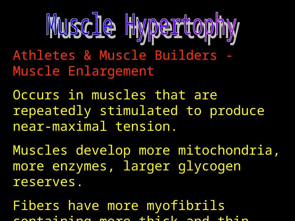
Athletes & Muscle Builders - Muscle Enlargement
Occurs in muscles that are repeatedly stimulated to produce near-maximal tension.
Muscles develop more mitochondria, more enzymes, larger glycogen reserves.
Fibers have more myofibrils containing more thick and thin filaments.
Number of fibers does not change, but the muscle diameter increases.
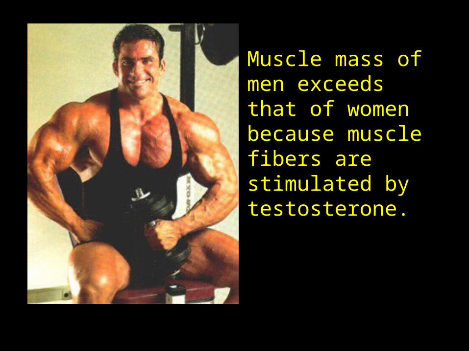
Muscle mass of men exceeds that of women because muscle fibers are stimulated by testosterone.