Embed Size (px)
Citation preview

www.elsevier.com/locate/jconrel
Journal of Controlled Rele
Review
Recent advances on chitosan-based micro- and nanoparticles
in drug deliveryB
Sunil A. Agnihotri, Nadagouda N. Mallikarjuna, Tejraj M. Aminabhavi*
Drug Delivery Division, Center of Excellence in Polymer Science, Karnatak University, Dharwad 580 003, India
Received 15 July 2004; accepted 12 August 2004
Abstract
Considerable research efforts have been directed towards developing safe and efficient chitosan-based particulate drug
delivery systems. The present review outlines the major new findings on the pharmaceutical applications of chitosan-based
micro/nanoparticulate drug delivery systems published over the past decade. Methods of their preparation, drug loading, release
characteristics, and applications are covered. Chemically modified chitosan or its derivatives used in drug delivery research are
discussed critically to evaluate the usefulness of these systems in delivering the bioactive molecules. From a literature survey, it
is realized that research activities on chitosan micro/nanoparticulate systems containing various drugs for different therapeutic
applications have increased at the rapid rate. Hence, the present review is timely.
D 2004 Elsevier B.V. All rights reserved.
Keywords: Microparticles; Nanoparticles; Chitosan; Chemically modified chitosan; Drug delivery
Contents
1. Introduction . . . . . . . . . . . . . . . . . . . . . . . . . . . . . . . . . . . . . . . . . . . . . . . . . . . . 6
2. Methods of preparation of micro/nanoparticles of chitosan . . . . . . . . . . . . . . . . . . . . . . . . . . . . 8
2.1. Emulsion cross-linking . . . . . . . . . . . . . . . . . . . . . . . . . . . . . . . . . . . . . . . . . . 8
2.2. Coacervation/precipitation . . . . . . . . . . . . . . . . . . . . . . . . . . . . . . . . . . . . . . . . . 9
2.3. Spray-drying . . . . . . . . . . . . . . . . . . . . . . . . . . . . . . . . . . . . . . . . . . . . . . . . 10
2.4. Emulsion-droplet coalescence method . . . . . . . . . . . . . . . . . . . . . . . . . . . . . . . . . . . 12
2.5. Ionic gelation . . . . . . . . . . . . . . . . . . . . . . . . . . . . . . . . . . . . . . . . . . . . . . . 12
2.6. Reverse micellar method . . . . . . . . . . . . . . . . . . . . . . . . . . . . . . . . . . . . . . . . . 14
2.7. Sieving method . . . . . . . . . . . . . . . . . . . . . . . . . . . . . . . . . . . . . . . . . . . . . . 15
0168-3659/$ - s
doi:10.1016/j.jco
B This pape
* Correspon
E-mail addr
ase 100 (2004) 5–28
ee front matter D 2004 Elsevier B.V. All rights reserved.
nrel.2004.08.010
r is CEPS Communication # 23.
ding author. Tel.: +91 836 2779983; fax: +91 836 2771275.
ess: [email protected] (T.M. Aminabhavi).
S.A. Agnihotri et al. / Journal of Controlled Release 100 (2004) 5–286
3. Drug loading into micro/nanoparticles of chitosan . . . . . . . . . . . . . . . . . . . . . . . . . . . . . . . 15
4. Drug release and release kinetics . . . . . . . . . . . . . . . . . . . . . . . . . . . . . . . . . . . . . . . . 16
5. Pharmaceutical applications of chitosan particulate systems . . . . . . . . . . . . . . . . . . . . . . . . . . . 18
5.1. Colon targeted drug delivery . . . . . . . . . . . . . . . . . . . . . . . . . . . . . . . . . . . . . . . 18
5.2. Mucosal delivery . . . . . . . . . . . . . . . . . . . . . . . . . . . . . . . . . . . . . . . . . . . . . 18
5.3. Cancer therapy . . . . . . . . . . . . . . . . . . . . . . . . . . . . . . . . . . . . . . . . . . . . . . 19
5.4. Gene delivery . . . . . . . . . . . . . . . . . . . . . . . . . . . . . . . . . . . . . . . . . . . . . . 20
5.5. Topical delivery . . . . . . . . . . . . . . . . . . . . . . . . . . . . . . . . . . . . . . . . . . . . . 22
5.6. Ocular delivery . . . . . . . . . . . . . . . . . . . . . . . . . . . . . . . . . . . . . . . . . . . . . . 22
5.7. Chitosan as a coating material . . . . . . . . . . . . . . . . . . . . . . . . . . . . . . . . . . . . . . 22
6. Chemically modified chitosans . . . . . . . . . . . . . . . . . . . . . . . . . . . . . . . . . . . . . . . . . 22
7. Conclusions . . . . . . . . . . . . . . . . . . . . . . . . . . . . . . . . . . . . . . . . . . . . . . . . . . . 24
Acknowledgements . . . . . . . . . . . . . . . . . . . . . . . . . . . . . . . . . . . . . . . . . . . . . . . . . . 24
References . . . . . . . . . . . . . . . . . . . . . . . . . . . . . . . . . . . . . . . . . . . . . . . . . . . . . . 24
1. Introduction
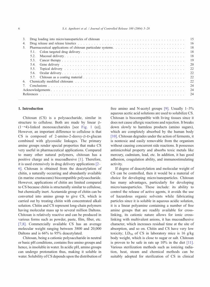
Chitosan (CS) is a polysaccharide, similar in
structure to cellulose. Both are made by linear h-(1Y4)-linked monosaccharides [see Fig. 1 (a)].
However, an important difference to cellulose is that
CS is composed of 2-amino-2-deoxy-h-d-glucancombined with glycosidic linkages. The primary
amine groups render special properties that make CS
very useful in pharmaceutical applications. Compared
to many other natural polymers, chitosan has a
positive charge and is mucoadhesive [1]. Therefore,
it is used extensively in drug delivery applications [2–
6]. Chitosan is obtained from the deacetylation of
chitin, a naturally occurring and abundantly available
(in marine crustaceans) biocompatible polysaccharide.
However, applications of chitin are limited compared
to CS because chitin is structurally similar to cellulose,
but chemically inert. Acetamide group of chitin can be
converted into amino group to give CS, which is
carried out by treating chitin with concentrated alkali
solution. Chitin and CS represent long-chain polymers
having molecular mass up to several million Daltons.
Chitosan is relatively reactive and can be produced in
various forms such as powder, paste, film, fiber, etc.
[7,8]. Commercially available CS has an average
molecular weight ranging between 3800 and 20,000
Daltons and is 66% to 95% deacetylated.
Chitosan, being a cationic polysaccharide in neutral
or basic pH conditions, contains free amino groups and
hence, is insoluble in water. In acidic pH, amino groups
can undergo protonation thus, making it soluble in
water. Solubility of CS depends upon the distribution of
free amino and N-acetyl groups [9]. Usually 1–3%
aqueous acetic acid solutions are used to solubilize CS.
Chitosan is biocompatible with living tissues since it
does not cause allergic reactions and rejection. It breaks
down slowly to harmless products (amino sugars),
which are completely absorbed by the human body
[10]. Chitosan degrades under the action of ferments, it
is nontoxic and easily removable from the organism
without causing concurrent side reactions. It possesses
antimicrobial property and absorbs toxic metals like
mercury, cadmium, lead, etc. In addition, it has good
adhesion, coagulation ability, and immunostimulating
activity.
If degree of deacetylation and molecular weight of
CS can be controlled, then it would be a material of
choice for developing micro/nanoparticles. Chitosan
has many advantages, particularly for developing
micro/nanoparticles. These include: its ability to
control the release of active agents, it avoids the use
of hazardous organic solvents while fabricating
particles since it is soluble in aqueous acidic solution,
it is a linear polyamine containing a number of free
amine groups that are readily available for cross-
linking, its cationic nature allows for ionic cross-
linking with multivalent anions, it has mucoadhesive
character, which increases residual time at the site of
absorption, and so on. Chitin and CS have very low
toxicity; LD50 of CS in laboratory mice is 16 g/kg
body weight, which is close to sugar or salt. Chitosan
is proven to be safe in rats up 10% in the diet [11].
Various sterilization methods such as ionizing radia-
tion, heat, steam and chemical methods can be
suitably adopted for sterilization of CS in clinical
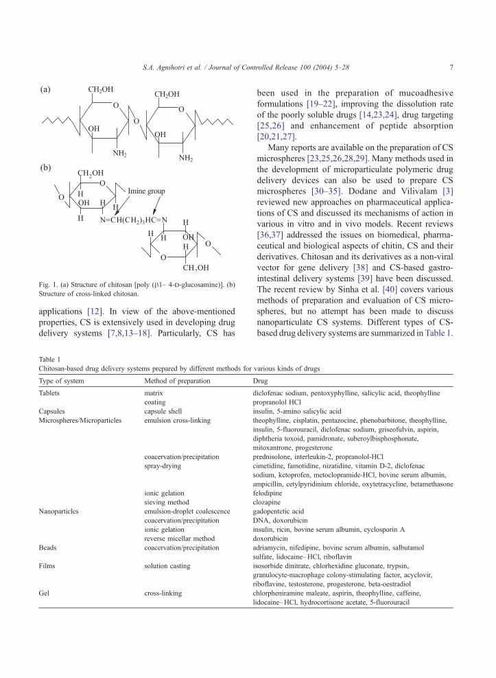
Fig. 1. (a) Structure of chitosan [poly (h1– 4-d-glucosamine)]. (b)
Structure of cross-linked chitosan.
S.A. Agnihotri et al. / Journal of Controlled Release 100 (2004) 5–28 7
applications [12]. In view of the above-mentioned
properties, CS is extensively used in developing drug
delivery systems [7,8,13–18]. Particularly, CS has
Table 1
Chitosan-based drug delivery systems prepared by different methods for v
Type of system Method of preparation D
Tablets matrix d
coating p
Capsules capsule shell i
Microspheres/Microparticles emulsion cross-linking t
i
d
m
coacervation/precipitation p
spray-drying c
s
a
ionic gelation f
sieving method c
Nanoparticles emulsion-droplet coalescence g
coacervation/precipitation D
ionic gelation i
reverse micellar method d
Beads coacervation/precipitation a
s
Films solution casting i
g
r
Gel cross-linking c
l
been used in the preparation of mucoadhesive
formulations [19–22], improving the dissolution rate
of the poorly soluble drugs [14,23,24], drug targeting
[25,26] and enhancement of peptide absorption
[20,21,27].
Many reports are available on the preparation of CS
microspheres [23,25,26,28,29]. Many methods used in
the development of microparticulate polymeric drug
delivery devices can also be used to prepare CS
microspheres [30–35]. Dodane and Vilivalam [3]
reviewed new approaches on pharmaceutical applica-
tions of CS and discussed its mechanisms of action in
various in vitro and in vivo models. Recent reviews
[36,37] addressed the issues on biomedical, pharma-
ceutical and biological aspects of chitin, CS and their
derivatives. Chitosan and its derivatives as a non-viral
vector for gene delivery [38] and CS-based gastro-
intestinal delivery systems [39] have been discussed.
The recent review by Sinha et al. [40] covers various
methods of preparation and evaluation of CS micro-
spheres, but no attempt has been made to discuss
nanoparticulate CS systems. Different types of CS-
based drug delivery systems are summarized in Table 1.
arious kinds of drugs
rug
iclofenac sodium, pentoxyphylline, salicylic acid, theophylline
ropranolol HCl
nsulin, 5-amino salicylic acid
heophylline, cisplatin, pentazocine, phenobarbitone, theophylline,
nsulin, 5-fluorouracil, diclofenac sodium, griseofulvin, aspirin,
iphtheria toxoid, pamidronate, suberoylbisphosphonate,
itoxantrone, progesterone
rednisolone, interleukin-2, propranolol-HCl
imetidine, famotidine, nizatidine, vitamin D-2, diclofenac
odium, ketoprofen, metoclopramide-HCl, bovine serum albumin,
mpicillin, cetylpyridinium chloride, oxytetracycline, betamethasone
elodipine
lozapine
adopentetic acid
NA, doxorubicin
nsulin, ricin, bovine serum albumin, cyclosporin A
oxorubicin
driamycin, nifedipine, bovine serum albumin, salbutamol
ulfate, lidocaine–HCl, riboflavin
sosorbide dinitrate, chlorhexidine gluconate, trypsin,
ranulocyte-macrophage colony-stimulating factor, acyclovir,
iboflavine, testosterone, progesterone, beta-oestradiol
hlorpheniramine maleate, aspirin, theophylline, caffeine,
idocaine–HCl, hydrocortisone acetate, 5-fluorouracil
S.A. Agnihotri et al. / Journal of Controlled Release 100 (2004) 5–288
However, the micro/nanoparticulate drug delivery
systems offer numerous advantages over the conven-
tional dosage forms. These include improved efficacy,
reduced toxicity and improved patient compliance
[35,41–43]. The present review addresses the recent
trends in the area of micro/nanoparticulate CS-based
drug delivery systems. Literature of the past decade has
been covered and results are critically evaluated.
Fig. 2. Schematic representation of preparation of chitosan
particulate systems by emulsion cross-linking method.
2. Methods of preparation of micro/nanoparticles
of chitosan
Different methods have been used to prepare CS
particulate systems. Selection of any of the methods
depends upon factors such as particle size require-
ment, thermal and chemical stability of the active
agent, reproducibility of the release kinetic profiles,
stability of the final product and residual toxicity
associated with the final product. Different methods
used in the preparation of CS micro/nanoparticles are
discussed in this review. However, selection of any of
these methods depends upon the nature of the active
molecule as well as the type of the delivery device.
Since we are concerned only with the micro/nano-
particulate systems of CS and its derivatives, we will
restrict our discussions only on these aspects.
2.1. Emulsion cross-linking
This method utilizes the reactive functional amine
group of CS to cross-link with aldehyde groups of the
cross-linking agent (see Fig. 1b). In this method, a
water-in-oil (w/o) emulsion is prepared by emulsify-
ing the CS aqueous solution in the oil phase. Aqueous
droplets are stabilized using a suitable surfactant. The
stable emulsion is cross-linked by using an appro-
priate cross-linking agent such as glutaraldehyde to
harden the droplets. Microspheres are filtered and
washed repeatedly with n-hexane followed by alcohol
and then dried [44]. By this method, size of the
particles can be controlled by controlling the size of
aqueous droplets. However, the particle size of final
product depends upon the extent of cross-linking
agent used while hardening in addition to speed of
stirring during the formation of emulsion. This
method is schematically represented in Fig. 2. The
emulsion cross-linking method has few drawbacks
since it involves tedious procedures as well as use of
harsh cross-linking agents, which might possibly
induce chemical reactions with the active agent.
However, complete removal of the un-reacted cross-
linking agent may be difficult in this process.
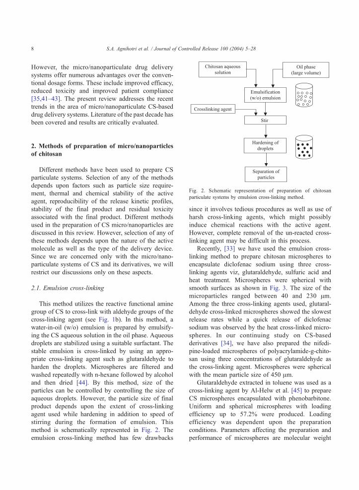
Recently, [33] we have used the emulsion cross-
linking method to prepare chitosan microspheres to
encapsulate diclofenac sodium using three cross-
linking agents viz, glutaraldehyde, sulfuric acid and
heat treatment. Microspheres were spherical with
smooth surfaces as shown in Fig. 3. The size of the
microparticles ranged between 40 and 230 Am.
Among the three cross-linking agents used, glutaral-
dehyde cross-linked microspheres showed the slowest
release rates while a quick release of diclofenac
sodium was observed by the heat cross-linked micro-
spheres. In our continuing study on CS-based
derivatives [34], we have also prepared the nifedi-
pine-loaded microspheres of polyacrylamide-g-chito-
san using three concentrations of glutaraldehyde as
the cross-linking agent. Microspheres were spherical
with the mean particle size of 450 Am.
Glutaraldehyde extracted in toluene was used as a
cross-linking agent by Al-Helw et al. [45] to prepare
CS microspheres encapsulated with phenobarbitone.
Uniform and spherical microspheres with loading
efficiency up to 57.2% were produced. Loading
efficiency was dependent upon the preparation
conditions. Parameters affecting the preparation and
performance of microspheres are molecular weight
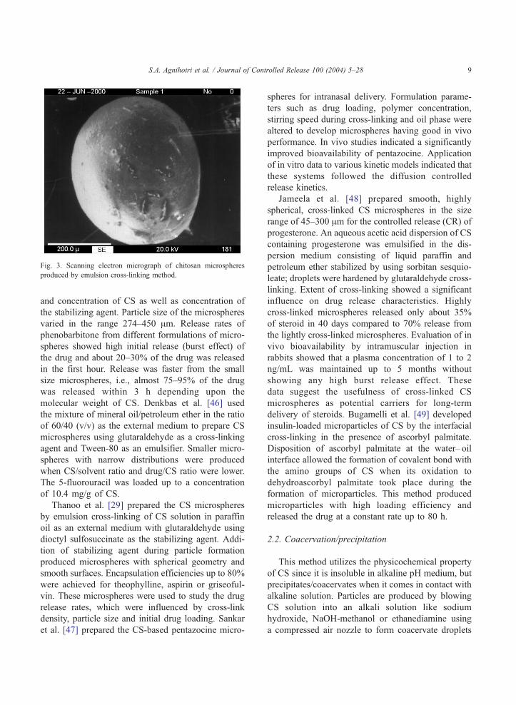
Fig. 3. Scanning electron micrograph of chitosan microspheres
produced by emulsion cross-linking method.
S.A. Agnihotri et al. / Journal of Controlled Release 100 (2004) 5–28 9
and concentration of CS as well as concentration of
the stabilizing agent. Particle size of the microspheres
varied in the range 274–450 Am. Release rates of
phenobarbitone from different formulations of micro-
spheres showed high initial release (burst effect) of
the drug and about 20–30% of the drug was released
in the first hour. Release was faster from the small
size microspheres, i.e., almost 75–95% of the drug
was released within 3 h depending upon the
molecular weight of CS. Denkbas et al. [46] used
the mixture of mineral oil/petroleum ether in the ratio
of 60/40 (v/v) as the external medium to prepare CS
microspheres using glutaraldehyde as a cross-linking
agent and Tween-80 as an emulsifier. Smaller micro-
spheres with narrow distributions were produced
when CS/solvent ratio and drug/CS ratio were lower.
The 5-fluorouracil was loaded up to a concentration
of 10.4 mg/g of CS.
Thanoo et al. [29] prepared the CS microspheres
by emulsion cross-linking of CS solution in paraffin
oil as an external medium with glutaraldehyde using
dioctyl sulfosuccinate as the stabilizing agent. Addi-
tion of stabilizing agent during particle formation
produced microspheres with spherical geometry and
smooth surfaces. Encapsulation efficiencies up to 80%
were achieved for theophylline, aspirin or griseoful-
vin. These microspheres were used to study the drug
release rates, which were influenced by cross-link
density, particle size and initial drug loading. Sankar
et al. [47] prepared the CS-based pentazocine micro-
spheres for intranasal delivery. Formulation parame-
ters such as drug loading, polymer concentration,
stirring speed during cross-linking and oil phase were
altered to develop microspheres having good in vivo
performance. In vivo studies indicated a significantly
improved bioavailability of pentazocine. Application
of in vitro data to various kinetic models indicated that
these systems followed the diffusion controlled
release kinetics.
Jameela et al. [48] prepared smooth, highly
spherical, cross-linked CS microspheres in the size
range of 45–300 Am for the controlled release (CR) of
progesterone. An aqueous acetic acid dispersion of CS
containing progesterone was emulsified in the dis-
persion medium consisting of liquid paraffin and
petroleum ether stabilized by using sorbitan sesquio-
leate; droplets were hardened by glutaraldehyde cross-
linking. Extent of cross-linking showed a significant
influence on drug release characteristics. Highly
cross-linked microspheres released only about 35%
of steroid in 40 days compared to 70% release from
the lightly cross-linked microspheres. Evaluation of in
vivo bioavailability by intramuscular injection in
rabbits showed that a plasma concentration of 1 to 2
ng/mL was maintained up to 5 months without
showing any high burst release effect. These
data suggest the usefulness of cross-linked CS
microspheres as potential carriers for long-term
delivery of steroids. Bugamelli et al. [49] developed
insulin-loaded microparticles of CS by the interfacial
cross-linking in the presence of ascorbyl palmitate.
Disposition of ascorbyl palmitate at the water– oil
interface allowed the formation of covalent bond with
the amino groups of CS when its oxidation to
dehydroascorbyl palmitate took place during the
formation of microparticles. This method produced
microparticles with high loading efficiency and
released the drug at a constant rate up to 80 h.
2.2. Coacervation/precipitation
This method utilizes the physicochemical property
of CS since it is insoluble in alkaline pH medium, but
precipitates/coacervates when it comes in contact with
alkaline solution. Particles are produced by blowing
CS solution into an alkali solution like sodium
hydroxide, NaOH-methanol or ethanediamine using
a compressed air nozzle to form coacervate droplets
S.A. Agnihotri et al. / Journal of Controlled Release 100 (2004) 5–2810
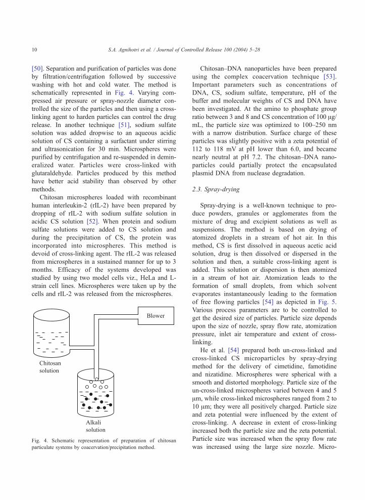
[50]. Separation and purification of particles was done
by filtration/centrifugation followed by successive
washing with hot and cold water. The method is
schematically represented in Fig. 4. Varying com-
pressed air pressure or spray-nozzle diameter con-
trolled the size of the particles and then using a cross-
linking agent to harden particles can control the drug
release. In another technique [51], sodium sulfate
solution was added dropwise to an aqueous acidic
solution of CS containing a surfactant under stirring
and ultrasonication for 30 min. Microspheres were
purified by centrifugation and re-suspended in demin-
eralized water. Particles were cross-linked with
glutaraldehyde. Particles produced by this method
have better acid stability than observed by other
methods.
Chitosan microspheres loaded with recombinant
human interleukin-2 (rIL-2) have been prepared by
dropping of rIL-2 with sodium sulfate solution in
acidic CS solution [52]. When protein and sodium
sulfate solutions were added to CS solution and
during the precipitation of CS, the protein was
incorporated into microspheres. This method is
devoid of cross-linking agent. The rIL-2 was released
from microspheres in a sustained manner for up to 3
months. Efficacy of the systems developed was
studied by using two model cells viz., HeLa and L-
strain cell lines. Microspheres were taken up by the
cells and rIL-2 was released from the microspheres.
Fig. 4. Schematic representation of preparation of chitosan
particulate systems by coacervation/precipitation method.
Chitosan–DNA nanoparticles have been prepared
using the complex coacervation technique [53].
Important parameters such as concentrations of
DNA, CS, sodium sulfate, temperature, pH of the
buffer and molecular weights of CS and DNA have
been investigated. At the amino to phosphate group
ratio between 3 and 8 and CS concentration of 100 Ag/mL, the particle size was optimized to 100–250 nm
with a narrow distribution. Surface charge of these
particles was slightly positive with a zeta potential of
112 to 118 mV at pH lower than 6.0, and became
nearly neutral at pH 7.2. The chitosan–DNA nano-
particles could partially protect the encapsulated
plasmid DNA from nuclease degradation.
2.3. Spray-drying
Spray-drying is a well-known technique to pro-
duce powders, granules or agglomerates from the
mixture of drug and excipient solutions as well as
suspensions. The method is based on drying of
atomized droplets in a stream of hot air. In this
method, CS is first dissolved in aqueous acetic acid
solution, drug is then dissolved or dispersed in the
solution and then, a suitable cross-linking agent is
added. This solution or dispersion is then atomized
in a stream of hot air. Atomization leads to the
formation of small droplets, from which solvent
evaporates instantaneously leading to the formation
of free flowing particles [54] as depicted in Fig. 5.
Various process parameters are to be controlled to
get the desired size of particles. Particle size depends
upon the size of nozzle, spray flow rate, atomization
pressure, inlet air temperature and extent of cross-
linking.
He et al. [54] prepared both un-cross-linked and
cross-linked CS microparticles by spray-drying
method for the delivery of cimetidine, famotidine
and nizatidine. Microspheres were spherical with a
smooth and distorted morphology. Particle size of the
un-cross-linked microspheres varied between 4 and 5
Am, while cross-linked microspheres ranged from 2 to
10 Am; they were all positively charged. Particle size
and zeta potential were influenced by the extent of
cross-linking. A decrease in extent of cross-linking
increased both the particle size and the zeta potential.
Particle size was increased when the spray flow rate
was increased using the large size nozzle. Micro-
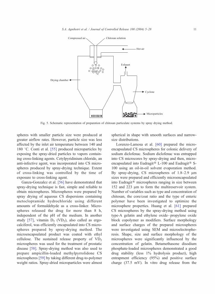
Fig. 5. Schematic representation of preparation of chitosan particulate systems by spray drying method.
S.A. Agnihotri et al. / Journal of Controlled Release 100 (2004) 5–28 11
spheres with smaller particle size were produced at
greater airflow rates. However, particle size was less
affected by the inlet air temperature between 140 and
180 8C. Conti et al. [55] produced microparticles by
exposing the spray-dried particles to vapors contain-
ing cross-linking agents. Cetylpyridinium chloride, an
anti-infective agent, was incorporated into CS micro-
spheres produced by spray-drying technique. Extent
of cross-linking was controlled by the time of
exposure to cross-linking agent.
Ganza-Gonzalez et al. [56] have demonstrated that
spray-drying technique is fast, simple and reliable to
obtain microspheres. Microspheres were prepared by
spray drying of aqueous CS dispersions containing
metoclopramide hydrochloride using different
amounts of formaldehyde as a cross-linker. Micro-
spheres released the drug for more than 8 h,
independent of the pH of the medium. In another
study [57], vitamin D2 (VD2), also called as ergo-
calciferol, was efficiently encapsulated into CS micro-
spheres prepared by spray-drying method. The
microencapsulated product was coated with ethyl
cellulose. The sustained release property of VD2
microspheres was used for the treatment of prostatic
disease [58]. Spray-drying method was also used to
prepare ampicillin-loaded methylpyrrolidone CS
microspheres [59] by taking different drug-to-polymer
weight ratios. Spray-dried microparticles were almost
spherical in shape with smooth surfaces and narrow-
size distributions.
Lorenzo-Lamosa et al. [60] prepared the micro-
encapsulated CS microspheres for colonic delivery of
sodium diclofenac. Sodium diclofenac was entrapped
into CS microcores by spray-drying and then, micro-
encapsulated into EudragitR L-100 and EudragitR S-
100 using an oil-in-oil solvent evaporation method.
By spray-drying, CS microspheres of 1.8–2.9 Amsizes were prepared and efficiently microencapsulated
into EudragitR microspheres ranging in size between
152 and 223 Am to form the multireservoir system.
Number of variables such as type and concentration of
chitosan, the core/coat ratio and the type of enteric
polymer have been investigated to optimize the
microsphere properties. Huang et al. [61] prepared
CS microspheres by the spray-drying method using
type-A gelatin and ethylene oxide– propylene oxide
block copolymer as modifiers. Surface morphology
and surface charges of the prepared microspheres
were investigated using SEM and microelectropho-
resis. Shape, size and surface morphology of the
microspheres were significantly influenced by the
concentration of gelatin. Betamethasone disodium
phosphate-loaded microspheres demonstrated a good
drug stability (less 1% hydrolysis product), high
entrapment efficiency (95%) and positive surface
charge (37.5 mV). In vitro drug release from the
S.A. Agnihotri et al. / Journal of Controlled Release 100 (2004) 5–2812
microspheres was related to gelatin content. Micro-
spheres containing gelatin/CS ratio of 0.4–0.6 (w/w)
showed a prolonged release up to 12 h.
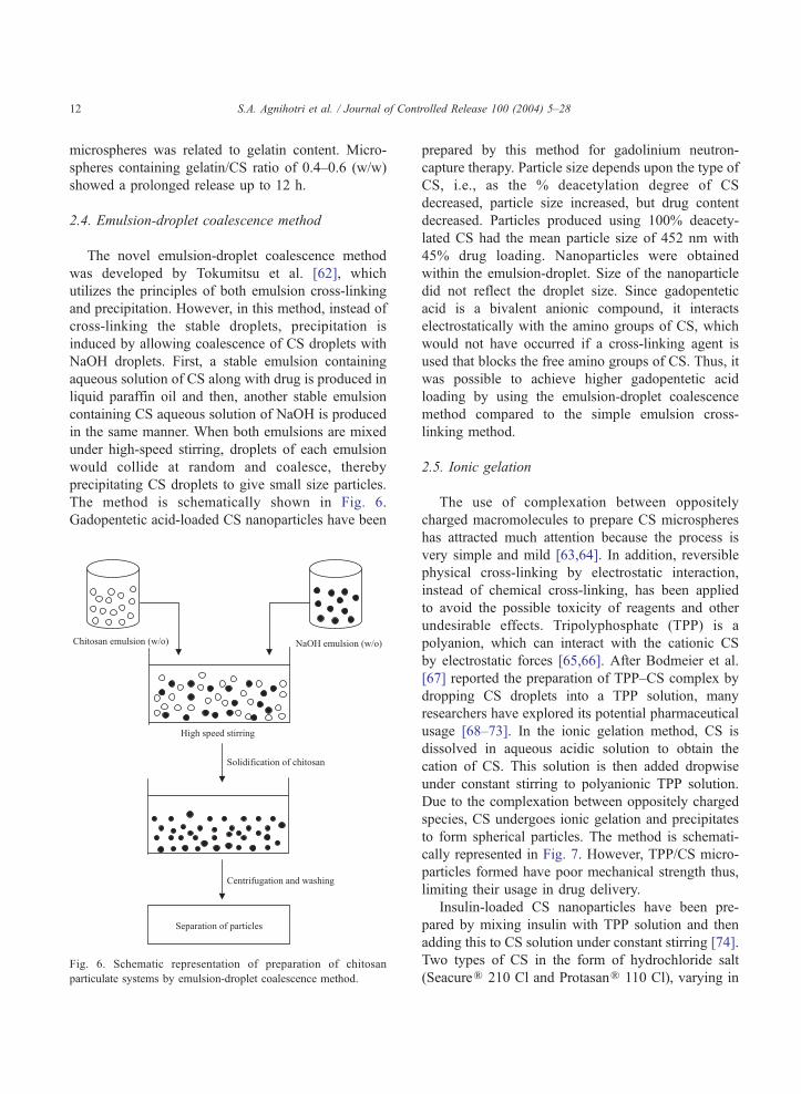
2.4. Emulsion-droplet coalescence method
The novel emulsion-droplet coalescence method
was developed by Tokumitsu et al. [62], which
utilizes the principles of both emulsion cross-linking
and precipitation. However, in this method, instead of
cross-linking the stable droplets, precipitation is
induced by allowing coalescence of CS droplets with
NaOH droplets. First, a stable emulsion containing
aqueous solution of CS along with drug is produced in
liquid paraffin oil and then, another stable emulsion
containing CS aqueous solution of NaOH is produced
in the same manner. When both emulsions are mixed
under high-speed stirring, droplets of each emulsion
would collide at random and coalesce, thereby
precipitating CS droplets to give small size particles.
The method is schematically shown in Fig. 6.
Gadopentetic acid-loaded CS nanoparticles have been
Fig. 6. Schematic representation of preparation of chitosan
particulate systems by emulsion-droplet coalescence method.
prepared by this method for gadolinium neutron-
capture therapy. Particle size depends upon the type of
CS, i.e., as the % deacetylation degree of CS
decreased, particle size increased, but drug content
decreased. Particles produced using 100% deacety-
lated CS had the mean particle size of 452 nm with
45% drug loading. Nanoparticles were obtained
within the emulsion-droplet. Size of the nanoparticle
did not reflect the droplet size. Since gadopentetic
acid is a bivalent anionic compound, it interacts
electrostatically with the amino groups of CS, which
would not have occurred if a cross-linking agent is
used that blocks the free amino groups of CS. Thus, it
was possible to achieve higher gadopentetic acid
loading by using the emulsion-droplet coalescence
method compared to the simple emulsion cross-
linking method.
2.5. Ionic gelation
The use of complexation between oppositely
charged macromolecules to prepare CS microspheres
has attracted much attention because the process is
very simple and mild [63,64]. In addition, reversible
physical cross-linking by electrostatic interaction,
instead of chemical cross-linking, has been applied
to avoid the possible toxicity of reagents and other
undesirable effects. Tripolyphosphate (TPP) is a
polyanion, which can interact with the cationic CS
by electrostatic forces [65,66]. After Bodmeier et al.
[67] reported the preparation of TPP–CS complex by
dropping CS droplets into a TPP solution, many
researchers have explored its potential pharmaceutical
usage [68–73]. In the ionic gelation method, CS is
dissolved in aqueous acidic solution to obtain the
cation of CS. This solution is then added dropwise
under constant stirring to polyanionic TPP solution.
Due to the complexation between oppositely charged
species, CS undergoes ionic gelation and precipitates
to form spherical particles. The method is schemati-
cally represented in Fig. 7. However, TPP/CS micro-
particles formed have poor mechanical strength thus,
limiting their usage in drug delivery.
Insulin-loaded CS nanoparticles have been pre-
pared by mixing insulin with TPP solution and then
adding this to CS solution under constant stirring [74].
Two types of CS in the form of hydrochloride salt
(SeacureR 210 Cl and ProtasanR 110 Cl), varying in
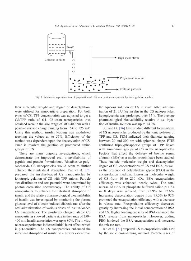
Fig. 7. Schematic representation of preparation of chitosan particulate systems by ionic gelation method.
S.A. Agnihotri et al. / Journal of Controlled Release 100 (2004) 5–28 13
their molecular weight and degree of deacetylation,
were utilized for nanoparticle preparation. For both
types of CS, TPP concentration was adjusted to get a
CS/TPP ratio of 6:1. Chitosan nanoparticles thus
obtained were in the size range of 300–400 nm with a
positive surface charge ranging from +54 to +25 mV.
Using this method, insulin loading was modulated
reaching the values up to 55%. Efficiency of the
method was dependent upon the deacetylation of CS,
since it involves the gelation of protonated amino
groups of CS.
There are many ongoing investigations, which
demonstrate the improved oral bioavailability of
peptide and protein formulations. Bioadhesive poly-
saccharide CS nanoparticles would seem to further
enhance their intestinal absorption. Pan et al. [75]
prepared the insulin-loaded CS nanoparticles by
ionotropic gelation of CS with TPP anions. Particle
size distribution and zeta potential were determined by
photon correlation spectroscopy. The ability of CS
nanoparticles to enhance the intestinal absorption of
insulin and the relative pharmacological bioavailability
of insulin was investigated by monitoring the plasma
glucose level of alloxan-induced diabetic rats after the
oral administration of various doses of insulin-loaded
CS nanoparticles. The positively charged, stable CS
nanoparticles showed particle size in the range of 250–
400 nm. Insulin association was up to 80%. The in vitro
release experiments indicated initial burst effect, which
is pH-sensitive. The CS nanoparticles enhanced the
intestinal absorption of insulin to a greater extent than
the aqueous solution of CS in vivo. After adminis-
tration of 21 I.U./kg insulin in the CS nanoparticles,
hypoglycemia was prolonged over 15 h. The average
pharmacological bioavailability relative to s.c. injec-
tion of insulin solution was up to 14.9%.
Xu and Du [76] have studied different formulations
of CS nanoparticles produced by the ionic gelation of
TPP and CS. TEM indicated their diameter ranging
between 20 and 200 nm with spherical shape. FTIR
confirmed tripolyphosphoric groups of TPP linked
with ammonium groups of CS in the nanoparticles.
Factors that affect the delivery of bovine serum
albumin (BSA) as a model protein have been studied.
These include molecular weight and deacetylation
degree of CS, concentrations of CS and BSA, as well
as the presence of polyethylene glycol (PEG) in the
encapsulation medium. Increasing molecular weight
of CS from 10 to 210 kDa, BSA encapsulation
efficiency was enhanced nearly twice. The total
release of BSA in phosphate buffered saline pH 7.4
in 8 days was reduced from 73.9% to 17.6%.
Increasing deacetylation degree from 75.5% to 92%
promoted the encapsulation efficiency with a decrease
in release rate. Encapsulation efficiency decreased
greatly by increasing the initial concentration of BSA
and CS. Higher loading capacity of BSA enhanced the
BSA release from nanoparticles. However, adding
PEG hindered the BSA encapsulation and increased
the release rate.
Ko et al. [77] prepared CS microparticles with TPP
by the ionic cross-linking method. Particle sizes of
S.A. Agnihotri et al. / Journal of Controlled Release 100 (2004) 5–2814
TPP-CS microparticles varied from 500 to 710 Amwith drug encapsulation efficiencies more than 90%.
Morphologies of TPP-CS microparticles have been
examined by SEM. As the pH of TPP solution
decreased and molecular weight of CS increased,
microparticles acquired better spherical shape having
smooth surface. Release of felodipine as a model drug
was affected by the preparation method. Chitosan
microparticles prepared at lower pH or higher
concentration of TPP solution resulted in a slower
release of felodipine. With a decreasing molecular
weight and concentration of CS solution, the drug
release increased. The release of drug from TPP-CS
microparticles decreased when the cross-linking time
was increased.
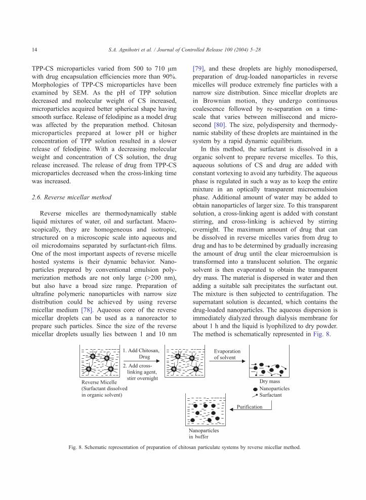
2.6. Reverse micellar method
Reverse micelles are thermodynamically stable
liquid mixtures of water, oil and surfactant. Macro-
scopically, they are homogeneous and isotropic,
structured on a microscopic scale into aqueous and
oil microdomains separated by surfactant-rich films.
One of the most important aspects of reverse micelle
hosted systems is their dynamic behavior. Nano-
particles prepared by conventional emulsion poly-
merization methods are not only large (N200 nm),
but also have a broad size range. Preparation of
ultrafine polymeric nanoparticles with narrow size
distribution could be achieved by using reverse
micellar medium [78]. Aqueous core of the reverse
micellar droplets can be used as a nanoreactor to
prepare such particles. Since the size of the reverse
micellar droplets usually lies between 1 and 10 nm
Fig. 8. Schematic representation of preparation of chitos
[79], and these droplets are highly monodispersed,
preparation of drug-loaded nanoparticles in reverse
micelles will produce extremely fine particles with a
narrow size distribution. Since micellar droplets are
in Brownian motion, they undergo continuous
coalescence followed by re-separation on a time-
scale that varies between millisecond and micro-
second [80]. The size, polydispersity and thermody-
namic stability of these droplets are maintained in the
system by a rapid dynamic equilibrium.
In this method, the surfactant is dissolved in a
organic solvent to prepare reverse micelles. To this,
aqueous solutions of CS and drug are added with
constant vortexing to avoid any turbidity. The aqueous
phase is regulated in such a way as to keep the entire
mixture in an optically transparent microemulsion
phase. Additional amount of water may be added to
obtain nanoparticles of larger size. To this transparent
solution, a cross-linking agent is added with constant
stirring, and cross-linking is achieved by stirring
overnight. The maximum amount of drug that can
be dissolved in reverse micelles varies from drug to
drug and has to be determined by gradually increasing
the amount of drug until the clear microemulsion is
transformed into a translucent solution. The organic
solvent is then evaporated to obtain the transparent
dry mass. The material is dispersed in water and then
adding a suitable salt precipitates the surfactant out.
The mixture is then subjected to centrifugation. The
supernatant solution is decanted, which contains the
drug-loaded nanoparticles. The aqueous dispersion is
immediately dialyzed through dialysis membrane for
about 1 h and the liquid is lyophilized to dry powder.
The method is schematically represented in Fig. 8.
an particulate systems by reverse micellar method.
S.A. Agnihotri et al. / Journal of Controlled Release 100 (2004) 5–28 15
Mitra et al. [81] have encapsulated doxorubicin–
dextran conjugate in CS nanoparticles prepared by
reverse micellar method. The surfactant sodium
bis(ethyl hexyl) sulfosuccinate (AOT), was dissolved
in n-hexane. To 40 mL of AOT solution (0.03 M), 100
AL of 0.1% CS solution in acetic acid, 200 ALdoxorubicin–dextran conjugate (6.6 mg/mL), 10 ALliquor ammonia and 10 AL of 0.01% glutaraldehyde
solution were added with continuous stirring at room
temperature. This procedure produced CS nanopar-
ticles encapsulating doxorubicin–dextran conjugate.
Solvent was removed by rotary evaporator and the dry
mass was resuspended in 5 mL of pH 7.4 Tris–Cl
buffer by sonication. To this, 1 mL of 30% CaCl2solution was added dropwise to precipitate the
surfactant as calcium salt of diethylhexyl sulfosucci-
nate. The precipitate was pelleted by centrifugation at
5,000 rpm for 30 min at 4 8C. The pellet was
discarded and the supernatant containing nanopar-
ticles was centrifuged at 60,000 rpm for 2 h to pellet
the nanoparticles. The pellet was dispersed in 5 mL of
pH 7.4 Tris–HCl buffer.
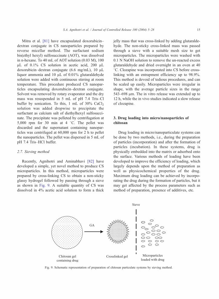
2.7. Sieving method
Recently, Agnihotri and Aminabhavi [82] have
developed a simple, yet novel method to produce CS
microparticles. In this method, microparticles were
prepared by cross-linking CS to obtain a non-sticky
glassy hydrogel followed by passing through a sieve
as shown in Fig. 9. A suitable quantity of CS was
dissolved in 4% acetic acid solution to form a thick
Fig. 9. Schematic representation of preparation of ch
jelly mass that was cross-linked by adding glutaralde-
hyde. The non-sticky cross-linked mass was passed
through a sieve with a suitable mesh size to get
microparticles. The microparticles were washed with
0.1 N NaOH solution to remove the un-reacted excess
glutaraldehyde and dried overnight in an oven at 40
8C. Clozapine was incorporated into CS before cross-
linking with an entrapment efficiency up to 98.9%.
This method is devoid of tedious procedures, and can
be scaled up easily. Microparticles were irregular in
shape, with the average particle sizes in the range
543–698 Am. The in vitro release was extended up to
12 h, while the in vivo studies indicated a slow release
of clozapine.
3. Drug loading into micro/nanoparticles of
chitosan
Drug loading in micro/nanoparticulate systems can
be done by two methods, i.e., during the preparation
of particles (incorporation) and after the formation of
particles (incubation). In these systems, drug is
physically embedded into the matrix or adsorbed onto
the surface. Various methods of loading have been
developed to improve the efficiency of loading, which
largely depends upon the method of preparation as
well as physicochemical properties of the drug.
Maximum drug loading can be achieved by incorpo-
rating the drug during the formation of particles, but it
may get affected by the process parameters such as
method of preparation, presence of additives, etc.
itosan particulate systems by sieving method.
S.A. Agnihotri et al. / Journal of Controlled Release 100 (2004) 5–2816
Both water-soluble and water-insoluble drugs can
be loaded into CS-based particulate systems. Water-
soluble drugs are mixed with CS solution to form a
homogeneous mixture, and then, particles can be
produced by any of the methods discussed before.
For instance, cisplatin was loaded [83] during the
formation of particles with encapsulation efficiency
as high as 99%. The initial concentration of
cisplatin and volume of glutaraldehyde had no
effect on the encapsulation efficiency. Drug encap-
sulation increased as the concentration of CS
increased. Water-insoluble drugs and drugs that
can precipitate in acidic pH solutions can be loaded
after the formation of particles by soaking the
preformed particles with the saturated solution of
drug.
Diclofenac sodium, which precipitates in acidic pH
conditions, has been loaded by the soaking method
[33]. In this method, loading depends upon the
swelling of particles in water. Percentage loading of
drug decreased with increasing cross-linking due to
decreased swelling. Water-insoluble drugs can also be
loaded using the multiple emulsion technique. In this
method, drug is dissolved in a suitable solvent and
then emulsified in CS solution to form an oil-in-water
(o/w) type emulsion. Sometimes, drug can be dis-
persed into CS solution by using a surfactant to get the
suspension. Thus, prepared o/w emulsion or suspen-
sion can be further emulsified into liquid paraffin to
get the oil-water-oil (o/w/o) multiple emulsion. The
resulting droplets can be hardened by using a suitable
cross-linking agent.
In a study by Jameela et al. [84], bovine serum
albumin (BSA) and diphtheria toxoid were loaded
into preformed glutaraldehyde cross-linked CS
microspheres by passive absorption from aqueous
solutions. This method is an alternative to loading
biological macromolecules that are sensitive to
organic solvents, pH, temperature, ultrasound, etc.
In vitro release of BSA showed a high burst effect.
Coating of particles with paraffin or polylactic acid
modulated the drug release. Diphtheria toxoid loaded
CS microspheres showed constant antibody titres for
5 months.
Hejazi and Amiji [85] have prepared CS micro-
spheres by ionic cross-linking and precipitation with
sodium sulfate. Two different methods were used
for drug loading. In method I, tetracycline was
mixed with CS solution before simultaneous cross–
linking and precipitation. In method II, drug was
incubated with the pre-formed microspheres for 48
h. Cumulative amount of tetracycline that was
released from CS microspheres and stability of drug
was examined in different pH media at 37 8C.Microspheres with a spherical shape having an
average diameter of 2– 3 Am were formed. When
drug was added to CS solution before cross-linking
and precipitation, only 8% (w/w) was optimally
incorporated in the final microsphere formulation.
When drug was incubated with the pre-formed
microspheres, a maximum of 69% (w/w) could be
loaded. About 30% of tetracycline either in solution
or when released from the microspheres was found
to degrade at pH 1.2 in 12 h. Preliminary results of
this study suggested that CS microspheres can be
used to incorporate antibiotic drugs, which may be
effective when administered locally in the stomach
against H. pylori.
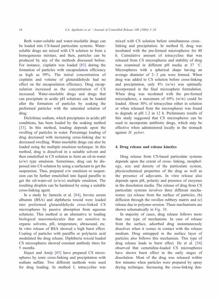
4. Drug release and release kinetics
Drug release from CS-based particulate systems
depends upon the extent of cross– linking, morphol-
ogy, size and density of the particulate system,
physicochemical properties of the drug as well as
the presence of adjuvants. In vitro release also
depends upon pH, polarity and presence of enzymes
in the dissolution media. The release of drug from CS
particulate systems involves three different mecha-
nisms: (a) release from the surface of particles, (b)
diffusion through the swollen rubbery matrix and (c)
release due to polymer erosion. These mechanisms are
shown schematically in Fig. 10.
In majority of cases, drug release follows more
than one type of mechanism. In case of release
from the surface, adsorbed drug instantaneously
dissolves when it comes in contact with the release
medium. Drug entrapped in the surface layer of
particles also follows this mechanism. This type of
drug release leads to burst effect. He et al. [54]
observed that cemetidine-loaded CS microspheres
have shown burst effect in the early stages of
dissolution. Most of the drug was released within
few minutes when particles were prepared by spray
drying technique. Increasing the cross-linking den-
Fig. 10. Mechanism of drug release from particulate systems.
S.A. Agnihotri et al. / Journal of Controlled Release 100 (2004) 5–28 17
sity can prevent the burst release. This effect can
also be avoided by washing microparticles with a
proper solvent, but it may lead to low encapsulation
efficiency.
Drug release by diffusion involves three steps.
First, water penetrates into particulate system, which
causes swelling of the matrix; secondly, the
conversion of glassy polymer into rubbery matrix
takes place, while the third step is the diffusion of
drug from the swollen rubbery matrix. Hence, the
release is slow initially and later, it becomes fast.
This type of release is more prominent in case of
hydrogels. Al-Helw et al. [45] observed a high
initial release of the drug in all the prepared
formulations. Nearly, 20– 30% of the incorporated
drug was released in the first hour. Release was
dependent on the molecular weight of CS and
particle size of the microspheres. The release rate
from microspheres prepared from high molecular
weight CS was slow compared to those prepared
from medium and low molecular weight CS. This
could be attributed to both lower solubility of high
molecular weight CS and higher viscosity of the gel
layer formed around the drug particles upon contact
with the dissolution medium. The release within the
first 3 h was fast (75– 95%) from microspheres
within the size range of 250– 500 Am, but for
particles in the size range of 500– 1,000 Am, drug
release was 56– 90% in 5 h. This is attributed to
large surface area available for dissolution with a
small particle size, thus favoring rapid release of the
drug compared to larger microspheres.
Kweon and Kang [86] prepared the CS-g–
poly(vinyl alcohol) matrix to study the release of
prednisolone under various conditions. Relationship
between the amount of drug release and square root
of time was linear indicating the diffusion-controlled
release. Drug release was controlled by the extent of
PVA grafting, heat treatment or cross-link density,
but it was less affected by the pH when compared to
plain chitosan. Ganza-Gonzalez et al. [56] analyzed
the drug release data using Higuchi equation [87].
Higuchi equation was used to describe the release of
a solute from a flat surface, but not from a sphere
[88], but the good fit obtained suggests that the
release rate depends upon the rate of diffusion
through the cross-linked matrix. Authors have also
fitted the release data to equations developed by Guy
et al. [89] to describe the diffusion from a sphere.
The most commonly used equation for diffusion-
controlled matrix system is an empirical equation used
by Ritger and Peppas [90], in which the early time
S.A. Agnihotri et al. / Journal of Controlled Release 100 (2004) 5–2818
release data can be fitted to obtain the diffusion
parameters,
Mt
Ml¼ ktn ð1Þ
Here, Mt/Ml is the fractional drug release at time t, k
is a constant characteristic of the drug-polymer
interaction and n is an empirical parameter character-
izing the release mechanism. Based on the diffusional
exponent [91], drug transport is classified as Fickian
(n=0.5), Case II transport (n=1), non-Fickian or
anomalous (0.5bnb1) and super Case II (nN1). Drug
release from the CS microspheres cross– linked with
glutaraldehyde, sulfuric acid and heat have shown
[33] different n values varying from 0.47 to 0.61. The
n values increase with increasing loading of diclofe-
nac sodium in different cross-linked formulations.
Recently, Agnihotri and Aminabhavi [82] analyzed
the dynamic swelling data of CS microparticles using
Eq. (1) to predict drug release from the water uptake
data of the microparticles cross-linked with (5.0, 7.5
and 10.0)�10– 4 mL of glutaraldehyde/mg of CS. It
was observed that as the cross-linking increases,
swelling of CS microparticles decreases. Values of n
obtained in the range of 0.160 to 0.249 indicating that
the release mechanism deviates from the Fickian
trend. The values of n are b0.5 due to the irregular
shaped particles and these decrease systematically
with increasing cross-linking.
In the swelling controlled release systems, drug is
dispersed within a glassy polymer. Upon contact with
biological fluid, the polymer swells, but no drug
diffusion occurs through the polymer phase. As the
penetrant enters the glassy polymer, glass transition
temperature of the polymer is lowered due to
relaxation of the polymer chains. Drug could diffuse
out of the swollen rubbery polymer. This type of
system is characterized by two moving boundaries:
the front separating the swollen rubbery portion and
the glassy region, which moves with a front velocity
and the polymer fluid interface. The rate of drug
release is controlled by the velocity and position of the
front dividing the glassy and rubbery portions of the
polymer.
Jameela et al. [48] have obtained a good correlation
fit for the cumulative drug released vs. square root of
time, demonstrating that the release from the micro-
sphere matrix is diffusion-controlled and obeys
Higuchi equation [87]. It was demonstrated that the
rate of release depends upon the size of microspheres.
Release from smaller size microspheres was faster
than those from the large size microspheres due to
smaller diffusional path length for the drug and the
larger surface area of contact of smaller particles with
the dissolution medium. Orienti et al. [92] studied the
correlation between matrix erosion and release
kinetics of indomethacin-loaded CS microspheres.
Release kinetics was correlated with the concentration
of CS in the microsphere and pH of the release
medium. At high concentrations of CS and at pH 7.4,
deviations from Fickian to zero order kinetics have
been observed. Variations induced by these parame-
ters on drug diffusion and solubility in the matrix
undergoing erosion have been analyzed.
5. Pharmaceutical applications of chitosan
particulate systems
Chitosan-based particulate systems are attracting
pharmaceutical and biomedical applications as poten-
tial drug delivery devices. Some important applica-
tions are discussed below.
5.1. Colon targeted drug delivery
Chitosan is a promising polymer for colon drug
delivery since it can be biodegraded by the colonic
bacterial flora [93,94] and it has mucoadhesive
character [1]. The pH-sensitive multicore microparti-
culate system containing CS microcores entrapped
into enteric acrylic microspheres was reported [60].
Sodium diclofenac was efficiently entrapped within
these CS microcores and then microencapsulated into
Eudragit L-100 and Eudragit S-100 to form a multi-
reservoir system. In vitro release study revealed no
release of the drug in gastric pH for 3 h and after the
lag-time, a continuous release for 8– 12 h was
observed in the basic pH.
5.2. Mucosal delivery
Nowadays, mucosal surfaces such as nasal, peroral
and pulmonary are receiving a great deal of attention
as alternative routes of systemic administration.
Chitosan has mucoadhesive properties and therefore,
S.A. Agnihotri et al. / Journal of Controlled Release 100 (2004) 5–28 19
it seems particularly useful to formulate the bioadhe-
sive dosage forms for mucosal administration (ocular,
nasal, buccal, gastro-enteric and vaginal-uterine ther-
apy) [95]. Nasal mucosa has high permeability and
easy access of drug to the absorption site. The
particulate delivery to peroral mucosa is easily taken
up by the Peyer’s patches of the gut associated
lymphoid tissue. Chitosan has been found to enhance
the drug absorption through mucosae without damag-
ing the biological system. Here, the mechanism of
action of CS was suggested to be a combination of
bioadhesion and a transient widening of the tight
junctions between epithelial cells [27].
Genta et al. [95] studied the influence of gluta-
raldehyde on drug release and mucoadhesive proper-
ties of CS microspheres. A new in vitro technique was
developed based on electron microscopy to study the
effect of polymer cross-link density on the mucoad-
hesive properties of CS microspheres modulating the
rate of theophylline release. The ability of insulin-
loaded CS nanoparticles to enhance the nasal absorp-
tion of insulin was investigated in a conscious rabbit
model. Chitosan nanoparticles enhanced the nasal
absorption of insulin to a greater extent than the
aqueous solution of CS [74]. van der Lubben et al.
[96] incorporated the model protein ovalbumin into
CS microparticles and the uptake of ovalbumin
associated with CS microparticles in murine Peyer’s
patches was demonstrated using confocal laser scan-
ning microscopy. In a further study, van der Lubben et
al. [97] investigated the ability of CS microparticles to
enhance both systemic and local immune responses
against diphtheria toxoid (DT) vaccine after the oral
and nasal administration in mice. Systemic and local
IgG and IgA immune responses against DT associated
to CS microparticles were strongly enhanced after the
oral delivery in mice.
Even though oral vaccination has numerous
advantages over the parenteral injection, degradation
of the vaccine in the gut and low uptake in the
lymphoid tissue of the gastrointestinal tract still
complicate the development of oral vaccines. In this
direction, van der Lubben et al. [98] prepared the CS
microparticles and characterized them for size, zeta
potential, morphology- and ovalbumin-loading as
well as release characteristics. The in vivo uptake of
CS microparticles by murine Peyer’s patches was
studied by using confocal laser scanning microscopy
(CLSM). Chitosan microparticles were prepared using
a precipitation/coacervation method. The size of CS
microparticles was 4.3±±0.7 Am and were positively
charged (20±1 mV). Since only microparticles smaller
than 10 Am can be taken up by M-cells of Peyer’s
patches, these microparticles were used as vaccination
systems. The CLSM studies showed that the model
antigen ovalbumin was entrapped within the CS
microparticles. Field emission scanning electron
microscopy demonstrated the porous structure of CS
microparticles, thus facilitating the entrapment of
ovalbumin. Ovalbumin loading in CS microparticles
was about 40%. Release studies have shown the low
release of ovalbumin within 4 h, but most of
ovalbumin (about 90%) remained entrapped in the
microparticles. Since CS microparticles are biode-
gradable, the entrapped ovalbumin was released after
intracellular digestion in Peyer’s patches. Initial in
vivo studies demonstrated that fluorescently labeled
CS microparticles can be taken up by the epithelium
of the murine Peyer’s patches. Since the uptake by
Peyer’s patches is an essential step in oral vaccination,
these results have shown that the porous CS micro-
particles developed are most promising vaccine
delivery systems.
5.3. Cancer therapy
Gadopentetic acid-loaded CS nanoparticles have
been prepared for gadolinium neutron-capture therapy
[62]. Their releasing properties and ability for long-
term retention of gadopentetic acid in the tumor
indicated that these nanoparticles are useful as intra-
tumoral injectable devices for gadolinium neutron-
capture therapy. The accumulation of gadolinium
loaded as gadopentetic acid (Gd-DTPA) in CS nano-
particles designed for gadolinium neutron-capture
therapy (Gd-NCT) for cancer have been evaluated in
vitro in cultured cells [99]. Using L929 fibroblast
cells, Gd accumulation for 12 h at 37 8C was
investigated at Gd concentrations lower than 40
ppm. The accumulation leveled above 20 ppm and
reached 18.0±2.7 (mean±S.D.) Ag Gd/106 cells at 40
ppm. Furthermore, the corresponding accumulations
in B16F10 melanoma cells and SCC-VII squamous
cell carcinoma, which were used in the previous Gd-
NCT trials in vivo were 27.1±2.9 and 59.8±9.8 Ag Gd/106 cells, respectively. This explains the superior
S.A. Agnihotri et al. / Journal of Controlled Release 100 (2004) 5–2820
growth-suppression in the in vivo trials using SCC-
VII cells. The accumulation of nanoparticles in these
cells was 100– 200 times higher in comparison to
dimeglumine gadopentetate aqueous solution (Mag-
nevistw), a magnetic resonance imaging contrast
agent. The endocytic uptake of nanoparticles was
suggested from TEM. These findings indicated that
nanoparticles had a high affinity to cells, thus
contributing to the long retention of Gd in tumor
tissue leading to significant suppression of tumor
growth in in vivo studies.
Tokumitsu et al. [100] demonstrated the potential
usefulness of Gd-NCT using gadolinium-loaded nano-
particles. The potential of gadolinium neutron-capture
therapy (Gd-NCT) for cancer was evaluated using CS
nanoparticles as a novel gadolinium device. The
nanoparticles incorporated with 1200 mg of natural
gadolinium were administered intratumorally twice in
mice-bearing subcutaneous B16F10 melanoma. The
thermal neutron irradiation was performed for the
tumor site, with the fluence of 6.32�1012 neutrons/
cm2, 8 h after the second gadolinium administration.
After irradiation, the tumor growth in the nano-
particle-administered group was significantly sup-
pressed compared to that in the gadopentetate
solution-administered group, despite radioresistance
of melanoma and the smaller Gd dose than that
administered in past Gd-NCT trials.
Jameela et al. [101] have prepared glutaraldehyde
cross-linked CS microspheres containing mitoxan-
trone. The antitumor activity was evaluated against
Ehrlich ascites carcinoma in mice by intraperitoneal
injections. The tumor inhibitory effect was followed
by monitoring the survival time and change in the
body weight of the animal for 60 days. Mean survival
time of animals which received free mitoxantrone was
2.1 days and this was increased to 50 days when
mitoxantrone was given via microspheres. In another
study [102], the in vitro release of mitoxantrone was
controlled for 4 weeks in phosphate buffer at 27 8C.Mitra et al. [81] have encapsulated doxorubicin–
dextran conjugate into long circulating CS nano-
particles. In an attempt to minimize cardiotoxicity of
doxorubicin, a conjugate with dextran was prepared
and encapsulated in CS nanoparticles. Size of the
nanoparticle was 100±10 nm, which favors enhanced
permeability and retention effect. Antitumor effect of
these doxorubicin– dextran-loaded nanoparticles was
evaluated in J774A.1 macrophage tumor cells
implanted in Balb/c mice. The in vivo efficacy of
these nanoparticles was determined by tumor regres-
sion and increased survival time compared to doxor-
ubicin– dextran conjugate and the free drug. These
results suggest that the system not only reduced the
side effects, but also improved its therapeutic efficacy
in the treatment of solid tumors.
Janes et al. [103] evaluated the potential of CS
nanoparticles as carriers for doxorubicin (DOX). The
challenge was to entrap a cationic, hydrophilic
molecule into nanoparticles formed by ionic gelation
of the positively charged CS. To achieve this
objective, the authors have masked the positive charge
of DOX by complexing it with dextran sulfate. This
modification doubled the DOX encapsulation effi-
ciency relative to controls and enabled real loadings
up to 4.0 wt.% of DOX. Authors also investigated the
possibility of forming a complex between CS and
DOX prior to the formation of particles. Despite low
complexation efficiency, no dissociation of the com-
plex was observed upon the formation of nano-
particles. Fluorimetric analysis of the in vitro drug
released showed the initial release phase, the intensity
of which was dependent upon the association mode,
followed by a very slow release. Evaluation of the
activity of DOX-loaded nanoparticles in cell cultures
indicated that those containing dextran sulfate were
able to maintain cytostatic activity relative to free
DOX, while DOX complexed with CS before the
nanoparticle formation showed a slightly decreased
activity. Additionally, confocal studies showed that
DOX was not released in the cell culture medium, but
entered the cells while being associated to nano-
particles. These studies have shown the feasibility of
CS nanoparticles to entrap DOX and to deliver it to
the cells in its active form.
5.4. Gene delivery
Gene therapy is a challenging task in the treatment
of genetic disorders. In case of gene delivery, the
plasmid DNA has to be introduced into the target
cells, which should get transcribed and the genetic
information should ultimately be translated into the
corresponding protein. To achieve this goal, number
of hurdles are to be overcome by the gene delivery
system. Transfection is affected by: (a) targeting the
S.A. Agnihotri et al. / Journal of Controlled Release 100 (2004) 5–28 21
delivery system to target cell, (b) transport through the
cell membrane, (c) uptake and degradation in the
endolysosomes and (d) intracellular trafficking of
plasmid DNA to the nucleus. Chitosan could interact
ionically with the negatively charged DNA and forms
polyelectrolyte complexes. In these complexes, DNA
becomes better protected against nuclease degradation
leading to better transfection efficiency.
DNA–CS nanoparticles have been prepared [53]
to examine the influence of several parameters on
their preparation. The transfection efficiency of CS-
DNA nanoparticles was cell-type dependent. Typi-
cally, it was 3 to 4 orders of magnitude, in relative
light units, higher than the background level in
HEK293 cells, and 2 to 10 times lower than that
achieved by LipofectAMINE–A–DNA complexes.
The presence of 10% fetal bovine serum did not
interfere with their transfection ability. The study
also developed three different schemes to conjugate
transferrin or KNOB protein to the nanoparticle
surface. The transferrin conjugation only yielded a
maximum of 4-fold increase in their transfection
efficiency in HEK293 cells and HeLa cells, whereas
KNOB conjugated nanoparticles could improve the
gene expression level in HeLa cells by 130-fold.
Conjugation of PEG on nanoparticles allowed
lyophilization without aggregation, and without loss
of bioactivity for at least 1 month in storage. The
clearance of PEGylated nanoparticles in mice follow-
ing i.v. administration was slower than the unmodi-
fied nanoparticles at 15 min, and with higher
depositions in kidney and liver. However, no differ-
ence was observed during the first hour.
Self-aggregates were prepared [104] by hydro-
phobic modification of CS with deoxycholic acid in
aqueous media. Self-aggregates have a small size
(mean diameter of 160 nm) with an unimodal size
distribution. Self-aggregates can form charge com-
plexes when mixed with plasmid DNA. The useful-
ness of self-aggregates/DNA complex for transfer of
genes into mammalian cells in vitro has been
suggested. Several transfection studies using chemi-
cally modified CS have been reported. Trimethyl CS
oligomers were examined for their potency as DNA
carriers [105]. Chitosan and lactosylated CS carriers
were investigated for their transfection efficiencies in
vitro [106]. Recently, galactosylated CS-g– dextran–
DNA complexes have been prepared [107]. Galactose
groups were chemically bound to CS for liver
specificity and dextran was grafted to increase the
stability of the complex in water. It was shown that this
system could efficiently transfect liver cells.
Chew et al. [108] studied the i.m. immunization
with full-length Der p 1 cDNA induced significant
humoral response to the left domain (approximately
corresponding to amino acids 1– 116), but not to the
right domain (approximately corresponding to amino
acids 117– 222) of Der p 1 allergen. Authors explored
the use of CS–DNA nanoparticles for oral immuniza-
tion to induce the immune responses specific to both
left and right domains of Der p 1. DNA constructs
pDer p 1 (1– 222) and pDer p 1 (114– 222), which
were complexed with CS and delivered orally
followed by an i.m. injection of pDer p 1 (1– 222)
after 13 weeks. Such an approach has successfully
primed Th1-skewed immune responses against both
domains of Der p 1. It was suggested that such a
strategy could be further optimized for more effica-
cious gene vaccination for full-length Der p 1.
Numerous studies have been reported on prophy-
lactic and therapeutic use of genetic vaccines for
combating a variety of infectious diseases in animal
models. Recent human clinical studies with the gene
gun have validated the concept of direct targeting of
dendritic cells (Langerhan’s cells) in the viable
epidermis of the skin. However, it is unclear whether
the gene gun technology or other needle-free devices
will become commercially viable. Cui and Mumper
[109] investigated the topical application of CS-
based nanoparticles containing plasmid DNA
(pDNA) as a potential approach to genetic immuni-
zation. Two types of nanoparticles were investigated:
(i) pDNA-condensed CS nanoparticles and (ii)
pDNA-coated on pre-formed cationic CS/carboxy-
methylcellulose (CMC) nanoparticles. These studies
have shown that both CS and a CS oligomer can
complex CMC to form stable cationic nanoparticles
for subsequent pDNA coating. Selected pDNA-
coated nanoparticles (with pDNA up to 400 mg/
mL) were stable to challenge with the serum.
Several different CS-based nanoparticles containing
pDNA resulted in both detectable and quantifiable
levels of luciferase expression in mouse skin 24 h
after topical application and significant antigen-
specific IgG titer to expressed h-galactosidase at
28 days.
S.A. Agnihotri et al. / Journal of Controlled Release 100 (2004) 5–2822
Borchard [110] has recently published a review on
the efficient non-viral gene delivery using cationic
polymers as DNA-condensing agents. The gene
delivery is dependent on several factors such as
complex size, complex stability, toxicity, immunoge-
nicity, protection against DNase degradation, intra-
cellular trafficking and processing of the DNA. The
review also examined the advances made in the
application of CS and CS derivatives to non-viral
gene delivery. It gives an overview of the transfection
studies performed by using CS as a transfection agent.
5.5. Topical delivery
Due to good bioadhesive property and ability to
sustain the release of the active constituents, CS has
been used in topical delivery systems. Bioadhesive CS
microspheres for topical sustained release of cetyl
pyridinium chloride have been evaluated [55].
Improved microbiological activity was shown by
these microparticulate systems. Conti et al. [111]
prepared microparticles composed of CS and designed
as powders for topical wound-healing properties.
Blank and ampicillin-loaded microspheres were pre-
pared by spray-drying technique. In vivo evaluation in
albino rats showed that both drug-loaded and blank
microspheres have shown good wound healing
properties.
5.6. Ocular delivery
De Campos et al. [112] investigated the potential of
CS nanoparticles as a new vehicle to improve the
delivery of drugs to ocular mucosa. Cyclosporin A
(CyA) was chosen as a model drug. A modified ionic
gelation technique was used to produce CyA-loaded
CS nanoparticles. These nanoparticles with a mean
size of 293 nm, a zeta potential of +37 mV, high CyA
association efficiency and loading of 73% and 9%,
respectively were obtained. The in vitro release
studies, performed under sink conditions, revealed
the fast release during the first hour followed by a
more gradual drug release during the 24-h period. The
in vivo experiments showed that after topical instilla-
tion of CyA-loaded CS nanoparticles to rabbits,
therapeutic concentrations were achieved in the
external ocular tissues (i.e., cornea and conjunctiva)
within 48 h while maintaining negligible or undetect-
able CyA levels in the inner ocular structures (i.e., iris/
ciliary body and aqueous humour), blood and plasma.
These levels were significantly higher than those
obtained following the instillation of CS solution
containing CyA and an aqueous CyA suspension. The
study indicated that CS nanoparticles could be used as
a vehicle to enhance the therapeutic index of the
clinically challenging drugs with potential application
at the extraocular level.
5.7. Chitosan as a coating material
Chitosan has good film forming properties and
hence, it is used as a coating material in drug delivery
applications. Chitosan-coated microparticles have
many advantages such as improvement of drug
payloads, bioadhesive property and prolonged drug
release properties over the uncoated particles. Chito-
san-coated microspheres composed of poly(lactic
acid)– poly(caprolactone) blends have been prepared
[113]. These microspheres showed good potential for
the targeted delivery of antiproliferative agents to treat
restenosis. Shu and Zhu [73] have prepared the
alginate beads coated with CS by three different
methods. The release of brilliant blue was not only
affected by CS density on the particle surface, but also
on the preparation method and other factors. Chiou et
al. [114] have used different molecular weight
chitosans for coating the microspheres. The initial
burst release was observed in the first hour with 50%
release of lidocaine. But, 19.2% release occurred at
25th hour for the un-coated particles and 14.6% at the
90th hour for the CS-coated microspheres.
6. Chemically modified chitosans
Various chemical modifications of CS have been
studied to alter its properties. N-Trimethyl chitosan
chloride (TMC), a quaternized CS derivative, has
been proven to effectively increase the permeation of
hydrophilic macromolecular drugs across- the
mucosal epithelia by opening the tight junctions
[115]. The study investigated the intestinal absorption
of octreotide when it is co-administered with a
polycationic absorption enhancer, TMC. Chitosan
succinate and CS phthalate were synthesized and
assessed as potential matrices for colon-specific orally
S.A. Agnihotri et al. / Journal of Controlled Release 100 (2004) 5–28 23
administered drug delivery applications. The prepared
matrices resisted the dissolution under acidic con-
ditions. On the other hand, improved drug release
profiles were observed in basic conditions. These
results suggested the suitability of the prepared
matrices in colon specific and orally administered
drug delivery applications [116]. In order to overcome
the low solubility of CS in neutral pH, which is the
major drawback to use this type of polymer as a
transfection agent, N-trimethylated and N-triethylated
oligosaccharides have been synthesized [105].
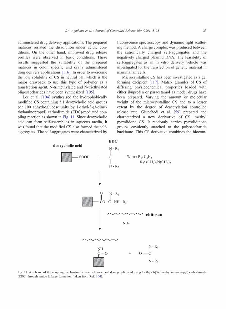
Lee et al. [104] synthesized the hydrophobically
modified CS containing 5.1 deoxycholic acid groups
per 100 anhydroglucose units by 1-ethyl-3-(3-dime-
thylaminopropyl) carbodiimide (EDC)-mediated cou-
pling reaction as shown in Fig. 11. Since deoxycholic
acid can form self-assemblies in aqueous media, it
was found that the modified CS also formed the self-
aggregates. The self-aggregates were characterized by
Fig. 11. A scheme of the coupling mechanism between chitosan and deox
(EDC) through amide linkage formation [taken from Ref. 104].
fluorescence spectroscopy and dynamic light scatter-
ing method. A charge complex was produced between
the cationically charged self-aggregates and the
negatively charged plasmid DNA. The feasibility of
self-aggregates as an in vitro delivery vehicle was
investigated for the transfection of genetic material in
mammalian cells.
Microcrystalline CS has been investigated as a gel
forming excipient [117]. Matrix granules of CS of
differing physicochemical properties loaded with
either ibuprofen or paracetamol as model drugs have
been prepared. Varying the amount or molecular
weight of the microcrystalline CS and to a lesser
extent by the degree of deacetylation controlled
release rate. Giunchedi et al. [59] prepared and
characterized a new derivative of CS: methyl
pyrrolidone CS. It randomly carries pyrrolidinone
groups covalently attached to the polysaccharide
backbone. This CS derivative combines the biocom-
ycholic acid using 1-ethyl-3-(3-dimethylaminopropyl) carbodiimide
S.A. Agnihotri et al. / Journal of Controlled Release 100 (2004) 5–2824
patibility of CS [118] and hydrophilic characteristics
of the pyrrolidinone moiety [119], being particularly
susceptible to the hydrolytic action of lysozyme
[120]. The microparticles were characterized by
S.E.M., particle size analyzer, DSC and in vitro
ampicillin release. Drug release characteristics
depend upon the nature of CS used.
Chen et al. [121] studied the modification of CS by
coupling with linoleic acid (LA) through 1-ethyl-3-(3-
dimethylaminopropyl) carbodiimide-mediated reac-
tion to increase its amphipathicity for improved
emulsification. The micelle formation of linoleic
acid-modified CS in 0.1 M acetic acid solution was
enhanced by O/W emulsification with methylene
chloride, an oil phase. Fluorescence spectra indicated
that without emulsification, the self-aggregation of
LA-CS occurred at the concentration of 1.0 g/L or
above, and with emulsification, self-aggregation was
greatly enhanced followed by a stable micelle
formation at 2.0 g/L. Addition of 1 M NaCl solution
promoted the self-aggregation of LA-CS particles
both with and without emulsification. The nanosize
micelles of LA-CS were formed ranging in size
between 200 and 600 nm. The LA-CS nanoparticles
were used to encapsulate the lipid soluble model
compound, retinal acetate, with 50% efficiency.
Chitosan was chemically modified [122] by graft
copolymerization of poly(ethylene glycol) diacrylate
macromonomer onto CS backbone. Microspheres
based on chitosan and polymer grafted chitosan were
prepared by a polymer dispersion technique. A
comparative study in relation to structural deviation
among CS and modified CS microspheres was
evaluated. These chemically modified CS micropar-
ticles were hydrophilic in nature and formed aggre-
gates. Chitosan derivative with galactose groups was
synthesized by introducing galactose group into the
amine group of CS [123]. The results indicated that
although acyl reaction on the part of amino groups of
CS took place, the degree of galactosylated substitu-
tion was 20%. Crystallinity, solubility, stability and
other physical properties were different from CS.
Microspheres of CS and galactosylated CS were
prepared by the physical precipitation and coacerva-
tion techniques, respectively. Microspheres of CS and
galactosylated CS were spherical in nature with an
average diameter of 0.54 and 1.05 Am and an average
zeta potential of +17 and +15 mV, respectively. It was
suggested that galactosylated CS microspheres could
be used for passive and active hepatic targeting.
7. Conclusions
Chitosan has the desired properties for safe use as a
pharmaceutical excipient. This has prompted accel-
erated research activities worldwide on chitosan micro
and nanoparticles as drug delivery vehicles. These
systems have great utility in controlled release and
targeting studies of almost all class of bioactive
molecules as discussed in this review. Recently,
chitosan is also extensively explored in gene delivery.
However, studies toward optimization of process
parameters and scale up from the laboratory to pilot
plant and then, to production level are yet to be
undertaken. Majority of studies carried out so far are
only in in vitro conditions. More in vivo studies need
to be carried out. Chemical modifications of chitosan
are important to get the desired physicochemical
properties such as solubility, hydrophilicity, etc. The
published literature indicates that in the near future,
chitosan-based particulate systems will have more
commercial status in the market than in the past.
Acknowledgements
Authors thank the University Grants Commission
(UGC), New Delhi, India for a major grant (F1-41/
2001/CPP-II) sanctioned to Karnatak University to
establish Center of Excellence in Polymer Science.
References
[1] P.C. Berscht, B. Nies, A. Liebendorfer, J. Kreuter, Incorpo-
ration of basic fibroblast growth factor into methylpyrrolidi-
none chitosan fleeces and determination of the in vitro release
characteristics, Biomaterials 15 (1994) 593–600.
[2] L. Illum, Chitosan and its use as a pharmaceutical excipient,
Pharm. Res. 15 (1998) 1326–1331.
[3] V. Dodane, V.D. Vilivalam, Pharmaceutical applications of
chitosan, Pharm. Sci. Technol. Today 1 (1998) 246–253.
[4] O. Felt, P. Buri, R. Gurny, Chitosan: a unique polysaccharide
for drug delivery, Drug Dev. Ind. Pharm. 24 (1998) 979–993.
[5] K.D. Yao, T. Peng, Y.J. Yin, M.X. Xu, Microcapsules/
microspheres related to chitosan, J. Macromol. Sci., Rev.
Macromol. Chem. Phys. C35 (1995) 155–180.
S.A. Agnihotri et al. / Journal of Controlled Release 100 (2004) 5–28 25
[6] H.S. Kas, Chitosan: properties, preparations and application
to microparticulate systems, J. Microencapsulation 14 (1997)
689–711.
[7] R.A.A. Muzzarelli, C. Jeuniauk, G.W. Gooday, Chitin in
Nature and Technology, Plenum, New York, 1986.
[8] G. Sjak-Braek, T. Anthonsen, P. Sandford, Chitin and
Chitosan, Elsevier, New York, 1992.
[9] T. Sannan, K. Kurita, Y. Iwakura, Studies on chitin, 2. Effect
of deacetylation on solubility, Makromol. Chem. 177 (1976)
3589–3600.
[10] S. Nicol, Life after death for empty shells, New Sci. 129
(1991) 46–48.
[11] K. Arai, T. Kinumaki, T. Fujita, Toxicity of chitosan, Bull.
Tokai Reg. Fish Lab. 43 (1968) 89–94.
[12] T. Chandy, C.P. Sharma, Chitosan as a biomaterial, Biomater.
Artif. Org. 18 (1990) 1–24.
[13] W.M. Hou, S. Miyazaki, M. Takada, T. Komai, Pharmaceut-
ical application of biomedical polymers. Part XVI. Sustained
release of indomethacin from chitosan, Chem. Pharm. Bull.
33 (1985) 3986–3992.
[14] S. Miyazaki, K. Ishii, T. Nadai, Pharmaceutical application of
biomedical polymers. Part IV. The use of chitin and chitosan
as drug carriers, Chem. Pharm. Bull. 29 (1981) 3067–3069.
[15] T. Handa, A. Kasai, H. Takenaka, S.Y. Lin, Y. Ando, Novel
method for the preparation of controlled-release theophylline
granules coated with a polyelectrolyte complex of sodium
polyphosphate-chitosan, J. Pharm. Sci. 74 (1985) 264–268.
[16] S. Miyazaki, H. Yamaguchi, C. Yokouchi, M. Takada, W.-M.
Hou, Sustained-release and intragastric-floating granules of
indomethacin using chitosan in rabbits, Chem. Pharm. Bull.
36 (1988) 4033–4038.
[17] Y. Sawayanagi, N. Nambu, T. Nagai, Use of chitosan for
sustained-release preparations of water-soluble drugs, Chem.
Pharm. Bull. 30 (1982) 4213–4215.
[18] S. Shiraishi, T. Imai, M. Otagiri, Controlled release of
indomethacin by chitosan– polyelectrolyte complex: optimi-
zation and in vivo/in vitro evaluation, J. Control. Release 25
(1993) 217–225.
[19] C.M. Lehr, J.A. Bouwstra, E.H. Schacht, H.E. Junginger, In
vitro evaluation of mucoadhesive properties of chitosan and
some other natural polymers, Int. J. Pharm. 78 (1992) 43–48.
[20] H.L. Luehen, C.M. Lehr, C.O. Rentel, A.B.J. Noach, A.G.
Boer, J.C. Verhoef, H.E. Junginger, Bioadhesive polymers for
the peroral delivery of peptide drugs, J. Control. Release 29
(1994) 329–338.
[21] L. Illum, N.F. Farraj, S.S. Davis, Chitosan as a novel nasal
delivery system for peptide drugs, Pharm. Res. 11 (1994)
1186–1189.
[22] T. Imai, S. Shiraishi, H. Saito, M. Otagiri, Interaction of
indomethacin with low molecular weight chitosan, and
improvements of some pharmaceutical properties of indome-
thacin by low molecular weight chitosans, Int. J. Pharm. 67
(1991) 11–20.
[23] I. Genta, F. Pavanetto, B. Conti, P. Giunchedi, U. Conte,
Spray-drying for the preparation of chitosan microspheres,
Proc. Int. Symp. Control. Release Bioact. Mater. 21 (1994)
616–617.
[24] Y. Sawayanagi, N. Nambu, T. Nagai, Enhancement of
dissolution properties of prednisolone from ground mix-
tures with chitin or chitosan, Chem. Pharm. Bull. 31
(1983) 2507–2509.
[25] J.M. Gallo, E.E. Hassan, Receptor-mediated magnetic
carriers: basis for targeting, Pharm. Res. 5 (1988) 300–304.
[26] E.E. Hassan, R.C. Parish, J.M. Gallo, Optimized formulation
of magnetic chitosan microspheres containing the anticancer
agent, oxantrazole, Pharm. Res. 9 (1992) 390–397.
[27] P. Artursson, T. Lindmark, S.S. Davis, L. lllum, Effect
of chitosan on the permeability of monolayers of
intestinal epithelial cells (CACO-2), Pharm. Res. 11
(1994) 1358–1361.
[28] J. Akbuga, G. Durmaz, Preparation and evaluation of cross-
linked chitosan microspheres containing furosemide, Int. J.
Pharm. 111 (1994) 217–222.
[29] B.C. Thanoo, M.C. Sunny, A. Jayakrishnan, Cross-linked
chitosan microspheres: preparation and evaluation as a matrix
for the controlled release of pharmaceuticals, J. Pharm.
Pharmacol. 44 (1992) 283–286.
[30] K.S. Soppimath, A.R. Kulkarni, T.M. Aminabhavi, Con-
trolled release of antihypertensive drug from the interpene-
trating network poly(vinyl alcohol) guar gum hydrogel
microspheres, J. Biomater. Sci., Polym. Ed. 11 (2000) 27–43.
[31] K.S. Soppimath, A.R. Kulkarni, T.M. Aminabhavi, Water
transport and drug release study of cross-linked guar gum
grafted polyacrylamide hydrogel microspheres for the con-
trolled release application, Eur. J. Pharm. Biopharm. 53
(2002) 87–98.
[32] A.R. Kulkarni, K.S. Soppimath, T.M. Aminabhavi, Con-
trolled release of cefadroxil using sodium alginate inter-
penetrating network with gelatin/egg albumin, Eur. J. Pharm.
Biopharm. 51 (2001) 127–133.
[33] S.G. Kumbar, A.R. Kulkarni, T.M. Aminabhavi, Cross-
linked chitosan microspheres for encapsulation of diclofenac
sodium: effect of cross-linking agent, J. Microencapsulation
19 (2002) 173–180.
[34] S.G. Kumbar, T.M. Aminabhavi, Synthesis and character-
ization of modified chitosan microspheres: effect of the
grafting ratio on the controlled release of nifedipine through
microspheres, J. Appl. Polym. Sci. 89 (2003) 2940–2949.
[35] K.S. Soppimath, T.M. Aminabhavi, A.R. Kulkarni, W.E.
Rudzinski, Biodegradable polymeric nanoparticles as drug
delivery devices, J. Control. Release 70 (2001) 1–20.
[36] D.K. Singh, A.R. Ray, Biomedical applications of chitin,
chitosan and their derivatives, J. Macromol. Sci., Rev.
Macromol. Chem. Phys. C40 (2000) 69–83.
[37] A.K. Singla, M. Chawla, Chitosan: some pharmaceutical and
biological aspects an update, J. Pharm. Pharmacol. 53 (2001)
1047–1067.
[38] W.G. Liu, K.D. Yao, Chitosan and its derivatives– a promis-
ing non-viral vector for gene transfection, J. Control. Release
83 (2002) 1–11.
[39] R. Hejazi, M. Amiji, Chitosan-based gastrointestinal delivery
systems, J. Control. Release 89 (2003) 151–165.
[40] V.R. Sinha, A.K. Singla, S. Wadhawan, R. Kaushik, R.
Kumria, K. Bansal, S. Dhawan, Chitosan microspheres as
S.A. Agnihotri et al. / Journal of Controlled Release 100 (2004) 5–2826
a potential carrier for drugs, Int. J. Pharm. 274 (2004)
1–33.
[41] J. Kreuter, Nanoparticles, in: J. Kreuter (Ed.), Colloidal Drug
Delivery Systems, Marcel Dekker, New York, 1994, pp.
219–342.
[42] L. Brannon Peppas, Recent advances on the use of
biodegradable microparticles and nanoparticles in the con-
trolled drug delivery, Int. J. Pharm. 116 (1995) 1–9.
[43] P. Couvreur, L. Grislain, V. Lenaerts, F. Brasseur, P. Guiot,
in: P. Guiot, P. Couvreur (Eds.), Polymeric Nanoparticles and
Microspheres, CRC Press, Boca Raton, FL, 1986.
[44] J. Akbuga, G. Durmaz, Preparation and evaluation of cross-
linked chitosan microspheres containing furosemide, Int. J.
Pharm. 11 (1994) 217–222.
[45] A.A. Al-Helw, A.A. Al-Angary, G.M. Mahrous, M.M. Al-
Dardari, Preparation and evaluation of sustained release
cross-linked chitosan microspheres containing phenobarbi-
tone, J. Microencapsulation 15 (1998) 373–382.
[46] E.B. Denkbas, M. Seyyal, E. Piskin, 5-Fluorouracil loaded
chitosan microspheres for chemoembolization, J. Micro-
encapsulation 16 (1998) 741–749.
[47] C. Sankar, M. Rani, A.K. Srivastava, B. Mishra, Chitosan
based pentazocine microspheres for intranasal systemic
delivery: development and biopharmaceutical evaluation,
Pharmazie 56 (2001) 223–226.
[48] S.R. Jameela, T.V. Kumary, A.V. Lal, A. Jayakrishnan,
Progesterone-loaded chitosan microspheres: a long acting
biodegradable controlled delivery system, J. Control. Release
52 (1998) 17–24.
[49] F. Bugamelli, M.A. Raggi, I. Orienti, V. Zecchi, Controlled
insulin release from chitosan microparticles, Arch. Pharm.
331 (1998) 133–138.
[50] K. Nishimura, S. Nishimura, H. Seo, N. Nishi, S. Tokura, I.
Azuma, Macrophage activation with multiporous beads
prepared from partially deacetylated chitin, J. Biomed. Mater.
Res. 20 (1986) 1359–1372.
[51] A. Berthod, J. Kreuter, Chitosan microspheres– improved
acid stability and change in physicochemical properties by
cross-linking, Proc. Int. Symp. Control. Release Bioact.
Mater. 23 (1996) 369–370.
[52] S. Ozbas-Turan, J. Akbuga, C. Aral, Controlled release of
interleukin-2 from chitosan microspheres, J. Pharm. Sci. 91
(2002) 124–125.
[53] H.Q. Mao, K. Roy, V.L. Troung-Le, K.A. Janes, K.Y. Lim, Y.
Wang, J.T. August, K.W. Leong, Chitosan DNA nano-
particles as gene delivery carriers: synthesis, characterization
and transfection efficiency, J. Control. Release 70 (2001)
399–421.
[54] P. He, S.S. Davis, L. Illum, Chitosan microspheres prepared
by spray drying, Int. J. Pharm. 187 (1999) 53–65.
[55] B. Conti, T. Modena, I. Genta, P. Perugini, C. Decarro, F.
Pavanetto, Microencapsulation of cetylpyridinium chloride
with a bioadhesive polymer, Proc. Int. Symp. Control.
Release Bioact. Mater. 25 (1998) 822–823.
[56] A. Ganza-Gonzalez, S. Anguiano-Igea, F.J. Otero-Espinar,
J.B. Mendez, Chitosan and chondroitin microspheres for
oral-administration controlled release of metoclopramide,
Eur. J. Pharm. Biopharm. 48 (1999) 149–155.
[57] X.Y. Shi, T.W. Tan, Preparation of chitosan/ethylcellulose
complex microcapsule and its application in controlled
release of vitamin D-2, Biomaterials 23 (2002) 4469–4473.
[58] C.W. Bishop, J.C. Knutson, C.R. Valliere, Method of treating
prostatic disease using delayed and/or sustained release
vitamin D formulation, US Pat. 5 795 882, 1998.
[59] P. Giunchedi, I. Genta, B. Conti, R.A.A. Muzzarelli, U.
Conte, Preparation and characterization of ampicillin loaded
methylpyrrolidinone chitosan and chitosan microspheres,
Biomaterials 19 (1998) 157–161.
[60] M.L. Lorenzo-Lamosa, C. Remunan-Lopez, J.L. Vila-Jato,
M.J. Alonso, Design of microencapsulated chitosan micro-
spheres for colonic drug delivery, J. Control. Release 52
(1998) 109–118.
[61] Y.C. Huang, M.K. Yeh, C.H. Chiang, Formulation factors in
preparing BTM-chitosan microspheres by spray drying
method, Int. J. Pharm. 242 (2002) 239–242.
[62] H. Tokumitsu, H. Ichikawa, Y. Fukumori, Chitosan– gado-
pentetic acid complex nanoparticles for gadolinium neutron
capture therapy of cancer: preparation by novel emulsion-
droplet coalescence technique and characterization, Pharm.
Res. 16 (1999) 1830–1835.
[63] A. Polk, B. Amsden, K.D. Yao, T. Peng, M.F.A. Goosen,
Controlled release of albumin from chitosan-alginate micro-
capsules, J. Pharm. Sci. 83 (1994) 178–185.
[64] L.S. Liu, S.Q. Liu, S.Y. Ng, M. Froix, T. Ohno, J. Heller,
Controlled release of interleukin-2 for tumor immunotherapy
using alginate:chitosan porous microspheres, J. Control.
Release 43 (1997) 65–74.
[65] Y. Kawashima, T. Handa, H. Takenaka, S.Y. Lin, Y. Ando,
Novel method for the preparation of controlled-release
theophylline granules coated with a polyelectrolyte complex
of sodium polyphosphate-chitosan, J. Pharm. Sci. 74 (1985)
264–268.
[66] Y. Kawashima, T. Handa, A. Kasai, H. Takenaka, S.Y. Lin,
The effect of thickness and hardness of the coating film on
the drug release of theophylline granules, Chem. Pharm.
Bull. 33 (1985) 2469–2474.
[67] R. Bodmeier, K.H. Oh, Y. Pramar, Preparation and evaluation
of drug-containing chitosan beads, Drug Dev. Ind. Pharm. 15
(1989) 1475–1494.
[68] S. Shirashi, T. Imai, M. Otagiri, Controlled release of
indomethacin by chitosan– polyelectrolyte complex: optimi-
zation and in vivo: in vitro evaluation, J. Control. Release 25
(1993) 217–225.
[69] A.D. Sezer, J. Akbuga, Controlled release of piroxicam from
chitosan beads, Int. J. Pharm. 121 (1995) 113–116.
[70] Z. Aydin, J. Akbuga, Chitosan beads for the delivery of
salmon calcitonin: preparation and characteristics, Int. J.
Pharm. 131 (1996) 101–103.
[71] P. Calvo, C. Remunan-Lopez, J.L. Vila-Jata, M.J. Alonso,
Chitosan and chitosan: ethylene oxide-propylene oxide block
copolymer nanoparticles as novel carriers for protein and
vaccines, Pharm. Res. 14 (1997) 1431–1436.
S.A. Agnihotri et al. / Journal of Controlled Release 100 (2004) 5–28 27
[72] P. Calvo, C. Remunan-Lopez, J.L. Vila-Jata, M.J. Alonso,
Novel hydrophilic chitosan-polyethylene oxide nanopar-
ticles as protein carriers, J. Appl. Polym. Sci. 63 (1997)
125–132.
[73] X.Z. Shu, K.J. Zhu, A novel approach to prepare tripoly-
phosphate/chitosan complex beads for controlled release drug
delivery, Int. J. Pharm. 201 (2000) 51–58.
[74] R. Fernandez-Urrusuno, P. Cavlo, C. Remunan-Lopez, J.L.
Vila-Jato, M.J. Alonso, Enhancement of nasal absorption of
insulin using chitosan nanoparticles, Pharm. Res. 16 (1999)
1576–1581.
[75] Y. Pan, Y. Li, H. Zhao, J. Zheng, H. Xu, G. Wei, J. Hao, F.
Cui, Chitosan nanoparticles improve the intestinal absorption
of insulin in vivo, Int. J. Pharm. 249 (2002) 139–147.
[76] Y. Xu, Y. Du, Effect of molecular structure of chitosan on
protein delivery properties of chitosan nanoparticles, Int. J.
Pharm. 250 (2003) 215–226.
[77] J.A. Ko, H.J. Park, S.J. Hwang, J.B. Park, J.S. Lee,
Preparation and characterization of chitosan microparticles
intended for controlled drug delivery, Int. J. Pharm. 249
(2002) 165–174.
[78] Y.S. Leong, F. Candau, Inverse microemulsion polymer-
ization, J. Phys. Chem. 86 (1982) 2269–2271.
[79] A. Maitra, Determination of size parameters of water–
Aerosol OT– oil reverse micelles from their nuclear magnetic
resonance data, J. Phys. Chem. 88 (1984) 5122–5125.
[80] P.L. Luisi, M. Giomini, M.P. Pileni, B.H. Robinson, Reverse
micelles as hosts for proteins and small molecules, Biochim.
Biophys. Acta 947 (1988) 209–246.
[81] S. Mitra, U. Gaur, P.C. Ghosh, A.N. Maitra, Tumor targeted
delivery of encapsulated dextran– doxorubicin conjugate
using chitosan nanoparticles as carrier, J. Control. Release
74 (2001) 317–323.
[82] S.A. Agnihotri, T.M. Aminabhavi, Controlled release of
clozapine through chitosan microparticles prepared by a
novel method, J. Control. Release 96 (2004) 245–259.
[83] J. Akbuga, N. Bergisadi, Effect of formulation variables on
cis-platin loaded chitosan microsphere properties, J. Micro-
encapsulation 16 (1999) 697–703.
[84] S.R. Jameela, A. Misra, A. Jayakrishnan, Cross-linked
chitosan microspheres as carriers for prolonged delivery of
macromolecular drugs, J. Biomater. Sci., Polym. Ed. 6 (1994)
621–632.
[85] R. Hejazi, M. Amiji, Stomach-specific anti-H. pylori therapy.
I: preparation and characterization of tetracyline-loaded
chitosan microspheres, Int. J. Pharm. 235 (2002) 87–94.
[86] D.K. Kweon, D.W. Kang, Drug-release behavior of chitosan-
g-poly(vinyl alcohol) copolymer matrix, J. Appl. Polym. Sci.
74 (1999) 458–464.
[87] T. Higuchi, Mechanism of sustained action medication, J.
Pharm. Sci. 52 (1963) 1145–1149.
[88] C. Washington, Drug release from microdisperse systems: a
critical review, Int. J. Pharm. 58 (1990) 1–12.
[89] R.H. Guy, J. Hadgraft, I.W. Kellaway, M.J. Taylor, Calcu-
lations of drug release rates from spherical particles, Int. J.
Pharm. 11 (1982) 199–207.
[90] P.L. Ritger, N.A. Peppas, A simple equation for description
of solute release. II. Fickian and anomalous release from
swellable devices, J. Control. Release 5 (1987) 37–42.
[91] N.A. Peppas, R.W. Korsmeyer, in: N.A. Peppas (Ed.),
Hydrogels in Medicine and Pharmacy, vol. 3, CRC Press,
Boca Raton, FL, 1987, p. 103.
[92] I. Orienti, K. Aiedeh, E. Gianasi, V. Bertasi, V. Zecchi,
Indomethacin loaded chitosan microspheres. Correlation
between the erosion process and release kinetics, J. Micro-
encapsulation 13 (1996) 463–472.
[93] D. Pantaleone, M. Yalpani, M. Scollar, Advances in chitin
and chitosan, in: C.J. Brine, P.A. Sandford, J.P. Zizakis,
Proceed. 5th Int. Conf. Chitin and Chitosan, Princeton, New
York, 1991.
[94] H. Zhang, I.A. Alsarra, S.H. Neau, An in vitro evaluation of a
chitosan-containing multiparticulate system for macromole-
cule delivery to the colon, Int. J. Pharm. 239 (2002) 197–205.
[95] I. Genta, M. Costantini, A. Asti, B. Conti, L. Montanari,
Influence of glutaraldehyde on drug release and mucoadhe-
sive properties of chitosan microspheres, Carbohydr. Polym.
36 (1998) 81–88.
[96] I.M. van der Lubben, F.A.J. Konings, G. Borchard, J.C.
Verhoef, H.E. Junginger, In vivo uptake of chitosan micro-
particles by murine Peyer’s patches: visualization studies
using confocal laser scanning microscopy and immuno-
histochemistry, J. Drug Target. 9 (2001) 39–47.
[97] I.M. van der Lubben, G. Kersten, M.M. Fretz, C. Beuvery,
J.C. Verhoef, H.E. Junginger, Chitosan microparticles for
mucosal vaccination against diphtheria: oral and nasal
efficacy studies in mice, Vaccine 21 (2003) 1400–1408.
[98] I.M. van der Lubben, J.C. Verhoef, A.C. van Aelst, G.
Borchard, H.E. Junginger, Chitosan microparticles for oral
vaccination: preparation, characterization and preliminary in
vivo uptake studies in murine Peyer’s patches, Biomaterials
22 (2001) 687–694.
[99] F. Shikata, H. Tokumitsu, H. Ichikawa, Y. Fukumori, In vitro
cellular accumulation of gadolinium incorporated into chito-
san nanoparticles designed for neutron-capture therapy of
cancer, Eur. J. Pharm. Biopharm. 53 (2002) 57–63.
[100] H. Tokumitsu, J. Hiratsuka, Y. Sakurai, T. Kobayashi, H.
Ichikawa, Y. Fukumori, Gadolinium neutron-capture therapy
using novel gadopentetic acid-chitosan complex nanopar-
ticles: in vivo growth suppression of experimental melanoma
solid tumor, Cancer Lett. 150 (2000) 177–182.
[101] S.R. Jameela, P.G. Latha, A. Subramoniam, A. Jayakrishnan,
Antitumor activity of mitoxantrone-loaded chitosan micro-
spheres against Ehrlich ascites carcinoma, J. Pharm. Phar-
macol. 48 (1996) 685–688.
[102] S.R. Jameela, A. Jayakrishnan, Glutaraldehyde cross-linked
chitosan microspheres as a long-acting biodegradable drug-
delivery vehicle-studies on the in-vitro release of mitoxan-
trone and in-vivo degradation of microspheres in rat muscle,
Biomaterials 16 (1995) 769–775.
[103] K.A. Janes, M.P. Fresneau, A. Marazuela, A. Fabra, M.J.
Alonso, Chitosan nanoparticles as delivery systems for
doxorubicin, J. Control. Release 73 (2001) 255–267.

S.A. Agnihotri et al. / Journal of Controlled Release 100 (2004) 5–2828
[104] K.Y. Lee, I.C. Kwon, Y.H. Kim, W.H. Jo, S.Y. Jeong,
Preparation of chitosan self-aggregates as a gene delivery
system, J. Control. Release 51 (1998) 213–220.
[105] B.I. Florea, M. Thanou, M. Geldof, C. Meaney, H.E.
Junginger, G. Borchard, Modified chitosan oligosaccharides
as transfection agents for gene therapy of cystic fibrosis,
Proc. Int. Symp. Control. Release Bioact. Mater. 27 (2000)
846–847.
[106] P. Erbacher, S. Zou, T. Beuinger, A.M. Steffan, J.S. Remy,
Chitosan-based vector/DNA complexes for gene delivery:
biophysical characteristics and transfection ability, Pharm.
Res. 15 (1998) 1332–1339.
[107] Y.K. Park, Y.H. Park, B.A. Shin, E.S. Choi, Y.R. Park, T.
Akaike, C.S. Cho, Galactosylated chitosan graft-dextran as
hepatocyte-targeting DNA carrier, J. Control. Release 69
(2000) 97–108.
[108] J.L. Chew, C.B. Wolfowicz, H.Q. Mao, K.W. Leong, K.Y.
Chua, Chitosan nanoparticles containing plasmid DNA
encoding house dust mite allergen, Der p 1 for oral
vaccination in mice, Vaccine 21 (2003) 2720–2729.
[109] Z. Cui, R.J. Mumper, Chitosan-based nanoparticles for
topical genetic immunization, J. Control. Release 75 (2001)
409–419.
[110] G. Borchard, Chitosans for gene delivery, Adv. Drug Deliv.
Rev. 52 (2001) 145–150.
[111] B. Conti, P. Giunchedi, I. Genta, U. Conte, The preparation
and in vivo evaluation of the wound-healing properties of
chitosan microspheres, STP Pharma. Sci. 10 (2000) 101–104.
[112] A.M. De Campos, A. Sanchez, M.J. Alonso, Chitosan
nanoparticles: a new vehicle for the improvement of the
delivery of drugs to the ocular surface. Application to
cyclosporin A, Int. J. Pharm. 224 (2001) 159–168.
[113] T. Chandy, R.F. Wilson, G.H.R. Rao, G.S. Das, Changes in
cisplatin delivery due to surface-coated poly(lactic acid)-
poly(q-caprolactone) microspheres, J. Biomater. Appl. 16
(2002) 275–291.
[114] S.H. Chiou, W.T. Wu, Y.Y. Huang, T.W. Chung, Effects of
the characteristics of chitosan on controlling drug release of
chitosan coated PLLA microspheres, J. Microencapsulation
18 (2001) 613–625.
[115] M. Thanou, J.C. Verhoef, P. Marbach, H.E. Junginger,
Intestinal absorption of octreotide: N-trimethyl chitosan
chloride (TMC) ameliorates the permeability and absorption
properties of the somatostatin analogue in vitro and in vivo, J.
Pharm. Sci. 89 (2000) 951–957.
[116] K. Aiedeh, M.O. Taha, Synthesis of chitosan succinate and
chitosan phthalate and their evaluation as suggested matrices
in orally administered, colon-specific drug delivery systems,
Archiv.der Pharmazie 332 (1999) 103–107.
[117] M. Sakkinen, U. Seppala, P. Heinanen, M. Marvola, In vitro
evaluation of microcrystalline chitosan (MCCh) as gel-
forming excipient in matrix granules, Eur. J. Pharm.
Biopharm. 54 (2002) 33–40.
[118] R.A.A. Muzzarelli, V. Baldassarre, F. Conti, G. Gazzanelli, V.
Vasi, P. Ferrara, G. Biagini, The biological activity of chitosan:
ultrastructural study, Biomaterials 8 (1988) 247–252.
[119] R.A.A. Muzzarelli, Depolymerization of methylpyrrolidinone
chitosan by lysozyme, Carbohydr. Polym. 19 (1992) 29–34.
[120] R.A.A. Muzzarelli, In vivo biochemical significance of
chitin-based medical items, in: S. Dumitriu, M. Szycher
(Eds.), Polymeric Materials for Biomedical Applications,
Marcel Dekker, New York, 1992, pp. 179–198.
[121] X.G. Chen, C.M. Lee, H.J. Park, O/W emulsification for the
self-aggregation and nanoparticle formation of linoleic acids
modified chitosan in the aqueous system, J. Agric. Food
Chem. 51 (2003) 3135–3139.
[122] K.L. Shanta, D.R.K. Harding, Synthesis and characterization
of chemically modified chitosan microspheres, Carbohydr.
Polym. 48 (2002) 247–253.
[123] C. Zhang, Q. Ping, Y. Ding, Y. Cheng, J. Shen, Synthesis,
characterization and microsphere formation of galactosylated
chitosan, J. Appl. Polym. Sci. 91 (2004) 659–665.


