Embed Size (px)
Citation preview
DOI: 10.1007/s00340-005-2056-2
Appl. Phys. B 82, 243–246 (2006)
Lasers and OpticsApplied Physics B
t. gaebel1,�
m. domhan1
c. wittmann1
i. popa1
f. jelezko1
j. rabeau2
a. greentree2
s. prawer2
e. trajkov3
p.r. hemmer3
j. wrachtrup1
Photochromism in single nitrogen-vacancydefect in diamond1 3rd Physical Institute, University of Stuttgart, Stuttgart, Germany2 School of Physics, University of Melbourne, Victoria, Australia3 Department of Electrical Engineering, Texas A&M University, Texas, USA
Received: 12 August 2005Published online: 30 November 2005 • © Springer-Verlag 2005
ABSTRACT Photochromism in single nitrogen-vacancy opti-cal centers in diamond is demonstrated. Time-resolved opticalspectroscopy shows that intense irradiation at 514 nm switchesthe nitrogen-vacancy defects to the negative form. This defectstate relaxes back to the neutral form under dark conditions.Temporal anticorrelation of photons emitted by the differentcharge states of the optical center unambiguously indicatesthat the nitrogen-vacancy defect accounts for both 575 nm and638 nm emission bands. Possible mechanism of photochromisminvolving nitrogen donors is discussed.
PACS 61.72.-y;61.72.Ji;03.67.-a
1 Introduction
The photophysics of color centers in diamond haveattracted interest during the last decade because of their pos-sible application in quantum information processing. Electronand nuclear spins associated with single nitrogen vacancy(N–V) defects in diamond are very promising qubit candi-dates because of the long coherence time and availability ofreliable control via well established ESR-technique [1]. Re-cently, optical readout of single spins associated with N–V de-fects in diamond was demonstrated [2]. The coherent controlof the electron spin of this defect and realization of a two qubitconditional quantum gate was reported [3, 4]. Additionally,the coupling to internal degrees of freedom, e.g., the hyper-fine coupling to nuclei, like 13C and 14N, was investigated [5].Although the N–V defect is one of the most studied color cen-ters, it is remarkable that its basic photophysical properties(e.g., the charge state) are not well established yet. Such un-certainty is related to the fact that the charge state of defectsin wide band gap materials is often affected by the proximityof donors (or acceptors). This explains why the concentrationof isolated substitutional nitrogen (which is a major electrondonor) defines the charge state of many optical centers in di-amond. The best-known example is the vacancy, which existsin two stable charge states [6]. Photoinduced electron transfercan often change the charge state of a defect.
� Fax: +49-711-685-5281, E-mail: [email protected]
The N–V defect is a naturally occurring feature in dia-mond containing nitrogen. It consists of a substitutional nitro-gen atom next to an adjacent vacancy in the diamond lattice.The defect can be produced by irradiation and subsequentannealing at temperatures above 550 ◦C of nitrogen rich dia-mond. Due to the radiation damage, vacancies are created inthe diamond lattice and the subsequent annealing treatmentleads to migration of the vacancies towards nitrogen atomscreating N–V defects. Alternatively, the center can be pro-duced in very pure diamond (with low nitrogen concentration)by N+ ion implantation [7, 8]. Depending on other impuritiesin the close surrounding acting as electron acceptors or donors(e.g. nitrogen), the defect can occur in two charge states, neu-tral (N–V)0 and negatively charged (N–V)−.
The (N–V)− shows a zero phonon line (ZPL) at 638 nm(1.945 eV) [9]. The properties of (N–V)− defects are wellstudied using hole burning, four-wave mixing and opti-cally detected magnetic resonance techniques [10]. Based onthese studies, a generally accepted six-electron model of the(N–V)− defect has been proposed. The energy level schemeconsists of a ground state, which is a spin triplet of 3A sym-metry, an excited spin triplet state of 3E symmetry, and anadditional metastable singlet state ( 1A). The ZPL absorptioncorresponds to 3A → 3E transition. The main photophysicalparameters, e.g., a large absorption cross section, a quan-tum yield close to unity (Φ = 0.99), and a short excited-statelifetime (τ = 11.6 ns) are suitable for single center detectionusing optical methods [1].
If electron donors are not available in the diamond lat-tice, the N–V center can be observed in its neutral chargestate (N–V)0. This charge state shows a ZPL lying at 575 nm(2.156 eV) and the effective electron spin of the neutral formof the defect is S = 1/2. The best indication, that both 575 nmand 638 nm optical bands belong to the different charge statesof the N–V center was given by heavy neutron irradiationof nitrogen-rich diamond [11]. A drastic change in the ab-sorption spectra after heavy neutron irradiation and annealingwas observed. The (N–V)− spectral signatures decrease andsimultaneously the sudden appearance of the (N–V)0 spec-tral signatures were observed. This effect is explained bya lowering of the Fermi level due to the irradiation and asa consequence the change of the charge state of the defect. Inaddition, the photochromic behavior of 575 nm and 638 nmemission bands was observed [12–14] and the relation be-
244 Applied Physics B – Lasers and Optics
tween them was studied [15, 16]. In this paper we extend thestudies of photochromism to a single N–V defect. Observa-tion of the photochromic effect in a single defect providesunambiguous proof that both spectral lines belong to differentcharge states, of a single N–V defect.
2 Experiment
The sample used is a natural type IIa diamond(Element Six) implanted with 14 keV N+
2 ions and subse-quently annealed for 3 hours at 900 ◦C. The ion dose usedwas approximately 2×10⁹ N+
2 ions cm−2, which creates an Ndoped layer about 20 nm thick in which the N concentrationis approximately 6 ×10¹⁴ N/cm3. Type IIa diamond is veryclean in respect to nitrogen impurities and most of the (N–V)defects created in this diamond originate from the implantednitrogen. Experiments were performed using a home-builtconfocal microscope. The fluorescence light from the opti-cal center was collected and directed to the detection chan-nel, which includes a spectrometer and a Hanbury-Brown andTwiss interferometer. The excitation wavelength of 514 nmwas blocked by a Notch 514 filter and a long-pass filter withcut-off at 570 nm. The interferometer was either used to meas-ure the autocorrelation of the photons arriving at the twodetectors, or, with additional filters (one detection arm a long-pass 667 nm for (N–V)− detection and the other detectionarm a band-pass 585 /80 nm for (N–V)0 detection) to meas-ure the cross-correlation between two charge states of defect.An acousto-optical modulator (AOM) was used for laser pulseshaping in time resolved experiments.
3 Results and Discussion
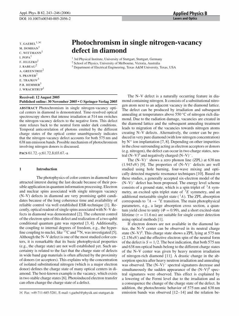
Single nitrogen vacancy defects have been detectedvia fluorescence using a confocal optical microscope operat-ing at room temperature. By choosing an appropriate implan-tation dose the spacing between defects in the diamond wasensured to be larger than the resolution of the optical micro-scope (0.3 µm). In this case the confocal image of the sampleshows distinct fluorescence spots corresponding to fluores-cence emission of the single N–V color centers. To check thatN–V centers were observed, a fluorescence spectrum of a spotwas taken (see Fig. 1). The spectrum shows two different ZPL,
FIGURE 1 Fluorescence spectra of a single fluorescence spot from the con-focal image. It shows two ZPLs, one at 575 nm indicating the (N–V)0 defectand one at 638 nm for the (N–V)− defect in diamond. The characteristicpronounced phonon side wings of the centers are visible as well
one at 575 nm for the (N–V)0 and the second ZPL at 638 nmfor the (N–V)− defect. There are also Stokes-shifted broadfeatures corresponding to characteristic phonon frequenciesof the center.
To check the number of fluorescence emitting centers inthe laser focus, the autocorrelation function g(2)(τ) has beenmeasured following the procedure described previously [17].The result of autocorrelation measurements performed ina spectral window corresponding to the (N–V)− state emis-sion (see experimental section) is shown in Fig. 2a. For a de-lay time τ = 0 the function shows a dip, indicating sub-Poissonian statistics of the emitted light. This behavior iscalled antibunching and can be explained by the fact that a sin-gle quantum object can not emit two photons at the same time.Note that the contrast of antibunching dip scales as 1/n wheren is the number of defects in focus. Hence it gives access tothe number of centers studied. The correlation curve shown inFig. 2a clearly shows the full contrast. Therefore only one sin-gle (N–V)−center is in the laser focus. The same result wasderived for the detection of the (N–V)0 fluorescence (data notshown).
At first glance one might conclude that in the fluorescencespot two defects: one (N–V)− defect and one (N–V)0 defectare present. Such an interpretation would be consistent with
FIGURE 2 Fluorescence autocorrelation function of a single (N–V)− de-fect, measured with the Hanbury-Brown and Twiss interferometer filteringout fluorescence from the (N–V)0 defect. The measured coincidences werenormalized and background corrected to obtain g(2)(τ). The smooth line rep-resents a single exponential fit function (for details see reference [17]). (a)Coincidences between (N–V)− and (N–V)0 center fluorescence. The distri-bution of interphoton delay times τ between a photon originating from thefluorescence of the (N–V)− center and a photon originating from the (N–V)0
center was measured. The value for τ = 0 indicates that both centers are onesingle quantum object (b)
GAEBEL et al. Photochromism in single nitrogen-vacancy defect in diamond 245
both the fluorescence emission spectra (both zero phononlines are visible) and the fluorescence correlation measure-ments. In this simple picture, both defects are independentfrom one another. As a consequence, even though the pho-tons emitted by each defect show antibunching, no correlationwould be expected between photons originating from differ-ent defects. On the other hand, if the light originates froma single NV center, then the NV− and NV0 emission should becorrelated.
Such cross-correlation measurements on photons, origi-nating from two different charge states have been performedas a next step to test this conjecture. In these experiments,the fluorescence originating from the (N–V)0 center was sepa-rated by appropriate optical filters and sent into one detectionarm of the interferometer. The second arm was tuned to thespectral emission window of the (N–V)− defect. The data ac-quisition was started by detection of a photon in the (N–V)−arm and stopped by the next photon arrival in the (N–V)0 arm.Data presented in Fig. 2b is a histogram of such interphotondelays. The coincidence rate is close to zero for τ = 0, indi-cating that the photons corresponding to the different chargestates of the N–V center are anticorrelated. This leads to thefollowing conclusions. First, the whole detected fluorescence(including (N–V)0 and (N–V)− bands) stems from a single de-fect. Second, continuous switching between two charge statesexists, because both bands are emitted by the same defect.Such photochromic behavior of N–V centers was previouslyreported for bulk samples where photoinduced changes oc-curred on a time scale on the order of seconds [12]. Althoughsuch photochromic behavior supports the assignment of both(575 nm and 638 nm) bands to a single N–V center [12] laterresults have shown a lack of direct correlation between thetwo spectral bands and indicated the need for further verifica-tion [13]. The present observation of photochromic switchingin a single defect resolves this uncertainty and establishes thatboth fluorescence bands (575 nm and 638 nm) originate fromthe same defect.
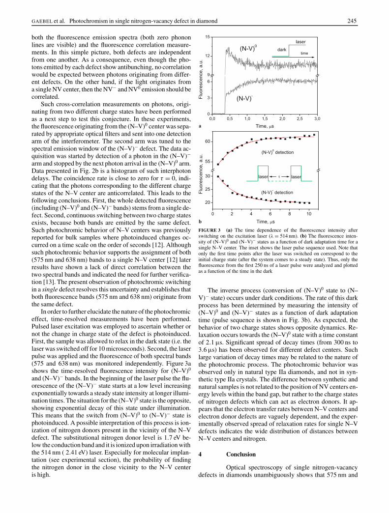
In order to further elucidate the nature of the photochromiceffect, time-resolved measurements have been performed.Pulsed laser excitation was employed to ascertain whether ornot the change in charge state of the defect is photoinduced.First, the sample was allowed to relax in the dark state (i.e. thelaser was switched off for 10 microseconds). Second, the laserpulse was applied and the fluorescence of both spectral bands(575 and 638 nm) was monitored independently. Figure 3ashows the time-resolved fluorescence intensity for (N–V)0
and (N–V)− bands. In the beginning of the laser pulse the flu-orescence of the (N–V)− state starts at a low level increasingexponentially towards a steady state intensity at longer illumi-nation times. The situation for the (N–V)0 state is the opposite,showing exponential decay of this state under illumination.This means that the switch from (N–V)0 to (N–V)− state isphotoinduced. A possible interpretation of this process is ion-ization of nitrogen donors present in the vicinity of the N–Vdefect. The substitutional nitrogen donor level is 1.7 eV be-low the conduction band and it is ionized upon irradiation withthe 514 nm ( 2.41 eV) laser. Especially for molecular implan-tation (see experimental section), the probability of findingthe nitrogen donor in the close vicinity to the N–V centeris high.
FIGURE 3 (a) The time dependence of the fluorescence intensity afterswitching on the excitation laser (λ = 514 nm). (b) The fluorescence inten-sity of (N–V)0 and (N–V)− states as a function of dark adaptation time for asingle N–V center. The inset shows the laser pulse sequence used. Note thatonly the first time points after the laser was switched on correspond to theinitial charge state (after the system comes to a steady state). Thus, only thefluorescence from the first 250 ns of a laser pulse were analyzed and plottedas a function of the time in the dark
The inverse process (conversion of (N–V)0 state to (N–V)− state) occurs under dark conditions. The rate of this darkprocess has been determined by measuring the intensity of(N–V)0 and (N–V)− states as a function of dark adaptationtime (pulse sequence is shown in Fig. 3b). As expected, thebehavior of two charge states shows opposite dynamics. Re-laxation occurs towards the (N–V)0 state with a time constantof 2.1 µs. Significant spread of decay times (from 300 ns to3.6 µs) has been observed for different defect centers. Suchlarge variation of decay times may be related to the nature ofthe photochromic process. The photochromic behavior wasobserved only in natural type IIa diamonds, and not in syn-thetic type IIa crystals. The difference between synthetic andnatural samples is not related to the position of NV centers en-ergy levels within the band gap, but rather to the charge statesof nitrogen defects which can act as electron donors. It ap-pears that the electron transfer rates between N–V centers andelectron donor defects are vaguely dependent, and the exper-imentally observed spread of relaxation rates for single N–Vdefects indicates the wide distribution of distances betweenN–V centers and nitrogen.
4 Conclusion
Optical spectroscopy of single nitrogen-vacancydefects in diamonds unambiguously shows that 575 nm and
246 Applied Physics B – Lasers and Optics
638 nm zero phonon lines belong to the different charge statesof nitrogen-vacancy defects in diamond. Upon 514 nm illu-mination the negative charge state appears to dominate. Thedefect can be converted into neutral charge state under darkconditions. Large spread of photochromic rates is consistentwith the hypothesis of N donors being involved in the pro-cess. However, additional work on doped diamond crystalsis required for precise identification of the electron donor re-sponsible for the photochromic effect.
ACKNOWLEDGEMENTS The work has been supported by theARO, DFG via SFB/TR 21 the graduate college “Magnetische Resonanz”,and the Landestiftung BW via the program “Atomoptik”.
REFERENCES
1 A. Gruber, A. Drabenstedt, C. Tietz, L. Fleury, J. Wrachtrup, C. vonBorczyskowski, Science 276, 2012 (1997)
2 F. Jelezko, T. Gaebel, I. Popa, A. Gruber, J. Wrachtrup, Phys. Rev. Lett.92, 076 401 (2004)
3 F. Jelezko, I. Popa, A. Gruber, C. Tietz, J. Wrachtrup, A. Nizovtsev, S.Kilin, Appl. Phys. Lett. 81, 2160 (2002)
4 F. Jelezko, T. Gaebel, I. Popa, M. Domhan, A. Gruber, J. Wrachtrup,Phys. Rev. Lett. 93, 130 501 (2004)
5 I. Popa, T. Gaebel, M. Domhan, C. Wittmann, F. Jelezko, J. Wrachtrup,Phys. Rev. B 70, 201 203 (2004)
6 G. Davies, Nature 269, 498 (1977)7 J. Meijer, B. Burchard, M. Domhan, C. Wittmann, T. Gaebel, I. Popa, F.
Jelezko, J. Wrachtrup, cond-mat/0505063 (2005) APL, in print8 J.R. Rabeau, G. Tamanyan, P. Reichart, D.N. Jamieson, S. Prawer,
F. Jelezko, T. Gaebel, I. Popa, M. Domhan, J. Wrachtrup, in prepara-tion
9 G. Davies, M. F. Hamer, Proc. R. Soc. Lon. Ser.-A 348, 285 (1976)10 A.M. Zaitsev, Optical Properties of Diamond (Springer, Berlin, Heidel-
berg, New York 2001)11 Y. Mita, Phys. Rev. B 53, 11 360 (1996)12 K. Iakoubovskii, G.J. Adriaenssens, M. Nesladek, J. Phys.-Condens.
Mat. 12, 189 (2000)13 I.N. Kupriyanov, V.A. Gusev, Y.N. Pal’yanov, Y.M. Borzdov, J. Phys.-
Condens. Mat. 12, 7843 (2000)14 N.B. Manson, J.P. Harrison, Diam. Relat. Mater. 14, 1705 (2005)15 J.W. Steeds, S.J. Charles, J. Davies, I. Griffin, Diam. Relat. Mater. 9, 397
(2000)16 W.Y. Wang, T. Moses, R. C. Linares, J. E. Shigley, M. Hall, J.E. Butler,
Gems & Gemology 39, 268 (2003)17 A. Beveratos, R. Brouri, T. Gacoin, J.P. Poizat, P. Grangier, Phys. Rev. A
64, 061 802 (2001)

![Title Photochromism and white long-lasting …...Title Photochromism and white long-lasting persistent luminescence in Bi[3+]-doped ZnGa[2]O[4] ceramics Author(s) Zhuang, Yixi; Ueda,](https://img.pdfslide.us/doc/110x75/5fca2348d932e01e9c134e64/title-photochromism-and-white-long-lasting-title-photochromism-and-white-long-lasting.jpg)